208 8 Pituitary Tumors IAN E. MCCUTCHEON Pituitary tumors are the most commonly occurring intracranial neoplasms. They are found in 8% to 24% of autopsied people, most of whom in life harbored small asymptomatic tumors that did not cause hor- monal disturbance (Kovacs et al., 1980; Parent et al., 1981; Teramoto et al., 1994; Tomita and Gates, 1999). Pituitary tumors can, however, produce pro- found physiologic upset by secreting supraphysiologic amounts of hormones or, when large, by compress- ing critical neural structures adjacent to their typical location in the sella turcica. The treatment of pituitary tumors has undergone a renaissance since the revival of transsphenoidal techniques by Guiot and Hardy 35 years ago. This pro- cess has been aided by the development of hormonal assaying techniques and radiographic imaging during the last three decades. Today, small pituitary tumors can be detected more easily and their hormonal ac- tivity can be assessed more accurately than has pre- viously been possible. Medical therapy, particularly for prolactin-secreting adenomas, but also for acromegaly, has developed sufficiently so that surgery can be avoided in some patients who can be man- aged with drugs alone. In general, pituitary tumors are diagnosed earlier and treated more effectively now than at any previous time. Nevertheless, a significant number of pituitary tumors deviate from the typical pattern of benign his- tology and slow growth. Pituitary adenomas can be invasive and penetrate adjacent dura to enter the sphenoid sinus, cavernous sinuses, or other parts of the skull base. These tu- mors can grow to a significant size and cause neu- rologic damage by provoking cranial neuropathies or optic nerve dysfunction; engulfing the carotid arter- ies and their tributaries; or impinging on the brain. Such aggressive variants behave as malignant tumors of the skull base. Even small, benign tumors may pro- duce a chronic, uncontrolled hypersecretion of pitu- itary hormone that causes profound physiologic changes over time. By any of these means, pituitary adenomas continue to pose an oncologic threat that warrants their inclusion in any list of clinically sig- nificant tumors of the central nervous system (CNS). PATHOLOGY Most pituitary gland tumors arise from the anterior portion of the gland known as the adenohypophysis. They are adenomas, tumors of the secretory elements, which in many cases produce and release one or more of the pituitary hormones produced by the an- terior lobe. A pseudocapsule, sharply demarcating them from the adjacent normal gland, which may be compressed by tumor, usually encloses these lesions. Although preferred locations for each of the different secretory types have been described within the ante- rior lobe, in actual fact any of the different subtypes can arise anywhere within the anterior lobe. Rare cases have been reported of adenomas arising in the pars tuberalis, a small extension of the anterior lobe along the distal anterior portion of the stalk (Roth- man et al., 1976), or of tumors arising within the sphenoid sinus, nasopharynx, or clivus from embry- onic rests of the pharyngeal pituitary (Hori et al., 3601_e08_p208-236 2/15/02 4:35 PM Page 208

Welcome message from author
This document is posted to help you gain knowledge. Please leave a comment to let me know what you think about it! Share it to your friends and learn new things together.
Transcript

208
8
Pituitary Tumors
IAN E. MCCUTCHEON
Pituitary tumors are the most commonly occurringintracranial neoplasms. They are found in 8% to 24%of autopsied people, most of whom in life harboredsmall asymptomatic tumors that did not cause hor-monal disturbance (Kovacs et al., 1980; Parent et al.,1981; Teramoto et al., 1994; Tomita and Gates,1999). Pituitary tumors can, however, produce pro-found physiologic upset by secreting supraphysiologicamounts of hormones or, when large, by compress-ing critical neural structures adjacent to their typicallocation in the sella turcica.
The treatment of pituitary tumors has undergonea renaissance since the revival of transsphenoidaltechniques by Guiot and Hardy 35 years ago. This pro-cess has been aided by the development of hormonalassaying techniques and radiographic imaging duringthe last three decades. Today, small pituitary tumorscan be detected more easily and their hormonal ac-tivity can be assessed more accurately than has pre-viously been possible. Medical therapy, particularlyfor prolactin-secreting adenomas, but also foracromegaly, has developed sufficiently so that surgerycan be avoided in some patients who can be man-aged with drugs alone.
In general, pituitary tumors are diagnosed earlierand treated more effectively now than at any previoustime. Nevertheless, a significant number of pituitarytumors deviate from the typical pattern of benign his-tology and slow growth.
Pituitary adenomas can be invasive and penetrateadjacent dura to enter the sphenoid sinus, cavernoussinuses, or other parts of the skull base. These tu-mors can grow to a significant size and cause neu-
rologic damage by provoking cranial neuropathies oroptic nerve dysfunction; engulfing the carotid arter-ies and their tributaries; or impinging on the brain.Such aggressive variants behave as malignant tumorsof the skull base. Even small, benign tumors may pro-duce a chronic, uncontrolled hypersecretion of pitu-itary hormone that causes profound physiologicchanges over time. By any of these means, pituitaryadenomas continue to pose an oncologic threat thatwarrants their inclusion in any list of clinically sig-nificant tumors of the central nervous system (CNS).
PATHOLOGY
Most pituitary gland tumors arise from the anteriorportion of the gland known as the adenohypophysis.They are adenomas, tumors of the secretory elements,which in many cases produce and release one ormore of the pituitary hormones produced by the an-terior lobe. A pseudocapsule, sharply demarcatingthem from the adjacent normal gland, which may becompressed by tumor, usually encloses these lesions.Although preferred locations for each of the differentsecretory types have been described within the ante-rior lobe, in actual fact any of the different subtypescan arise anywhere within the anterior lobe. Rarecases have been reported of adenomas arising in thepars tuberalis, a small extension of the anterior lobealong the distal anterior portion of the stalk (Roth-man et al., 1976), or of tumors arising within thesphenoid sinus, nasopharynx, or clivus from embry-onic rests of the pharyngeal pituitary (Hori et al.,
3601_e08_p208-236 2/15/02 4:35 PM Page 208
1999). When less than 10 mm in diameter, these tu-mors are called microadenomas; when larger, theyare called macroadenomas. A typical adenoma his-tologically shows loss of the normal acinar patternand its intervening reticulin network. The cells arefairly uniform in appearance, with small nuclei anda small amount of cytoplasm (Fig. 8–1).
Older schemes of classification, which have reliedon standard hematoxylin and eosin staining to pro-duce an appearance of basophilic, acidophilic, orchromophobe staining, have little functional signifi-cance and are now obsolete. Current pathologic anal-ysis of pituitary adenomas relies heavily on immuno-histochemistry (Asa, 1998). The six major hormonesof the anterior lobe (prolactin, growth hormone, thy-rotropin [TSH], luteinizing hormone [LH], follicle-stimulating hormone [FSH], and adrenocorticotropin[ACTH]) can be detected by applying a polyclonal ormonoclonal antiserum to tumor sections and then ex-posing the sections to a secondary antibody linked toreagents that give a colorizing reaction. The demon-stration of hormonal production does not necessar-ily correlate with hormone secretion, and a numberof clinically nonfunctional tumors, which produce nodetectable rise in serum hormone levels, have beenfound to contain hormone, usually FSH or LH, thathas been synthesized in small amounts but not se-creted or secreted inefficiently (Black et al., 1987;Daneshdoost et al., 1993; Sano and Yamada, 1994).In addition, pituitary tumors in many cases producehormones in a disorderly fashion, with an imbalancein the production of the �- and �-subunits, which(in the case of TSH, LH, and FSH) must link cova-
lently to produce a bioactive molecule. Some tumorsproduce hormone that is detectable through conven-tional techniques but is biologically inactive (Katznel-son et al., 1992; Trouillas et al., 1991) or hyperac-tive (Gesundheit et al., 1989). In most patients,however, a reasonable correlation exists between theclinical endocrine status of the patient, serum hor-mone levels, and the hormones demonstrated withinthe tumor through immunohistochemistry.
The spectrum of tumors that are truly inactive hasnarrowed greatly with the development of more sen-sitive hormone detection techniques. Such tumors arecalled null-cell adenomas (Kovacs et al., 1980).These tumors stain negatively for all hormones andcontain none of the granules detected by electron mi-croscopy in typical secretory tumors. It has been sug-gested that they arise from cells of gonadotrophic lin-eage, although their origin remains controversial. Asubset of these tumors contains large numbers of mi-tochondria visible by electron microscopy, which arecalled onocytomas (Bauserman et al., 1978). Onco-cytomas are typically large when diagnosed and af-fect males more frequently than females (Silbergeldet al., 1993).
It is now apparent that many of the tumors tradi-tionally called nonsecreting actually do secrete hor-mone in amounts too small to be of clinical signifi-cance (Asa et al., 1992; Greenman et al., 1998), orthey may produce the �-subunit or chromogranin(Nobels et al., 1993). These two secreted products,neither of which has hormonal activity, can be de-tected in the blood of some patients with clinicallynonfunctional adenomas. The �-subunit is one of the
Pituitary Tumors 209
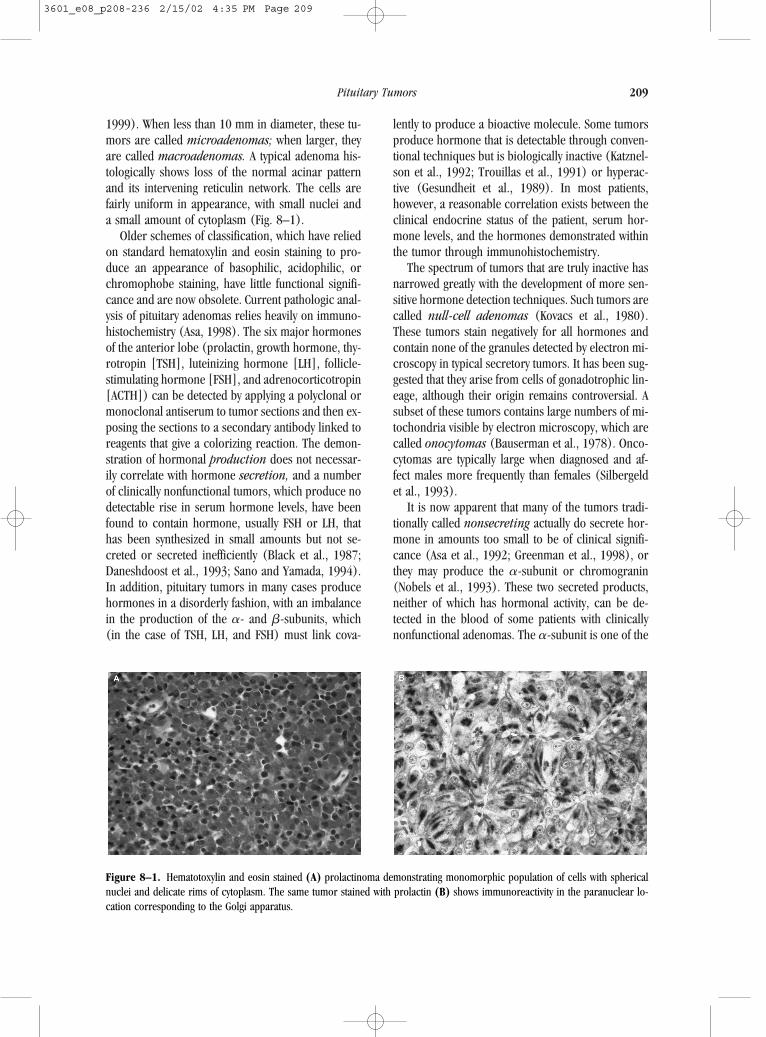
Figure 8–1. Hematotoxylin and eosin stained (A) prolactinoma demonstrating monomorphic population of cells with sphericalnuclei and delicate rims of cytoplasm. The same tumor stained with prolactin (B) shows immunoreactivity in the paranuclear lo-cation corresponding to the Golgi apparatus.
3601_e08_p208-236 2/15/02 4:35 PM Page 209

two subunits necessary for hormonal activity in gly-coprotein hormones; alone, however, the �-sub-unit has no hormonal function. Chromogranin isproduced by a variety of neuroendocrine tumors,including most pituitary tumors. These two prod-ucts may be the only molecular species availablefor surveillance after surgery or other types of ther-apy in patients with hormonally inactive adenomas.The most common nonfunctional adenomas in pa-tients under 40 years of age are silent corticotropicand gonadotropic adenomas, and their oncocyticvariants typically occur in patients over age 40years.
NATURAL HISTORY
Although pituitary adenomas are rare in children andfairly common in elderly patients at autopsy, the trueprevalence of pituitary tumors in the different decadesof life is not known. One preliminary study found fo-cal abnormality of the pituitary suggestive of tumor in10% of asymptomatic adults undergoing high-resolu-tion scans (Hall et al., 1994). The expected rate ofchange in small or large adenomas is also not known.Although one study of patients with prolactin-secret-ing microadenomas found that many do not changein size over time (March et al., 1981), its authorsused now-outdated techniques for radiologic surveil-lance. Another survey that examined untreated hy-perprolactinemia found a gradual increase in tumorsize in only 20% of patients, but did not address thequestion of the rate of tumor growth (Schlechte et al.,1989). In patients with clinically nonfunctional tu-mors in whom a partial surgical removal has beenachieved, careful radiographic surveillance of the tu-mor remnant has shown regrowth in one-third, witha mean time to detection of 5.4 years (Turner et al.,1999). However, another study showed only 6% ofendocrine-inactive tumor remnants recurring within5 years (Lillehei et al., 1998). As the odds of tumorprogression weigh heavily on decisions to use (orwithhold) postoperative radiotherapy, such informa-tion has great practical value. However, these deter-minations have never been addressed adequately forpatients with other forms of pituitary tumor. Mosttherapeutic decisions in pituitary tumor algorithmsare based on logical, although not clearly proven, as-sumptions that the tumor will enlarge over time and
that present hormone levels predict future hormonelevels in the untreated state.
CLINICAL PRESENTATION
Pituitary tumors cause a panoply of signs and symp-toms that can be grouped into four categories: (1) compression of adjacent normal gland, (2) hor-monal hypersecretion, (3) visual disturbance, and(4) headache.
Compression of Adjacent Normal Gland
Patients may present with hypopituitarism, that is,impairment of the normal function of the varioushormonal axes subserved by the anterior pituitary.Particularly vulnerable is the pituitary-gonadal axis,in which minor disturbances of the cycling of FSHor LH can affect libido and fertility in both sexes andthe menstrual cycle of women. Also vulnerable tolocal pressure effects are the pituitary-thyroid andpituitary-adrenal axes. Patients may therefore pres-ent with secondary hypothyroidism or a relativehypocortisolism predisposing them to addisoniancrisis.
In adults, low levels of prolactin are not thoughtto be significant. Traditionally, the same assumptionhas been held regarding growth hormone. During thepast decade, however, the clinical effects of growthhormone deficiency on body composition and bonemetabolism—and the benefits of treating such defi-ciency—have become widely accepted. Diabetes in-sipidus caused by insufficient production of antidi-uretic hormone rarely occurs, but, when it does, itheralds granulomatous involvement of the skull base(or a metastasis to the pituitary from a systemic can-cer) rather than a pituitary adenoma in most patientswho present with the disorder.
Hormonal Hypersecretion
Pituitary tumors may produce excess amounts of oneor more pituitary hormones, which can induce symp-toms relative to the specific hormone present in ex-cess. Growth hormone–secreting tumors produce thesyndrome of acromegaly, which is characterized byenlargement of the distal extremities and a coarsen-ing of facial features from bony overgrowth in the
210 PRIMARY CENTRAL NERVOUS SYSTEM TUMORS
3601_e08_p208-236 2/15/02 4:35 PM Page 210

skull. Patients with these tumors are prone to cardiacdisease and diabetes mellitus and if untreated (or un-successfully treated) have a significantly shortenedlife expectancy, with an observed-to-expected mor-tality ratio of l.6 to 3.3 (Wright et al., 1970; Hold-away and Rajasoorya, 1999).
Adrenocorticotropin-secreting tumors producehypercortisolism and present with the findings ofCushing’s disease. The protean nature of this diseasereflects the importance of cortisol in many organ sys-tems. Patients with Cushing’s disease show changesin body habitus caused by excess fat deposits, givingthem the classic “buffalo hump” and moon facies. Pa-tients also tend to have abdominal striae, osteoporo-sis, and diabetes mellitus; show muscle weakness,particularly in the proximal distribution; and may ex-hibit psychiatric disturbances (McCutcheon and Old-field, 1992).
The effects of ACTH-secreting tumors can be sub-tle in some patients and may present only as a ten-dency to arterial hypertension or as a very slowchange in skin texture and facial contour. Patientswith thyrotropin-secreting tumors have typical se-quelae of the hyperthyroid state, including heat in-tolerance, nervousness, and cardiac dysrhythmias.
Prolactinomas cause galactorrhea in men andwomen and menstrual irregularity in women, whichin many cases leads to infertility. A chronic, indirectsuppression of estrogen caused by excess prolactinalso predisposes the patient to osteoporosis. Men withprolactinomas may show a decreased sex drive andare prone to infertility. Gonadotropin-secreting tu-mors producing FSH, LH, or both, have a similar ef-fect on the menstrual cycle, fertility, potency, and sexdrive as do prolactinomas, but do not cause galact-orrhea.
Visual Disturbance
Patients with suprasellar extension of a clinically non-functional tumor usually come to medical attentionbecause of visual loss. Any pituitary tumor can causedecreasing vision if it grows large enough to com-press the visual pathways. Typically a macroadenomaextending above the sella to the optic chiasm causesa defect in the bitemporal fields that begins in the up-per quadrants and can progress to complete bitem-poral hemianopia. Because of the anatomic variabil-ity in the placement of the chiasm relative to the
pituitary stalk, a variety of presentations have beennoted. Von Willebrandt’s knee (where nerve tractsfrom the contralateral retina occupy part of the prox-imal optic nerve anterior to the chiasm) allows pa-tients occasionally to present with a junctional sco-toma, and some tumors are more eccentric to theright or left, thus causing a diversity of nonhomony-mous field cuts. Long-standing compression of theoptic nerves or chiasm can produce optic atrophy,with a resulting permanent loss of visual acuity. Inaddition, a tumor that extends into the cavernous si-nus may, in its later phases of growth, cause distur-bances of third, fourth, or sixth cranial nerve func-tion, resulting in diplopia and ptosis.
Headache
Many patients with pituitary tumors present withheadache. These patients often have chronic, refrac-tory headaches that lead a physician to request a brainscan, which may disclose an unsuspected lesion inthe pituitary fossa. Although headache is logical in pa-tients with large tumors spilling out of the sella tur-cica and invading or compressing the pain-sensitivedura, its presence in patients with small, noninvasivetumors must be regarded as coincidental in the ab-sence of any other logical explanation. Suggestionsthat such tumors cause headache by raising intrasel-lar pressure are intriguing but have not been provensufficiently to gain general acceptance (Arafah et al.,2000). Occasionally, pituitary adenomas are found inpatients who present with persistent headache after aminor head injury and in whom imaging performedonce the headache begins reveals a small pituitarytumor that had been previously undetected.
In a few patients, headache indicates suddenchanges in the size and structural integrity of the pi-tuitary tumor. A small percentage of pituitary adeno-mas hemorrhage and produce an apoplectic syn-drome of acute headache and sudden neurologicdeterioration caused either by direct compression ofthe hypothalamus or its vascular supply or by diffuseeffects of bleeding across the diaphragma sellae intothe subarachnoid space (Randeva et al., 1999). Inaddition, such patients may show sudden hypopitu-itarism from acute compression of the normal gland.Occasionally, a patient with a clinically nonfunctional,previously unsuspected adenoma will present in thisfashion and will experience diplopia, headache, and
Pituitary Tumors 211
3601_e08_p208-236 2/15/02 4:35 PM Page 211
a sudden decrease in vision. Such patients require ur-gent surgical decompression if vision is to be recov-ered or preserved.
LABORATORY INVESTIGATIONS
The standard laboratory work-up for a patient sus-pected of having a pituitary tumor involves measure-ment of an array of hormones that provide direct andindirect indices of pituitary and tumor function (Table8–1). The typical biochemical survey includes serumlevels of prolactin, TSH, LH, and FSH. Growth hor-mone may also be measured, but this is only neces-sary if there is clinical evidence of acromegaly. If it is measured, then somatomedin-C (insulin-likegrowth factor-I) should also be determined becauseit provides a better picture of growth hormone se-cretion over time. Some endocrinologists have advo-cated measuring insulin-like growth factor bindingproteins (most specifically IGFBP-3) as a measure ofdisease activity in acromegaly, but this remains con-troversial and has not gained widespread acceptanceas a replacement for insulin-like growth factor-I sur-veillance (DeHerder et al., 1995; Paramo et al., 1997;
Halperin et al., 1999). Normal levels of LH and FSHdiffer in men and women, and also differ in womenduring the various phases of the menstrual cycle.Measurement of thyrotropin levels should be accom-panied by measurement of triiodothyronine and L-thy-roxine so that elevations in TSH caused by primarypituitary hypersecretion can be distinguished from el-evations caused by primary thyroid insufficiency.
Adrenocorticotropic hormone is not generallymeasured directly; rather, the activity of the pituitary-adrenal axis is determined by measurement of corti-sol, one of the main adrenal hormones produced inresponse to adrenocorticotropic stimulation. Bothcortisol and ACTH are heavily subject to diurnalrhythms, and it is generally considered best for co-herent and consistent interpretation of serum corti-sol levels to collect them at 8 AM, when they are rel-atively high. For women, estradiol levels are checked.These are fairly sensitive indices of gonadal failurebut must be interpreted in conjunction with levels ofFSH and LH.
Prolactin is measured for all patients. Elevationsof prolactin may occur because of hypersecretion bytumor cells but can also be caused by anatomic dis-tortion of the pituitary stalk by the tumor mass. Thisdistortion results in inadequate flow through the por-tal-hypophyseal system of those dopaminergic factorsthat exert a tonic inhibition on prolactin production.When the stalk is compressed, this inhibition is re-leased, and prolactin levels rise. Because the eleva-tion caused by a prolactin-secreting tumor is usuallysignificantly higher than that caused by a nonfunc-tional tumor disturbing the stalk, it is usually possi-ble to distinguish between the two by a careful anal-ysis of prolactin levels over time.
Other important information on the hormonal con-dition of the patient can be obtained by the use ofprovocative hormonal tests. In these tests, a syntheticstimulatory factor (such as the corticotrophin-re-leasing factor) is administered intravenously to thepatient, and the hormonal response is evaluated overtime. Such tests provide a dynamic indication of theability of the gland to push hormone levels abovebaseline values. In subtle cases of hormone dysfunc-tion, and particularly in patients with Cushing’s syn-drome where these tests can help with the differen-tial diagnosis and localization of the hypersecretorysource, the use of provocative tests may be consid-ered. They also have value for the assessment of nor-mal pituitary function in patients recovering from sur-
212 PRIMARY CENTRAL NERVOUS SYSTEM TUMORS
Table 8–1. Laboratory Work-Up for Patients Suspected ofHaving a Pituitary Tumor
Prolactin
Thyrotropin
Triiodothyronine
L-thyroxine
Luteinizing hormone
Follicle-stimulating hormone
Growth hormone, if clinical evidence of acromegaly
Somatomedin-C (insulin-like growth factor-I)
Cortisol
Estradiol levels in women
Prolactin
Provocative hormonal tests (tailor to clinical situation: critical in Cushing’s disease)
Serum electrolytes (serum and urine osmolality, fluid intake and output, urine specific gravity when diabetes is suspected)
Measurement of �-subunit for patients with clinically nonfunctional adenomas or those with a thyrotropin-secreting adenoma
3601_e08_p208-236 2/15/02 4:35 PM Page 212

gery for a pituitary tumor. In the postoperative set-ting, basal hormone values are remeasured after asuitable interval. This helps both to establish the pres-ence or absence of continuing hypersecretion by re-sidual tumor and to assess the adequacy of normalgland function, because such function may be dam-aged by the gentle manipulation required to separatea tumor from the adjacent normal gland. After sur-gery pre-existing hormone deficits will be restored inapproximately 50% of patients, and 20% develop newdeficits of anterior pituitary function (Webb et al.,1999).
The function of the posterior lobe is assessed bymeasurement of serum electrolytes and, in caseswhere there is a high suspicion of diabetes insipidus,by checking serum and urine osmolality and byrecording fluid intake and output and urine specificgravity. It is exceedingly rare for a patient with a pi-tuitary tumor to present with diabetes insipidus. If asellar mass is seen on a scan and diabetes insipidusforms part of the clinical presentation, it is highly pos-sible that the tumor is not a pituitary adenoma, butrather a metastatic tumor arising in the lung, breast,prostate, or another organ system. Diabetes insipidusoccurs much more often in the postoperative period,when about 15% of patients show this phenomenoneither transiently or permanently. The production ofvasopressin by the pituitary is more sensitive thanother hormonal axes to the trauma of surgery. Spe-cial vigilance is necessary during the first week aftersurgery, because patients with partial diabetes in-sipidus can become dehydrated and hypernatremic ifthe condition goes unrecognized and if no hormonesupplementation is given. Surveillance for hypona-tremia is also required during the first 2 weeks aftersurgery, as 2% (Hensen et al., 1999) to 21% (Olsonet al., 1997) of patients develop a transient dysregu-lation of vasopressin release that can be clinically sig-nificant.
Measurement of the �-subunit has been consid-ered experimental but is gaining acceptance. It is nowrecognized that some tumors are pure �-subunit–secreting adenomas (Ridgway et al., 1981). As a rule,this measurement should be taken for all patients withclinically nonfunctional adenomas (Warnet et al.,1994). The other group in whom the �-subunit hasparticular relevance is the occasional patient whopresents with a TSH-secreting adenoma. Measure-ment of the �-subunit/TSH molar ratio in this instanceis valuable in sorting out those patients with a “syn-
drome of inappropriate TSH secretion” who have apituitary tumor from those who have pituitary resis-tance to the effects of TSH (Gesundheit et al., 1989;McCutcheon et al., 1990).
RADIOLOGIC EVALUATION
The preferred radiologic technique for pituitary tu-mor detection and follow up is magnetic resonanceimaging (MRI). Magnetic resonance imaging has nowsupplanted previous methods of sellar tomographyand computed tomography (CT), which are less sen-sitive and should only be used when MRI is unavail-able (Webb et al., 1992). An occasional patient is stillbrought to medical attention by the incidental findingof erosion or enlargement of the sellar boundarieson a skull radiograph. Such patients generally havelarge nonfunctional tumors, and the most accurateway of defining the anatomic boundaries of the tu-mor is with a MRI scan. In years past, concerns aboutcarotid artery aneurysms that masquerade as pituitarytumors led to the occasional use of angiograms forsuch patients. This too is now unnecessary, as stan-dard MRI shows nicely the distinction between theflow void of the carotid artery within the cavernoussinus and the tumor within the sella and adjacent areas.
Tumors as small as 3 mm in diameter now repre-sent the limits of detection of MRI scans enhanced bygadolinium (Lundin and Bergstrom, 1992). Pituitarytumors are generally seen on contrast scans as hy-pointense to isointense areas against a slightly hy-perintense area of normal gland (Fig. 8–1). This sit-uation is the reverse of that seen with glial tumors ofthe cerebral parenchyma, where enhancement is seenwithin the tumor and within adjacent areas of ede-matous brain with an impaired blood–brain barrier.There is no blood–brain barrier in the pituitary gland,and by virtue of this pattern of enhancement the ves-sels within the tumor would seem to be less imme-diately leaky to contrast than those of the normalgland. The best delineation is therefore seen in scansdone early (i.e., from 2 to 5 minutes after gadolin-ium injection) (Hayashi et al., 1995).
There are still a number of patients in whom nodistinct adenoma is seen but in whom biochemicaltests suggest the presence of such a lesion. This cir-cumstance is particularly relevant in Cushing’s dis-ease, where tumors may be very small, yet produce
Pituitary Tumors 213
3601_e08_p208-236 2/15/02 4:35 PM Page 213
a profound physiologic upset because of the primacyof ACTH in a number of the physiologic processes rel-evant to homeostasis. Other secretory tumors tend tocome to attention only when they are larger. Patientswith Cushing’s disease frequently have obvious hyper-cortisolism on biochemical and clinical examination,but can have a normal MRI of the sella turcica. Al-though a few such occult tumors become visible whendynamic scanning is performed during contrast infu-sion, this technique is not widely available (Bartynskiand Lin, 1997). The possibility of misdiagnosis of ec-topic secretion of ACTH for pituitary hypersecretionemphasizes the importance of rigorous biochemicaltesting, which should be able to distinguish betweenthese two etiologies. In addition, even tumors largerthan 3 mm in diameter can escape detection by scansif their signal characteristics are isointense to those ofnormal gland. The selection of patients for surgery whohave been shown to secrete hormone in excess shouldbe on the basis of biochemical evidence first and, sec-ond, on radiologic evidence.
TREATMENT
Rationale for Treatment
Determining the best way to treat pituitary adenomasdepends on the predicted nature of the pathology.Hormonally active tumors usually require suppres-sion by surgery, medical means, and/or radiationtherapy to prevent the long-term sequelae of hyper-secretion. In particular, patients with Cushing’s dis-ease or acromegaly rarely reach a normal life ex-pectancy because of the deleterious effects that excessACTH and growth hormone exert on a variety of or-gan systems, particularly the cardiovascular system.Patients with prolactinomas may require therapy toreverse the infertility imposed by such tumors and toprevent the accelerated osteoporosis that occurs inpatients with chronically elevated prolactin levels(Klibanski et al., 1988). Those with TSH-secretingadenomas suffer an intractable form of hyperthy-roidism with attendant cardiac irritability and also re-quire definitive treatment. Clinically nonfunctional tu-mors often present because of chiasmal compressioncausing visual field defects. The presence of an in-creasing field cut is a strong indication for surgicaldecompression, a statement that applies equally topatients with secretory tumors of whatever type. For
pituitary tumors as a group, statistics show a twofoldexcess mortality relative to the population at large,but the reason for this difference is unclear (Nilssonet al., 2000).
The goals of therapy for pituitary tumor include
• Elimination of hormonal excess by tumor sec-tion
• Prevention of optic nerve dysfunction caused bysuprasellar extension of tumor
• Restoration or preservation of normal pituitaryfunction
• Avoidance of therapeutic complications• Long-term remission without biochemical or
radiographic evidence for regrowth of tumor
Any treatment that is chosen must be judged by thesestandards in assessing its efficacy and suitability fora given patient.
Surgical Approaches
Entry to the sella turcica is achieved most safely bythe transsphenoidal approach in patients with smalltumors without extrasellar extension. This method al-lows the surgeon to avoid entering the intracranialcompartment and has the advantage of midline tra-jectory that circumvents critical structures on eitherside of the gland (Hardy, 1969, 1991). The incisionin such an operation can be made in the sublabial(below the lip) area over the maxilla just beneath theinferior extent of the pyriform apertures, a route thatallows direct exposure of the virtual space betweenthe cartilaginous nasal septum and the medial nasalmucosa (mucoperichondrium). A tunnel createdwithin this virtual space leads directly to the vomer,sphenoid sinus, and sella. Alternatively, an endonasalroute that avoids an external incision can be chosen,but this requires either a wide nasal cavity or inci-sion of the nasal ala to allow room for a speculumwithin the nose (Griffith and Veerapen, 1987). In theendonasal approach, the incision is made in the nasalmucosa at the posterior edge of the cartilaginous sep-tum where it meets the bony septum. Transsphenoidalapproaches should be chosen for tumors confined tothe sella, those with relatively minor suprasellar ex-tension, or those that extend into the sphenoid sinus.Tumors with cavernous sinus extension are frequentlyexcised by the transsphenoidal route, but completeresection should not be expected as the contents ofthe cavernous sinus usually elude full inspection.
214 PRIMARY CENTRAL NERVOUS SYSTEM TUMORS
3601_e08_p208-236 2/15/02 4:35 PM Page 214

The transcranial approach is reserved for pa-tients with larger tumors that elevate the diaphragmasellae more than 2 cm above its normal level or thatextend laterally from their intrasellar origin. Often,a pterional or combined pterional/subfrontal cran-iotomy is used, and a bone flap is fashioned that ex-tends as close as possible to the floor of the ante-rior cranial fossa (Tindall and Tindall, 1987). Thesylvian fissure is then split medially, the frontal lobeis elevated, the temporal lobe is slightly depressedwith malleable retractors, and the suprasellar andsellar areas are visualized. The operating micro-scope is an essential tool in this small and anatom-ically complex region when transcranial andtranssphenoidal procedures are performed. Intra-operative fluoroscopy is vital as well in transsphe-noidal procedures and allows localization of the po-sition of instruments relative to the bony landmarksof the skull base.
If cavernous sinus exploration or exenteration isdesired, a fronto-orbito-zygomatic craniotomy can beperformed. This procedure involves a more extensivebony removal that includes the orbital rim and roof,anterior clinoid, and malar eminence (Al-Mefty andSmith, 1990). The zygoma is usually cut free and al-lowed to fall down with the temporalis muscle, a tac-tic that grants the surgeon additional exposure overa fairly wide field of view. If carotid resection isplanned, we prefer to expose the internal carotidartery in the neck in case a sudden rupture occursrequiring arterial tamponade; others unroof thepetrous segment of the carotid instead. Before anycavernous sinus exploration is done, a preoperativecarotid balloon occlusion test must be performed,which predicts the ability of the patient to toleratecarotid resection. This clinically qualitative test maybe enhanced by the performance of single-photonemission CT or xenon-CT scans that measure cere-bral blood flow in the ipsilateral and contralateralcerebral hemispheres (Origitano et al., 1994). Thesetechniques continue to evolve and may offer hope forthose patients with extensive disease in the cavernoussinus or skull base who, until recently, were offeredradiation therapy as palliative treatment but for whomlittle other therapy was available (MacKay andHosobuchi, 1978).
Other variations on these themes have been usedover the years. The transethmoidal approach has hadsome proponents but is generally not used becauseit requires a facial incision and provides an angled,
imperfect view that makes anatomic landmarks moredifficult to identify than does the midline transsphe-noidal approach (Kirchner, 1984). Endoscopic exci-sion of pituitary tumors has been reported and is gain-ing increasing popularity as part of the trend in allsurgical disciplines toward reducing incision size andthe “invasiveness” of any given procedure. Althoughimprovement in the quality of endoscopic instru-mentation has fostered its use in pituitary surgery, andseveral medical centers have been enthusiastic in re-porting its advantages (Jho, 1999; Sheehan et al.,1999; Jarrahy et al., 2000), no prospective compar-ison of its efficacy in achieving complete resection ofadenomas, large or small, has yet been undertaken.Even though less disruption of the nasal septum oc-curs during endoscopic transsphenoidal surgery, pa-tients are still subject to the risks of cerebrospinalfluid rhinorrhea and endocrine disturbance, so it isnot clear why use of the endoscope should lead toshorter stays in hospital after operation, as some au-thors have proposed (Jho, 1999). It seems appro-priate to use endoscopy as a tool for examining ar-eas within the sella turcica that currently elude directinspection by the surgeon. In particular, in many pa-tients, the medial wall of the cavernous sinus is poorlyseen or not seen at all, and adherent tumor can beleft there after an otherwise successful operation. Itis likely that endoscopy will become an additional,quite valuable tool for maximizing tumor resection,but that open procedures using the operative micro-scope will continue to be used for many patients(Jankowski et al., 1992).
Computer-assisted navigation has also been ap-plied to pituitary surgery during the past 5 years. Suchdevices provide the surgeon with a pointer (typicallytracked optically), the location of which is deter-mined by a workstation and displayed on a screenshowing the operative field on relevant multiplanarMRI slices. This technology is still evolving and is notyet ready to replace fluoroscopy as the standard lo-calizing tool in transsphenoidal surgery (McCutcheonet al., 2001). However, it is quite useful in locatingsuch surgically relevant structures as the carotid ar-teries and sphenoid septations, in planning the sur-gical trajectomy, and in locating lateral or inferior ex-tensions of pituitary tumors invading the skull base(Sandeman and Moufid, 1998). Such tools, togetherwith endoscopy, will become important aids to saferand more complete removal of pituitary tumors dur-ing the next decade.
Pituitary Tumors 215
3601_e08_p208-236 2/15/02 4:35 PM Page 215
SPECIFIC TUMOR TYPES
Table 8–2 summarizes specific types of pituitary tumors.
Prolactin-Secreting Adenomas
Approximately 40% of pituitary adenomas secreteprolactin, and such tumors are more common thanany other type of pituitary neoplasm. In contrast tothe reliance on surgery as the primary treatment forother forms of pituitary tumor, these tumors are usu-ally treated medically before surgery is considered.
Drug Therapy
Various dopamine analogues that inhibit prolactin release are available, the most common beingbromocriptine (Parlodel). About 70% to 80% of patients respond to this drug, but significant side ef-fects occur in 20% to 30% of patients, which limitsits use (Molitch et al., 1985). Side effects associatedwith bromocriptine include nausea, vomiting, ortho-static hypotension, and, in some patients, psychoticreactions. The drug may be stopped, therefore, be-cause of a failure to arrest tumor growth and bringprolactin levels to normal or because the patient can-not tolerate the drug. An alternative long-acting do-pamine agonist called quinagolide (or CV 205-502)has been developed. This agent has a higher affinitythan bromocriptine for dopamine receptors. Ap-proximately one-half of adenomas that resistbromocriptine therapy respond to quinagolide (Brueet al., 1992b). Quinagolide also invokes side effectsand has caused weight loss and psychiatric break-down in some patients, but has fewer side effects thanbromocriptine (Glaser et al., 1994; Merola et al.,1994). Other long-acting bromocriptine analogueshave also been studied that may lower the incidenceof side effects (Brue et al., 1992a; Maraschini et al.,1991; Jamrozik et al., 1996); however, their role and
that of quinagolide, in the clinical management ofthese patients has been eclipsed by cabergoline, an-other long-acting dopaminergic agent released foruse in the United States in 1998.
Cabergoline (Dostinex) was developed as a D2-receptor–specific agonist, by virtue of which it hasless of a tendency to provoke side effects (operatingthrough the D1 as well as the D2 receptor) than ei-ther bromocriptine or quinagolide. Because of thisand because it is given once or twice weekly, patientsare more likely to be compliant in taking the drug. Itnormalizes prolactin levels in 80% to 90% of patientsand causes tumor shrinkage in two-thirds (Verhelstet al., 1999). In a series of patients resistant tobromocriptine, prolactin became normal in 70% ofpatients taking cabergoline; and, of patients intoler-ant of bromocriptine’s side effects, 84% toleratedcabergoline and normalized their prolactin levels.Dose escalation improves these numbers even further(Colao et al., 2000). Comparison of cabergoline withquinagolide shows that the two drugs normalize pro-lactin in an equal proportion of patients, but that agreater chance of tumor shrinkage occurs with caber-goline (Di Sarno et al., 2000). Because of such results, cabergoline is now the drug of choice for medical treatment of prolactinomas, althoughbromocriptine remains a well-established (and sig-nificantly less expensive) alternative.
The response of patients with prolactinomas tomedical therapy depends on tumor size and prolactinlevel. Patients with smaller tumors and lower pro-lactin levels respond best. If the prolactin level be-fore treatment is greater than 1000 ng/ml, the levelsusually fall with dopamine agonist therapy, but do notnormalize (Hardy, 1984). Most tumors cause pro-lactin levels between 50 and 250 ng/ml. From 65%to 80% of patients taking bromocriptine in this rangewill normalize their prolactin levels, and over time tu-mor reduction occurs in 76% (Brue et al., 1992a;Maraschini et al., 1991). Cabergoline’s statistics, al-luded to above, are comparable or better.
The general assumption is that patients treatedmedically require life-long therapy to avoid regrowthof suppressed tumor; however, in some patients, thetumor disappears completely and does not recurwhen bromocriptine is stopped. After withdrawal ofcabergoline, approximately 10% of patients exhibitthis phenomenon, which appears to be confined tothose with microadenomas (Di Sarno et al., 2000).Unfortunately, it is only possible to identify these pa-
216 PRIMARY CENTRAL NERVOUS SYSTEM TUMORS
Table 8–2. Specific Pituitary Tumor Types
Clinically nonfunctional adenomas (incidentalomas)
Gonadotropin-secreting adenomas
Growth hormone–secreting adenomas
Adrenocorticotropin-secreting adenomas
Thyrotropin-secreting adenomas
3601_e08_p208-236 2/15/02 4:35 PM Page 216

tients after the fact. This dilemma cannot be resolvedfully at this time, but does suggest a clinical approachof stopping the drug at intervals and following hor-mone levels and scans for any sign of regrowth or re-turn of hypersecretion. It is likely that the tumor willregain its activity in the majority of patients who areevaluated long enough and, in those patients, treat-ment must begin anew.
Surgery
Surgery is used only if medical treatment fails or ifthe patient cannot tolerate the side effects of the treat-ment. There has been some evidence to support theidea that pretreatment with bromocriptine enhancessurgical results (Hubbard et al., 1987) and opposingevidence that it makes tumor excision more difficultby inducing fibrosis within the tumor (Bevan et al.,1987). Although patients may wish to temporize andavoid surgery, it is best not to continue trials of med-ical therapy for more than 1 year without declaringsuccess or failure and deciding whether surgery willor will not be indicated.
The success of surgical treatment depends on tu-mor size and preoperative hormone level, both ofwhich predict the invasiveness of the tumor in ques-tion. Microadenomas are more readily excised andgenerally show prolactin levels of 50 ng/ml to 100ng/ml. In the series of Randall et al. (1983), pro-lactin levels normalized after surgery in 88% of thosewith preoperative levels less than 100 ng/ml, but inonly 43% of those with levels greater than 100 ng/ml.Patients with prolactin levels greater than 350 ng/mlshould be considered to have an invasive tumor, andall will harbor macroadenomas. In the very extensiveseries of Hardy (1984), the immediate remission rate was 86% for patients with levels less than 250ng/ml but only 6% for those with levels greater than1000 ng/ml.
Given the predominant role of medical therapy forprolactinomas, it is now uncommon to operate onpatients with small tumors who have not receivedprior dopamine agonist therapy. It has been suggestedthat such pre-treatment compromises the success rateof surgical removal. In one surgical series in whichall patients had been previously given bromocriptine,only 45% of patients with microadenomas and only17% of those with macroadenomas had normal lev-els of prolactin postoperatively (Soule et al., 1996).Such results may reflect bromocriptine-induced fi-
brosis within the tumor, as well as a selection biassuch that more difficult cases were more likely to bereferred for surgery.
Despite initial success in most surgical series, asignificant number of patients relapse when studiedlong-term after surgery (Hardy, 1984). Hardy’s re-currence rate was 22% during the first 5 years aftersurgery. Others have shown recurrence rates of 17%to 50% during the first 2 years after surgery for mi-croadenomas and 20% to 80% for macroadenomasduring the same time period (Rodman et al., 1983;Serri et al., 1983). In Wilson’s modern series, morethan 90% of patients with microadenomas (and morethan 80% of those with small macroadenomas)showed initial remission, as did 40% of those withprolactin levels above 200 ng/ml or with large, inva-sive tumors (Tyrrell et al., 1999). During 15 years offollow up, about 15% of those in remission relapsedto hyperprolactinemia.
In Adams’ surgical series of microprolactinomaswith a mean follow-up period of 70 months, only 1of 32 patients recurred. It is certainly possible to nor-malize prolactin in most patients with a small, en-capsulated tumor and to expect a sustained remis-sion, but the persistence of remission has not beensufficiently studied for periods longer than 5 years.Because many patients in surgical series are between25 and 40 years old and may be expected to live forseveral decades, more data on long-term rates of re-currence are needed. Tumor reactivation may occurfor several reasons, including the regrowth of resid-ual microscopic clusters of tumor cells, the presenceof an underlying genetic mutation predisposing to tu-mor growth, or the presence of a mechanism of hy-pothalamic overdrive that continues unabated aftertumor excision.
Radiation Therapy
Radiation therapy is useful for patients in whom suchregrowth recurs and who wish to avoid further sur-gery. It is also a valuable adjunct for patients in whoma complete excision cannot be achieved because tu-mor has infiltrated adjacent dura, neural structures,or the cavernous sinus. Usually a fractionated dose of45 to 50 Gy is given over 4 to 5 weeks by a limited-field technique that excludes structures outside a 4cm window centered on the sella turcica. The draw-backs of radiotherapy are its gradual effect and itsability to damage parapituitary structures contained
Pituitary Tumors 217
3601_e08_p208-236 2/15/02 4:35 PM Page 217
within the field as well as late effects that can occur.Several years usually pass before maximum tumor re-gression occurs, and 2 to 10 years may be requiredfor normalization of prolactin levels. The success rateof sellar irradiation within this time frame has beenreported to be as low as 30% (Sheline et al., 1984;Williams et al., 1994), although others have reportedan 83% rate of local control over 10 years (Sasaki etal., 2000). Larger prolactinomas (i.e., with volume�30 cm3), however, show particularly poor localcontrol rates and may require specialized techniquesor repeat surgery (Isobe et al., 2000).
New methods of irradiation include one that linksbeam delivery with stereotactic localization, allowingdelivery of a highly concentrated, accurately focuseddose of radiation to an intracranial target. Althoughthe name “radiosurgery” has been applied to thismethod, no surgical incision is involved. Rather, thismethod involves attachment of a stereotactic ring tothe patient’s head by a neurosurgeon who assists aradiotherapist in planning the isodose contours, acomplicated process that requires specialized com-puters and precise three-dimensional representationof the target. Either a standard radiotherapy gantrywith a stationary target (LINAC) or multiple station-ary sources of irradiation all focused simultaneouslyon the target (gamma knife) may be used. In theory,this method reduces the risk of radiation damage toadjacent structures, particularly the medial temporallobes and the optic chiasm, while maximizing radia-tion dose. The pituitary gland is still included in theradiation field because of its intimate relation withany intrasellar tumor. In addition, if the cavernous si-nus is included within the radiation field, 10% to 15%of patients will develop a cranial neuropathy within 3years of radiosurgical treatment (Tishler et al., 1993).
The function of the normal gland is at risk withboth stereotactic and conventional radiation tech-niques, and as many as 100% of patients treated con-ventionally show pituitary insufficiency when studiedfor 10 years (Littley et al., 1989). The administrationof a stereotactic dose of 40 to 70 Gy by charged par-ticle beam (no longer much used since the advent ofthe gamma knife) yielded a 10% to 30% incidence ofpost-treatment hypopituitarism, and 60% of patientswith prolactin-secreting tumors had normalized pro-lactin levels within 1 year after treatment (Levy et al.,1991). Although such techniques have been availablefor a number of years in Europe and North Americaand were widely used during the 1990s, good long-
term statistics are still unavailable. Most treatmentcenters that perform radiosurgery for pituitary tumorsuse the gamma knife system in which the target (i.e.,the tumor) is placed at the center of a fixed, spheri-cal array of radiation sources. LINAC-based systems,which utilize a roving gantry arm to deliver the radi-ation beam, can also be employed. However, thegamma knife has a dosimetric advantage in treatingpituitary adenomas, as it can achieve a greater con-formity index than the LINAC in this anatomic region(Plowman and Doughty, 1999).
In one small series of patients with micropro-lactinomas, radiosurgery caused normalization ofprolactin levels in 23%, and a decrease (but not nor-malization) in 62%, during 1 year of follow up (Kimet al., 1999). In a larger series, similar results wereachieved but correction of endocrinopathy occurredmore frequently in patients with acromegaly or Cush-ing’s disease than in those with prolactin-secreting tu-mors (Pan et al., 1998). As radiosurgery can only bedone in patients with microadenomas or tumors withlateral (rather than superior) extension, many pa-tients do not qualify for it and must be treated surgi-cally and/or with more conventional radiotherapeu-tic techniques. For some, fractionated conformalirradiation may offer increased accuracy of targeting,with reduction in the volume of normal tissue exposedto irradiation. However, only preliminary experiencehas been reported with its use for pituitary tumors,so it is impossible to conclude whether it will provesafer or more effective than conventional limited-fieldtechniques (Jalali et al., 2000; Perks et al., 1999).
Summary of Therapeutic Approach
In our current practice, we treat patients with pro-lactinomas first with cabergoline, unless a trial of thedrug shows side effects the patient cannot tolerate. Inyears past, such patients were offered quinagolide asan alternative or they chose surgery. Quinagolide hasbeen withdrawn from use in clinical trials in theUnited States despite its published clinical utility; itremains available in Europe and in Canada. Patientswith progressive visual loss are also treated with sur-gery, but those with a macroadenoma and visual fieldcut can sometimes be controlled with dopamine ag-onists, and surgery can thereby be avoided. Radio-therapy is reserved for the relatively few patientswhose tumors regrow after surgery or whose tumorscannot be completely excised, as proven by persis-
218 PRIMARY CENTRAL NERVOUS SYSTEM TUMORS
3601_e08_p208-236 2/15/02 4:35 PM Page 218

tent elevation of prolactin despite maximal removalof tumor.
No Treatment
Prolactinomas, gonadotropin-secreting adenomas,and clinically nonfunctional adenomas comprise themajority of pituitary tumors and do not generally pro-voke hormonal derangements affecting long-termsurvival. What are the chances that a small tumor inthis group that does not affect chiasmal function willgrow at all if simply left untreated? Patients withmacroadenomas often require treatment to preserveor improve vision and to control (in prolactinomas)high levels of hormone secretion that impair fertilityand menstruation. These conditions must be cor-rected in those patients who wish to conceive.
Microadenomas, however, have been shown insome instances not to change for as long as 20 years,and their ability to secrete excess hormone may fadeor actually increase over time. Spontaneous involu-tion of such tumors, sometimes as a result of anapoplectic event, may occasionally occur. In a studyof 30 patients with small tumors in whom no treat-ment was given, 14 showed no change in prolactinlevels over time; 6 showed a gradual increase; and10 showed a decrease (Schlechte et al., 1989). Forpatients with prolactin-secreting microadenomas andfor those with small nonfunctional tumors, it is rea-sonable to follow those in whom some menstruationis present and child-bearing is not desired, with in-tervention reserved for those in whom tumor growthor increased secretory activity occurs.
Clinically Nonfunctional Adenomas
The category of clinically nonfunctional adenomashas been redefined in recent years as techniques inpathologic analysis have become more powerful. Itnow includes patients with true null cell adenomasthat secrete no hormone of any kind; those with �-subunit–secreting adenomas that produce no activehormone; and those with tumors that, although se-cretory, produce quantities of hormone too small toeffect clinical change in endocrine function. Many ofthe patients in the last category have tumors that, byimmunohistochemical analysis, produce FSH or LH(Asa et al., 1992; Young et al., 1996). These gonad-otropin-secreting adenomas were previously thoughtto be quite rare. It is now recognized that a spectrum
exists that, at its lower end, includes patients previ-ously classified as nonsecreting and at its high endincludes those with detectable elevations of FSHand/or LH in peripheral blood. Each category is ad-dressed here.
About 25% to 33% of pituitary adenomas are clin-ically nonfunctional and produce symptoms only bycompressing the pituitary and parasellar structuresand, eventually, causing frank hypopituitarism. Thiscondition often goes unrecognized, except in retro-spect, until compression of the visual pathways leadsto the diagnosis of a sellar lesion. Approximately one-half of all pituitary adenomas stain on immunocyto-chemistry for one or more of the glycoprotein hor-mones or their subunits (� or �) (Black et al., 1987;Daneshdoost et al., 1993; Sano and Yamada, 1994).They are not clinically active because of inefficienthormone release or because they produce hormonalspecies of low bioactivity. Between 20% and 30% ofpatients with endocrine-silent pituitary adenomas dohave increased levels of serum �-subunit, which maybe useful in some cases as a marker of tumor activ-ity (Oppenheim et al., 1990; Nobels et al., 1993).Such hypersecretion of �-subunit is most common inlarger tumors (Warnet et al., 1994).
As for any large pituitary tumor, treatment includeshormone replacement to correct deficiency of nor-mal pituitary function. There is no available medicaltreatment that effectively suppresses the growth ofthese tumors. However, octreotide has been shownto cause improvement in visual field deficits in clini-cally nonfunctional macroadenomas, although it hasno effect in 50% of such tumors (Warnet et al., 1997).Although nonfunctional tumors are quite common,only a few large surgical series have been published,and all such series include both null cell adenomasand weakly gonadotropin-secreting tumors under the“nonsecreting” or “nonfunctional” heading. Ebersoldet al. (1986) reported results from the excision of100 nonfunctioning adenomas, of which 82 weregreater than 2 cm in diameter. Most of these patientshad preoperative visual impairment that generally im-proved after surgery. Because of the high prevalenceof invasive features in such tumors, 50% were in-completely resected and required postoperative ra-diotherapy. Regrowth occurred in 18% of those whoreceived radiation after surgery and in 12% of those(presumably smaller tumors) treated with surgeryalone. In Hardy’s series of 126 patients treated over25 years, vision improved in 75% of those who had
Pituitary Tumors 219
3601_e08_p208-236 2/15/02 4:35 PM Page 219
visual impairment before surgery, and 21% regrewduring a mean follow-up period of 6.4 years (Com-tois et al., 1991). In addition, preoperative hypopi-tuitarism (present in �75% overall) improved in41% of those with deficits in the pituitary-adrenal axisand in 14% of those with hypogonadism.
As with prolactinomas, radiotherapy is generallyrecommended when obvious residual tumor remainsafter surgery or when regrowth occurs and such treat-ment has not previously been given. Halberg and She-line (1987) administered radiotherapy to 140 pa-tients with clinically nonfunctional tumors. Of these,23 were poor surgical candidates who received irra-diation alone, 37 were treated by surgery only, and80 underwent combined treatment. A long follow-upperiod (up to 20 years) was achieved. All patientstreated with irradiation alone showed arrested growthof tumor; tumor recurred in 75% of those treatedwith surgery and radiotherapy and in 100% of pa-tients treated with surgery alone. Such results suggestthat many tumors in this category are invasive and areincompletely resected.
An opposing school of more conservative bent hasarisen during the past 10 years. The volume of re-sidual tumor in patients undergoing incomplete re-section may be small and sit well away from the op-tic nerves or chiasm and may be followed upaccurately with MRI. One group evaluated such non-irradiated patients with serial imaging and found thattumor in only 6% regrew after 5 years (Lillehei et al.,1998). A second report by others showing a recur-rence rate of 18% at 5 years and 44% at 10 years ismore pessimistic even than their own earlier report,which had suggested a much lower incidence of re-currence (Bradley et al., 1994; Turner et al., 1999).In our practice we strive for maximum resection ofintrasellar and suprasellar tumors and follow up non-functional tumors (even if they are grossly invasive)without further treatment unless atypical pathologicfindings suggest anaplasia. If a tumor regrows, ourfirst inclination is to operate if it is technically feasi-ble and only to irradiate if a second instance of re-currence follows or if the pathologic appearance ofthe tumor has changed since the first resection. Inthis way we defer or avoid altogether the risks of de-layed loss of pituitary function or of radiation-inducedoptic neuropathy (which is rare, but devastating anddifficult to arrest when it happens).
Patients with small, clinically nonfunctional tumorsconfined to the sella are followed up clinically and
radiographically; surgery is performed only if tumorgrowth is confirmed on serial MRI scans or if thereis confusion about whether the tumor is truly non-functional. We prefer to avoid radiotherapy as a pri-mary modality because of the high incidence of hy-popituitarism after its use.
Patients with “incidentalomas,” that is, clinicallyoccult but radiologically detectable adenomas, gen-erally discovered when patients are scanned afterthey complain of headache or suffer minor headtrauma, are frequently seen in the neurosurgicalclinic in this era of easily available MRI scans(Donovan and Corenblum, 1995). Although a min-imalist approach has been suggested in which onlyscreening for hyperprolactinemia is performed(King et al., 1997), longitudinal studies show that15% of these patients have partial hypopituitarismand that 3% of incidentally discovered microade-nomas and 26% of larger tumors grew during amean follow-up period of 2.7 years (Feldkamp etal., 1999). We do a full endocrine screen on suchpatients and scan them yearly for several years,partly in response to the need to alleviate the pa-tient’s anxiety and partly to identify the minority withendocrine deficits and/or active tumors. Any patientwith a tumor causing an asymptomatic visual fieldloss, however, is offered surgery.
Gonadotropin-Secreting Adenomas
Gonadotropin-secreting adenomas produce no spe-cific clinical syndrome and are treated much likeother clinically nonfunctional adenomas (Snyder,1997). When large, they more often present becauseof visual impairment and hypopituitarism than endo-crine excess. Hypogonadism in such patients may re-flect impairment of normal pituitary function throughmass effect or may relate to the elevation of FSH or,less commonly, LH produced by the tumor itself.Some tumors secrete gonadotropins in vitro but pro-duce no detectable change in hormone levels in vivoexcept an occasional elevation of the �-subunit (Asaet al., 1992; Snyder et al., 1984). Others producesmall but measurable increases in serum levels of LH,but not consistently enough to allow its use as a tu-mor marker (Greenman et al., 1998). In addition, tu-mor immunostaining does not correlate well withserum gonadotropin levels, which further weakensthe utility of studying those levels for any clinical pur-pose (Ho et al., 1997).
220 PRIMARY CENTRAL NERVOUS SYSTEM TUMORS
3601_e08_p208-236 2/15/02 4:35 PM Page 220

The main reason for distinguishing this group fromother nonfunctional tumors lies in the potential thatmedical therapy may prove effective against them. Be-cause dopamine suppresses gonadotropin secretionin the normal gland, bromocriptine has been usedwith occasional success for patients who have go-nadotropin-secreting adenomas (Lamberts et al.,1987). In addition, innovative therapy using LH-re-leasing hormone and its antagonists has been usedfor a few patients. LH-releasing hormone increasessecretion in most patients, but inhibits it in a few, per-haps by desensitizing the tumor (Klibanski et al.,1989). Antagonists to LH-releasing hormone haveyielded mixed results but may eliminate the pre-sumptive hypothalamic stimulus to tumor growth. Asmentioned above for clinically nonfunctional tumors,somatostatin analogues have also been used in thissubset of patients to obtain clinical improvement, andless often tumor shrinkage. In general, most patientswith gonadotropin-secreting tumors are treated likethose with other clinically nonfunctional tumors.
Growth Hormone–Secreting Adenomas
Medical Treatment
The results of drug therapy in treating growth hormone–secreting adenomas are less predictablethan those achieved in treating prolactinomas.Bromocriptine, cabergoline, short-acting somato-statin analogues (SMS 201-995, octreotide acetate),or long-acting analogues (lanreotide) have beenused. Bromocriptine normalizes growth hormone lev-els in 20% of patients, but doses at the higher end ofthe accepted range (20 mg/day) are often necessary,producing a greater incidence of side effects (Wasset al., 1977). Most such tumors will not shrink withbromocriptine, although octreotide has been re-ported to induce tumor regression in 50% of patientsand reduces growth hormone levels in a majority ofcases (Barkan et al., 1988; Arosio et al., 1995; New-man et al., 1998). Because some patients achieve per-sistent biochemical and clinical improvement overlong periods of time with chronic somatostatin ther-apy, this drug can be used as a medical alternative tosurgery. Somatostatin has the disadvantages of a sub-cutaneous route of administration and causes gall-stones and gastrointestinal upset in some patients.
We use octreotide occasionally as a preoperativeadjunct because it will shrink tumor in 50% of pa-
tients to whom it is given, which may make completesurgical excision more feasible. Unlike bromocrip-tine for prolactinomas, octreotide does not cause his-tologic changes that impede tumor removal. Shrink-age occurs during the first 2 weeks of treatment, if itis to occur at all, so prolonged courses give no addedbenefit (Lucas-Morante et al., 1994). Pre-surgicaltreatment with octreotide does make more completeremoval possible in some cases, but the data are notconclusive and studies have been published that bothconfirm (Stevenaert and Beckers, 1993; Lucas-Morante et al., 1994) and deny (Kristof et al., 1999)its utility before surgery for macroadenomas. We havenot been impressed with the degree of shrinkage,however, and have become less inclined to pretreatpatients unless lateral extension of tumor is present.Somatostatin may also be used to suppress growthhormone production in patients with residual tumorafter surgery, a setting in which we are much morelikely to apply it. The chance of biochemical nor-malization does not, however, depend on the dosechosen (Ezzat et al., 1995). These results can be im-proved by continuous infusion, but this is expensiveand rarely used in most medical centers (Tamura etal., 1998).
If medical therapy is chosen, several alternativesto octreotide are available. Long-acting analogues ofoctreotide (lanreotide, somatostatin-LAR) are simi-larly efficacious, but may be better tolerated as theyare given much less frequently (two to four times permonth) (Verhelst et al., 2000). Although bromocrip-tine has limited utility, cabergoline is more effectiveand induces a degree of biochemical normalizationand tumor shrinkage comparable with that achievedby the somatostatin analogues (Cozzi et al., 1998; Abset al., 1998). As cabergoline is given orally, it mayvery well become more popular than octreotide orlanreotide. Data have not yet been accumulated on itsuse in the preoperative setting.
The newest drug applied to acromegaly is pegvi-soment, a growth hormone analogue that binds to andblocks the action of the growth hormone receptor. Ina major prospective randomized trial involving in-pa-tients given pegvisoment for 12 weeks, IGF-I normal-ized in 89% of patients receiving the highest dosetested (20 mg/day subcutaneously) (Trainer et al.,2000). Although side effects seem minimal and thesedata are promising, it is not known whether it will beeffective in long-term use or whether loss of negativefeedback due to lower circulating levels of IGF-I will
Pituitary Tumors 221
3601_e08_p208-236 2/15/02 4:35 PM Page 221

promote growth of the (otherwise untreated) ade-noma responsible for the ongoing growth hormoneexcess. This drug will be further studied and is ex-pected to join cabergoline and lanreotide as the med-ical therapies of choice in the future.
Surgery
Surgery is the primary mode of treatment for mostpatients with pituitary acromegaly. Success rates varywidely among series, and when examining their ratesof induced remission, it is important to considerwhich biochemical criteria the authors used. Thelargest surgical series reported so far involved 254patients treated at the University of California, SanFrancisco as described by Abosch et al. (1998). Inthat series, growth hormone levels less than 5 ng/mlwere achieved in 76% of patients; 29% of patients re-quired postoperative radiotherapy for residual tumor.In a previous paper, these authors compared theirown statistics with those of 30 other surgical seriesinvolving a total of 1360 patients (Ross and Wilson,1988). Overall growth hormone levels of less than 5ng/ml were achieved in 60% of patients. Other serieshave based successful outcome on achieving growthhormone levels of less than 10 ng/ml (Laws et al.,1987), but the most vigorous modern definition of“cure” requires a level of �2.3 ng/ml (Melmed etal., 1998; Levitt et al., 1995). Only a few have fol-lowed up somatomedin-C levels, and none has ap-plied provocative testing as a criterion for cure (Kaoet al., 1992; Tindall et al., 1993; Abosch et al., 1998).In the series by Tindall et al. (1993), sustained post-operative levels less than 5 ng/ml were achieved in88% of patients and in 82% when somatomedin-C lev-els were included in the analysis.
Other series have comparable results, particularlyif a single, experienced surgeon has performed all in-cluded operations (Ahmed et al., 1999). About 90%of patients with microadenomas, and 40% to 50% ofthose with macroadenomas, achieve a postsurgicalremission.
Sustained remission after surgery should be ex-pected in 60% to 80% of patients overall. The sizeand location of the tumor affects the ease with whichtumor excision is achieved. Fortunately, most of thesetumors present as microadenomas and can be iden-tified at surgery and selectively excised. Tumors withsuprasellar extension can still be cured, but thechance for leaving residual tumor is greater in such
cases. In patients with cavernous sinus tumor, resid-ual tumor almost always remains, even when a tran-scranial approach is used together with skull-basetechniques for cavernous sinus entry that have evolvedover the past 10 years (Al-Mefty and Smith, 1990;Origitano et al., 1994). We currently enter the cav-ernous sinus rarely, with trepidation, and only if tu-mor mass within it must be debulked to decompressthe optic nerve; the risk of cranial neuropathy is high,radiosurgery is available to control focal areas of tu-mor, and pituitary tumor within the cavernous sinusoften goes for years without clinically significant al-teration in carotid diameter or cranial nerve function.
Treatment failures should lead to the considerationof several possibilities. Some patients will, of course,have residual tumor. If a complete excision of a mi-croadenoma has been done, however, the possibilityof primary hypothalamic gangliocytoma secretinggrowth hormone–releasing hormone should be con-sidered, as should the possibility that growth hormoneor growth hormone–releasing hormone secretion isectopic (usually from the pancreas) (Asa et al., 1987;Melmed et al., 1985). Although the great majority ofpatients with acromegaly have pituitary pathology, thosewho do not can be identified by measuring the plasmalevel of growth hormone–releasing hormone. If thelevel is greater than 300 ng/ml, an ectopic source ofsecretion is likely. In such patients, evidence of pitu-itary enlargement may appear on MRI scans, and thepatient may undergo inappropriate transsphenoidalsurgery. Patients in whom a somatotroph hyperplasiais found but no adenoma should have growth hor-mone–releasing hormone levels measured and, if ele-vated, the source should be identified by radiologicstudies of the chest and abdomen.
Radiation Therapy
Radiation therapy has been used in the past for manypatients as a primary treatment for acromegaly. Thedose given is similar to that used for other forms ofpituitary tumor, and the incidences of hypopitu-itarism, optic nerve damage, and cerebral ra-dionecrosis are similar as well. Both conventional ir-radiation and proton-beam bombardment have beenused and produce comparable results, although mor-bidity may be somewhat higher with the latter (East-man et al., 1979; Kliman et al., 1987; Lüdecke et al.,1989). As with other tumors, the main drawback isthe gradual effect of such therapy. In most patients,
222 PRIMARY CENTRAL NERVOUS SYSTEM TUMORS
3601_e08_p208-236 2/15/02 4:35 PM Page 222

growth hormone levels decline slowly over the firstyear and may continue to do so for as long as 10years after treatment. In 70% of irradiated patients,growth hormone levels less than 10 ng/ml are even-tually reached in 5 years, although the response slipsto 40% at 10 years (Clarke et al., 1993; Plataniotis etal., 1998; Kokubo et al., 2000). In normal individu-als, growth hormone levels range from 0.25 to 0.7ng/ml, and most authors now consider a level of 2.3ng/ml or less as remission. A truly rigorous bio-chemical definition would include normalization ofgrowth hormone and somatomedin-C levels as wellas a normal oral glucose tolerance test and thy-rotropin-releasing hormone stimulation test(Melmed, 1990; Melmed et al., 1998). These data aregenerally absent in reported series. Even these crite-ria are imperfect, as 40% of patients with persistentlyabnormal dynamic responses show no radiographicregrowth of tumor during the decade after surgery(Ross and Wilson, 1988).
Radiosurgery must also be considered in the ar-ray of options available for treating acromegaly. Dataon its efficacy in this specific disease are buried withingeneral series of pituitary tumors treated with thismodality and are scanty. Two studies have been pub-lished that focus on its role in acromegaly. In one,96% of 79 patients treated achieved normal growthhormone levels over 3 years after treatment (Zhanget al., 2000). Increasing tumor shrinkage was seenover the same time period. Follow up was insufficientin this study to allow conclusions about long-termcontrol. In another study of 16 patients with recur-rent acromegaly, growth hormone and IGF-I normal-ized after 1.4 years, on average; in a comparisongroup who received conventional fractionated irradi-ation, the mean time to normalization was 7.1 years(Landolt et al., 1998). Thus, radiosurgery may actmore quickly, but the relative risks (over time) forradiosurgery versus fractionated radiation are notwell understood. In addition, the interesting obser-vation has been made that acromegalic patients tak-ing octreotide during radiosurgery show a slower,less complete response to irradiation. Thus, oc-treotide may act as a radioprotectant (Landolt et al.,2000).
Preferred Treatment
At our institution, the general treatment schema foracromegaly includes the preoperative and postoper-
ative measurement of growth hormone and so-matomedin-C levels. We use provocative testing onlyin borderline cases. For a few patients (usually withtumors extending laterally or with large suprasellarcomponents) a trial of somatostatin analogue therapyis given for up to 3 months, and the scans are re-peated to check for tumor shrinkage. Half of thoseso treated show partial regression of the tumor andan unpredictable degree of correction in biochemi-cal abnormalities. Only those in whom somatomedin-C and growth hormone levels normalize (or nearlyso, as long as clinical symptoms substantially regress)are offered continuing medical therapy. The majorityof patients undergo surgery at this point, and then ra-diotherapy is used with conventional limited-fieldtechniques for any residual active tumor. Octreotide-LAR is used to control continuing hypersecretionwhile the patient waits for the irradiation to take effect.
Adrenocorticotropin-SecretingAdenomas
Approximately 4% to 10% of pituitary tumors secreteadrenocorticotropin, and another 5% produce thehormone but do not secrete it in significant amounts.Those in the former category develop Cushing’s dis-ease, the name for a hypercortisolism (Cushing’s syn-drome) of pituitary source. About 70% of adults withtrue hypercortisolism have such a pituitary tumor.
Much endocrinologic effort has been expended in-venting and validating tests that identify hypercorti-solism as present and identify its ultimate source asthe pituitary gland, the adrenals, or an ectopic tumorsecreting adrenocorticotropin, such as in the lung. Adiagnosis of Cushing’s disease is sometimes difficultto make, because MRI scans fail to show a tumor in20% to 30% of patients who have an ACTH-secretingadenoma in the pituitary. In years past, as many as10% of patients with Cushing’s disease were treatedwith bilateral adrenalectomy as a measure of somedesperation to eliminate the target organ on whichACTH acts and thereby eliminate the clinical effectsof hypercortisolism. In this circumstance, the natu-rally occurring negative feedback exerted by cortisolon the normal gland (and on the pituitary tumor) iseliminated, and hypersecretion of ACTH increasesdramatically. In the 15% to 25% of adrenalectomizedpatients in whom Nelson’s syndrome develops, a dra-matic growth of pituitary adenoma occurs, although
Pituitary Tumors 223
3601_e08_p208-236 2/15/02 4:35 PM Page 223
it may be delayed for a number of years (Nelson etal., 1960; Moore et al., 1976; Nagesser et al., 2000).At least one case has now been reported in which thissyndrome was successfully treated with cabergoline,suggesting that it may be as useful in a subset of pa-tients with hypercortisolism as it is in those with prolactin-secreting tumors (Pivonello et al., 1999).
The biochemical assessment of patients with Cush-ing’s disease must take into account the diurnal vari-ation in levels both of ACTH and cortisol normally re-leased by the pituitary gland. Although this episodicsecretion still occurs when a pituitary adenoma ispresent, neoplastic corticotrophs are relatively in-sensitive to negative feedback from any glucocorti-coid, whether endogenous or exogenous. Within thespectrum of tumor autonomy, only 33% have any realmeasure of hypothalamic control exerted on them(Van Cauter and Refetoff, 1985).
The bewildering array of diagnostic tests used toestablish the presence and source of hypercortisolismin a patient suspected of having Cushing’s syndromeis beyond the scope of this chapter. Several excellentreviews cover the diagnostic approach in detail andexplain how to interpret test results (Kaye and Crapo,1990; McCutcheon and Oldfield, 1992). In general,one recognizes cortisol excess by determining bloodlevels in relation to time of day or by collecting urineover 24 hours to eliminate diurnal variations. Evensalivary sampling may be done when multiple mea-surements of cortisol are required (Mosnier-Pudar etal., 1995). If confusion persists (as it often does)even after several assays of blood and urine have beenperformed, provocative tests are used to clarify thepresence of a true hypercortisolism; they also giveclues to the etiology of the excess. The most popularof these is the dexamethasone suppression test, whichhas several variations and is accurate about 90% ofthe time (Liddle, 1960; Nieman et al., 1986). Stimu-lation tests using corticotrophin-releasing factor arefrequently used and show a normal or excessive risein ACTH in patients with Cushing’s disease, but littleresponse in patients with tumor of the adrenal glandor other sites. This test, like the dexamethasone sup-pression test, is misleading approximately 10% of thetime (Nieman et al., 1986).
In the past 10 years, a great deal of attention hasbeen given to bilateral sampling of the inferior pet-rosal sinus. In theory, this technique can detect cen-tral/peripheral gradients that confirm the presence ofpituitary tumor and also can be used to lateralize thetumor within the gland to the right or left half. In
practice, the test is useful for confirming the pres-ence of a tumor within the pituitary gland, but inter-mixing between the two cavernous sinuses leads tonumerous cases of inappropriate lateralization(Mamelak et al., 1996). It is now apparent that therate of false lateralization approaches 40%. The useof corticotrophin-releasing factor stimulation withthis test increases its diagnostic accuracy to nearly100% when a central-to-peripheral ACTH gradient�3.0 is used as the criterion for diagnosis (Oldfieldet al., 1991; Kaltsas et al., 1999). Direct sampling ofhormone levels in cavernous sinus blood performedduring surgery may also be used to localize occultmicroadenomas, but has practical limitations giventhe time required to perform the analyses; false neg-atives may still occur, especially in the absence of cor-ticotrophin-releasing hormone stimulation (Dopp-man et al., 1995; Graham et al., 1999).
Surgery
Surgical excision is the most popular method of erad-icating these tumors. Because many are microade-nomas, transsphenoidal techniques have proven use-ful and can achieve a cure rate of 88%, as reportedby Mampalam et al. (1988) from a series of 221 suchpatients. About 5% of these patients showed eventualrecurrence of hypersecretion, with a mean time to re-currence of almost 4 years. Those in whom an in-trasellar exploration showed no tumor were curedusing biochemical criteria a little less than 50% of thetime. Some of these patients had hemihypophysec-tomy based on petrosal sinus sampling, which asmentioned above is an imperfect method of deter-mining tumor location within the gland. Others hadcomplete hypophysectomy, which remains a validtreatment for patients with this ultimately life-threat-ening disease. The morbidity among all patients inthis series was 9% with a mortality of 1%. Such sta-tistics are fairly representative of those reported fromother surgical series of ACTH-secreting pituitary tu-mors (Burke et al., 1990; Chandler et al., 1987;Bochicchio et al., 1995). Only two-thirds of patientswith macroadenomas achieve remission after surgery,and they also tend to relapse sooner than those withsmaller tumors (Blevins et al., 1998a).
Some patients in whom Cushing’s disease is bio-chemically identified but in whom surgery fails to ef-fect remission have been misdiagnosed and actuallyharbor other lesions causing cortisol excess. It is there-fore important to review rigorously the evidence from
224 PRIMARY CENTRAL NERVOUS SYSTEM TUMORS
3601_e08_p208-236 2/15/02 4:35 PM Page 224

which the diagnosis was made before performing sur-gery to re-explore the sella and perhaps to perform acomplete hypophysectomy. Reoperation certainly hasits place in the treatment of Cushing’s disease and canraise the rate of remission by 20% or more (Ram etal., 1994). If surgery is redone in this setting and anadenoma is found that had previously been overlooked,it is still better to excise it selectively, together with amargin of adjacent normal pituitary, than to extirpatethe entire gland. This approach results in remission ofhypercortisolism in about half of these patients withmuch less risk of hypopituitarism.
Radiation and Other Therapy
Radiotherapy is not often used as the primary treat-ment as this tumor, like others, is slow to respond toit. This group of patients, however, needs a fairlyquick and definitive elimination of the hormonal ex-cess to correct physiologic derangement in multipleorgan systems. Adrenal ablation is now reserved forpatients for whom other attempts to ablate the sourceof adrenocorticotropin, either surgically or medically,have failed. When radiotherapy is used after incom-plete surgical resection, 80% of patients achieve re-mission, most within 2 years (Estrada et al., 1997).
Preferred Treatment
Currently, we approach patients suspected of havingCushing’s disease with a rigorous and lengthy endocrineevaluation before any treatment is considered. Thework-up includes repeated sampling of blood and col-lection of 24 hour urine volumes to prove hypersecre-tion. Even if the first one or two samples fail to showany abnormality, patients with appropriate symptomsshould be repeatedly studied because some will harbora tumor that secretes cortisol episodically, which is easyto miss with a less than strenuous diagnostic work-up(Loh, 1999). Most patients have dexamethasone sup-pression testing and then petrosal sinus sampling be-fore and after stimulation by corticotrophin-releasingfactor. Only then is surgery considered.
In the great majority of patients, a transsphenoidalapproach is used and the sella is explored vigorously.For patients with Cushing’s disease, the frequency ofcerebrospinal fluid leak and of intraoperative bleed-ing from the cavernous sinus should be higher thanfor other patients because a more aggressive explo-ration is often necessary to find and completely re-move small tumors (or areas of corticotroph hyper-plasia) that may lurk unseen in the far lateral areas
of a normal-appearing gland. The concept that suchtumors occur in the “central median wedge” of thegland and that a search for occult tumor should bedirected first to the area of the middle third of thegland is somewhat erroneous. These tumors can oc-cur in any part of the anterior lobe and have evenbeen found in the posterior lobe or outside the gland(Pluta et al., 1999), and the stakes for the patient arehigh enough to make a higher incidence of (treat-able) cerebrospinal fluid leakage acceptable. Patientswho remain hypercortisolemic even after such a vig-orous exploration and perhaps after excision of his-tologically confirmed tumor should be reviewed as-siduously. If any doubt exists about the diagnosis,appropriate tests should be redone. If, despite theseprecautions, the pituitary is still thought to be thesource of ACTH excess, reoperation may be neededwith the intent to perform a hemihypophysectomy ormore complete removal of the anterior lobe (Fried-man et al., 1989). Only after two surgical proceduresshould radiotherapy, adrenalectomy, or medical sup-pression of the adrenals with ketoconazole or mi-totane be entertained.
Thyroid-Stimulating Hormone Secreting Adenomas
Tumors that secrete TSH are rare and represent lessthan 1% of large series of pituitary tumors. Their rar-ity is probably exaggerated somewhat by the tendencyof physicians to misdiagnose as primary hyperthy-roidism, which is much more common, the hyper-thyroid syndrome they typically produce. Patients withTSH-secreting adenomas are usually falsely diagnosedwith primary thyroid disease for years and are oftentreated with thyroid ablation without success. This de-lay frequently allows TSH-secreting tumors to enlargeand makes surgical removal more difficult. In theory,the loss of negative homeostatic feedback caused bythyroid ablation in patients with such tumors mightbe expected to make the tumor more aggressive, ashappens in the occasional case of Nelson’s syndromethat develops after adrenal ablation in patients withCushing’s disease. Only small series of these patientshave been reported, supporting the idea that these tu-mors are aggressive and that surgery for TSH-secret-ing adenomas carries a higher risk of perioperativedeath and/or neurologic morbidity than occurs withother tumor types (McCutcheon et al., 1990). Thelargest series currently on record are composed of24 patients treated in Europe (Losa et al., 1999) and
Pituitary Tumors 225
3601_e08_p208-236 2/15/02 4:35 PM Page 225
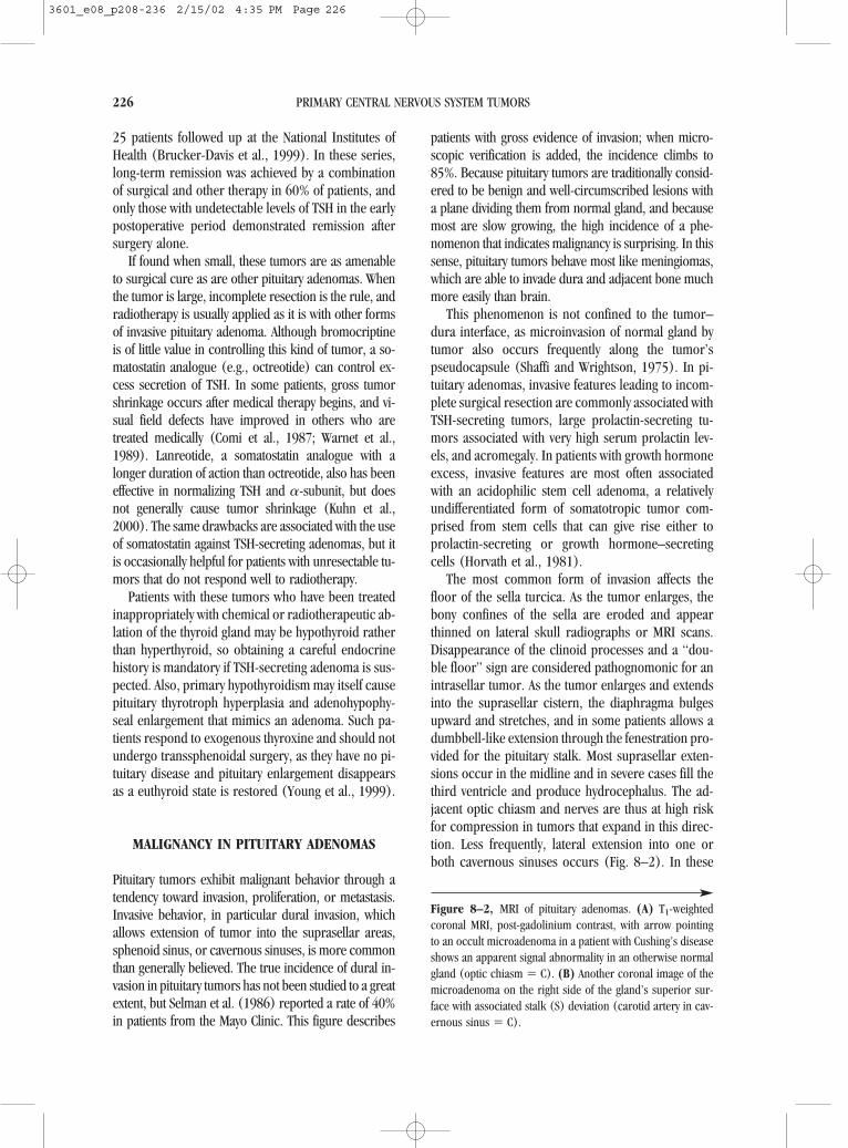
25 patients followed up at the National Institutes ofHealth (Brucker-Davis et al., 1999). In these series,long-term remission was achieved by a combinationof surgical and other therapy in 60% of patients, andonly those with undetectable levels of TSH in the earlypostoperative period demonstrated remission aftersurgery alone.
If found when small, these tumors are as amenableto surgical cure as are other pituitary adenomas. Whenthe tumor is large, incomplete resection is the rule, andradiotherapy is usually applied as it is with other formsof invasive pituitary adenoma. Although bromocriptineis of little value in controlling this kind of tumor, a so-matostatin analogue (e.g., octreotide) can control ex-cess secretion of TSH. In some patients, gross tumorshrinkage occurs after medical therapy begins, and vi-sual field defects have improved in others who aretreated medically (Comi et al., 1987; Warnet et al.,1989). Lanreotide, a somatostatin analogue with alonger duration of action than octreotide, also has beeneffective in normalizing TSH and �-subunit, but doesnot generally cause tumor shrinkage (Kuhn et al.,2000). The same drawbacks are associated with the useof somatostatin against TSH-secreting adenomas, but itis occasionally helpful for patients with unresectable tu-mors that do not respond well to radiotherapy.
Patients with these tumors who have been treatedinappropriately with chemical or radiotherapeutic ab-lation of the thyroid gland may be hypothyroid ratherthan hyperthyroid, so obtaining a careful endocrinehistory is mandatory if TSH-secreting adenoma is sus-pected. Also, primary hypothyroidism may itself causepituitary thyrotroph hyperplasia and adenohypophy-seal enlargement that mimics an adenoma. Such pa-tients respond to exogenous thyroxine and should notundergo transsphenoidal surgery, as they have no pi-tuitary disease and pituitary enlargement disappearsas a euthyroid state is restored (Young et al., 1999).
MALIGNANCY IN PITUITARY ADENOMAS
Pituitary tumors exhibit malignant behavior through atendency toward invasion, proliferation, or metastasis.Invasive behavior, in particular dural invasion, whichallows extension of tumor into the suprasellar areas,sphenoid sinus, or cavernous sinuses, is more commonthan generally believed. The true incidence of dural in-vasion in pituitary tumors has not been studied to a greatextent, but Selman et al. (1986) reported a rate of 40%in patients from the Mayo Clinic. This figure describes
patients with gross evidence of invasion; when micro-scopic verification is added, the incidence climbs to85%. Because pituitary tumors are traditionally consid-ered to be benign and well-circumscribed lesions witha plane dividing them from normal gland, and becausemost are slow growing, the high incidence of a phe-nomenon that indicates malignancy is surprising. In thissense, pituitary tumors behave most like meningiomas,which are able to invade dura and adjacent bone muchmore easily than brain.
This phenomenon is not confined to the tumor–dura interface, as microinvasion of normal gland bytumor also occurs frequently along the tumor’spseudocapsule (Shaffi and Wrightson, 1975). In pi-tuitary adenomas, invasive features leading to incom-plete surgical resection are commonly associated withTSH-secreting tumors, large prolactin-secreting tu-mors associated with very high serum prolactin lev-els, and acromegaly. In patients with growth hormoneexcess, invasive features are most often associatedwith an acidophilic stem cell adenoma, a relativelyundifferentiated form of somatotropic tumor com-prised from stem cells that can give rise either to prolactin-secreting or growth hormone–secretingcells (Horvath et al., 1981).
The most common form of invasion affects thefloor of the sella turcica. As the tumor enlarges, thebony confines of the sella are eroded and appearthinned on lateral skull radiographs or MRI scans.Disappearance of the clinoid processes and a “dou-ble floor” sign are considered pathognomonic for anintrasellar tumor. As the tumor enlarges and extendsinto the suprasellar cistern, the diaphragma bulgesupward and stretches, and in some patients allows adumbbell-like extension through the fenestration pro-vided for the pituitary stalk. Most suprasellar exten-sions occur in the midline and in severe cases fill thethird ventricle and produce hydrocephalus. The ad-jacent optic chiasm and nerves are thus at high riskfor compression in tumors that expand in this direc-tion. Less frequently, lateral extension into one orboth cavernous sinuses occurs (Fig. 8–2). In these
226 PRIMARY CENTRAL NERVOUS SYSTEM TUMORS
Figure 8–2, MRI of pituitary adenomas. (A) T1-weightedcoronal MRI, post-gadolinium contrast, with arrow pointingto an occult microadenoma in a patient with Cushing’s diseaseshows an apparent signal abnormality in an otherwise normalgland (optic chiasm � C). (B) Another coronal image of themicroadenoma on the right side of the gland’s superior sur-face with associated stalk (S) deviation (carotid artery in cav-ernous sinus � C).
3601_e08_p208-236 2/15/02 4:35 PM Page 226

227
3601_e08_p208-236 2/15/02 4:35 PM Page 227

228
3601_e08_p208-236 2/15/02 4:36 PM Page 228

patients, the dura is physically breached by tumor,which gradually fills the venous sinus spaces and en-cases the carotid artery and cranial nerves. In somepatients, tumor also breaches the lateral wall of thecavernous sinus and encroaches directly on the me-dial temporal lobe. Usually, it simply expands the cav-ernous sinus, and cranial nerve dysfunction occursonly in later stages of the disease. Narrowing of thecarotid artery is seen as tumor progression occursand can be devastating to patients with poor collat-eral flow through an anatomically incomplete circleof Willis. Such invasion, whether superiorly, inferi-orly, or laterally directed, usually occurs in the ab-sence of the cytologic hallmarks of malignancy. Itmay, however, correlate with alterations in p53 sta-tus, epidermal growth factor receptor activity, or pro-tein kinase C activity; and proliferation indices (suchas the MIB-1 labeling index) correlate well with thedegree of invasiveness and are highest in frankly met-astatic pituitary tumors (Blevins et al., 1998b).
A much smaller proportion of tumors representsthe very rare phenomenon of metastasis, and theseare considered true pituitary carcinomas. Suchmetastases have been reported in both cerebrospinaland extraneural sites. Approximately 120 cases havebeen described, which represent an infinitesimal frac-tion of all pituitary tumors. In about 50% of patients,tumors disseminate within the neural axis and on his-tologic examination generally exhibit the cytologicfeatures of malignancy, including pleomorphism, nu-clear atypia, and mitotic figures. Some patients dem-onstrate true leptomeningeal spread of tumor andhave positive cytology on examination of cere-brospinal fluid (Ogilvy and Jakubowski, 1973). Mostsuch patients have clinically nonfunctional tumors, al-though acromegaly, hypoprolactinemia, and Cush-ing’s disease have all been reported in this setting(Ogilvy and Jakubowski, 1973; Petterson et al., 1992;Pernicone et al., 1997).
A similar number of patients with pituitary carci-noma have metastasis to extracranial and extraspinalsites. About 50% of these have ACTH-secreting tu-
mors, and some are associated with Nelson’s syn-drome. Approximately 50% of pituitary carcinomasmetastasize to the liver with lesser numbers metasta-sizing to bones, lungs, and lymph nodes (Mountcas-tle et al., 1989). It has been suggested that metasta-tic pituitary tumors that spread within the CNS tendto enlarge upward and laterally, whereas those thatmetastasize outside the CNS erode through the floorof the sella, thus gaining access to parasellar soft tis-sues, which permits lymphatic and/or hematogenousspread (Scheithauer, 1984).
Overall, pituitary carcinomas are nonfunctionalabout half the time and secrete ACTH in 22% of pa-tients, growth hormone in 13%, and prolactin in 11%(Mountcastle et al., 1989). Nonfunctional and ACTH-secreting adenomas are thus overrepresented. Al-though TSH-secreting tumors are known for theirclinical tenacity, only one has been reported withfrank metastasis (McCutcheon et al., 1990). Becausesome patients were reported before the advent of pro-lactin assays or routine immunocytochemical stain-ing, it is possible that nonfunctional cases have beenoverestimated and that some were actually pro-lactinomas or gonadotropin-secreting tumors (Mc-Cutcheon et al., 2000).
Many cases of pituitary carcinoma begin as histo-logically benign, slow-growing adenomas, thenprogress over a number of years to a more aggres-sive state. A few are diagnosed de novo as carcino-mas. In Nelson’s syndrome and in patients who haveTSH-secreting tumors and have undergone thyroidablation, pituitary carcinomas actually occur quite in-frequently. This fact argues against the importance ofrelease of feedback inhibition as a factor promotingmalignancy in pituitary tumors. Although radiother-apy causes sarcomatous change in some patients withbenign adenomas (Waltz and Brownell, 1966), morethan 50% of patients with carcinomas have not hadsellar exposure to irradiation, and its role as a trans-forming agent remains unclear.
Even with aggressive treatment, survival is short inthese patients. Kaiser et al. (1983) reviewed 15 pa-tients and found a mean survival time of 1.4 years af-ter the discovery of metastasis; the longest survivaltime was 4.8 years. One reason these statistics are sodismal is that a true oncologic approach to these neo-plasms has not been applied in most patients. By def-inition, such an approach would include radical re-section of the primary site and of any surgicallyaccessible metastatic sites followed by a combinationof radiotherapy and cytotoxic chemotherapy. Because
Pituitary Tumors 229
Figure 8–2, (continued) (C) T1-weighted coronal MRI,post-gadolinium contrast, showing macroadenoma (T) ex-tending into the sphenoid sinus and obscuring normal pitu-itary gland (optic nerves � O, stalk � S, cavernous sinus �C). (D) T1-weighted sagittal MRI, post-gadolinium contrast,showing an invasive macroadenoma (T) filling the sphenoidsinus and destroying the skull base and projecting into the na-sopharynx (optic chiasm � C).
3601_e08_p208-236 2/15/02 4:36 PM Page 229

such patients are rare, chemotherapy protocols havenot been developed for them. Isolated, temporarysuccess has been reported in one patient who re-ceived 5-fluorouracil, adriamycin, and cyclophos-phamide over two courses before tumor relapse oc-curred (Mixson et al., 1993). A patient with aprolactin-secreting pituitary carcinoma has also beenreported in whom improvement of vision occurredwith radiographic arrest of tumor growth during fourcycles of combined lomustine, procarbazine, andetoposide (Petterson et al., 1992).
A combination of lomustine and 5-fluorouracil hasbeen given to seven patients with aggressive pituitarytumors, four of whom had frank carcinoma (Kaltsaset al., 1998). Some temporary success was obtainedin stabilizing symptoms or shrinking tumors, but alldied of their disease from 3 to 65 months after start-ing treatment. Based on chromogranin positivity anda histologic similarity with paraganglioma, we haveused a sarcoma regimen for such lesions with lim-ited success (McCutcheon et al., 2000).
The development of skull-base approaches nowmakes resection of cavernous sinus disease feasible,and this approach might be used for young patientswho can tolerate a radical cavernous sinus exenter-ation. If cavernous sinus exploration is done withoutcarotid and cranial nerve excision, residual tumor isbound to remain, and regrowth will ineluctably oc-cur. Given the dismal natural history of the disease,whenever possible patients with pituitary carcinomashould undergo as aggressive an operation as neu-rosurgical technical constraints permit.
PITUITARY APOPLEXY
Pituitary tumors can also do significant harm to pa-tients when sudden expansion occurs because of in-tratumoral hemorrhage. This phenomenon, known aspituitary apoplexy, probably represents infarctionand subsequent bleeding within a tumor whose an-giogenic capacity is insufficient to keep pace with itsproliferative capacity. Hemorrhage into the infarctedareas therefore occurs within tumor, not in the adja-cent gland, and the gland may recover lost functiondue to this sudden compression if the tumor and clotare surgically excised without delay (Arafah et al.,1990). Patients who are not operatively treated usu-ally fail to recover from hypopituitarism induced byacute compression, and their pituitary function may
actually worsen with time. Some patients present withhemorrhage into a previously undiagnosed, clinicallynonfunctional adenoma. In severe cases, bleedingbreaks through into the subarachnoid space and pro-duces a typical syndrome of subarachnoid hemor-rhage. In that circumstance, the risk of vasospasmdepends on the amount of blood in the basal cisterns,just as in patients with aneurysmal rupture that hascaused a subarachnoid hemorrhage. These patientscan be left with subtle neurophysiologic impairmentfrom cerebral microinfarcts caused by vasospasm insmall perforating vessels and should be treatedprospectively with hyperdynamic therapy during therecovery phase after transsphenoidal decompressionof the sellar contents.
For patients who have macroadenomas, pituitaryapoplexy usually presents with the sudden onset ofheadache, nausea, visual loss, or diplopia. Some pa-tients do not lose pituitary function, but in most pa-tients two or more hormonal axes are impaired. Oneseries reported 9 of 13 patients with hypopituitarismbefore surgery (Arafah et al., 1990). Surgery may cor-rect endocrine and visual abnormalities in a signifi-cant percentage of patients. Long-term hormone re-placement with corticosteroids (58%), thyroxine(45%), and testosterone (43% of males) was neces-sary in one recent series (Randeva et al., 1999). Inmost of these patients, decreasing vision demands rel-atively urgent surgical decompression (certainly,within a week of the ictus) in which the goal is to de-compress parasellar structures, remove tumor, andrestore pituitary function.
CONCLUSION
Pituitary tumors act malignantly if they produce a hor-monal excess that promotes premature death in af-flicted patients; if they invade or compress parasellarstructures, particularly the visual pathways; or if theybleed or metastasize. Many are now detected, how-ever, by MRI and by sensitive biochemical tests be-fore they achieve symptomatic size. Although a gen-eral reclassification of these tumors according tohormonal production has been ongoing for the past15 years, a true understanding of their pathogenesishas not been achieved. Medical, surgical, and radio-biologic approaches to treating these lesions havebeen greatly refined, but a significant number of themcannot be cured by currently available means. Al-
230 PRIMARY CENTRAL NERVOUS SYSTEM TUMORS
3601_e08_p208-236 2/15/02 4:36 PM Page 230

though more aggressive surgery is now possible, itsometimes achieves its success at the expense of neu-rologic or pituitary function, and the real hope for fu-ture therapeutic advances lies in a better under-standing of the molecular mechanisms that subservethe proliferative and invasive nature of these diverseneoplasms.
REFERENCES
Abosch A, Tyrrell JB, Lamborn KR, Hannegan LT, AppleburyCB, Wilson CB. 1998. Transsphenoidal microsurgery forgrowth hormone-secreting pituitary adenomas: initial out-come and long-term results. J Clin Endocrinol Metab83:3411–3418.
Abs R, Verhelst J, Maiter D, et al. 1998. Cabergoline in thetreatment of acromegaly: a study of 64 patients. J Clin En-docrinol Metab 83:374–378.
Ahmed S, Elsheikh M, Stratton IM, Page RC, Adams CB, WassJA. 1999. Outcome of transsphenoidal surgery foracromegaly and its relationship to surgical experience. ClinEndocrinol 50:561–567.
Al-Mefty O, Smith RR. 1990. Tailoring the cranio-orbital ap-proach. Keio J Med 39:217–224.
Arafah BM, Harrington JF, Madhoun ZT, Selman WR. 1990.Improvement in pituitary function following surgical de-compression for pituitary tumor apoplexy. J Clin EndocrinolMetab 71:323–328.
Arafah BM, Prunty D, Ybarra J, Hlavin ML, Selman WR. 2000.The dominant role of increased intrasellar pressure in thepathogenesis of hypopituitarism, hyperprolactinemia, andheadaches in patients with pituitary adenomas. J Clin En-docrinol Metab 85:1789–1793.
Arosio M, Macchelli S, Rossi CM, Casati G, Biella O, Faglia G.1995. Effects of treatment with octreotide in acromegalicpatients: a multicenter Italian study—Italian MulticenterOctreotide Study Group. Eur J Endocrinol 133:430–439.
Asa SL. 1998. Tumors of the Pituitary Gland. Washington, DC:Armed Forces Institute of Pathology.
Asa SL, Cheng Z, Ramyar L, et al. 1992. Human pituitary nullcell adenomas and oncocytomas in vitro: effects of adeno-hypophysiotropic hormones and gonadal steroids on hor-monal secretion and tumor cell morphology. J Clin En-docrinol Metab 74:1128–1134.
Asa SL, Singer W, Kovacs K, et al. 1987. Pancreatic endocrinetumour producing growth hormone-releasing hormone as-sociated with multiple endocrine neoplasia type 1 syn-drome. Acta Endocrinol (Copenh) 115:331–337.
Barkan AL, Lloyd RV, Chandler WF, et al. 1988. Preoperativetreatment of acromegaly with long-acting somatostatin ana-log SMS 201-995: shrinkage of invasive pituitary macroade-nomas and improved surgical remission rate. J Clin En-docrinol Metab 67:1040–1048.
Bartynski WS, Lin L. 1997. Dynamic and conventional spin-echo MR of pituitary microlesions. AJNR Am J Neuroradiol18:965–972.
Bauserman SC, Hardman JM, Schochet SS, Earle KM. 1978.Pituitary oncocytoma. Indispensable role of electron mi-croscopy in its identification. Arch Pathol Lab Med102:456–459.
Bevan JS, Adams CBT, Burke CW, et al. 1987. Factors in theoutcome of transsphenoidal surgery for prolactinoma andnon-functioning pituitary tumor, including pre-operativebromocriptine therapy. Clin Endocrinol (Oxf) 26:541–556.
Black PM, Hsu DW, Klibanski A, et al. 1987. Hormone pro-duction in clinically nonfunctioning pituitary adenomas. JNeurosurg 66:244–250.
Blevins LS, Christy JH, Khajavi M, Tindall GT. 1998a. Outcomesof therapy for Cushing’s disease due to adrenocorti-cotropin-secreting pituitary macroadenomas. J Clin En-docrinol Metab 83:63–67.
Blevins LS, Verity DK, Allen G. 1998b. Aggressive pituitary tu-mors. Oncology (Huntingt) 12:1307–1312.
Bochicchio D, Losa M, Buchfelder M. 1995. Factors influenc-ing the immediate and late outcome of Cushing’s diseasetreated by transsphenoidal surgery: a retrospective study bythe European Cushing’s Disease Survey Group. J Clin En-docrinol Metab 80:3114–3120.
Bradley KM, Adams CB, Potter CP, Wheeler DW, Anslow PJ, BurkeCW. 1994. An audit of selected patients with non-functioningpituitary adenoma treated by transsphenoidal surgery withoutirradiation. Clin Endocrinol (Oxf) 41:655–659.
Brucker-Davis F, Oldfield EH, Skarulis MC, Doppman JL, Wein-traub BD. 1999. Thyrotropin-secreting pituitary tumors: di-agnostic criteria, thyroid hormone sensitivity, and treatmentoutcome in 25 patients followed at the National Institutesof Health. J Clin Endocrinol Metab 84:476–486.
Brue T, Lancranjan I, Louvet JP, Dewailly D, Roger P, JaquetP. 1992a. A long-acting repeatable form of bromocriptineas long-term treatment of prolactin-secreting macroadeno-mas: a multicenter study. Fertil Steril 57:74–80.
Brue T, Pellegrini I, Gunz G, et al. 1992b. Effects of the do-pamine agonist CV 205-502 in human prolactinomas re-sistant to bromocriptine. J Clin Endocrinol Metab74:577–584.
Burke CW, Adams CB, Esiri MM, Morris C, Bevan JS. 1990.Transsphenoidal surgery for Cushing’s disease: does whatis removed determine the endocrine outcome? Clin En-docrinol (Oxf) 33:525–537.
Chandler WF, Schteingart DE, Lloyd RV, McKeever PE, Ibarra-Perez G. 1987. Surgical treatment of Cushing’s disease. JNeurosurg 66:204–212.
Clarke SD, Woo SY, Butler EB, et al. 1993. Treatment of se-creting pituitary adenoma with radiation therapy. Radiology188:759–763.
Colao A, Di Sarno A, Landi ML, et al. 2000. Macroprolactin-oma shrinkage during cabergoline treatment is greater innaïve patients than in patients pretreated with other dopa-mine agonists: a prospective study in 110 patients. J ClinEndocrinol Metab 85:2247–2252.
Comi RJ, Gesundheit N, Murray L, Gorden P, Weintraub BD.1987. Response of thyrotropin-secreting pituitary adeno-mas to a long-acting somatostatin analogue. N Engl J Med317:12–17.
Comtois R, Beauregard H, Somma M, Serri O, Aris-Jilwan N,Hardy J. 1991. The clinical and endocrine outcome to trans-
Pituitary Tumors 231
3601_e08_p208-236 2/15/02 4:36 PM Page 231

sphenoidal microsurgery of nonsecreting pituitary adeno-mas. Cancer 68:860–866.
Cozzi R, Attanasio R, Barausse M, et al. 1998. Cabergoline inacromegaly: a renewed role for dopamine agonist treat-ment? Eur J Endocrinol 139:516–521.
Daneshdoost L, Gennarelli TA, Bashey HM, et al. 1993. Iden-tification of gonadotroph adenomas in men with clinicallynonfunctioning adenomas by the luteinizing hormone betasubunit response to thyrotropin-releasing hormone. J ClinEndocrinol Metab 77:1352–1355.
De Herder WW, van der Lely AJ, Janssen JA, Uitterlinden P,Hofland LJ, Lamberts SW. 1995. IGFBP-3 is a poor param-eter for assessment of clinical activity in acromegaly. ClinEndocrinol (Oxf) 43:501–505.
Di Sarno A, Landi ML, Marzullo P, et al. 2000. The effect ofquinagolide and cabergoline, two selective dopamine re-ceptor type 2 agonists, in the treatment of prolactinomas.Clin Endocrinol (Oxf) 53:53–60.
Donovan LE, Corenblum B. 1995. The natural history of thepituitary incidentaloma. Arch Intern Med 155:181–183.
Doppman JL, Nieman LK, Chang R, et al. 1995. Selective ve-nous sampling from the cavernous sinuses is not a morereliable technique than sampling from the inferior petrosalsinuses in Cushing’s syndrome. J Clin Endocrinol Metab80:2485–2489.
Eastman RC, Gorden P, Roth J. 1979. Conventional supervolt-age irradiation is an effective treatment for acromegaly. JClin Endocrinol Metab 48:931–940.
Ebersold MJ, Quast LM, Laws ER, Scheithauer B, Randall RV.1986. Long-term results in transsphenoidal removal of non-functioning pituitary adenomas. J Neurosurg 64:713–719.
Estrada J, Boronat M, Mielgo M, et al. 1997. The long-termoutcome of pituitary irradiation after unsuccessfultranssphenoidal surgery in Cushing’s disease. N Engl J Med336:172–177.
Ezzat S, Redelmeier DA, Gnehm M, Harris AG. 1995. A prospec-tive multicenter octreotide dose response study in the treat-ment of acromegaly. J Endocrinol Invest 18:364–369.
Feldkamp J, Santen R, Harms E, Aulich A, Modder U,Scherbaum WA. 1999. Incidentally discovered pituitary le-sions: high frequency of macroadenomas and hormone-se-creting adenomas—results of a prospective study. Clin En-docrinol 51:109–113.
Friedman RB, Oldfield EH, Nieman LK. 1989. Repeattranssphenoidal surgery for Cushing’s disease. J Neurosurg71:520–527.
Gesundheit N, Petrick PA, Nissim M, et al. 1989. Thyrotropin-secreting pituitary adenomas: clinical and biochemical het-erogeneity. Case reports and follow-up of nine patients. AnnIntern Med 111:827–835.
Glaser B, Nesher Y, Barziliai S. 1994. Long-term treatment ofbromocriptine-intolerant prolactinoma patients with CV205-502. J Reprod Med 39:449–454.
Graham KE, Samuels MH, Nesbit GM, et al. 1999. Cavernoussinus sampling is highly accurate in distinguishing Cush-ing’s disease from the ectopic adrenocorticotropin syn-drome and in predicting intrapituitary tumor location. J ClinEndocrinol Metab 84:1602–1610.
Greenman Y, Tordjman K, Somjen D, et al. 1998. The use ofbeta-subunits of gonadotrophin hormones in the follow-up
of clinically non-functioning pituitary tumors. Clin En-docrinol (Oxf) 49:185–190.
Griffith HB, Veerapen R. 1987. A direct transnasal approach tothe sphenoid sinus. Technical note. J Neurosurg 66:140–142.
Halberg FE, Sheline GE. 1987. Radiotherapy of pituitary tu-mors. Endocrinol Metab Clin North Am 16:667–684.
Hall WA, Luciano MG, Doppman JL, Patronas NJ, Oldfield EH.1994. Pituitary magnetic resonance imaging in normal hu-man volunteers: occult adenomas in the general popula-tion. Ann Intern Med 120:817–820.
Halperin I, Casamitjana R, Flores L, Fernandez-Balsells M, Vi-lardell E. 1999. The role of IGF binding protein-3 as a pa-rameter of activity in acromegalic patients. Eur J Endocrinol141:145–148.
Hardy J. 1969. Transsphenoidal microsurgery of the normaland pathological pituitary. Clin Neurosurg 16:185–217.
Hardy J. 1984. Transsphenoidal microsurgery of prolactino-mas. In: Black PM, Zervas NT, Ridgway EC, et al. (eds), Se-cretory Tumors of the Pituitary Gland. New York: RavenPress, p 73.
Hardy J. 1991. Atlas of Transsphenoidal Microsurgery in Pi-tuitary Tumors. New York: Igaku Shoin.
Hensen J, Henig A, Fahlbusch R, et al. 1999. Prevalence, pre-dictors and patterns of postoperative polyuria and hypona-tremia in the immediate course after transsphenoidal sur-gery for pituitary adenomas. Clin Endocrinol 50:431–439.
Hayashi S, Ito K, Shimada M, et al. 1995. Dynamic MRI withslow injection of contrast material for the diagnosis of pi-tuitary adenoma. Radiat Med 13:167–170.
Ho DM, Hsu CY, Ting LT, Chiang H. 1997. The clinicopatho-logical characteristics of gonadotroph cell adenoma: a studyof 118 cases. Hum Pathol 28:905–911.
Holdaway IM, Rajasoorya C. 1999. Epidemiology of acrome-galy. Pituitary 2:29–41.
Hori A, Schmidt D, Rickels E. 1999. Pharyngeal pituitary: de-velopment, malformation, and tumorigenesis. Acta Neu-ropathol (Berl) 98:262–272.
Horvath E, Kovacs K, Singer W, et al. 1981. Acidophil stem celladenoma of the human pituitary: clinicopathological anal-ysis of 15 cases. Cancer 47:761–771.
Hubbard JL, Scheithauer BW, Abboud CF, Laws ER. 1987. Pro-lactin-secreting adenomas: the preoperative response tobromocriptine treatment and surgical outcome. J Neuro-surg 67:816–821.
Isobe K, Ohta M, Yasuda S. 2000. Postoperative radiation ther-apy for pituitary adenoma. J Neurooncol 48:135–140.
Jalali R, Brada M, Perks JR, et al. 2000. Stereotactic confor-mal radiotherapy for pituitary adenomas: technique andpreliminary experience. Clin Endocrinol (Oxf) 52:695–702.
Jamrozik SI, Bennet AP, James-Deidier A, et al. 1996. Treat-ment with long acting repeatable bromocriptine (Parlodel-LAR*) in patients with macroprolactinomas: long-termstudy in 29 patients. J Endocrinol Invest 19:472–479.
Jankowski R, Auque J, Simon C, Marchal JC, Hepner H, Way-off M. 1992. Endoscopic pituitary tumor surgery. Laryngo-scope 102:198–202.
Jarrahy R, Berci G, Shahinian HK. 2000. Assessment of the ef-ficacy of endoscopy in pituitary adenoma resection. ArchOtolaryngol Head Neck Surg 126:1487–1490.
232 PRIMARY CENTRAL NERVOUS SYSTEM TUMORS
3601_e08_p208-236 2/15/02 4:36 PM Page 232
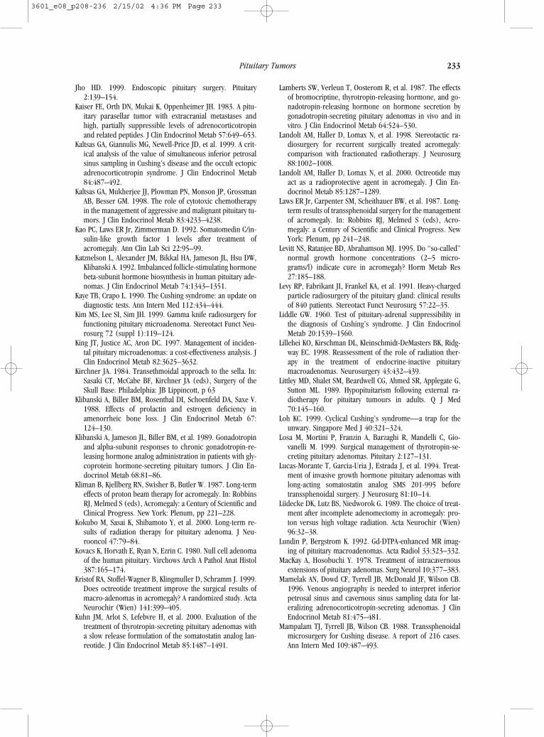
Jho HD. 1999. Endoscopic pituitary surgery. Pituitary2:139–154.
Kaiser FE, Orth DN, Mukai K, Oppenheimer JH. 1983. A pitu-itary parasellar tumor with extracranial metastases andhigh, partially suppressible levels of adrenocorticotropinand related peptides. J Clin Endocrinol Metab 57:649–653.
Kaltsas GA, Giannulis MG, Newell-Price JD, et al. 1999. A crit-ical analysis of the value of simultaneous inferior petrosalsinus sampling in Cushing’s disease and the occult ectopicadrenocorticotropin syndrome. J Clin Endocrinol Metab84:487–492.
Kaltsas GA, Mukherjee JJ, Plowman PN, Monson JP, GrossmanAB, Besser GM. 1998. The role of cytotoxic chemotherapyin the management of aggressive and malignant pituitary tu-mors. J Clin Endocrinol Metab 83:4233–4238.
Kao PC, Laws ER Jr, Zimmerman D. 1992. Somatomedin C/in-sulin-like growth factor 1 levels after treatment ofacromegaly. Ann Clin Lab Sci 22:95–99.
Katznelson L, Alexander JM, Bikkal HA, Jameson JL, Hsu DW,Klibanski A. 1992. Imbalanced follicle-stimulating hormonebeta-subunit hormone biosynthesis in human pituitary ade-nomas. J Clin Endocrinol Metab 74:1343–1351.
Kaye TB, Crapo L. 1990. The Cushing syndrome: an update ondiagnostic tests. Ann Intern Med 112:434–444.
Kim MS, Lee SI, Sim JH. 1999. Gamma knife radiosurgery forfunctioning pituitary microadenoma. Stereotact Funct Neu-rosurg 72 (suppl 1):119–124.
King JT, Justice AC, Aron DC. 1997. Management of inciden-tal pituitary microadenomas: a cost-effectiveness analysis. JClin Endocrinol Metab 82:3625–3632.
Kirchner JA. 1984. Transethmoidal approach to the sella. In:Sasaki CT, McCabe BF, Kirchner JA (eds), Surgery of theSkull Base. Philadelphia: JB Lippincott, p 63
Klibanski A, Biller BM, Rosenthal DI, Schoenfeld DA, Saxe V.1988. Effects of prolactin and estrogen deficiency in amenorrheic bone loss. J Clin Endocrinol Metab 67:124–130.
Klibanski A, Jameson JL, Biller BM, et al. 1989. Gonadotropinand alpha-subunit responses to chronic gonadotropin-re-leasing hormone analog administration in patients with gly-coprotein hormone-secreting pituitary tumors. J Clin En-docrinol Metab 68:81–86.
Kliman B, Kjellberg RN, Swisher B, Butler W. 1987. Long-termeffects of proton beam therapy for acromegaly. In: RobbinsRJ, Melmed S (eds), Acromegaly: a Century of Scientific andClinical Progress. New York: Plenum, pp 221–228.
Kokubo M, Sasai K, Shibamoto Y, et al. 2000. Long-term re-sults of radiation therapy for pituitary adenoma. J Neu-rooncol 47:79–84.
Kovacs K, Horvath E, Ryan N, Ezrin C. 1980. Null cell adenomaof the human pituitary. Virchows Arch A Pathol Anat Histol387:165–174.
Kristof RA, Stoffel-Wagner B, Klingmuller D, Schramm J. 1999.Does octreotide treatment improve the surgical results ofmacro-adenomas in acromegaly? A randomized study. ActaNeurochir (Wien) 141:399–405.
Kuhn JM, Arlot S, Lefebvre H, et al. 2000. Evaluation of thetreatment of thyrotropin-secreting pituitary adenomas witha slow release formulation of the somatostatin analog lan-reotide. J Clin Endocrinol Metab 85:1487–1491.
Lamberts SW, Verleun T, Oosterom R, et al. 1987. The effectsof bromocriptine, thyrotropin-releasing hormone, and go-nadotropin-releasing hormone on hormone secretion bygonadotropin-secreting pituitary adenomas in vivo and invitro. J Clin Endocrinol Metab 64:524–530.
Landolt AM, Haller D, Lomax N, et al. 1998. Stereotactic ra-diosurgery for recurrent surgically treated acromegaly:comparison with fractionated radiotherapy. J Neurosurg88:1002–1008.
Landolt AM, Haller D, Lomax N, et al. 2000. Octreotide mayact as a radioprotective agent in acromegaly. J Clin En-docrinol Metab 85:1287–1289.
Laws ER Jr, Carpenter SM, Scheithauer BW, et al. 1987. Long-term results of transsphenoidal surgery for the managementof acromegaly. In: Robbins RJ, Melmed S (eds), Acro-megaly: a Century of Scientific and Clinical Progress. NewYork: Plenum, pp 241–248.
Levitt NS, Ratanjee BD, Abrahamson MJ. 1995. Do “so-called”normal growth hormone concentrations (2–5 micro-grams/l) indicate cure in acromegaly? Horm Metab Res27:185–188.
Levy RP, Fabrikant JI, Frankel KA, et al. 1991. Heavy-chargedparticle radiosurgery of the pituitary gland: clinical resultsof 840 patients. Stereotact Funct Neurosurg 57:22–35.
Liddle GW. 1960. Test of pituitary-adrenal suppressibility inthe diagnosis of Cushing’s syndrome. J Clin EndocrinolMetab 20:1539–1560.
Lillehei KO, Kirschman DL, Kleinschmidt-DeMasters BK, Ridg-way EC. 1998. Reassessment of the role of radiation ther-apy in the treatment of endocrine-inactive pituitarymacroadenomas. Neurosurgery 43:432–439.
Littley MD, Shalet SM, Beardwell CG, Ahmed SR, Applegate G,Sutton ML. 1989. Hypopituitarism following external ra-diotherapy for pituitary tumours in adults. Q J Med70:145–160.
Loh KC. 1999. Cyclical Cushing’s syndrome—a trap for theunwary. Singapore Med J 40:321–324.
Losa M, Mortini P, Franzin A, Barzaghi R, Mandelli C, Gio-vanelli M. 1999. Surgical management of thyrotropin-se-creting pituitary adenomas. Pituitary 2:127–131.
Lucas-Morante T, Garcia-Uria J, Estrada J, et al. 1994. Treat-ment of invasive growth hormone pituitary adenomas withlong-acting somatostatin analog SMS 201-995 beforetranssphenoidal surgery. J Neurosurg 81:10–14.
Lüdecke DK, Lutz BS, Niedworok G. 1989. The choice of treat-ment after incomplete adenomectomy in acromegaly: pro-ton versus high voltage radiation. Acta Neurochir (Wien)96:32–38.
Lundin P, Bergstrom K. 1992. Gd-DTPA-enhanced MR imag-ing of pituitary macroadenomas. Acta Radiol 33:323–332.
MacKay A, Hosobuchi Y. 1978. Treatment of intracavernousextensions of pituitary adenomas. Surg Neurol 10:377–383.
Mamelak AN, Dowd CF, Tyrrell JB, McDonald JF, Wilson CB.1996. Venous angiography is needed to interpret inferiorpetrosal sinus and cavernous sinus sampling data for lat-eralizing adrenocorticotropin-secreting adenomas. J ClinEndocrinol Metab 81:475–481.
Mampalam TJ, Tyrrell JB, Wilson CB. 1988. Transsphenoidalmicrosurgery for Cushing disease. A report of 216 cases.Ann Intern Med 109:487–493.
Pituitary Tumors 233
3601_e08_p208-236 2/15/02 4:36 PM Page 233

Maraschini C, Moro M, Masala A, et al. 1991. Chronic treat-ment with parlodel LAR of patients with prolactin-secretingtumours. Different responsiveness of micro- and macro-prolactinomas. Acta Endocrinol (Copenh) 125:494–501.
March CM, Kletzky OA, Davajan V, et al. 1981. Longitudinalevaluation of patients with untreated prolactin-secreting pi-tuitary adenomas. Am J Obstet Gynecol 139:835–844.
McCutcheon IE, Kitagawa RS, DeMasi PF, et al. 2001. Frame-less stereotactic navigation in transsphenoidal surgery:comparison with fluoroscopy. Neurosurgery (in press).
McCutcheon IE, Oldfield EH. 1992. Cortisol: regulation, dis-orders, and clinical evaluation. In: Barrow DL, Selman WR(eds), Neuroendocrinology. Baltimore: Williams & Wilkins,pp 117–173.
McCutcheon IE, Pieper DR, Fuller GN, Benjamin RS, FriendKE, Gagel RF. 2000. Pituitary carcinoma containing gonad-otropins: treatment by radical excision and cytotoxic che-motherapy: case report. Neurosurgery 46:1233–1240.
McCutcheon IE, Weintraub BD, Oldfield EH. 1990. Surgicaltreatment of thyrotropin-secreting pituitary adenomas. JNeurosurg 73:674–683.
Melmed S. 1990. Acromegaly. N Engl J Med 322:966–977.Melmed S, Ezrin C, Kovacs K, Goodman RS, Frohman LA. 1985.
Acromegaly due to secretion of growth hormone by an ec-topic pancreatic islet-cell tumor. N Engl J Med 312:9–17.
Melmed S, Jackson I, Kleinberg D, Klibanski A. 1998. Currenttreatment guidelines for acromegaly. J Clin EndocrinolMetab 83:2646–2652.
Merola B, Sarnacchiaro F, Colao A, et al. 1994. Positive re-sponse to compound CV 205-502 in hyperprolactinemic pa-tients resistant to or intolerant of bromocriptine. GynecolEndocrinol 8:175–181.
Mixson AJ, Friedman TC, Katz DA, et al. 1993. Thyrotropin-se-creting pituitary carcinoma. J Clin Endocrinol Metab76:529–533.
Molitch ME, Elton RL, Blackwell RE, et al. 1985. Bromocrip-tine as primary therapy for prolactin-secreting macroade-nomas: results of a prospective multicenter study. J Clin En-docrinol Metab 60:698–705.
Moore TJ, Dluhy RG, Williams GH, Cain JP. 1976. Nelson’s syn-drome: frequency, prognosis, and effect of prior pituitaryirradiation. Ann Intern Med 85:731–734.
Mosnier-Pudar H, Thomopoulos P, Bertagna X, Fournier C,Guiban D, Luton JP. 1995. Long-distance and long-term fol-low-up of a patient with intermittent Cushing’s disease by sali-vary cortisol measurements. Eur J Endocrinol 133:313–316.
Mountcastle RB, Roof BS, Mayfield RK, et al. 1989. Pituitaryadenocarcinoma in an acromegalic patient: response tobromocriptine and pituitary testing: a review of the litera-ture on 36 cases of pituitary carcinoma. Am J Med Sci298:109–118.
Nagesser SK, van Seters AP, Kievit J, Hermans J, Krans HM, vande Velde CJ. 2000. Long-term results of total adrenalectomyfor Cushing’s disease. World J Surg 24:108–113.
Nelson DH, Meakin JW, Thorn GW. 1960. ACTH-producing tu-mors following adrenalectomy for Cushing’s syndrome. AnnIntern Med 52:560–569.
Newman CB, Melmed S, George A, et al. 1998. Octreotide asprimary therapy for acromegaly. J Clin Endocrinol Metab83:3034–3040.
Nieman LK, Chrousos GP, Oldfield EH, Avgerinos PC, CutlerGB, Loriaux DI. 1986. The ovine corticotropin-releasinghormone stimulation test and the dexamethasone suppres-sion test in the differential diagnosis of Cushing’s syndrome.Ann Intern Med 105:862–867.
Nilsson B, Gustavasson-Kadaka E, Bengtsson BA, Jonsson B.2000. Pituitary adenomas in Sweden between 1958 and1991: incidence, survival, and mortality. J Clin EndocrinolMetab 85:1420–1425.
Nobels FR, Kwekkeboom DJ, Coopmans W, et al. 1993. A com-parison between the diagnostic value of gonadotropins, al-pha-subunit, and chromogranin-A and their response tothyrotropin-releasing hormone in clinically nonfunctioning,alpha-subunit–secreting, and gonadotroph pituitary adeno-mas. J Clin Endocrinol Metab 77:784–789.
Ogilvy KM, Jakubowski J. 1973. Intracranial dissemination ofpituitary adenomas. J Neurol Neurosurg Psychiatry 36:199–205.
Oldfield EH, Doppman JL, Nieman LK, et al. 1991. Petrosal si-nus sampling with and without corticotropin-releasing hor-mone for the differential diagnosis of Cushing’s syndrome.N Engl J Med 325:897–905 (erratum in N Engl J Med326:1172, 1992).
Olson BR, Gumowski J, Rubino D, Oldfield EH. 1997. Patho-physiology of hyponatremia after transsphenoidal pituitarysurgery. J Neurosurg 87:499–507.
Oppenheim DS, Kana AR, Sangha JS, Klibanski A. 1990. Preva-lence of alpha-subunit hypersecretion in patients with pi-tuitary tumors: clinically nonfunctioning and somatotrophadenomas. J Clin Endocrinol Metab 70:859–864.
Origitano TC, Al-Mefty O, Leonetti JP, DeMonte F, ReichmanOH. 1994. Vascular considerations and complications incranial base surgery. Neurosurgery 35:351–363.
Pan L, Zhang N, Wang E, Wang B, Xu W. 1998. Pituitary ade-nomas: the effect of gamma knife radiosurgery on tumorgrowth and endocrinopathies. Stereotact Funct Neurosurg70:119–126.
Paramo C, Andrade O MA, Fluiters E, Luna R, de la Fuente J,Garcia-Mayor RV. 1997. Comparative study of insulin-likegrowth factor-I (IGF-I) and IGF-binding protein-3 (IGFBP-3) level and IGF-I/IGFBP-3 ratio measurements and theirrelationship with an index of clinical activity in the man-agement of patients with acromegaly. Metabolism 46:494–498.
Parent AD, Bebin J, Smith RR. 1981. Incidental pituitary ade-nomas. J Neurosurg 54:228–231.
Perks JR, Jalali R, Cosgrove VP, et al. 1999. Optimization ofstereotactically-guided conformal treatment planning of sellar and parasellar tumors, based on normal brain dosevolume histograms. Int J Radiat Oncol Biol Phys 45:507–513.
Pernicone PJ, Scheithauer BW, Sebo TJ, et al. 1997. Pituitarycarcinoma: a clinicopathologic study of 15 cases. Cancer79:804–812.
Petterson T, MacFarlane IA, MacKenzie JM, Shaw MD. 1992.Prolactin secreting pituitary carcinoma. J Neurol NeurosurgPsychiatry 55:1205–1206.
Pivonello R, Faggiano A, Di Salle F, et al. 1999. Complete re-mission of Nelson’s syndrome after 1-year treatment withcabergoline. J Endocrinol Invest 22:860–865.
234 PRIMARY CENTRAL NERVOUS SYSTEM TUMORS
3601_e08_p208-236 2/15/02 4:36 PM Page 234
Plataniotis GA, Kouvaris JR, Vlahos L, Papavasiliou C. 1998.Radiation therapy alone for growth hormone–producing pi-tuitary adenomas. Acta Oncol 37:97–99.
Plowman PN, Doughty D. 1999. Stereotactic radiosurgery, X:clinical isodosimetry of gamma knife versus accelerator X-knife for pituitary and acoustic tumours. Clin Oncol (R CollRadiol) 11:321–329.
Pluta RM, Nieman L, Doppman JL, et al. 1999. Extrapituitaryparasellar microadenoma in Cushing’s disease. J Clin En-docrinol Metab 84:2912–2923.
Ram Z, Nieman LK, Cutler GB, Chrousas GP, Doppman JL, Old-field EH. 1994. Early repeat surgery for persistent Cushing’sdisease. J Neurosurg 80:37–45.
Randall RV, Laws ER, Abboud CF, Ebersold MJ, Kao PC, Schei-thauer BW. 1983. Transsphenoidal microsurgical treatmentof prolactin-producing pituitary adenomas: results in 100patients. Mayo Clin Proc 58:108–121.
Randeva HS, Schoebel J, Byrne J, Esiri M, Adams CB, Wass JA.1999. Classical pituitary apoplexy: clinical features, man-agement and outcome. Clin Endocrinol (Oxf) 51:181–188.
Ridgway EC, Klibanski A, Ladenson PW, et al. 1981. Pure al-pha-secreting pituitary adenomas. N Engl J Med 304:1254–1259.
Rodman EF, Molitch ME, Post KD, Biller BJ, Reichlin S. 1983.Long-term follow-up of transsphenoidal selective ade-nomectomy for prolactinoma. JAMA 252:921–924.
Ross DA, Wilson CB. 1988. Results of transsphenoidal micro-surgery for growth hormone–secreting pituitary adenomain a series of 214 patients. J Neurosurg 68:854–867.
Rothman LM, Sher J, Quencer RM, Tenner MS. 1976. In-tracranial ectopic pituitary adenoma. Case report. J Neuro-surg 44:96–99.
Sandeman D, Moufid A. 1998. Interactive image-guided pitu-itary surgery. An experience of 101 procedures. Neu-rochirurgie 44:331–338.
Sano T, Yamada S. 1994. Histologic and immunohistochem-cial study of clinically non-functioning pituitary adenomas:special reference to gonadotropin-positive adenomas.Pathol Int 44:697–703.
Sasaki R, Murakami M, Okamoto Y, et al. 2000. The efficacyof conventional radiation therapy in the management of pi-tuitary adenoma. Int J Radiat Oncol Biol Phys 47:1337–1345.
Scheithauer BW. 1984. Surgical pathology of the pituitary: theadenomas. Part I. Pathol Annu 19:317–374.
Schlechte J, Dolan K, Sherman B, Chapler F, Luciano A. 1989.The natural history of untreated hyperprolactinemia: aprospective analysis. J Clin Endocrinol Metab 68:412–418.
Selman WR, Laws ER, Scheithauer BW, Carpenter SM. 1986.The occurrence of dural invasion in pituitary adenomas. JNeurosurg 64:402–407.
Serri O, Rasio E, Beauregard H, Hardy J, Somma M. 1983. Re-currence of hyperprolactinemia after selective transsphe-noidal adenomectomy in women with prolactinoma. N EnglJ Med 309:280–283.
Shaffi OM, Wrightson P. 1975. Dural invasion by pituitary tu-mors. NZ Med J 81:386–390.
Sheehan MT, Atkinson JL, Kasperbauer JL, Erickson BJ, Nip-poldt TB. 1999. Preliminary comparison of the endoscopictransnasal vs. the sublabial transseptal approach for clini-
cally non-functioning pituitary macroadenomas. Mayo ClinProc 74:661–670.
Sheline GE, Grossman A, Jones AE, Besser GM. 1984. Radia-tion therapy for prolactinomas. In: Black PM, Zervas NT,Ridgway EC, Martin JB (eds), Secretory Tumors of the Pi-tuitary Gland. New York: Raven Press, pp 93–108.
Silbergeld DL, Mayberg MR, Berger MS, Ali-Osman F, Kelly WA,Shaw CM. 1993. Pituitary oncocytomas: clinical features,characteristics in cell culture, and treatment recommenda-tions. J Neurooncol 16:39–46.
Snyder PJ. 1997. Gonadotroph and other clinically nonfunc-tioning pituitary adenomas. Cancer Treat Res 89:57–72.
Snyder PJ, Bashey HM, Kim SU, Chappel SC. 1984. Secretionof uncombined subunits of luteinizing hormone by go-nadotroph cell adenomas. J Clin Endocrinol Metab59:1169–1175.
Soule SG, Farhi J, Conway GS, Jacobs HS, Powell M. 1996. Theoutcome of hypophysectomy for prolactinomas in the eraof dopamine agonist therapy. Clin Endocrinol (Oxf)44:711–716.
Stevenaert A, Beckers A. 1993. Presurgical octreotide treat-ment in acromegaly. Acta Endocrinol (Copenh) 129:18–20.
Tamura M, Yokoyama N, Abe Y, et al. 1998. Preoperative treat-ment of growth hormone-producing pituitary adenoma withcontinuous subcutaneous infusion of octreotide. Endocr J45:269–275.
Teramoto A, Hirakawa K, Sanno N, Osamura Y. 1994. Inci-dental pituitary lesions in 1,000 unselected autopsy speci-mens. Radiology 193:161–164.
Tindall GT, Oyesiku NM, Watts NB, Clark RV, Christy JH, AdamsDA. 1993. Transsphenoidal adenomectomy for growth hor-mone-secreting pituitary adenomas in acromegaly: outcomeanalysis and determinants of failure. J Neurosurg78:205–215.
Tindall GT, Tindall SC. 1987. Pterional approach. In: ApuzzoMLJ (ed), Surgery of the Third Ventricle. Baltimore:Williams & Wilkins, p. 440–461.
Tishler RB, Loeffler JS, Lunsford LD, et al. 1993. Tolerance ofcranial nerves of the cavernous sinus to radiosurgery. Int JRadiat Oncol Biol Phys 27:215–221.
Tomita T, Gates E. 1999. Pituitary adenomas and granular celltumors. Incidence, cell type, and location of tumor in 100pituitary glands at autopsy. Am J Clin Pathol 111:817–825.
Trainer PJ, Drake WM, Katznelson L, et al. 2000. Treatment ofacromegaly with the growth hormone–receptor antagonistpegvisomant. N Engl J Med 342:1171–1177.
Trouillas J, Sassolas G, Loras B, et al. 1991. Somatotropic adenomas without acromegaly. Pathol Res Pract 187:943–949.
Turner HE, Stratton IM, Byrne JV, Adams CB, Wass JA. 1999.Audit of selected patients with nonfunctioning pituitary ade-nomas treated without irradiation—a follow-up study. ClinEndocrinol 51:281–284.
Tyrrell JB, Lamborn KR, Hannegan LT, Applebury CB, WilsonCB. 1999. Transsphenoidal microsurgical therapy of pro-lactinomas: initial outcomes and long-term results. Neuro-surgery 44:254–261.
Van Cauter E, Refetoff S. 1985. Evidence for two subtypes ofCushing’s disease based on the analysis of episodic corti-sol secretion. N Engl J Med 312:1343–1349.
Pituitary Tumors 235
3601_e08_p208-236 2/15/02 4:36 PM Page 235
Verhelst J, Abs R, Maiter D, et al. 1999. Cabergoline in thetreatment of hyperprolactinemia: a study in 455 patients. JClin Endocrinol Metab 84:2518–2522.
Verhelst JA, Pedroncelli AM, Abs R, et al. 2000. Slow-releaselanreotide in the treatment of acromegaly: a study in 66 pa-tients. Eur J Endocrinol 143:577–584.
Waltz TA, Brownell B. 1966. Sarcoma: a possible late result ofeffective radiation therapy for pituitary adenoma. Report oftwo cases. J Neurosurg 24:901–907.
Warnet A, Harris AG, Renard E, Martin D, James-Deidier A,Chaumet-Riffaud P. 1997. A prospective multicenter trial ofoctreotide in 24 patients with visual defects caused by non-functioning and gonadotropin-secreting pituitary adeno-mas. French Multicenter Octreotide Study Group. Neuro-surgery 41:786–797.
Warnet A, Porsova-Dutoit I, Lahlou N, et al. 1994. Glycopro-tein hormone alpha-subunit secretion in prolactinomas andin non-functioning adenomas: relation with the tumour size.Clin Endocrinol (Oxf) 41:177–184.
Warnet A, Timsit J, Chanson P, et al. 1989. The effect of so-matostatin analogue on chiasmal dysfunction from pituitarymacroadenomas. J Neurosurg 71:687–690.
Wass JAH, Thorner MO, Morris DV, et al. 1977. Long-term treat-ment of acromegaly with bromocriptine. BMJ 1:875–878.
Webb SM, Rigla M, Wagner A, Oliver B, Bartumeus F. 1999.Recovery of hypopituitarism after neurosurgical treatmentof pituitary adenomas. J Clin Endocrinol Metab 84:3696–3700.
Webb SM, Ruscalleda J, Schwarzstein D, et al. 1992. Comput-erized tomography versus magnetic resonance imaging: acomparative study in hypothalamic-pituitary and parasellarpathology. Clin Endocrinol (Oxf) 36:459–465.
Williams M, van Seters AP, Hermans J, Leer JW. 1994. Evalu-ation of the effects of radiotherapy on macroprolactinomasusing the decline rate of serum prolactin levels as a dy-namic parameter. Clin Oncol (R Coll Radiol) 6:102–109.
Wright AD, Hill DM, Lowy C, Fraser TR. 1970. Mortality inacromegaly. Q J Med 39:1–16.
Young M, Kattner K, Gupta K. 1999. Pituitary hyperplasia re-sulting from primary hypothyroidism mimicking macro-adenomas. Br J Neurosurg 13:138–142.
Young WF, Scheithauer BW, Kovacs KT, Horvath E, Davis DH,Randall RV. 1996. Gonadotroph adenoma of the pituitarygland: a clinicopathological analysis of 100 cases. Mayo ClinProc 71:649–656.
Zhang N, Pan L, Wang EM, Dai JZ, Wang BJ, Cai PW. 2000.Radiosurgery for growth hormone-producing pituitary ade-nomas. J Neurosurg 93:6–9.
236 PRIMARY CENTRAL NERVOUS SYSTEM TUMORS
3601_e08_p208-236 2/15/02 4:36 PM Page 236
Related Documents