PIM2 & PIM3 for the ANZPIC Registry Information Booklet Version Mar2016

Welcome message from author
This document is posted to help you gain knowledge. Please leave a comment to let me know what you think about it! Share it to your friends and learn new things together.
Transcript

PIM2 & PIM3 for the ANZPIC Registry
Information Booklet
Version Mar2016

(page intentionally left blank)
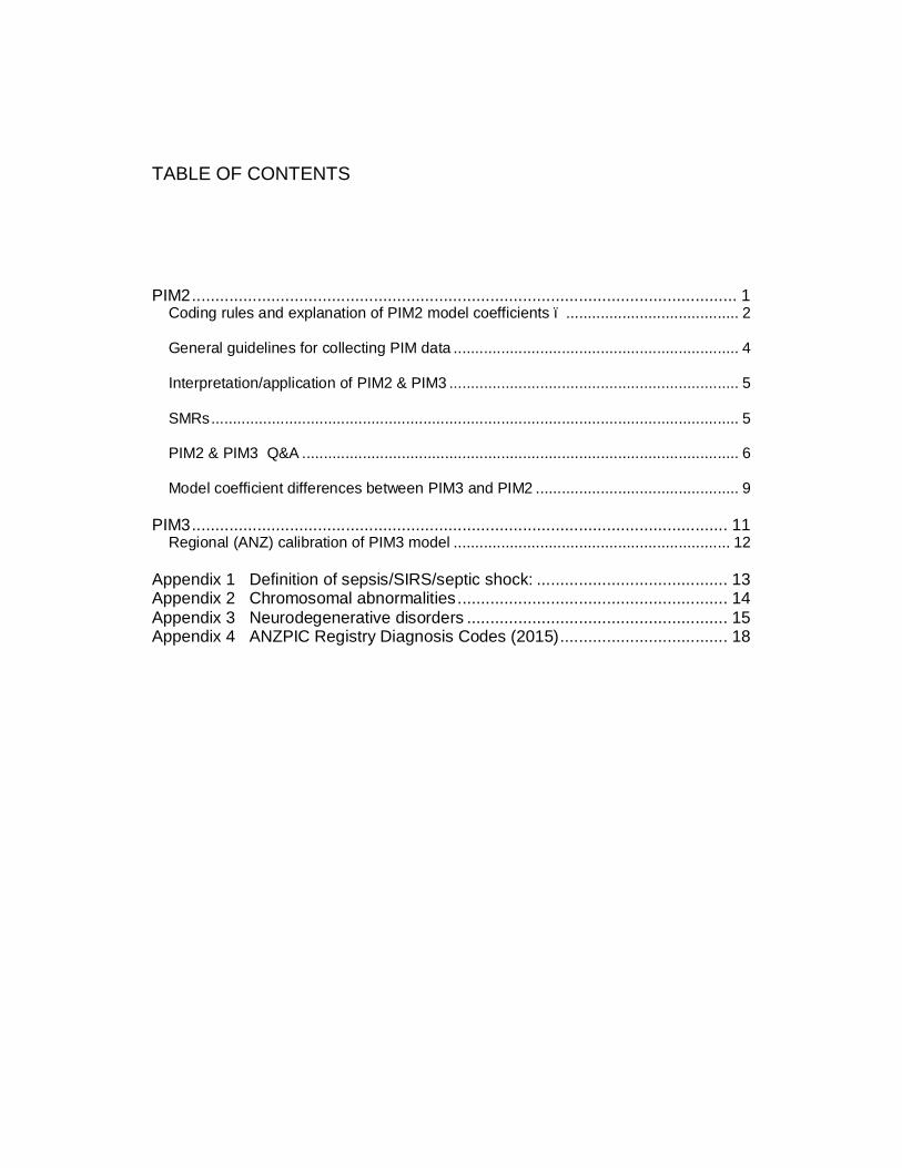
TABLE OF CONTENTS
PIM2 ..................................................................................................................... 1 Coding rules and explanation of PIM2 model coefficients – ........................................ 2
General guidelines for collecting PIM data .................................................................. 4
Interpretation/application of PIM2 & PIM3 ................................................................... 5
SMRs .......................................................................................................................... 5
PIM2 & PIM3 Q&A ..................................................................................................... 6
Model coefficient differences between PIM3 and PIM2 ............................................... 9
PIM3 ................................................................................................................... 11 Regional (ANZ) calibration of PIM3 model ................................................................ 12
Appendix 1 Definition of sepsis/SIRS/septic shock: ......................................... 13 Appendix 2 Chromosomal abnormalities .......................................................... 14 Appendix 3 Neurodegenerative disorders ........................................................ 15 Appendix 4 ANZPIC Registry Diagnosis Codes (2015) .................................... 18

- 1 -
PIM2
Reference: A. Slater et al. PIM2 : a revised version of the Paediatric Index of Mortality. Intensive Care Med 2003 ;29:278- 85.
PIM2 is calculated from the information collected at the time a child is admitted to your ICU. Because PIM2 describes how ill the child was at the time you started intensive care, the observations to be recorded are those made at or about the time of first face-to-face (not telephone) contact between the patient and a doctor from your intensive care unit (or a doctor from a specialist paediatric transport team). Use the first value of each variable measured within the period from the time of first contact to 1 hour after arrival in your ICU. The first contact may be in your ICU, your emergency department, a ward in your own hospital, or in another hospital (e.g. on a retrieval). If information is missing, see the documentation for each field (model coefficient) for the correct coding practice.
Calculation of PIM2 (and PIM2 risk of death%)–
PIM2val= (0.01395 * (absolute(SBP-120))) + (3.0791 * Pupils) + (0.2888 * (100*FiO2/PaO2)) + (0.1040 * (absolute Base Excess)) + (1.3352 * MechVent) – (0.9282 * Elective) – (1.0244 * Recovery) + (0.7507 * Bypass) + (1.6829 * HRdiag) – (1.5770 * LRdiag) -4.8841
PIM2 risk of death = ePIM2val / (1 + ePIM2val )
Example of PIM2 calculation A patient with hypoplastic left heart syndrome is admitted to intensive care for recovery following an elective Norwood procedure. At the time of admission he is ventilated. The first recorded systolic blood pressure is 55 mmHg, PaO2 is 110 mmHg, FiO2 0.5, base excess -6.0. The pupils are reactive to light. (The low risk diagnoses do not apply to this case).
PIM2val = (0.01395* [absolute(55–120)) + (3.0791*0) + (0.2888* (100*0.5/110)) + (0.104* |–6.0|) + (1.3352* 1) – (0.9282* 1) – (1.0244* 1) +(0.7507 * 1) + (1.6829* 1) – (1.5770 * 0) – 4.8841
= –1.4059
Probability of Death = exp (–1.4059) / [1+exp (–1.4059)]
= 0.1969 or 19.7%
- 2 -
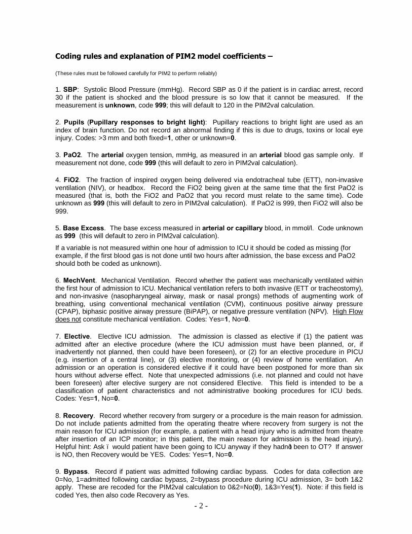
Coding rules and explanation of PIM2 model coefficients –
(These rules must be followed carefully for PIM2 to perform reliably)
1. SBP: Systolic Blood Pressure (mmHg). Record SBP as 0 if the patient is in cardiac arrest, record 30 if the patient is shocked and the blood pressure is so low that it cannot be measured. If the measurement is unknown, code 999; this will default to 120 in the PIM2val calculation.
2. Pupils (Pupillary responses to bright light): Pupillary reactions to bright light are used as an index of brain function. Do not record an abnormal finding if this is due to drugs, toxins or local eye injury. Codes: >3 mm and both fixed=1, other or unknown=0.
3. PaO2. The arterial oxygen tension, mmHg, as measured in an arterial blood gas sample only. If measurement not done, code 999 (this will default to zero in PIM2val calculation).
4. FiO2. The fraction of inspired oxygen being delivered via endotracheal tube (ETT), non-invasive ventilation (NIV), or headbox. Record the FiO2 being given at the same time that the first PaO2 is measured (that is, both the FiO2 and PaO2 that you record must relate to the same time). Code unknown as 999 (this will default to zero in PIM2val calculation). If PaO2 is 999, then FiO2 will also be 999.
5. Base Excess. The base excess measured in arterial or capillary blood, in mmol/l. Code unknown as 999 (this will default to zero in PIM2val calculation).
If a variable is not measured within one hour of admission to ICU it should be coded as missing (for example, if the first blood gas is not done until two hours after admission, the base excess and PaO2 should both be coded as unknown).
6. MechVent. Mechanical Ventilation. Record whether the patient was mechanically ventilated within the first hour of admission to ICU. Mechanical ventilation refers to both invasive (ETT or tracheostomy), and non-invasive (nasopharyngeal airway, mask or nasal prongs) methods of augmenting work of breathing, using conventional mechanical ventilation (CVM), continuous positive airway pressure (CPAP), biphasic positive airway pressure (BiPAP), or negative pressure ventilation (NPV). High Flow does not constitute mechanical ventilation. Codes: Yes=1, No=0.
7. Elective. Elective ICU admission. The admission is classed as elective if (1) the patient was admitted after an elective procedure (where the ICU admission must have been planned, or, if inadvertently not planned, then could have been foreseen), or (2) for an elective procedure in PICU (e.g. insertion of a central line), or (3) elective monitoring, or (4) review of home ventilation. An admission or an operation is considered elective if it could have been postponed for more than six hours without adverse effect. Note that unexpected admissions (i.e. not planned and could not have been foreseen) after elective surgery are not considered Elective. This field is intended to be a classification of patient characteristics and not administrative booking procedures for ICU beds. Codes: Yes=1, No=0.
8. Recovery. Record whether recovery from surgery or a procedure is the main reason for admission. Do not include patients admitted from the operating theatre where recovery from surgery is not the main reason for ICU admission (for example, a patient with a head injury who is admitted from theatre after insertion of an ICP monitor; in this patient, the main reason for admission is the head injury). Helpful hint: Ask – would patient have been going to ICU anyway if they hadn’t been to OT? If answer is NO, then Recovery would be YES. Codes: Yes=1, No=0.
9. Bypass. Record if patient was admitted following cardiac bypass. Codes for data collection are 0=No, 1=admitted following cardiac bypass, 2=bypass procedure during ICU admission, 3= both 1&2 apply. These are recoded for the PIM2val calculation to 0&2=No(0), 1&3=Yes(1). Note: if this field is coded Yes, then also code Recovery as Yes.

- 3 -
10. HRdiag. PIM2 High Risk Diagnosis (ANZPICR field PIM_UC). Specific conditions** associated with increased mortality risk. Codes for data collection are - 0 None 1 Cardiac arrest out of hospital - requires either documented absent pulse or the requirement for
external cardiac massage (do not include past history of cardiac arrest). 2 Severe combined immune deficiency - requires the documented diagnosis of SCID. 3 Leukaemia or lymphoma after 1st induction. Include only cases where the admission is related
to leukaemia or lymphoma, or the therapy for these conditions. 4 Spontaneous cerebral haemorrhage - haemorrhage must be spontaneous (for example, from
an aneurysm or AVM). Do not include traumatic cerebral haemorrhage or intracranial haemorrhage that is not intracerebral (eg subdural haemorrhage).
5 Cardiomyopathy or myocarditis - requires the documented diagnosis of myocarditis or cardiomyopathy.
6 Hypoplastic left heart syndrome – at any age on admission, but include only cases where a Norwood procedure, or equivalent, is required in the neonatal period to sustain life. If a subsequent heart transplant, then this diagnosis and high risk indicator no longer apply.
7 HIV infection - requires the document diagnosis of HIV. 8 Code no longer in use (2009 onwards) 9 Neurodegenerative disorder - requires a history of progressive loss of milestones (even if no
specific condition has been diagnosed) or a diagnosis where this will inevitably occur. 10 Liver failure - acute or chronic, is the main reason for ICU admission. Include patients
admitted for recovery following liver transplantation for acute or chronic liver failure. 11 Cardiac arrest in hospital - preceding ICU admission requires either documented absent pulse
or the requirement for external cardiac massage (do not include past history of cardiac arrest) These are recoded for the PIM2val calculation to 0=No(0), 1-11=Yes(1).
11. LRdiag. PIM2 Low Risk Diagnosis (ANZPICR field PIM_LR). Specific conditions** associated with decreased mortality risk - which must be the main reason for admission. Codes for data collection are – 0 None 1 Asthma is the main reason for ICU admission. 2 Bronchiolitis is the main reason for admission. Include children who present either with
respiratory distress or central apnoea where the clinical diagnosis is bronchiolitis. 3 Croup is the main reason for ICU admission. 4 Obstructive sleep apnoea (OSA) is the main reason for ICU admission. Include admissions
following adenoidectomy &/or tonsillectomy where OSA is the main or underlying reason for ICU admission (and also code as recovery from surgery).
5 Diabetic keto-acidosis (DKA) is the main reason for ICU admission. These are recoded for the PIM2val calculation to 0=No(0), 1-5=Yes(1). ** See Appendix 4 for current ANZPIC Registry diagnosis list

- 4 -
General guidelines for collecting PIM data
1. Do not over-diagnose the specified conditions - if there is any doubt, do not record a specified condition. For example: do not code cerebral haemorrhage for intracerebral bleeding associated with trauma ; impaired cardiac function associated with sepsis or surgery should not be coded as cardiomyopathy ; and a static disability should not be coded as neurodegenerative (even if it is severe) unless there is progressive ongoing loss of milestones.
2. You should record the first value of each variable from the time of first contact up to one hour after arrival in your ICU (not the worst value). If a variable is not measured within one hour of admission to ICU it should be coded as unknown (for example, if the first blood gas is not done until two hours after admission, the base excess and PaO2 should both be coded as unknown). Missing data is treated as being normal when PIM is calculated.
3. The PIM equation is used to calculate the PIMval. If you are using your own software to calculate the PIMval, and if any information is missing, that variable should add nothing to PIMval. For example, if the base excess is missing, the value of "(0.0671 * (absolute Base Excess))" for PIM3 should be set to zero. The exception to this rule is for the PaO2/FiO2 ratio in PIM3, where a value of 0.23 has been clinically regarded as being a better “normal” substitute than the zero value in the PIM2 model. (see section on differences between PIM3 and PIM2)
4. Record the FiO2 being given at the same time that the first PaO2 is measured (that is, both the FiO2 and PaO2 that you record must relate to the same time). PaO2 must be arterial.
5. Read very carefully the definition for the ELECTIVE field - noting in particular that unexpected ICU admissions (i.e. not planned and that could not have been foreseen) after elective surgery are not classed as Elective.
6. The pupils are only recorded as fixed if both are >3 mm, and both are fixed, and the finding is not caused by drugs or toxins or direct injury to the eye.
7. If systolic blood pressure is not measured in the first hour, record 999 - do not record zero.
8. If the field RS_HR124 (mechanical ventilation within the first hour of ICU admission) is “yes” then there must be a corresponding episode of respiratory support (either invasive or non-invasive) recorded. Episodes of HFNC do not qualify as mechanical respiratory support.
9. When carrying out a self-audit at your site, randomly sample about every 20th admission to your ICU and get another person to collect the PIM2/PIM3 data independently a second time, so that you can check the accuracy of your data.
10. As the accurate recording of physiological measurements has a major impact on the calculated PIM value, it is important that each site develop a recording mechanism to minimize the risk of data interpretation or data entry errors. For example, if the values are to be extracted directly from hand-written observation charts, good local practice would be for the attending clinical staff to write the numerical values as well as plot them. As the values are taken from the first observation only, this should not add to workload.
11. You should include all admissions to your ICU, not just selected cases. For specialist PICUs this should be every admission regardless of age; for other ICUs, this should be all children whose age at admission is less than 16 years.
- 5 -
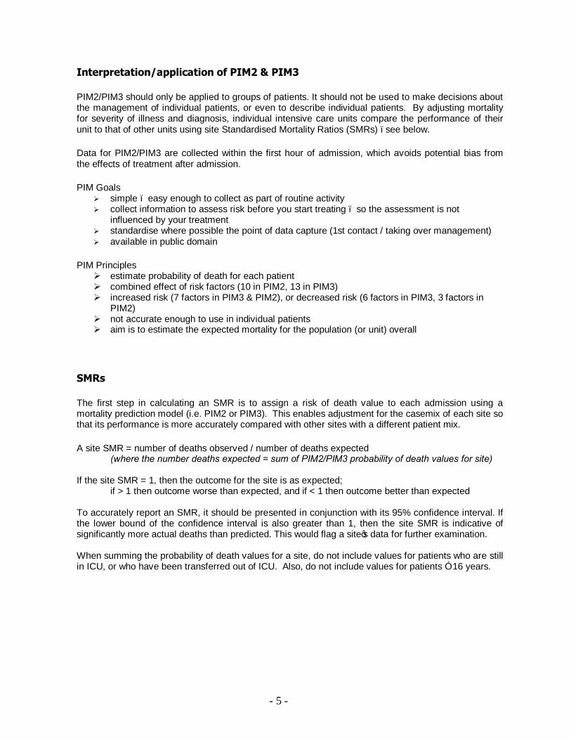
Interpretation/application of PIM2 & PIM3
PIM2/PIM3 should only be applied to groups of patients. It should not be used to make decisions about the management of individual patients, or even to describe individual patients. By adjusting mortality for severity of illness and diagnosis, individual intensive care units compare the performance of their unit to that of other units using site Standardised Mortality Ratios (SMRs) –see below.
Data for PIM2/PIM3 are collected within the first hour of admission, which avoids potential bias from the effects of treatment after admission.
PIM Goals Ø simple – easy enough to collect as part of routine activity Ø collect information to assess risk before you start treating – so the assessment is not
influenced by your treatment Ø standardise where possible the point of data capture (1st contact / taking over management) Ø available in public domain
PIM Principles Ø estimate probability of death for each patient Ø combined effect of risk factors (10 in PIM2, 13 in PIM3) Ø increased risk (7 factors in PIM3 & PIM2), or decreased risk (6 factors in PIM3, 3 factors in
PIM2) Ø not accurate enough to use in individual patients Ø aim is to estimate the expected mortality for the population (or unit) overall
SMRs
The first step in calculating an SMR is to assign a risk of death value to each admission using a mortality prediction model (i.e. PIM2 or PIM3). This enables adjustment for the casemix of each site so that its performance is more accurately compared with other sites with a different patient mix.
A site SMR = number of deaths observed / number of deaths expected (where the number deaths expected = sum of PIM2/PIM3 probability of death values for site) If the site SMR = 1, then the outcome for the site is as expected; if > 1 then outcome worse than expected, and if < 1 then outcome better than expected To accurately report an SMR, it should be presented in conjunction with its 95% confidence interval. If the lower bound of the confidence interval is also greater than 1, then the site SMR is indicative of significantly more actual deaths than predicted. This would flag a site’s data for further examination. When summing the probability of death values for a site, do not include values for patients who are still in ICU, or who have been transferred out of ICU. Also, do not include values for patients ≥ 16 years.

- 6 -
PIM2 & PIM3 Q&A Q: Clarification about what is “first contact” in collecting physiologic values for PIM when patient is a retrieval or first seen elsewhere in hosp. A: PIM values can be taken in period of first contact up to end of first hour in ICU. First contact to be defined as ICU staff (or retrieval team) taking over management of patient – e.g. if ICU medical staff review a patient on the ward or in ED and admission is not considered necessary at that time, then this review does not constitute “first contact” for PIM measurements if the patient is admitted to ICU at a later time. Q: Please clarify PIM2/PIM3 timing requirements for physiological variables in retrievals A: For retrieved patients, use the 1st measurement recorded after time of first face to face contact with the retrieval team doctor up until 1 hour post admission to ICU. (e.g. if retrieval takes 3 hours from 1st face to face contact until admission then use the first measurement during the 4 hour period.) Note that this may result in some values recorded at different times (eg first SBP available during retrieval, but first PaO2/FiO2 ratio not done until ICU admission). 1. SBP Q: For SBP should we use arterial measurements in preference to cuff measurements? A: If there is an arterial line in situ with an appropriate wave form, use the SBP recorded from the arterial line in preference to SBP measured non-invasively. Q: Should we delete SBPs during agitation or crying? A: Use the first SBP recorded in the observation chart. 2. Pupils Q: What about a child that has received atropine during intubation? A: If 1 or both pupils were reactive to light before atropine then do NOT code as fixed to light. 3. PaO2 Q: What if the patient has a mixing lesion? Should PaO2 and FiO2 be recorded as 0? A: Record values measured (not 0). Q. What to do with PaO2 if the patient has a right to left shunt (e.g. congenial heart disease). Do you still use PaO2 in the same way (100*FiO2/PaO2) for the calculations or do you consider it to be unknown and then use 0.23 as its value in PIM3? A: The actual FiO2/PaO2 is used irrespective of whether or not there is cyanotic congenital heart disease. In the development of PIM3 a number of approaches to treating this group of patients differently were investigated but any modification to the model for these patients did not improve the performance of the model. Therefore they are treated the same way as other patients. 4. FiO2 Q: FiO2 measurement – can it only be via ETT or headbox? A: If FiO2 can be measured accurately then it can be recorded, e.g. BiPAP and CPAP with mask sealed tightly. Record FiO2=0.21 for spontaneous breathing of room air, e.g. patient has pneumonia and you are drawing PaO2s but they are breathing on their own with no supplemental O2. 5. Base Excess Q: Can the values from a venous blood gas be used if no arterial/cap value is available?
- 7 -
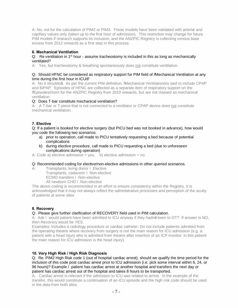
A: No, not for the calculation of PIM2 or PIM3. These models have been validated with arterial and capillary values only (taken up to the first hour of admission). This restriction may change for future PIM models if research supports its inclusion, and the ANZPIC Registry is collecting venous base excess from 2012 onwards as a first step in this process. 6. Mechanical Ventilation Q: Re ventilation in 1st hour - assume tracheostomy is included in this as long as mechanically ventilated? A: Yes, but tracheostomy & breathing spontaneously does not constitute ventilation. Q: Should HFNC be considered as respiratory support for PIM field of “Mechanical Ventilation at any time during the first hour in ICU”? A: No it shouldn’t. As per the current PIM definition, “Mechanical Ventilation” is said to include CPAP and BiPAP. Episodes of HFNC are collected as a separate item of respiratory support on the “Episodes” form for the ANZPIC Registry from 2010 onwards, but are not classed as mechanical ventilation. Q: Does T-bar constitute mechanical ventilation? A: A T-bar or T-piece that is not connected to a ventilator or CPAP device does not constitute mechanical ventilation. 7. Elective Q: If a patient is booked for elective surgery (but PICU bed was not booked in advance), how would you code the following two scenarios:
a) prior to operation, call made to PICU tentatively requesting a bed because of potential complications
b) during elective procedure, call made to PICU requesting a bed (due to unforeseen complications during operation)
A: Code a) elective admission = yes, b) elective admission = no. Q: Recommended coding for elective/non-elective admissions in other queried scenarios. A: Transplants, living donor – Elective Transplants, cadaveric – Non-elective ECMO transfers – Non-elective All newborn CHD – Non-elective The above coding is recommended in an effort to ensure consistency within the Registry. It is acknowledged that it may not always reflect the administrative processes and perception of the acuity of patients at some sites. 8. Recovery Q: Please give further clarification of RECOVERY field used in PIM calculation. A: Ask – would patient have been admitted to ICU anyway if they hadn’t been to OT? If answer is NO, then Recovery would be YES. Examples: Includes a radiology procedure or cardiac catheter. Do not include patients admitted from the operating theatre where recovery from surgery is not the main reason for ICU admission (e.g. a patient with a head injury who is admitted from theatre after insertion of an ICP monitor; in this patient the main reason for ICU admission is the head injury). 10. Very High Risk / High Risk Diagnosis Q: Re. PIM2 High Risk code 1 (out of hospital cardiac arrest), should we qualify the time period for the inclusion of this code post cardiac arrest prior to ICU admission (i.e. pick some interval within 6, 24, or 96 hours)? Example – patient has cardiac arrest at another hospital and transfers the next day or patient has cardiac arrest out of the hospital and takes 8 hours to be transported. A: Cardiac arrest is relevant if the admission to ICU was related to arrest. In the example of the transfer, this would constitute a continuation of an ICU episode and the high risk code should be used in the data from both sites.
- 8 -
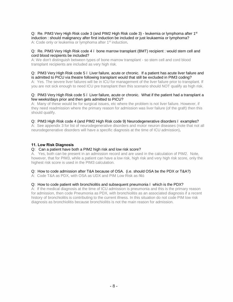
Q: Re. PIM3 Very High Risk code 3 (and PIM2 High Risk code 3) - leukemia or lymphoma after 1st induction : should malignancy after first induction be included or just leukaemia or lymphoma? A: Code only or leukemia or lymphoma after 1st induction. Q: Re. PIM3 Very High Risk code 4 – bone marrow transplant (BMT) recipient : would stem cell and cord blood recipients be included? A: We don't distinguish between types of bone marrow transplant - so stem cell and cord blood transplant recipients are included as very high risk. Q: PIM3 Very High Risk code 5 – Liver failure, acute or chronic. If a patient has acute liver failure and is admitted to PICU via theatre following transplant would that still be excluded in PIM3 coding? A: Yes. The severe liver failures will be in ICU for management of the liver failure prior to transplant. If you are not sick enough to need ICU pre transplant then this scenario should NOT qualify as high risk. Q: PIM3 Very High Risk code 5 – Liver failure, acute or chronic. What if the patient had a transplant a few weeks/days prior and then gets admitted to PICU? A: Many of these would be for surgical issues, etc where the problem is not liver failure. However, if they need readmission where the primary reason for admission was liver failure (of the graft) then this should qualify. Q: PIM3 High Risk code 4 (and PIM2 High Risk code 9) Neurodegenerative disorders – examples? A: See appendix 3 for list of neurodegenerative disorders and motor neuron diseases (note that not all neurodegenerative disorders will have a specific diagnosis at the time of ICU admission). 11. Low Risk Diagnosis Q: Can a patient have both a PIM2 high risk and low risk score? A. Yes, both can be present in an admission record and are used in the calculation of PIM2. Note, however, that for PIM3, while a patient can have a low risk, high risk and very high risk score, only the highest risk score is used in the PIM3 calculation. Q: How to code admission after T&A because of OSA. (i.e. should OSA be the PDX or T&A?) A: Code T&A as PDX, with OSA as UDX and PIM Low Risk as “4”. Q: How to code patient with bronchiolitis and subsequent pneumonia – which is the PDX? A: If the medical diagnosis at the time of ICU admission is pneumonia and this is the primary reason for admission, then code Pneumonia as PDX, with bronchiolitis as an associated diagnosis if a recent history of bronchiolitis is contributing to the current illness. In this situation do not code PIM low risk diagnosis as bronchiolitis because bronchiolitis is not the main reason for admission.

- 9 -
Model coefficient differences between PIM3 and PIM2 1. For the PaO2/FiO2 ratio term, if either or both PaO2 and FiO2 are not recorded or unknown, the calculated ratio’s default value in the PIM3 model is now 0.23 (derived from the normal value of PaO2 in air ((0.21*100)/90), and not zero as in the PIM2 model. 2. The Recovery and Bypass fields are collected using the same format as PIM2, but are no longer used as individual terms in PIM3. Instead they are combined centrally with the procedure code in the PDx field to create 3 new yes/no terms in the calculation of PIM3 – (1) Recovery from a bypass cardiac procedure (Recov_CardBypPr), (2) Recovery from a non-bypass cardiac procedure (Recov_CardNonBypPr), and (3) recovery from a non-cardiac procedure (Recov_NonCardPr). Recov_CardBypPr. This is a new term and is centrally created after data submission. Codes are : Yes=1, No=0, where “yes” is generated if Recovery=yes and Bypass prior to admission = yes and PDx is any of(1900-1999). Recov_CardNonBypPr. This is a new field and is centrally created after data submission. Codes are : Yes=1, No=0, where “yes” is generated if Recovery=1 and Bypass prior to admission = no and PDx is any of(1900-1999,1102,1106,1107). Recov_NonCardPr. This is a new field and is centrally created after data submission. Codes are : Yes=1, No=0, where “yes” is generated if Recovery=1 and PDx is any of(1100,1101,1103-1105,1108-1899). 3. There are now 3 “risk” indicator fields – Very High Risk (VHRdiag), High Risk (HRdiag) and Low Risk (LRdiag). VHRdiag is a new field, while HRdiag and LRdiag have different inclusions in PIM3 compared to their use in PIM2. 4. Only one of VHRdiag, HRdiag and LRdiag can be included in the calculation of PIM3val, with the most severe risk over-riding the lesser risks. This is different to the use of HRdiag & LRdiag in PIM2 where both could be used in the calculation if applicable. 5. VHRdiag. PIM3 Very High Risk Diagnosis (ANZPICR field PIM3_VHR). Specific conditions** associated with increased mortality risk. Codes for data collection are - 0 None 1 Cardiac arrest preceding ICU admission. Includes both in-hospital and out-of-hospital arrest.
Requires either documented absent pulse or the requirement for external cardiac massage (do not include past history of cardiac arrest).
2 Severe combined immune deficiency (SCID) - requires the documented diagnosis of SCID. 3 Leukaemia or lymphoma after 1st induction. Include only cases where the admission is related
to leukaemia or lymphoma, or the therapy for these conditions. 4 Bone marrow transplant (BMT) recipient. Include cases where the BMT has occurred during
the current hospital admission. 5 Liver failure - acute or chronic, is the main reason for ICU admission. DO NOT Include
patients admitted for recovery following liver transplantation for acute or chronic liver failure. (coding of liver transplant patients is different from PIM2)
6 Necrotising enterocolitis is the main reason for ICU admission.(see HRdiag code 6 instead) Code 6 for VHRdiag is no longer used.
7 SCID and BMT recipient. 8 Leukaemia or lymphoma after 1st induction and BMT recipient. These are recoded for the PIM3val calculation to 0=No(0), 1-5,7,8=Yes(1).

- 10 -
6. HRdiag. PIM3 High Risk Diagnosis (ANZPICR field PIM3_HR). Specific conditions** associated with increased mortality risk. Codes for data collection are -
0 None 1 Spontaneous cerebral haemorrhage - haemorrhage must be spontaneous (for example, from
an aneurysm or AVM). Do not include traumatic cerebral haemorrhage or intracranial haemorrhage that is not intracerebral (eg subdural haemorrhage).
2 Cardiomyopathy or myocarditis - requires the documented diagnosis of myocarditis or cardiomyopathy.
3 Hypoplastic left heart syndrome – any age, but include only cases where a Norwood procedure, or equivalent, is required in the neonatal period to sustain life. If a subsequent heart transplant, then this diagnosis and high risk indicator no longer apply.
4 Neurodegenerative disorder - requires a history of progressive loss of milestones (even if no specific condition has been diagnosed) or a diagnosis where this will inevitably occur.
5 Septic shock. As defined by the International Pediatric Sepsis Consensus Conference, 2002. Requires the presence of the systemic inflammatory response syndrome (SIRS) and suspected or proven infection and cardiovascular organ dysfunction. Collected as HR code by ANZPIC Registry but not used in PIM3 calculation.
6 Necrotising enterocolitis is the main reason for ICU admission. Include patients where an acute episode of NEC is the main reason for admission. DO NOT include patients where the admission is for management of the sequelae such as strictures, revision of stomas, etc..
These are recoded for the PIM3val calculation to 0=No(0), 1-6=Yes(1). 7. LRdiag. PIM3 Low Risk Diagnosis (ANZPICR field PIM3_LR). Specific conditions** associated with
decreased mortality risk. Note that these conditions must be the main reason for ICU admission to be eligible for the low risk code. Codes for data collection are -
0 None 1 Asthma is the main reason for ICU admission. 2 Bronchiolitis is the main reason for ICU admission. Include children who present either with
respiratory distress or central apnoea where the clinical diagnosis is bronchiolitis. 3 Croup is the main reason for ICU admission. 4 Obstructive sleep apnoea. Include patients admitted following adenoidectomy and/or
tonsillectomy in whom obstructive sleep apnoea (OSA) is the main reason for ICU admission (and also code as recovery from surgery).
5 Diabetic ketoacidosis (DKA) is the main reason for ICU admission. 6 Seizures. Include patients who require admission primarily due to status epilepticus, epilepsy,
febrile convulsion, or other epileptic syndrome where admission is required either to control seizures or to recover from the effects of seizures or treatment.
These are recoded for the PIM3val calculation to 0=No(0), 1-6=Yes(1). ** See Appendix 4 for current ANZPIC Registry diagnosis list

- 11 -
PIM3
Reference: Straney L, Clements A, Parslow R, Pearson G, Shann F, Alexander J, Slater A, for the ANZICS Paediatric Study Group and the Paediatric Intensive Care Audit Network (2013) Paediatric Index of Mortality 3 : an updated model for predicting mortality in Pediatric Intensive Care. Ped Crit Care Med. 2013 Sep; 14(7): 673-81.
PIM3 is calculated from the information collected at the time a child is admitted to your ICU. Because PIM3 describes how ill the child was at the time you started intensive care, the observations to be recorded are those made at or about the time of first face-to-face (not telephone) contact between the patient and a doctor from your intensive care unit (or a doctor from a specialist paediatric transport team). Use the first value of each variable measured within the period from the time of first contact/first taking over management to 1 hour after arrival in your ICU. The first contact may be in your ICU, your emergency department, a ward in your own hospital, or in another hospital (e.g. on a retrieval). If information is missing, see the documentation for each field (model coefficient) for the correct coding practice.
Calculation of PIM3 (and PIM3 risk of death%)–
PIM3val= (3.8233 * Pupils) – (0.5378 * Elective) + (0.9763 * MechVent) + (0.0671 * (absolute Base Excess)) – (0.0431*SBP) + (0.1716*(SBP*SBP/1000)) + (0.4214 * (100*FiO2/PaO2)) – (1.2246*Recov_CardBypPr) – (0.8762*Recov_CardNonBypPr) – (1.5164*Recov_NonCardPr) + (1.6225* VHRdiag) + (1.0725*HRdiag) – (2.1766*LRdiag) – 1.7928
PIM3 risk of death = ePIM3val / (1 + ePIM3val )
Example of PIM3 calculation
A six year old girl receiving chemotherapy for relapsed leukemia presents to the emergency department of your hospital with febrile neutropenia. Initial observations are temperature 39oC, heart rate 160 beats/min, respiratory rate 30 breaths/min and SBP 65mmHg. A doctor from your ICU assesses her before admission and at the time the SBP is 70 mmHg, and she has reactive pupils. She is admitted to ICU, intubated and ventilated and an arterial blood gas is performed within the first hour. The PaO2 is 65mmHg in FiO2 of 0.7, the base excess is -12 mmol/l. Interpretation. The admission is not elective, or for recovery from a procedure. She is ventilated within the first hour of admission. The first SBP recorded after first contact with the ICU doctor in the emergency department was 70 mmHg. She has a very high risk condition (leukemia after first induction).
PIM3val = (3.8233*0) + (-0.5378*0) + (0.9763*1) + (0.0671*12) - (0.0431*70) + (0.1716*(70*70/1000)) + (0.4214*(100*(0.7/65)) - (1.2246*0) - (0.8762*0) - (1.5164*0) + (1.6225*1) + (1.0725*0) - (2.1766*0) - 1.7928
= -0.11114
Probability of Death = exp(-0.11114)/(1+exp(-0.11114))
= 0.4722 or 47.22%

- 12 -
Regional (ANZ) calibration of PIM3 model While the published PIM3 model has been developed using data from multiple countries and provides an international comparison, it is important to also have coefficients based on regional data when comparing SMRs for units in a country or region. An ANZ calibration of the PIM3 model has been generated using ANZPIC Registry data from 2012 and 2013, and is called PIM3-anz13. The details of the ANZ model are as follows -
Calculation of PIM3-anz13 (and PIM3-anz13 risk of death%)–
PIM3-anz13_val= (4.371172 * Pupils) – (0.5164336 * Elective) + (0.6634843 * MechVent) + (0.0740947 * (absolute Base Excess)) – (0.0296888 * SBP) + (0.0964949 * (SBP*SBP/1000)) + (0.5181944 * (100*FiO2/PaO2)) – (1.866951*Recov_CardBypPr) – (1.318171*Recov_CardNonBypPr) – (1.572421*Recov_NonCardPr) + (1.993498* VHRdiag) + (1.368355*HRdiag) – (2.401701*LRdiag) – 2.299542
PIM3-anz13 risk of death = ePIM3-anz13_val / (1 + ePIM3-anz13_val )

- 13 -
Appendix 1 Definition of sepsis/SIRS/septic shock: SIRS: widespread inflammatory response that may or may not be associated with infection. The
presence of 2 or more of the following criteria (one of which must be abnormal temperature or
leukocyte count):
► Core temp > 38.5°C or < 36°C
► WBC increased or decreased for age, or > 10% immature neutrophils
► Tachycardia (HR > 2 SD above normal for age OR for children < 1 yr, HR < 10th percentile for age )
► Bradycardia
► Mean respiratory rate > 2 SD above normal for age
Sepsis: SIRS in the presence of, or as a result of, suspected or proven infection.
Septic shock: is sepsis with cardiovascular dysfunction despite the administration of > 40ml/kg of isotonic fluid in one hour.

- 14 -
Appendix 2 Chromosomal abnormalities
Chromosomal abnormalities: Code for conditions that can be detected on routine chromosomal analyses (including FISH studies), e.g. Trisomies, translocations, deletions.
Examples of most common chromosomal abnormalities: Trisomies. Down syndrome is among the most common of these disorders, affecting about 1 in 800 to 1000 live-born babies. Babies also can be born with extra copies of chromosomes 13 or 18. These trisomies are usually much more severe than Down syndrome, but fortunately less common, each affecting about 1 in 5000 babies. Babies with trisomies 13 or 18 generally have severe mental retardation and many physical birth defects.
Turner syndrome is a sex chromosome abnormality that affects about 1 in 2500 girls. Girls with Turner syndrome have only one X chromosome, instead of the normal two. About 1 in 1000 to 2000 females has an extra X chromosome, referred to as triple X.
Klinefelter syndrome is a sex chromosome abnormality that affects about 1 in 600 to 800 boys. Boys with Klinefelter syndrome have two, or occasionally more, X chromosomes along with their Y chromosome.

- 15 -
Appendix 3 Neurodegenerative disorders For the purposes or recording a neurodegenerative disorder in the PIM high risk field, a patient requires a history of progressive loss of milestones (even if no specific condition has yet been diagnosed), or a diagnosis where this will inevitably occur.
Alpers’ Disease (Synonym: Progressive Sclerosing Poliodystrophy). Alpers’ disease is a rare, genetically determined disease of the brain that causes progressive degeneration of grey matter in the cerebrum. The first sign of the disease usually begins early in life with convulsions. Other symptoms are developmental delay, progressive mental retardation, hypotonia (low muscle tone), spasticity (stiffness of the limbs), dementia, and liver conditions such as jaundice and cirrhosis that can lead to liver failure. Optic atrophy may also occur, often causing blindness. Researchers believe that Alpers’ disease is caused by an underlying metabolic defect. Some patients have mutations in mitochondrial DNA.
Cerebro-Oculo-Facio-Skeletal Syndrome (Synonyms: Pena Shokeir II Syndrome, Cockayne Syndrome Type II). Cerebro-oculo-facio-skeletal (COFS) syndrome is a paediatric, genetic, degenerative disorder that involves the brain and the spinal cord. It is characterized by craniofacial and skeletal abnormalities, severely reduced muscle tone, and impairment of reflexes. Symptoms may include large, low-set ears, small eyes, microcephaly (abnormal smallness of the head), micrognathia (abnormal smallness of the jaws), clenched fists, wide-set nipples, vision impairments, involuntary eye movements, and mental retardation, which can be moderate or severe. Respiratory infections are frequent. COFS is diagnosed at birth.
Leigh’s Disease (Synonym: Subacute Necrotizing Encephalomyelopathy (SNEM)). Leigh’s disease is a rare inherited neurometabolic disorder characterized by degeneration of the central nervous system. Leigh’s disease can be caused by mutations in mitochondrial DNA or by deficiencies of the enzyme pyruvate dehydrogenase. Symptoms of Leigh’s disease usually begin between the ages of 3 months to 2 years and progress rapidly. In most children, the first signs may be poor sucking ability and loss of head control and motor skills. Monomelic Amyotrophy (Synonyms: Benign Focal Amyotrophy, Hirayama Syndrome, O’Sullivan-McLeod Syndrome). Monomelic amyotrophy (MMA) is characterized by progressive degeneration and loss of motor neurons, the nerve cells in the brain and spinal cord that are responsible for controlling voluntary muscles. It is characterized by weakness and wasting in a single limb, usually an arm and hand rather than a foot and leg. There is no pain associated with MMA. While some physicians contend that mild sensory loss may be associated with this disease, many experts suggest that such symptoms actually indicate a cause other then MMA. MMA occurs in males between the ages of 15 and 25. Onset and progression are slow. MMA is seen most frequently in Asia, particularly in Japan and India; it is much less common in North America. In most cases, the cause is unknown, although there have been a few published reports linking MMA to traumatic or radiation injury. There are also familial forms of MMA. Diagnosis is made by physical exam and medical history. Electromyography (EMG), a special recording technique that detects electrical activity in muscles, shows a loss of the nerve supply, or denervation, in the affected limb; MRI and CT scans may show muscle atrophy. People believed to have MMA should be followed by a neuromuscular disease specialist for a number of months to make certain that no signs of other motor neuron diseases develop.
- 16 -
Progressive Multifocal Leukoencephalopathy (PML) is an infrequent disorder of the nervous system that primarily affects individuals with suppressed immune systems (including, allograft recipients such as kidney transplant patients; patients with cancers such as leukaemia or lymphoma; and nearly 10% of patients with acquired immune deficiency syndrome (AIDS). The disorder, which is caused by a common human polyomavirus, JC virus, is characterized by demyelination or destruction of the myelin sheath that covers nerve cells. The myelin sheath is the fatty covering – which acts as an insulator – on nerve fibres in the brain. Symptoms of PML include mental deterioration, vision loss, speech disturbances, ataxia (inability to coordinate movements), paralysis, and, ultimately, coma reflecting the multifocal distribution of brain lesions. In rare cases, seizures may occur. Motor Neuron Diseases (include as PIM high risk diagnosis of neurodegenerative disorder) Motor neuron diseases (MNDs) are progressive, degenerative disorders that affect nerves in the upper or lower parts of the body. Some of the diseases are inherited, while others may be acquired. Common MNDs include amyotrophic lateral sclerosis (ALS), progressive muscular atrophy, and postpolio syndrome.
Amyotrophic Lateral Sclerosis (Synonym: Lou Gehrig’s Disease). Amyotrophic lateral sclerosis (ALS) is a rapidly progressive, invariably fatal neurological disease that attacks the neurons responsible for controlling voluntary muscles. In ALS, both the upper motor neurons and the lower motor neurons degenerate or die, ceasing to send messages to muscles. Unable to function, the muscles gradually weaken, waste away, and twitch. Eventually the ability of the brain to start and control voluntary movement is lost. Individuals with ALS lose their strength and the ability to move their arms, legs, and body. When muscles in the diaphragm and chest wall fail, individuals lose the ability to breathe without ventilatory support.
Spinal Muscular Atrophy Spinal muscular atrophy (SMA) is a genetic, motor neuron disease caused by progressive degeneration of motor neurons in the spinal cord. The disorder causes weakness and wasting of the voluntary muscles. Weakness is often more severe in the legs than in the arms. The childhood SMAs are all autosomal recessive diseases. The gene for SMA has been identified and accurate diagnostic tests exist. There are many types of SMA; some of the more common types are described below.
SMA type 1, also called Werdnig-Hoffmann disease, is evident before birth or within the first few months of life. There may be a reduction in fetal movement in the final months of pregnancy. Symptoms include floppiness of the limbs and trunk, feeble movements of the arms and legs, swallowing and feeding difficulties, and impaired breathing. Affected children never sit or stand and usually die before the age of 2. Symptoms of SMA type II usually begin between 3 and 15 months of age. Children may have respiratory problems, floppy limbs, decreased or absent deep tendon reflexes, and twitching of arm, leg, or tongue muscles. These children may learn to sit but will never be able to stand or walk. Life expectancy varies. Symptoms of SMA type III (Kugelberg-Welander disease) appear between 2 and 17 years of age, and include abnormal manner of walking; difficulty running, climbing stairs, or rising from a chair; and slight tremor of the fingers. Progressive spinobulbar muscular atrophy or Kennedy syndrome may occur between 15 and 60 years of age. Features of this type may include weakness of muscles in the tongue and face, difficulty swallowing, speech impairment, and excessive development of the mammary glands in males. The course of the disorder is usually slowly progressive. Kennedy syndrome is an X-linked recessive disorder. Congenital SMA with arthrogryposis (persistent contracture of joints with fixed abnormal posture of the limb) is a rare disorder. Manifestations include

- 17 -
severe contractures, curvature of the spine, chest deformity, respiratory problems, an unusually small jaw, and drooping upper eyelids. Rett syndrome Rett syndrome is a childhood neurodevelopmental disorder characterized by normal early development followed by loss of purposeful use of the hands, distinctive hand movements, slowed brain and head growth, gait abnormalities, seizures, and mental retardation. Rett syndrome is caused by mutations in the MECP2 gene, which is found on the X chromosome. The MECP2 gene contains instructions for the synthesis of a protein called methyl cytosine binding protein 2 (MeCP2), which acts as one of the many biochemical switches that tell other genes when to turn off and stop producing their own unique proteins. Because the MECP2 gene does not function properly in those with Rett syndrome, insufficient amounts or structurally abnormal forms of the protein are formed.
- 18 -
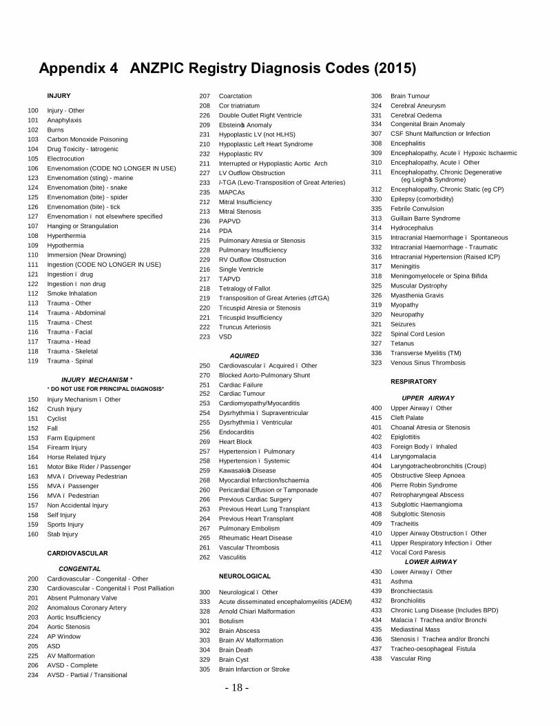
Appendix 4 ANZPIC Registry Diagnosis Codes (2015)
INJURY 100 Injury - Other 101 Anaphylaxis 102 Burns 103 Carbon Monoxide Poisoning 104 Drug Toxicity - Iatrogenic 105 Electrocution 106 Envenomation (CODE NO LONGER IN USE) 123 Envenomation (sting) - marine 124 Envenomation (bite) - snake 125 Envenomation (bite) - spider 126 Envenomation (bite) - tick 127 Envenomation – not elsewhere specified 107 Hanging or Strangulation 108 Hyperthermia 109 Hypothermia 110 Immersion (Near Drowning) 111 Ingestion (CODE NO LONGER IN USE) 121 Ingestion – drug 122 Ingestion – non drug 112 Smoke Inhalation 113 Trauma - Other 114 Trauma - Abdominal 115 Trauma - Chest 116 Trauma - Facial 117 Trauma - Head 118 Trauma - Skeletal 119 Trauma - Spinal INJURY MECHANISM * * DO NOT USE FOR PRINCIPAL DIAGNOSIS*
150 Injury Mechanism – Other 162 Crush Injury 151 Cyclist 152 Fall 153 Farm Equipment 154 Firearm Injury 164 Horse Related Injury 161 Motor Bike Rider / Passenger 163 MVA – Driveway Pedestrian 155 MVA – Passenger 156 MVA – Pedestrian 157 Non Accidental Injury 158 Self Injury 159 Sports Injury 160 Stab Injury CARDIOVASCULAR CONGENITAL 200 Cardiovascular - Congenital - Other 230 Cardiovascular - Congenital – Post Palliation 201 Absent Pulmonary Valve 202 Anomalous Coronary Artery 203 Aortic Insufficiency 204 Aortic Stenosis 224 AP Window 205 ASD 225 AV Malformation 206 AVSD - Complete 234 AVSD - Partial / Transitional
207 Coarctation 208 Cor triatriatum 226 Double Outlet Right Ventricle 209 Ebstein’s Anomaly 231 Hypoplastic LV (not HLHS) 210 Hypoplastic Left Heart Syndrome 232 Hypoplastic RV 211 Interrupted or Hypoplastic Aortic Arch 227 LV Outflow Obstruction 233 l-TGA (Levo-Transposition of Great Arteries) 235 MAPCAs 212 Mitral Insufficiency 213 Mitral Stenosis 236 PAPVD 214 PDA 215 Pulmonary Atresia or Stenosis 228 Pulmonary Insufficiency 229 RV Outflow Obstruction 216 Single Ventricle 217 TAPVD 218 Tetralogy of Fallot 219 Transposition of Great Arteries (dTGA) 220 Tricuspid Atresia or Stenosis 221 Tricuspid Insufficiency 222 Truncus Arteriosis 223 VSD AQUIRED 250 Cardiovascular – Acquired – Other 270 Blocked Aorto-Pulmonary Shunt 251 Cardiac Failure 252 Cardiac Tumour 253 Cardiomyopathy/Myocarditis 254 Dysrhythmia – Supraventricular 255 Dysrhythmia – Ventricular 256 Endocarditis 269 Heart Block 257 Hypertension – Pulmonary 258 Hypertension – Systemic 259 Kawasaki’s Disease 268 Myocardial Infarction/Ischaemia 260 Pericardial Effusion or Tamponade 266 Previous Cardiac Surgery 263 Previous Heart Lung Transplant 264 Previous Heart Transplant 267 Pulmonary Embolism 265 Rheumatic Heart Disease 261 Vascular Thrombosis 262 Vasculitis
NEUROLOGICAL 300 Neurological – Other 333 Acute disseminated encephalomyelitis (ADEM) 328 Arnold Chiari Malformation 301 Botulism 302 Brain Abscess 303 Brain AV Malformation 304 Brain Death 329 Brain Cyst 305 Brain Infarction or Stroke
306 Brain Tumour 324 Cerebral Aneurysm 331 Cerebral Oedema 334 Congenital Brain Anomaly 307 CSF Shunt Malfunction or Infection 308 Encephalitis 309 Encephalopathy, Acute – Hypoxic Ischaemic 310 Encephalopathy, Acute – Other 311 Encephalopathy, Chronic Degenerative
(eg Leigh’s Syndrome) 312 Encephalopathy, Chronic Static (eg CP) 330 Epilepsy (comorbidity) 335 Febrile Convulsion 313 Guillain Barre Syndrome 314 Hydrocephalus 315 Intracranial Haemorrhage – Spontaneous 332 Intracranial Haemorrhage - Traumatic 316 Intracranial Hypertension (Raised ICP) 317 Meningitis 318 Meningomyelocele or Spina Bifida 325 Muscular Dystrophy 326 Myasthenia Gravis 319 Myopathy 320 Neuropathy 321 Seizures 322 Spinal Cord Lesion 327 Tetanus 336 Transverse Myelitis (TM) 323 Venous Sinus Thrombosis RESPIRATORY UPPER AIRWAY 400 Upper Airway – Other
415 Cleft Palate 401 Choanal Atresia or Stenosis 402 Epiglottitis 403 Foreign Body – Inhaled 414 Laryngomalacia 404 Laryngotracheobronchitis (Croup) 405 Obstructive Sleep Apnoea 406 Pierre Robin Syndrome 407 Retropharyngeal Abscess 413 Subglottic Haemangioma 408 Subglottic Stenosis 409 Tracheitis 410 Upper Airway Obstruction – Other 411 Upper Respiratory Infection – Other 412 Vocal Cord Paresis LOWER AIRWAY 430 Lower Airway – Other 431 Asthma 439 Bronchiectasis 432 Bronchiolitis 433 Chronic Lung Disease (Includes BPD) 434 Malacia – Trachea and/or Bronchi 435 Mediastinal Mass 436 Stenosis – Trachea and/or Bronchi 437 Tracheo-oesophageal Fistula 438 Vascular Ring
- 19 -
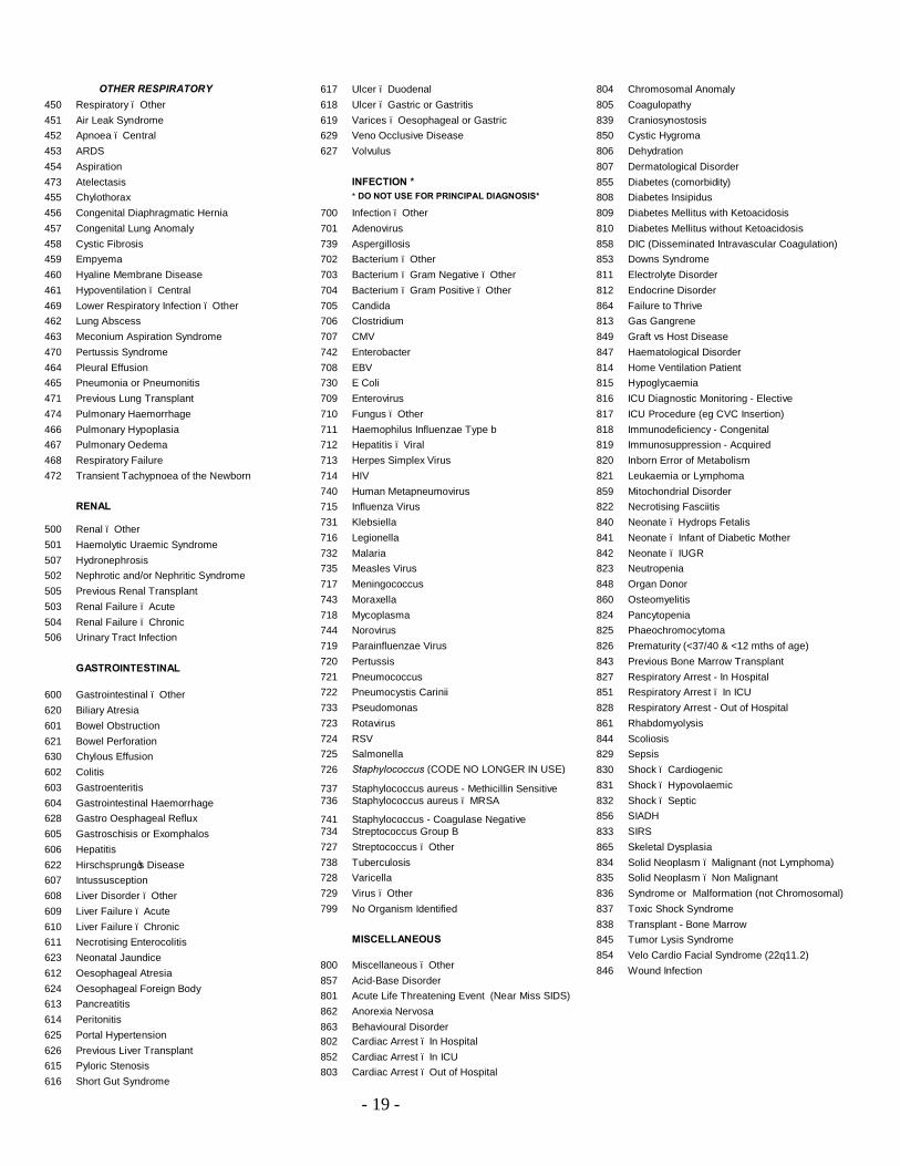
OTHER RESPIRATORY 450 Respiratory – Other 451 Air Leak Syndrome 452 Apnoea – Central 453 ARDS 454 Aspiration 473 Atelectasis 455 Chylothorax 456 Congenital Diaphragmatic Hernia 457 Congenital Lung Anomaly 458 Cystic Fibrosis 459 Empyema 460 Hyaline Membrane Disease 461 Hypoventilation – Central 469 Lower Respiratory Infection – Other 462 Lung Abscess 463 Meconium Aspiration Syndrome 470 Pertussis Syndrome 464 Pleural Effusion 465 Pneumonia or Pneumonitis 471 Previous Lung Transplant 474 Pulmonary Haemorrhage 466 Pulmonary Hypoplasia 467 Pulmonary Oedema 468 Respiratory Failure 472 Transient Tachypnoea of the Newborn
RENAL 500 Renal – Other 501 Haemolytic Uraemic Syndrome 507 Hydronephrosis 502 Nephrotic and/or Nephritic Syndrome 505 Previous Renal Transplant 503 Renal Failure – Acute 504 Renal Failure – Chronic 506 Urinary Tract Infection GASTROINTESTINAL 600 Gastrointestinal – Other 620 Biliary Atresia 601 Bowel Obstruction 621 Bowel Perforation 630 Chylous Effusion 602 Colitis 603 Gastroenteritis 604 Gastrointestinal Haemorrhage 628 Gastro Oesphageal Reflux 605 Gastroschisis or Exomphalos 606 Hepatitis 622 Hirschsprung’s Disease 607 Intussusception 608 Liver Disorder – Other 609 Liver Failure – Acute 610 Liver Failure – Chronic 611 Necrotising Enterocolitis 623 Neonatal Jaundice 612 Oesophageal Atresia 624 Oesophageal Foreign Body 613 Pancreatitis 614 Peritonitis 625 Portal Hypertension 626 Previous Liver Transplant 615 Pyloric Stenosis 616 Short Gut Syndrome
617 Ulcer – Duodenal 618 Ulcer – Gastric or Gastritis 619 Varices – Oesophageal or Gastric 629 Veno Occlusive Disease 627 Volvulus INFECTION * * DO NOT USE FOR PRINCIPAL DIAGNOSIS*
700 Infection – Other 701 Adenovirus 739 Aspergillosis 702 Bacterium – Other 703 Bacterium – Gram Negative – Other 704 Bacterium – Gram Positive – Other 705 Candida 706 Clostridium 707 CMV 742 Enterobacter 708 EBV 730 E Coli 709 Enterovirus 710 Fungus – Other 711 Haemophilus Influenzae Type b 712 Hepatitis – Viral 713 Herpes Simplex Virus 714 HIV 740 Human Metapneumovirus 715 Influenza Virus 731 Klebsiella 716 Legionella 732 Malaria 735 Measles Virus 717 Meningococcus 743 Moraxella 718 Mycoplasma 744 o
Norovirus 719 Parainfluenzae Virus 720 Pertussis 721 Pneumococcus 722 Pneumocystis Carinii 733 Pseudomonas 723 Rotavirus 724 RSV 725 Salmonella 726 Staphylococcus (CODE NO LONGER IN USE)
737 Staphylococcus aureus - Methicillin Sensitive 736 Staphylococcus aureus – MRSA
741 Staphylococcus - Coagulase Negative 734 Streptococcus Group B 727 Streptococcus – Other 738 Tuberculosis 728 Varicella 729 Virus – Other 799 No Organism Identified MISCELLANEOUS 800 Miscellaneous – Other 857 Acid-Base Disorder 801 Acute Life Threatening Event (Near Miss SIDS) 862 Anorexia Nervosa 863 Behavioural Disorder 802 Cardiac Arrest – In Hospital 852 Cardiac Arrest – In ICU 803 Cardiac Arrest – Out of Hospital
804 Chromosomal Anomaly 805 Coagulopathy 839 Craniosynostosis 850 Cystic Hygroma 806 Dehydration 807 Dermatological Disorder 855 Diabetes (comorbidity) 808 Diabetes Insipidus 809 Diabetes Mellitus with Ketoacidosis 810 Diabetes Mellitus without Ketoacidosis 858 DIC (Disseminated Intravascular Coagulation) 853 Downs Syndrome 811 Electrolyte Disorder 812 Endocrine Disorder 864 Failure to Thrive 813 Gas Gangrene 849 Graft vs Host Disease 847 Haematological Disorder 814 Home Ventilation Patient 815 Hypoglycaemia 816 ICU Diagnostic Monitoring - Elective 817 ICU Procedure (eg CVC Insertion) 818 Immunodeficiency - Congenital 819 Immunosuppression - Acquired 820 Inborn Error of Metabolism 821 Leukaemia or Lymphoma 859 Mitochondrial Disorder 822 Necrotising Fasciitis 840 Neonate – Hydrops Fetalis 841 Neonate – Infant of Diabetic Mother 842 Neonate – IUGR 823 Neutropenia 848 Organ Donor 860 Osteomyelitis 824 Pancytopenia 825 Phaeochromocytoma 826 Prematurity (<37/40 & <12 mths of age) 843 Previous Bone Marrow Transplant 827 Respiratory Arrest - In Hospital 851 Respiratory Arrest – In ICU 828 Respiratory Arrest - Out of Hospital 861 Rhabdomyolysis 844 Scoliosis 829 Sepsis 830 Shock – Cardiogenic 831 Shock – Hypovolaemic 832 Shock – Septic 856 SIADH 833 SIRS 865 Skeletal Dysplasia 834 Solid Neoplasm – Malignant (not Lymphoma) 835 Solid Neoplasm – Non Malignant 836 Syndrome or Malformation (not Chromosomal) 837 Toxic Shock Syndrome 838 Transplant - Bone Marrow 845 Tumor Lysis Syndrome 854 Velo Cardio Facial Syndrome (22q11.2) 846 Wound Infection

- 20 -
POST PROCEDURAL DIAGNOSES MISCELLANEOUS / ANAESTHETIC 1100 Post Procedure - Other 1101 Anaesthetic Complication 1106 Cardiac Catheter – Balloon Septostomy 1102 Cardiac Catheter – Diagnostic 1107 Cardiac Catheter – Interventional 1103 Ex-prem, Post GA 1104 Invasive Radiology Procedure 1105 Massive Intraop Transfusion (> 1 blood vol) 1109 PEG (Percutaneous Endoscopic Gastrostomy) 1108 Post operative bleeding NEUROSURGERY 1300 Neurosurgery – Other 1301 Craniotomy – Anterior Fossa 1302 Craniotomy – Posterior Fossa 1303 CSF Shunt Insertion or Revision 1304 Decompression - Cranial 1310 Decompression - Craniocervical 1305 Decompression - Spinal Cord 1306 Hemispherectomy or Lobectomy 1307 ICP Monitor or Vent. Drain Insertion 1308 Intracranial Haematoma Evacuation 1309 Repair Myelomeningocoele 1311 Third Ventriculostomy
THORACIC SURGERY 1400 Thoracic Surgery - Other 1401 Diaphragm Plication 1402 Diaphragm Repair 1403 Lung Biopsy 1404 Lung Decortication 1405 Oesophageal Atresia Repair 1406 Pneumonectomy or Lobectomy 1407 Thoracic Tumour Resection 1410 Thoracoscopy 1408 Tracheo-oesophageal Fistula Repair 1409 Tracheopexy 1411 VATS procedure
ENT SURGERY 1500 ENT - Other 1501 Adenoidectomy and/or Tonsillectomy 1502 Choanal Atresia Repair 1503 Cricoid Split 1510 Dilatation of Airway 1504 Laryngeal Reconstruction 1505 Laryngobronchoscopy 1508 Laryngoplasty 1507 Retopharyngeal Abscess Drainage 1509 Tracheal Reconstruction / Tracheoplasty 1506 Tracheostomy ABDOMINAL / GENERAL SURGERY 1600 General Surgery – Other 1621 Abdominal Aorta Surgery 1601 Abdominal Tumour Resection 1602 Appendicectomy
1603 Bladder Extrophy Repair 1604 Burns Surgery 1605 Fundoplication 1606 Gastroschisis or Exomphalos Repair 1607 GI Endoscopy and/or Sclerotherapy 1608 Intussusception Repair 1609 Kasai 1610 Laparotomy 1615 Laparotomy – Bowel Obstruction 1616 Laparotomy – Bowel Perforation 1617 Laparotomy – GI Haemorrhage 1618 Laparotomy – Necrotising Enterocolitis 1619 Laparotomy – Peritonitis 1620 Laparotomy – Trauma 1611 Transplant – Kidney 1612 Transplant – Liver 1613 Transplant - Small Bowel 1614 Urogenital Surgery – Other 1622 Wound Debridement CRANIOFACIAL SURGERY 1700 Craniofacial Surgery – Other 1706 Cleft Palate Repair 1701 Cranial Vault Reshaping 1702 Dental Surgery 1703 Facial Cleft Repair 1704 Mandibular Mobilisation 1705 Midface Mobilisation ORTHOPAEDIC SURGERY 1800 Orthopaedic Surgery - Other 1801 Fracture Fixation 1802 Spinal Instrumentation
CARDIAC SURGERY (with RACHS Risk Categories, where applicable)
1955 Annuloplasty (RC 3) 1929 Aortic valve replacement (RC 3) 1906 Aortic valvotomy - valvuloplasty >30d old (RC 2) 1958 Aortic valvotomy - valvuloplasty ≤30d old (RC 4) 1902 Aortopexy (RC 1) 1933 Aortoplasty (not arch) (RC 3) 1953 Arterial switch operation (RC 3) 1966
Arterial switch operation with pulmonary artery band removal (RC 4)
1968 Arterial switch operation with repair of sub PS (RC 4) 1967 Arterial switch operation with VSD closure (RC 4) 1913 ASD and VSD repair (RC 2)
1914 ASD primum repair (RC 2) 1901 ASD surgery (incl ASD secundum, sinus venosus ASD, patent foramen ovale closure) (RC 1) 1962 Atrial septectomy (RC 4) 1952 Atrial switch operation (RC 3) 1965 Atrial switch operation with repair of sub PS (RC 4) 1964 Atrial switch operation with VSD closure (RC 4) 1998 Cardiac Surgery Closed – Other 1999 Cardiac Surgery Open – Other 1988 Chest Closure 1942 Closure of semilunar valve, aortic or Pulmonary (RC 3) 1904 Coarctation repair >30d of age (RC 1) 1924 Coarctation repair ≤30d of age (RC 2) 1927 Common atrium closure (RC 2)
1979 Damus-Kaye-Stansel procedure (RC 6) 1974 Double switch (RC 4) 1989 ECMO cannulation/exploration 1990 Emergency chest opening 1957 Excision of intracardiac tumour (RC 3) 1986 Fontan conversion 1946 Fontan procedure (RC 3) 1987 Fontan take-down 1921 Glenn shunt (RC 2) 1994 Heart Transplant 1995 Heart Lung Transplant 1959 Konno procedure (RC 4) 1931 Left ventricular outflow tract patch (RC 3) 1944 Left ventricular to pulmonary artery conduit (RC 3) 1928 Left ventricular to right atrial shunt repair (RC 2) 1996 Lung Transplant 1935 Mitral valve replacement (RC 3) 1934 Mitral valvotomy - valvuloplasty (RC 3) 1977 Norwood (Stage 1 repair of hypoplastic left heart syndrome) (RC 6) 1997 PA Plasty or Repair 1992 Pacemaker insertion/replacement 1905 Partially anomalous pulmonary venous connection surgery (RC 1) 1903 PDA surgery >30d of age (RC 1) 1993 PDA surgery ≤ 30 days 1948 Pulmonary artery banding (RC 3) 1911 Pulmonary outflow tract augmentation (RC 2) 1909 Pulmonary valve replacement (RC 2) 1908 Pulmonary valvotomy - valvuloplasty (RC 2) 1991 Pulmonary venous stenosis repair 1954 Reimplantation of anomalous pulmonary artery (RC 3) 1941 Repair anomalous coronary art. with intrapulmonary tunnel (Takeuchi) (RC 3) 1940 Repair of anomalous coronary artery without intrapulmonary tunnel (RC 3) 1923 Repair of AP window (RC 2) 1956 Repair of Coarctation and VSD closure (RC 3) 1960 Repair of complex anomaly (single ventricle) by VSD enlargement (RC 4) 1950 Repair of Cor triatriatum (RC 3) 1912 Repair of coronary AV fistula (RC 2)
1945 Repair of double-outlet right ventricle with or without repair of right ventricular obstruction (RC 3) 1971 Repair of hypoplastic or interrupted arch with VSD closure (RC 4) 1970 Repair of hypoplastic or interrupted arch without VSD closure (RC 4) 1925 Repair of pulmonary artery stenosis (RC 2) 1949 Repair of tetralogy of Fallot with pulmonary atresia (RC 3) 1920 Repair of total anomalous pulmonary veins >30d of age (RC 2) 1947 Repair of transitional or complete atrioventricular canal with or without valve replacement (RC 3) 1963 Repair of transposition-VSD sub PS (Rastelli) (RC 4) 1969 Repair of truncus arteriosus (RC 4) 1976 Repair of truncus arteriosus and interrupted arch (RC 5) 1918 Repair of unspecified septal defect (RC 2) 1910 Right ventricular infundibulectomy (RC 2)

- 21 -
1943 Right ventricular to pulmonary artery conduit (RC 3) 1930 Ross procedure (RC 3) 1977 Stage 1 repair of hypoplastic left heart syndrome (Norwood) (RC 6) 1978 Stage 1 repair of nonhypoplastic left heart syndrome conditions (RC 6) 1907 Sub aortic stenosis resection (RC 2) 1951 Systemic to pulmonary artery shunt (RC 3) 1961 Total repair of anomalous pulmonary veins ≤30d of age (RC 4) 1919 Total repair of tetralogy of Fallot (RC 2) 1926 Transection of pulmonary artery (RC 2) 1994 Transplant – Heart
1995 Transplant – Heart Lung 1996 Transplant – Lung 1972 Transverse arch graft (RC 4) 1938 Tricuspid valve replacement (RC 3) 1939 Tricuspid valve repositioning for Ebstein anomaly >30d of age (RC 3) 1975 Tricuspid valve repositioning for neonatal Ebstein ≤30d of age (RC 5) 1937 Tricuspid valvotomy - valvuloplasty (RC 3) 1973 Unifocalisation for tetralogy of Fallot - pulmonary atresia (RC 4) 1936 Valvectomy of tricuspid valve (RC 3) 1906 Valvotomy - valvuloplasty (Aortic) >30d of age (RC 2)
1958 Valvotomy - valvuloplasty (Aortic) ≤30d of age (RC 4) 1934 Valvotomy - valvuloplasty (Mitral) (RC 3) 1908 Valvotomy - valvuloplasty (Pulmonary) (RC 2) 1937 Valvotomy - valvuloplasty (Tricuspid) (RC 3) 1922 Vascular ring surgery (RC 2) 1932 Ventriculomyotomy (RC 3) 1917 VSD closure and pulmonary artery band removal (RC 2) 1916 VSD closure and pulmonary valvotomy or infundibular resection (RC 2) 1913 VSD and ASD repair (RC 2) 1915 VSD repair (RC 2)
Related Documents