Mallmann, Holger Stepan, S. Ananth Karumanchi and Thomas Benzing Kreyssig, Linda Hemphill, Alan C. Rigby, Santosh Khedkar, Tom H. Lindner, Peter Wiebke Schaarschmidt, Alexander Jank, Angela Kribs, Oliver A. Cornely, Claudia Ravi Thadhani, Tuelay Kisner, Henning Hagmann, Verena Bossung, Stefanie Noack, Preeclampsia Pilot Study of Extracorporeal Removal of Soluble Fms-Like Tyrosine Kinase 1 in ISSN: 1524-4539 Copyright © 2011 American Heart Association. All rights reserved. Print ISSN: 0009-7322. Online 72514 Circulation is published by the American Heart Association. 7272 Greenville Avenue, Dallas, TX published online August 1, 2011 Circulation 3 http://circ.ahajournals.org/content/early/2011/08/01/CIRCULATIONAHA.111.03479 located on the World Wide Web at: The online version of this article, along with updated information and services, is http://www.lww.com/reprints Reprints: Information about reprints can be found online at [email protected] 410-528-8550. E-mail: Fax: Kluwer Health, 351 West Camden Street, Baltimore, MD 21202-2436. Phone: 410-528-4050. Permissions: Permissions & Rights Desk, Lippincott Williams & Wilkins, a division of Wolters http://circ.ahajournals.org//subscriptions/ Subscriptions: Information about subscribing to Circulation is online at at AHA National Center on August 1, 2011 http://circ.ahajournals.org/ Downloaded from

Welcome message from author
This document is posted to help you gain knowledge. Please leave a comment to let me know what you think about it! Share it to your friends and learn new things together.
Transcript

Mallmann, Holger Stepan, S. Ananth Karumanchi and Thomas BenzingKreyssig, Linda Hemphill, Alan C. Rigby, Santosh Khedkar, Tom H. Lindner, PeterWiebke Schaarschmidt, Alexander Jank, Angela Kribs, Oliver A. Cornely, Claudia
Ravi Thadhani, Tuelay Kisner, Henning Hagmann, Verena Bossung, Stefanie Noack,Preeclampsia
Pilot Study of Extracorporeal Removal of Soluble Fms-Like Tyrosine Kinase 1 in
ISSN: 1524-4539 Copyright © 2011 American Heart Association. All rights reserved. Print ISSN: 0009-7322. Online
72514Circulation is published by the American Heart Association. 7272 Greenville Avenue, Dallas, TX
published online August 1, 2011Circulation
3http://circ.ahajournals.org/content/early/2011/08/01/CIRCULATIONAHA.111.03479
located on the World Wide Web at: The online version of this article, along with updated information and services, is
http://www.lww.com/reprintsReprints: Information about reprints can be found online at [email protected]. E-mail:
Fax:Kluwer Health, 351 West Camden Street, Baltimore, MD 21202-2436. Phone: 410-528-4050. Permissions: Permissions & Rights Desk, Lippincott Williams & Wilkins, a division of Wolters http://circ.ahajournals.org//subscriptions/Subscriptions: Information about subscribing to Circulation is online at
at AHA National Center on August 1, 2011http://circ.ahajournals.org/Downloaded from

Pilot Study of Extracorporeal Removal of Soluble Fms-LikeTyrosine Kinase 1 in Preeclampsia
Ravi Thadhani, MD, MPH; Tuelay Kisner, MD*; Henning Hagmann, MD*; Verena Bossung, MD;Stefanie Noack, RN; Wiebke Schaarschmidt, MD; Alexander Jank, MD; Angela Kribs, MD;Oliver A. Cornely, MD; Claudia Kreyssig, MD; Linda Hemphill, MD; Alan C. Rigby, PhD;Santosh Khedkar, PhD; Tom H. Lindner, MD; Peter Mallmann, MD; Holger Stepan, MD;
S. Ananth Karumanchi, MD; Thomas Benzing, MD
Background—Targeted therapies to stabilize the clinical manifestations and prolong pregnancy in preeclampsia do notexist. Soluble fms-like tyrosine kinase 1 (sFlt-1), an alternatively spliced variant of the vascular endothelial growthfactor receptor 1, induces a preeclampsia-like phenotype in experimental models and circulates at elevated levels inhuman preeclampsia. Removing sFlt-1 may benefit women with very preterm (�32 weeks) preeclampsia.
Methods and Results—We first show that negatively charged dextran sulfate cellulose columns adsorb sFlt-1 in vitro. In5 women with very preterm preeclampsia and elevated circulating sFlt-1 levels, we next demonstrate that a singledextran sulfate cellulose apheresis treatment reduces circulating sFlt-1 levels in a dose-dependent fashion. Finally, weperformed multiple apheresis treatments in 3 additional women with very preterm (gestational age at admission 28, 30,and 27�4 weeks) preeclampsia and elevated circulating sFlt-1 levels. Dextran sulfate apheresis lowered circulatingsFlt-1, reduced proteinuria, and stabilized blood pressure without apparent adverse events to mother and fetus.Pregnancy lasted for 15 and 19 days in women treated twice and 23 days in a woman treated 4 times. In each, there wasevidence of fetal growth.
Conclusions—This pilot study supports the hypothesis that extracorporeal apheresis can lower circulating sFlt-1 in verypreterm preeclampsia. Further studies are warranted to determine whether this intervention safely and effectivelyprolongs pregnancy and improves maternal and fetal outcomes in this setting. (Circulation. 2011;124:00-00.)
Key Words: angiogenic factor � apheresis � preeclampsia
Preeclampsia is a devastating medical complication ofpregnancy associated with significant maternal and fetal
morbidity and mortality.1 The risk is highest in very preterm(�32 weeks) preeclampsia when the infant mortality rate is70 times higher than at term.2,3 Delivery of the placentaremains the only effective means to treat preeclampsia.Randomized trials have tested nonspecific interventions in-cluding antihypertensive agents; however, the ability of theseand other interventions to prevent or stabilize the clinicalmanifestations and prolong pregnancy in preterm preeclamp-sia is limited.4–6 The underlying pathogenesis of preeclamp-sia has remained elusive, hampering successful developmentof targeted interventions for the condition.
Clinical Perspective on p ●●●
The prevailing hypothesis suggests that preeclampsiainvolves a placental factor that circulates to distal organsand causes damage to the vasculature.7 We and others havesuggested that excess placental derived soluble fms-liketyrosine kinase 1 (sFlt-1) or the soluble vascular endothe-lial growth factor (VEGF) receptor 1, an alternativelyspliced variant of VEGF receptor 1, mediates the signs andsymptoms of preeclampsia, and elevated circulating levelsare associated with clinical preeclampsia.8 –11 CirculatingsFlt-1 levels in very preterm preeclampsia are among thehighest observed.12,13 Soluble fms-like tyrosine kinase 1
Received March 29, 2011; accepted June 27, 2011.From the Division of Nephrology, Department of Medicine, Massachusetts General Hospital, Harvard Medical School, Boston, MA (R.T., L.H.); Renal
Division and Department of Medicine and Center for Molecular Medicine (T.K., H.H., T.B.), Department of Obstetrics and Gynecology (V.B., P.M.),Department of Neonatology (A.K.), Department of Internal Medicine and Clinical Trials Center, ZKS Koln (O.A.C.), and Cologne Excellence Clusteron Cellular Stress Responses in Aging-Associated Diseases (O.A.C., T.B.), all at the University of Cologne, Cologne, Germany; Department of Obstetrics(W.S., A.J., H.S.) and Division of Nephrology, Department of Internal Medicine, Neurology, and Dermatology (C.K., T.L.), University Hospital Leipzig,Leipzig, Germany; Department of Medicine (A.C.R., S.K., S.A.K.) and Obstetrics and Gynecology (S.A.K.), Beth Israel Deaconess Medical Center,Harvard Medical School, Boston, MA; Howard Hughes Medical Institute, Chevy Chase, MD (S.A.K.).
*Drs Kisner and Hagmann contributed equally to this article.The online-only Data Supplement is available with this article at http://circ.ahajournals.org/cgi/content/full/CIRCULATIONAHA.111.034793/DC1.Correspondence to Ravi Thadhani, MD, MPH, Bullfinch 127, Boston, MA 02114 or to Thomas Benzing, MD, Kerpener Str. 62, 50937 Cologne,
Germany. E-mail [email protected] or [email protected]© 2011 American Heart Association, Inc.
Circulation is available at http://circ.ahajournals.org DOI: 10.1161/CIRCULATIONAHA.111.034793
1 at AHA National Center on August 1, 2011http://circ.ahajournals.org/Downloaded from
acts by inhibiting local VEGF signaling in target organsincluding the kidney, brain, and liver where VEGF isconstitutively expressed.14 A preeclampsia-like phenotypeafter anti-VEGF chemotherapies supports this hypothe-sis.15 Although other mediators likely exist,16 an interven-tion targeting circulating sFlt-1 may benefit women withvery preterm preeclampsia. Because circulating sFlt-1levels are in excess in preeclampsia, rather than add anagent to target sFlt-1, a potentially attractive approachwould be to subtract sFlt-1 from maternal circulation.4
Removal of toxic circulating factors in preeclampsia hasmet with limited success. Women undergoing hemodialy-sis to remove small molecules through convection anddiffusion have elevated rates of preeclampsia.17 Similarly,plasmapheresis to remove circulating antibodies and im-mune complexes has not prolonged pregnancy in pretermpreeclampsia.18 Because circulating sFlt-1 represents�20% of the total body sFlt-1 burden,19 we hypothesizedthat a selective adsorption column would create a concen-tration gradient and augment its removal. Herein wecharacterize the charge of sFlt-1, perform in vitro experi-ments to test commercially available devices that couldadsorb circulating sFlt-1, and then describe our earlyexperience in treating women with very preterm pre-eclampsia with the goal of prolonging pregnancy.
MethodsBiochemical Properties of sFlt-1To characterize the charge of sFlt-1, the isoelectric point (pI) forsFlt-1 protein (gene bank ID AAC50060.1) was calculated usingExPasy software (http://expasy.org/tools/pi_tool.html). As a compar-ison, we also calculated the pI for 2 abundant plasma proteins,�-fibrinogen and albumin. Although previous mutagenesis studiessuggested that the fourth immunoglobulin-like domain of sFlt-1contributes to its heparin-binding properties,20 more recent studieshave revealed that the third immunoglobulin-like domain may play amore important role.21 To examine the spatial structure of theheparin-binding epitopes of sFlt-1, we modeled the thirdimmunoglobulin-like domain of the sFlt-1 protein (amino acids 230to 327) and calculated the electrostatic surface potential using proteinstructure prediction software (Prime, www.schrodinger.com).
In Vitro Experiments With Negatively ChargedApheresis ColumnsApheresis columns adsorb circulating proteins on the basis ofelectrostatic interactions. To determine whether commerciallyavailable dextran sulfate cellulose (DSC) columns bind sFlt-1 invitro, we spiked 25 �g of recombinant sFlt-1 protein (R&DSystems, Minneapolis, MN) into 2 U of discarded human wholeblood. In another experiment we used 50 mL of human amnioticfluid, which is known to carry endogenous sFlt-1 isoforms.22
Blood was perfused through a DSC column (DL-75, Kaneka,Osaka, Japan); pre- and postcolumn levels were measured usingan automated sFlt-1 assay (inter- and intra-assay coefficients ofvariation �3%; optimal range 10 to 85 000 pg/mL; Elecsys sFlt-1Assay, Roche Diagnostics, Germany)13; and sFlt-1 reductionratios ((pre–sFlt-1�post–sFlt-1)/pre–sFlt-1) were expressed as apercentage. Similar studies also were performed with commer-cially available heparin-based low-density lipoprotein (LDL)columns (B. Braun, Germany).
Development of Apheresis Protocolsfor PreeclampsiaWe modified existing extracorporeal apheresis protocols to ad-dress specific features of preeclampsia.23–26 Preeclampsia is
characterized by intravascular volume depletion.27 Therefore,standard flow rates (80 to 150 mL/min) were lowered to 40 to 60mL/min to avoid hypotension. Heparin anticoagulation is stan-dard to prevent the columns from clotting. However, heparin hasbeen known to release sFlt-1 into the systemic circulation,28 andheparin–sFlt-1 complexes may interfere with binding to DSCcolumns. Furthermore, DSC columns transiently lower coagula-tion factors such as plasma fibrinogen.23,25 Preeclampsia canprogress to a life-threatening condition within hours, necessitat-ing emergent Caesarean section. To maximize sFlt-1 clearanceand minimize the risk for excess bleeding should emergentsurgery be necessary, we adapted regional citrate anticoagulationprotocols from hemodialysis and LDL apheresis.29,30
We then examined maternal and fetal safety and potential efficacy(lowering circulating sFlt-1 levels) of 1 treatment of extracorporealDSC apheresis in 5 women meeting the definition of very pretermpreeclampsia: presenting at �32 weeks of gestation; systolic ordiastolic blood pressure �140 mm Hg or �90 mm Hg, respectively,on 2 occasions at least 4 hours apart; proteinuria defined as 24-hourtotal protein excretion of �300 mg or �2� on dipstick testing, or aprotein-to-creatinine ratio �0.35 g protein/gram creatinine. Alldemonstrated circulating sFlt-1 levels at least 2 SDs above the upperlimit of normal on the basis of published gestational age references.13
Other requirements included the absence of intrauterine growthrestriction (defined as �5th centile for gestational age), chronichypertension or preexisting renal disease, and HELLP (hemolysis,elevated liver enzymes, low platelets) syndrome. We finally refinedour protocol (online-only Data Supplement Table I) to standardizethe intervention for 3 additional women with very preterm pre-eclampsia and elevated circulating sFlt-1 levels with the goal toprolong pregnancy for at least 14 days.
The potential risks and benefits of the procedure were explained indetail to each patient, and each provided written informed consent.The study was approved by the Cologne Regional Board (Bezirksr-egierung) in March of 2010, and 2 Institutional Ethics Committeeswhere the study would take place, Cologne University, Cologne,Germany, in January of 2010 (Project Code 09-258) and UniversityHospital Leipzig, Leipzig, Germany, in April of 2010 (Project Code108-10-08032010). The study was also approved by the PartnersHuman Research Committee for the secondary use of researchsamples and data analysis (Protocol 2010-P-000487).
Results
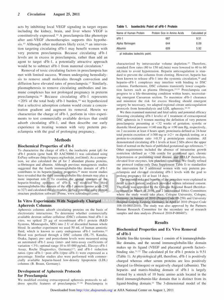
Biochemical Properties and Ex Vivo Removalof sFlt-1Soluble fms-like tyrosine kinase 1 consists of 6 immunoglobulin-like domains, and the second immunoglobulin-like domainmakes up its ligand (VEGF and placental growth factor)–binding site.31,32 The calculated pI for sFlt-1 protein is 9.51(Table 1). At physiological pH, therefore, sFlt-1 is positivelycharged whereas other serum proteins are less positivelycharged (�-fibrinogen) or negatively charged (albumin). Theheparin- and matrix-binding domain of sFlt-1 is largelyformed by a stretch of 10 basic amino acids located in thethird immunoglobumin-like domain that is distinct from itsligand-binding domain.21 The 3-dimensional model of the
Table 1. Isoelectric Point of sFlt-1 Protein
Name of Human Protein Protein Size in Amino Acids Calculated pI
sFlt-1 687 9.51
Alpha fibrinogen 644 8.09
Albumin 609 5.92
pI indicates isolectric point.
2 Circulation August 25, 2011
at AHA National Center on August 1, 2011http://circ.ahajournals.org/Downloaded from

third immunoglobulin-like loop reveals a patch of basicamino acids around R275 and R279 (Figure 1).
Dextran sulfate, a polyanionic derivative of dextran linking2 sulfate groups to a single unit of glucose, binds positivelycharged particles in circulation such as the apolipoproteinB– containing lipoprotein LDL. Low-density lipoproteinapheresis with DSC has been used safely in children andadults with homozygous familial hypercholesterolemia(FH).24,33 In pregnant women with FH, treatments havecontinued without major complications.26,34,35 To determinewhether negatively charged DSC columns adsorb circulatingsFlt-1, we performed ex vivo studies with recombinant sFlt-1protein spiked into human whole blood. As shown in Table 2,�80% of the circulating sFlt-1 protein was removed after 2runs through a DSC column. Because several naturallyoccurring sFlt-1 isoforms circulate in humans,36 we also used
amniotic fluid (a rich source of endogenous sFlt-1) in thesame experiments and found similar results (Table 3), sug-gesting that DSC columns would be suitable for sFlt-1adsorption in vivo.
Single Apheresis Treatment LowersCirculating sFlt-1To test whether DSC columns adsorb sFlt-1 in humans, wefirst performed a single treatment in 5 women and there-after treated 3 additional women multiple times. Fivewomen with very preterm preeclampsia and elevatedcirculating sFlt-1 levels underwent a single apheresistreatment (Table 4). During apheresis, the only apparentside effect noted in 3 women was a transient reduction insystolic blood pressure (maximum 20 to 30 mm Hg) within30 minutes of initiating the procedure. This immediatelyresponded to normal saline infusion, with no womanrequiring �0.5 to 1.0 L after this initial episode. Nosubject experienced bleeding complications, and oxygensaturations remained above 98% throughout. Fetal moni-toring was notable for transient (3 to 5 minutes) reductionin fetal heart beats (average 150 to 130/min) concomitantwith maternal blood pressure reduction; this also immedi-ately recovered after normal saline infusion. Uterine con-tractions were not observed, and clinical indications foremergent Caesarean section were absent. After apheresis,plasma fibrinogen levels were reduced by �20% andreturned to baseline levels within 24 hours in all women.Circulating sFlt-1 levels appeared to be lowered in adose-dependent fashion as the correlation (Spearman)between percentage sFlt-1 reduction and apheresis dura-tion in minutes was 0.80, P�0.10. We were unable to useplacental growth factor measurements to follow the effectsof sFlt-1 reduction during DSC apheresis because certainheparin-binding isoforms of placental growth factor wouldbind DSC columns, rendering these levels uninterpretable.
Changes in sFlt-1 Levels in Untreated WomenWe next examined admission sFlt-1 levels and subsequentprolongation of pregnancy in 7 contemporaneous womenwith very preterm preeclampsia from the same clinicalsites but who did not undergo apheresis treatments (online-only Data Supplement Table II). Admission sFlt-1 valuesranged from 8 028 to 18 738 pg/mL, with a mean of 14 123pg/mL. Prolongation of pregnancy with expectant manage-
Figure 1. Model of the third immunoglobulin domain of sFlt-1protein (AAC50060.1). Spatial structure of the heparin-bindingepitopes of sFlt-1 is shown using a computer-generated modelas described in Methods. A stretch of 10 amino acids from 272to 281 that constitutes the major heparin-binding domain ishighlighted. The positive amino acids (lysine, K, and arginine, R)are labeled. Blue and red region indicate regions of positive andnegative charges, respectively.
Table 2. Ex Vivo Removal of Recombinant sFlt-1 Protein byDSC Column
Experiment 1 sFlt-1 in pg/mL Reduction Ratio, %
Baseline (pre) 4558
Run 1 (post)* 805.5 82.3
Run 2 (post)* 699.7 84.6
*Each run represents passing of 1 entire blood volume (2 units of discardedhuman whole blood) through the column.
Table 3. Ex Vivo Removal of Amniotic Fluid sFlt-1 Protein byDSC Column
Experiment With DL75(Dextran Sulfate)* sFlt-1 in pg/ml Reduction Ratio, %
Baseline (pre) 4576
Run 1 (post)† 2273 50.3
Run 2 (post)† 1611 64.8
Run 3 (post)† 1185 74.1
*Experiment performed as described in Methods with 50 mL of humanamniotic fluid spiked into 2 units of discarded human blood.
†Each run represents passing of 1 entire blood volume (2 units of discardedhuman whole blood) through the column.
Thadhani et al sFlt-1 Removal in Preeclampsia 3
at AHA National Center on August 1, 2011http://circ.ahajournals.org/Downloaded from

ment and changes in sFlt-1 levels are shown in Figure 2.Average pregnancy prolongation was 3.6 days (range 1 to6 days).
Apheresis Treatments to Prolong PregnancyTo prolong pregnancy for at least 14 days after admission,we then offered �1 treatment to 3 additional women withsimilar characteristics, namely very preterm preeclampsia.We targeted 5 to 6 L whole blood (�1.0 to 1.5 timesplasma volume)37 exchanges and a 25% to 35% reductionof circulating sFlt-1 levels with each treatment. We usedcirculating sFlt-1 levels to guide the timing of eachtreatment. Obstetricians could decide to deliver at any timeon the basis of standard indications. The 3 patients aredescribed below.
Patient 1A 32-year-old gravida 1 para 0 woman (Figure 3A through3C) presented at 28�0 weeks of gestation with a singlefetus, hypertension, and proteinuria. Blood pressure was170/106 and 147/101 mm Hg (4 hours apart), and urinedipstick demonstrated 3� proteinuria. She had no historyof chronic hypertension or diabetes mellitus, she was anonsmoker, and prior prenatal visits were unremarkable.Physical examination was notable for pedal edema. Esti-mated fetal weight was 976 g, which was at the 9th centilefor gestational age. Umbilical artery Doppler and amnioticfluid volume measures were normal. Uterine Dopplerdemonstrated a positive notching sign with an umbilico-placental resistance above the 95th percentile. Fetal heartrate was 140 to 150 bpm. Laboratory findings were normal
Table 4. Characteristics of 5 Women With Very Preterm Preeclampsia Who Underwent a SingleExtracorporeal Apheresis Treatment*
Patient 1 Patient 2 Patient 3 Patient 4 Patient 5
Characteristics on day of apheresis
Maternal age, y 22 26 37 34 20
Body mass index, kg/m2 25.9 24.3 29.7 21.8 31.8
Gestational age, weeks�days 29�0 31�3 29�0 24�6 28�3
Systolic blood pressure, mm Hg 186 150 223 211 163
Diastolic blood pressure, mm Hg 96 104 116 104 103
Laboratory tests on day of apheresis
Hemoglobin, g/L 13.2 12.3 12.0 11.1 10.7
Platelets, �109/L 276 199 172 203 237
Creatinine, �mol/L 61 53 57 52 37
SGOT, U/L 87 31 20 � 26
SGPT, U/L 64 24 18 39 25
LDH, U/L 265 185 232 186 189
Pre- and post-pheresis measurements
sFlt-1 before, pg/ml† 14232 13628 12490 18168 8818
sFlt-1 after, pg/ml 12138 11249 10126 14251 6360
% sFlt-1 reduction 14.7 17.5 18.9 21.6 27.9
Urine PC ratio before 446 2983 2568 1168 6583
Urine PC ratio after (nadir) � 1250 1026 618 2500
Fibrinogen before, g/L 3.5 4.0 3.6 2.0 3.6
Fibrinogen after, g/L � 3.7 � 1.4 3.0
Volume treated, mL 1916 2500 5900 5459 4300
Apheresis duration, min 60 108 105 134 120
Duration of pregnancy after pheresis, d 2 4 3 0 7
Reason for delivery‡ Worseningpreeclampsia
Worseningpreeclampsia
HELLPsyndrome
HELLPsyndrome
Worsening of preeclampsiaand evidence of slowing
fetal heart rate
*Missing values (�).†Normal reference values of sFlt-1: median 1449 pg/ml (quartile 1 to quartile 3, 1028–1968 pg/mL) between 24 and 28 weeks of
gestation; median 1934 pg/mL (quartile 1 to quartile 3, 1222–2818 pg/mL) between 29 and 33 weeks of gestation. Ninety-fifth centilevalues at these 2 time periods are 3890 pg/mL and 6688 pg/mL, respectively.16
‡Worsening preeclampsia included new onset of headache, visual changes, requirement of additional medications to control bloodpressure, worsening proteinuria, and worsening edema; HELLP (hemolysis, elevated liver enzymes, low platelets) syndrome included newevidence of hemolysis, rising liver function tests, reduction of platelets, and epigastric pain.
SGOT indicates serum glutamic oxalacetic transaminase; SGPT, serum glutamic pyruvic transaminase; LDH, lactate dehydrogenase;sFlt-1, soluble fms-like tyrosine kinase 1; and PC, protein-to-creatinine ratio.
4 Circulation August 25, 2011
at AHA National Center on August 1, 2011http://circ.ahajournals.org/Downloaded from

(Table 5) except for elevated sFlt-1 levels at 12 865pg/mL. She received 2 doses of betamethasone. Two dayslater 24-hour urine revealed 6.4 g of protein, and circulat-ing sFlt-1 levels rose to 16 236 pg/mL. She was receivingmethyldopa 250 mg 4 times daily.
On hospital day 4, she agreed to undergo 2 extracorpo-real apheresis sessions. On average, blood pressure was186/110 mm Hg, urine protein-to-creatinine ratio was 4.5,and sFlt-1 levels were 18 755 pg/mL. During the apheresistreatment, blood pressure remained stable (range 140/107to 182/120 mm Hg), and fetal heart rate ranged from 130 to160 bpm. Over 110 minutes, 6.0 L of whole blood wasexchanged. After apheresis, circulating sFlt-1 was loweredby �30% (to 13 167 pg/mL), and urine protein-to-creati-nine ratio fell to 3.4 within 12 hours. Preapheresis plasmafibrinogen was 1.9 g/L. It dropped to 1.6 g/L afterapheresis, then returned to 2.0 g/L within 24 hours. Theentire procedure was well tolerated.
Over the ensuing days, blood pressure remained stableas did her blood pressure medication requirements. Shereported improved pedal edema. By hospital day 8, circu-lating sFlt-1 and protein-to-creatinine ratio climbed to18 934 pg/mL and 6.2, respectively. Given the rise insFlt-1 to pretreatment levels, she underwent a secondapheresis treatment. Starting blood pressure was 160/118 mm Hg. At �30 minutes into the treatment, bloodpressure reached a nadir of 125/74 mm Hg but recoveredwith 500 mL of normal saline to 134/75 mm Hg (within 5minutes) and 142/103 (within 15 minutes). Fetal heart ratewent from 160 to 130 bpm, but this too immediatelyrecovered with normal saline. No further reductions inblood pressure or fetal heart rate were noted, and apheresiscontinued uninterrupted, exchanging 6.0 L of whole blood.After apheresis, circulating sFlt-1 levels were lowered by34% (to 12 475 pg/mL), and within 12 hours protein-to-creatinine ratio decreased to 3.2. Plasma fibrinogen levels
went from 2.3 g/L to 2.0 g/L after apheresis and returnedto 2.2 g/L within 12 hours.
On hospital day 11, estimated fetal weight was 1097 g.By hospital day 15, at 30�0 weeks of gestation, circulatingsFlt-1 levels and protein-to-creatinine ratio climbed to25 415 pg/mL and 6.1, respectively. In the face of an acute4-kg weight gain, headaches, and rising diastolic bloodpressures (120 mm Hg) despite increasing medications,she was delivered by Caesarean section. At delivery,Apgar scores were 7/9/9, and the newborn girl weighed995 g. Twelve hours after delivery, maternal blood pres-sure remained elevated at 177/115 mm Hg, and at 48hours, when circulating sFlt-1 levels were 2118 pg/mL,blood pressure was 158/123 mm Hg. Within 24 hours,however, urine protein-to-creatinine ratio dropped to 2.6.She was discharged on day 25. The newborn requiredminimal monitoring and remained in the neonatal intensivecare unit for 55 days, after which she was discharged inexcellent condition without any notable abnormalities. Ather 4-week follow-up, her blood pressures were normaland her newborn weighed 1465 g and had achievedexpected developmental milestones.
Patient 2A 40-year-old gravida 2 para 0 woman (Figure 4A through4C) presented at 30�0 weeks with significant hyperten-sion, proteinuria, and pedal edema. She was carryingdichorionic-diamniotic twins after receiving in vitro fertil-ization therapy. Blood pressure was 140/90 mm Hg and161/92 mm Hg (4 hours apart), and urine dipstick revealed2� protein. She was admitted for very preterm preeclamp-sia. Her past medical history was unremarkable, and familyhistory was only notable for paternal diabetes mellitus. Shewas a nonsmoker, and prenatal visits routinely demon-strated normal blood pressure and urine dipstick results.She was taking prenatal vitamins. Physical examinationwas notable for hypertension and pedal edema. Ultrasoundshowed 2 fetuses, estimated weights 1329 and 1327 g, withnormal growth, amniotic fluid volume, and umbilicalperfusion. Fetal heart rates were 130 to 150 bpm in eachwith normal oscillation and sporadic accelerations, uterinecontractions were absent, and there was no evidence ofdecelerations. Laboratory findings revealed no evidence ofHELLP syndrome (Table 5). Two doses of betamethasonewere administered. Blood sFlt-1 levels were 29 068 pg/mL. A 24-hour urine collection demonstrated 786 mg ofprotein, and a urine protein-to-creatinine ratio of 1.0.Blood pressures were stable.
On hospital day 6, she agreed to undergo 2 extracorpo-real apheresis treatments. Blood pressure was 187/81 mm Hg. Fetal ultrasound revealed normal arterial per-fusion in both umbilical cords, and cardiotocography wasotherwise normal. By 30 minutes into apheresis, her bloodpressure dropped to 90/50 mm Hg, which recovered to144/60 mm Hg within 5 minutes after 1.0 L of normalsaline. Fetal ultrasound and cardiotocography did notreveal alterations. She continued with uninterruptedapheresis for 120 minutes, exchanging 5.1 L of wholeblood. For the remainder of the treatment, blood pressure
Figure 2. Plot of baseline and predelivery sFlt-1 (pg/mL) levelsand prolongation time (days) in 7 untreated patients with verypreterm preeclampsia. The red circles represent the baselinelevels of sFlt-1 for each patient whereas the black arrowheadsrepresent predelivery levels (y axis) and the days of prolongation(x axis) after admission. sFlt-1 indicates soluble fms-like tyrosinekinase 1.
Thadhani et al sFlt-1 Removal in Preeclampsia 5
at AHA National Center on August 1, 2011http://circ.ahajournals.org/Downloaded from

averaged 150/65 mm Hg and fetal heart rates averagedbetween 130 and 150 bpm for both fetuses. CirculatingsFlt-1 levels were lowered by 26% (to 17 933 pg/mL) afterapheresis, and protein-to-creatinine ratio dropped to 0.55within 24 hours. Plasma fibrinogen levels went from 3.2g/L to 2.7 g/L after apheresis, then rose to 3.0 g/L within12 hours.
She received no blood pressure medications, and she hadno complaints. By hospital day 11, circulating sFlt-1 levelsand urine protein-to-creatinine ratio climbed to 23 075pg/mL and 1.9, respectively. Given the rise in sFlt-1 levels,she underwent a second treatment. She experienced noepisodes of hypotension (nadir 166/72 mm Hg at 30minutes) or changes in fetal heart rates. Estimated fetalweights were 1674 and 1626 g. Apheresis continued for120 minutes, exchanging 6.0 L of whole blood. After thesecond treatment, circulating sFlt-1 levels were lowered by34% (to 15 197 pg/mL), and within 12 hours urine protein-to-creatinine ratio decreased to 0.79. Plasma fibrinogenlevels went from 3.4 g/L to 2.8 g/L after apheresis, thenreturned to 3.3 g/L within 12 hours.
Over the next 7 days, she remained stable, requiring noblood pressure medications. However, circulating sFlt-1levels climbed in conjunction with proteinuria. At 32�5weeks of gestation, 19 days after hospital admission, herplacental membranes spontaneously ruptured and she un-derwent uncomplicated Caesarean section. Five-minuteApgar scores were 9 for fetus 1 and 7 for fetus 2, and fetalweights were 1498 and 1485 g, respectively. At 12 hoursafter delivery, circulating sFlt-1 levels dropped to 226pg/mL, yet blood pressure remained elevated: 155/85 mm Hg at 12 and 160/80 mm Hg at 24 hours. By day 7,
Figure 3. Changes in maternal circulating sFlt-1 levels (pg/mL)(A), protein-creatinine ratio (g/g) (B), and blood pressure(mm Hg) (C) in patient 1 over the course of 15 days of inpatienthospitalization starting at gestational age 28 weeks. Dotted ref-erence lines indicate days on which apheresis was performed.Bars on panel C show average daily systolic blood pressure (topwhisker), average daily diastolic blood pressure (bottom whis-ker), and average daily mean arterial pressure [(2�diastolicblood pressure�systolic blood pressure)/3] (black circle). Nor-mal reference values for sFlt-1: median 1449 pg/mL (quartile 1toquartile 3, 1028 to 1968 pg/mL) between 24 and 28 weeks ofgestation; median 1934 pg/mL (quartile 1 to quartile 3, 1222 to
2818 pg/mL) between 29 and 33 weeks of gestation. Ninety-fifthcentile values at these 2 time periods are 3890 pg/mL and 6688pg/mL, respectively.13 sFlt-1 indicates soluble fms-like tyrosinekinase 1.
Table 5. Characteristics of 3 Patients With Very PretermPreeclampsia Who Underwent >2 ExtracorporealApheresis Treatments
Patient 1 Patient 2 Patient 3
Characteristics prior to pheresis
Maternal age, y 32 40 35
Body mass index, kg/m2 26.5 27.4 28.4
Gestational age, weeks�days 28�3 30�0 27�5
Systolic blood pressure, mm Hg) 186 187 175
Diastolic blood pressure, mm Hg 110 81 100
Laboratory tests prior to pheresis
Hemoglobin, g/L 12.9 11.2 11.1
Platelets, �109/L 234 222 244
Creatinine, �mol/L 67 41 57
SGOT, U/L 36 53 64
SGPT, U/L 27 43 62
LDH, U/L 247 195 263
SGOT indicates serum glutamic oxalacetic transaminase; SGPT, serumglutamic pyruvic transaminase; and LDH, lactate dehydrogenase.
6 Circulation August 25, 2011
at AHA National Center on August 1, 2011http://circ.ahajournals.org/Downloaded from

blood pressure was 135/80 mm Hg. In contrast, urineprotein-to-creatinine ratio just before delivery was 18.1,and within 24 hours after delivery it was 1.4. Neithernewborn required ventilator support other than continuouspositive airway pressure. On follow-up at 5 weeks, theirweights were 2330 and 2300 g, and their expected devel-opmental milestones were achieved.
Patient 3A 35-year-old gravida1 para 0 woman (Figure 5A through5C) presented at 27�4 weeks with hypertension, protein-uria, and pedal edema. Initial blood pressure was 177/95 mm Hg, which was confirmed several hours later, andurine protein-to-creatinine ratio was 2.3. She was admittedfor very preterm preeclampsia. Her medical history wasnotable for Hashimoto’s thyroiditis, and her family historywas notable for paternal and maternal hypertension. Shewas a nonsmoker, and previous prenatal visits demon-strated normal blood pressure and urine dipstick results.She had no history of chronic hypertension. Physicalexamination was notable for hypertension and pedaledema. Ultrasound showed a singleton fetus in breechpresentation with an estimated weight of 811 g. Amnioticfluid volume and placenta appeared normal. Dopplerultrasound revealed an abnormal uterine Doppler withincreased resistance but no notching. Fetal perfusion in theumbilical artery, middle cerebral artery, and ductus veno-sus was normal. Fetal heart rate was 140 to 160 bpm, andthere was no evidence of uterine contractions or deceler-ations. Laboratory findings revealed no evidence ofHELLP syndrome (Table 5). Two doses of betamethasonewere administered.
On hospital day 3, she agreed to undergo serial extra-corporeal apheresis treatments. Blood pressure was 175/105 mm Hg without antihypertensive medications. Fetalultrasound revealed normal umbilical perfusion, and car-diotocography was otherwise normal. By 23 minutes intotreatment, her blood pressure dropped to 93/50 mm Hg,which recovered to 126/51 mm Hg (within 5 minutes) after1.0 L of normal saline. She continued with uninterruptedapheresis, but after 90 minutes of treatment her bloodpressure dropped again. Apheresis was then stopped afterexchanging a total 3.2 L of whole blood. Ultrasoundexamination showed unchanged umbilical artery perfusionvalues and normal fetal movements. After apheresis, sFlt-1levels dropped together with her urine protein-to-creati-nine ratio. Her blood pressure remained stable with valuesof �175/100 mm Hg without medications. By hospital day7 (28�3 weeks), sFlt-1 levels and urine protein-to-creati-nine ratio climbed.
A second treatment was performed on day 7. This time,1 to 2 L of saline were administered slowly at the start ofthe treatment. The procedure was better tolerated with nopronounced hypotensive episodes. Apheresis continued for110 minutes, exchanging a total of 4.0 L of whole blood.After the second treatment, circulating sFlt-1 levels werelowered to 10 345 pg/mL, and within 12 hours, urineprotein-to-creatinine ratio also decreased. Plasma fibrino-gen levels went from 4.4 g/L to 3.1 g/L after apheresis,
Figure 4. Changes in maternal circulating sFlt-1 levels (pg/mL)(A), protein-creatinine ratio (g/g) (B), and blood pressure(mm Hg) (C) in patient 2 over the course of 19 days of inpatienthospitalization starting at gestational age 30 weeks. Dotted ref-erence lines indicate days on which apheresis was performed.Bars on panel C show average daily systolic blood pressure (topwhisker), average daily diastolic blood pressure (bottom whis-ker), and average daily mean arterial pressure [(2�diastolicblood pressure�systolic blood pressure)/3] (black circle). seeFigure 3 legend for sFlt-1 reference ranges. sFlt-1 indicates sol-uble fms-like tyrosine kinase 1.
Thadhani et al sFlt-1 Removal in Preeclampsia 7
at AHA National Center on August 1, 2011http://circ.ahajournals.org/Downloaded from

then increased to 3.4 g/L within 12 hours. Ultrasounddemonstrated fetal weight gain (estimated at 1013 g).Amniotic fluid amount and fetal perfusion indices werewithin the normal range. Over the next 7 days, she waswithout complaints. Beginning at week 29�1, she receiveda daily dose of 32 mg methylprednisolone intravenouslybecause of slightly increased liver enzymes. Pedal edemawas present but stable.
A third treatment was performed on day 14. She experi-enced no drops in blood pressure, and apheresis continued for115 minutes, exchanging 4.0 L of whole blood. After treat-ment 3, circulating sFlt-1 levels were lowered by 23% (to11 755 pg/mL), and within 12 hours, urine protein-to-creati-nine ratio also decreased from 3.5 to 2.2. Plasma fibrinogenlevels went from 5.2 to 3.7 g/L after apheresis, then climbedto 3.9 g/L within 12 hours. A fourth uncomplicated treatmentwas performed on day 18. Apheresis continued for 126minutes, exchanging 4.5 L of whole blood. Circulating sFlt-1levels were lowered by 25%, and within 12 hours, urineprotein-to-creatinine ratio was lowered from 3.7 to 2.5.
Twenty-three days after hospital admission, at 30�6weeks of gestation, she developed regular contractionswith cervical effacement. She underwent uncomplicatedCaesarean section. Five-minute Apgar score was 7/9/9, andthe fetal weight was 1140 g. At 12 hours after delivery,circulating sFlt-1 levels dropped to 1464 pg/mL. However,her blood pressure remained elevated at 144/98 mm Hg.Blood pressure returned to normal values (110/75 mm Hg)by 24 to 48 hours after delivery. Her newborn receivedpositive airway pressure support in the neonatal intensivecare unit for 5 days, after which he was sent to a regularneonatal care bed. The patient was discharged 4 days afterdelivery. The newborn was discharged approximately 9weeks later, weighing 2710 g, and with normal hearing,respiratory, cardiac, and neurological development.
DiscussionThe antiangiogenic factor sFlt-1 is one possible candidateinvolved in mediating the clinical manifestations of pre-eclampsia. By understanding the charged nature of sFlt-1 andmodifying existing extracorporeal apheresis methods to opti-mize its removal, we demonstrated that apheresis with DSCcolumns reduces circulating sFlt-1 in a dose-dependent fash-ion in women with very preterm preeclampsia. In 3 additionalpatients with very preterm preeclampsia, repeated apheresistreatments reduced circulating levels of sFlt-1 and protein-uria, stabilized blood pressure, and potentially prolongedpregnancy without apparent adverse events occurring toeither mother or fetus.
We chose extracorporeal methods to remove sFlt-1.However, other strategies to target circulating sFlt-1 in-clude administration of VEGF (its natural ligand) or aneutralizing antibody or inhibition of its translation withsmall interfering RNA technologies.4 Preeclampsia doesnot occur naturally in animals. Therefore, encouragingresults in animals may not immediately translate intosuccessful human therapeutics. Because each extra week inutero markedly lowers fetal morbidity and mortality,38 anyshort-term intervention for very preterm preeclampsia
Figure 5. Changes in maternal circulating sFlt-1 levels (pg/mL)(A), protein-creatinine ratio (g/g) (B), and blood pressure(mm Hg) (C) in patient 3 over the course of 23 days of inpatienthospitalization starting at gestational age 27 weeks�4 days.Dotted reference lines indicate days on which apheresis wasperformed. Bars on panel C show average daily systolic bloodpressure (top whisker), average daily diastolic blood pressure(bottom whisker), and average daily mean arterial pressure[(2�diastolic blood pressure�systolic blood pressure)/3] (blackcircle). see Figure 3 legend for sFlt-1 reference ranges. sFlt-1indicates soluble fms-like tyrosine kinase 1.
8 Circulation August 25, 2011
at AHA National Center on August 1, 2011http://circ.ahajournals.org/Downloaded from

should aim to prolong pregnancy without compromisingmaternal safety. The intervention should stabilize or im-prove maternal and fetal parameters without leaving resid-ual effects. The intervention should be easily titrated giventhe potential for acute deterioration of mother, fetus, orboth. Extracorporeal adsorption of circulating sFlt-1 ful-fills several of these criteria. This strategy also does notdeplete VEGF, which does not circulate to any large extentand which is critical in preeclampsia in maintaining localvascular integrity.39,40
Maternal blood pressure appeared to be stabilized butnot markedly decreased after apheresis therapy. In additionto apheresis itself, withholding medications before treat-ments and saline administration during treatments alsomay have modified blood pressure. In experimentalsFlt-1–induced preeclampsia, immediate VEGF-121 ad-ministration lowers blood pressure only after severaldays.39 In humans, delivery of the placenta normalizessFlt-1 levels almost immediately,11 yet maternal bloodpressure returns to normal only after days to weeks. Thislag was evident in our 3 patients. Maternal hypertension inpreeclampsia may result from renal endothelial cell dam-age, and the time required to heal this lesion may explainthe delayed response. In women treated serially, sFlt-1levels were lowered by 17% to 34%. Although furthersFlt-1 reductions may have lowered maternal blood pres-sure, we were concerned about fetal growth retardationfrom aggressive blood pressure reduction.41 Perceiving theneed for more blood flow in preeclampsia, the fetal-placental unit may release excess sFlt-1 into the maternalcirculation to sustain blood pressure.42 This likely explainsthe rebound in circulating sFlt-1 levels in the days aftereach apheresis.
The role of sFlt-1 in normal placentation remainsunknown. Genetically engineered mice that lack placentalsFlt-1 do not develop placental abnormalities,43 and casereports highlight natural reductions in circulating sFlt-1with continuation of human pregnancy to term.44,45 Thus,lowering sFlt-1 to levels near normal does not appear to beharmful. Regardless, we designed our methods to avoidsignificant reduction in circulating sFlt-1, maternal bloodpressure, and placental blood flow, and in this context,maternal blood pressure stabilized, fetal compromise wasclinically absent, and fetal weight gain was evident.
Protein-to-creatinine ratios rose and fell in conjunctionwith sFlt-1 levels. Rescue therapy with VEGF-121 in exper-imental preeclampsia also demonstrates a more immediateimprovement in proteinuria, as does discontinuation of anti-VEGF therapies in cancer.39,46 Although blood pressurelowering reduces proteinuria, apheresis did not result insustained blood pressure reduction. Therefore, reduction ofcirculating sFlt-1 levels with improvement of proteinuriaafter apheresis suggests a more dynamic effect of circulatingsFlt-1 on the integrity of fenestrated glomerular endotheliumand proteinuria. However, the specific mechanisms involvedremain unknown.47 Improvement of proteinuria may becomean important early signal for efficacy of this intervention.
Expectant management in preterm preeclampsia in-creases the risk of maternal and fetal morbidity and
mortality, but in highly selected women this may beappropriate.48 Limited data suggest that in very pretermpreeclampsia without fetal growth restriction, expectantmanagement extends pregnancy by 7 to 14 days.5,48
Among contemporaneous controls that met the criteria forvery preterm preeclampsia, the average prolongation ofpregnancy was �4 days, similar to our 5 patients treatedonce. Furthermore, all controls had a singleton pregnancy,but the risk for adverse outcomes is markedly higher intwin pregnancies. A pregnancy with twins is infrequentlyprolonged beyond a few days with expectant manage-ment.49 We excluded those with fetal growth restriction(�5th centile for gestational age). With 2 apheresis treat-ments, the patient with a singleton fetus remained pregnantfor 15 days and the patient with twins remained pregnantfor 19 days. With 4 treatments, our last patient remainedpregnant for 23 days after admission. None showed evi-dence of fetal compromise. Although it is tempting tospeculate that apheresis prolonged pregnancy and thatlonger or additional treatments would have prolongedpregnancy even further, we treated only a limited numberof patients, and only randomized trials with additionalpatients will allow definitive conclusions on this importantend point.
Our work leaves certain questions unanswered. We did notinclude patients with a severely growth-restricted fetus orwith other evidence of fetal compromise, and thus we do notknow if this therapy would benefit or harm the fetus in thissetting. We cannot exclude the possibility that DSC apheresisremoves substances such as LDL cholesterol and C-reactiveprotein or substances that have a heparin-binding domain andexhibit angiogenic properties (eg, fibroblast growth factors)and that this contributed to our findings. We also do not knowif apheresis cleared substances altered in preeclampsia suchas auto-antibodies against the angiotensin receptor.50 Datafrom one patient, however, suggested that DSC apheresisdoes not reduce endothelin-1 but modestly reduces circulat-ing soluble endoglin (online-only Data Supplement TableIII), similar to what has been reported in patients with FH.51
The specificity of sFlt-1 removal may be improved bydeveloping extracorporeal columns with anti-sFlt-1 antibod-ies covalently bound to beads, similar to methods developedin LDL apheresis.52 Such specificity may reduce the fre-quency of treatments when compared to the 1 to 2 treatments/week we employed to avert further elevations in circulatingsFlt-1 levels. Other adsorption therapies may benefit womenwith preeclampsia. Heparin-mediated LDL apheresis hasbeen attempted in preeclampsia with moderate success.53 Ourex vivo studies suggest that heparin-based columns would beinferior to DSC columns in removing circulating sFlt-1(online-only Data Supplement Table IV).
Novel drugs directed at improving maternal and fetalhealth are infrequently developed. Risk aversion and limitedmarket size are among the many reasons.54 Modifying ap-proved interventions with known safety profiles such asdemonstrated here with extracorporeal DSC apheresis is aviable and attractive alternative.55 In this hypothesis-generating study we provide 1 potential intervention for verypreterm preeclampsia characterized by elevated circulating
Thadhani et al sFlt-1 Removal in Preeclampsia 9
at AHA National Center on August 1, 2011http://circ.ahajournals.org/Downloaded from

sFlt-1. Further studies with additional patients are warrantedto fully assess whether the intervention studied herein safelyand effectively prolongs pregnancy and improves maternaland fetal outcomes in very preterm preeclampsia.
AcknowledgmentsWe would like to thank the patients who agreed to participate in thisstudy. We would especially like to thank Drs Samuel O. Thier,Dennis A. Ausiello, and M. Amin Arnaout for their support. Finally,we thank Kathy Lucchesi for her assistance with the protocol, andRameh Follette with her assistance in early in vitro studies.
Sources of FundingThis work was funded by the Discovery Fund from the Departmentof Medicine, Massachusetts General Hospital, to Dr Thadhani. DrThadhani’s salary is supported by several federal and foundationgrants. This work also received support from Forschungspool Klinis-che Studien, Faculty of Medicine, University of Cologne. Dr Cornelyis supported by the German Federal Ministry of Research andEducation (Bundesministerium fur Bildung und Forschung Grant01KN1106). Dr Benzing received funding from the German Re-search Foundation (BE2212). Dr Karumanchi is supported by theHoward Hughes Medical Institute.
DisclosuresDr Thadani is coinventor of patents related to diagnostics in theprediction of preeclampsia that have been out-licensed to diag-nostic companies and has financial interest in Aggamin LLC. DrKarumanchi is coinventor of multiple patents related to the use ofangiogenic proteins for the diagnosis and therapy of preeclamp-sia. These patents have been licensed to multiple companies. DrKarumanchi is a consultant to Roche Diagnostics and BeckmanCoulter and has financial interest in Aggamin LLC. The remain-ing authors report no conflicts.
References1. Redman CW, Sargent IL. Latest advances in understanding preeclampsia.
Science. 2005;308:1592–1594.2. Alexander GR, Kogan M, Bader D, Carlo W, Allen M, Mor J. US birth
weight/gestational age–specific neonatal mortality: 1995–1997 rates forwhites, hispanics, and blacks. Pediatrics. 2003;111:e61–e66.
3. Mathews TJ, MacDorman MF. Infant mortality statistics from the 2004period linked birth/infant death data set. Natl Vital Stat Rep. 2007;55:1–32.
4. Thadhani R. Inching towards a targeted therapy for preeclampsia.Hypertension. 2010;55:238–240.
5. Odendaal HJ, Pattinson RC, Bam R, Grove D, Kotze TJ. Aggressive orexpectant management for patients with severe preeclampsia between28–34 weeks’ gestation: a randomized controlled trial. Obstet Gynecol.1990;76:1070–1075.
6. Sibai BM, Mercer BM, Schiff E, Friedman SA. Aggressive versusexpectant management of severe preeclampsia at 28 to 32 weeks’ ges-tation: a randomized controlled trial. Am J Obstet Gynecol. 1994;171:818–822.
7. Roberts JM, Taylor RN, Musci TJ, Rodgers GM, Hubel CA, McLaughlinMK. Preeclampsia: an endothelial cell disorder. Am J Obstet Gynecol.1989;161:1200–1204.
8. Ahmad S, Ahmed A. Elevated placental soluble vascular endothelialgrowth factor receptor-1 inhibits angiogenesis in preeclampsia. Circ Res.2004;95:884–891.
9. Chaiworapongsa T, Romero R, Espinoza J, Bujold E, Mee Kim Y,Goncalves LF, Gomez R, Edwin S. Evidence supporting a role forblockade of the vascular endothelial growth factor system in the patho-physiology of preeclampsia. Young Investigator Award. Am J ObstetGynecol. 2004;190:1541–1550.
10. Levine RJ, Maynard SE, Qian C, Lim KH, England LJ, Yu KF,Schisterman EF, Thadhani R, Sachs BP, Epstein FH, Sibai BM, SukhatmeVP, Karumanchi SA. Circulating angiogenic factors and the risk ofpreeclampsia. N Engl J Med. 2004;350:672–683.
11. Maynard SE, Min JY, Merchan J, Lim KH, Li J, Mondal S, LibermannTA, Morgan JP, Sellke FW, Stillman IE, Epstein FH, Sukhatme VP,
Karumanchi SA. Excess placental soluble fms-like tyrosine kinase 1(sFlt1) may contribute to endothelial dysfunction, hypertension, and pro-teinuria in preeclampsia. J Clin Invest. 2003;111:649–658.
12. Chaiworapongsa T, Romero R, Kim YM, Kim GJ, Kim MR, Espinoza J,Bujold E, Goncalves L, Gomez R, Edwin S, Mazor M. Plasma solublevascular endothelial growth factor receptor-1 concentration is elevatedprior to the clinical diagnosis of pre-eclampsia. J Matern Fetal NeonatalMed. 2005;17:3–18.
13. Verlohren S, Galindo A, Schlembach D, Zeisler H, Herraiz I, Moertl MG,Pape J, Dudenhausen JW, Denk B, Stepan H. An automated method forthe determination of the sFlt-1/PIGF ratio in the assessment of pre-eclampsia. Am J Obstet Gynecol. 2010;202:161e1–161e11.
14. Maynard S, Epstein FH, Karumanchi SA. Preeclampsia and angiogenicimbalance. Annu Rev Med. 2008;59:61–78.
15. Feldman DR, Baum MS, Ginsberg MS, Hassoun H, Flombaum CD,Velasco S, Fischer P, Ronnen E, Ishill N, Patil S, Motzer RJ. Phase I trialof bevacizumab plus escalated doses of sunitinib in patients with meta-static renal cell carcinoma. J Clin Oncol. 2009;27:1432–1439.
16. Levine RJ, Lam C, Qian C, Yu KF, Maynard SE, Sachs B, Sibai B,Epstein FH, Romero R, Thadhani R, Karumanchi SA; CPEP StudyGroup. Soluble endoglin and other circulating antiangiogenic factors inpreeclampsia. N Engl J Med. 2006;355:992–1005.
17. Davison JM. Dialysis, transplantation, and pregnancy. Am J Kidney Dis.1991;17:127–132.
18. Martin JN Jr, Perry KG Jr, Roberts WE, Norman PF, Files JC, Blake PG,Morrison JC, Wiser WL. Plasma exchange for preeclampsia: II. Unsuc-cessful antepartum utilization for severe preeclampsia with or withoutHELLP syndrome. J Clin Apher. 1994;9:155–161.
19. Wu FT, Stefanini MO, Mac Gabhann F, Popel AS. A compartment modelof VEGF distribution in humans in the presence of soluble VEGFreceptor-1 acting as a ligand trap. PLoS One. Published online April 8,2009. doi: 10.1371/journal.pone.0005108. 2009;4:e5108.
20. Park M, Lee ST. The fourth immunoglobulin-like loop in the extracellulardomain of FLT-1, a VEGF receptor, includes a major heparin-bindingsite. Biochem Biophys Res Commun. 1999;264:730–734.
21. Holash J, Davis S, Papadopoulos N, Croll SD, Ho L, Russell M, BolandP, Leidich R, Hylton D, Burova E, Ioffe E, Huang T, Radziejewski C,Bailey K, Fandl JP, Daly T, Wiegand SJ, Yancopoulos GD, Rudge JS.VEGF-Trap: a VEGF blocker with potent antitumor effects. Proc NatlAcad Sci U S A. 2002;99:11393–11398.
22. Vuorela P, Helske S, Hornig C, Alitalo K, Weich H, Halmesmaki E.Amniotic fluid–soluble vascular endothelial growth factor receptor-1 inpreeclampsia. Obstet Gynecol. 2000;95:353–357.
23. Bambauer R, Schiel R, Latza R. Low density lipoprotein apheresis intreatment of hyperlipidemia: experience with four different technologies.Ther Apher. 2000;4:213–217.
24. Bosch T, Schmidt B, Kleophas W, Gillen C, Otto V, Passlick-Deetjen J,Gurland HJ. LDL hemoperfusion–a new procedure for LDL apheresis:first clinical application of an LDL adsorber compatible with humanwhole blood. Artif Organs. 1997;21:977–982.
25. Klingel R, Fassbender T, Fassbender C, Gohlen B. From membranedifferential filtration to lipidfiltration: technological progress in low-density lipoprotein apheresis. Ther Apher Dial. 2003;7:350–358.
26. Teruel JL, Lasuncion MA, Navarro JF, Carrero P, Ortuno J. Pregnancy in apatient with homozygous familial hypercholesterolemia undergoing low-density lipoprotein apheresis by dextran sulfate adsorption. Metabolism.1995;44:929–933.
27. Soffronoff EC, Kaufmann BM, Connaughton JF. Intravascular volumedeterminations and fetal outcome in hypertensive diseases of pregnancy.Am J Obstet Gynecol. 1977;127:4–9.
28. Sela S, Natanson-Yaron S, Zcharia E, Vlodavsky I, Yagel S, Keshet E.Local retention versus systemic release of soluble VEGF receptor-1 aremediated by heparin-binding and regulated by heparanase. Circ Res.2011;108:1063–1070.
29. Bosch T, Heinemann O, Duhr C, Wendler T, Keller C, Fink E, KirschnerT, Klebert S, Samtleben W. Effect of low-dose citrate anticoagulation onthe clinical safety and efficacy of direct adsorption of lipoproteins (DALIapheresis) in hypercholesterolemic patients: a prospective controlledclinical trial. Artif Organs. 2000;24:790–796.
30. Palsson R, Niles JL. Regional citrate anticoagulation in continuous veno-venous hemofiltration in critically ill patients with a high risk of bleeding.Kidney Int. 1999;55:1991–1997.
31. Davis-Smyth T, Presta LG, Ferrara N. Mapping the charged residues inthe second immunoglobulin-like domain of the vascular endothelial
10 Circulation August 25, 2011
at AHA National Center on August 1, 2011http://circ.ahajournals.org/Downloaded from
growth factor/placenta growth factor receptor Flt-1 required for bindingand structural stability. J Biol Chem. 1998;273:3216–3222.
32. Kendall RL, Thomas KA. Inhibition of vascular endothelial cell growthfactor activity by an endogenously encoded soluble receptor. Proc NatlAcad Sci U S A. 1993;90:10705–10709.
33. Zwiener RJ, Uauy R, Petruska ML, Huet BA. Low-density lipoproteinapheresis as long-term treatment for children with homozygous familialhypercholesterolemia. J Pediatr. 1995;126:728–735.
34. Klingel R, Gohlen B, Schwarting A, Himmelsbach F, Straube R. Differ-ential indication of lipoprotein apheresis during pregnancy. Ther ApherDial. 2003;7:359–364.
35. Kroon AA, Swinkels DW, van Dongen PW, Stalenhoef AF. Pregnancy ina patient with homozygous familial hypercholesterolemia treated withlong-term low-density lipoprotein apheresis. Metabolism. 1994;43:1164–1170.
36. Heydarian M, McCaffrey T, Florea L, Yang Z, Ross MM, Zhou W,Maynard SE. Novel splice variants of sFlt1 are upregulated in pre-eclampsia. Placenta. 2009;30:250–255.
37. Kaplan AA. A simple and accurate method for prescribing plasmaexchange. ASAIO Trans. 1990;36:M597–M599.
38. ACOG Practice Bulletin. Assessment of risk factors for preterm birth.Clinical management guidelines for obstetrician-gynecologists. Number31, October 2001. (Replaces Technical Bulletin number 206, June 1995;Committee Opinion number 172, May 1996; Committee Opinion number187, September 1997; Committee Opinion number 198, February 1998;and Committee Opinion number 251, January 2001). Obstet Gynecol.2001;98:709–716.
39. Li Z, Zhang Y, Ying Ma J, Kapoun AM, Shao Q, Kerr I, Lam A,O’Young G, Sannajust F, Stathis P, Schreiner G, Karumanchi SA, ProtterAA, Pollitt NS. Recombinant vascular endothelial growth factor 121attenuates hypertension and improves kidney damage in a rat model ofpreeclampsia. Hypertension. 2007;50:686–692.
40. Tsatsaris V, Goffin F, Munaut C, Brichant JF, Pignon MR, Noel A,Schaaps JP, Cabrol D, Frankenne F, Foidart JM. Overexpression of thesoluble vascular endothelial growth factor receptor in preeclampticpatients: pathophysiological consequences. J Clin Endocrinol Metab.2003;88:5555–5563.
41. von Dadelszen P, Ornstein M, Bull S, Logan A, Koren G, Magee L. Fallin mean arterial pressure and fetal growth restriction in pregnancy hyper-tension: a meta-analysis. The Lancet. 2000;355:87–92.
42. Haig D. Genetic conflicts in human pregnancy. Q Rev Biol. 1993;68:495–532.
43. Hirashima M, Lu Y, Byers L, Rossant J. Trophoblast expression offms-like tyrosine kinase 1 is not required for the establishment of thematernal-fetal interface in the mouse placenta. Proc Natl Acad Sci U S A.2003;100:15637–15642.
44. Hladunewich MA, Steinberg G, Karumanchi SA, Levine RJ, Keating S,Kingdom J, Keunen J. Angiogenic factor abnormalities and fetal demisein a twin pregnancy. Nat Rev Nephrol. 2009;5:658–662.
45. Stepan H, Faber R. Elevated sFlt1 level and preeclampsia withparvovirus-induced hydrops. N Engl J Med. 2006;354:1857–1858.
46. Eremina V, Jefferson JA, Kowalewska J, Hochster H, Haas M, WeisstuchJ, Richardson C, Kopp JB, Kabir MG, Backx PH, Gerber HP, Ferrara N,Barisoni L, Alpers CE, Quaggin SE. VEGF inhibition and renalthrombotic microangiopathy. N Engl J Med. 2008;358:1129–1136.
47. Smithies O. Why the kidney glomerulus does not clog: a gel permeation/diffusion hypothesis of renal function. Proc Natl Acad Sci U S A. 2003;100:4108–4113.
48. Sibai BM, Barton JR. Expectant management of severe preeclampsiaremote from term: patient selection, treatment, and delivery indications.Am J Obstet Gynecol. 2007;196:514 e511–e519.
49. Morcel K, Lavoue V, Beuchee A, Le Lannou D, Poulain P, Pladys P.Perinatal morbidity and mortality in twin pregnancies with dichorionicplacentas following assisted reproductive techniques or ovarian inductionalone: a comparative study. Eur J Obstet Gynecol Reprod Biol. 2010;153:138–142.
50. Dechend R, Homuth V, Wallukat G, Kreuzer J, Park JK, Theuer J,Juepner A, Gulba DC, Mackman N, Haller H, Luft FC. AT(1) receptoragonistic antibodies from preeclamptic patients cause vascular cells toexpress tissue factor. Circulation. 2000;101:2382–2387.
51. Blaha M, Cermanova M, Blaha V, Jarolim P, Andrys C, Blazek M, Maly J,Smolej L, Zajic J, Masin V, Zimova R, Rehacek V. Elevated serumsoluble endoglin (sCD105) decreased during extracorporeal eliminationtherapy for familial hypercholesterolemia. Atherosclerosis. 2008;197:264–270.
52. Stoffel W, Demant T. Selective removal of apolipoprotein B-containingserum lipoproteins from blood plasma. Proc Natl Acad Sci U S A. 1981;78:611–615.
53. Wang Y, Walli AK, Schulze A, Blessing F, Fraunberger P, Thaler C,Seidel D, Hasbargen U. Heparin-mediated extracorporeal low densitylipoprotein precipitation as a possible therapeutic approach in pre-eclampsia. Transfus Apher Sci. 2006;35:103–110.
54. Fisk NM, Atun R. Market failure and the poverty of new drugs inmaternal health. PLoS Med. 2008;5:e22.
55. Boguski MS, Mandl KD, Sukhatme VP. Drug discovery Repurposingwith a difference. Science. 2009;324:1394–1395.
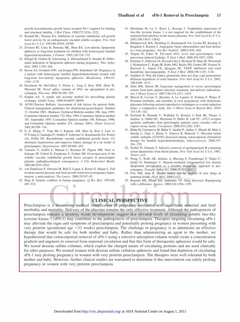
CLINICAL PERSPECTIVEPreeclampsia is a devastating medical complication of pregnancy associated with significant maternal and fetalmorbidity and mortality. Delivery of the placenta remains the only effective treatment. Although the pathogenesis ofpreeclampsia remains a mystery, recent developments suggest that elevated levels of circulating soluble fms-liketyrosine kinase 1 (sFlt-1) may contribute to the pathogenesis of preeclampsia. Therapies targeting circulating sFlt-1may alleviate the signs and symptoms of preeclampsia and potentially prolong pregnancy in women presenting withvery preterm (gestational age �32 weeks) preeclampsia. The challenge in pregnancy is to administer an effectivetherapy that would be safe for both mother and baby. Rather than administering an agent to the mother, wehypothesized that extracorporeal removal of sFlt-1 using a selective adsorption column would create a concentrationgradient and augment its removal from maternal circulation and that this form of therapeutic apheresis would be safe.We tested dextran sulfate columns, which exploit the charged nature of circulating proteins and are used clinicallyfor other purposes. We treated women with dextran sulfate cellulose apheresis and found that depletion of circulatingsFlt-1 may prolong pregnancy in women with very preterm preeclampsia. The therapies were well tolerated by bothmother and baby. However, further clinical studies are warranted to determine if this intervention can safely prolongpregnancy in women with very preterm preeclampsia.
Thadhani et al sFlt-1 Removal in Preeclampsia 11
at AHA National Center on August 1, 2011http://circ.ahajournals.org/Downloaded from
Related Documents