Magnetic Resonance Imaging (MRI) Screening for High Risk Patients Ellen Warner M.D. Division of Medical Oncology Sunnybrook & Women’s College Health Sciences Center Toronto, Ontario, Canada

PILOT STUDY OF AN INFORMATION AID FOR WOMEN AT LOW TO MODERATE ...
Jul 05, 2015
Welcome message from author
This document is posted to help you gain knowledge. Please leave a comment to let me know what you think about it! Share it to your friends and learn new things together.
Transcript

Magnetic Resonance Imaging (MRI) Screening
for High Risk Patients
Ellen Warner M.D.
Division of Medical OncologySunnybrook & Women’s College Health
Sciences CenterToronto, Ontario, Canada

Each year in the U.S. alone:
• 5.3 million affected
• 40,000 deaths

Motor Vehicle Injuries
Breast Cancer
Primary Prevention:• obey traffic laws • tamoxifen• don’t drink & drive • oophorectomy
Secondary Prevention: • seat belts ± air bags • breast screening

Is MRIScreening
of the Breast
an EffectiveSeat BeltFor High
RiskWomen?

Definition of ‘High Risk’
• Known BRCA mutation carrier
or• Close relative of mutation carrier
or
• Family history suggestive of inherited predisposition

Cumulative Risk of Breast Cancer
0%
10%
20%
30%
40%
50%
60%
70%
30 40 50 60 70
Age
1. Antoniou et al. Am J Hum Genet, 20032. SEER Cancer Stats Review, 2004. BRCA1
BRCA1 +oophorectomy
generalpopulation
no familymutation

High Risk Screening Guidelines
U.S. (NCCN, 2004)
U.K. (NICE, 2004)
France (Eisinger, 2004)
Mammography (annual)
25+ 30+ 30+
CBE (q 6months) 25+ - 20-25+
BSE (monthly) 18+ - -
Ultrasound (annual)
- - 30+ (dense breasts)

Mammography Screening for High Risk Women
The Ideal• 100% sensitivity• DCIS
• invasive ≤ 1cm,
node -ve
The Reality• 50% sensitivity
• DCIS rarely found
• 50% > 1 cm
• 40% node +ve
Brekelmans et al. JCO, 2001
Scheuer et al. JCO, 2002
Komenaka et al. Cancer, 2004

Limitations of Mammographyfor ‘High Risk’ Screening
• young age = dense breasts


Mammographic Visibility of Palpable Breast Cancers
0%
20%
40%
60%
80%
100%
BRCA1
sporadic
ChangLancet, ‘99
GoffinJNCI ‘01
Tilanus -Linthorst Int J Cancer ‘02
P=.03P=.01 P=.01

Limitations of Mammographyfor HBC Surveillance
• young age = dense breasts
• tumour pathology (BRCA1)
– less DCIS
– fleshy, ‘pushing’ borders
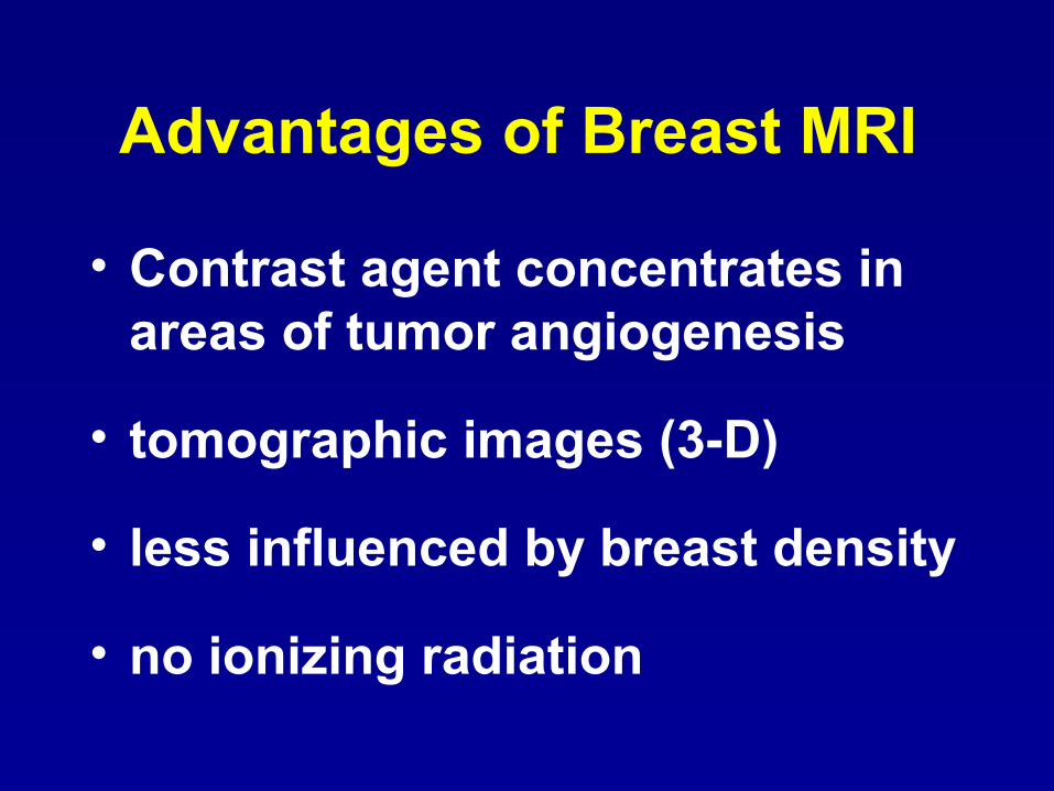
Advantages of Breast MRI
• Contrast agent concentrates in areas of tumor angiogenesis
• tomographic images (3-D)
• less influenced by breast density
• no ionizing radiation

Disadvantages of MRI
• $$$
• lower specificity• biopsy more difficult
• logistics – menstrual phase– weight
• claustrophobia

Breast MRI Screening Studiesfor High Risk Women
Kriege et al. The Netherlands
Kuhl, et al. Bonn, Germany
Leach et al. U.K.
Podo et al. Italy
Schnall, Lehman et al. U.S.
Warner, Plewes, et al. Toronto, Canada

Breast MRI Screening Studiesfor High Risk Women
Similarities• prospective, non-randomized• not restricted to mutation carriers• annual mammography + MRI
Differences• single / multiple centers• patient population• additional modalities• MRI technique

Dutch National Study Kriege et al. NEJM 351: 427, 2004.
• 6 centers
• unaffected women
• ages 25-70
• ≥ 15% lifetime risk • MRI + mammography + CBE

Dutch National Study: Results• 1909 women
– 358 mutation carriers – mean age 40
– mean # screens = 2
•
4 (9%) interval cancers!
• 45 evaluable cancers
• 39 invasive, 6 DCIS• 50% in carriers• 50% 1st screen

Sensitivity of Individual Modalities
Dutch Study: Results
71%
40%
18%
0%
20%
40%
60%
80%
MRI Mam CBE

Sensitivity: Invasive vs. In-Situ
Dutch Study: Results
17%
80%
33%
83%
0%
20%
40%
60%
80%
100%
Invasive In-Situ
MRI
Mammography
n=39 n=6

False Positives
Recalls Biopsies
MRI 10% 5.8%
Mammography 5% 1.7%
Dutch Study: Results

Invasive Tumor Stage
40%
48%
43%
14% 12%
32%
37%
49%25%
0%
20%
40%
60%
80%
100%
Study Control 1 Control 2
> 2 cm
1.1 - 2 cm
< 1 cm
Dutch Study: Results
n=45 n=1500 n=45
21% node + 52% node + 56% node +

Toronto StudyWarner et al. JAMA 292: 1317, 2004
• single center
• affected & unaffected women• ages 25 - 65
• >25% lifetime risk
• MRI + mammography + CBE + US

The Toronto Study
Medical Biophysics
Donald Plewes PhD.
Martin Yaffe PhD.
Elizabeth Ramsay MSc
Cameron Piron MSc
Medical Imaging
Petrina Causer M.D.
Roberta Jong M.D.
Belinda Curpen M.D.
Joan Glazier MRT
Garry Detzler MRT
Caron Murray MRT
Joanne Muldoon MRT
Study Co-ordinator
Kimberley Hill, BScGenetics
Steven Narod M.D.
Sandra Messner M.D.
Wendy Meschino M.D.
Andrea Eisen M.D.
Pathology
John Wong M.D.
Judit Zubovits M.D.
General Surgery
Glen Taylor M.D.
Claire Holloway M.D.
Frances Wright M.D.
Biostatistics
Gerrit DeBoer PhD
Alice Chung BSc
Funding
CBCRANBCFAmersham HealthPapoff Family
Nurse Examiner Marg Cutrara R.N.

Toronto Study: Results• 437 women
– 318 BRCA mutation carriers
– mean age 43
– mean # screens = 3
Only 1 interval cancer!
• 37 cancers – 32 in carriers – mean age 48 (34-64) – 28 invasive (2 lobular), 9 DCIS

Sensitivity of Individual Modalities
Toronto Study: Results
84%
30%8%
33%
0%
20%
40%
60%
80%
100%
MRI Mam CBE US
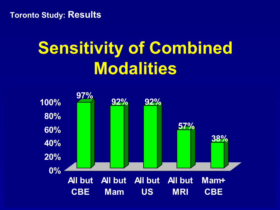
Sensitivity of CombinedModalities
Toronto Study: Results
97%92% 92%
57%
38%
0%
20%
40%
60%
80%
100%
All butCBE
All butMam
All butUS
All butMRI
Mam+CBE

Sensitivity: Invasive vs. In-Situ
Toronto Study: Results
86%78%
25%33%
50%
0%0%
20%
40%
60%
80%
100%
invasive In-Situ
MRI
MMG
US
n=28 n=9


Sensitivity by Age
Toronto Study: Results
80%88%
24%35%
29%
45%
0%
20%
40%
60%
80%
100%
<50 (n=20) 50+ (n=17)
MRI
MMG
US

Toronto Study:: Results
Sensitivity by Year of Screening
89%79%
28% 32%28%42%
0%
20%
40%
60%
80%
100%
year 1 (n=18) year 2-5 (n=19)
MRI
MMG
US

False Positives: Recalls
Toronto Study: Results
1% 1%
19%
9%
2%2% 2%
6%
0.00%
5.00%
10.00%
15.00%
20.00%
Year 1 Years 2 - 5
MRI
M
CBE
US

False Positives: Biopsies
Toronto Study: Results
11%
5%6% 6%3%
16%
10%
4% 3%1% 1% 0
0%
5%
10%
15%
20%
Year 1 Year 2 Years 3-5
Any
MRI
M
US

74%
43%
22%
32%
3%
25%
0%
20%
40%
60%
80%
100%
Toronto the Netherlands
> 2 cm
1.1 - 2 cm
< 1 cm
Invasive Tumour Size

Toronto Study: Results
Yr. # cancers DCIS Mean Invasive Size Node +
1 18 22% 1.1 (0.4 - 3.0) cm 3
2 9 11% 1.2 (0.4 - 2.0) cm 1
3-5 9 44% 0.8 (0.7 - 1.0) cm 0
Tumor Stage by Year
No recurrences to date. Median f/u 3yrs. (range 1 to 7)

Effect of MRI Screening on Survival
0
5
10
15
1 2 3 4 5
years
log
cell
num
ber
M e t s
MRI
mammo
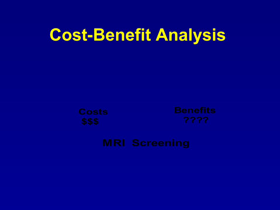
Costs$$$
MRI Screening
Benefits????
Cost-Benefit Analysis

Cost-Benefit Estimate
$$$
• 62 million women
ages 30-60 in U.S.
• 1% high risk (620,000)
• $1200 per screen
____________________
$744 million/year
• 620,000 high risk
• 1% (6,200) have cancer• mortality 30% → 10%• 1240 more cured • mean years saved = 25________________________
31,000 life years saved
$24,000 / year of life saved

Summary
Breast MRI for high risk women:
• most sensitive screening modality
• finds cancers at an earlier stage
• has acceptable specificity
• saves lives?

Other Research Questions
• Optimal MRI screening schedule for subgroups? – age– mutation status
– breast density
• Role of other screening modalities?
• Role of MRI for other high risk women?– Atypical hyperplasia, LCIS– Chest irradiation < age 30– Very dense breasts
Related Documents