..................................................................................................................................................................................... ..................................................................................................................................................................................... PI3Kg inhibition reduces blood pressure by a vasorelaxant Akt/L-type calcium channel mechanism Daniela Carnevale 1 , Carmine Vecchione 1 , Giada Mascio 1 , Giovanni Esposito 2 , Giuseppe Cifelli 1 , Katiuscia Martinello 3,4 , Alessandro Landolfi 1 , Giulio Selvetella 1 , Paolo Grieco 2 , Antonio Damato 1 , Elio Franco 1 , Hannelore Haase 5 , Angelo Maffei 1 , Elisa Ciraolo 6 , Sergio Fucile 3,4 , Giacomo Frati 1 , Orazio Mazzoni 2† , Emilio Hirsch 6 , and Giuseppe Lembo 1,7 * 1 Department of Angiocardioneurology, IRCCS Neuromed, Pozzilli, Isernia, Italy; 2 Department of Pharmaceutical Chemistry, ‘Federico II’ University of Naples, Italy; 3 Department of Physiology and Pharmacology, ‘Sapienza’ University of Rome, Italy; 4 Department of Molecular Pathology, IRCCS Neuromed, Pozzilli, Isernia, Italy; 5 Max-Delbru ¨ck-Center (MDC) for Molecular Medicine, Berlin, Germany; 6 Department of Genetics, Biology and Biochemistry, Molecular Biotechnology Center, University of Turin, Italy; and 7 Department of Molecular Medicine, ‘Sapienza’ University of Rome, Rome, Italy Received 16 May 2011; revised 10 October 2011; accepted 24 October 2011; online publish-ahead-of-print 27 October 2011 Time for primary review: 29 days Aims The lipid and protein kinase phosphoinositide 3-kinase g (PI3Kg) is abundantly expressed in inflammatory cells and in the cardiovascular tissue. In recent years, its role in inflammation and in cardiac function and remodelling has been unravelled, highlighting the beneficial effects of its pharmacological inhibition. Furthermore, a role for PI3Kg in the regulation of vascular tone has been emphasized. However, the impact of this signalling in the control of blood pres- sure is still poorly understood. Our study investigated the effect of a selective inhibition of PI3Kg, obtained by using two independent small molecules, on blood pressure. Moreover, we dissected the molecular mechanisms involved in control of contraction of resistance arteries by PI3Kg. Methods and results We showed that inhibition of PI3Kg reduced blood pressure in normotensive and hypertensive mice in a concentra- tion-dependent fashion. This effect was dependent on enhanced vasodilatation, documented in vivo by decreased per- ipheral vascular resistance, and ex vivo by vasorelaxing effects on isolated resistance vessels. The vasorelaxation induced by PI3Kg inhibition relied on blunted pressure-induced Akt phosphorylation and a myogenic contractile response. Molecular insights revealed that PI3Kg inhibition affected smooth muscle L-type calcium channel current density and calcium influx by impairing plasma membrane translocation of the a1C L-type calcium channel subunit responsible for channel open-state probability. Conclusion Overall our findings suggest that PI3Kg inhibition could be a novel tool to modulate calcium influx in vascular smooth muscle cells, thus relaxing resistance arteries and lowering blood pressure. ----------------------------------------------------------------------------------------------------------------------------------------------------------- Keywords Phosphoinositide 3-kinase g † Blood pressure † Resistance artery † Myogenic tone † L-type calcium channel 1. Introduction Phosphoinositide 3-kinase g (PI3Kg) is a lipid and protein kinase mainly expressed in bone marrow-derived cells and in the cardiovas- cular tissue 1 and, once activated, generates phosphatidylinositol-3,4,5- trisphosphate to recruit and activate downstream signalling molecules. In recent years, several studies have dissected the role played by PI3Kg in different pathophysiological contexts involving inflammatory responses. In particular, it has been demonstrated that mice with PI3Kg inactivated by genetic or pharmacological approaches are pro- tected from disease development in a number of inflammation and autoimmune disease models. 2,3 This recent evidence has prompted † Deceased * Corresponding author. Tel: +39 0865 915244; fax: +39 0865 927575, Email: [email protected], [email protected] Published on behalf of the European Society of Cardiology. All rights reserved. & The Author 2011. For permissions please email: [email protected]. Cardiovascular Research (2012) 93, 200–209 doi:10.1093/cvr/cvr288 at Universita di Napoli on October 1, 2012 http://cardiovascres.oxfordjournals.org/ Downloaded from

Welcome message from author
This document is posted to help you gain knowledge. Please leave a comment to let me know what you think about it! Share it to your friends and learn new things together.
Transcript

. . . . . . . . . . . . . . . . . . . . . . . . . . . . . . . . . . . . . . . . . . . . . . . . . . . . . . . . . . . . . . . . . . . . . . . . . . . . . . . . . . . . . . . . . . . . . . . . . . . . . . . . . . . . . . . . . . . . . . . . . . . . . . . . . . . . . . . . . . . . . . . . . . . . . . . . . . . . . . . . . . . . . . . . . . . . . . . . . . . . .
. . . . . . . . . . . . . . . . . . . . . . . . . . . . . . . . . . . . . . . . . . . . . . . . . . . . . . . . . . . . . . . . . . . . . . . . . . . . . . . . . . . . . . . . . . . . . . . . . . . . . . . . . . . . . . . . . . . . . . . . . . . . . . . . . . . . . . . . . . . . . . . . . . . . . . . . . . . . . . . . . . . . . . . . . . . . . . . . . . . . .
PI3Kg inhibition reduces blood pressureby a vasorelaxant Akt/L-type calciumchannel mechanismDaniela Carnevale1, Carmine Vecchione1, Giada Mascio1, Giovanni Esposito2,Giuseppe Cifelli1, Katiuscia Martinello3,4, Alessandro Landolfi1, Giulio Selvetella1,Paolo Grieco2, Antonio Damato1, Elio Franco1, Hannelore Haase5, Angelo Maffei1,Elisa Ciraolo6, Sergio Fucile3,4, Giacomo Frati1, Orazio Mazzoni2†, Emilio Hirsch6,and Giuseppe Lembo1,7*
1Department of Angiocardioneurology, IRCCS Neuromed, Pozzilli, Isernia, Italy; 2Department of Pharmaceutical Chemistry, ‘Federico II’ University of Naples, Italy; 3Department ofPhysiology and Pharmacology, ‘Sapienza’ University of Rome, Italy; 4Department of Molecular Pathology, IRCCS Neuromed, Pozzilli, Isernia, Italy; 5Max-Delbruck-Center (MDC) forMolecular Medicine, Berlin, Germany; 6Department of Genetics, Biology and Biochemistry, Molecular Biotechnology Center, University of Turin, Italy; and 7Department of MolecularMedicine, ‘Sapienza’ University of Rome, Rome, Italy
Received 16 May 2011; revised 10 October 2011; accepted 24 October 2011; online publish-ahead-of-print 27 October 2011
Time for primary review: 29 days
Aims The lipid and protein kinase phosphoinositide 3-kinase g (PI3Kg) is abundantly expressed in inflammatory cells and inthe cardiovascular tissue. In recent years, its role in inflammation and in cardiac function and remodelling has beenunravelled, highlighting the beneficial effects of its pharmacological inhibition. Furthermore, a role for PI3Kg in theregulation of vascular tone has been emphasized. However, the impact of this signalling in the control of blood pres-sure is still poorly understood. Our study investigated the effect of a selective inhibition of PI3Kg, obtained by usingtwo independent small molecules, on blood pressure. Moreover, we dissected the molecular mechanisms involved incontrol of contraction of resistance arteries by PI3Kg.
Methodsand results
We showed that inhibition of PI3Kg reduced blood pressure in normotensive and hypertensive mice in a concentra-tion-dependent fashion. This effect was dependent on enhanced vasodilatation, documented in vivo by decreased per-ipheral vascular resistance, and ex vivo by vasorelaxing effects on isolated resistance vessels. The vasorelaxationinduced by PI3Kg inhibition relied on blunted pressure-induced Akt phosphorylation and a myogenic contractileresponse. Molecular insights revealed that PI3Kg inhibition affected smooth muscle L-type calcium channelcurrent density and calcium influx by impairing plasma membrane translocation of the a1C L-type calciumchannel subunit responsible for channel open-state probability.
Conclusion Overall our findings suggest that PI3Kg inhibition could be a novel tool to modulate calcium influx in vascular smoothmuscle cells, thus relaxing resistance arteries and lowering blood pressure.
- - - - - - - - - - - - - - - - - - - - - - - - - - - - - - - - - - - - - - - - - - - - - - - - - - - - - - - - - - - - - - - - - - - - - - - - - - - - - - - - - - - - - - - - - - - - - - - - - - - - - - - - - - - - - - - - - - - - - - - - - - - - - - - - - - - - - - - - - - - - - - - - - - - - - - - - - - -Keywords Phosphoinositide 3-kinase g † Blood pressure † Resistance artery † Myogenic tone † L-type calcium channel
1. IntroductionPhosphoinositide 3-kinase g (PI3Kg) is a lipid and protein kinasemainly expressed in bone marrow-derived cells and in the cardiovas-cular tissue1 and, once activated, generates phosphatidylinositol-3,4,5-trisphosphate to recruit and activate downstream signalling molecules.
In recent years, several studies have dissected the role played byPI3Kg in different pathophysiological contexts involving inflammatoryresponses. In particular, it has been demonstrated that mice withPI3Kg inactivated by genetic or pharmacological approaches are pro-tected from disease development in a number of inflammation andautoimmune disease models.2,3 This recent evidence has prompted
† Deceased
* Corresponding author. Tel: +39 0865 915244; fax: +39 0865 927575, Email: [email protected], [email protected]
Published on behalf of the European Society of Cardiology. All rights reserved. & The Author 2011. For permissions please email: [email protected].
Cardiovascular Research (2012) 93, 200–209doi:10.1093/cvr/cvr288
at Universita di N
apoli on October 1, 2012
http://cardiovascres.oxfordjournals.org/D
ownloaded from

the task of developing small molecule inhibitors of PI3Kg, to attain theappropriate isoform selectivity profile.
So far, selective pharmacological inhibitors of PI3Kg have been usedto treat cardiovascular diseases with a substantial inflammatory com-ponent, such as myocardial ischaemia4,5 and atherosclerosis.6 Further-more, we have recently demonstrated a beneficial role of PI3Kginhibition on cardiac remodelling in response to pressure overload,a stimulus in which challenges to cardiac and inflammatory cells arefinely intertwined.7 We have also described that mice lacking PI3Kgare protected from hypertension induced by chronic administrationof angiotensin II (AngII),8 thus suggesting an additional role of PI3Kgin the regulation of vascular tone.
Nevertheless, the impact of PI3Kg inhibition on blood pressure(BP) is still poorly explored. The availability of transgenic mice andpharmacological approaches to inhibit PI3Kg can help to disclose itsrole in BP regulation and then unveil mechanistic insights responsiblefor this effect. Here we investigated the effect of two independentsmall molecules inhibiting PI3Kg, focusing on their impact on BPand the molecular mechanisms recruited. For these purposes, weevaluated concentration-dependent effects of PI3Kg inhibitors onBP levels, in normotensive and hypertensive mice. Then, we exploredhow PI3Kg inhibition exerted haemodynamic effects, by analysingcardiac output and total peripheral vascular resistance, the two com-ponents on which BP depends. Insights into the actions of PI3Kginhibition on vascular function, obtained in isolated vessels, finallyprompted us to dissect molecular mechanisms residing in the vascularsmooth muscle component, regulating calcium influx.
2. Methods
2.1 AnimalsC57BL/6 mice (8–12 weeks old) were purchased from Charles River(Calco, Italy). Genetically engineered mice for PI3Kg were generated aspreviously described.9
According to the New Directive 2010/63/EU, special attention was paidto animal welfare and to minimize animal suffering. Anaesthetized micewere closely monitored during the procedure to assure that they weremaintained in the proper anaesthetic plane. In particular, the anaestheticefficacy was assessed by pinching the tail of the animal as well as by check-ing the body temperature and the respiratory rate. Any reaction from theanimal, hypothermia, or increase of the respiratory rate, indicated that theanaesthesia was too light and that additional anaesthetic should be given.
All experiments were conducted in conformity with European Com-munities Council Directive and approved by our Institution.
2.2 Synthesis of (Z)-5-(5-nitrofuryl-2-idene)-1,3-thiazolidine-2,4-dione GE21The detailed procedure for GE21 synthesis and for the assay of PI3K lipidkinase activity used to test GE21 selectivity is provided in the Supplemen-tary material online, Methods.
2.3 GE21 plasma levelsThe quantitative analysis in blood samples, collected by tail nicks at theindicated time points, was performed using the same chromatographicmethod coupled to ultraviolet detection, described for GE21 synthesis.
2.4 Administration of drugsAS605240, from Merck Serono (Geneva, Switzerland)2 was used aspreviously described.3
GE21 was dissolved in saline for i.p. injections and in water for oraladministration, and used at the doses indicated in figures. Chronic admin-istration was by daily i.p. injections or intragastrically by gavage, asindicated in figure legends. GE21 administration did not result in anysign of toxicity (see Table 2), and a long-lasting treatment of 3 monthsdid not affect the viability of the mice (data not shown).
For Akt inhibition, we used 1L6-hydroxymethyl-chiro-inositol-2-(R)-2-O-methyl-3-O-octadecyl-sn-glycerocarbonate (Calbiochem #124005)at the doses indicated in figures.
2.5 Blood pressure measurementsBlood pressure was evaluated in conscious mice by tail-cuff plethysmo-graphy (Visitech Systems, Apex, NC, USA).
2.6 Cardiac output and total peripheralresistanceCardiac catheterization was performed using a 1.4 French high-fidelitymicromanometer catheter (Millar Instruments, Houston, TX, USA) inmice anaesthetized with a mixture of ketamine (110 mg/kg) and xylazine(20 mg/kg), before animals were killed with an overdose of sodium pento-barbital (250 mg/kg), as previously described.10 Cardiac output was mea-sured and total peripheral resistance calculated by dividing the meanarterial pressure by cardiac output.
2.7 Clinical chemistry analysisGE21 was administered by a daily oral treatment at the maximal doseused in the in vivo studies (50 mg/kg/day) for 2 weeks. Mice were killedwith an overdose of sodium pentobarbital (250 mg/kg) and serum wascollected from the trunk. Clinical chemistry analyses were performedby Dimension EXL (Siemens Heathcare Diagnostic Inc., Deerfield, IL,USA) apparatus. Red (RBCs) and white blood cells (WBCs) were manu-ally counted in plasma samples by May-Grunwald Giemsa staining and ac-quiring images using a DMI4000B Leica microscope (Leica Microsystems,Wetzlar, Germany).
2.8 Evaluation of locomotor activityLocomotor activity was recorded by radiotelemetric transmitters(TA11PA-C20; Data Sciences International, St. Paul, MN, USA). Micewere anaesthetized with a mixture of ketamine (110 mg/kg) and xylazine(20 mg/kg), and radiotelemetric transmitters were inserted subcutaneous-ly on the right flank. After 2 days recovery from surgery, continuous 24 hlocomotor activity was monitored. Data were stored and analysed usingDSI Ponemah 4.9 software (Data Sciences International, St. Paul, MN,USA).
2.9 Evaluation of vascular functionThe myogenic tone of second-order mesenteric arteries, isolated frommice killed with an overdose of sodium pentobarbital (250 mg/kg) wasdetermined with a pressure arteriograph with stepwise perfusion pres-sure. Mesenteric arteries were subjected to a perfusion pressure of100 mmHg for functional studies, and placed in a wire myograph fordetermination of structure, as previously described.10 Where indicated,the endothelium was mechanically removed and endothelial functiontested by evaluating the response to acetylcholine.
The percentage of myogenic tone was calculated with the followingformula:
(internal diameter Ca2+- free Krebs − internal active diameter)internal diameter Ca2+- free Krebs
[ ]× 100.
For all studies vessels, were equilibrated and maintained in Krebs solution(mM: NaCl, 120; KCl, 4.7; MgSO4 . 7H2O, 2.5; NaHCO3, 25; KH2PO4, 1.1;glucose, 6; and CaCl2 . 2H2O, 2.5).
PI3Kg inhibition and hypertension 201 at U
niversita di Napoli on O
ctober 1, 2012http://cardiovascres.oxfordjournals.org/
Dow
nloaded from

2.10 Smooth muscle cellsRat aortic smooth muscle cells (RASMCs), provided by ATTC (AmericanType Culture Collection, Sesto San Giovanni, Milan, Italy), clone A10,were maintained in Dulbecco’s modified Eagle’s medium supplementedwith 10% fetal bovine serum (378C; air supplemented with 5% CO2)and harvested at confluence. Treatments were given in serum-freemedium as indicated in the figures. GE21 (1025 M) was incubated for30 min before stimulation with AngII (1 mmol/L).
2.11 Evaluation of membrane currentMembrane current recordings were made on voltage-clamped RASMCswith a standard patch-clamp technique using a Multiclamp 700A patch-clamp amplifier (Molecular Devices, Sunnyvale, CA, USA) according topreviously described methods.11 Peak current density was expressed asthe maximal amplitude of the Ba2+ current per capacitance unit (inpicoamperes per picofarad). Current densities were recorded in the con-tinuous presence of tetrodotoxin (10 mmol/L) to avoid activation ofvoltage-activated Na+ channels, and with Cs+ replacing K+ in the intracel-lular solution. Currents were completely and reversibly inhibited bynifedipine (3 mmol/L).
2.12 Fluorescent measurement of Ca21
RASMCs were serum starved, mounted in chamber slides and loaded withfluo-4 AM (Invitrogen Molecular Probes, Monza, Italy; 1 mmol/L) for 1 h at378C and then stimulated as indicated above in section 2.10. Real-timemovies were obtained with Leica DMI4000B (Leica Microsystems, CMSGmbH, Wetzlar, Germany) and relative fluorescence intensities at F0
(basal fluorescence) and at F (peak fluorescence) were measured usingLAS Application Suite and reported as DF ¼ (F 2 F0)/F0. AngII-inducedfluorescence was completely inhibited by nifedipine (3 mmol/L).
2.13 Protein analysisProteins were extracted as previously described.12 In brief, vessels andcells were homogenized in a lysis buffer containing the following: Tris,50 mmol/L (pH 7.4); NaCl, 150 mmol/L; EDTA, 1 mmol/L; sodium deox-ycholate, 0.5%; NaF, 10 mmol/L; Na3VO4, 1 mmol/L; NP-40, 1%; and pro-tease inhibitors. Following a centrifugation step, samples were separatedby SDS–PAGE and blotted on a nitrocellulose membrane. Primary anti-bodies used were as follows: anti-phospho-(Ser473) Akt (1:500 dilution)and anti-total-Akt (1:1000 dilution) from Cell Signaling Technology, Inc.,Danvers, MA, USA; anti-CACNA1C (a1C subunit, L-Type Ca2+
channel; 1:300 dilution) from Novus Biologicals, Littleton, CO, USA;
Figure 1 Two small molecules inhibiting PI3Kg lower BP in mice. (A) Three-dimensional graphical representation of GE21, the(Z)-5-(5-nitrofuryl-2-idene)-1,3-thizoline-2,4-dione, a novel PI3Kg inhibitor. GE21 structure obtained by a simulated annealing method. (B and C)Effects of AS605240 (B) and GE21 (C) in C57BL/6 mice (n ¼ 24), injected i.p. daily for 5 days at different doses (10, 20 and 40 mg/kg forAS605240 and GE21). Changes in systolic blood pressure (DSBP) on days 1 and 5 are shown. *P , 0.001 vs. vehicle; #P , 0.001 vs. mediumdose; §P , 0.001 vs. highest dose. (D and E) Effects of AS605240 (D) and GE21 (E) i.p. administration in PI3Kg+/+ and PI3Kg2/2 mice (n ¼ 20)at the highest dose tested, given for 5 days. *P , 0.001 vs. basal and #P , 0.001 vs. PI3Kg+/+.
D. Carnevale et al.202 at U
niversita di Napoli on O
ctober 1, 2012http://cardiovascres.oxfordjournals.org/
Dow
nloaded from
and anti-p-Ser monoclonal antibody (4A3; 1:200 dilution), Santa Cruz Bio-technology, Inc., Santa Cruz, CA, USA. The b2 subunit was immunopre-cipitated with the previously characterized antibody.12
Protein detection was performed with AmershamTM ECL Plus WesternBlotting Detection kit (GE Healthcare Europe GmbH, Milan, Italy) anddensitometry was obtained with the NIH Image 1.61 software (developedat the U.S. National Institutes of Health).
2.14 Statistical analysisStatistical significance was calculated with one-way ANOVA (for compari-son of multiple groups) and two-way ANOVA (for multiple groups withfactorial design), followed by Bonferroni post hoc test (data are presented
as means+ SEM). For comparisons of two groups, Mann–Whitney U testwas applied. The number of independent experiments and P value areindicated in each figure legend. Statistical analyses were performed withGraphPad software PRISM5 (GraphPad Software Inc., La Jolla, CA, USA).
3. Results
3.1 Pharmacological inhibition of PI3Kglowers blood pressure in normotensiveand hypertensive miceWe investigated BP effects of a pharmacological inhibition of PI3Kgobtained by using two independent small molecules: AS605240,already commercially available, and GE21, a novel one. We developedthis latter analogue inhibitor by designing molecules that should fitinto the ATP-binding pocket of the enzyme.13 GE21 (Figure 1A) wasthe most selective molecule synthesized, with an EC50 for PI3Kglower than that needed to inhibit other PI3K isoforms (Table 1).
The effects of PI3Kg inhibition on BP were analysed in normoten-sive C57BL/6 mice. PI3Kg inhibitors (AS605240 in Figure 1B and GE21in Figure 1C) showed a similar concentration-dependent hypotensiveeffect, compared with vehicle alone, after an i.p. treatment over5 days. Then, to demonstrate specificity of this pharmacological strat-egy, we treated PI3Kg+/+ and PI3Kg2/2 mice chronically, at the
. . . . . . . . . . . . . . . . . . . . . . . . . . . . . . . . . . . . . . . . . . . . . . . . . . . . . . . . . . . . . . . . . . . . . . . . . . . . . . . . . . . .
Table 1 EC50 values of GE21 against PI3K isoforms,calculated from enzyme inhibition assay
PI3Ka PI3Kb PI3Kg PI3Kd
EC50 (mmol/L) 18.2 + 1.3 18.1 + 1.4 5.3 + 1.4 84.2 + 1.3
Emax (% ofinhibition)
108.4 + 7.4 98.3 + 7.4 102.7 + 7.1 114.6 + 7.6
n 24 24 24 24
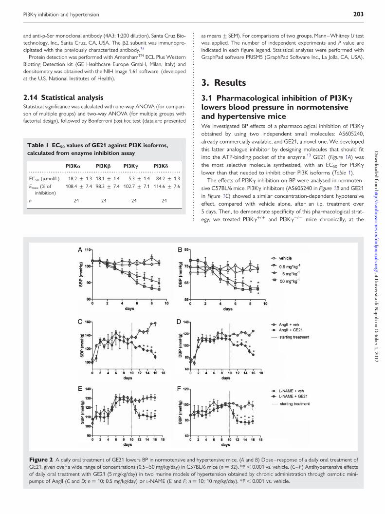
Figure 2 A daily oral treatment of GE21 lowers BP in normotensive and hypertensive mice. (A and B) Dose–response of a daily oral treatment ofGE21, given over a wide range of concentrations (0.5–50 mg/kg/day) in C57BL/6 mice (n ¼ 32). *P , 0.001 vs. vehicle. (C–F) Antihypertensive effectsof daily oral treatment with GE21 (5 mg/kg/day) in two murine models of hypertension obtained by chronic administration through osmotic mini-pumps of AngII (C and D; n ¼ 10; 0.5 mg/kg/day) or L-NAME (E and F; n ¼ 10; 10 mg/kg/day). *P , 0.001 vs. vehicle.
PI3Kg inhibition and hypertension 203 at U
niversita di Napoli on O
ctober 1, 2012http://cardiovascres.oxfordjournals.org/
Dow
nloaded from

maximal effective dose and time, determined in the dose–responseexperiment. We confirmed that basal BP was comparable in bothmouse strains and, as expected, both AS605240 (Figure 1D) andGE21 (Figure 1E) reduced BP levels in PI3Kg+/+ but not inPI3Kg2/2 mice.
A daily oral treatment of GE21 demonstrated, even with this routeof administration, a concentration-dependent lowering effect on bothsystolic BP (SBP; Figure 2A) and diastolic BP (DBP; Figure 2B), whichbecame statistically significant 5 days after the start of the treatmentand was sustained until the end of our analysis.
When we evaluated the efficacy of PI3Kg inhibition as an antihyper-tensive strategy, we found that the daily oral treatment completelynormalized BP levels in two murine models in which hypertensionwas induced by chronic administration of AngII (SBP, Figure 2C; andDBP, Figure 2D) or L-NAME, a nitric oxide synthase inhibitor (SBP,Figure 2E; and DBP, Figure 2F).
Overall, this first piece of evidence demonstrated that PI3Kg inhib-ition had a clear antihypertensive effect, and the fact that this wasaccomplished using two independent molecules, AS605240 andGE21, further strengthens our findings.
3.2 PI3Kg inhibition does not influencehealth of miceChronic oral treatment with GE21, at the maximal dose used in ourexperiments (50 mg/kg/day) and for a maximal observation period of2 weeks, showed no relevant sign of drug toxicity on clinical chemistryanalysis (Table 2). The locomotor activity of GE21- and vehicle-treated mice was comparable (Figure 3A).
Next, we measured the GE21 plasma concentrations reached aftera daily oral treatment given at the dose of 5 mg/kg/day (the dosageused for antihypertensive purposes), and found that it peaked 1 h
. . . . . . . . . . . . . . . . . . . . . . . . . . . . . . . . . . . . . . . . . . . . . . . . . . . . . . . . . . . . . . . . . . . . . . . . . . . . . . . . . . . .
Table 2 Peripheral haematological and clinical chemistryvalues in vehicle- or GE21 (1022 M)-treated mice, andanimal weights before and after treatment
Units Vehicle GE21
Total bilirubin mg/dL 0.21 + 0.1 0.22 + 0.1
Creatinine mEq/L 0.26 + 0.1 0.24 + 0.1
Aspartateaminotransferase
IU/L 64 + 11 72 + 18
Alanine aminotransferase IU/L 37 + 7 44 + 7
Creatine kinase IU/L 871 + 166 1060 + 409
Alkaline phosphatase IU/L 68 + 28 60 + 23
Insulin mIU/mL 1.8 + 0.3 2.4 + 0.6
RBCs ×1012/L 8.6 + 0.6 8.9 + 0.5
WBCs ×109/L 5.2 + 1 4.7 + 1
Weight gain g +0.37 + 0.5 +0.58 + 0.6
Data are expressed as means + SEM of n ¼ 5–10 mice for each group.
Figure 3 Chronic treatment with the maximal dose of GE21 does not affect general health of the mice. (A) Effects of 2 weeks treatment with1022 M GE21 on locomotor activity (LMA), evaluated by radiotelemetry (n ¼ 8). (B) Concentration of GE21 in the plasma after daily oral treatment(5 mg/kg/day; n ¼ 5). (C) An example of chromatographic analysis for GE21 levels in blood (peak corresponding to 49 h, i.e. 1 h from intake).(D) Glucose tolerance test on mice orally administered with GE21 (5 mg/kg/day) or vehicle alone after a 6 h fasting by i.p. injection of glucose(2 g/kg) at time zero (n ¼ 10).
D. Carnevale et al.204 at U
niversita di Napoli on O
ctober 1, 2012http://cardiovascres.oxfordjournals.org/
Dow
nloaded from

after drug administration (Figure 3B), as evaluated by chromatographicanalysis (Figure 3C), at levels compatible with the reported EC50
for PI3Kg.As other isoforms of the PI3K family have been demonstrated to be
crucial in insulin signalling, we investigated whether GE21 treatmentmay interfere with glucose metabolism. We performed a glucose tol-erance test in mice given GE21 orally at the maximal dose (50 mg/kg/day) or given vehicle alone. As shown in Figure 3D and in Table 2, wefound no difference in fasting glucose and insulin levels between thetwo groups and, more importantly, i.p. glucose injection inthe GE21-treated mice resulted in an increase in glycaemia at15–30 min, comparable to that of vehicle-treated mice. This resultindicated that chronic administration of GE21, even at a dose higherthan that used to produce a hypotensive effect, does not interferewith other PI3K isoforms controlling glucose metabolism.
3.3 PI3Kg inhibitors exert smooth musclevasorelaxant effects by inhibitingpressure-mediated Akt activationOur next aim was to characterize the mechanisms through whichPI3Kg inhibitors exert antihypertensive effects. In this regard, weshould mention that PI3Kg is expressed in cardiac as well as in vascu-lar cells, making it conceivable that its inhibition may affect one orboth of them. To verify whether the decrease in BP induced byPI3Kg inhibition was dependent on a negative effect on cardiac func-tion or on decreased vascular resistance, we measured cardiac outputand calculated total peripheral resistance, before and after drug injec-tion. PI3Kg inhibition significantly reduced vascular resistance(Figure 4A) without modifying cardiac output (5588+329 vs.5307+215 mL/min; n ¼ 5 mice per group; not significant).
Figure 4 PI3Kg inhibition evokes vasorelaxant effects through modulation of calcium influx. (A) Changes in total peripheral resistance (DTPR)evoked by GE21 (4 mg/kg) or vehicle in C57BL/6 mice (n ¼ 10). *P , 0.01 vs. vehicle. (B) Effect of GE21 on intact or endothelium-denuded mesen-teric arteries from C57BL/6 mice (n ¼ 10), compared with vehicle alone (n ¼ 5). *P , 0.01 vs. vehicle. (C ) Changes in the internal diameter of mes-enteric arteries (n ¼ 12) from PI3Kg+/+ and PI3Kg2/2 mice with increasing doses of GE21. *P , 0.01 vs. PI3Kg2/2. (D) Changes in the internaldiameter of mesenteric arteries (n ¼ 10) from C57Bl/6 mice with increasing doses of AS605240, compared with vehicle. *P , 0.01 vs. vehicle.(E and F) Myogenic response obtained in mesenteric arteries from PI3Kg+/+ and PI3Kg2/2 mice (E) and from C57BL/6 mice (F) treated withGE21 (1024 M) or vehicle alone. *P , 0.01 vs. PI3Kg+/+ or vs. vehicle.
PI3Kg inhibition and hypertension 205 at U
niversita di Napoli on O
ctober 1, 2012http://cardiovascres.oxfordjournals.org/
Dow
nloaded from

This finding prompted us to explore actions of PI3Kg on vasculartissue. In particular, we evaluated the effects of PI3Kg inhibition onisolated resistance vessels, i.e. mesenteric arteries. As shown inFigure 4B, GE21 exerted a concentration-dependent increase in theinternal diameter of perfused mesenteric arteries, indicating a clearvasorelaxing effect, which remained completely unaffected byremoval of the endothelium. This latter finding was confirmedby the lack of concentration-dependent vasorelaxation to acetylcho-line (data not shown). Thus, as GE21 was still able to induce vasore-laxant effects in the absence of endothelium, PI3Kg inhibition shouldrecruit a mechanism of action in smooth muscle cells to provokevasodilatation.
Interestingly, PI3Kg2/2 mice had a different structure of the mes-enteric arteries. In particular, they had greater media thickness(see Supplementary material online, Figure S1A) and media cross-sectional area (see Supplementary material online, Figure S1B), accom-panied by an increased internal diameter (see Supplementary materialonline, Figure S1C ). However, the internal diameter of PI3Kg2/2 mes-enteric arteries was not further increased by GE21 compared withPI3Kg+/+ (Figure 4C), thus supporting a selective action of GE21 onthe g isoform. Like our analogue inhibitor GE21, AS605240 induceda comparable concentration-dependent vasorelaxation in mesentericarteries (Figure 4D).
The maximal dose of PI3Kg inhibitor that induced a vasorelaxingeffect showed kinetics of vasodilatation that reached a plateau in15–20 min, as shown for GE21 in Supplementary material online,Figure S2A.
A more detailed characterization of the influence of PI3Kg signallingin the smooth muscle was obtained by studying the regulation of thevascular myogenic tone developed in response to stepwise
increases in perfusion pressure.14 We found that absence of PI3Kgor its pharmacological inhibition by GE21 counteractedthe pressure-induced contractile myogenic response starting at25 mmHg (Figure 4E and F; see Supplementary material online,Figure S2B), thus demonstrating that PI3Kg signalling is crucial toestablish a vascular myogenic response to pressure.
A downstream target of PI3Kg signalling, Akt, has been also pro-posed to be among the main pathways involved in vascular mechan-otransduction. In this regard, we detected that, at the maximalinhibition of perfusion pressure-induced vascular myogenic tone(100 mmHg), Akt phosphorylation was higher than that observed invessels treated with GE21 and in mice lacking PI3Kg (Figure 5A). Strik-ingly, when we inhibited Akt phosphorylation, we evoked a significantvasorelaxant effect, comparable to that induced by GE21 (Figure 5B).Interestingly, we found that Akt inhibition impaired the myogenicresponse to increasing steps in perfusion pressure (Figure 5C).Finally, when we tested both GE21 and Akt inhibitor on the myogenicresponse of PI3Kg2/2 arteries, they failed to exert any effect (seeSupplementary material online, Figure S2C ). These findings suggestedthat PI3Kg inhibition influences vascular tone by blunting its down-stream Akt signalling.
3.4 PI3Kg inhibitors reducenifedipine-sensitive whole-cell currentsand cytosolic Ca21 in vascular smoothmuscle cellsIt is known that the PI3Kg/Akt pathway could drive calcium influx insmooth muscle cells through increased L-type calcium channel(LTCC) open probability;8,15– 18 thus, we reasoned that
Figure 5 PI3Kg inhibition counteracts pressure-induced Akt activation in resistance vessels. (A) Effect of GE21 (1024 M) or vehicle on Akt phos-phorylation in mesenteric arteries from C57BL/6 mice, perfused at 100 mmHg. Representative western blot (left) and quantification of phospho/totalratio (mean+ SEM; right) of n ¼ 5 independent experiments. *P , 0.01 vs. vehicle and PI3Kg+/+, respectively. (B) Effect of GE21 (1024 M) or Aktinhibitor (1025 M) on changes in the internal diameter of mesenteric arteries from C57BL/6 mice (n ¼ 4), perfused at 100 mmHg, compared withvehicle alone. Data are means+ SEM. *P , 0.01 vs. vehicle. (C) Myogenic response obtained in mesenteric arteries from from C57BL/6 micetreated with Akt inhibitor (1025 M) or vehicle alone. *P , 0.01 vs. vehicle.
D. Carnevale et al.206 at U
niversita di Napoli on O
ctober 1, 2012http://cardiovascres.oxfordjournals.org/
Dow
nloaded from

Figure 6 Molecular mechanism involved in vasorelaxation evoked by PI3Kg inhibition. (A) Effect of GE21 (1024 M) or vehicle on Akt phosphor-ylation in vascular smooth muscle cells in control conditions and stimulated with AngII (1 mM). Representative western blot (top) and quantification(bottom) of n ¼ 5 independent experiments. *P , 0.01 vs. control–vehicle and #P , 0.01 vs. AngII–vehicle. (B) Current densities in RASMCs evokedby depolarization (from 250 to 0 mV, 300 ms) in control conditions and with AngII (1 mM), after 30 min pre-treatment with GE21 (1024 M) orvehicle. Representative traces (top) and quantification (bottom) of n ¼ 8 independent experiments. *P , 0.01 vs. control–vehicle and #P , 0.01vs. AngII–vehicle. (C) Changes in peak fluorescence intensity (F/F0) in vascular smooth muscle cells, loaded with fluo-4 AM, evoked by AngII(1 mM), after 30 min pre-treatment with GE21 (1025 M) or vehicle. Representative images of peak fluorescence (left panel) and quantification(right panel) of n ¼ 4 independent experiments. *P , 0.01 vs. AngII–vehicle. (D) Effect of GE21 (1024 M) or vehicle on a1C membrane/cytoplasmratio in RASMCs in control conditions and stimulated with AngII (1 mM). Representative western blot (top) and quantification (bottom) of n ¼ 3independent experiments. *P , 0.01 vs. control–vehicle and #P , 0.01 vs. AngII–vehicle. (E) Effect of GE21 (1024 M) or vehicle on immunopreci-pitated b2 serine phosphorylation, in control conditions and stimulated with AngII (1 mM). Representative western blot (top) and quantification ofphosphorylated/total ratio (bottom) of n ¼ 5 independent experiments. *P , 0.01 vs. control–vehicle and #P , 0.01 vs. AngII–vehicle.
PI3Kg inhibition and hypertension 207 at U
niversita di Napoli on O
ctober 1, 2012http://cardiovascres.oxfordjournals.org/
Dow
nloaded from

pharmacological inhibition of this target could affect such a mechan-ism. The best way to study whether PI3Kg inhibition affects LTCCregulation in smooth muscle cells, thus controlling muscle contrac-tion, was to dissect the molecular mechanisms in isolated smoothmuscle cells. In this experimental setting, AngII was able to activateAkt phosphorylation in a PI3Kg-dependent manner, as pre-treatmentof cells with GE21 blunted AngII-recruited Akt phosphorylation(Figure 6A).
Likewise, GE21 significantly blunted smooth muscle nifedipine-sensitive voltage-dependent current density and consequently[Ca2+]i transients induced by AngII (Figure 6B and C; see Supplemen-tary material online, Figure S3 and Movies S1 and S2). Overall, theresults obtained in the isolated whole vessel and in isolated smoothmuscle cells suggested that GE21-induced impairment in vascularmyogenic contraction could be ascribed to an impairment of Ca2+
influx through LTCCs.The LTCC open probability is finely tuned by the regulation of
subunit assembly in the plasma membrane. In particular, the transloca-tion in the plasma membrane of Cava1C, the pore-forming subunit, isnecessary for increased open probability, whereas the Cavb2 adaptivesubunit, when phosphorylated, antagonizes Cava1C degradation,allowing its translocation.17,18 Interestingly, in smooth muscle cellswe found that AngII stimulation induced translocation in the plasmamembrane of Cava1C (Figure 6D) and that this process was impairedby PI3Kg inhibition with GE21 (Figure 6D). In addition, GE21 bluntedAngII-induced phosphorylation of Cavb2 (Figure 6E).
Overall, our data reveal that pharmacological inhibition of PI3Kginduces vasorelaxation through an action on an intracellularpathway finely modulating LTCC functional regulation and calciuminflux, by regulating subunit assembly in the plasma membrane (seemodel in Supplementary material online, Figure S4).
4. DiscussionIn this study, we provide evidence that pharmacological inhibition ofPI3Kg reduces arterial BP, by exerting a significant relaxant actionon the smooth muscle component of resistance vessels. This effectis mediated through a fine regulation of the LTCC, a pivotal molecularswitch for calcium influx in smooth muscle cells, by PI3Kg inhibition.
These results uncover novel potential applications for PI3Kg inhibi-tors and also better delineate the haemodynamic contribution of thisstrategy to the beneficial effects already observed on overloadedhearts.7 Indeed, until now, PI3Kg has gained increasing attentiononly as a promising pharmacological target for the treatment ofinflammation,2– 6 and successively, for protection from maladaptivecardiac remodelling.7 Here we present new insights suggesting thatPI3Kg inhibition could be exploited to develop novel antihypertensivetreatments.
The antihypertensive effect of PI3Kg inhibition was obtained withtwo independent molecules, AS605240 and GE21, thus clearlyproving that this molecular target is of outmost importance in BPcontrol. Moreover, the fact that these molecules failed to lower BPin mice with genetic ablation of PI3Kg demonstrates their selectivityof action.
The effect of PI3Kg inhibitors on BP is realized by reducing totalperipheral resistance, thus depicting the involvement of a vascularmechanism. Indeed, PI3Kg inhibitors evoked a concentration-dependent relaxant effect in resistance arteries, a mechanism demon-strated to be relevant for efficacy of antihypertensive treatments. On
this issue, it should be considered that the relaxant effect of PI3kginhibition could be not only limited to the mesenteric compartment,but also extended to other resistance beds, such as the renal andskeletal muscle ones.
Our results also clarify that the vasorelaxation induced by PI3Kginhibition is independent from effects on endothelium and has to beascribed to an impact on smooth muscle function. Our data reveal,for the first time, that inhibition of PI3Kg, as well as of its downstreamsignalling, Akt, is able to impair the vascular myogenic contractileresponse markedly, in an experimental setting for isolated vesselswhere the myogenic response to perfusion pressure represents themain component of vascular tone, in the absence of any neurohu-moral influence. In this regard, it is tempting to speculate thatPI3Kg/Akt signalling could be one of the main pathways recruitedby pressure-induced mechanical stress. Our novel data on PI3Kgadd a further piece of knowledge on how the mechanical stimulus res-iding in pressure itself could be sensed by the vessel. The signallingpathway activated by the vessel will in turn allow the myogenic con-tractile response counteracting the increasing BP. In contrast, it is wellknown that the PI3Kg isoform is activated by the bg subunit of G-protein coupled receptor (GPCR). Among these, Angiotensin II re-ceptor type 1 (AT1R) in vascular myocytes has been demonstratedto be responsible for PI3Kg activation that, in turn, regulatesLTCCs.19 This result is in agreement with our data obtained in iso-lated vascular smooth muscle cells, in which the proposed mechanismof LTCCs through PI3Kg inhibition, has been recruited by AngII. Ac-cordingly, our results obtained in isolated resistance arteries, throughperfusion pressure, recapitulate the same effects. Cellular mechano-transduction of pressure could be mediated by a membrane receptorand associated second messengers. In particular, AT1Rs are requiredfor mechanosensitivity, even in the absence of the ligand AngII (thecondition reproduced in the isolated vessel, without neurohumoralinfluences). These receptors, when mechanically activated, adopt anactive conformation, allowing G-protein coupling and thus bg activa-tion.20 Taken together, these mechanisms through AT1R/bg could ac-tivate PI3Kg signalling, thus regulating LTCCs and the myogenicresponse in both a ligand-dependent and –independent manner.
Although PI3Kg inhibitors lower BP, genetic ablation of PI3Kgcounteracts only agonist-induced hypertension, without affectingbasal BP. This may seem counterintuitive, but a perusal of the dataindicates that PI3Kg2/2 mice have a larger lumen of isolated resist-ance vessels accompanied by a marked enhancement in cardiacmuscle contractility, as documented by our previous observations.21
Thus, the reduced peripheral resistance, coupled to increased myo-cardial contractility, could explain the development of normal basalBP in PI3Kg2/2 mice.
A main role in myogenic vascular contraction is played by calciuminflux through LTCCs,14 and we have previously reported that PI3Kgrepresents an upstream signalling in response to AngII, modulatingAkt-dependent calcium influx through LTCCs.15 Interestingly, herewe extend those observations, demonstrating that PI3Kg signalling,converging on Akt phosphorylation, is also recruited by pressure.Finally, the fact that an Akt inhibitor induces a vasorelaxant effectcomparable to that observed with a PI3Kg inhibitor suggests thatthe PI3Kg/Akt pathway has a crucial role in regulating vascularcontraction.
Akt is of key importance for the structural organization and func-tionality of the LTCC complex at the plasma membrane.18 In particu-lar, regulation of LTCC activity is directly related to the Akt-mediated
D. Carnevale et al.208 at U
niversita di Napoli on O
ctober 1, 2012http://cardiovascres.oxfordjournals.org/
Dow
nloaded from

phosphorylation of the accessory/chaperone subunit Cavb2, which inturn, protects the pore-forming Cava1C subunit from the proteolyticdegradation system and results in increased density of Cava1C inplasma membrane and consequent LTCC open probability.18 Herewe show that PI3Kg inhibitors interfere with this signalling pathwayby impairing both Cavb2 phosphorylation and Cava1C translocationin the plasma membrane of smooth muscle cells, thus explainingtheir vasorelaxant effects, which are responsible for their antihyper-tensive properties.
Overall, we clarify that PI3Kg inhibition has important haemo-dynamic effects that need to be considered when this moleculartarget is considered for a therapeutic strategy. Moreover, our findingsstrongly indicate that PI3Kg inhibitors could be further studied as apotential tool to treat hypertension. It is noteworthy to emphasizethat very recently PI3Kg emerged as a new locus influencing pulsepressure and mean arterial pressure in a human genome-wide studyon a very large population (over �120 000 individuals).22 Thus, thisrecent discovery of genome-wide association of PI3Kg and BPencourages the possibility of translating to humans our previous8
and present findings on the pathophysiological role of PI3Kg in BPregulation.
Moreover, PI3Kg signalling is also a promising pharmacologicaltarget for fighting inflammation,2 – 4 even in atherosclerosis,6 andthis represents a consistent pathophysiological trait of hypertension-induced organ damage;23 therefore, the use of PI3Kg inhibitorscould be an appealing strategy with combined beneficial effects forlimiting cardiovascular risk. On this particular issue, the possibility ofhaving a common therapeutic target for both inflammation and hyper-tension is particularly intriguing, because the use of classical non-steroidal anti-inflammatory drugs is frequently reported to dampenthe BP-lowering actions of various antihypertensive medications,24,25
in contrast to that observed with PI3Kg inhibitors.
Supplementary materialSupplementary material is available at Cardiovascular Research online.
AcknowledgementsWe thank Massimiliano De Lucia, Stefania Fardella, FatimaCampopiano and Fabio Pallante for technical assistance.
Conflict of interest: none declared.
FundingThis work was supported by Italian Ministry of Health ‘Ricerca corrente’,‘Cinquepermille’, ‘Ricerca finalizzata 2007’, by ‘Sapienza’ University‘Ateneo Federato 2008’ and by ‘Fondazione Roma’ to G.L.
References1. Vanhaesebroeck B, Guillermet-Guibert J, Graupera M, Bilanges B. The emerging
mechanisms of isoform-specific PI3K signalling. Nat Rev Mol Cell Biol 2010;11:329–411.
2. Camps M, Ruckle T, Ji H, Ardissone V, Rintelen F, Shaw J et al. Blockade of PI3Kg sup-presses joint inflammation and damage in mouse models of rheumatoid arthritis. NatMed 2005;11:936–943.
3. Barber DF, Bartolome A, Hernandez C, Flores JM, Redondo C, Fernandez-Arias Cet al. PI3Kg inhibition blocks glomerulonephritis and extends lifespan in a mousemodel of systemic lupus. Nat Med 2005; 11:933–935.
4. Doukas J, Wrasidlo W, Noronha G, Dneprovskaia E, Fine R, Weis S et al. Phosphoi-nositide 3-kinase g/d inhibition limits infarct size after myocardial ischemia/reperfu-sion injury. Proc Natl Acad Sci USA 2006;103:19866–19871.
5. Siragusa M, Katare R, Meloni M, Damilano F, Hirsch E, Emanueli C et al. Involvementof phosphoinositide 3-kinase g in angiogenesis and healing of experimental myocar-dial infarction in mice. Circ Res 2010;106:757–768.
6. Fougerat A, Gayral S, Gourdy P, Schambourg A, Ruckle T, Schwarz MK et al. Geneticand pharmacological targeting of phosphoinositide 3-kinase-g reduces atherosclerosisand favors plaque stability by modulating inflammatory processes. Circulation 2008;117:1310–1317.
7. Damilano F, Franco I, Perrino C, Schaefer K, Azzolino O, Carnevale D et al. Distincteffects of leukocyte and cardiac phosphoinositide 3-kinase g activity in pressureoverload-induced cardiac failure. Circulation 2011;123:391–399.
8. Vecchione C, Patrucco E, Marino G, Barberis L, Poulet R, Aretini A et al. Protectionfrom angiotensin II-mediated vasculotoxic and hypertensive response in mice lackingPI3Kg. J Exp Med 2005;201:1217–1228.
9. Costa C, Barberis L, Ambrogio C, Manazza AD, Patrucco E, Azzolino O et al.Negative feedback regulation of Rac in leukocytes from mice expressing a consti-tutively active phosphatidylinositol 3-kinase g. Proc Natl Acad Sci USA 2007;104:14354–14359.
10. Zacchigna L, Vecchione C, Notte A, Cordenonsi M, Dupont S, Maretto S et al.Emilin1 links TGF-b maturation to blood pressure homeostasis. Cell 2006;124:929–942.
11. Macrez N, Mironneau C, Carricaburu V, Quignard JF, Babich A, Czupalla C et al. Phos-phoinositide 3-kinase isoforms selectively couple receptors to vascular L-type Ca2+
channels. Circ Res 2001;89:692–699.12. Haase H, Alvarez J, Petzhold D, Doller A, Behlke J, Erdmann J et al. Ahnak is critical
for cardiac CaV1.2 calcium channel function and its b-adrenergic regulation. FASEB J2005;19:1969–1977.
13. Walker EH, Pacold ME, Perisic O, Stephens L, Hawkins PT, Wymann MP et al. Struc-tural determinants of phosphoinositide 3-kinase inhibition by wortmannin, LY294002,quercetin, myricetin, and staurosporine. Mol Cell 2000;6:909–919.
14. Hill MA, Meininger GA, Davis MJ, Laher I. Therapeutic potential of pharmacologicallytargeting arteriolar myogenic tone. Trends Pharmacol Sci 2009;30:363–374.
15. Vecchione C, Carnevale D, Di Pardo A, Gentile MT, Damato A, Cocozza G et al.Pressure-induced vascular oxidative stress is mediated through activation ofintegrin-linked kinase 1/bPIX/Rac-1 pathway. Hypertension 2009;54:1028–1034.
16. Le Blanc C, Mironneau C, Barbot C, Henaff M, Bondeva T, Wetzker R et al. Regulationof vascular L-type Ca2+ channels by phosphatidylinositol 3,4,5-trisphosphate. Circ Res2004;95:300–307.
17. Viard P, Butcher AJ, Halet G, Davies A, Nurnberg B, Heblich F et al. PI3K promotesvoltage-dependent calcium channel trafficking to the plasma membrane. Nat Neurosci2004;7:939–946.
18. Catalucci D, Zhang DH, DeSantiago J, Aimond F, Barbara G, Chemin J et al. Akt reg-ulates L-type Ca2+ channel activity by modulating Cava1 protein stability. J Cell Biol2009;184:923–933.
19. Quignard JF, Mironneau J, Carricaburu V, Fournier B, Babich A, Nurnberg B et al.Phosphoinositide 3-kinase g mediates angiotensin II-induced stimulation of L-typecalcium channels in vascular myocytes. J Biol Chem 2001;276:32545–32551.
20. Patel A, Sharif-Naeini R, Folgering JR, Bichet D, Duprat F, Honore E. Canonical TRPchannels and mechanotransduction: from physiology to disease states. Pflugers Arch2010;460:571–581.
21. Crackower MA, Oudit GY, Kozieradzki I, Sarao R, Sun H, Sasaki T et al. Regulation ofmyocardial contractility and cell size by distinct PI3K-PTEN signaling pathways. Cell2002;110:737–749.
22. Wain LV, Verwoert GC, O’Reilly PF, Shi G, Johnson T, Johnson AD et al. Genome-wide association study identifies six new loci influencing pulse pressure and meanarterial pressure. Nat Genet 2011;43:1005–1011. doi: 10.1038/ng.922.
23. Cohuet G, Struijker-Boudier H. Mechanisms of target organ damage caused by hyper-tension: therapeutic potential. Pharmacol Ther 2006;111:81–98.
24. Chan AT, Manson JE, Albert CM, Chae CU, Rexrode KM, Curhan GC et al. Nonster-oidal antiinflammatory drugs, acetaminophen, and the risk of cardiovascular events.Circulation 2006;113:1578–1587.
25. Brophy JM. Cardiovascular risk associated with celecoxib. N Engl J Med 2005;352:2648–2650.
PI3Kg inhibition and hypertension 209 at U
niversita di Napoli on O
ctober 1, 2012http://cardiovascres.oxfordjournals.org/
Dow
nloaded from
Related Documents