Physiology of Dendrites • Passive electrical properties • Active properties of dendrites • How dendrites transform their inputs • Dendrites as axon-like output elements • Spines • Special physiological features • Behavior in plasticity • Changes in disease and aging

Welcome message from author
This document is posted to help you gain knowledge. Please leave a comment to let me know what you think about it! Share it to your friends and learn new things together.
Transcript

Physiology of Dendrites
• Passive electrical properties• Active properties of dendrites• How dendrites transform their inputs• Dendrites as axon-like output elements• Spines
• Special physiological features• Behavior in plasticity• Changes in disease and aging

.
Segev I J Neurophysiol 2006;95:1295-1297
©2006 by American Physiological Society
Wilfrid Rall
Modeling electrotonic properties
http://www.genesis-sim.org/GENESIS/Tutorials/cnslecs/cns2a.html

length constantmembrane resistance
internal resistance
• thin dendrites have short length constants (large ri)
• leaky dendrites have short length constants (small rm)
The length constant is the distance at which 37% of Vmax has been reached during the fall of voltage
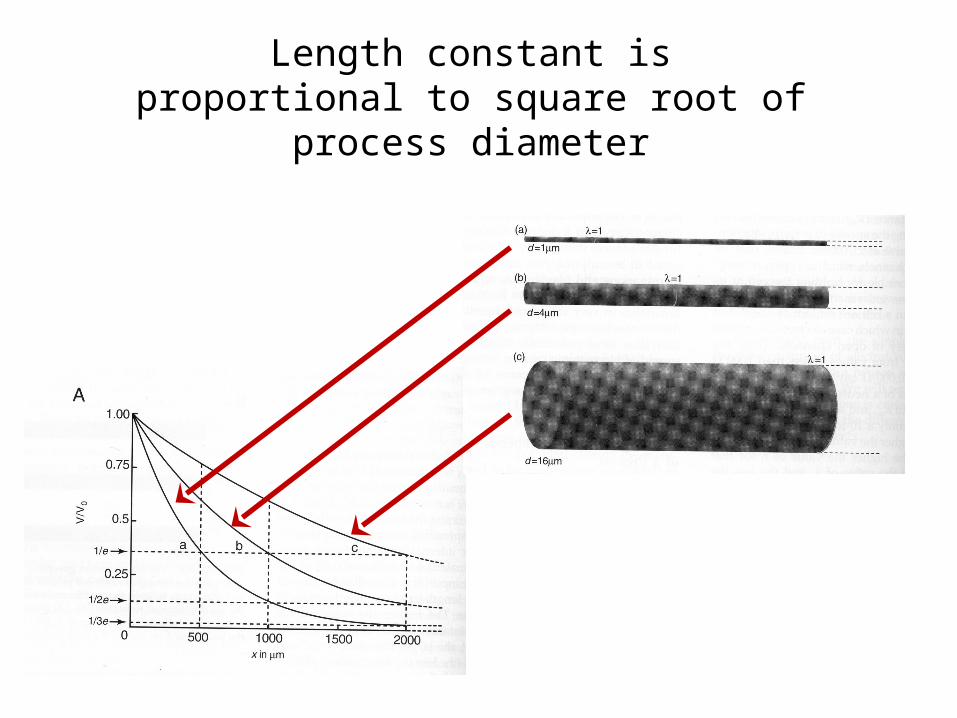
Length constant is proportional to square root of process diameter
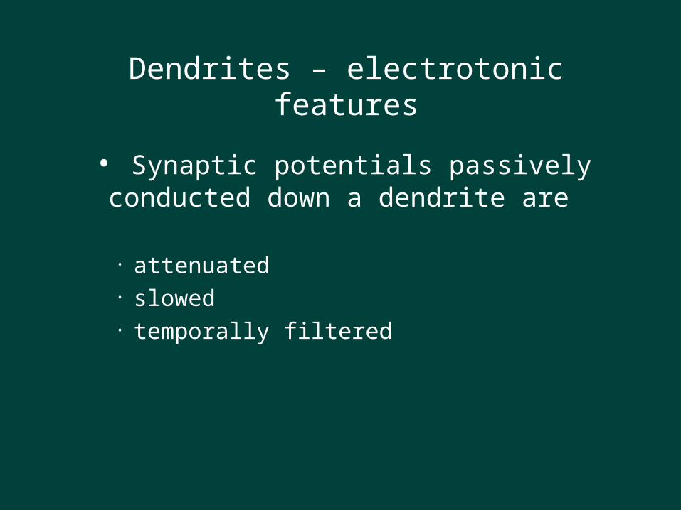
Dendrites – electrotonic features
• Synaptic potentials passively conducted down a dendrite are
• attenuated• slowed• temporally filtered

Slowing and attenuation of distal inputs

Dendrites – electrotonic features
• Temporal summation of synaptic inputs• nearly synchronous inputs summate (but non-
linearly)• inputs widely separated in time do not interact
• Spatial summation of synaptic inputs• nearby inputs summate (but non-linearly)• widely separated input interact only weakly

Spatial SummationTemporal Summation

A potential problem:dendritic filtering
Because of the leaky cable structure of dendrites, inputs fade away with distance.
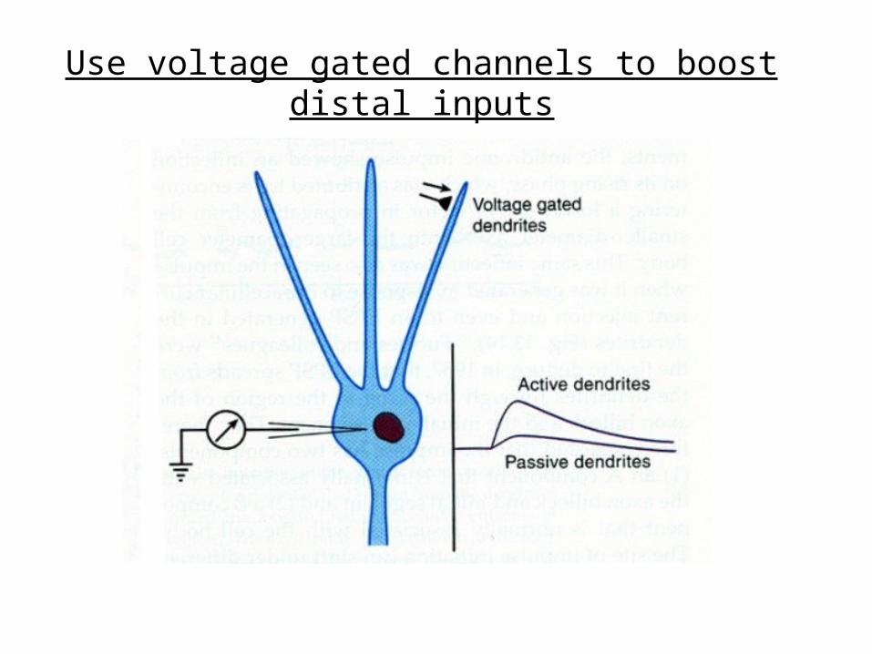
Can distal inputs influence spiking?

Mitral cells of olfactory bulbdriven effectively by distal input

Possible solutions:
Passive Properties
• Increase length constant and lower capacitance.
• Increase size of EPSPs distally.
Active Properties
• Voltage-dependent ion-channels could boost the signal along the way.
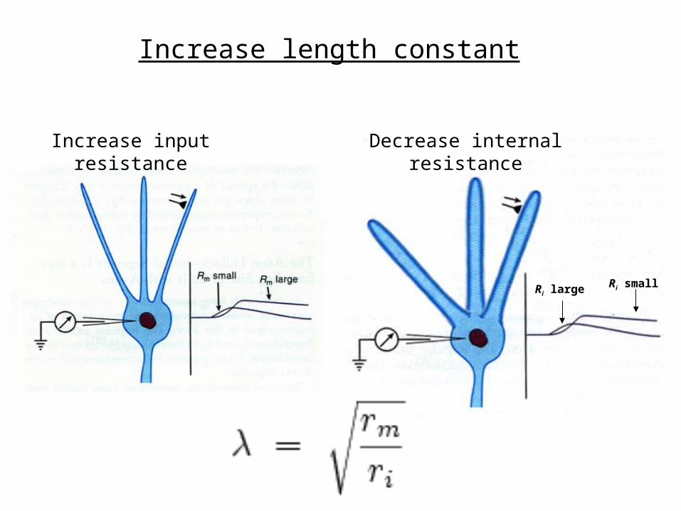
Spines – electrotonic featuresIncrease length constant
Increase input resistance Decrease internal resistance
Ri large Ri small

Problem: changes in morphology are not always practical:
In order for the length constant to double, the diameter of the dendrite has to increase by a factor of four.
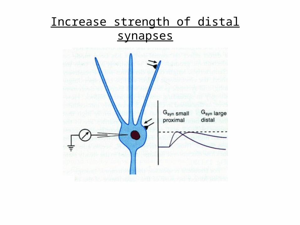
Spines – electrotonic featuresIncrease strength of distal synapses
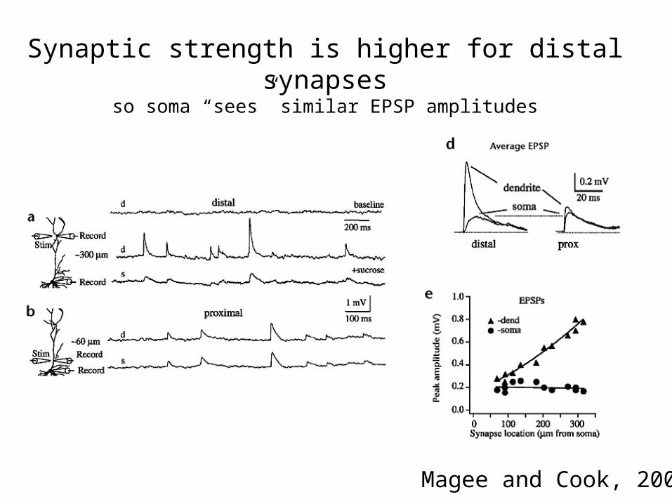
Magee and Cook, 2000
Synaptic strength is higher for distal synapsesso soma “sees” similar EPSP amplitudes

Vm = -60mV
25 mV
7.5 mV
67.5 mV
Vrev = 0 mV
However, distal inputs can only be so big…
So what is a poor dendrite to do?
Spines – electrotonic features
• Small neck, high input resistance• maximizes synaptic potentials
• Low capacitance• maximizes frequency response
• Impedance mismatch with dendritic trunk results in asymmetric effects
• spine voltage has relatively little effect on dendrite (local action)• dendritic voltage significantly influences spine
Use voltage gated channels to boost distal inputs
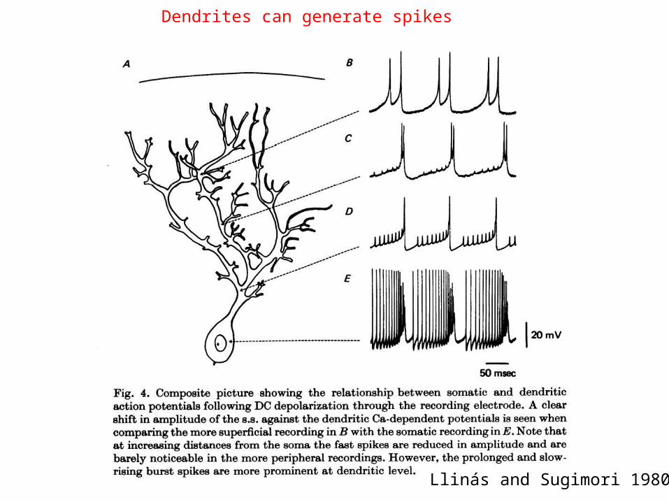
Dendrites can generate spikes
Llinás and Sugimori 1980

Active properties vary within and between neurons
• Purkinje cells• P-type calcium channels• Few sodium channels• Little backpropagation of spikes
• Cortical pyramidal cells• Calcium and sodium channels• Robust backpropagation of spikes•
• Some neurons have minimal active properties
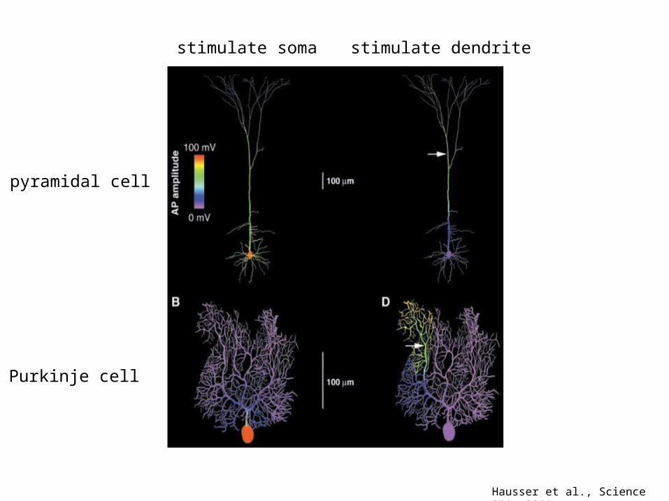
Hausser et al., Science 290, 2000
stimulate soma stimulate dendrite
pyramidal cell
Purkinje cell
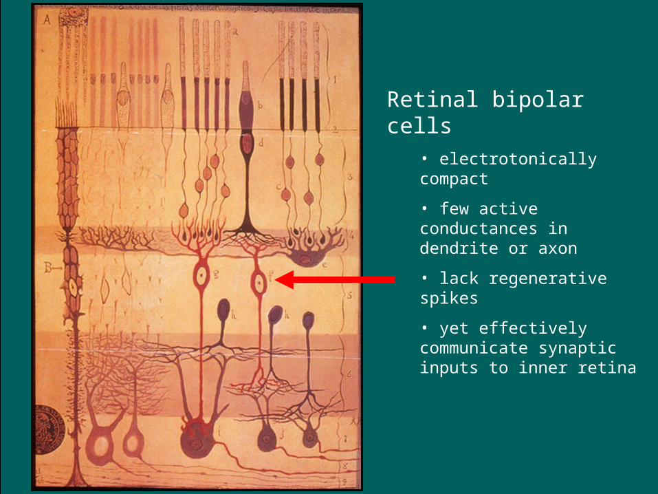
Retinal bipolar cells• electrotonically compact
• few active conductances in dendrite or axon
• lack regenerative spikes
• yet effectively communicate synaptic inputs to inner retina
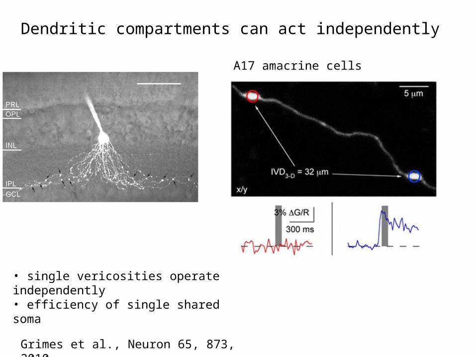
Grimes et al., Neuron 65, 873, 2010
Dendritic compartments can act independently
A17 amacrine cells
• single vericosities operate independently• efficiency of single shared soma

Horizontal cells: uncoupled dendritic and axonal compartments

Non-linear properties of dendrites serve diverse functions
• Boost synaptic responses in graded fashion
• Thresholding (non-linear amplification of stronger
inputs)
• Propagate spikes in anterograde or retrograde
direction•

Dendritic spikes propagate in both directions
Forward Propagation Backpropagation

Early evidence for somadendritic spikes
Eccles, 1957

Hausser et al., Science 290, 2000
Contemporary evidence for dendritic spikes

Backpropagation – functional roles
• Pyramidal-cells• boost somadendritic spike so it invades the dendritic tree• reset membrane potential for new inputs• depolarize spines
• gate NMDA receptors• coincidence detection for Hebbian increase in synaptic
strength
• Mitral cells and dentate granule cells • trigger release from presynaptic dendrites

Direction of information flow in dendrites affected by many factors
• Extent and complexity of branching (electrotonic factors)
• Distribution of excitatory and inhibitory synapses• Distribution of voltage gated channels• Interaction among all of these factors
•

Spines – special features
• Narrow neck high input resistance• maximizes EPSP evoked by synaptic conductance
• Low capacitance • maximizes frequency response
• Impedance mismatch where neck meets shaft• spine has trouble strongly influencing parent dendrites• voltage fluctuations in shaft do influence spine

Spines – role in plasticity
• Big changes in spine form and motility in development
• Enriched environments increase spine number
• LTP more and bigger spines •
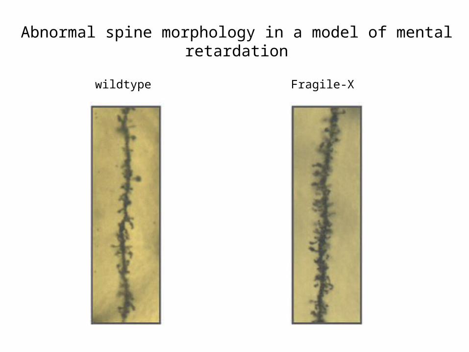
Abnormal spine morphology in a model of mental retardation
wildtype Fragile-X

www.neurostructural.org/images/nine.jpg
normal Alzheimer’s
Spine loss in neurodegenerative diseases

Dendrites as presynaptic elements

excit
inhib
sum
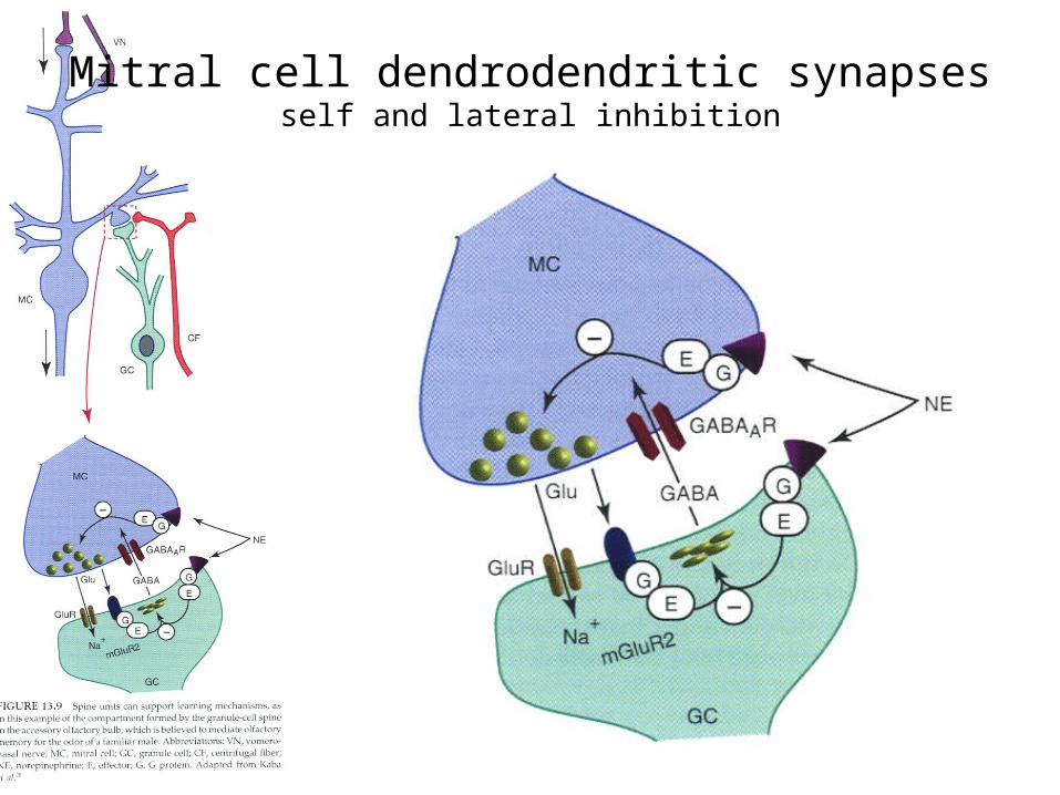
Mitral cell dendrodendritic synapsesself and lateral inhibition


Lin and Koleske, 2010
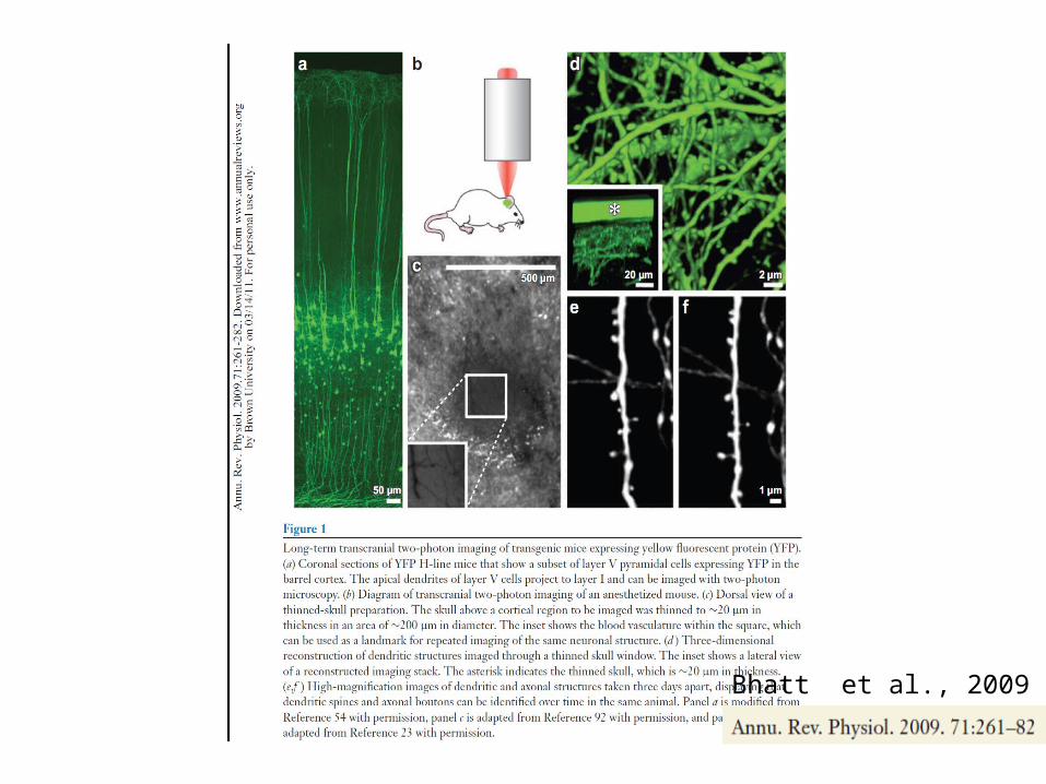
Bhatt et al., 2009
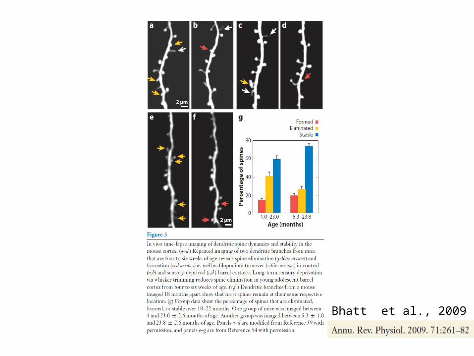
Bhatt et al., 2009

Most retinal amacrine cells lack axons

Gap junction - substrate for electrical synapses
Wagner, C. Kidney International (2008) 73, 547–555

A-type and B-type horizontal cells in the rabbit retina have different dye-coupling properties.
O'Brien J J et al. J. Neurosci. 2006;26:11624-11636
©2006 by Society for Neuroscience

Cx50 plaques occur at dendritic crossings in calbindin-labeled A-type horizontal cells.
O'Brien J J et al. J. Neurosci. 2006;26:11624-11636
©2006 by Society for Neuroscience

Grimes et al., Neuron 65, 873, 2010
Spines – electrotonic features
• Small neck, high input resistance• maximizes synaptic potentials
• Low capacitance• maximizes frequency response
• Impedance mismatch with dendritic trunk results in asymmetric effects
• spine voltage has relatively little effect on dendrite (local action)• dendritic voltage significantly influences spine
Spines – role in plasticity
• Spine morphology changes during development• @
• Enriched environments and training increase spine numbers
• @
• Long-term potentiation• increases spine numbers• increase spine volume in single spines monitored over time

Direction of Dendritic Spikes is Bidirectional
Forward Propagation Backpropagation

Active Dendritic Properties Summary:
• Active conductances are present in dendrites.• Not uniform expression within dendrites or between neurons.• Boost subthreshold EPSPs.• Generate dendritic spikes.• Lead to non-linear synaptic integration.• Backpropagate somatic action potentials: open NMDAR, increase dendritic Ca++ levels.
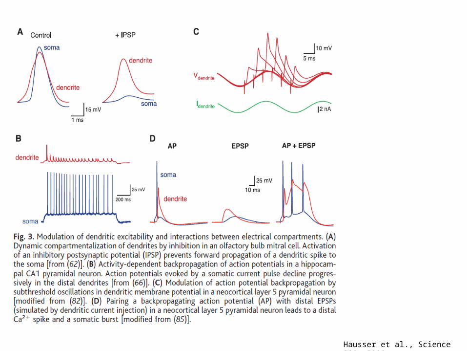
Hausser et al., Science 290, 2000

Hausser et al., Science 290, 2000


Related Documents