PHYSIOLOGICAL EFFECTS OF CADMIUM ON JUVENILE PACIFIC OYSTERS, CRASSOSTREA GIGAS, THUNBERG. Rosalind Hand B.Sc. (Tas.) Being a thesis submitted to the University of Tasmania in partial fulfilment of the degree of Bachelor of Science with Honours in Zoology. March, 1993.

Welcome message from author
This document is posted to help you gain knowledge. Please leave a comment to let me know what you think about it! Share it to your friends and learn new things together.
Transcript

PHYSIOLOGICAL EFFECTS OF CADMIUM ON JUVENILE PACIFIC OYSTERS, CRASSOSTREA GIGAS, THUNBERG.
Rosalind Hand B.Sc. (Tas.)
Being a thesis submitted to the University of Tasmania in partial fulfilment of the
degree of Bachelor of Science with Honours in Zoology.
March, 1993.
ACKNOWLEDGEMENTS
I would like to thank both of my supervisors, Dr. Christian Garland and Dr. David Ritz
for their guidance and encouragement throughout the course of this investigation.
I am indebted to Dr. Robert Dineen and the staff of the Department of Environment and
Planning Laboratory for graphite furnace atomic absorption spectrophotometer
analyses. I am also grateful to Dr. Alan Bray, of the Department of Agricultural
Science, for the use of his atomic absorption spectrophotometer throughout the year.
I wish to thank Shellfish Culture for the loan of equipment as well as providing oyster
spat for experiments. The Tasmanian Shellfish Company also kindly provided oysters
for the initial stages of the project.
My thanks to Dr. Dennis Mackey, of the CSIRO, for his helpful advice and for the use
of the computer program, Mineql.
Dr. Leon Barmuta provided invaluable assistance with statistical analyses. Finally, I
would like to thank the technical staff of the Zoology and Agricultural Science
Departments for their assistance throughout the project.

CONTENTS
Page
ABSTRACT 1
1. GENERAL INTRODUCTION 2
2. GENERAL EXPERIMENTAL METHODS 4
2.1 EXPE~NTALSYSTEM 4
2.2 WATER ANALYSIS 6
2.3 MICROALGAL CUL1URE 7
2.3.1 Materials and Methods 7 2.3.2 Results 9
2.3.3 Discussion 11
3. LETHAL CADMIUM EXPOSURE 12
3.1 INTRODUCTION 12
3.2 MATERIALS AND METHODS 13
3.2.1 Range assay 13
3. 2. 2 Defmitive assay 14
3.3 RESULTS 16 3.3.1 Range assay 16 3. 3. 2 Defmitive assay 18
3.4 DISCUSSION 22
4. EFFECfS OF SUBLETHAL CADMIUM EXPOSURE 24
4.1 INTRODUCTION 24
4.2 MATERIALS AND METHODS 25 4.2.1 Chronic exposure 25 4. 2. 2 Physiological effects 25
4.3 RESULTS 27 4.3.1 Chronic exposure 28 4. 3. 2 Physiological effects 34

5.
6.
GENERAL DISCUSSION
LITERATURE CITED
APPENDIX
LITERATURE REVIEW
45
48
55
57

ABSTRACT
1. "Small" C. gigas spat (2.8 mm) were more sensitive to acute cadmium exposure
in seawater than larger spat (3.5 mm), with a 96 hour LCso value of 10.29 and 28.43
mgL-1 total cadmium respectively.
2. Cadmium toxicity to juvenile C. gigas was more closely related to the free
cadmium ion concentration than to the total cadmium concentration. The 96 hour LCso in
terms of free ion concentration was 0.25 and 0.27 mgL-1 for "small" and "large" spat
respectively.
3. EDTA reduced the toxicity of cadmium to C. gigas spat by reducing the external
free ion concentration.
4 . Chronic exposure of C. gigas spat to sublethal concentrations of cadmium
-retarded growth of both shell and soft tissue;
-caused an apparent increase in shell abnormalities;
-affected heart contraction rate and caused cardiac arryhthmia;
-slowed the rate of filtration of microalgae (spat feed rates);
-slowed the behavioural response to tactile and light stimuli;
5. The cadmium bioaccumulation factors of spat exposed to 0, 10, 50 and 250 J..lgL-1
cadmium were 1.5 x 10⁴, 3 x 10³, 1.3 x 10³ and 1.5 x 10³ respectively. These values are
similar to those previously reported by Ward (1983) for adult oysters (Saccostrea
commercialis ).
6 . Accumulation of cadmium in spat soft tissue was linearly related to the external
cadmium concentration. This is similar to the pattern of cadmium accumulation by other
species of adult bivalves.
1
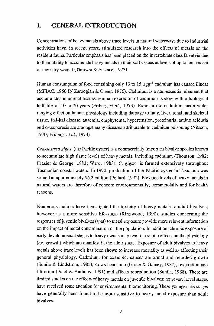
1. GENERAL INTRODUCTION
Concentrations of heavy metals above trace levels in natural waterways due to industrial
activities have, in recent years, stimulated research into the effects of metals on the
resident fauna. Particular emphasis has been placed on the invertebrate class Bivalvia due
to their ability to accumulate heavy metals in their soft tissues at levels of up to ten percent
of their dry weight (Thrower & Eustace, 1973).
Human consumption of food containing only 13 to 15 ~gg-1 cadmium has caused illness
(MFIAC, 1950 IN Zaroogian & Cheer, 1976). Cadmium is a non-essential element that
accumulates in animal tissues. Human excretion of cadmium is slow with a biological
half-life of 10 to 30 years (Friberg et al., 1974). Exposure to cadmium has a wide
ranging effect on human physiology including damage to lung, liver, renal, and skeletal
tissue. Itai-itai disease, anaemia, emphysema, hypertension, proteinuria, amino aciduria
and osteoporosis are amongst many diseases attributable to cadmium poisoning (Nilsson,
1970; Friberg et al., 1974).
Crassostrea gigas (the Pacific oyster) is a commercially important bivalve species known
to accumulate high tissue levels of heavy metals, including cadmium (Thomson, 1982;
Frazier & George, 1983; Ward, 1983). C. gigas is farmed extensively throughout
Tasmanian coastal waters. In 1990, production of the Pacific oyster in Tasmania was
valued at approximately $6.2 million (Pollard, 1992). Elevated levels of heavy metals in
natural waters are therefore of concern environmentally, commercially and for health
reasons.
Numerous authors have investigated the toxicity of heavy metals to adult bivalves;
however, as a more sensitive life-stage (Ringwood, 1990), studies concerning the
responses of juvenile bivalves (spat) to metal exposure provide more relevent information
on the impact of metal contamination on the population. In addition, chronic exposure of
early developmental stages to heavy metals may result in subtle effects on the physiology
(eg. growth) which are manifest in the adult stage. Exposure of adult bivalves to heavy
metals above trace levels has been shown to increase mortality as well as affecting their
general physiology. Cadmium, for example, causes abnormal and retarded growth
(Sunila & Lindstrom, 1985), slows heart rate (Grace & Gainey, 1987), respiration and
filtration (Patel & Anthony, 1991) and affects reproduction (Sunila, 1988). There are
limited studies on the effects of heavy metals on juvenile bivalves; however, larval stages
have received some attention for environmental biomonitoring. These younger life-stages
have generally been found to be more sensitive to heavy metal exposure than adult
bivalves.
2
The relative proportions of different forms of a metal influence its effect in the
environment. Metal uptake by bivalves is from food, water and sedimentary sources.
Despite frequently high levels of metals in marine sediments, uptake is predominantly
from the surrounding water (B1yan et al., 1985). Recommended limits for levels of trace
metals in the environment are generally based on the total concentration. This is
misleading where only specific forms of a metal are toxic and the concentration of those
forms depend not only on the total metal concentration, but also on the physico-chemical
conditions present. For example, the toxicity of cadmium to the grass shrimp
Palaemonetes pugio is related to the free cadmium ion concentration rather than the total
concentration (Sunda et al., 1978). The free ion concentration of a metal in solution is
highly dependent on the salinity of the water; ie. with decreasing salinity, the free ion
concentration conesponding to a certain total cadmium concentration increases due to a
reduction in the formation of chloro-complexes (Engel & Fowler, 1979). Cadmium in sea
water is predominantly present as inorganic chloro-complexes, in particular, CdChO and
CdCl+ with only a small proportion of the total cadmium present in the form of free
cadmium ions (Zirino & Yamamoto, 1972).
In recent years, a widespread incidence of an abnormal shell growth of C. gigas spat,
termed curly back, has been noted in the commercial shellfish industry. Curly back is
characterised by an upward growth of the posterior, ventral shell valve; that is, growth is
opposite to the "normal" direction of shell growth (pers. obs.). It is possible that chronic
exposure to sublethal concentrations of metals may be responsible for this growth defect.
A range of shell abnormalities has been reported in adult mussels, Mytilus edulis, in
response to acute cadmium exposure (Sunila & Lindstrom, 1985). The observed growth
abnormality as well as previous reports of high levels of heavy metals in Tasmanian
waterways (eg. Bloom & Ayling, 1977; Tasmanian Department of Environment, 1987),
prompted an interest in the effect~ of cadmium on the juvenile Pacific oyster.
In this thesis I will address two main areas of research:
1. lethal toxicity of cadmium to juvenile C. gigas (including the specific form of
cadmium that is toxic to spat).
2. the effects of chronic sublethal exposure to cadmium on the physiology of C.
gigas spat.
3
2.. GENERAL EXPERIMENTAL METHODS
Outlined in the following chapter are materials and methods pertinent to experiments on
both lethal and sublethal cadmium exposure. More specific materials and methods for
each experiment are described in the relevant chapters.
2. 1 EXPERIMENTAL SYSTEM
A static experimental system comprising a series of 400 mL vessels was used for tests of
both lethal and sublethal effects of cadmium. Polyethylene treatment vessels were used to
minimise loss of cadmium from solution by adsorption to the vessel walls. Gentle
aeration promoted constant exposure of spat to the cadmium solution as well as reducing
oxygen depletion and toxic metabolite accumulation in the vicinity of the spat.
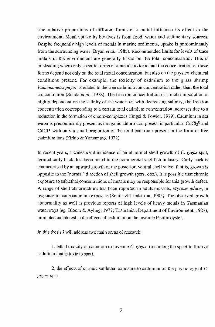
Temperature of treatment solutions was maintained at 20 ± 0.5 OC by means of a
circulating water bath housed within a controlled temperature cabinet (Fig. 2.1). Salinity,
temperature, dissolved oxygen and pH were monitored during all experiments. Treatment
solutions were made by dilution (in oceanic water) of stock solutions of 1gL-1
CdCl2.2112H20 (AnalaR *). Stock solutions were made up in deionized water with
1mUL HN03 (AnalaR*) to maintain cadmium solubility.
Exposure of spat to metals (other than experimental doses) was avoided at all stages of
handling and treatment using standard bioassay procedures (APHA, 1989). Prior to use,
all plastic and glassware (treatment vessels, sample bottles, aeration lines, volumetric
glassware etc.) was washed, soaked for a minimum of 48 hours in 10% nitric acid then
rinsed twice in deionised water to remove any contaminating metals. Oceanic water used
throughout the experiment was stored in a clean tank and filtered to 0.45 Jlm before use.
All water pipes and fittings were plastic.
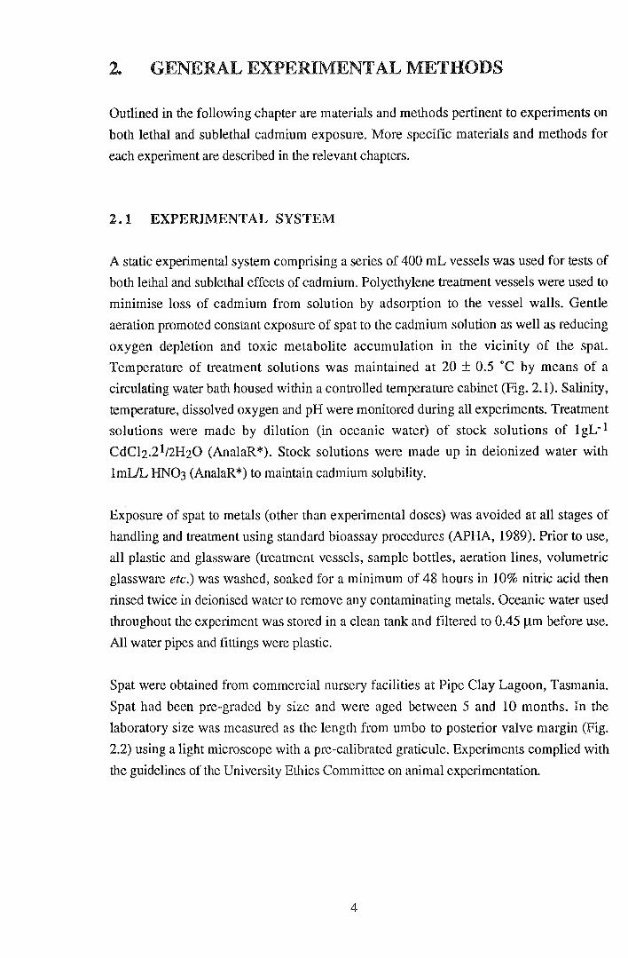
Spat were obtained from commercial nursery facilities at Pipe Clay Lagoon, Tasmania.
Spat had been pre-graded by size and were aged between 5 and 10 months. In the
laboratory size was measured as the length from umbo to posterior valve margin (Fig.
2.2) using a light microscope with a pre-calibrated graticule. Experiments complied with
the guidelines of the University Ethics Committee on animal experimentation.
4

2.2 WATER ANALYSIS
Concentrations of cadmium treatment solutions (for lethal exposure) and stock solutions
were verified with flame atomic absorption spectrophotometry (flame AAS) using a
Unicam SP 1900 Atomic Absorption Spectrophotometer. Treatment solutions were
preserved with 10 mLIL HN03 and stored in 250 mL, acid soaked (see Section 2.1),
polyethyene sample bottles at 5 OC until analysis by AAS.
Three replicates of each sample were measured for total cadmium (without digestion) by
AAS using an air-acetylene flame. Dilution of more concentrated solutions was necessary
for accurate determination of cadmium concentration. AAS operating conditions used for
determination of cadmium concentration are shown in Table 1.1. Calibration curves were
prepared from a series of cadmium standards of 0.5, 1, 2, 4 and 10 mgL -1 (as
CdCh.21t2H20) in deionized water with 10 mLIL HN03.
Table 1.1 Flame AAS operating conditions for measurement of cadmium
concentration in water.
WAVELENGTH SLIT WIDTH LAMP CURRENT SENSITIVITY CPIIMUMRANGE
228.8 nm 0.2 nm 3mA 0.02 mgL-1 0.2-1.8mgL-1
To avoid instrument dtift, calibration was checked every 12 samples. Matrix interference
was assessed using the method of known additions. Sample recovery was found to be
between 98 and 102 % indicating that matrix interference was insignificant (APHA,
1989).
Limited variation (3.67 ± 1.58 %) occurred between estimated concentrations of
cadmium in water and measured initial cadmium concentrations for all lethal exposure
trials. Similarly, cadmium concentrations in exposure vessels varied by only 1.72 ± 0.66
% over the 48 hours between water changes.
Filtered oceanic (control treatment) water was measured for trace levels of cadmium by
graphite furnace AAS (analysis courtesy of Dept. of Environment) using ammonium
oxalate as a matrix modifier. Initial cadmium levels were found to be below 0.2 ~gL-1 for
all experiments.
6
2. 3 MICROALGAL CULTURE
Microalgae are an essential food source required for long-tenn maintenance of bivalve
molluscs in artificial systems. For experimental purposes feed rates, food quality and
algal species composition may be controlled by feeding from laboratory monocultures of
microalgae. The following section outlines the procedures used for culture of microalgae.
Algae were provided to juvenile oysters during experiments on effects of exposure to
sublethal cadmium concentrations and during assessment of spat feeding rates.
Spat were fed a mixed diet of Isochrysis sp. -clone T-ISO (Prymnesiophyceae) and
Nannochloris atomus (Chlorophyceae). Both species are readily cultured under
controlled conditions and are commonly used for bivalve nutrition. Isochrysis sp. is a
particularly good source of nutrition for juvenile molluscs (Brown et al., 1989).
2.3.1 Materials and Methods
Algal production was based on the batch culture methods ofGuillard (1975). Axenic 150
mL stock cultures of algae were obtained from CSIRO Marine Laboratories, Hobart.
Initial stock cultures were subcultured into 3 x 250 mL Erlenmeyer flasks containing
approximately 150 mL of f/2 medium (see Appendix, Guillard, 1975). Auxilliary stock
cultures were maintained by repeated subculturing into 250 mL flasks of f/2 medium
every 14 to 20 days. Algal production was scaled up to 5 L Erlenmeyer flasks by
inoculation with approximately 200 mL of stock culture. The routine for maintenance of
algal cultures and culture vessel design is shown in Figures 2.3 and 2.4.
f12 medium was made from nutrient stock solutions (Guillard, 1975) with oceanic water
which had been pre-filtered through a series of filters to 0.4 ~m. All media were
autoclaved at 105 kPa and 121 OC for 30 minutes several days prior to inoculation with
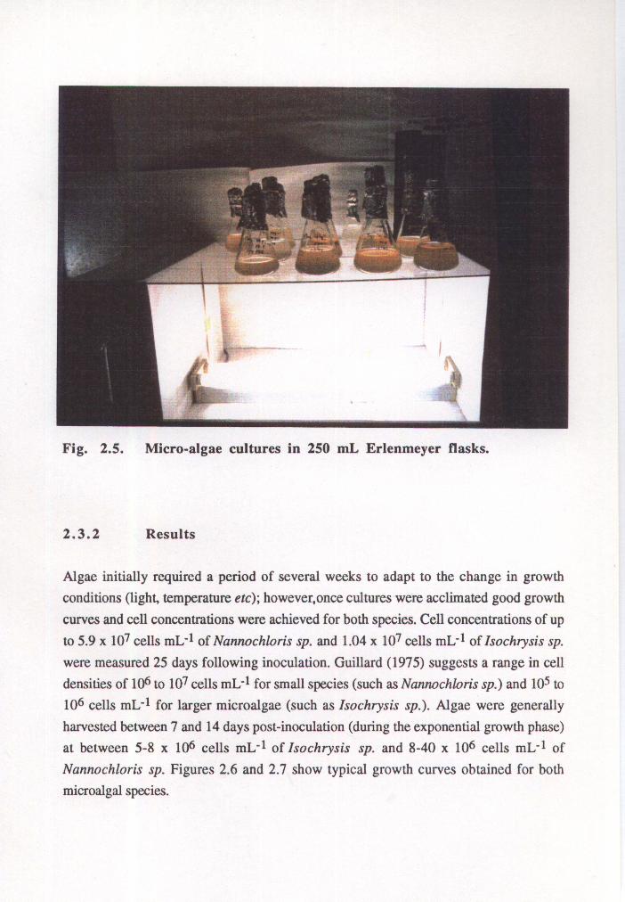
algae. Cultures were illuminated with fluorescent lights (Fig. 2.5) on a 12 hour day:night
cycle. Aeration of the 2 and 5 L flasks was provided through silicon tubing with in-line
0.2 j.Lm cellulose acetate membrane filters.
Being less susceptible to bacterial colonisation, Nannochloris sp. was cultured in 10 L
open aerated culture vessels. Culture vessels with 10 L of f/2 medium were inoculated
with 1 L of Nannochloris sp. culture every 7 to 14 days depending on culture densities
and algal demand. All algae were harvested during the exponential growth phase and cell
densities detennined prior to feeding using a Neubauer haemocytorneter.
7

~ ~ !J 200 ml starter cultures
250 ml stock cultures
DayO Day7-14
Fig. 2.3. Routine used for culture
air outlet
cotton filter in syringe tube
• 5 L cultures
~ !J!J !J Auxilliary 250 ml stock cultures
Day 14-21
of micro-algae.
foil dust cap
r=i-~r----- neoprene stopper
~--- silicon tubing
J--~~""'""'"T<---- glass tubing
air inlet
Fig. 2.4. Vessel design for culture of microalgae in SL Erlenmeyer
flasks.
9000
........ _j
~ ~ w (,) ....... 1:: 0 700 4:i (11 .... .... 1:: Q) (,) c: 0 u
6000
5000+-~--r-~-,--~-r~~,-~--r---~
4 6 8 10 12 14 16
Time (days)
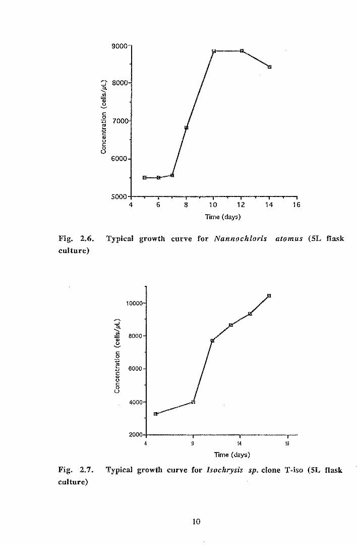
Fig. 2.6. Typical growth curve for Nannochloris atomus (SL flask
culture)
........ _j
~ ~ w (,) '-'
c: 0
·.;:::; rn .... .... c: Q) (,)
c: 0 u
10000
8000
6000
4000
4 9 14
Time (days)
19
Fig. 2.7. Typical growth curve for lsochrysis sp. clone T-iso (SL flask
culture)
10
2.3.3 Discussion
Jeffrey et al (1990) recommended harvesting of microalgae during the exponential
growth phase at between 1-3 x 10⁶ cells mL-1 for golden-brown flagellates (eg.
Isochrysis sp.) and 1 x 10⁶ cells mL-1 for green flagellates (eg. Nannochloris atomus).
Cell densities achieved during culture were higher than these suggested values probably
due to the small sizes of culture vessels (maximum 10 L). Growth rates and cell densities
of both microalgal species were normal to high compared with published values
(Guillard, 1975; Jeffrey et al., 1990). Harvesting during the exponential growth phase
maximises the culture's cell density whilst reducing the risk of contamination by bacteria,
ciliates etc. as well as limiting toxic algal metabolites (Guillard, 1975). The biochemical
composition of microalgae alters during the different phases of growth; generally the
biochemical composition is more nutritious to bivalves during the early exponential
growth phase (Whyte, 1987).
A mixed algal diet such as that used here is generally more nutritionally beneficial to
mariculture species than feeding a single species of algae (Jeffrey et al., 1990). For
example, the two species used complement each other to provide a diet rich in the
polyunsaturated fatty acids known to be essential to C gigas spat (La~ngdon & Waldock,
1981). Overall, good algal culture growth rates and high culture concentrations combined
with rapid algal clearance by control spat (Section 4.3.1) indicated that adequate
microalgal culture techniques were employed.
11
3 LETHAL CADMIUM EXPOSURE
3.1 INTRODUCTION
Acute toxicity of heavy metals is conventionally assessed in terms of the "median lethal
concentration" (LCso), that is, the total concentration of the relevant metal that causes
fifty percent mortality in a given exposure time (frequently 48 or 96 hours) (eg. Eisler,
1971; Calabrese et al., 1973). More recently attention has been paid to the specific forms
of metals that are toxic to various organisms (eg. Zamuda & Sunda, 1982; Patel and
Anthony, 1991). Sunda et al. (1978) found that when exposed to cadmium the mmtality
of the grass shrimp Palaemonetes pugio was related to the free cadmium ion
concentration rather than the total cadmium concentration. Similarly, accumulation of
copper by the oyster Crassostrea virginica has been found to be related to the free cupric
ion activity rather than the total copper exposure concentration (Zamuda & Sunda, 1982).
For experimental purposes, chelating agents (eg. humic acid, nitrilotriacetic acid) allow
variation of the free ion concentration of metals whilst maintaining a constant total metal
concentration in solution (Sunda et al., 1978).
LCso values have limited use for direct assessment of the effects of toxicants in the
environment where organisms are often chronically exposed to sublethal toxicant
concentrations under fluctuating physico-chemical conditions. Generally, LCsos are now
used as a preliminary means of assessing toxicity before more extensive toxicity
investigations such as chronic exposure experiments are used. Chronic exposure tests
provide more ecologically relevant information of toxicity (Beaumont et al., 1987).
However, LCsos do provide a valuable means of assessing relative toxicities between
different toxicants or (in this case different forms of a toxicant) as well as the relative
sensitivities between different species or life-stages.
Several methods are available for determination of LCsos and their confidence limits. The
more common analytical techniques are the arithmetic graph method, the logarithmic
method and probit analysis (Reish & Oshida, 1986; APHA, 1989). The favoured, more
precise method of LCso determination is probit analysis which maximises use of the
linear portion of the typical sigmoid dose-response curve, thus minimising the influence
of extremes in tolerance to the relevant toxicant on the calculated LCso.
12

The following aspects of acute toxicity of cadmium to two sizes of C. gigas spat were
examined:
1) 96 hour LCso determination using probit analysis.
2) the influence of free cadmium ion concentration on cadmium toxicity
3) general physiological responses to cadmium exposure
3.2 MATERIALS AND METHODS
3.2.1 Range assay
An initial range assay was conducted to detennine the approximate test range required for
determination of the 96 hour LCso of C. gigas spat exposed to cadmium. Procedures
were based on those outlined in APHA (1989). Spat were exposed to three
concentrations of cadmium as CdCh.2112H20 in sea water in a log progression of 2, 20
and 200 mgL-1 with sea water as the control (control treatments were arbitrarily labelled 0
mgL-1 despite containing trace levels of cadmium (see Section 2.2)). The lowest
treatment concentration was selected to correspond to the approximated 96 hour LCso for
juvenile C. gigas of 2 mgL-1 (Watling, 1978). Spat of commercial size grading "1500"
were acclimated for 24 hours to experimental conditions of salinity and temperature, sized
and placed in experimental vessels (section 2.1). Duplicate treatments of each exposure
concentration were set up in 400 mL vessels with 50 spat in each vessel. Treatment
solutions were renewed at 48 hours to avoid cadmium depletion and build-up of toxic
metabolic products. Spat were not fed dming periods of either acclimation or cadmium
exposure. Mortality and morphological observations were recorded at 48 and 96 hours.
Any dead spat were noted and removed from the treatment vessel after 48 hours of
exposure to treatment solutions. Mortality was assessed using a light microscope by
observing spat for the presence of heart contractions, filtering activity and behavioural
responses to light and tactile stimuli (eg. shell gaping, valve adduction, etc). After 96
hours spat mortality was plotted against the log of cadmium exposure concentration and
the approximate concentration conesponding to 50 % mortality was determined.
3.2.2 Definitive assay
Ninety-six hour LCsos were determined for 2 sizes of spat of commercial grading "1500"
and "1800"; these will subsequently be referred to as "small" and "large" spat.
13
Measurement of 200 spat in each size group, by light microscopy, gave mean sizes of
2.84 ± 0.03 mm and 3.46 ± 0.03 mm for "small" and "large" spat respectively. Prior to
treatment, spat were acclimated to expelimental conditions of salinity and temperature.
Spat were not fed for two days prior to and dming experiments.
An LCso of approximately 10 mgL -1 for "small" spat was expected following analysis of
the range assay data. Cadmium treatment concentrations were selected on an approximate
log scale to encompass the expected LCso of both sizes of spat. Triplicate treatment
vessels of 0, 2, 5, 10, 20, and 50 mgL-1 cadmium were set up for each size group with
50 spat per 400 mL vessel (see Section 2.1 ).
To investigate the effect of free cadmium ion concentration on spat mortality the
experimental system was replicated for both "small" and "large" spat with the addition of
an artificial chelating agent ie. 5 x 1Q5 M ethylene diaminetetraacetic acid (EDTA, di
sodium salt). EDT A enabled control of the free cadmium ion concentration. The program
"MINEQL" (Westall et. al., 1976) was used to determine free ion concentration in the
resulting solutions (Table 3.1). This program accounts for interactions between the
various ions present in water at equilibrium (including any added compounds eg. EDTA)
and provides the final concentrations and forms of these ions. Figure 3.1 shows the
relationship between free cadmium ion concentration and the total cadmium concentration
used in the experimental systems. "pCd2+" is the free ion concentration transformed to a
scale analogous to the pH scale, where:
pCd2+ =-log [Cd2+].
Treatment solutions were renewed and any dead spat recorded and removed at 48 hours.
Mortality and morphological observations were recorded at 48 and 96 hours.
14
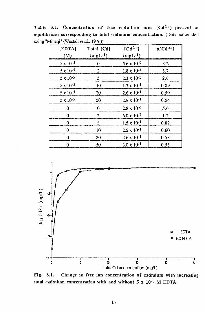
Table 3.1: Concentration of free cadmium ions (Cdl+) present at
equilibrium corresponding to total cadmium concentration. ·(Data calculated
using "M" 1" (W tall l 1976)) rneqJ es eta.,
+ N "'0 u 0) 0
0
[EDTA]
(M)
5 X lQ-5
5 X IQ-5
5 X IQ-5
5 x w-5 5 x w-5 5 x w-s
0
0
0
0
0
0
10
Total [Cd] [Cd2+] p[Cd2+]
(mgL-1) (m_gL·l)
0
2
5
10
20
50
0
2
5
10
20
50
5.6 X I0-9 8.2
1.8 X I0-4 3.7
2.3 X I0-3 2.6
1.3 X 10⁻¹ 0.89
2.6 X 10⁻¹ 0.59
2.9 X lQ-1 0.54
2.8 X 10⁻⁶ 5.6
6.0 X 10⁻² 1.2
1.5 X 10⁻¹ 0.82
2.5 X 10⁻¹ 0.60
2.6 X I0-1 0.58
3.0 X 10⁻¹ 0.53
~ ~ ~
total Cd concentration (mg/L)
m + EDTA
• NOEDTA
50
Fig. 3.1. Change in free ion concentration of cadmium with increasing
total cadmium concentration with and without 5 x 10·5 M EDTA.
15
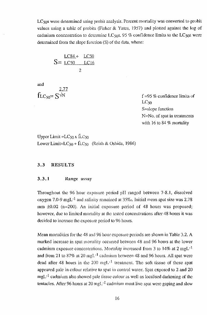
LCsos were determined using probit analysis. Percent mortality was converted to probit
values using a table of probits (Fisher & Yates, 1957) and plotted against the log of
cadmium concentration to determine LCsos. 95 % confidence limits to the LCsos were
determined from the slope function (S) of the data, where:
LC84+ LC50
S= LC50 LC16
2
and
Upper Limit =LCso x fLcso
Lower Limit=LCso + fLcso (Reish & Oshida, 1986)
3.3 RESULTS
3.3.1 Range assay
f =95% confidence limits of
LCso
S=slope function
N=No. of spat in treatments
with 16 to 84% mortality
Throughout the 96 hour exposure period pH ranged between 7-8.1, dissolved
oxygen 7.0-9 mgL-1 and salinity remained at 35%o. Initial mean spat size was 2.78
mm ±0.02 (n=200). An initial exposure period of 48 hours was proposed;
however, due to limited mortality at the tested concentrations after 48 hours it was
decided to increase the exposure period to 96 hours.
Mean mortalities for the 48 and 96 hour exposure periods are shown in Table 3.2. A
marked increase in spat mortality occurred between 48 and 96 hours at the lower
cadmium exposure concentrations. Mortality increased from 3 to 14% at 2 mgL-1
and from 21 to 87% at 20 mgL-1 cadmium between 48 and 96 hours. All spat were
dead after 48 hours in the 200 mgL-1 treatment. The soft tissue of these spat
appeared pale in colour relative to spat in control water. Spat exposed to 2 and 20
mgL-1 cadmium also showed pale tissue colour as well as localised darkening of the
tentacles. After 96 hours at 20 mgL-1 cadmium most live spat were gaping and slow
16
to respond to the microscope light or to mechanical stimulation whilst most control
spat retracted into the shell and closed when subjected to the same stimuli. A similar
but less dramatic response was observed after exposure to 2 mgL-1 cadmium.
Table 3.2. Mean percent mortality of spat after acute exposure to
cadnrlu d t m: range assay a a. Cadmum % Mortality- % Mortality-
concentration (mgL-1) 48 hours 96 hours
0 2 4
2 3 14
20 21 87
200 100 100
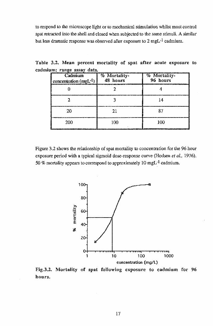
Figure 3.2 shows the relationship of spat mortality to concentration for the 96 hour
exposure period with a typical sigmoid dose-response curve (Hodsen et al., 1976).
50% mortality appears to correspond to approximately 10 mgL-1 cadmium.
1 10 100 1000 concentration (mg/l)
Fig.3.2. Mortality of spat following exposure to cadmium for 96
hours.
17
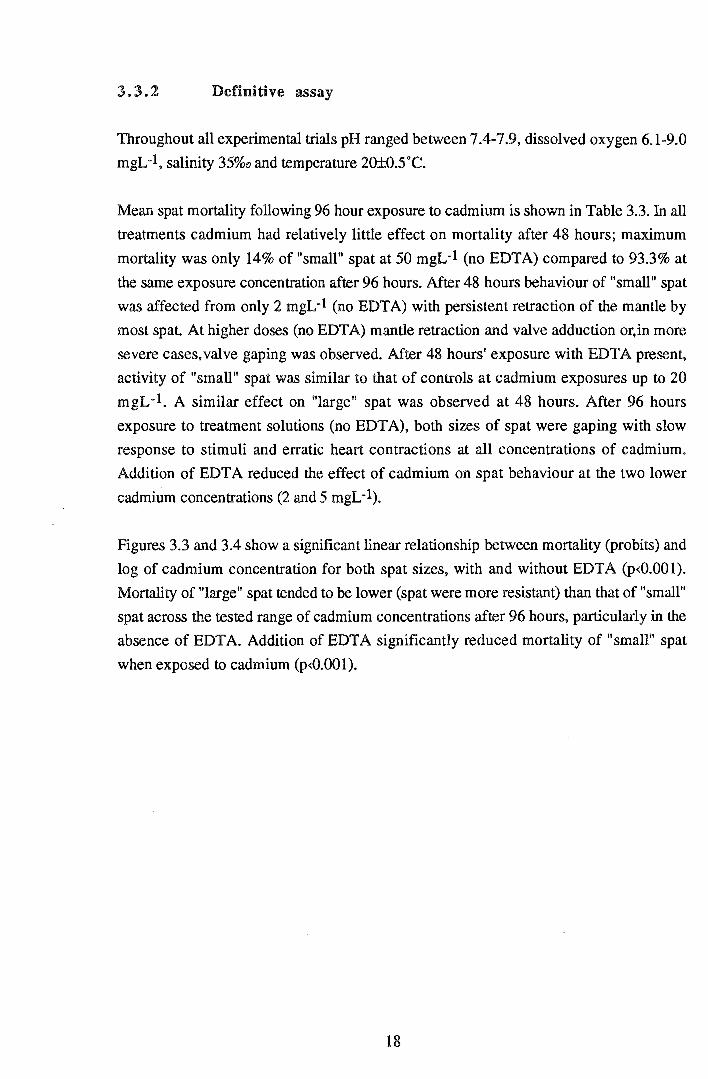
3.3.2 Definitive assay
Throughout all experimental trials pH ranged between 7.4-7.9, dissolved oxygen 6.1-9.0
mgL-1, salinity 35%o and temperature 20±0.5"C.
Mean spat mortality following 96 hour exposure to cadmium is shown in Table 3.3. In all
treatments cadmium had relatively little effect on mortality after 48 hours; maximum
mortality was only 14% of "small" spat at 50 mgL-1 (no EDTA) compared to 93.3% at
the same exposure concentration after 96 hours. After 48 hours behaviour of "small" spat
was affected from only 2 mgL-1 (no EDTA) with persistent retraction of the mantle by
most spat. At higher doses (no EDTA) mantle retraction and valve adduction or, in more
severe cases, valve gaping was observed. After 48 hours' exposure with EDTA present,
activity of "small" spat was similar to that of controls at cadmium exposures up to 20
mgL-1. A similar effect on "large" spat was observed at 48 hours. After 96 hours
exposure to treatment solutions (no EDT A), both sizes of spat were gaping with slow
response to stimuli and erratic heart contractions at all concentrations of cadmium.
Addition of EDTA reduced the effect of cadmium on spat behaviour at the two lower
cadmium concentrations (2 and 5 mgL-1).
Figures 3.3 and 3.4 show a significant linear relationship between mortality (pro bits) and
log of cadmium concentration for both spat sizes, with and without EDTA (p<O.OOl).
Mortality of "large" spat tended to be lower (spat were more resistant) than that of "small"
spat across the tested range of cadmium concentrations after 96 hours, particularly in the
absence of EDTA. Addition of EDTA significantly reduced mortality of "small" spat
when exposed to cadmium (p<O.OOl).
18
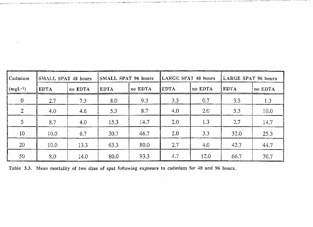
Cadmium SMALL SPAT 48 hours SMALL SPAT 96 hours LARGE SPAT 48 hours LARGE SPAT 96 hours
(mgL-1) EDTA no EDTA EDTA no EDTA EDTA no EDTA EDTA no EDTA
0 2.7 7.3 8.0 9.3 3.3 0.7 3.3 1.3 I
2 4.0 4.6 5.3 8.7 4.0 2.0 5.3 10.0 '
5 8.7 4.0 15.3 14.7 2.0 1.3 2.7 14.7 .•
10 10.0 6.7 30.7 46.7 2.0 3.3 32.0 25.3 !
20 10.0 13.3 63.3 80.0 2.7 4.0 42.7 44.7 '
50 8.0 14.0 80.0 93.3 4.7 12.0 66.7 70.7
Table 3.3. Mean mortality of two sizes of spat following exposure to cadmium for 48 and 96 hours.
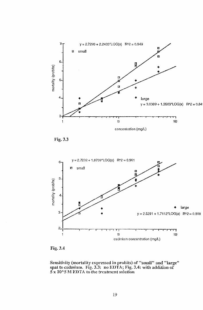
Fig. 3.3
Fig. 3.4
y = 2.7290 + 2.2433*LOG(x) R"2 = 0.949
1:1 small
• large y = 3.0369 + 1.3503*LOG(x) R"2 == 0.841
'i I' I 10 100
concentration (mg/L)
y = 2.7232 + 1.8739*LOG(x) R"2::: 0.961
1:1 small
• large
y = 2.5291 + 1.7112*LOG(x) R"2 = 0.818
10 100 cadmium concentration (mg/L)
Sensitivity (mortality expressed in probits) of "small" and "large" spat to cadmium. Fig. 3.3: no EDTA; Fig. 3.4: with addition of 5 x 10"5 M EDTA to the treatment solution
19
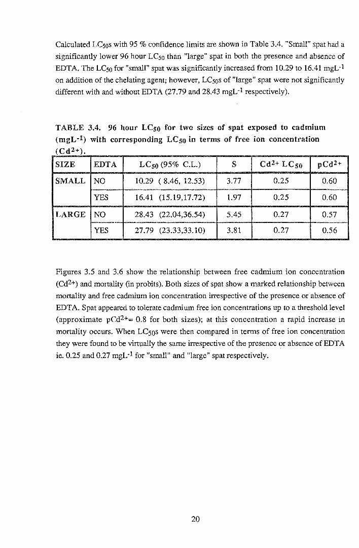
Calculated LCsos with 95% confidence limits are shown in Table 3.4. "Small" spat had a
significantly lower 96 hour LCso than "large" spat in both the presence and absence of
EDT A. The LCso for "small" spat was significantly increased from 10.29 to 16.41 mgL-1
on addition of the chelating agent; however, LCsos of "large" spat were not significantly
different with and without EDTA (27.79 and 28.43 mgL-1 respectively).
TABLE 3.4. 96 hour LCso for two sizes of spat exposed to cadmium
(mgL·l) with corresponding LCso in terms of free ion concentration (Cd2+).
SIZE EDTA LCso (95% C.L.) s Cd2+ LCso pCd2+
SMALL NO 10.29 ( 8.46, 12.53) 3.77 0.25 0.60
YES 16.41 (15.19,17.72) 1.97 0.25 0.60
LARGE NO 28.43 (22.04,36.54) 5.45 0.27 0.57
YES 27.79 (23.33,33.10) 3.81 0.27 0.56
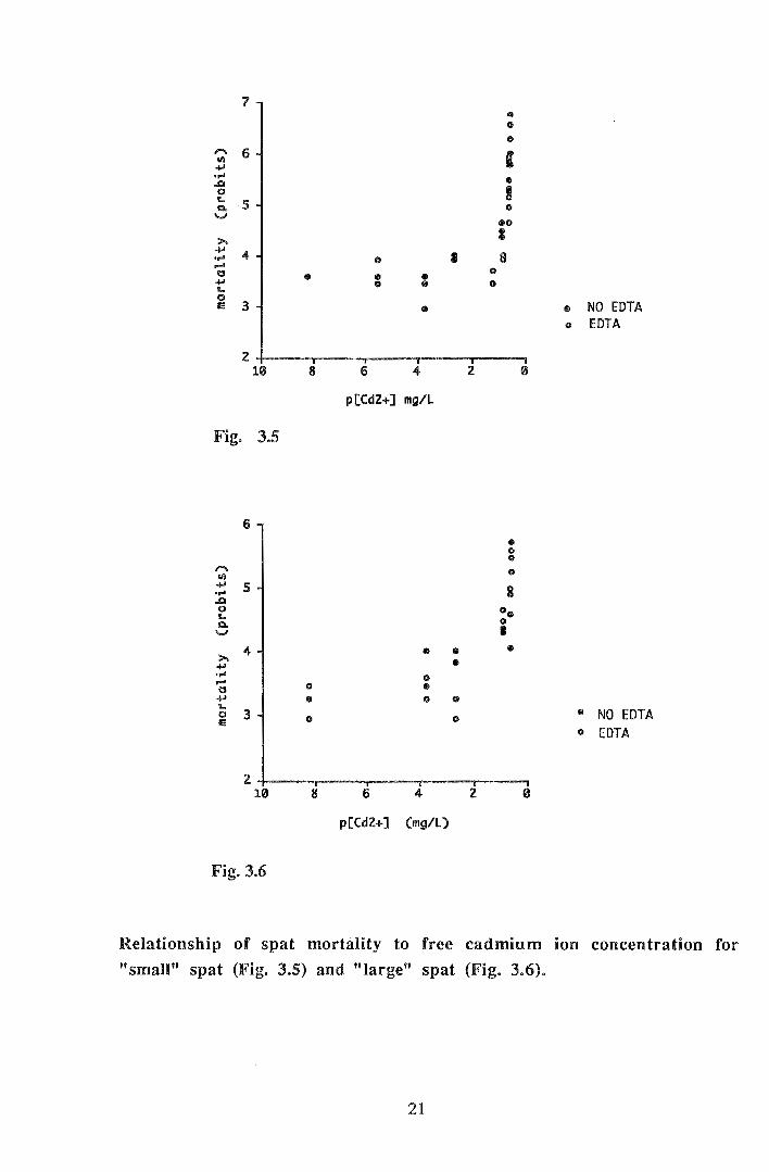
Figures 3.5 and 3.6 show the relationship between free cadmium ion concentration
(Cd2+) and mortality (in probits). Both sizes of spat show a marked relationship between
mortality and free cadmium ion concentration irrespective of the presence or absence of
EDTA. Spat appeared to tolerate cadmium free ion concentrations up to a threshold level
(approximate pCd2+::::: 0.8 for both sizes); at this concentration a rapid increase in
mortality occurs. When LCsos were then compared in terms of free ion concentration
they were found to be virtually the same irrespective of the presence or absence of EDT A
ie. 0.25 and 0.27 mgL-1 for "small" and "large" spat respectively.
20
7 • 0
0 ,.... 6 I eli ..... ..... " ..1:1
0 ! L Q, 5 0 '-' eo
>. I .....
4 I 8 ..... 0 ..... 0 a 0 • ..... 0 • 0
L 0 IE 3 • • NO EDTA
0 EDTA
2 10 8 6 4 2 0
p[Cd2+] mg/L
Fig. 3.5
6
• 0 0 ,.... 0 eli ..... 5 8 ....
..D 0 o., L a. 0
'-' I
4 • .. ill >. Ill ..... .....
0 ..... 0 • a ..... • 0 0
L • NO EDTA 0 3 IE (I 0
0 EDTA
z 10 8 6 4 2
p[Cd2+] (mg/L)
Fig. 3.6
Relationship of spat mortality to free cadmium ion concentration for
"small" spat (Fig. 3.5) and "large" spat (Fig. 3.6).
21

3.4 DISCUSSION
Results of the range assay provided preliminary information on the effects and toxicity of
cadmium to C. gigas spat. These results indicated an approximate 96 hour LCso of 10
mgL-1 (total cadmium) for spat. Both range assay and definitive assay results indicated an
adverse effect of exposure to cadmium on the behaviour and physiology of spat Ninety
six hour exposure to as low as 2 mgL-1 cadmium resulted in impaired behavioural
responses to light and tactile stimuli. General physiology was also affected with erratic
heart contractions, persistent valve gaping and increased mortality. The effects of chronic
cadmium exposure on spat physiology will be discussed in detail in Section 4. The
cumulative effect of cadmium on physiology (eg. Ringwood, 1989) was illustrated by a
dramatic increase in mortality between 48 and 96 hours compared to mortality between 0
and 48 hours.
Previously used ctiteria for mortality of bivalves may have overestimated mortality rate.
For example, Watling (1978) assessed mortality of C. gigas as the number of oysters
with gaping shells. The results presented here showed that following exposure to
cadmium, live spat frequently showed persistent gaping of valves. Use of several criteria
was necessary for precise measurement of spat mortality. A longer exposure period than
the 96 hour criterion used here is recommended for more accurate determination of
cadmium toxicity to C. gigas spat.
The difference in the toxicity of cadmium to the two spat sizes was surprisingly large.
However, greater tolerance of larger spat compared with smaller spat to cadmium
exposure is consistent with the findings of Watling (1978) who found greater tolerance of
64 day C. gigas spat (8.3 mm) than that of three month spat (4.1 mm). Conversely,
Sunila (1981 in McLusky et al., 1986) found greater resistance of small mussels (Mytilus
edulis) to cadmium than larger ones. Vadation in spat sensitivity due to size may be due
to a combination of factors. Uptake of cadmium by adult C. gigas is inversely related to
body size (Boyden, 1977); thus, the smaller spat were likely to have been subjected to
higher tissue concentrations of cadmium. The faster metabolic rate rate of smaller spat
and greater relative surface area would cause faster accumulation and therefore sensitivity
to cadmium compared to larger spat. Previous metal exposure history of spat may also
account for size-specific differences in sensitivity to cadmium. Exposure of bivalves to
heavy metals causes induction of specific metal-binding proteins such as metallothioneins
(Ringwood, 1992). Older spat are therefore more likely to possess higher levels of these
proteins which allow detoxification of accumulated metal.
22
Ninety-six hour LCsos in terms of total cadmium were 10.29 and 28.43 mgL-1 for
"small" and "large" spat respectively. In tenns of free cadmium ion concentration LCsos
were 0.25 and 0.27 mgL-1 respectively. These values are comparable to, although a little
higher than previously published LCso values for bivalves. The LCso for C. virginica
embryos is reported to be 3.8 mgL-1 total cadmium (48 hour LCso) (Calabrese et al.,
1973) while that for the adult mussel Mytilus edulis is 25 mgL-1 (Eisler, 1971) or 1.62
mgL-1 forM. e. planulatus (Ahsanullah, 1976). Watling (1978) estimated a 96 hour
LC50 for 3 month C. gigas spat of 2 mgL-1; however, her results were intended only to
illustrate relative tolerances of different life-stages and were not a precise measurement of
acute toxicity. These values are not strictly comparable due to differences in experimental
conditions, life-stages and species. Surprisingly, no short-term cadmium LCso value for
adult C. gigas could be found in the available literature.
Previously, acute toxicity of cadmium to bivalves has been assessed in terms of the total
cadmium exposure concentration (eg. Watling, 1978; Ringwood, 1990). However, when
percent mortality was related to the free cadmium ion exposure concentration they were
found to be closely related. EDTA reduced mortality of both sizes of spat, by a reduction
in the free cadmium ion concentration to which spat were exposed at a set total cadmium
concentration. Indeed, a distinct threshold level of free ion (pCd2+=0.8) above which
mortality dramatically increased was evident for both sizes of spat irrespective of the
presence or absence of EDTA. In addition, when the calculated LCsos for both sizes of
spat were expressed in terms of the corresponding free ion concentrations they were
found to be virtually the same. Thus, as the free ion concentration is closely related to the
total concentration of cadmium, measurements of acute toxicity in terms of total cadmium
are misleading by masking the effects of free ions on an organism. This clear relationship
between spat mortality and free cadmium ion concentration has important implications for
both commercial and environmental situations. It is of particular relevance under
conditions which promote high levels of free ion (eg. low salinity) despite levels of total
cadmium below recommended limits.
23

4.. EFFECTS OF SUBLETHAL CADMIUM EXPOSURE
4.1 INTRODUCTION
Studies concerning the effects of chronic exposure to sublethal levels of heavy metals
provide more realistic inf01mation on their biological impact in the environment and on
commercial operations than studies of acute toxicity. Levels of cadmium in natural
waterways range from trace levels in oceanic waters of approximately 0.05JlgL-1 (Riley,
1971) to levels as high as 20-30 JlgL-1 in the King River which flows into Macquruie
Harbour, Tasmania (Tasmanian Department of Environment, 1987). The Tasmanian
recommended limit for cadmium in marine water is 5 JlgL-1 (Tasmanian Department of
Environment, 1986). The USEP A ( 1986) chronic criterion for marine waters is 9.3 JlgL-
1 total cadmium. That is, the average concentration of total cadmium should not exceed
9.3 JlgL-1 over a 4 day peliod more than once in three years. By their own admission, the
USEPA (1986) suggest that the criteria for cadmium may be underprotective for sensitive
species and at low salinities. Free cadmium ion concentrations in the environment vary
according to the physico-chemical conditions, particularly pH, salinity, humic matelials
and total metal concentration (Mantoura et al., 1978; Sunda et al., 1978). Values for free
cadmium ion concentration present in the environment are several orders of magnitude
less than the total cadmium concentration. For example, Jackson & Morgan (1978)
reported a typical seawater free ion concentration of 9.8 x lQ-4 JlgL-1 (pCd2+=3.0)
corresponding to a total cadmium concentration of9.6 x lQ-2 JlgL-1.
Reduced growth and shell abnormalities (eg. Sunila & Lindstrom, 1985) have been
reported following acute exposure of adult bivalves to heavy metals; however, little is
known of the effects of chronic exposure to metals on the juvenile oyster. Chronic
exposure to sublethal doses of cadmium may exert a range of subtle effects on the
physiology of juvenile bivalves which in turn affect their growth and survival. For
example, acute exposure of C. gigas spat to lethal doses of cadmium (see Section 3.3)
appeared to cause erratic heart contraction, reduced algal clearance rates and impaired
behavioural responses. It is possible that long-term exposure to sublethal concentrations
of heavy metals may have a chronic effect on these physiological processes which in turn
affect growth and survival of spat in the environment. Chronic metal exposure may also
be responsible for the shell growth abnormality termed "curly back" observed in the
commercial oyster industry (Section 1).
24

The following hypotheses were investigated:
1. that chronic exposure to cadmium causes growth abnormalities in juvenile C.
gigas.
2. that chronic exposure to cadmium produces a range of adverse, subtle effects
on the physiology of juvenile C. gigas.
4.2 MATERIALS AND METHODS
4. 2. 1 Chronic exposure
Spat of a commercial sizing of 1500 J-Lm (grading mesh size) were acclimated over 96
hours to experimental conditions of temperature and salinity. At the commencement of the
experiment 50 spat were sized and placed in treatment vessels containing 0, 10, 50 and
250 J-LgL-1 total cadmium, in tliplicate. The lowest dose was chosen to correspond to the
USEPA (1986) chronic criterion for marine waters. Each vessel contained microalgae as
20 x 104 cells mL-1 Nannochloris atomus and 10 x 1Q4 cells mL-lfsochrysis sp. (T
iso). Spat were exposed to treatment solutions for a total of 32 days. Solutions and
microalgae were replaced every 48 hours and dissolved oxygen, salinity and pH
measured at each water change. Temperature was monitored daily.
Every four days spat were gently rinsed to remove faeces and other organic matter then
each measured for size. Any dead spat were recorded and removed from the treatment
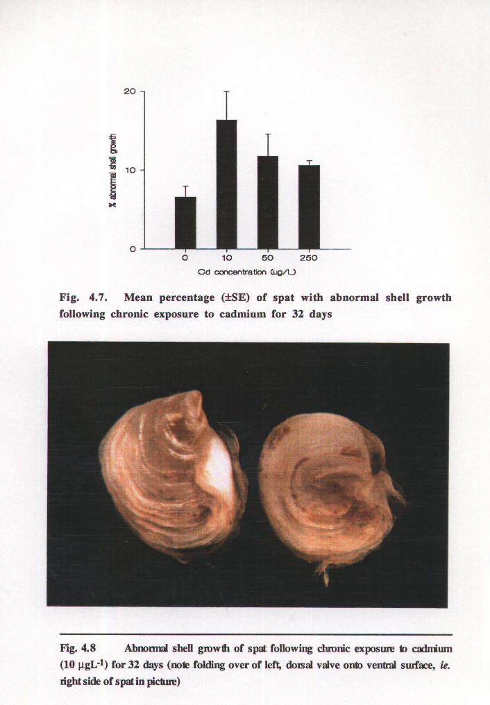
vessel. On day 32 spat were measured for length and the percent with abnormal shell
growth in each replicate determined. This was classed by an abnormal folding of the shell
margin onto the ventral valve surface as seen in Figure 4.8. Spat were then transferred to
untreated salt water for measurements of physiological effects following chronic
cadmium exposure. Repeated measures analysis of variance (ANOV A) and Tukey
Kramer HSD comparison of the regression slopes of size over time for each exposure
concentration were used to assess the effect of cadmium on length. The computer
program, Systat (1992), was used for all statistical analyses.
4.2.2 Physiological effects
Unless otherwise indicated, all results were analysed using ANOVA and, where the null
hypothesis was rejected, Tukey-Kramer HSD pairwise comparison of means.
25
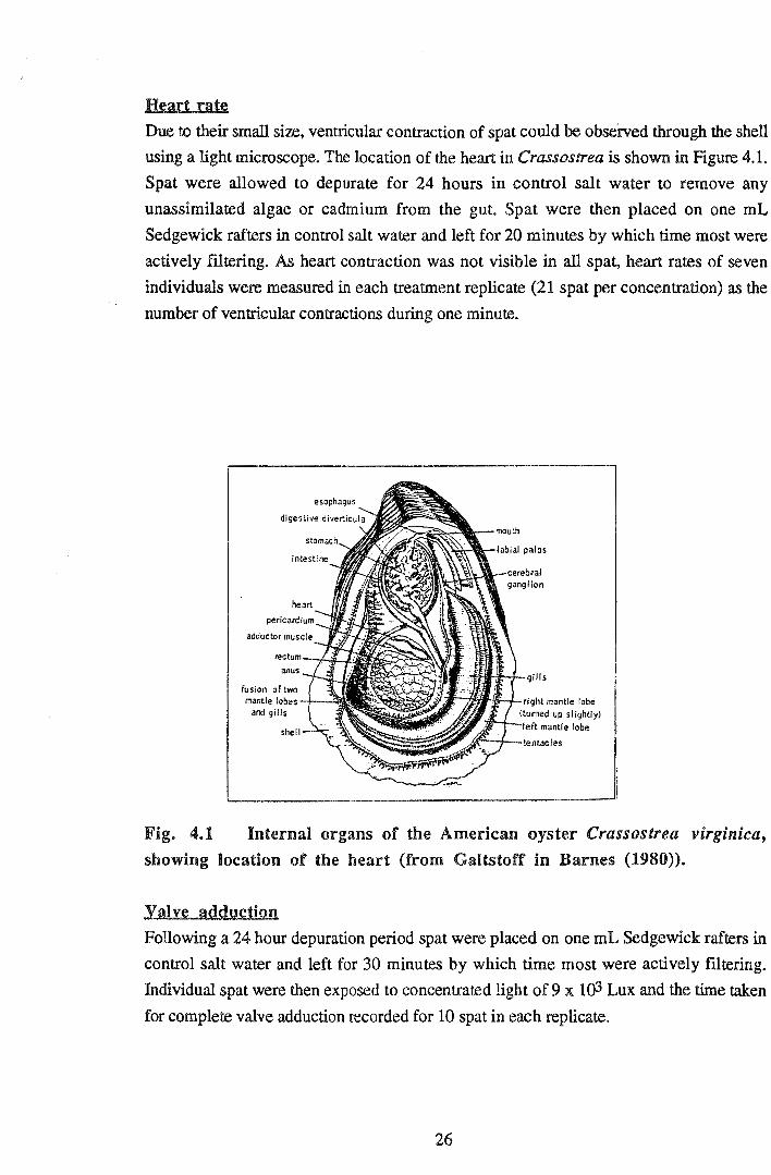
Heart rat~
Due to their small si:z.e, ventricular contraction of spat could be observed through the shell
using a light microscope. The location of the heart in Crassostrea is shown in Figure 4.1.
Spat were allowed to depurate for 24 hours in control salt water to remove any
unassimilated algae or cadmium from the gut. Spat were then placed on one mL
Sedgewick rafters in control salt water and left for 20 minutes by which time most were
actively flltering. As heart contraction was not visible in all spat, heart rates of seven
individuals were measured in each treatment replicate (21 spat per concentration) as the
number of ventricular contractions during one minute.
't--fRf-ij;.;_J._right mantle lobe (turned up slightly)
ft mantle lobe
Fig. 4.1 Internal organs of the American oyster Crassostrea virgmzca,
showing location of the heart (from Galtstoff in Barnes (1980)).
Yalve adduction
Following a 24 hour depuration period spat were placed on one mL Sedgewick rafters in
control salt water and left for 30 minutes by which time most were actively filtering.
Individual spat were then exposed to concentrated light of9 :x 103 Lux and the time taken
for complete valve adduction recorded for 10 spat in each replicate.
26
Feed rate
Feed rates were measured as rate of microalgal clearance. Prior to the experiment spat
were allowed to depurate for 48 hours in control salt water. The microalgal species
lsochrysis sp. (T-iso) was then added to each of the 12 culture vessels to achieve an
initial algal concentration of 10 x 1 ()4 cells mL-1. Three counts of algal concentration were
then measured for each vessel at regular intervals over 24 hours using a Neubauer
haemocytometer. Gentle aeration maintained the algal cells in suspension throughout the
experiment. Temperature was maintained at 20 ± 0.5 OC. Results were analysed using
ANOV A for compadson of regression slopes of cell concentration over time.
Bioaccumulation
Following physiological tests spat soft tissue was dissected from the shells using surgical
stainless steel forceps. Soft tissue and shells were rinsed in deionised water and dried for
96 hours at 60°C. Spat dry tissue was weighed on a microbalance in groups of ten spat
(five groups per replicate). Following nitric acid digestion of soft tissue, bioaccumulation
of cadmium by spat from each replicate was measured using graphite furnace AAS
(analysis courtesy of Department of Environment). A comparable weight of standard
oyster tissue (US National Bureau of Standards (NBS) #1566) was taken through the
digestion procedure and analysed for cadmium content using AAS, as a calibration check.
Due to difficulties in the acid digestion step shell was not analysed for cadmium content.
However, accumulation of cadmium by the shell of bivalves is generally very low
compared to soft tissue accumulation (eg. see Ringwood, 1991). The tissue concentration
factor for spat over 32 days was determined from the ratio of tissue cadmium
concentration to exposure concentration of cadmium.
4.3 RESULTS
Throughout the exposure period pH ranged between 7.8-8.3, dissolved oxygen 5.7-6.4
mgL-1, salinity 35 %o and temperature 20 ± 0.5 °C. Spat mortality was low throughout
the experiment with a mean mortality of 3.3%, 1.3%, 2.6% and 11.3% at 0, 10, 50 and
250 ~gL-1 cadmium respectively. ANOV A indicated no significant effect of cadmium
concentration on spat mortality (p=0.278).
27

4.3.1 Chronic exposure
Growth
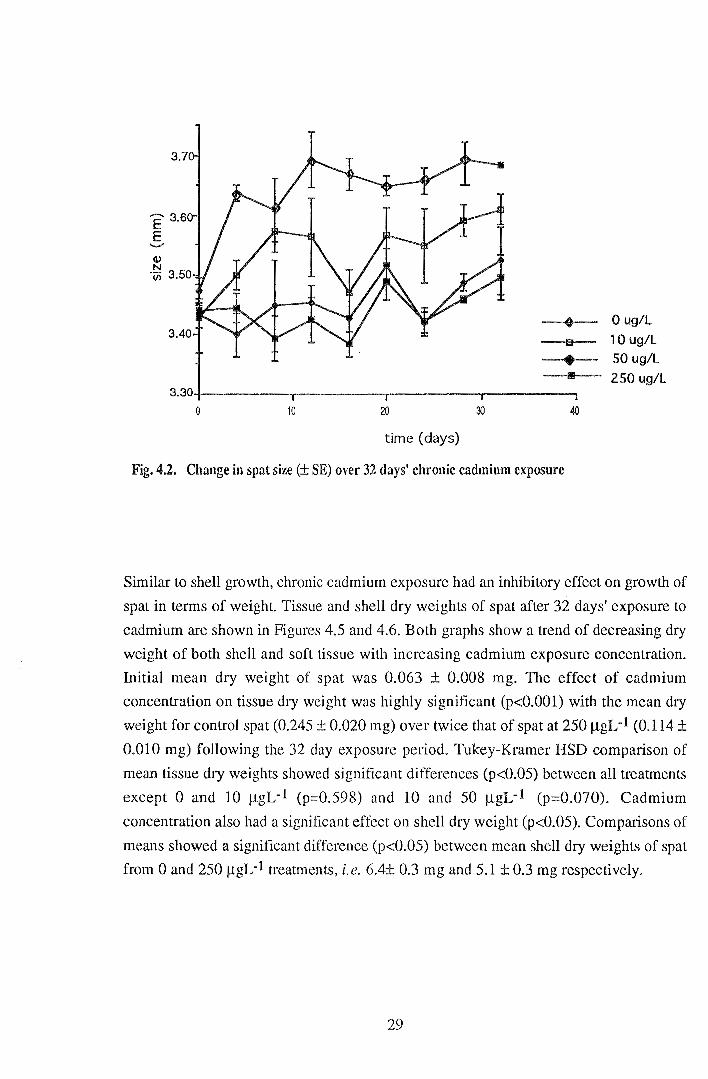
Figure 4.2 shows the sizes of spat over the 32 day exposure period. Growth curves
indicate that growth of spat exposed to 50 and 250 J..LgL-1 cadmium was limited over the
32 day exposure period relative to growth of controls. Over 32 days spat grew by 192
and 180 J.Lm at 0 and 10 J..LgL-1 respectively compared to only 89 and 56 J.Lm at
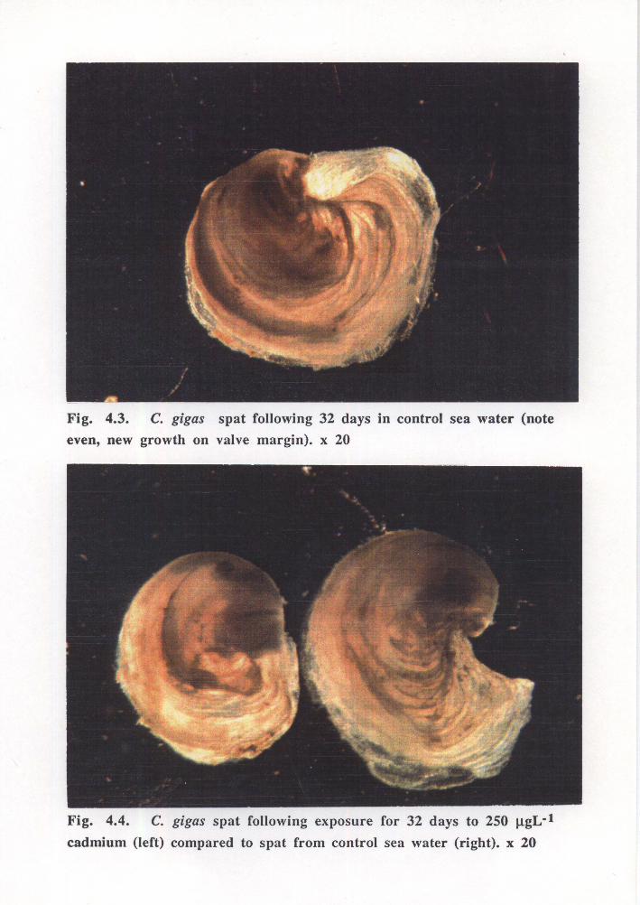
concentrations of 50 and 250 J.LgL-1 respectively. Differences between shell growth of
control spat and spat from the highest cadmium concentration were visible by day 32
(Figures 4.3 and 4.4). From day four it was noted that the limited shell growth of spat
exposed to cadmium was weak and fragmented, therefore susceptible to breakage; this
may account for the fluctuations in size over time of spat exposed to cadmium. Spat at 0
J.LgL~l grew rapidly to day 12 followed by a levelling off of growth rate to day 32.
Control spat were observed to be active with even new growth and rapidly cleared added
microalgae compared to spat exposed to cadmium. Activity of spat exposed to 10 J.LgL-1
appeared similar to that of controls over the 32 days with even new growth to
approximately day 8. However, an increase in abnormal shell growth was apparent after
further exposure (see results for "shell abnormalities"). At higher sublethal doses of
cadmium (50 and 250 J.LgL-1) spat appeared to be affected from as early as day four.
Observations were noted of pale soft tissue, localised darkening of the tentacles, weak,
fragmented shell growth and less faecal production when compared to control spat.
Interestingly, after 24 days' exposure spat exposed to 50 J.Lg cadmium/L appeared to
develop unfragmented, even new shell growth. At 250 J..LgL-1 activity of spat appeared
suppressed compared to that of controls. This was particularly obvious around day 28
with many spat showing persistent retraction of the mantle into the shell. Gut and soft
tissue were pale in colour compared to spat at lower doses of cadmium. Repeated
measures ANOV A indicated a significant difference in spat size with cadmium
concentration (p<0.01) and over time (p<0.001). A significant time*concentration
interaction (p<0.001) was also found ie. differences between the profiles of size over
time (growth rates) at each exposure concentration. Comparison of regression slopes for
growth at each cadmium concentration showed a significant difference between growth
rate between 0 and 250 J.LgL-1 (p<0.05) and between 10 and 250 J.LgL-1 (p<0.05).
28
E' 3.e
E ...._,.
3.30
0 10 20
time (days)
~-
8
• 1111
30 40
Fig. 4.2. Change in spat size (± SE) over 32 days' chronic cadmium exposure
0 ug/L
10 ug/L
50 ug/L
250 ug/L
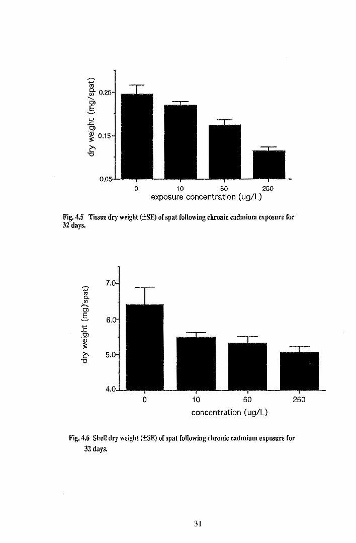
Similar to shell growth, chronic cadmium exposure had an inhibitory effect on growth of
spat in terms of weight. Tissue and shell dry weights of spat after 32 days' exposure to
cadmium are shown in Figures 4.5 and 4.6. Both graphs show a trend of decreasing dry
weight of both shell and soft tissue with increasing cadmium exposure concentration.
Initial mean dry weight of spat was 0.063 ± 0.008 mg. The effect of cadmium
concentration on tissue dry weight was highly significant (p<O.OOl) with the mean dry
weight for control spat (0.245 ± 0.020 mg) over twice that of spat at 250 JlgL-1 (0.114 ±
0.010 mg) following the 32 day exposure period. Tukey-Kramer HSD comparison of
mean tissue dry weights showed significant differences (p<0.05) between all treatments
except 0 and 10 !lgL-1 (p=0.598) and 10 and 50 JlgL-1 (p=0.070). Cadmium
concentration also had a significant effect on shell dry weight (p<0.05). Comparisons of
means showed a significant difference (p<0.05) between mean shell dry weights of spat
from 0 and 250 JlgL-1 treatments, i.e. 6.4± 0.3 mg and 5.1 ± 0.3 mg respectively.
29
.... ..c C)
·~ 0.15
~ "0
0 10 50 250 exposure concentration ( ug/L)
Fig. 4.5 Tissue dry weight (±SE) of spat following chronic cadmium exposure for 32 days.
......... +-' co 0.. VI
.>-... C)
E 6 .......-
.... ..c C) ·w 3: ~ 5
"0
0 10 50 250
concentration ( ug/L)
Fig. 4.6 SheU dry weight (±SE) of spat following chronic cadmium exposure for 32 days.
31
Shell abnormalities
Discrete differences in shell growth were observed within and between treatments (see
Fig.s 4.7 & 4.8) allowing accurate determination of the percentage of spat with abnormal·
shell growth. The mean percentage of spat with shell abnormalities was increased in all
treatments following chronic exposure to cadmium when compared to control spat. At the
lowest cadmium concentration of 10 J..LgL-1 a notably greater proportion of spat with
abnormal shell growth was evident compared to spat at the higher sublethal doses of 50
and 250 J.LgL -1. Despite the increase in spat shell abnormalities following cadmium
exposure the effect of cadmium concentration was not significant (p=0.119).
32
4.3.2 Physiological effects
H~art rate
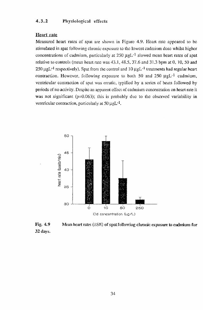
Measured heart rates of spat are shown in Figure 4.9. Heart rate appeared to be
stimulated in spat following chronic exposure to the lowest cadmium dose whilst higher
concentrations of cadmium, particularly at 250 f.lgL-1 slowed mean heart rates of spat
relative to controls (mean heart rate was 43.1, 48.5, 37.6 and 31.3 bpm at 0, 10, 50 and
250 f.lgL-1 respectively). Spat from the control and 10 f.lgL-1 treatments had regular heart
contraction. However, following exposure to both 50 and 250 f.lgL-1 cadmium,
ventricular contraction of spat was erratic, typified by a series of beats followed by
periods of no activity. Despite an apparent effect of cadmium concentration on heart rate it
was not significant (p=0.063); this is probably due to the observed variability in
ventricular contraction, particularly at 50 f.lgL-1.
Fig. 4.9
32 days.
"2 .E 'd3 cr; <D
8 <D
"§ ....... ro Q)
_c
45
40
35
0 10 50 250
Cd concentration (ug/U
l\1ean heart rntes (±SE) of spat following chronic exposure to cadmiwn for
34
valve adduction
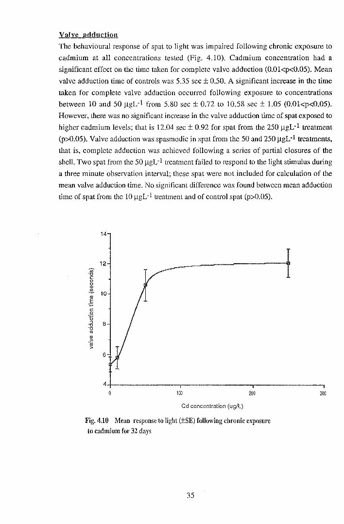
The behavioural response of spat to light was impaired following chronic exposure to
cadmium at all concentrations tested (Fig. 4.10). Cadmium concentration had a
significant effect on the time taken for complete valve adduction (0.01<p<0.05). Mean
valve adduction time of controls was 5.35 sec ± 0.50. A significant increase in the time
taken for complete valve adduction occurred following exposure to concentrations
between 10 and 50 JlgL-1 from 5.80 sec ± 0.72 to 10.58 sec ± 1.05 (0.0l<p<0.05).
However, there was no significant increase in the valve adduction time of spat exposed to
higher cadmium levels; that is 12.04 sec± 0.92 for spat from the 250 JlgL-1 treatment
(p>0.05). Valve adduction was spasmodic in spat from the 50 and 250 JlgL-1 treatments,
that is, complete adduction was achieved following a series of partial closures of the
shell. Two spat from the 50 j...lgL-1 treatment failed to respond to the light stimulus during
a three minute observation interval; these spat were not included for calculation of the
mean valve adduction time. No significant difference was found between mean adduction
time of spat from the 10 jlgL-1 treatment and of control spat (p>0.05).
12 en -o c: 0 0 (I)
~ (I)
10 E ·.;::; c: 0 gu :::s 8 -o -o IU (I)
> Iii >
6
0 100 200
Cd concentration (ug/L)
Fig. 4.10 Mean response to light (±SE) following chronic exposure to cadmium for 32 days
35
300
F~~d rate
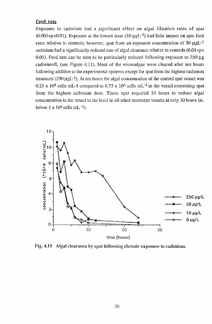
Exposure to cadmium had a significant effect on algal filtration rates of spat
(0.001<p<0.01). Exposure at the lowest dose (10 JlgL-1) had little impact on spat feed
rates relative to controls; however, spat from an exposure concentration of 50 J..LgL-1
cadmium had a significantly reduced rate of algal clearance relative to controls (O.Ol<p<
0.05). Feed rate can be seen to be particularly reduced following exposure to 250 J..Lg
cadmium/L (see Figure 4.11 ). Most of the microalgae were cleared after ten hours
following addition to the experimental systems except for spat from the highest cadmium
treatment (250 JlgL-1). At ten hours the algal concentration of the control spat vessel was
0.25 x 10⁴ cells mL-1 compared to 6.75 x 10⁴ cells mL-1 in the vessel containing spat
from the highest cadmium dose. These spat required 24 hours to reduce algal
concentration in the vessel to the level in all other treatment vessels at only 10 hours (ie.
below 1 x 10⁴ cells mL -1).
-....1
E1 ....... Cll
a; <J
v < 0 .... "" ........
t: 0 .... «< .... ..... c:: Q) <J t: 0 <J
ol---~~~~~~~~~ 0 10 20
time (hours)
30
1111
250 pg/L
50 pg/L
10 pg/L
a 0 pg/L
Fig. 4.11 Algal clearance by spat following chronic exposure to cadmium.
36

Accumulation
Analysis of standard oyster tissue for cadmium concentration confirmed the precision of
the tissue digestion procedure and AAS analytical techniques. Measured tissue cadmium
concentration was 4.2J.LgL-l compared to the NBS certified value (with 95% confidence
limits) of 3.5J.LgL-1 ± 0.4. Some variation of determined levels of cadmium occurred due
to the low weight of oyster tissue; that is, results were susceptible to trace levels of
contamination. Figure 4.12 shows the level of accumulation of cadmium in spat soft
tissue after exposure to cadmium for 32 days followed by a three day depuration period.
A highly significant linear relationship was found between tissue cadmium concentration
and exposure concentration (p<0.001). Spat accumulated 30 J.Lgg-1 ± 4.0 after only 32
days at the lowest exposure concentration (10 J.!gL-1). The concentration factor of spat at
each exposure concentration is shown in Table 4.1 with published values for adult
oysters at corresponding exposure concentrations. Bioconcentration factors of C. gigas
spat were similar to those of adult bivalves, particularly at the lowest exposure
concentration.
500,
~ 40 Cl 2.. c: 0 -~ c (]) u c: 8
30
~ 20 (/1
·~
10
0
y = 2.6689 + 1.4559x R112 = 0.894
100 200
exposure concentration (ug!l)
Fig. 4.12. Soft tissue accumulation of cadmium following chronic
exposure for 32 days.
37
300
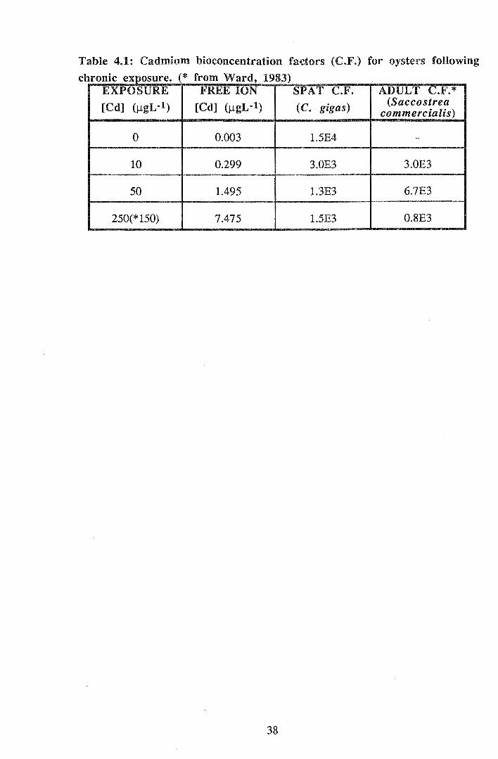
Table 4.1: Cadmium bioconcentration factors (C.F.) for oysters following
chronic exposure. (* from Ward, 1983) EXPOSURE FREE ION SPAT C.F. ADULT C.F.*
[Cd] (J..tgL·l) [Cd] (J.lgL·l) (C. gigas) (Saccostrea commercialis)
0 0.003 1.5E4 -
10 0.299 3.0E3 3.0E3
50 1.495 1.3E3 6.7E3
250(*150) 7.475 1.5E3 0.8E3
38

4.4 DISCUSSION
The mean mortality of control spat of 3.3 % over the 32 day exposure period is well
below the APHA (1989) recommended maximum for toxicity tests of 10 %. The low
overall mortality indicates that experimental conditions were suitable for maintenance of
spat. Cadmium had no significant effect on mortality at the selected concentrations over
the 32 day exposure period. It should be noted that growth rates were found to be slower
than in previous studies. For example, Shuster and Pringle (1969) reported growth of
control oysters (C. virginica) of 300 !lm over a four week period compared to 192 J..Lm in
the present study. Slow shell growth of control spat was particularly obvious after day 12
following rapid initial growth. In any population of spat there are both fast and slow
growing individuals. Spat available for these experiments were from healthy nursery
graded stock known to have slow growth rates. In addition, these growth rates are not
directly comparable to other studies where different species, ages and developmental
stages of oyster are used. By day 32 it was evident that new growth of cadmium treated
spat in these experiments had occurred predominantly on the left, anterior valve margin
(see Fig. 4.8). Thus, the generally accepted size criterion for spat (length) did not account
for much of the new shell growth. In future studies involving exposure to contaminants
spat size should be measured in terms of shell width as well as length. The reduction in
control growth rates after day 12 may have been due to an increased energy requirement
due to greater size of spat. In these experiments algal feed rates were kept constant
throughout the exposure period, rather than increased, for experimental purposes and
may have limited growth of larger spat. Crowding of spat may have also limited growth
rates. Fmiher studies are required in order to determine the factors responsible for slow
growth rates and optimal spat husbandry conditions for laboratory experiments.
Despite slow growth rates of control and treated spat, a significant effect of chronic
cadmium exposure on growth was found. Over the 32 day exposure period cadmium
significantly inhibited both shell growth (by length and weight) and soft tissue growth
(by weight). The inhibitory effect of cadmium on spat growth (in terms of both length
and weakness of shell) was more severe at higher concentrations of cadmium. This is
consistent with the findings of Watling (1983) who reported reduced growth rates of C.
gigas with increasing heavy metal exposure concentration. Even at 10 J..LgL-1
(approximately equal to the USEPA (1986) chronic criterion) spat grew more slowly than
controls, with a greater percentage having abnormal shell growth. This has important
implications for both environmental and commercial situations where spat may be
exposed to such a level of cadmium (equivalent to a free cadmium ion concentration of
0.003 !-lgL-1) particularly under conditions oflow salinity.
39
Carbonic anhydrase and alkaline phosphatase are two enzymes believed to be involved in
shell deposition in molluscs (Wagge, 1951; Wilbur & Saleuddin, 1983). The activity of
alkaline phosphatase increases in C. gigas between 50 and 150 days post-settlement
(George & Coombs, 1975). Spat used throughout the present experiments were aged
between approximately 150 and 300 days. Thus, cadmium exposure may be inhibiting
shell growth of spat by blocking the active site of metalloenzymes such as alkaline
phosphatase involved in shell deposition. In addition, cadmium may be interfering with
the normal increase in metalloenzyme activity of young spat. Indeed, Evtushenko et al.
(1986) found inhibition of alkaline phosphatase (as well as other metalloenzymes) in
adult scallops (Mizuhopecten yessoensis) following exposure to 0.5 mgL-1 cadmium for
60 days. Ringwood (1992) postulated that acute exposure to 20 J.tgL-1 copper for 24
hours interfered with zinc metallothionein synthesis in oyster embryos. She suggested
that this affected activation of alkaline phosphatase and carbonic anhydrase thus blocking
deposition of the larval shell. A similar mechanism may be involved in the observed
effect of chronic cadmium exposure on C. gigas spat. The observed change in shell
growth following 24 days exposure to 50 J.tgL-1 cadmium may be due to induction of
cadmium metallothioneins; that is, spat developed the ability to detoxify accumulated
cadmium. Chronic cadmium exposure also affects calcium homeostasis in bivalves
(Regoli et al., 1991); the concentration of calcium in the extrapallial fluid of molluscs is
highly important to the amount and composition of deposited shell (Wilbur & Saleuddin,
1983).
Shell abnormalities have been rep01ted in bivalve embryos (Ringwood, 1992) and adults
(Sunila & Lindstrom, 1985) following exposure to heavy metals. The effect of cadmium
on shell shape is probably mediated via a similar mechanism to the effect on shell growth
(eg. by blockage of metalloenzyme active sites). Seed (1968) found that mussels (M.
edulis) may grow more along the shell midline when exposed to adverse environmental
conditions. I observed a similar effect on oyster spat exposed to cadmium. Seed
proposed that this is due to retraction of the mantle thus exposing the free mantle edge to
the shell midline resulting in localised shell secretion. The higher proportion of spat with
shell abnormalities at the lowest cadmium concentration (10 J.tgL-1) than at 50 and 250
J.tgL-1 was simply due to the very limited shell growth of spat at the higher
concentrations. In order to determine if cadmium has a significant effect on shell
abnormalities in spat, further experiments are required to investigate the effect of even
lower concentrations of cadmium for a longer exposure period. The growth abnormality
observed was similar (but more severe) to the condition referred to as "curly back" in
commercial spat. "Curly back" is characterised by a more gradual growth of shell (in
particular the posterior valve margins) in the direction of the ventral valve. No
intermediate forms of the abnormal growth condition were apparent in this study.
40

Cadmium exposure caused pale tissue colour and localised darkening of spat tentacles.
Pale tissue colour of oysters was also reported by Shuster and Pringle (1969) following
chronic exposure to cadmium. However, localised darkening of the tentacles does not
appear to have been previously noted in bivalves following exposure to heavy metals.
These dark patches may con·espond to cadmium detoxification sites (eg. amoebocyte or
metallothionein-bound cadmium) in C. gigas spat. Thomson et al. (1985) reported
localisation of zinc, calcium and iron (note that no analysis for cadmium was made by
these authors) in membrane-limited vesicles at the "outer edge of the mantle". Further
analysis of the tentacles following metal exposure is required before a valid explanation
for these dark patches can be proposed.
Measurement of heart rate in adult bivalves generally involves removal of part of a valve
for either direct observation of the heart (eg. Scott & Major, 1972) or for insertion of
electrodes that are sensitive to heart contraction (Grace & Gainey, 1987; Grace &
Kenyon, 1990). Such procedures are likely to cause some stress to the animal thereby
influencing heart rate. Due to the thin shell, I was able to measure heart rates of oyster
spat with minimal disturbance by direct observation using a light microscope. Heart rates
of the juvenile oysters were higher than those reported for other adult bivalves. For
example, control spat had a mean heart rate of 43 bpm (beats per minute) compared with
that of adult C. gigas of 21 bpm (20-25 OC) (Uesaka et al., 1987) and adult M. edulis of
24-26 bpm (17 "C) (Helm & Trueman, 1967). Heart rates of invertebrates are generally
faster for smaller individuals (Vernberg & Vernberg, 1972). Chronic exposure of spat to
sublethal cadmium concentrations appeared to cause bradycardia and erratic (arrhythmic)
heart contraction. Similarly, short-tenn exposure of adult M. edulis. to copper causes a
reduction in heart rate and "burst activity" (irregular heart contraction) (Scott & Major,
1972; Grace & Gainey, 1987; Gainey & Kenyon, 1990). At the lowest cadmium
concentration mean heart rate appeared to be slightly higher than that of controls. A lack
of studies on the effect of chronic metal exposure on heart rate in bivalves is apparent.
Reduction of heart rate in response to metal exposure has previously been attributed to
"avoidance behaviour" ie. valve closure which reduces heart rate and serves to reduce
metal uptake by both avoiding the contaminated medium and reducing circulation
(Davenport & Redpath, 1984; Gainey & Kenyon, 1990). Metal exposure may also inhibit
cardia-regulatory chemicals such as adenylate cyclase (Gainey & Kenyon, 1990). Heart
rate was measured in control water following chronic exposure to cadmium rather than in
metal-contaminated water. Thus, the apparent effect of exposure to cadmium on heart
contraction of spat was probably due to a more chronic physiological effect rather than a
direct response to the metal (eg. valve closure).
41
Davenport & Manley (1978) found that following exposure to 20 )lgL-1 copper for 10
days M. edulis exhibited a reduced behavioural response to further copper exposure.
They described this effect as "acclimation" to raised levels of copper. It is more likely that
this represented an impaired physiological response of spat rather than acclimation to
conditions. Our results showed an impaired behavioural response of spat to light
following chronic cadmium exposure. Cadmium appeared to have a distinct impact on
either the photoreceptive detection or response mechanism of spat with no further effect
following exposure to greater than 50 )lgL-1. The reduced behavioural response may be
due to the influence of cadmium on calcium homeostasis in bivalves (Regoli et al., 1991)
which would intetfere with adduction of the shelL Intracellular free calcium concentration
is important in excitation and contraction of the adductor muscle in bivalves (Muneoka &
Twarog, 1983). Alternatively, cadmium may interfere with the photoreceptive pathway of
C. gigas spat; however, the former explanation is more plausible as spat were detecting
the light stimulus. The mechanism by which oysters may be able to detect light is unclear.
It has previously been suggested that the mantle tentacles of oysters may be able to detect
light (Yonge, 1960). In addition, Paparo (1987) discusses photoreception in C. virginica
by neuronal pigments. Further research is required for an understanding of the
photoreceptive pathway of C. gigas .
Chronic exposure to cadmium had a dose-dependent inhibitory effect on spat feed rates,
with a significant effect on algal clearance (relative to controls) following exposure to
only 50 )lgL-1 for 32 days. Similarly, exposure to 1200 )..LgL-1 cadmium for 96 hours
reduced the filtration rate of clams (Anadara granosa) by at least fifty percent (Patel &
Anthony, 1991). It should be noted here that algal feeding rates of bivalves are closely
related but not strictly comparable to filtration rates. For example, the presence of algae
stimulates filtration by M. edulis (Manley, 1983). There are few studies concerning the
effects of heavy metals on bivalve feed rates. Inhibition of filtration by bivalves in
response to heavy metal exposure may be due to either a behavioural response to the
external metal (eg. valve adduction) which reduces filtration, or a physiological effect
(eg. on gill structure or ciliary activity). In the presence of only 2 )lgL-1 copper, valve
movements and the activity of both the inhalant and exhalant siphons of M. edulis are
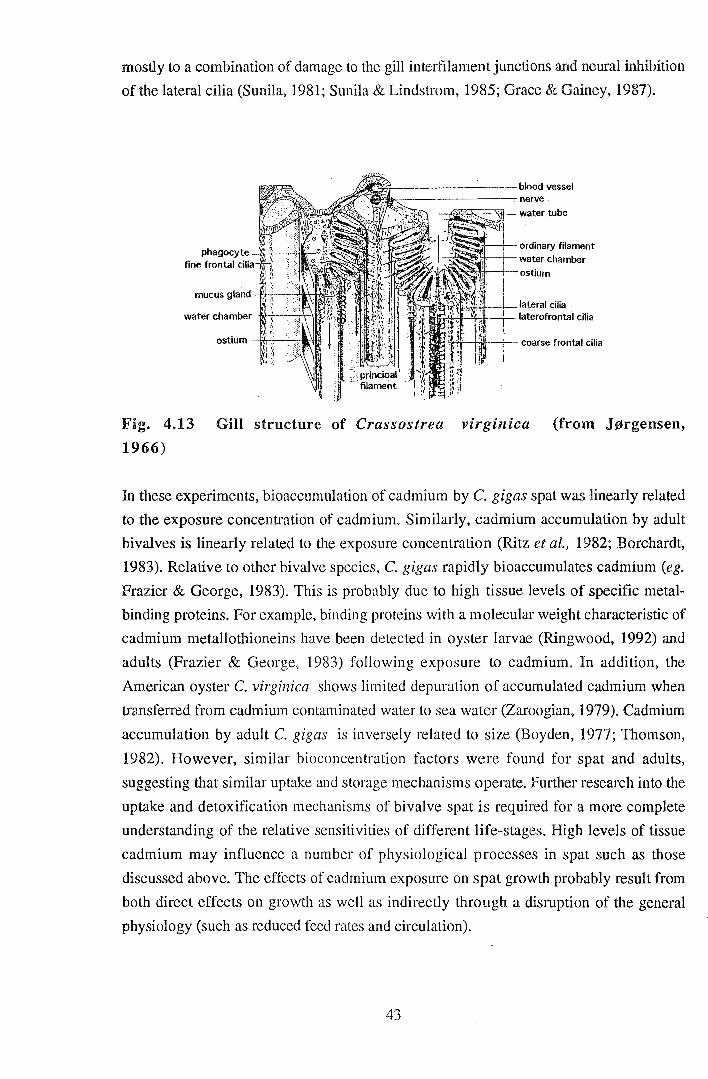
affected which results in reduced ventilation rates (Manley, 1983). The laterofrontal and
frontal cilia of lamellibranch gills (Jorgensen, 1966) are involved in trapping and
transporting food pmticles, whilst the lateral cilia maintain a water current across the gill
surface (Barnes, 1980). Thus, ciliary inhibition would reduce the rate of removal of
microalgae from water. Chronic exposure of M. edulis to copper cause histopathological
changes to the gill epithelium as well as inhibiting ciliary activity (Brown & Newell,
1972). The effect of copper exposure on filtration rates of M. edulis is probably due
42
mostly to a combination of damage to the gill interfilament junctions and neural inhibition
of the lateral cilia (Sunil a, 1981; Sunila & Lindstrom, 1985; Grace & Gainey, 1987).
+--------blood vessel ~--------nerve
--"'gg~::s;:\1-- water tube
!
ordinary filament water chamber
-ostium
:, i lateral cilia ·~· · ... ;, ·; + laterofrontal cilia
I, I I .
Fig. 4.13 Gill structure of Crassostrea virginica (from J~rgensen,
1966)
In these experiments, bioaccumulation of cadmium by C. gigas spat was linearly related
to the exposure concentration of cadmium. Similarly, cadmium accumulation by aduh
bivalves is linearly related to the exposure concentration (Ritz et al., 1982; Borchardt,
1983). Relative to other bivalve species, C. gigas rapidly bioaccumulates cadmium (eg.
Frazier & George, 1983). This is probably due to high tissue levels of specific metal
binding proteins. For example, binding proteins with a molecular weight characteristic of
cadmium metallothioneins have been detected in oyster larvae (Ringwood, 1992) and
adults (Frazier & George, 1983) following exposure to cadmium. In addition, the
American oyster C. virginicct shows limited depuration of accumulated cadmium when
transferred from cadmium contaminated water to sea water (Zaroogian, 1979). Cadmium
accumulation by adult C. gigas is inversely related to size (Boyden, 1977; Thomson,
1982). However, similar bioconcentration factors were found for spat and adults,
suggesting that similar uptake and storage mechanisms operate. Further research into the
uptake and detoxification mechanisms of bivalve spat is required for a more complete
understanding of the relative sensitivities of different life-stages. High levels of tissue
cadmium may influence a number of physiological processes in spat such as those
discussed above. The effects of cadmium exposure on spat growth probably result from
both direct effects on growth as well as indirectly through a disruption of the general
physiology (such as reduced feed rates and circulation).
43

In conclusion, chronic exposure to sublethal concentrations of cadmium had an effect on
the general physiology of juvenile C. gigas. Relative to control spat, exposure to
concentrations as low as 10 ~gL-1 total cadmium (equivalent to a free cadmium ion
concentration of 0.003 ).lgL -1) for only 32 days had an effect on growth rate, heart
contraction, algal filtration rate and behavioural response to light. Exposure to sublethal
cadmium concentrations also caused an increase in the proportion of spat with abnormal
shell growth. A growth abnormality similar but more severe than "curly back" was
observed following exposure to only 10 ).lgL-1 cadmium for 32 days. Longer term
exposure to lower cadmium concentrations (for example, the 5 ).lgL-1 limit allowed in
Tasmanian waters) may well be responsible for the growth abnormality observed in the
Tasmanian oyster industry.
44

5. GENERAL DISCUSSION
When compared to previously published LCso values for other bivalve species, juvenile
C. gigas were less sensitive to acute cadmium exposure than expected. C. gigas spat had
LCsos of 10.29 and 28.43 mgL-1 total cadmium for "small" and "large" spat respectively.
The Hawaiian bivalve, lsognomon californicum has a 48 hour LCso of 16.5 mgL-1
(Ringwood, 1990); however, 96 hour LCsos for molluscs are generally lower than the
values obtained here for C. gigas spat (see Table 5.1). A surprisingly large difference
between total cadmium toxicity to the two sizes of spat was noted; however, when LCsos
were compared in terms of free ion concentration the difference was minimal (0.25 and
0.27 mgL-1 respectively). Addition of EDTA to cadmium treatment solutions, reduced the
toxicity to spat by a reduction in the free ion concentration to which they were exposed.
The toxicity of cadmium to juvenile Pacific oysters was found to be highly related to the
free ion concentration as opposed to the total cadmium concentration.
Table 5.1: Acute toxicity of cadmium to several species of mollusc in
terms of the 96 hour median lethal concentration.
SPECIES LCso (mgL-1) SALINITY 1EMPERA1URE REFERENCE.
(%o) (C)
C. gigas spat 10.3-28.4 35 20 present study
C. virginica 3.8 (48 hour) 25 26 Calabrese et al.,
embryos (1973) I. californicum, spat 16.5 (48 hour) 34 24 Ringwood (1990) adults 27.5
Mya arenaria 2.2 20 20 Eisler (1971)
Mytilus. edulis 25 20 20 Eisler (1971)
M. e. planulatus 1.62 18.5 33 Ahsanullah (1976)
N assatius obsoletus 10.5 20 20 Eisler (1971)
Urosdpinx cinerea 6.6 20 20 Eisler (1971)
The observed relationship between cadmium toxity to C. gigas spat and the free
cadmium ion concentration may be understood through a knowledge of the mechanism of
metal uptake by bivalves. If metal uptake is reduced, both tissue accumulation of the
metal and physiological effects due to exposure (eg. mortality) will be reduced. Copper
accumulation by the oyster, C. virginica is dependent on the free cupric ion
concentration. Zamuda and Sunda (1982) suggested that this relationship is due to
45
equilibria between copper~carrying ligands at the cell surface and soluble copper in the
external medium. If copper uptake into the cell is governed by the amount of copper
bound to ligands at the surface, then it is also directly related to the free ion concentration
in the external medium. Chelators (eg. EDTA) compete with these ligands for the
available copper. A similar mechanism for cadmium would explain the relationship
between free cadmium ion concentration and C. gigas mortality. Cadmium is
predominantly present as chloride complexes in salt water (Riley, 1971); CdCh passes
through cell membranes 1Q3 times faster than the free cadmium ion (Gutnecht, 1981 in
Nelson & Donkin, 1985). Thus it is probable that cadmium uptake by C. gigas is
facilitated. That is, the free cadmium ion concentration controls the rate of cadmium
binding to ligand sites and thus the rate of facilitated uptake as opposed to controlling
direct uptake of the free cadmium ion. Addition of EDTA reduced the free ion
concentration and tlms the cadmium available for binding at the cell surface of cadmium
uptake sites (eg. gill and digestive gland epithelia).
The observed relationship between spat mortality and free cadmium ion concentration is
of particular concern where total cadmium levels are within recommended limits whilst
physico-chemical conditions promote high free ion concentrations. Commercial bivalve
hatcheries frequently add EDT A to larval culture systems; however, there is limited
control of metal levels in culture water for bivalves beyond metamorphosis. Cadmium
has previously been shown to leach from black polyethylene and galvanised piping
(Nilsson, 1970). Polyethylene piping, in particular, is frequently used to supply seawater
to bivalve hatcheries and nurseries. Sources of cadmium contamination in the
environment include pesticides, phosphate fertilisers, paints, anti-corrosion cadmium
plating (including boat-fittings) and galvanised iron. Industrial activities such as mining
and electrolytic processes (eg. the Electrolytic Zinc Company in Tasmania) contribute
significant levels of cadmium to the environment (Nilsson, 1970; Friberg et al., 1974).
The Tasmanian recommended limit for cadmium in marine water is 5 JlgL-1 (Tasmanian
Department of Environment, 1986); chronic exposure of oysters to levels of cadmium
around 5 JlgL-1 may well affect their appearance and physiology, particularly where
conditions promote relatively high levels of free ions. The recently published Australian
Water Quality Guidelines suggest a more conservative level for marine waters of2 JlgL-1
(ANZECC, 1992). Use of the ANZECC limit is recommended for Tasmanian waters,
particularly in areas used for aquaculture. As I have shown, exposure to only 10 JlgL -1
total cadmium (or a free ion concentration of 0.003 ~gL-1) for 32 days had an adverse
effect on growth rate, heart contraction, algal filtration rate, behavioural response to light
and caused an increase in spat with abnormal shell growth. Recommended environmental
limits for heavy metals based on tl1e total metal concentration provide an unreliable guide
46

for protection of waterways. Spat may well be exposed to free cadmium ion
concentrations of around 0.003 )lgL-1 in the environment, particularly in estuarine waters
where lower salinity promotes a high free ion concentration relative to that in oceanic
water. In environmental and commercial culture situations where oysters are exposed to
such concentrations of cadmium for longer periods, from an earlier age and under a range
of physico-chemical conditions these effects on the physiology of juvenile C. gigas
would have important implications for their survival and growth.
47

6. LITERATURE CITED
Ahsanulla, M. (1976). Acute toxicity of cadmium and zinc to seven invertebrate species
from Western Port, Victoria. Aust. J. Mar. Freshwater Res., 27, 187-96.
ANZECC, (1992). Australian water quality guidelines for fresh and marine waters.
Australian and New Zealand Environment and Conservation Council.
APHA., (1989). Standard methods for the examination of water and waste water. 17th
ed. joint publishers: American Public Health Association: American Water Works
Association: Water Pollution Control Federation.
Barnes, R. D. (1980). Invertebrate Zoology. 4th ed. Saunders College, Philadelphia
1089 pp.
Beaumont, A. R., Tserpes, G. & Budd, M.D. (1987). Some effects of copper on the
veliger larvae of the mussel Mytilus edulis and the scallop Pecten maximus (Mollusca,
Bivalvia). Marine Environ. Res. 21, 299-309.
Bloom, H. & Ayling, G. M. (1977). Heavy metals in the Derwent Estuary. Env.
Geology, 2, 3-22.
Boyden, C. R. (1977). Effect of size upon metal content of shellfish. J. Mar. Biol. Soc.
U.K., 57, 675-714.
Borchardt, T. (1983). Influence of cadmium on the kinetics of cadmium uptake and loss
via food and seawater in Mytilus edulis. Mar. Bioi., 76, 67-76.
Brown, M. R., Jeffrey, S. W. & Garland, C. D (1989). Nutritional aspects of
microalgae used in mariculture; a literature review. CSIRO Marine Laboratories Report
205.
Brown, B. & Newell, R. (1972). The effect of copper and zinc on the metabolism of the
mussel Mytilus edulis. Mar. Biol. 16, 108-118.
Bryan, G. W., Langston, W. J., Hummerstone, L. G. & Burt, G. R. (1985). A guide to
the assessment of heavy metal contamination in estuaries using biological indicators.
Occasional paper No.4, March 1985. Mar. Bioi. Assoc. U.K.
Calabrese, A., Collier, R. S. Nelson, D. A. & Macinnes, J. R. (1973). The toxicity of
heavy metals to embryos of the American oyster Crassostrea virginica. Mar. Biol., 18,
162-166.
48
Davenport, J. & Redpath, K.J. (1984). Copper and the mussel Mytilus edulis. L. In
Toxins, Drugs and Pollutants in Marine Animals (L. Bolis, Zadunaisky & R. Gilles,
eds) pp 176-189, Springer-Verlag, Berlin.
Davenport, J. & Manley, A. (1978). The detection of heightened sea-water copper
concentrations by the mussel Mytilus edulis. J. mar. biol. Ass. U.K., 58, 843-850.
Eisler, R. (1971). Cadmium poisoning in Fundulus heteroclitus (Pisces:
Cyprinodontidae) and other marine organisms. J. Fish. Res. Board Can. 28, 1225-34.
Engel, D.W. & Fowler, B.A. (1979). Factors influencing cadmium accumulation and its
toxicity to marine organisms, Env. Health Perspectives, 28, 81-88.
Evtushenko, Z.S., Belcheva, N.N. and Lukyanova, O.N. (1986). Cadmium
accumulation in organs of the scallop Mizuhopecten yessoensis--II. Subcellular
distribution of metals and metal-binding proteins. Comp. Biochem. Physiol., 83C, 377-
383.
Fisher, Sir R. A. & Yates, F. (1957). Statistical tables for biological, agricultural and
medical research. 5th ed.,Oliver & Boyd Ltd., Edinburgh.
Frazier, J. M. & George, S. G. (1983). Cadmium kinetics in oysters -a comparative
study of Crassostrea gigas and Ostrea edulis. Mar. Biol., 16, 55-61.
Friberg, L., Piscator, M., Nordberg, G. F. & Kjellstrom, T. (1974). Cadmium in the
environment. 2nd ed. CRC Press, Cleveland, Ohio. 248 pp.
George, S.G & Coombes, T. L. (1975). A comparison of trace-metal and
metalloenzyme profiles in different molluscs and during development of the oyster.
p.433-449. In: Proceedings of the 9th European Marine Biology Symposium. Barnes,
H. (ed.) Aberdeen University Press
Grace, A.L. & Gainey, L.F., Jr. (1987). The effects of copper on the heart rate and
filtration rate of Mytilis edulis. Mar. Pollut. Bull. 18, 87-91.
Gainey, L.F., Jr. & Kenyon, J.R. (1990). The effects of reserpine on copper induced
cardiac inhibition in Mytilus edulis. Comp. Biochem. Physiol., 95C, 177-179.
49

Guillard, R.R.L. (1975). Culture of phytoplankton for feeding marine invertebrates. In
Culture of Marine Invertebrate Animals, W.L.Smith and M.H. Chanley, eds., (1975)
pp.29-60, Plenum Press, New York.
Helm, M.M. & Trueman, E.R. (1967). The effect of exposure on the heart rate of the
mussel, Mytilus edulis L. Comp. Biochem. Physiol., 21, 171-177.
Hodson, P. V., Ross, C. W., Niimi, A. J. & Spry, D. J. (1976). Statistical
considerations in planning aquatic bioassays. Proc. 3rd Aquatic Toxicity Workshop,
Halifax, N. S., Nov. 2-3, 1976. Environmental Protection Service Technical Report No.
EPS-5-AR-77-1, Canada. pp.15-31.
Jeffrey, S.W., Garland, C.D. & Brown, M.R. (1990). Microalgae in Australian
mariculture. In Biology of marine plants, eds. Clayton, M.N. & King, R.J. chapter 18.,
Longman Cheshire, Pty Ltd, Australia.
J¢rgensen, C. B. (1966). Biology of suspension feeding. Pergamon Press, Oxford.
Langdon, C.J. & Waldock, M.J. (1981). The effect of algal and artificial diets on the
growth and fatty acid composition of Crassostrea gigas spat. J. mar. bioi. Assoc. U.K.,
61, 431-448.
Manly, A. R. (1983). The effects of copper on the behaviour, respiration, filtration and
ventilation activity of Mytilus edulis. J. Mar. Biol. Assoc. U.K., 63, 205-222.
McLusky, D. S., Bryant, V. & Campbell, R. (1986). The effects of temperature and
salinity on the toxicity of heavy metals to marine and estuarine invertebrates. Oceano gr.
Mar. Biol. Ann. Rev., 24, 481-520.
Muneoka, Y. & Twarog, B.M. (1983). In The Mollusca. Vol. 4. Physiology Part 1.,
eds. Saleuddin, A.S.M & Wilbur, K.M., chapter 2., Academic Press., Inc., New York.
523 pp.
Nelson, A. & Donkin, P. (1985).Processes of bioaccumulation: the importance of
chemical speciation. Mar. Pollut. Bull., 16 (4) 164-169.
Nilsson, R. (1970). Aspects on the toxicity of cadmium and its compounds; a review.
Swedish Ecological Research Committe, Bulletin no. 7, Natural Science Research
Council, Sweden. 58pp.
50
Paparo, A. A. (1987). Ciliary/decarboxylase activities as indicators ofPTZ, photoperiod
and Ca-ionophore effect on Crassostrea virginica (Gmelin).Comp. Biochem. Physiol. A
Comp. Phys., 88(3), 471-474.
Patel, B. & Anthony, K. (1991). Uptake of cadmium in tropical marine lamellibranchs,
and effects on physiological behaviour. Mar. Bioi., 108, 457-470.
Pollard, J. (1992). Tasmanian Year Book, 1992. No. 23. Australian Bureau of
Statistics.
Regoli, F., Orlando, E., Mauri, M., Nigro, M. & Cognetti, G. A. (1991). Heavy metal
accumulation and calcium content in the bivalve Donacilla cornea. Mar. Ecol. Pro g.
Ser., 74, 219-224.
Reish, D. J. & Oshida, P. S. (1986). Manual of methods in aquatic environment
research. Prt 10 Short term static bioassays. FAO Fisheries Technical Paper No. 247,
FAO, Rome, 62pp.
Riley, J.P. (1971). The major and minor elements in sea water. In.: Introduction to
marine chemistry. eds. Riley, J.P. & Chester, R., Academic Press, London.
Ringwood, A. H. (1989). Accumulation of cadmium by larvae and adults of an
Hawaiian bivalve, Isognomon californicum, during chronic exposure. Mar. Bioi., 102,
499-504.
Ringwood, A. H. (1990). The relative sensitivities of different life stages of !so gnomon
caiifornicum to cadmium toxicity. Arch. Environ. Contam. Toxicol., 19, 338-340.
Ringwood, A. H. (1992). Bivalve development and variation in metallothionein
expression during metal exposures. Environs, December 1992,7.
Ritz, D., Swain, R. & Elliott, N. (1982). Use of the mussel Mytilus edulis planulatus in
monitoring heavy metal Aust. J. F. Wat. Res., 33, 491-506.
Scott, D. M. & Major, C. W. (1972). The effect of copper (II) on survival, respiration
and heart rate in the common blue mussel, Mytilus edulis. Bioi. Bull., 143, 679-688.
51
Seed, R. (1968). Factors influencing shell shape in the mussel Mytilu edulis., J. mar.
biol. Ass. U.K., 48, 561-584.
Shuster, C. (Jr) & Pringle, B. (1969). Trace metal accumulation by the American
Eastern oyster, Crassostrea virginica. Proc. Nat. Shellfish. Assoc., 59, 91-103.
Sumner, C. E. (1972) Oysters and Tasmania. Tasmanian Fisheries Research, Sea
Fisheries Division, Department of Agriculture, 6 (2), 1-15.
Sunda, W. G., Engel, D. W. & Thuotte, R. M. (1978). Effect of chemical speciation on
toxicity of cadmium to grass shrimp, Palaemonetes pugio:: importance of free cadmium
ion, Env. Sci. Tech., 12(4), 409-413.
Sunila, I. & Lindstrom, R. (1985). Survival, growth and shell deformities of copper
and cadmium-exposed mussels (Mytilus edulis L.) in brackish water. Estuar., Coast.
Shelf Sci., 21, 555-565.
Sunila, I. (1988). Pollution-related histopathological changes in the mussel Mytilus
edulis L. in the Baltic Sea. In: Marine Environ. Res., 24. Responses of marine
organisms to pollutants. Roesijadi, G. & Spies, R. B. (eds.) Elsevier Applied Science,
England, 356 pp.
SYSTAT (1992): Statistics, Version 5.2 Edition. Evanston, IL:SYSTAT, Inc.,
1992.724 pp.
Tasmanian Department of the Environment (1986). Guidelines on minimum desirable
ambient water quality for receiving waters in Tasmania.
Tasmanian Department of the Environment (1987). Department of the Environment
report for the year 1986-87. Parliament of Tasmania. Government Printer.
Thomson, J. D. (1982). Metal concentration changes in growing Pacific oysters,
Crassostrea gigas, cultivated in Tasmania, Australia. Mar. Bioi., 67, 135-142.
Thomson, J. D., Pirie, B. J. S & George, S. G. (1985). Cellular metal distribution in
the Pacific oyster, Crassostrea gigas (Thun.) determined by quantitative X-ray
microprobe analysis. J. Exp. Mar. Biol. Ecol., 85, 37-45.
52
Thrower, S.J. & Eustace, I.J. (1973). Heavy metal accumulation in oysters grown in
Tasmanian waters. Food Tech. in Australia., 25, 546-553.
Uesak:a, H., Yamagishi, H. & Ebara, A. (1987). Stretch-mediated interaction beyween
the auricle and ventricle in an oyster Crassostrea gigas. Comp. Biochem. Physiol., 88A,
221-227.
USEPA (1986). Quality criteria for water. EPA 440/5-86-001, U S Environmental
Protection Agency,. Washington, D.C.
Vernberg, W. B. & Vernberg, F. J. (1972). Environmental physiology of marine
animals. Springer-Verlag.
Wagge, L.E. (1951). The activity of amoebocytes and of alkaline phosphatases during
the regeneration of the shell in the snail, Helix aspersa. Q. J. Microscop. Sci., 92(3),
307-321.
Ward, T.J. (1983). Laboratory study of the accumulation and distribution of cadmium in
the Sydney rock oyster Saccostrea commercialis., Aust. J. Mar. Freshwater Res., 33,
33-44.
Watling, H. R. (1978). Effect of cadmium on larvae and spat of the oyster Crassostrea
gigas (Thunberg). Trans. roy. Soc. S. Afr., 43 (2) 125-134.
Watling, H. R. (1983). Comparative study of the effects of metals on the settlement of
Crassostrea gigas. Bull. Environ. Contam. Toxicol., 31, 344-351.
Westall, J. C., Zachary, J. L. & Morel, F. M. (1976). MINEQL: A computer program
for the calculation of chemical equilibrium composition of aqueous systems. Mass. Inst.
Technol. R. M. Parsons Lab. Tech. Note 18.
Whyte, J.N.C. (1987). Biochemical composition and energy content of six species of
phytoplankton used in mariculture of bivalves. Aquaculture, 60, 231-241.
Wilbur, K.M. & Saleuddin, A.S.M (1983). Shell formation In. The Mollusca. Vol. 4.
Physiology Part 1., eds. Saleuddin, A.S.M & Wilbur, K.M., Academic Press., Inc.,
New York. 523 pp.
Yonge, C. M. (1960). Oysters. A New Naturalist Special Volume. Collins, London.
53

Zamuda, C. D. & Sunda, W. G. (1982). Bioavailability of dissolved copper to the
American oyster Crassostrea virginica. I. Importance of chemical speciation. Mar. Bioi.,
66, 77-82.
Zaroogian, G.E. & Cheer, S. (1976). Accumulation of cadmium by the American
oyster, Crassostrea virginica. Nature, 261, 408-409.
Zirino, A. & Yamamoto, S. (1972). A pH-dependent model for the chemical speciation
of copper, zinc, cadmium, and lead in seawater. Limnol. & Oceano gr., 17, 661-671.
54
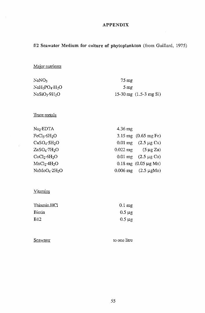
APPENDIX
f/2 Seawater Medium for culture of phytoplankton (from Guillard, 1975)
Major nutrients
NaN03
NaH2P04·H20
NaSi03·9H20
Trace metals
Na2·EDTA
FeCl3·6H20
CuS04·5H20
ZnS04·?H20
CoCh·6H20
MnCh·4H20
NaMo04·2H20
Vitamins
Thiamin.HCl
Biotin
B12
Seawater
75mg
5mg
15-30 mg (1.5-3 mg Si)
4.36 mg
3.15 mg (0.65 mgFe)
0.01 mg (2.5 !lg Cu)
0.022 mg (5 !lg Zn)
0.01 mg (2.5 !lg Co)
0.18 mg (0.05 !lg Mn)
0.006 mg
0.1 mg
0.5 !lg
0.5 !lg
to one litre
55
(2.5 !lgMo)


PHYSIOLOGICAL EFFECTS OF HEAVY METALS ON BIVALVES, WITH PARTICULAR EMPHASIS ON
JUVENILE STAGES
A review of the literature submitted to the Department of Zoology, University of
Tasmania in partial fulfilment of the degree of Bachelor of Science with Honours.
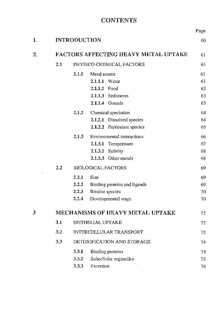
CONTENTS
Page
1. INTRODUCTION 60
2. FACTORS AFFECTING HEAVY METAL UPTAKE 61
2.1 PHYSICO-CHEMICAL FACTORS 61
2.1.1 Metal source 61
2.1.1.1 Water 61
2.1.1.2 Food 62
2.1.1.3 Sediments 63
2.1.1.4 Gonads 63
2.1.2 Chemical speciation 64 2.1.2.1 Dissolved species 64
2.1.2.2 Particulate species 65
2.1.3 Environmental interactions 66 2.1.3.1 Temperature 67 2.1.3.2 Salinity 68 2.1.3.3 Other metals 68
2.2 BIOLOGICAL FACTORS 69
2.2.1 Size 69 2.2.2 Binding proteins and ligands 69 2.2.3 Bivalve species 70 2.2.4 Developmental stage 70
3 MECHANISMS OF HEAVY METAL UPTAKE 72
3.1 EPITHELIAL UPTAKE 72
3.2 INTERCELLULAR TRANSPORT 73
3.3 DETOXIFICATION AND STORAGE 74
3.3.1 Binding proteins 74 3.3.2 Subcellular organelles 75 3.3.3 Excretion 76
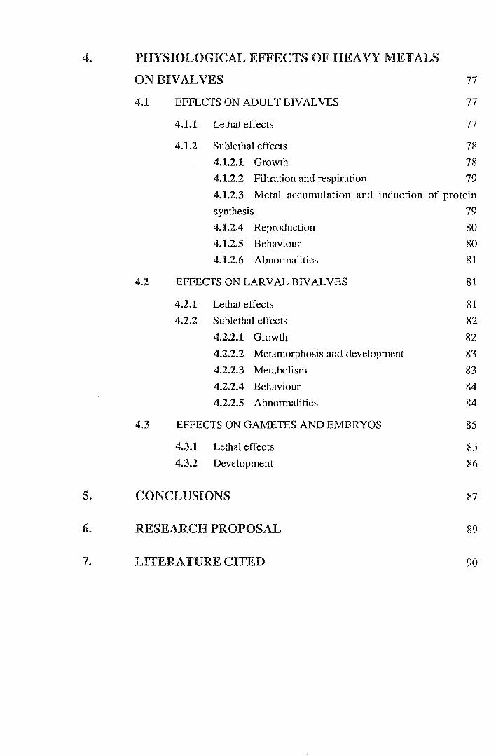
4. PHYSIOLOGICAL EFFECTS OF HEAVY METALS
ON BIVALVES 77
4.1 EFFECTS ON ADULT BIVALVES 77
4.1.1 Lethal effects 77
4.1.2 Sublethal effects 78 4.1.2.1 Growth 78 4.1.2.2 Filtration and respiration 79 4.1.2.3 Metal accumulation and induction of protein
synthesis 79 4.1.2.4 Reproduction 80 4.1.2.5 Behaviour 80 4.1.2.6 Abnormalities 81
4.2 EFFECTS ON LARVAL BIVALVES 81
4.2.1 Lethal effects 81 4.2.2 Sublethal effects 82
4.2.2.1 Growth 82
4.2.2.2 Metamorphosis and development 83 4.2.2.3 Metabolism 83 4.2.2.4 Behaviour 84 4.2.2.5 Abnormalities 84
4.3 EFFECTS ON GAMETES AND EMBRYOS 85
4.3.1 Lethal effects 85 4.3.2 Development 86
5. CONCLUSIONS 87
6. RESEARCH PROPOSAL 89
7. LITERATURE CITED 90

1. INTRODUCTION
The capacity of bivalves to bioaccumulate heavy metals is well documented (George
and Coombs, 1975; Phillips, 1976; Cooper et al., 1982). Concern over aquatic pollution
from anthropogenic activities combined with the ability of bivalves to bioaccumulate
metals has stimulated numerous studies into interactions between bivalves and heavy
metals (George, 1980).
Research has focused on physiological effects (eg. Brown and Newell 1972; Manley,
1983), accumulation and detoxification (George, 1980) and biomonitoring (eg. Ritz et
al., 1982). Effects of heavy metals on growth and mortality have received particular
attention. Recent studies have examined biochemical aspects of heavy metal uptake and
detoxification by adult bivalves (Langston et al., 1989; Weber, 1990).
Adult bivalves, particularly commercial species, have previously received considerable
attention; however, little information is available on metal uptake by larval stages
especially at chronic and/or sub-lethal exposure levels (Calabrese et al., 1973;
Ringwood, 1989). Nevertheless, developments in hatchery technology have ensured a
reliable supply of juvenile bivalves heightening interest in studies of early life stages
(Calabrese et al., 1973). Hatchery reared bivalve larvae and embryos are convenient for
controlled laboratory experiments due to size, availability and sensitivity (ASFA, 1976;
Ringwood, 1991). The use of bivalve larvae and embryos for bioassays has received
attention as a sensitive, efficient means of assessing contamination (ASFA, 1976).
This review discusses the physiological effects of heavy metals on marine bivalves and
contributing factors, in particular, uptake, accumulation and depuration. Where
information is available, emphasis is placed on juvenile bivalves. The effects of some
of the more toxic heavy metals, especially mercury, cadmium, copper and zinc
(McLusky et al., 1986), are discussed in detail.
60
2. FACTORS AFFECTING HEAVY METAL UPTAKE
Relative to other invertebrates marine bivalves are tolerant to a range of environmental
conditions due mainly to avoidance behaviour (Akberali and Trueman, 1985) and
effective detoxification strategies (George, 1980); this accounts for their colonization
success in estuarine and coastal environments (Akberali and Trueman, 1985).
Bioavailability of metals is determined by their relative concentrations as ions, organic
and inorganic complexes and combined with particulate matter (Engel et al., 1981).
Heavy metal uptake is controlled by both environmental conditions and biological
factors (Simkiss and Mason, 1983). Different factors affect uptake of individual metals
to varying degrees (George, 1980).
2.1 PHYSICO-CHEMICAL FACTORS
2.1.1 Metal source
Marine heavy metal uptake by bivalves is from three sources: water, food and
sediments. The relative contribution of each source varies according to environmental
conditions, metal form and bivalve species. For example, relative uptake of mercury
from salt water, phytoplankton and suspended sediment by M. edulis is approximately
in the ratio of 10:5:1. This ratio varies with environmental conditions (King and
Davies, 1987). The source of a heavy metal also affects accumulation levels by
different organs. Figure 1 shows the internal organs of a bivalve (Geloina erosa). The
digestive gland and to a lesser extent the kidneys take up most of the metals obtained
from phytoplankton feeding by M. edulis, sedimentary metals are taken up by the
kidney and metal uptake from water is highest in the gills, kidney and digestive gland
(King and Davies, 1987). Uptake pathways will be discussed in Section 3.
2.1.1.1 Water
Unlike secondary consumers such as the flounder (Riisgard and Hansen, 1990), the
concentration effect of metal accumulation from food is less important than uptake
from water, particularly for filter feeders (Mohlenberg and Riisgard, 1988). Filter
feeding bivalves such as Mytilus edulis take up metals at an initial rate proportional to
61
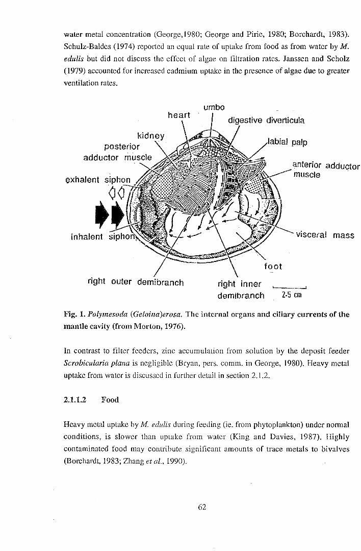
water metal concentration (George,1980; George and Pirie, 1980; Borchardt, 1983).
Schulz-Baldes (1974) reported an equal rate of uptake from food as from water by M.
edulis but did not discuss the effect of algae on filtration rates. Janssen and Scholz
(1979) accounted for increased cadmium uptake in the presence of algae due to greater
ventilation rates.
kidnt:Y posterior
adductor muscle
right outer demibranch
umbo
right inner demibranch
anterior adductor muscle
visceral mass
z.s em
Fig. 1. Polymesoda (Ge/oina)erosa. The internal organs and ciliary currents of the
mantle cavity (from Morton, 1976).
In contrast to filter feeders, zinc accumulation from solution by the deposit feeder
Scrobicularia plana is negligible (Bryan, pers. comm. in George, 1980). Heavy metal
uptake from water is discussed in further detail in section 2.1.2.
2.1.1.2 Food
Heavy metal uptake by M. edulis during feeding (ie. from phytoplankton) under normal
conditions, is slower than uptake from water (King and Davies, 1987). Highly
contaminated food may contribute significant amounts of trace metals to bivalves
(Borchardt, 1983; Zhang et al., 1990).
62
2.1.1.3 Sediments
Despite the high metal content of sediments relative to other sources (Bryan et al.,
1985) metal uptake from them by bivalves is generally low. Bivalves, in particular
deposit feeders such as scallops and clams, may take up metals via the digestive gland
from ingested sediments (George, 1980). The biogenic and interstitial water
components of marine sediments provide the more bioavailable sources of sedimentary
metal (Bryan, 1976; Bryan et a/.,1985). Sediment composition may determine the
availability of metals for accumulation (Cooke et al., 1979). For example, the uptake of
lead by the deposit feeder S. plana is related to the lead:iron ratio in surface sediments.
High iron levels may increase binding of lead to iron oxide, thus decreasing lead uptake
in the digestive tract (Luoma and Bryan, 1978). On the other hand, mercury uptake by
S. plana and Macoma balthica is related to the mercury:total organic matter ratio of the
surface sediments (Langston, 1982).
Filter feeders accumulate metals to a lesser extent from sediments. Greig and Wenzloff
(1978) found copper levels in Crassostrea virginica were related to sedimentary levels.
They suggested that uptake of copper occurs from sediments as well as from
phytoplankton and water. Cadmium and silver, however, were not taken up directly
from sediments. Brooks and Rumsby (1965) found uptake of chromium, iron,
manganese and nickel is predominantly by ingestion of sedimentary matter of small
particle size.
2.1.1.4 Gonads
Heavy metal levels in bivalve larvae and embryos are partially attributable to uptake by
the parental gonads and gametes (Frazier, 1975; Stiles et al., 1991). Copper, cadmium
and zinc may be directly incorporated into the gametes of C. virginica (Frazier, 1975).
Although low compared with total metal uptake, copper and zinc concentrations in C.
gigas eggs may be comparable to those in the gills (Thomson et al., 1985). Levels of
contamination influence the gonad uptake of heavy metals by certain bivalve species.
Greig et al., (1975) found constant levels of copper and cadmium in the eggs of C.
virginica adults from different environments and no transfer of silver, lead and zinc to
the oocytes was detected. In contrast, Akberali and Trueman (1985) state that gamete
metal uptake is stimulated by high metal levels. In a study by Roesijadi et al. (1982),
M. edulis larvae from adults exposed to mercury, had greater cytosolic mercury
concentrations than controls. The hard clam, Mercenaria mercenaria, exhibits
63
significantly different gonad metal concentration between polluted and relatively
unpolluted environments (Stiles et al., 1991). Embryos from contaminated adults,
cultured in reference sea water develop more chromosomal abnormalities than those
spawned from uncontaminated adults. Thus metal uptake by prespawned gametes
probably affected subsequent development of the fertilized embryos.
2.1.2 Chemical speciation
2.1.2.1 Dissolved species
Bioavailability of a dissolved metal species is a function of its activity (Riley, 1971). In
bivalve studies metal uptake and toxicity are frequently expressed as a function of the
total dissolved metal concentration (Brooks and Rumsby, 1965; Brereton et al., 1973).
However, in natural systems heavy metal accumulation is largely determined by free
metal ion concentration. For example, an increase in salinity lowers cadmium toxicity
by reducing the concentration of free ions (Engel et al., 1981). Mantoura et al. (1978)
found the trace metals zinc and manganese exist predominantly as free ions; however,
free metal ion concentrations are generally low in marine environments (Nelson and
Donkin, 1985). Metal ions are predominantly present as complexed species (Coombs
and George, 1977). Table 1 shows levels of trace metals in the Derwent River estuary,
Tasmania.
Inorganic metal speciation in salt water is largely determined by competitive binding
with the major anions: Ct, C032-, OH-, S04
2-, F (Riley, 1971) and Br- (Kennish,
1989). Relative bioaccumulation of cadmium salts by bivalves is in the order of
sulphate> iodide> acetate> chloride> nitrate> carbonate (Patel and Anthony, 1991).
Simkiss (1983) proposed that inorganic complexes of heavy metals (including copper,
zinc, cadmium and mercury) are more bioavailable than other metal species due to
greater lipid solubility in cell membranes. Conversely, Bryan et al. (1985) describe the
organic mercury species as more bioavailable than inorganic mercury complexes.
Dissolved organic metal complexes typically occur as rnetal-chelates, particularly in
estuarine zones with high levels of dissolved organic matter (Riley, 1971; Engel et al.,
1981). Organic complexes may provide a significant source of heavy metals to marine
invertebrates; for example, Mohlenberg and Riisgard (1988) found increased uptake of
organic mercury with age in Cardium sp. Conversely, organic chelators such as humic
acid and the synthetic chelator EDTA (ethylenediaminetetraacetic acid) usually reduce
heavy metal uptake (Knezovich et al., 1981; Bryan et al., 1985). However, George and
64
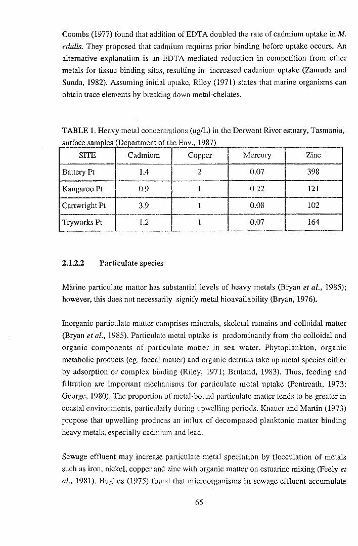
Coombs (1977) found that addition of EDTA doubled the rate of cadmium uptake in M.
edulis. They proposed that cadmium requires prior binding before uptake occurs. An
alternative explanation is an EDTA-mediated reduction in competition from other
metals for tissue binding sites, resulting in increased cadmium uptake (Zamuda and
Sunda, 1982). Assuming initial uptake, Riley (1971) states that marine organisms can
obtain trace elements by breaking down metal-chelates.
TABLE 1. Heavy metal concentrations (ug/L) in the Derwent River estuary, Tasmania,
su rf 1 (D f h E 1987) ace samp:es epartment o · t e nv.,
SITE Cadmium Copper Mercury Zinc
Battery Pt 1.4 2 0.07 398
Kangaroo Pt 0.9 1 0.22 121
Cartwright Pt 3.9 1 0.08 102
Tryworks Pt 1.2 1 0.07 164
2.1.2.2 Particulate species
Marine particulate matter has substantial levels of heavy metals (Bryan et al., 1985);
however, this does not necessarily signify metal bioavailability (Bryan, 1976).
Inorganic particulate matter comprises minerals, skeletal remains and colloidal matter
(Bryan et al., 1985). Particulate metal uptake is predominantly from the colloidal and
organic components of particulate matter in sea water. Phytoplankton, organic
metabolic products (eg. faecal matter) and organic detritus take up metal species either
by adsorption or complex binding (Riley, 1971; Bruland, 1983). Thus, feeding and
filtration are important mechanisms for particulate metal uptake (Pentreath, 1973;
George, 1980). The proportion of metal-bound particulate matter tends to be greater in
coastal environments, particularly duting upwelling periods. Knauer and Martin (1973)
propose that upwelling produces an influx of decomposed planktonic matter binding
heavy metals, especially cadmium and lead.
Sewage effluent may increase particulate metal speciation by flocculation of metals
such as iron, nickel, copper and zinc with organic matter on estuarine mixing (Feely et
al., 1981). Hughes (1975) found that microorganisms in sewage effluent accumulate
65

zinc and cadmium. Sewage effluent reduced the toxicity of copper to the copepod
Euchaetajaponica in a study by Lewis et al. (1972), probably by chelation.
Particulate metals vary in bioavailability accoi·ding to type, concentration and source.
Inorganic particulate metal complexes are generally less bioavailable than organic
particulate species (eg. Riisgard & Hansen, 1990). Copper (Zamuda and Sunda, 1982)
and cadmium (Engel et al., 1981) uptake is normally related to the concentration of free
ions. However, in a short term study Carpene and George (1981) reported 50%
particulate cadmium uptake by diffusion across M.edulis gills.
Particulate species are arbitrarily defined as those which are retained on a 0.4um filter
(Amdurer et al., 1981). This by definition excludes colloidal species. Precipitates of
colloidal metal hydroxides are taken up by molluscs by pinocytosis, particularly at the
gili epithelium (George et al., 1976; George, 1982).
2.1.3 Environmental interactions
Heavy metal uptake is influenced both directly and indirectly by environmental
conditions such as temperature, salinity, pH, redox potential and other metals (George,
1980). Estuarine organisms, in particular, are affected by constant exposure to
fluctuating environmental conditions (Jones, 1977). Environmental parameters interact
with metal uptake by influencing metal bioavailability in the environment or by
affecting the physiology and thus the uptake mechanisms of the organism itself
(George, 1980).
66
2.1.3.1 Temperature
Elevated temperatures generally increase heavy metal uptake by bivalve molluscs
(Bryan, 1976; Jackim et al., 1977; Elliott, 1982). For example, Denton and Burdon
Janes (1981) examined the interaction of temperature and salinity with metal uptake in
Saccostrea echinata. An increase in cadmium and mercury uptake due to elevated
temperatures was augmented by concmTently lowering the salinity. Nevertheless, high
temperature maintained this effect at higher salinities. M. edulis accumulation of
cadmium is influenced by temperature at low salinities (Phillips 1976). Copper
accumulation by Donacilla cornea is stimulated by high temperatures, whilst cadmium
and manganese uptake is not significantly affected (Regoli et al., 1991).
Conversely, low temperature may suppress metal uptake in sensitive life stages of
bivalves even resulting in lead ioss by C. virginica larvae (Zaroogian et al., 1979).
Accumulation of cadmium and copper by C. virginica (Zaroogian, 1979) and lead and
zinc by M. edulis (Phillips, 1976) is not affected by low temperatures. Elliott (1982)
reported lower metal uptake by M. edulis planulatus in warmer conditions. He
concluded that the winter acclimatised mussels had a reduced capacity to accumulate
metals at warm temperatures.
High temperature induced heavy metal uptake by bivalve molluscs may be a
physiological response, for example, greater ventilation rates which increase uptake at
the gills (Denton and B urdon-J ones, 1981) or increased solute movement across cell
membranes (Cairns et al., 1975). Alternatively, Simkiss and Watkins (1988) suggested
that the effect of temperature on zinc uptake in M. edulis is a function of temperature
fluctuations rather than the absolute temperature. A change in temperature causes zinc
mobilization or detoxification resulting in additional uptake to replace "lost" zinc.
67
2.1.3.2 Salinity
Salinity has been shown to have an inverse relationship to heavy metal uptake in
bivalves (Jackim et al., 1977). Cadmium uptake by M. edulis is stimulated by low
salinity (Phillips, 1976; Engel et al., 1981). Phillips (1976) reported reduced lead
uptake and constant zinc uptake in response to low salinity. This was explained by
lowered filtration rates or changed ion flux. However, zinc uptake was affected by
salinity in a study by Thomson (1982). As discussed in section 2.1.3.1 salinity also
interacts with temperature to change heavy metal uptake rates.
High salinity reduces the more bioavailable free metal ions in marine systems by the
formation of chloro-complexes (Mantoura et al., 1978; McLusky et al., 1986). George
et al. (1977) proposed that low salinity may cause increased cadmium uptake by the gill
surface due to increased ion nux or the swelling of the cell membrane exposing a
carrier or membrane pore.
2.1.3.3 Other metals
Several examples of the influence of other metal species on heavy metal uptake are
discussed in section 2.1.1.3. Uptake may also be influenced by competition between
heavy metals for binding proteins; for example, cadmium, copper and zinc compete for
binding sites on the metallothionein molecule (discussed in section 3). Exposure to
either of the alternative metals increases copper and zinc uptake by M. edulis
planulatus and inhibits cadmium uptake (Elliott, 1982; Patel and Anthony, 1991). The
extent of metal interaction depends on the concentrations of the metals involved (Elliott
et al., 1986). Metal induction of metallothionein synthesis accounts for increased
copper and zinc uptake. In contrast, Phillips (1976) found no change in net uptake of
cadmium, lead and zinc by M. edulis exposed to other metals and no interaction
between the metals. Copper uptake by M. edulis is very susceptible to environmental
conditions, changing with exposure to other metals and changes in salinity and
temperature (Phillips, 1976).
68
2.2 BIOLOGICAL FACTORS
2.2.1 Size
The relationship between age or weight and metal uptake varies between metals and
bivalve species. Several authors have examined the dynamics of metal accumulation
with respect to size. For example, cadmium uptake is inversely related to body size of
the clam Macoma balthica (McLeese and Ray, 1984) and C. gigas (Boyden, 1977).
Cadmium content is inversely related to dry weight of M. edulis (Cossa et al., 1980); in
contrast cadmium content is directly related to dry weight of C. virginica (Zaroogian,
1980). Organic mercury content increases linearly with age and weight in cockles
(Mohlenberg & Riisgard, 1988). No relationship exists between copper content and dry
weight of the scallop Argopecten iradians (Zaroogian and Johnson, 1983) whilst
Dermott and Lum (1986) reported greater accumulation of copper and zinc by Elliptio
complanata during periods of fastest growth. Although metal content and
concentration provide an idea of metal uptake by bivalves these values cannot strictly
be compared to rates of uptake.
2.2.2 Binding proteins and ligands
George (1980) suggested that nett uptake of metals is regulated by intracellular binding
ligands. Metal ions bound by ligands, possibly derived from mucus, increase metal
uptake by M. edulis. Mucus sheets of M. edulis may affect the membrane permeability
at uptake sites or serve to transport ions to the gut (Simkiss and Mason, 1983).
Naturally, saturation of binding ligands reduces metal uptake (George and Pirie, 1980).
Metal transport across membranes of M. edulis is probably by passive diffusion with
little control of rates or metal type (George, 1980). Bivalves generally exhibit limited
control of metal uptake. However, Bryan (pers. comm. in George, 1980) has observed
control of zinc accumulation from solution by the deposit feeder Scrobicularia plana.
Relative levels of different metal binding proteins in bivalves vary between species and
environment. In several species, exposure to certain heavy metals results in increased
levels of low molecular weight binding proteins (including metallothionein-like
proteins) and high molecular weight binding proteins (including metalloenzymes)
(Harrison et al., 1988; Bebianno and Langston, 1991). The metallothionein-like
molecule in M. edulis binds zinc, cadmium and copper with a greater affinity for
cadmium (Bremner, 1977; Bebianno and Langston, 1991). Talbot and Magee (1978)
69

proposed that cadmium induction of metallothionein-like proteins in M. edulis aids the
organism in control of cadmium, copper and zinc uptake. Interspecific differences in
uptake may be due to metallothionein-like protein levels. For example, the gastropod
Littorina littorea has naturally high metallothionein levels; metallothionein may be
induced in M. edulis but is not produced by Macoma balthica. Cadmium is bound to
high molecular weight proteins in Macoma balthica , this may account for relatively
slow uptake compared to the rate of uptake by M. edulis and L. littorea (Langston et
al., 1989).
2.2.3 Bivalve Species
Differences in metabolic rate, binding proteins, behaviour and feeding strategies may
account for interspecific differences in metal uptake. As discussed in section 2.2.2
differing metallothionein levels may account for different rates of metal uptake
between M. edulis, M. balthica and L. littorea (Langston et al., 1989). C. gigas has a
greater rate of cadmium accumulation than 0. edulis (Frazier and George, 1983). Patel
and Anthony (1991) examined six bivalve species and found highest cadmium
accumulation occurred in arcid clams, followed by venerid clams then mytilid mussels.
Feeding strategy, for example the filtration rate of filter feeders such as M. edulis and
C. gigas, is an important factor affecting relative rates of metal uptake (Janssen and
Scholz, 1979; George, 1980). As discussed in section 2.1.1.3, metal uptake by deposit
feeders such as S. plana and M. balthica is affected by sediment composition.
2.2.4 Developmental stage
Bivalve embryos and larvae take up heavy metals at greater rates than adults.
Ringwood (1991) found highest accumulation rates in embryos of Isognomon
californicum followed by larvae then adults. She discounts the hypothesis posed in a
previous paper (Ringwood, 1989) that larvae have superior accumulation rates to adults
due to higher metabolic rates. Ringwood proposes that the greater exposed surface
area:volume ratio of embryos and larvae is responsible for higher rates of uptake. For
example, the gill buds and foot of I. californicwn pediveligers may account for higher
rates of metal uptake when compared to veligers (Ringwood, 1991). It is also likely that
the faster filtration rates of juvenile stages contribute to differences in relative rates of
metal uptake.
70

Adult bivalves change metal accumulation rates during and following the spawning
period. Delhaye and Cornet (1975) reported increased copper uptake during the
spawning period of M. edulis due to greater metabolic rates. Following spawning
bivalves generally have suppressed metal accumulation (Zaroogian et al., 1979;
Zaroogian, 1980; Zaroogian and Johnson, 1983).
71
3 MECHANISMS OF HEAVY METAL UPTAKE
3.1 EPITHELIAL UPTAKE
Bivalve heavy metal uptake occurs predominantly at the gill and digestive gland
epithelia (Janssen and Scholz, 1979; George, 1980; Elliott, 1982). Uptake at other
epithelial surfaces is generally slower (George, 1980). In an early study by Nakahara
and Bevelander ( 1967), particulate metal uptake occurred in the pallial space of two
bivalves across the epithelial mantle and by wandering amoebocytes. The site of
epithelial uptake of heavy metals is largely detennined by the metal species involved.
Particulate metal species are mostly taken up at the gut epithelium by endocytosis
(Nakahara and Bevelander, 1967; George et al., 1980; Simkiss and Mason, 1983).
Pinocytotic uptake of metal species has also been shown to occur at the gill epithelium
(Coombs, 1977) and the mantle (George, 1980). In addition to endocytosis, particulate
metals are transported across the digestive gland membrane by preferential chelation.
Soluble metal species, such as cadmium, zinc and lead, may be taken up either directly
(by passive diffusion) or following adsorption onto mucus, usually at the gut or gill
surface (Hobden, 1969; George, 1980; Elliott, 1982). Mucus production is inducible by
metal exposure (Scott and Major, 1972; Sunila, 1984). It has also been suggested that
soluble metal species, in particular cadmium and copper, may pass through the
epithelium after being bound by a canier molecule (Elliott, 1982; Zamuda and Sunda,
1982) for example, glycoproteins, phospholipids and metallothionein (George, 1982).
Cadmium and copper compete for binding sites on carrier compounds (George and
Coombs, 1977). Ringwood (1991) suggested cadmium uptake by Isognomon
califomicum pediveligers may occur at the foot and gill buds in addition to the velum.
The internal fate of heavy metals is similar irrespective of uptake mechanisms. High
levels of metals are incorporated into membrane-bound vesicles (Janssen and Scholz,
1979) which may eventually be integrated into lysosomes (Nakahara and Bevelander,
1967; Ruddel and Rains, 1975; George et al., 1980). Metals are exocytosed from the
epithelial cell as vesicles or bound metal. They may then be transported in the
haemolymph bound to proteins (George and Pirie, 1980; Robinson and Ryan, 1988) or
to low molecular weight compounds (Coombs, 1974), or phagocytosed by amoebocytes
(Coombs, 1977; George and Pitie, 1980; Simkiss and Mason, 1983).
72
3.2 INTERCELLULAR TRANSPORT
Janssen and Scholz (1979) showed that cadmium and mercury are transported
predominantly in the blood plasma of M. edulis rather than in amoebocytes. In
comparison, approximately half the haemolymph zinc in M. edulis is transported in
amoebocytes and half bound to high molecular weight proteins. Such binding proteins
tend to aggregate in the presence of metal ions, thus may serve to accumulate metals for
endocytosis into storage tissue amoebocytes (George and Pirie, 1980). Similarly, metals
are transported in the quahog clam M. mercenaria bound to high molecular weight
proteins (Robinson and Ryan, 1988) whilst low molecular weight proteins bind 40% of
the total zinc in 0. edulis. Coombs (1974) postulated that low molecular weight
proteins enable circulation of an exchangeable supply of zinc for metabolic processes,
particulariy enzyme systems. Binding proteins, especially metallothioneins, may be
specific for metals as well as for deposition sites (Simkiss and Mason, 1983).
Amoebocytes transport heavy metals in the haemolymph from epithelial cells to
various tissues, in particular the bivalve kidney, for storage and eventual excretion
(George et al., 1980; George and Pirie, 1980). This transport mechanism is important
for essential metals such as copper and zinc in M. edulis (George and Pirie, 1980).
Ruddel and Rains (1975) reported significant levels of copper and zinc in C. gigas
haemocytes. They proposed that the basophilic and acidophilic haemocytes transfer
copper and zinc to traumatised tissue possibly to prevent pathogenic attack and to
control the inflammatory response (Ruddel, 1971; Ruddel and Rains, 1975).
Alternatively, amoebocyte transport enables removal of toxic metals by diapedesis
across the epithelium (Ruddel, 1971).
SER coelomocyte cells in larval oysters (Elston, 1980) may possibly transport
dissolved metals secreted into the visceral cavity from the gut, whilst phagocytes may
take up particulate metals from the visceral cavity. However, although Elston (1980)
examined transport of dissolved and particulate matter by larval coelomocytes, metal
transport was not included.
73
3.3 DETOXIFICATION AND STORAGE
3.3.1 Binding proteins
In addition to heavy metal transport, proteins detoxify metals by fanning stable protein
metal complexes. In particular, numerous authors have discussed the role of
metallothionein-like molecules (subsequently referred to as metallothioneins) in metal
detoxification by bivalve molluscs (Talbot and Magee, 1978; George et al., 1979;
Langston et al., 1989). Bivalve metallothioneins are sulphur containing proteins with
the capacity to bind specific metals, notably copper, cadmium, mercury and zinc
(Roesijadi, 1980; Simkiss and Mason, 1983). Metallothioneins may be induced in
certain species by exposure to metals. For example, cadmium induces metallothionein
production in M. edulis but not in the clam Macoma balthica (Langston et al., 1989).
Howard and Nickless (1978) found no evidence of metallothionein production in the
cockle Cardium edule or the scallop Chlamys opercularis. Amongst other species,
metallothioneins have been detected in M. edulis (Noel-Lambot, 1976), Protothaca
staminea (Roesijadi, 1980) and possibly Mercenaria mercenaria (Carmichael et al.,
1980) and Ostrea sp. (Casterline and Yip, 1975). A lack of literature concerning metal
binding proteins in bivalve larvae is evident.
Heavy metal uptake may be affected by competition between cadmium, copper and
zinc for binding sites on the metallothionein molecule (Elliott, 1982). Conversely,
Bremner (1977) suggested that zinc actually stabilizes the copper metallothionein
molecule in animals. Metal detoxification by metallothioneins is a saturatable process.
For example, M. edulis binds mercury primarily to high molecular weight proteins, but
under chronic exposure metallothionein accounts for further detoxification. This
eventually becomes saturated so M. edulis reverts to high molecular weight protein
binding of mercury (Roesijadi, 1982). High molecular weight protein binding of heavy
metals is dependent on previous exposure, ambient metal levels and inherent
metallothioneinlevels (Hanison et al., 1988). High molecular weight (HMW) proteins,
in particular ferritin, bind significant levels of copper and zinc in the bivalve Saccostrea
cuccullata (Webb et al., 1985).
Low molecular weight compounds, for example taurine and homarine, reversibly bind
copper and zinc in C. gigas and 0. edulis (Coombs, 1974; Howard and Nickless, 1977)
and cadmium in M. mercenaria (Langston et al., 1989). The pattern of cadmium
accumulation exhibited by both larval and adult I. californicwn after cadmium
exposure for 14 days suggests storage is by very low molecular weight (VLMW)
74
binding proteins (Ringwood, 1991). These compounds represent a more available store
of metals to the organism than HMW compounds. Binding proteins occur in the
haemolymph and tissues. Cellular binding proteins may be taken up by lysosomes and
broken down to release the bound metal (George, 1982).
Unlike /. californicum, 0. edulis larvae do not appear to accumulate metals. An
increase in zinc metalloenzyme activity in older 0. edulis spat, coincides with
development of the ability to bioaccumulate zinc (George and Coombs, 1975). The
different metal accumulation mechanisms of bivalve larvae compared to adults may
contribute to their relatively higher rates of uptake (Ringwood, 1991)
3.3.2 Subcellular organelles
Bivalve blood amoebocytes and tissue cells sequester significant levels of heavy metals
in subcellular organelles. The scallop Pecten maximus stores copper associated with
mitochondria, lysosomes, microsomes, granules and cytoplasm (George et al., 1980).
Lysosomes may take up heavy metals from proteins and other organelles such as
pinocytic vesicles (George, 1982). Subcellular storage and detoxification is
predominantly as insoluble granules in membrane-bound vesicles formed from
lysosomes (George and Pirie, 1980; Simkiss and Mason, 1983; Pirie et al., 1984). In
bivalves, a major proportion of these vesicles are located in the kidney (Carmichael et
al., 1980; Elliott, 1982). However, Thomson et al. (1985) showed that more than 90%
of total copper and zinc in C. gigas is stored in amoebocyte vesicles. Metal
detoxification varies between metals and tissues. For example, the gill epithelium of C.
gigas stores less copper and zinc in granules than in the cytoplasm whilst calcium and
iron are sequestered in membrane-bound granules (Thomson et al., 1985). In M. edulis
metals are stored temporarily in lysosomcs of the digestive gland before excretion
(Janssen and Scholz, 1979). By virtue of its essential role in enzyme systems zinc is
more evenly distributed between tissues than other metals which tend to accumulate in
the kidney (Carmichael et al., 1980).
Placopecten magellanicus (Fowler et al., 1988), M. mercenaria and Argopecten
irradiens (Doyle et at., 1931; Carmichael et al., 1980) possess inorganic kidney
granules of calcium and magnesium phosphates associated with trace metals (George,
1982). Fowler et al. (1988) found that the availability of these 'concretions' in P.
magellanicus determines copper and cadmium sequestration by cadmium binding
protein. Concretions are exocytosed from the kidney under high environmental copper
75
exposure, thus metal storage is restricted to cadmium binding proteins.
Metals are also deposited in bivalve shells and the byssal threads of M. edulis (George,
1980; Dermott and Lum, 1986). Davies and Paul (1986) suggested that tributyl tin
(TBT) is stored in the adductor muscle of Pecten maximus.
3.3.3 Excretion .
Bivalve metal excretion is mostly achieved via the following mechanisms:
1. Exocytosis (diapedesis): amoebocytes containing heavy metals migrate to bivalve
epithelial surfaces where they may be exocytosed to the environment (George and
Pirie, 1980). Diapedesis of phagocytes across the velar surface is an important
excretory process in C. virginica larvae (Elston, 1980) although whether metal
excretion occurs via diapedesis is unknown.
2. Urine: metals are excreted from the bivalve kidney in urine by vesicle extrusion or
direct secretion (Schulz-Baldes, 1977; George et al., 1980). For example, M. edulis
excretes most heavy metals into the kidney lumen as particulate urine (George and
Pirie, 1980).
3. Faeces: Janssen and Scholz (1979) estimated that 1/3 to 1/2 of the cadmium taken up
in food by M. edulis is lost as granules or undigested algae in the faeces. Faeces
account for excretion of significant levels of particulate and colloidal metal species
(George, 1980).
4. Spawning: authors of several studies have discussed the loss of heavy metals during
spawning (Cunningham and Tripp, 1975; Frazier, 1975; Lowe and Moore, 1979).
Zaroogian (1980) and Zaroogian and Johnson (1983) reported a drop in metal content
of adults after spawning. However, prespawned bivalve eggs have a low membrane
permeability (Ringwood, 1991), thus, spawning probably plays a minor role in metal
excretion.
76
4.. PHYSIOLOGICAL EFFECTS OF HEAVY METALS ON BIVALVES
The physiological impact of a metal species on marine bivalves is a function of the
uptake rates, which are in turn dependent on biological and environmental factors
(section 2). In addition, metal accumulation and detoxification mechanisms (section 3)
determine the effects on an organism's physiology.
Effects of metals on bivalves are frequently measured as median lethal concentrations
(LC50 values) over 48 or 96 hour exposures, i.e. the concentration of a toxicant that
results in 50 % mortality over a certain time period. However, sub-lethal or chronic
measurements may provide more information about the action of metals in the
environment on an organism's physiology (McLusky et al., 1986; APHA, 1989).
4.1 EFFECTS ON ADULT BIVALVES
4.1.1 Lethal effects
Metals are involved in a number of physiological processes; in particular, copper and
zinc are essential metals involved in bivalve enzyme systems, respiratory proteins and
redox systems (Simkiss and Mason, 1983). Substitution or displacement of an essential
metal by an influx of another metal may have toxic effects due to enzyme inhibition or
protein denaturation (Alzieu et al., 1982; Simkiss and Mason, 1983). Regoli et al.
(1991) reported disruption of calcium homeostasis in the bivalve Donacilla cornea due
to copper and cadmium accumulation. An increase in calcium will affect a number of
metabolic processes; notably membrane transport, muscle contraction and respiration
(Bayne, et al., 1976; Stryer, 1981). Mortality of M. edulis is an exponential function of
the initial copper concentration, with a critical concentration between approximately
0.3 and 1 mg/L. Delhaye and Cornet (1975) found the median lethal time for mussels
during the spawning period decreased from 9 days at 0.3 mg/L to 3 or 4 days at 1 mg!L.
Mortality was also heightened during the reproductive period
The 24 hour LC50 forM. edulis exposed to cadmium (4 mg/1) is 10 times that for
copper (0.4 mg/1) (Lewis et al., 1972). Cadmium (a non-essential element) competes,
under high concentrations, with copper in metalloproteins (George and Coombs, 1977).
Above trace levels both metals are highly toxic to bivalves such as Mya arenaria
77
(Eisler, 1977) and C. virginica (Schuster and Pringle, 1969).
Toxicity is generally heightened by an increase in temperature or a decrease in salinity
(McLusky et al., 1986). This is particularly important for bivalves exposed to low
metal concentrations; for example, survival of the scallop Pecten maximus at low
mercury concentrations decreases significantly under conditions of low salinity and
high temperature (Paul and Davies, 1986). Similarly, M. arenaria survival increases at
colder temperatures when exposed to zinc or copper (Eisler, 1977). This effect may
partially explain the observation that mortality of oysters exposed to high
environmental copper concentrations is greatest during Summer and Spring (Mandelli,
1975).
4.1.2 Sublethal effects
4.1.2.1 Growth
Heavy metals may affect growth directly or by initially affecting another aspect of
metabolism such as filtration rates and ventilation (Manley, 1983; Sunila and
Lindstrom, 1985). Direct metal accumulation probably accounts for longer term
disruption of shell growth (Manley et al., 1984). Heavy metals, including copper,
cadmium and zinc, above trace levels generally suppress growth of adult bivalves (eg.
Schuster and Pringle, 1969; Manley eta!., 1984; Sunila and Lindstrom, 1985).
However, exposure to low levels of copper (Paul and Davies, 1986) and cadmium
(Stromgen, 1982) may enhance bivalve growth.
78
4.1.2.2 Filtration and respiration
Copper, cadmium, mercury and zinc have all been shown to reduce oxygen
consumption in marine invertebrates (Brown and Newell, 1972; Vernberg and
Vernberg, 1972; Patel and Anthony, 1991). This may be a response to metal-suppressed
filtration rates (Brown and Newell, 1972; Manley, 1983; Patel and Anthony, 1991) or
to disruption of mitochondrial processes (Vernberg and Vernberg, 1972; Akberali et
al., 1984). Metal exposure may affect mitochondrial metabolism by reducing the
number of mitochondria (Vernberg and Vernberg, 1972), disrupting ATP levels or
mitochondrial membrane transport (Akberali and Earnshaw, 1982; Akberali et al.,
1984).
Abel (1976) reported a 50% reduction in filtration rate of M. edulis following exposure
to 0.15 mg/1 CuS04. However, short-term exposure ofM. edulis to 0.5 mg/1 Cu
inhibited respiration but allowed ciliary movement in the gills (Delhaye and Cornet,
1975). Nonetheless, chronic metal exposure or high environmental levels may inhibit
ciliary movement (Brown and Newell, 1972). Oxygen consumption may also be
influenced by behavioural responses to metal exposure such as sealing the valves and
mantle tissue or exhalent siphon restriction (Manley, 1983).
4.1.2.3 Metal accumulation and induction of protein synthesis
Heavy metal storage is discussed in section 3.4.2. Metal exposure influences
subsequent metal accumulation by affecting initial epithelial uptake (sections 2 and 3)
and internal processing such as availability of metal binding sites, internal membrane
transport and binding protein concentrations.
Formation of another series of proteins, the "heat shock" or "stress" proteins, is induced
by acute metal exposure in the gills and haemocytes of M. edulis (Steinert and
Pickwell, 1988). These proteins generally suppress normal protein synthesis and confer
stress resistance on the organism (Lindquist, 1986; Steinert and Pickwell, 1988).
Further research into the formation and effects of stress proteins in bivalves is required.
The respiratory protein haemoglobin is induced by cadmium exposure in Scapharca
inaequivalvis under anoxic conditions (Weber eta!., 1990).
79
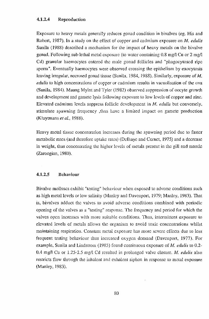
4.1.2.4 Reproduction
Exposure to heavy metals generally reduces gonad condition in bivalves (eg. His and
Robert, 1987). In a study on the effect of copper and cadmium exposure on M. edulis
Sunila (1988) described a mechanism for the impact of heavy metals on the bivalve
gonad. Following sub-lethal metal exposure (to water containing 0.8 mg/1 Cu or 2 mg/1
Cd) granular haemocytes entered the male gonad follicles and "phagocytosed ripe
sperm". Eventually haemocytes were observed crossing the epithelium by exocytosis
leaving irregular, necrosed gonad tissue (Sunila, 1984, 1988). Similarly, exposure of M.
edulis to high concentrations of copper or cadmium results in vacuolisation of the ova
(Sunila, 1984). Maung Myint and Tyler (1982) observed suppression of oocyte growth
and development and gamete lysis following exposure to low levels of copper and zinc.
Elevated cadmium levels suppress follicle development in M. edulis but conversely,
stimulate spawning frequency ,thus have a limited impact on gamete production
(Kluytmans et al., 1988).
Heavy metal tissue concentration increases during the spawning period due to faster
metabolic rates (and therefore uptake rates) (Delhaye and Comet, 1975) and a decrease
in weight, thus concentrating the higher levels of metals present in the gill and mantle
(Zaroogian, 1980).
4.1.2.5 Behaviour
Bivalve molluscs exhibit "testing" behaviour when exposed to adverse conditions such
as high metal levels or low salinity (Manley and Davenport, 1979; Manley, 1983). That
is, bivalves adduct the valves to avoid adverse conditions combined with periodic
opening of the valves as a "testing" response. The frequency and period for which the
valves open increases with more suitable conditions. Thus, intermittent exposure to
elevated levels of metals allows the organism to avoid toxic concentrations whilst
maintaining respiration. Constant metal exposure has more severe effects due to less
frequent testing behaviour thus increased oxygen demand (Davenport, 1977). For
example, Sunila and Lindstrom (1985) found continuous exposure of M. edulis to 0.2-
0.4 mg/1 Cu or 1.25-2.5 mg/1 Cd resulted in prolonged valve closure. M. edulis also
restricts flow through the inhalent and exhalent siphon in response to metal exposure
(Manley, 1983).
80
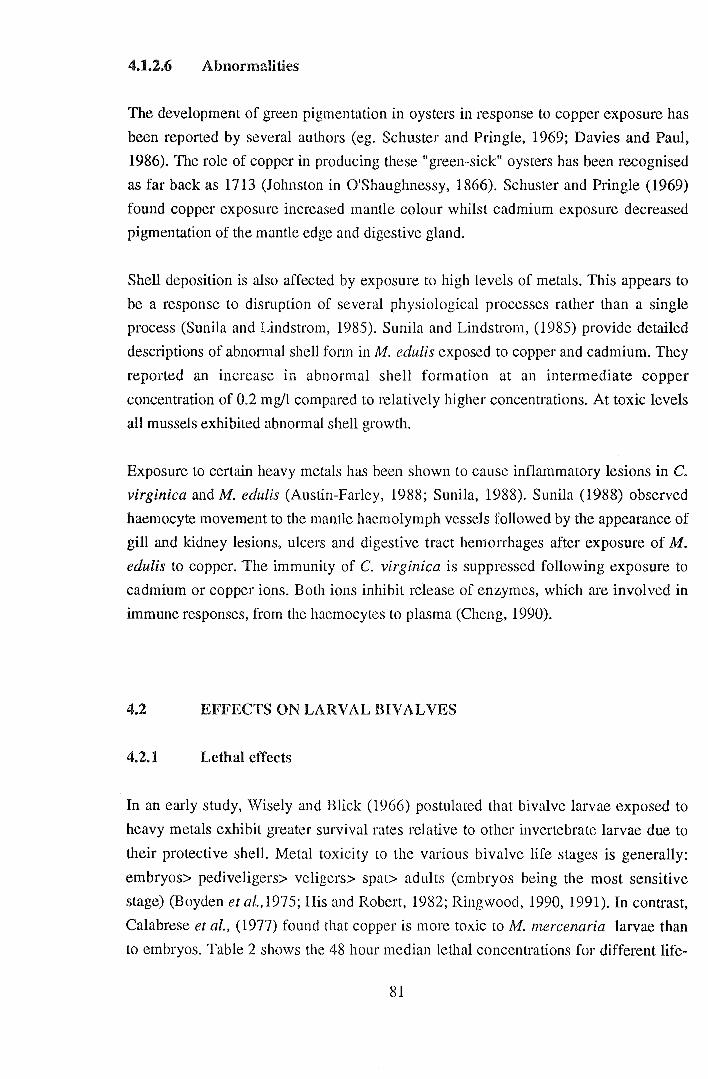
4.1.2.6 Abnormalities
The development of green pigmentation in oysters in response to copper exposure has
been reported by several authors (eg. Schuster and Pringle, 1969; Davies and Paul,
1986). The role of copper in producing these "green-sick" oysters has been recognised
as far back as 1713 (Johnston in O'Shaughnessy, 1866). Schuster and Pringle (1969)
found copper exposure increased mantle colour whilst cadmium exposure decreased
pigmentation of the mantle edge and digestive gland.
Shell deposition is also affected by exposure to high levels of metals. This appears to
be a response to disruption of several physiological processes rather than a single
process (Sunila and Lindstrom, 1985). Sunila and Lindstrom, (1985) provide detailed
descriptions of abnormal shell form in M. edulis exposed to copper and cadmium. They
reported an increase in abnormal shell formation at an intermediate copper
concentration of 0.2 mg/1 compared to relatively higher concentrations. At toxic levels
all mussels exhibited abnormal shell growth.
Exposure to certain heavy metals has been shown to cause inflammatory lesions in C.
virginica and M. edulis (Austin-Farley, 1988; Sunila, 1988). Sunila (1988) observed
haemocyte movement to the mantle haemolymph vessels followed by the appearance of
gill and kidney lesions, ulcers and digestive tract hemorrhages after exposure of M.
edulis to copper. The immunity of C. virginica is suppressed following exposure to
cadmium or copper ions. Both ions inhibit release of enzymes, which are involved in
immune responses, from the haemocytes to plasma (Cheng, 1990).
4.2 EFFECTS ON LARVAL BIVALVES
4.2.1 Lethal effects
In an early study, Wisely and Blick (1966) postulated that bivalve larvae exposed to
heavy metals exhibit greater survival rates relative to other invertebrate larvae due to
their protective shell. Metal toxicity to the various bivalve life stages is generally:
embryos> pediveligers> veligers> spat> adults (embryos being the most sensitive
stage) (Boyden et a/.,1975; His and Robert, 1982; Ringwood, 1990, 1991). In contrast,
Calabrese et al., (1977) found that copper is more toxic toM. mercenaria larvae than
to embryos. Table 2 shows the 48 hour median lethal concentrations for different life-
81
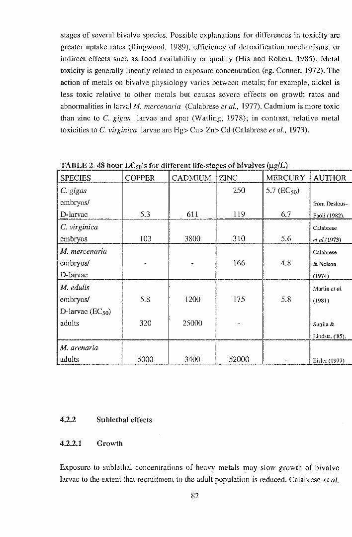
stages of several bivalve species. Possible explanations for differences in toxicity are
greater uptake rates (Ringwood, 1989), efficiency of detoxification mechanisms, or
indirect effects such as food availability or quality (His and Robert, 1985). Metal
toxicity is generally linearly related to exposure concentration (eg. Conner, 1972). The
action of metals on bivalve physiology varies between metals; for example, nickel is
less toxic relative to other metals but causes severe effects on growth rates and
abnormalities in larval M. mercenaria (Calabrese et al., 1977). Cadmium is more toxic
than zinc to C. gigas . larvae and spat (Watling, 1978); in contrast, relative metal
toxicities to C. virginica larvae are Hg> Cu> Zn> Cd (Calabrese et al., 1973).
TABLE2 48h . our LC I f d'ff l'f so s or I erent 1 e-stal!es o fb' ( !L) Iva ves J.lgl
SPECIES COPPER CADMIUM ZINC MERCURY AUTHOR
C. gigas 250 5.7 (ECso)
embryos/ from Deslous-
D-larvae 5.3 611 119 6.7 Paoli (1982).
C. virginica Calabrese
embryos 103 3800 310 5.6 et al.(l973)
M. mercenaria Calabrese
embryos/ - - 166 4.8 & Nelson
D-larvae (1974)
M. edulis Martin et al.
embryos/ 5.8 1200 175 5.8 (1981)
D-larvae (ECso)
adults 320 25000 - Sunila &
Lindstr. ('85).
M. arenaria
adults 5000 3400 52000 - Eisler ( 1977)
4.2.2 Sublethal effects
4.2.2.1 Growth
Exposure to sublethal concentrations of heavy metals may slow growth of bivalve
larvae to the extent that recruitment to the adult population is reduced. Calabrese et al.
82
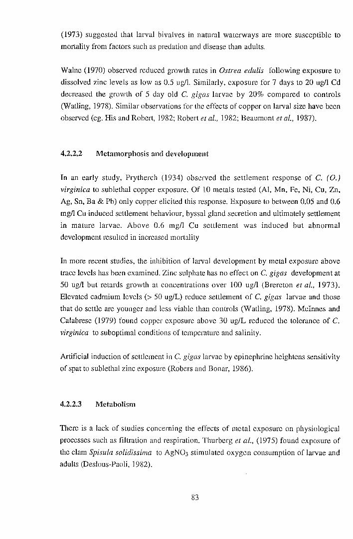
(1973) suggested that larval bivalves in natural waterways are more susceptible to
mortality from factors such as predation and disease than adults.
Walne (1970) observed reduced growth rates in Ostrea edulis following exposure to
dissolved zinc levels as low as 0.5 ug/1. Similarly, exposure for 7 days to 20 ug/1 Cd
decreased the growth of 5 day old C. gigas larvae by 20% compared to controls
(Watling, 1978). Similar observations for the effects of copper on larval size have been
observed (eg. His and Robert, 1982; Robert et al., 1982; Beaumont et al., 1987).
4.2.2.2 Metamorphosis and development
In an early study, Prytherch (1934) observed the settlement response of C. ( 0.)
virginica to sublethal copper exposure. Of 10 metals tested (Al, Mn, Fe, Ni, Cu, Zn,
Ag, Sn, Ba & Pb) only copper elicited this response. Exposure to between 0.05 and 0.6
mg/1 Cu induced settlement behaviour, byssal gland secretion and ultimately settlement
in mature larvae. Above 0.6 mg/1 Cu settlement was induced but abnormal
development resulted in increased mortality
In more recent studies, the inhibition of larval development by metal exposure above
trace levels has been examined. Zinc sulphate has no effect on C. gigas development at
50 ug/1 but retards growth at concentrations over 100 ug/1 (Brereton et al., 1973).
Elevated cadmium levels (> 50 ug/L) reduce settlement of C. gigas larvae and those
that do settle are younger and less viable than controls (Watling, 1978). Mcinnes and
Calabrese (1979) found copper exposure above 30 ug/L reduced the tolerance of C.
virginica to suboptimal conditions of temperature and salinity.
Artificial induction of settlement in C. gigas larvae by epinephrine heightens sensitivity
of spat to sublethal zinc exposure (Robers and Bonar, 1986).
4.2.2.3 Metabolism
There is a lack of studies concerning the effects of metal exposure on physiological
processes such as filtration and respiration. Thurberg et al., (1975) found exposure of
the clam Spisula solidissima to AgN03 stimulated oxygen consumption of larvae and
adults (Deslous-Paoli, 1982).
83
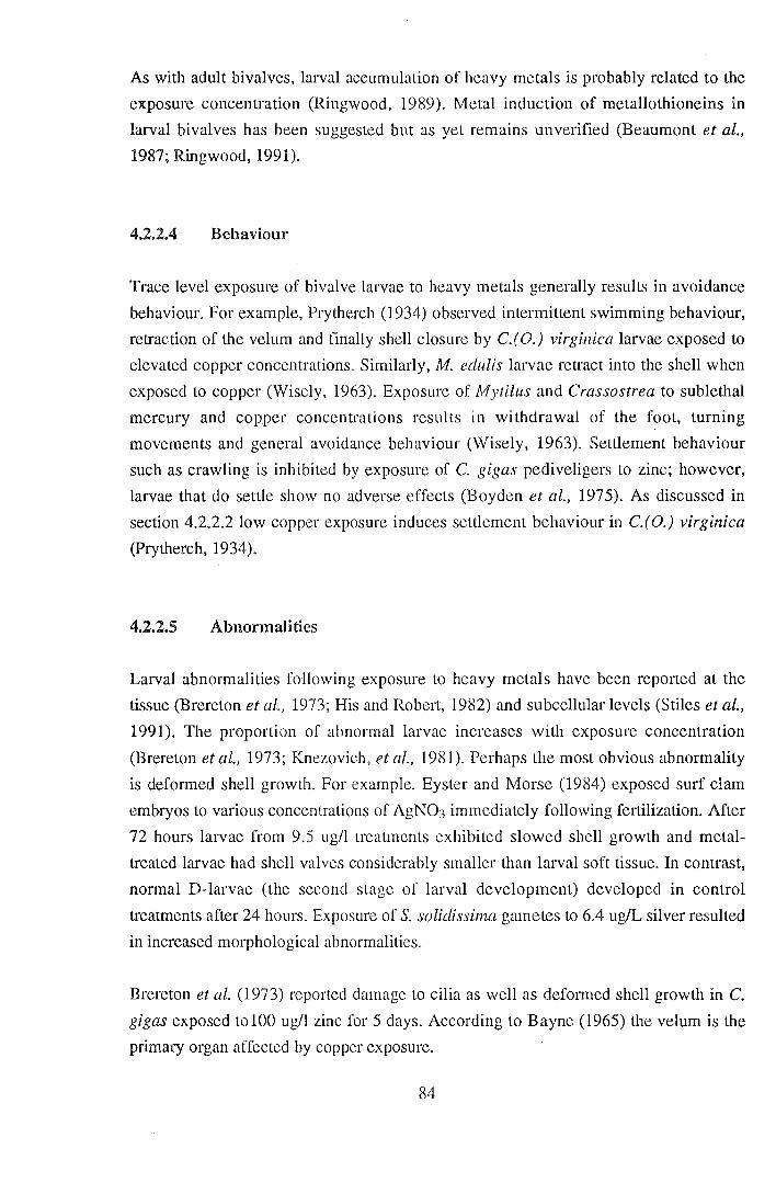
As with adult bivalves, larval accumulation of heavy metals is probably related to the
exposure concentration (Ringwood, 1989). Metal induction of metallothioneins in
larval bivalves has been suggested but as yet remains unverified (Beaumont et al.,
1987; Ringwood, 1991).
4.2.2.4 Behaviour
Trace level exposure of bivalve larvae to heavy metals generally results in avoidance
behaviour. For example, Prytherch (1934) observed intermittent swimming behaviour,
retraction of the velum and finally shell closure by C.(O.) virginica larvae exposed to
elevated copper concentrations. Similarly, M. edulis larvae retract into the shell when
exposed to copper (Wisely, 1963). Exposure of Mytilus and Crassostrea to sublethal
mercury and copper concentrations results in withdrawal of the foot, turning
movements and general avoidance behaviour (Wisely, 1963). Settlement behaviour
such as crawling is inhibited by exposure of C. gigas pediveligers to zinc; however,
larvae that do settle show no adverse effects (Boyden et al., 1975). As discussed in
section 4.2.2.2 low copper exposure induces settlement behaviour in C.(O.) virginica
(Prytherch, 1934).
4.2.2.5 Abnormalities
Larval abnormalities following exposure to heavy metals have been reported at the
tissue (Brereton eta!., 1973; His and Robert, 1982) and subcellular levels (Stiles et al.,
1991). The proportion of abnormal larvae increases with exposure concentration
(Brereton et al., 1973; Knezovich, et al., 1981). Perhaps the most obvious abnormality
is deformed shell growth. For example. Eyster and Morse (1984) exposed surf clam
embryos to various concentrations of AgN03 immediately following fertilization. After
72 hours larvae from 9.5 ug/1 treatments exhibited slowed shell growth and metal
treated larvae had shell valves considerably smaller than larval soft tissue. In contrast,
normal D-larvae (the second stage of larval development) developed in control
treatments after 24 hours. Exposure of S. solidissima gametes to 6.4 ug/L silver resulted
in increased morphological abnormalities.
Brereton et al. (1973) reported damage to cilia as well as deformed shell growth in C.
gigas exposed to 100 ug/l zinc for 5 days. According to Bayne (1965) the velum is the
primary organ affected by copper exposure.
84
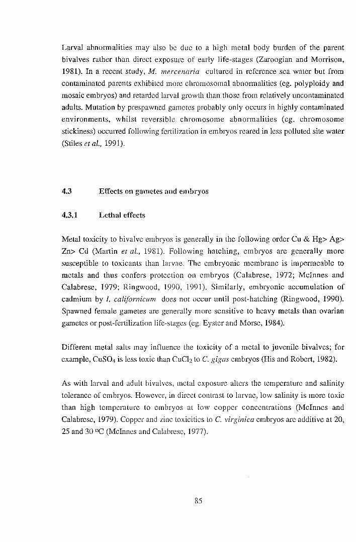
Larval abnormalities may also be due to a high metal body burden of the parent
bivalves rather than direct exposure of early life-stages (Zaroogian and Morrison,
1981). In a recent study, M. mercenaria cultured in reference sea water but from
contaminated parents exhibited more chromosomal abnormalities (eg. polyploidy and
mosaic embryos) and retarded larval growth than those from relatively uncontaminated
adults. Mutation by prespawned gametes probably only occurs in highly contaminated
environments, whilst reversible chromosome abnormalities (eg. chromosome
stickiness) occurred following fertilization in embryos reared in less polluted site water
(Stiles et al., 1991).
4.3 Effects on gametes and embryos
4.3.1 Lethal effects
Metal toxicity to bivalve embryos is generally in the following order Cu & Hg> Ag>
Zn> Cd (Martin ei al., 1981). Following hatching, embryos are generally more
susceptible to toxicants than larvae. The embryonic membrane is impermeable to
metals and thus confers protection on embryos (Calabrese, 1972; Mcinnes and
Calabrese, 1979; Ringwood, 1990, 1991). Similarly, embryonic accumulation of
cadmium by I. californicum does not occur until post-hatching (Ringwood, 1990).
Spawned female gametes are generally more sensitive to heavy metals than ovarian
gametes or post-fertilization life-stages (eg. Eyster and Morse, 1984).
Different metal salts may influence the toxicity of a metal to juvenile bivalves; for
example, CuS04 is less toxic than CuCh to C. gigas embryos (His and Robert, 1982).
As with larval and adult bivalves, metal exposure alters the temperature and salinity
tolerance of embryos. However, in direct contrast to larvae, low salinity is more toxic
than high temperature to embryos at low copper concentrations (Mcinnes and
Calabrese, 1979). Copper and zinc toxicities to C. virginica embryos are additive at 20,
25 and 30 OC (Mcinnes and Calabrese, 1977).
85

4.3.2 Development
Copper slows embryonic cell division and development to the D-larval stage (His and
Robert, 1982; Johnson, 1988). For example, development of C. gigas embryos is
slowed above 5ug/l (Knezovich et al., 1991) and most cell division is arrested at
500ug/l Cu (His and Robert, 1982).
Exposure of adult M. edulis to copper (0.05 ug/L) suppresses gametogenesis, zinc (0.2
ug/L) is less toxic but nevertheless suppresses oocyte development and cadmium (0.05
mg/L) only influences early gonad development (Maung Myint and Tyler, 1982).
Akberali et al. (1984) reported significant stimulation (47%) ofrespiration by M. edulis
oocytes at 0.5mM Cu2+ and inhibition (49%) of sperm respiration by Zn2+.
The cytogenetic effects of metals on bivalve embryos are discussed briefly in section
4.2.2.5.
86

5. CONCLUSIONS
The physiological responses of bivalves to heavy metal exposure is determined by both
environmental and biological factors. Initial metal uptake and accumulation by bivalves
largely depends on the chemical form of the metal (particularly the free ion
concentration) as well as its physical source ie. water, food or sediments (Riley, 1971;
George, 1980). The latter is particularly important to uptake by different species and
life-stages with different feeding strategies. Metal speciation and source also affects the
specific sites of accumulation. Dissolved metal species mostly accumulate in the gills
and kidney, whilst particulate species arc taken up predominantly by the digestive
gland and stored in the kidney. Bivalve metal accumulation from phytoplankton is
generally considered to be relatively minor compared to dissolved metals (King and
Davies, 1987); however, high levels of phytoplankton contamination have resulted in
significant uptake via feeding (eg. Zhang et al., 1990).
The amount of metal accumulation by bivalves generally increases with adult size but
bivalve embryos and larvae take up metals at superior rates to adults (Ringwood, 1991).
A lack of knowledge concerning chronic accumulation of metals by larval bivalves is
evident. Metals are known to accumulate in adult bivalves in intracellular vesicles
located in tissues or haemocytes (George and Pirie, 1980). Specific binding proteins
and other compounds sequester metals within cells or in the haemolymph (Simkiss and
Mason, 1983). Bivalve metal excretion is predominantly via urine and faecal
production as well as by granulocyte diapedesis across the epithelial surfaces (George,
1980; George and Pirie, 1980).
Heavy metals above trace level concentrations exert a range of adverse effects on the
physiology of bivalve molluscs. Exposure to sub-lethal concentrations, particularly to
essential metals such as copper and zinc, may be beneficial to the growth and
development of bivalves (eg Paul and Davies, 1986). However, chronic exposure
generally counterracts any short term stimulus to bivalve physiology (eg. Watling,
1978).
Toxic effects of heavy metals on bivalves are generally measured as 48 or 96 hour
LCso's. A general order of decreasing metal toxicity to bivalves is Cu> Hg> Zn> Cd
(see Table 1). This order varies slightly between bivalve species and life-stages,
probably due to differences in metabolism and feeding strategies.
Sub-lethal effects provide more spedfic information about the action of heavy metals
87
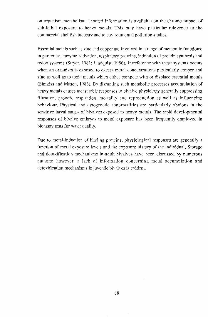
on organism metabolism. Limited information is available on the chronic impact of
sub-lethal exposure to heavy metals. This may have particular relevence to the
commercial shellfish industry and to environmental pollution studies.
Essential metals such as zinc and copper are involved in a range of metabolic functions;
in particular, enzyme activation, respiratory proteins, induction of protein synthesis and
redox systems (Stryer, 1981; Lindquist, 1986). Interference with these systems occurs
when an organism is exposed to excess metal concentrations particularly copper and
zinc as well as to toxic metals which either compete with or displace essential metals
(Simkiss and Mason, 1983). By disrupting such metabolic processes accumulation of
heavy metals causes measurable responses in bivalve physiology generally suppressing
filtration, growth, respiration, mortality and reproduction as well as influencing
behaviour. Physical and cytogenetic abnormalities are particularly obvious in the
sensitive larval stages of bivalves exposed to heavy metals. The rapid developmental
responses of bivalve embryos to metal exposure has been frequently employed in
bioassay tests for water quality.
Due to metal-induction of binding proteins, physiological responses are generally a
function of metal exposure levels and the exposure history of the individual. Storage
and detoxification mechanisms in adult bivalves have been discussed by numerous
authors; however, a lack of information concerning metal accumulation and
detoxification mechanisms in juvenile bivalves is evident.
88
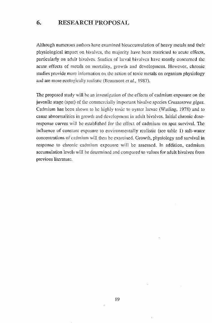
6. RESEARCH PROPOSAL
Although numerous authors have examined bioaccumulation of heavy metals and their
physiological impact on bivalves, the majority have been restricted to acute effects,
particularly on adult bivalves. Studies of larval bivalves have mostly concerned the
acute effects of metals on mortality, growth and development. However, chronic
studies provide more itiformation on the action of toxic metals on organism physiology
and are more ecologically realistic (Beaumont et al., 1987).
The proposed study will be an investigation of the effects of cadmium exposure on the
juvenile stage (spat) of the commercially important bivalve species Crassostrea gigas.
Cadmium has been shown to be highly toxic to oyster larvae (Watling, 1978) and to
cause abnormalities in growth and development in adult bivalves. Initial chronic dose
response curves will be established for the effect of cadmium on spat survival. The
influence of constant exposure to environmentally realistic (see table 1) salt-water
concentrations of cadmium will then be examined. Growth, physiology and survival in
response to chronic cadmium exposure will be assessed. In addition, cadmium
accumulation levels will be determined and compared to values for adult bivalves from
previous literature.
89

6. LITERATURE CITED
Abel, P.D. (1976). Effect of some pollutants on the filtration rate of Mytilus. Mar.
Pollut. Bull., 7(12), 228-231.
Akberali, H.B. & Earnshaw, M. J. (1982). A possible role for mitochondrial Ca2+ in
the contraction of the mollusc siphon induced by Cu2+. Comp. Biochem. Physiol.,
73C, 395-398.
Akberali, H.B., Earnshaw, M. J. & Marriott, K. R. M. (1984). The action of heavy
metals on the gametes of the marine mussel, Mytilus edulis (L.). I. Copper-induced
uncoupling of respiration in the unfertilized egg. Comp. Biochem. Physiol., 77C, 289-
294.
Akberali, H.B. & Trueman, E. R., (1985). Effects of environmental stress on marine
bivalve molluscs. Advances Mar. Bioi., 22, 101-198.
Alzieu, C., Thibaud, Y., Heral, M. & Boutier, B., (1982). Evaluation des risques dus a
l'emploi des peintures anti-salissures dans les zones conchylicoles. Rev. Trav. Inst.
Peches Marit., 44 (4), 303-348
Amdurer, M., Adler, D. & Santschi P. H., (1981). Studies of the chemical forms of
trace elements in sea water using a radiotracer. In: Trace metals in sea water, Wong,
C.S., Boyle, E., Bruland, K.W., Burton, J.D. & Goldberg, E. D. (eds). Plenum Press,
New York. 920 pp.
APHA., (197 6). Standard methods for the examination of water and waste water. 14th
ed. American Public Health Association 1193 pp.
Austin-Farley, C. (1988). Histochemistry as a tool for examining possible pathologic
cause-and-effect relationships between heavy metal and inflammatory lesions in
oysters, Crassostrea virginica, 271-275. In: Marine Environ. Res., 24. Responses of
marine organisms to pollutants. Roesijadi, G. & Spies, R. B. (eds.) Elsevier Applied
Science, England. 356 pp.
Bayne, B. L. (1965). Growth and delay of metamorphosis of the larvae of Mytilus
edulis (L.). Ophelia, 2, 1-47.
90

Bayne, B. L., Widdows, J. & Thompson, R. J. (1976). Physiology: II. In: Marine
mussels: their ecology and physiology. Bayne (ed.) Cambridge University Press,
Cambridge. 506 pp.
Beaumont, A. R., Tserpes, G. & Budd, M. D. (1987). Some effects of copper on the
veliger larvae of the mussel Mytilus edulis and the scallop Pecten maximus (Mollusca,
Bivalvia). Marine Environ. Res. 21, 299-309.
Bebianno, M. J. & Langston, W. J. (1991). Metallothionein induction in Mytilus edulis
exposed to cadmium. Mar. Bioi., 108,91-96.
Borchardt, T. (1983). Influence of cadmium on the kinetics of cadmium uptake and loss
via food and seawater in Mytilus edulis. Mar. Bioi., 76, 67-76.
Boyden, C. R., Watling, H. & Thornton, I. (1975). Effect of zinc on the settlement of
the oyster Crassostrea gigas. Mar. Bioi., 31, 227-234.
Boyden, C. R. (1977). Effect of size upon metal content of shellfish. J. Mar. Bioi. Soc.
U.K., 57,675-714.
Bremner, I. (1977). Measurement and toxicity of metallic and organic species.
Tra1,1sport and storage of trace metals in animals. Proc. Anal. Div. Chem. Soc., 14, 218-
219.
Brereton, A., Lord, H., Thornton, I. & Webb, J. S. (1973). Effect of zinc on growth and
development of larvae of the Pacific oyster Crassostrea gigas. Mar. Biol., 19, 96-101.
Brookes, R. R. & Rumsby, M.G. (1965). The biogeochemistry of trace element uptake
by some New Zealand bivalves. Limnol. Oceanogr. 10, 521-527.
Brown, B. & Newell, R. (1972). The effect of copper and zinc on the metabolism of the
mussel Mytilus edulis. Mar. Biol. 16, 108-118.
Bruland, K. W. (1983). Trace elements in sea-water. In: Chemical oceanography vol.
8. 2nd ed., Riley, J.P. & Chester, R. (eds) Academic Press, London.
Bryan, G. W. (1976). Heavy metal contamination in the sea. In: Marine Pollution ,
Johnston, R. (ed.) Academic Press, London. 729 pp.
91

Bryan, G. W., Langston, W. J., Hummerstone, L. G. & Burt, G. R. (1985). A guide to
the assessment of heavy metal contamination in estuaries using biological indicators.
Occasional paper No.4, March 1985. Mar. Bioi. Assoc. U.K.
Cairns, J., Heath, A. G., & Parker, B. C.(1975). The effect of temperature upon the
toxicity of chemicals to aquatic organisms. Hydrobiologia, 47, 135-171.
Calabrese, A. (1972). How some pollutants affect embryos and larvae of the American
oyster and hard-shell clam. Mar. Fish. Rev., 34 (11-12) 66-77.
Calabrese, A., Collier, R. S. Nelson, D. A. & Macinnes, J. R. (1973). The toxicity of
heavy metals to embryos of the American oyster Crassostrea virginica. Mar. Bioi., 18,
162-166.
Calabrese, A, Macinnes, J. R., Nelson, D. A. &Miller, J. E. (1977). Survival and
growth of bivalve larvae under heavy-metal stress. Mar. Biol., 41, 179-184.
Carmichael, N. G., Squibb, K. S., Engel, D. W. & Fowler, B. A. (1980). Metals in the
molluscan kidney: uptake and subcellular distribution of 109Cd, 54 Mn and 65zn by the
clam, Mercenaria mercenaria. Comp. Biochem. Physiol., 65A, 203-206.
Carpene, E. & George, S. G. (1981). Absorption of cadmium by gills of Mytilus edulis
(L.). Mol. Physiol. 1, 23-34.
Casterline, J. L. & Yip, G. (1975). The distribution and binding of cadmium in oyster,
soybean and rat liver and kidney. Arch. Environm. Contam. Toxicol., 3, 319-329.
Cheng, T. C. (1990). Effects of in vivo exposure of Crassostrea virginica to heavy
metals on haemocyte viability and activity levels of lysosomal enzymes. In: Pathology
in Marine Science, Perkins, F. 0. & Cheng, T. C. (eds.) Academic Press Inc., San
Diego, 538 pp.
Connor, P.M. (1972). Acute toxicity of heavy metals to some marine larvae. Mar. Poll.
Bull. 3 (12) 190-192.
Coombs, T. L. (1974). Zinc and copper in the soft tissues of Ostrea edulis. Mar. Bioi.
28, 1-10.
92

Coombes, T. L. (1977). Uptake and storage mechanisms of heavy metals in marine
organisms. Proc. Analyt. Div. Chern. Soc. 14,219-222.
Coombs,T. L. & George, S. G. (1977). Mechanisms of immobilization and
detoxification of metals in marine organisms. In: Physiology and behaviour of marine
organisms. 12th European Symposium on Marine Biology. McLusky, D. S. & Berry, A.
J. (eds.) Pergamon Press, Scotland. 388 pp.
Cooper, R. J., Langlois, D. & Olley, J. (1982). Heavy metals in Tasmanian shellfish. 1-
Monitoring heavy metal contamination in the Derwent estuary: use of oysters and
mussels. J. App. Toxicol., 2, (2) 99-109.
Cossa, D., Bourget, E., Pouliot, D., Piuze, J. & Chanut, J. P. (1980). Geographical and
seasonal variations in the relationship between trace metal content and body weight in
Mytilus edulis. Mar. Bioi., 58, 7-14.
Cunningham, P. A. & Tripp, M. R. (1975). Accumulation, tissue distribution and
elimination of 203HgC12 and CH3203HgC1 in the tissues of the American oyster
Crassostrea virginica. Mar. Bioi., 31, 321-344.
Dav~nport, J. (1977). A study of the effects of copper applied continuously and
discontinuously to specimens of Mytilus edulis (L.) exposed to steady and fluctuating
salinity levels. J. Mar. Bioi. Assoc. U.K., 57, 63-74.
Davies, I. M. & Paul, J. D. (1986). Accumulation of copper and nickel from anti
fouling compounds during cultivation of scallops (Pecten maximus L.) and Pacific
oysters (Crassostrea gigas Thun.) Aquaculture 55, 93-102.
Delhaye, W. & Cornet, D. (1975). Contribution to the study of the effect of copper on
Mytilus edulis during reproductive period. Comp. Biochem. Physiol., SOA, 511-518.
Denton, G. R. W & Burdon-Jones, C. (1981). Influence of temperature and salinity on
the uptake, distribution and depuration of mercury, cadmium and lead by the black-lip
oyster Saccostrea echinata .. Mar. Bioi., 64,317-326.
Department of the Environment (1987). Report for the year 1986-87. Government
printer, Tasmania.
93
Dermott, R. M. & Lum, K. R.(1986). Metal concentrations in the annual shell layers of
the bivalve Elliptio complanata. Env. Pollution, (Series B) 12, 131-143.
Deslous-Paoli, J. (1982). Toxicite des elements metalliques dissous pour les larves
d'organismes marins: donnees bibliographiques. Rev. Trav.lnst. Peches Marit., 45, 73-
83.
Doyle, L. J., Blake, N.J., Woo, C. and Yevich, P. (1931). Recent biogenic phosphorite:
concretions in mollusc kidneys. Science, 199, 1431-1433.
Eisler, R.(1977). Toxicity evaluation of a complex metal mixture to the softshell clam
Mya arenaria. Mar. Bioi., 43,265-276.
Elliott, N. G. (1982). Biomonitoring of heavy metals in sea water. Thesis (Ph. D)
University of Tasmania, 192 pp.
Elliott, N. G., Swain, R. and Ritz, D. A. (1986). Metal interaction during accumulation
by the mussel Mytilus edulis planulatus. Mar. Bioi., 93, 395-399.
Elston, R. (1980). Functional morphology of the coelomocytes of the larval oysters
(CrCfSSostrea virginica and Crassostrea gigas). J. Mar. Bioi. Ass. U.K., 60, 947-957.
Engel, D. W., Sunda, W. G. and Fowler, B. A. (1981). Factors affecting trace metal
uptake and toxicity to estuarine organisms. I. Environmental Parameters. In: Biological
monitoring of marine pollutants. Vernberg, J., Calabrese, A., Thurberg, F. P. and
Vernberg, W. B. (eds.). Academic Press.
Eyster, L. S. & Morse, M. P. (1984). Development of the surf clam (Spisula
solidissima) following exposure of gametes, embryos, and larvae to silver. Arch.
Environ. Contam. Toxicol., 13,641-646.
Feeley, R. A., Massoth, G. J. & Lamb, M. F. (1981). The effect of sewage effluents on
the flocculation of major and trace elements in a stratified estuary. In: Trace metals in
sea water, Wong, C.S., Boyle, E., Bruland, K.W., Burton, J. D. & Goldberg, E. D.
(eds). Plenum Press, New York, 920 pp.
94

Fowler, B. A., Gould, E., Garvey, J. S. & Bakewell, W. E. (1988). Comparative studies
on the 45000 Dalton cadmium-binding protein (45 K Cd BP) from the scallop
Placopecten magellanicus: Immunological properties and copper competition studies.
(Abstract) p. 171-172. In: Marine Environ. Res., 24. Responses of marine organisms to
pollutants. Roesijadi, G. & Spies, R. B. (eds.) Elsevier Applied Science, England. 356
pp.
Frazier, J. M. (1975). The dynamics of metals in the American oyster, Crassostrea
virginica. I. Seasonal effects. Science, 16 (3) 162-171.
Frazier, J. M. & George, S. G. (1983). Cadmium kinetics in oysters -a comparative
study of Crassostrea gigas and Ostrea edulis. Mar. Bioi., 76, 55-61.
George, S. G. (1980). Correlation of metal accumulation in mussels with the
mechanisms of uptake, metabolism and detoxification: a review. Thalassia
Jugoslavica, 16, 347-365.
George, S. G. (1982). Subcellular accumulation and detoxication of metals in aquatic
animals. In: Physiological mechanisms of marine pollutant toxicity. Vernberg, W. B.,
Calabrese, A., Thurberg, F. P. & Vernberg, F. J. (eds.) Academic Press.
Ge9rge, S. G., Carpene, E. & Coombs, T.L. (1977). The effect of salinity on the uptake
of cadmium by the common mussel Mytilus edulis (L.). In: Physiology and behaviour
of marine organisms. 12th European Symposium on Marine Biology. McLusky, D. S.
& Berry, A. J. (eds.) Pergamon Press, Scotland.
George S. G., Carpene, E., Coombs, T.L., Overnell, J. & Youngson, A. (1979).
Characterisation of cadmium-binding proteins from mussels, Mytilus edulis (L.),
exposed to cadmium. Biochim. Biophys. Acta, 580, 225-233.
George, S.G & Coombes, T. L. (1975). A comparison of trace-metal and
metalloenzyme profiles in different molluscs and during development of the oyster.
p.433-449. In: Proceedings of the 9th European Marine Biology Symposium. Barnes,
H. (ed.) Aberdeen University Press. 760 pp.
George, S. G. & Coombs, T.L. (1977). The effects of chelating agents on the uptake
and accumulation of cadmium by Mytilus edulis. Mar. Bioi., 39, 261-268.
95
George, S. G., Pirie, B. J. S. & Coombs, T.L. (1976). The kinetics of accumulation and
excretion of ferric hydroxide in Mytilus edulis (L.) and its distribution in the tissues. J.
Exp. Mar. Biol. Ecol., 23,71-84.
George, S. G., Pirie, B. J. S. & Coombs, T.L. (1980). Isolation and elemental analysis
of metal-rich granules from the kidney of the scallop, Pecten maximus (L.). J. Exp.
Mar. Bioi. Ecol., 42, 143-156.
George, S. G. & Pirie, B. J. S. (1980). Metabolism of zinc in the mussel, Mytilus edulis
(L.): a combined ultrastructural and biochemical study. J. Mar. Bioi. Assoc. U.K., 60,
575-590.
Greig, R. A., Nelson, B. A. & Nelson, D. A. (1975). Trace metal content in the
American oyster. Marine Poll. Bull., 6, 72-73.
Greig, R. A. & Wenzloff, D. R. (1978). Metal accumulation and depuration in the
American oyster Crassostrea virginica. Bull. Environ. Contam. Toxicol., 20, 499-504.
Harrison, F .L., Lam, J. R. & Novacek, J (1988). Partitioning of metals among metal
binding proteins in the bay mussel, Mytilus edulis. In: Marine Environ. Res., 24.
Responses of marine organisms to pollutants. Roesijadi, G. & Spies, R. B. (eds.)
Elst?vier Applied Science.
His, E. & Robert, R. (1982). Le danger des traitements par le sulfate de cuivre en zone
conchylicole: toxicite vis-a-vis des oeufs et des jeunes larves de Crassostrea gigas.
Rev. Trav. lnst. Peches Marit., 45, (2) 117-125.
His, E. & Robert, R. (1985). Developpement des veligeres de Crassostrea gigas dans le
bassin d'Arcachon. Etudes sur les mortalites larvaires. Rev. Trav. Inst. Peches Marit.,
47, 63-88.
His, E. & Robert, R. (1987). Comparative effects of two antifouling paints on the oyster
Crassostrea gigas. Mar. Bioi., 95, 83-86.
Hobden, D. J. (1969). Iron metabolism in Mytilus edulis. II. Uptake and distribution of
radioactive iron. J. Mar. Bioi. Assoc. U.K., 49, 661-668.
96

Howard, A. G. & Nickless, G. (1977). Heavy metal complexation in polluted molluscs.
II. Oysters (Ostrea edulis & Crassostrea gigas). Chem.-Biol.lnterr., 17, 257-263.
Howard, A. G. & Nickless, G. (1978). Heavy metal complexation in polluted molluscs.
III. Periwinkles (Littorina littorea), cockles (Cardium edule) and scallops(Chlamys
opercularis). Chem.-Biol.lnterr., 23, 227-231.
Hughes, J. P. (1975). Zinc and cadmium accumulation by sewage microorganisms.
B.Sc. Honours Thesis, University of Tasmania.
Kennish, M. J. (1989). Practical handbook of marine science. CRC Press. 710 pp.
Jackim, E., Morrison, G. & Steele, R. (1977). Effects of environmental factors on
radiocadmium uptake by four species of marine bivalves. Mar. Bioi., 40, 303-308.
Janssen, H. H. & Scholz, N. (1979). Uptake and cellular distribution of cadmium in
Mytilus edulis. Mar. Bioi., 55, 133-141.
Johnson, D. (1988). Development of Mytilus edulis embryos: a bioassay for polluted
waters. Mar. Ecol. -Prog. Ser., 46, 135-138.
Jones, M. B. (1977).Influence of salinity and temperature on the toxicity of mercury to
marine and brackish water isopods (Crustacea). Est. Coast. Mar. Sci., 1, 425-431.
King, D. G. & Davies, I. M. (1987). Laboratory and field studies of the accumulation of
inorganic mercury by the mussel Mytilus edulis (L.). Marine Poll. Bull., 18 (1) 40-45.
Kluytmans, J. H., Brands, F. & Zandee, D. I. (1988). Interractions of cadmium with the
reproductive cycle of Mytilus edulis (L.). In: Marine Environ. Res., 24. Responses of
marine organisms to pollutants. Roesijadi, G. & Spies, R. B. (eds.) Elsevier Applied
Science, England. 356 pp.
Knauer, G. & Martin, J. (1973). Seasonal variations of cadmium, copper, manganese,
lead and zinc in the water and phytoplankton in Monterey Bay, California. Limnol.
Oceanogr., 18 (4) 597-604.
97
Knezovich, J. P., Harrison, F. L. & Tucker, J. S. (1991). The influence of organic
chelators on the toxicity of copper to embryos of the Pacific oyster, Crassostrea gigas.
Arch. Envir. Contam. Toxicol., 10, 241-249.
Langston, W. J. (1982). The distribution of mercury in British estuarine sediments and
its availability to the deposit feeding bivalves. J. Mar. Bioi. Assoc. U.K., 62, 667-684.
Langston, W. J., Bebbianno, M. J & Zhou M. (1989). A comparison of metal-binding
proteins and cadmium metabolism in the marine molluscs Littorina littorea
(Gastropoda), Mytilus edulis andMacoma balthica (Bivalvia). Mar. Environ. Res., 28,
195-200.
Lewis, A. G., Whitfield, P. H. & Ramnarine, A. (1972). Some particulate and soluble
agents affecting the relationship between metal toxicity and organism survival in the
calanoid copepod Euchaetajaponica. Mar. Bioi., 17, 215-221.
Lindquist, S. (1986). The heat shock response. Ann. Rev. Biochem., 55, 1151-1191.
Lowe, D. M. & Moore, M. N. (1979). The cytochemical distributions of zinc (Zn II)
and iron (Fe III) in the common mussel, Mytilus edulis and their relationship with
lysosomes. J. Mar. Bioi. Assoc. U.K., 59, 851-858.
Luoma, S. N. & Bryan, G. W (1978). Factors controlling the availability of sediment
bound lead to the estuarine bivalve Scrobicularia plana. J. Mar. Bioi. Assoc. U.K., 58,
793-802.
Manly, A. R. (1983). The effects of copper on the behaviour, respiration, filtration and
ventilation activity of Mytilus edulis. J. Mar. Bioi. Assoc. U.K., 63, 205-222.
Manly, A. R. & Davenport, J. (1979). Behavioural responses of some marine bivalves
to heightened salt water copper concentrations. Bull. Environ. Contam. Toxicoi., 22,
739-744.
Manly, A. R., Gruffydd, L. D. & Almada-Villela, P. C. (1984). The effect of copper
and zinc on the shell growth of Mytilus edulis measured by a laser diffraction
technique. J. Mar. Bioi. Assoc. U.K., 64,417-427.
98

Mantoura, R. F. C., Dickson, A. & Riley, J.P. (1978). The complexation of metals with
humic materials in natural waters. Estuar. Coast. Mar. Sci., 6, 387-408.
Martin, M., Osborn, K., Billig, P. & Glickstein, N. (1981). Toxicities of ten metals to
Crassostrea gigas embryos and Cancer magister larvae. Mar. Poll. Bull., 12 (9) 305-
308.
Maung Myint, U. & Tyler, P. (1982). Effects of temperature, nutritive and metal
stressors on the reproductive biology of Mytilus edulis. Mar. Bioi., 67, 209-223.
Macinnes, J. R. & Calabrese, A. (1979). Combined effects of salinity, temperature and
copper on embryos and early larvae of the American oyster, Crassostrea virginica.
Arch. Environ. Contam. Toxicol., 8, 553-562.
McLeese, D. W & Ray, S. (1984). Uptake and excretion of Cd, Cd-EDTA and Zn by
Macoma balthica. Bull. Environ. Contam. Tocicol., 32, 85-92.
McLusky, D. S., Bryant, V. & Campbell, R. (1986). The effects of temperature and
salinity on the toxicity of heavy metals to marine and estuarine invertebrates.
Oceanogr. Mar. Bioi. Ann. Rev., 24,481-520.
Mohlenberg, F. & Riisgard, H. U. (1988). Partitioning of inorganic and organic
mercury in cockles Cardium edule (L.) and C. glaucum (Bruguiere) from a chronically
polluted area: influence of size and age. Environ. Pollut., 55, 137-148.
Morton, B. (1976). The biology and functional morphology of the Southeast Asian
mangrove bivalve, Polymesoda (Geloina) erosa (Solander, 1786) (Bivalvia:
Corbiculidae). Can. J. Zoo!., 54, 482-500.
Nakahara, H. & Bevelander, G. (1967). Ingestion of particulate matter by the outer
surface cells of the mollusc mantle. J. Morph., 122, 139-146.
Nelson, A. & Donkin, P. (1985).Processes of bioaccumulation: the importance of
chemical speciation. Mar. Pollut. Bull., 16 (4) 164-169.
Noel-Lambot, F. (1976). Distribution of cadmium, zinc and copper in the mussel
Mytilus edulis. Existence of cadmium-binding proteins similar to metallothioneins.
Experentia, 32 (1) 324-326.
99
O'Shaughnessy, A. W. E. (1866). On green oysters The annals and magazine of natural
history. Series 3. 18, 221-228.
Patel, B. & Anthony, K. (1991). Uptake of cadmium in tropical marine lamellibranchs,
and effects on physiological behaviour. Mar. Bioi., 108, 457-470.
Paul, J. D. & Davies, I. M. (1986). Effects of copper- and tin-based anti-fouling
compounds on the growth of scallops (Pecten maximus) and oysters (Crassostrea
gigas). Aquaculture, 54, 191-203.
Pentreath, R. J. (1973). The accumulation of 65zn, 54Mn, 58Co and 59Fe by the mussel
Mytilus edulis. J. Mar. Bioi. Assoc. U.K., 53, 127-143.
Phillips, D. J. H. (1976). The common mussel Mytilus edulis as an indicator of
pollution by zinc, cadmium, lead, and copper. I. Effects of environmental variables on
uptake of metals. Mar. Bioi., 38, 59-69.
Pirie, B. J. S., George, S. G., Lytton, D. G. & Thomson, J.D. (1984). Metal-containing
blood cells of oysters: ultrastructure, histochemistry and X-ray microanalysis. J. Mar.
Bioi. Assoc. U.K., 64, 115-123.
Prytherch, H. F. (1934). The role of copper in the setting, metamorphosis and
distribution of the American oyster, Ostrea virginica. Ecol. mono gr., 4 (1) 47-107.
Regoli, F., Orlando, E., Mauri, M., Nigro, M. & Cognetti, G. A. (1991). Heavy metal
accumulation and calcium content in the bivalve Donacilla cornea. Mar. Ecol. Prog.
Ser., 74, 219-224.
Riisgard, H. U. & Hansen, S. (1990). Biomagnification of mercury in a grazing food
chain: algal cells P haeodactylum tricornatum, mussels My til us edulis and flounders
Platichthysflesus studied by means of a stepwise reduction- CVAA method. Mar. Ecol.
Prog. Ser., 62, 259-270.
Riley, J.P. (1971). The major and minor elements in sea water. In.: Introduction to
marine chemistry. Riley, J.P. & Chester, R. (eds.) Academic Press, London.
100

Ringwood, A. H. (1989). Accumulation of cadmium by larvae and adults of an
Hawaiian bivalve, Isognomon californicum, during chronic exposure. Mar. Bioi., 102,
499-504.
Ringwood, A. H. (1990). The relative sensitivities of different life stages of Isognomon
californicum to cadmium toxicity. Arch. Environ. Contam. Toxicol., 19, 338-340.
Ringwood, A. H. (1991). Short-term accumulation of cadmium by embryos, larvae, and
adults of an Hawaiian bivalve, Isognomon californicum. J. Exp. Mar. Bioi. Ecol., 149,
55-66.
Ritz, D., Swain, R. & Elliott, N. (1982). Use of the mussel Mytilus edulis planulatus in
monitoring heavy metal levels in sea water. Aust. J. mar freshw. Res., 33 (3) 491-506.
Robers, S. K. & Bonar, D. (1986). Inhibition of metamorphosis in Crassostrea gigas
by zinc. Am. Zoot. 26, Abstract 49.
Robert, R., His, E. & Maurer, D. (1982). L'unite d'ecophysiologie et de molysmologie
larvaire des bivalves d'interet commercial du laboratoire I.S.T.P.M. d'Arcachon. Rev.
Trav. Inst. Peches marit., 45 (3) 197-209.
Robinson, R. E. & Ryan, D. K. (1988). Transport of cadmium and other metals in the
blood of the bivalve mollusc Mercenaria mercenaria. Mar. Bioi., 97, 101-109.
Roesijadi, G. (1980). Influence of copper on the clam Protothaca staminea: effects on
gills and occurrence of copper-binding proteins. Bioi. Bull., 158, 233-247.
Roesijadi, G. (1982). Uptake and incorporation of mercury into mercury-binding
proteins of Mytilus edulis gills as a function of time. Mar. Bioi., 66, 151-157.
Roesijadi, G., Calabrese, A. & Nelson, D. A. (1982). Mercury-binding proteins of
Mytilus edulis. In: Physiological mechanisms of marine pollutant toxicity. Vernberg,
W. B., Calabrese, A., Thurberg, F. P. & Vernberg, F. J. (eds.) Academic Press,.
Ruddel, C. L. (1971). Elucidation of the nature and function of the granular oyster
amoebocytes through histochemical studies of normal and traumatized oyster tissues.
Histochemie, 26, 98-112.
101
Ruddel, C. L. & Rains, D. W. (1975). The relationship between zinc, copper and the
basophils of two crassostreid oysters, C. gigas and C. virginica. Comp. Biochem.
Physiol., 51A, 585-591.
Schulz-Baldes, M .. (1974). Lead transport in the common mussel Mytilus edulis. In:
Physiology and behaviour of marine organisms. 12th European Symposium on Marine
Biology. McLusky, D. S. & Berry, A. J. (eds.) Pergamon Press, Scotland. 388 pp.
Schuster, C. (Jr) & Pringle, B. (1969). Trace metal accumulation by the American
Eastern oyster, Crassostrea virginica. Proc. Nat. Shellfish. Assoc., 59, 91-103.
Scott, D. M. & Major, C. W. (1972). The effect of copper (II) on survival, respiration
and heart rate in the common blue mussel, Mytilus edulis. Bioi. Bull., 143, 679-688.
Simkiss, K. (1983). Lipid solubility of heavy metals in saline solutions. J. Mar. Bioi.
Assoc. U.K., 63, 1-7.
Simkiss, K. & Mason, A. Z. (1983). Metal ions: metabolic and toxic effects. In: The
mollusca, 2, Environmental biochemistry and physiology, Hochachka, P. W. (ed)
Academic Press, New York.
Simkiss, K. & Watkins, B. (1988). Metal ion activities in oscillating temperatures. In:
Marine Environ. Res., 24. Responses of marine organisms to pollutants. Roesijadi, G.
& Spies, R. B. (eds.) Elsevier Applied Science.
Steinert, S. A. & Pickwell, G. V. (1988). Expression of heat shock proteins and
metallothionein in mussels exposed to heat stress and metal ion challenge. In: Marine
Environ. Res., 24. Responses of marine organisms to pollutants. Roesijadi, G. & Spies,
R. B. (eds.) Elsevier Applied Science, Engalnd. 356 pp.
Stromgen, T. (1982). Effect of heavy metals (Zn, Hg, Cu, Cd, Pb, Ni) on the length
growth of Mytilus edulis. Mar. Bioi., 72, 69-72.
Stryer, L. (1981). Biochemistry. 2nd ed. W. H. Freeman & Co., San Francisco. 949pp.
Styles, S., Choromanski, J., Nelson, D., Miller, J., Greig, R. & Sennefelder, G. (1991).
Early reproductive success of the hard clam (Mercenaria mercenaria) from five sites
in Long Island sound. Estuaries, 14 (3) 332-342.
102

Sunila, I. & Lindstrom, R. (1985). Survival, growth and shell deformities of copper
and cadmium-exposed mussels (Mytilus edulis L.) in brackish water. Estuar., Coast.
Shelf Sci., 21, 555-565.
Sunila, I. (1984). Copper- and cadmium-induced histological changes in the mantle of
Mytilus edulis L. (Bivalvia). Limnologica, 15 (2) 523-527.
Sunila, I. (1988). Pollution-related histopathological changes in the mussel Mytilus
edulis L. in the Baltic Sea. In: Marine Environ. Res., 24. Responses of marine
organisms to pollutants. Roesijadi, G. & Spies, R. B. (eds.) Elsevier Applied Science,
England, 356 pp.
Talbot, V. & Magee, R. J. (1978). Naturally- occurring heavy metal binding proteins in
inverebrates. Arch. Environ. Contam. Toxicol., 7, 73-81.
Thomson, J.D., Pirie, B. J. S & George, S. G. (1985). Cellular metal distribution in the
Pacific oyster, Crassostrea gigas (Thun.) determined by quantitative X-ray microprobe
analysis. J. Exp. Mar. Biol. Ecol., 85, 37-45.
Thomson, J. D. (1982). Metal concentration changes in growing Pacific oysters,
Crassostrea gigas, cultivated in Tasmania, Australia. Mar. Bioi., 67, 135-142.
Thurberg, F.P., Cable, W. D., Dawson, M. A., Mcinnes, J. R. & Wenzloff, D. R.
(1975). Respiratory response of larval, juvenile, and adult surf clam, Spisula
solidissima, to silver. In: Respiration of marine organisms. Cech, J. J. et al. (eds) The
Research Institute of the Guld of Maine. In: Deslous-Paoli, J. M. (1982). Toxicite des
elements metalliques dissous pour les larves d'organismes marins: donnees
bibliographiques. Rev. Trav. Inst. Peches Marit., 45, 73-83.
Vernberg, W. B. & Vernberg, F. J. (1972). Environmental physiology of marine
animals. Springer-Verlag.
Walne, P. R. (1970). Present problems in the culture of the larvae of Ostrea edulis.
Helgolander Wiss. Meeresunters 20, 514-524.
Watling, H. R. (1978). Effect of cadmium on larvae and spat of the oyster Crassostrea
gigas (Thunberg). Trans. roy. Soc. S. Afr., 43 (2) 125-134.
103

Webb, J., Macey, D. J. & Talbot, V. (1985). Identification of ferritin as a major high
molecular weight Zn-binding protein in the tropical rock oyster, Saccostrea cucculata.
Arch. Environ. Contam. Toxicol., 14,403-407.
Weber, R. E. (1990). Effects of mercury on the functional properties of haemoglobins
from the bivalve mollusc Scapharca inaequivalvis. J. Exp. Mar. Bioi. Ecol., 144, 39-
48.
Weber, R. E., Lykke-Madsen, M., Bang, A., De Zwaan, A. & Cortesi, P. (1990).
Effects of cadmium on anoxic survival, haematology, erythrocytic volume regulation
and haemoglobin-oxygen affinity in the marine bivalve Scapharca inaequivalvis. J.
Exp. Mar. Bioi. Ecol., 144, 29-38.
Wisely, B. (1963). Detection and avoidance of a cuprous oxide antifouling paint by
bivalve and gastropod larvae (Mollusca). Aust. J. Mar. F. Wat. Res., 14 (1) 60-69.
Wisely, B. & Blick, R. A. P. (1966). Mortality of marine invertebrate larvae in
mercury, copper and zinc solutions. Aust. J. Mar. F. Wat. Res., 18,63-72.
Zamuda, C. D. & Sunda, W. G. (1982). Bioavailability of dissolved copper to the
American oyster Crassostrea virginica. I. Importance of chemical speciation. Mar.
Bioi., 66, 77-82.
Zaroogian, G. E. (1980). Crassostrea virginica as an indicator of cadmium pollution.
Mar. Biol., 58, 275-284.
Zaroogian, G. E. & Johnson, M. (1983). Copper accumulation in the bay scallop,
Argopecten irradians. Arch. Environ. Contam. Toxicol., 12, 127-133.
Zaroogian, G. E., Morrison, G. & Heltsche, J. F. (1979). Crassostrea virginica as an
indicator of lead pollution. Mar. Bioi., 52, 189-196.
Zaroogian, G. E. & Morrison, G. (1981). Effect of cadmium body burdens in adult
Crassostrea virginica on fecundity and viability of larvae. Bull. Environ. Contam.
Toxicol., 27, 344-348.
104
Zhang, G., Hu, M., Huang, Y. & Harrison, P. (1990). Selenium uptake and
accumulation in marine phytoplankton and transfer of selenium to the clam Puditapes
philippnarum. Mar. Environ. Res., 30, 179-190.
105
Related Documents