ISJ 9: 184-199, 2012 ISSN 1824-307X REVIEW Physiological and molecular responses of bivalves to toxic dinoflagellates C Manfrin 1 , G De Moro 1 , V Torboli 1 , P Venier 2 , A Pallavicini 1 , M Gerdol 1 1 Department of Life Sciences, University of Trieste, Trieste, Italy 2 Department of Biology, University of Padua, Padua, Italy Accepted October 31, 2012 Abstract Dinoflagellates and other microalgae can produce a wide spectrum of toxic molecules, which are the main responsible of shellfish poisoning syndromes. During seasonal harmful algal blooms (HABs), many filter-feeding marine invertebrates, including bivalve molluscs, can accumulate phycotoxins at extremely high levels, thus representing a serious threat to human health. Furthermore, HABs also have a severe impact on the aquaculture sector due to the forced prolonged closure of large harvesting areas. Although the targets and mechanism of action of many phycotoxins have been extensively studied on vertebrate model organisms, so far just a little attention has been focused on their effects on marine invertebrates. Here we provide an overview about the molecular response of marine bivalves to phycotoxins, with a particular focus on toxins produced by dinoflagellates. Even though large-scale genomic and proteomic approaches on molluscs are still hindered by the limited molecular knowledge of these organisms, a few studies exploiting the most recent technological advances provide promising perspectives for a better comprehension of the mechanisms involved in shellfish toxicity and for the identification of molecular markers of contamination. Key Words: bivalve; phycotoxin; harmful algal bloom; dinoflagellates; microarray; gene expression; RNA-seq Introduction The largest component of the universe of algae, estimated to comprise between one and ten million different species, is represented by unicellular microalgae. Some of them can produce, through complex and not completely understood biochemical processes, toxic compounds of various chemical composition and modes of action, collectively named ___________________________________________________________________________ Corresponding author: Alberto Pallavicini Department of Life Sciences University of Trieste Via Giorgieri 5, 34127 Trieste, Italy E-mail: [email protected] 184 List of abbreviations: ASP: Amnesic Shellfish Poisoning; AZA/AZT: Azaspiracid/ Azaspiracid Shellfish Poisoning; pBTx: Brevetoxin; DA: Domoic Acid; DSP/DST: Diarrhetic Shellfish Poisoning/Toxin; DTX: Dinophysistoxin; GYM: Gymnodimine; HAB: Harmful Algal Bloom; NSP: Neurotoxic Shellfish Poisoning; OA: Okadaic Acid; PLTX: Palytoxin; PSP/PST: Paralytic Shellfish Poisoning/Toxin, PTX: Pectenotoxin; SPX: Spirolides; STX: Saxitoxin; YTX: Yessotoxin. phycotoxins. In response to favorable environmental conditions, toxic microalgae can proliferate and/or aggregate to form dense concentrations, called “Harmful Algal Blooms” (HABs). Phycotoxins are responsible of a number of human illnesses associated with the consumption of contaminated seafood and, in some cases, with respiratory exposure to aerosolized toxins. In fact, filter-feeding shellfish, zooplankton, and herbivorous fishes can ingest these algae and act as vectors to humans either directly (e.g. shellfish) or through further food web transfer to higher trophic levels. Although seasonal micro-algal blooms are considered as a natural phenomenon, their frequency of occurrence appears to have increased in the recent years. Certainly, the worsening of the hygienic characteristic of the aquaculture areas, the transportation of ship ballast water, water eutrophication and global climate changes are factors which altogether provide favorable conditions for the spreading and the growth of toxic algae, thus contributing to the increase of threats for human by the consumption of contaminated shellfish. Unfortunately, algal toxins are not detectable by sight or smell and contaminated seafood appears normal and in most cases they are heat stable and thereby largely unaffected by cooking.

Welcome message from author
This document is posted to help you gain knowledge. Please leave a comment to let me know what you think about it! Share it to your friends and learn new things together.
Transcript

ISJ 9: 184-199, 2012 ISSN 1824-307X
REVIEW Physiological and molecular responses of bivalves to toxic dinoflagellates C Manfrin1, G De Moro1, V Torboli1, P Venier2, A Pallavicini1, M Gerdol1 1Department of Life Sciences, University of Trieste, Trieste, Italy
2Department of Biology, University of Padua, Padua, Italy
Accepted October 31, 2012
Abstract Dinoflagellates and other microalgae can produce a wide spectrum of toxic molecules, which are
the main responsible of shellfish poisoning syndromes. During seasonal harmful algal blooms (HABs), many filter-feeding marine invertebrates, including bivalve molluscs, can accumulate phycotoxins at extremely high levels, thus representing a serious threat to human health. Furthermore, HABs also have a severe impact on the aquaculture sector due to the forced prolonged closure of large harvesting areas. Although the targets and mechanism of action of many phycotoxins have been extensively studied on vertebrate model organisms, so far just a little attention has been focused on their effects on marine invertebrates. Here we provide an overview about the molecular response of marine bivalves to phycotoxins, with a particular focus on toxins produced by dinoflagellates. Even though large-scale genomic and proteomic approaches on molluscs are still hindered by the limited molecular knowledge of these organisms, a few studies exploiting the most recent technological advances provide promising perspectives for a better comprehension of the mechanisms involved in shellfish toxicity and for the identification of molecular markers of contamination. Key Words: bivalve; phycotoxin; harmful algal bloom; dinoflagellates; microarray; gene expression; RNA-seq
Introduction
The largest component of the universe of algae,
estimated to comprise between one and ten million different species, is represented by unicellular microalgae. Some of them can produce, through complex and not completely understood biochemical processes, toxic compounds of various chemical composition and modes of action, collectively named ___________________________________________________________________________
Corresponding author: Alberto Pallavicini Department of Life Sciences University of Trieste Via Giorgieri 5, 34127 Trieste, Italy E-mail: [email protected]
184
List of abbreviations: ASP: Amnesic Shellfish Poisoning; AZA/AZT: Azaspiracid/ Azaspiracid Shellfish Poisoning; pBTx: Brevetoxin; DA: Domoic Acid; DSP/DST: Diarrhetic Shellfish Poisoning/Toxin; DTX: Dinophysistoxin; GYM: Gymnodimine; HAB: Harmful Algal Bloom; NSP: Neurotoxic Shellfish Poisoning; OA: Okadaic Acid; PLTX: Palytoxin; PSP/PST: Paralytic Shellfish Poisoning/Toxin, PTX: Pectenotoxin; SPX: Spirolides; STX: Saxitoxin; YTX: Yessotoxin.
phycotoxins. In response to favorable environmental conditions, toxic microalgae can proliferate and/or aggregate to form dense concentrations, called “Harmful Algal Blooms” (HABs). Phycotoxins are responsible of a number of human illnesses associated with the consumption of contaminated seafood and, in some cases, with respiratory exposure to aerosolized toxins. In fact, filter-feeding shellfish, zooplankton, and herbivorous fishes can ingest these algae and act as vectors to humans either directly (e.g. shellfish) or through further food web transfer to higher trophic levels.
Although seasonal micro-algal blooms are considered as a natural phenomenon, their frequency of occurrence appears to have increased in the recent years. Certainly, the worsening of the hygienic characteristic of the aquaculture areas, the transportation of ship ballast water, water eutrophication and global climate changes are factors which altogether provide favorable conditions for the spreading and the growth of toxic algae, thus contributing to the increase of threats for human by the consumption of contaminated shellfish. Unfortunately, algal toxins are not detectable by sight or smell and contaminated seafood appears normal and in most cases they are heat stable and thereby largely unaffected by cooking.
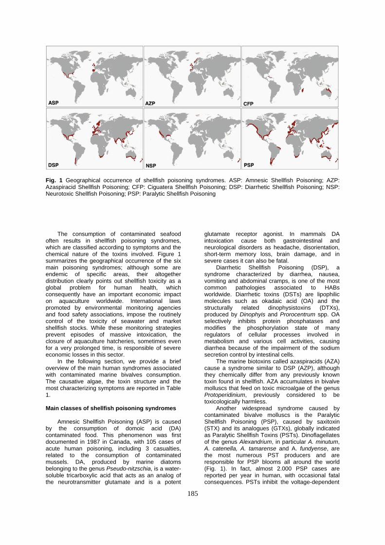
Fig. 1 Geographical occurrence of shellfish poisoning syndromes. ASP: Amnesic Shellfish Poisoning; AZP: Azaspiracid Shellfish Poisoning; CFP: Ciguatera Shellfish Poisoning; DSP: Diarrhetic Shellfish Poisoning; NSP: Neurotoxic Shellfish Poisoning; PSP: Paralytic Shellfish Poisoning
The consumption of contaminated seafood often results in shellfish poisoning syndromes, which are classified according to symptoms and the chemical nature of the toxins involved. Figure 1 summarizes the geographical occurrence of the six main poisoning syndromes; although some are endemic of specific areas, their altogether distribution clearly points out shellfish toxicity as a global problem for human health, which consequently have an important economic impact on aquaculture worldwide. International laws promoted by environmental monitoring agencies and food safety associations, impose the routinely control of the toxicity of seawater and market shellfish stocks. While these monitoring strategies prevent episodes of massive intoxication, the closure of aquaculture hatcheries, sometimes even for a very prolonged time, is responsible of severe economic losses in this sector.
In the following section, we provide a brief overview of the main human syndromes associated with contaminated marine bivalves consumption. The causative algae, the toxin structure and the most characterizing symptoms are reported in Table 1.
Main classes of shellfish poisoning syndromes
Amnesic Shellfish Poisoning (ASP) is caused
by the consumption of domoic acid (DA) contaminated food. This phenomenon was first documented in 1987 in Canada, with 105 cases of acute human poisoning, including 3 casualties, related to the consumption of contaminated mussels. DA, produced by marine diatoms belonging to the genus Pseudo-nitzschia, is a water-soluble tricarboxylic acid that acts as an analog of the neurotransmitter glutamate and is a potent
glutamate receptor agonist. In mammals DA intoxication cause both gastrointestinal and neurological disorders as headache, disorientation, short-term memory loss, brain damage, and in severe cases it can also be fatal.
Diarrhetic Shellfish Poisoning (DSP), a syndrome characterized by diarrhea, nausea, vomiting and abdominal cramps, is one of the most common pathologies associated to HABs worldwide. Diarrhetic toxins (DSTs) are lipophilic molecules such as okadaic acid (OA) and the structurally related dinophysistoxins (DTXs), produced by Dinophyis and Prorocentrum spp. OA selectively inhibits protein phosphatases and modifies the phosphorylation state of many regulators of cellular processes involved in metabolism and various cell activities, causing diarrhea because of the impairment of the sodium secretion control by intestinal cells.
The marine biotoxins called azaspiracids (AZA) cause a syndrome similar to DSP (AZP), although they chemically differ from any previously known toxin found in shellfish. AZA accumulates in bivalve molluscs that feed on toxic microalgae of the genus Protoperidinium, previously considered to be toxicologically harmless.
Another widespread syndrome caused by contaminated bivalve molluscs is the Paralytic Shellfish Poisoning (PSP), caused by saxitoxin (STX) and its analogues (GTXs), globally indicated as Paralytic Shellfish Toxins (PSTs). Dinoflagellates of the genus Alexandrium, in particular A. minutum, A. catenella, A. tamarense and A. fundyense, are the most numerous PST producers and are responsible for PSP blooms all around the world (Fig. 1). In fact, almost 2.000 PSP cases are reported per year in human, with occasional fatal consequences. PSTs inhibit the voltage-dependent
185
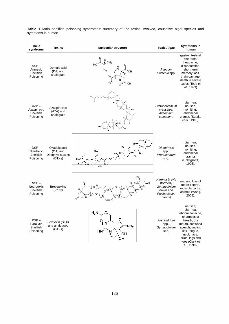
Table 1 Main shellfish poisoning syndromes: summary of the toxins involved, causative algal species and symptoms in human
Toxic syndrome Toxins Molecular structure Toxic Algae Symptoms in
human
ASP – Amnesic Shellfish
Poisoning
Domoic acid (DA) and
analogues
Pseudo-nitzschia spp.
gastrointestinal disorders, headache,
disorientation, short-term
memory loss, brain damage, death in severe cases (Todd et
al., 1993)
AZP – Azaspiracid
Shellfish Poisoning
Azaspiracids (AZA) and analogues
Protoperidinium crassipes, Azadinium spinosum.
diarrhea, nausea, vomiting,
abdominal cramps (Satake
et al., 1998).
DSP – Diarrhetic Shellfish
Poisoning
Okadaic acid (OA) and
Dinophysistoxins (DTXs)
Dinophysis spp.,
Prorocentrum spp.
diarrhea, nausea, vomiting,
abdominal cramps
(Hallegraeff, 1995).
NSP – Neurotoxic Shellfish
Poisoning
Brevetoxins (PbTx)
Karenia brevis (formerly
Gymnodinium breve and
Ptychodiscus brevis)
nausea, loss of motor control,
muscular ache, asthma (Wang,
2008).
PSP – Paralytic Shellfish
Poisoning
Saxitoxin (STX) and analogues
(GTXs)
Alexandrium spp.,
Gymnodinium spp.
nausea, diarrhea,
abdominal ache, shortness of breath, dry
mouth, confused speech, tingling
lips, tongue, neck, face,
arms, legs and toes (Clark et
al., 1999).
186

CFP – Ciguatera
Fish Poisoning
Only palytoxin (PLTX) and
analogues in bivalves, other
toxins are responsible of CFP in fishes (ciguatoxin,
maitotoxin, etc.)
Ostreopsis spp. (PLTX, ovatoxin and analogues)
lethargy, muscle spasms, tremor
myalgia, cyanosis,
respiratory distress,
rhabdomyolysis, death in severe cases (Ramos
and Vasconcelos,
2010)
Others Yessotoxins (YTXs)
Lingolodinium polyedrum, Gonyaulax spinifera,
Prorocentrum reticulatum.
diarrhea (Tubaro et al., 2010)
Others Pectenotoxins (PTXs)
Dinophysis spp.
diarrhea (Draisci et al., 1996)
Others
Spirolides (SPXs) and
Gymnodimine (GYMs)
Karenia selliformis (GYMs)
Alexandrium ostenfeldii
(SPXs)
neurological symptoms
(Munday et al., 2004)
sodium channel conductance causing the blockade of neuronal activity. Symptoms include nausea, diarrhea, abdominal ache, shortness of breath, dry mouth, confused speech, tingling or burning sensations.
Neurotoxic shellfish poisoning (NSP) is caused by brevetoxins (PbTx); the main symptoms are gastrointestinal and neurological, including nausea,
loss of motor control and muscular ache. The formation of toxic aerosol by wave action produces respiratory asthma-like symptoms, even though no fatalities have been reported so far. PbTx is produced by Karenia brevis and binds with high affinity to the voltage-dependent sodium channel, altering its sensitivity and inhibiting its inactivation.
The sixth and last group of shellfish poisoning
187

188
is the Ciguatera Fish Poisoning (CFP), an intoxication caused by a heterogeneous group of toxins, including ciguatoxin, maitotoxin, palytoxin (PLTX) and others. While the main concerns for human health derive from the consumption of contaminated fish, PLTX and its analogues, produced by species of the Ostreopsis genus, can also be accumulated in bivalves. Likewise ciguatoxin, PLTX also lowers the threshold for the opening of voltage-gated sodium channels in synapses of the nervous system, determining severe neurological disorders, including lethargy, muscle spasms, myalgia, cyanosis, respiratory distress, rhabdomyolysis and even death in severe cases.
Beside the toxins classifiable among the six large classes listed above, microalgae can produce an extremely broad spectrum of additional toxic compounds. Some species of dinoflagellates, such as Lingulodinium polyedrum, Gonyaulax spinifera and Protoceratium reticulatum produce yessotoxins (YTXs), structurally related to PbTx and ciguatoxins. YTXs cause diarrhea and therefore they were initially wrongly classified as DSTs, only to be later assigned to a novel independent group after the discovery of their different mechanism of action. Pectenotoxins (PTXs) belong to a group of polyether-lactone toxins that, like YTXs cause symptoms similar to DSP. They are exclusively produced by Dinophysis spp., which have a large distribution worldwide. Gymnodimines (GYMs) and spirolides (SPXs) produced by Karenia selliformis and Alexandrium ostefeldii, respectively are emerging lipophilic marine toxins that belong to a heterogeneous group of macrocyclic compounds called cyclic imines. Since their discovery in the early 1990s, GYMs and SPXs are well known due their fast acting toxicity in mouse bioassay, by blocking the nicotinic receptors and causing neurological symptoms.
Moreover, dinoflagellates are certainly capable of producing several harmful compounds which have not been fully characterized yet. As a matter of fact, the biosynthetic potential of microalgae is underestimated and many algal natural products, including toxins, remain yet to be discovered. Therefore it is not surprising that the number of phycotoxins isolated from marine microalgae continues to increase exponentially.
Nevertheless, dinoflagellates are not the only source of marine toxins, as certain diatoms (such as the DA producing Pseudo-nitzschia spp.) and several species of seawater Cyanobacteria produce compounds hazardous to human health. While these toxins are extremely diverse both by chemical composition and by physiological effects, these aspects will be not discussed in the present review, which is mainly focused on the effects of toxic dinoflagellates. For a more comprehensive overview suggesting the reading of more specific literature on the topic.
Toxicological studies on human and other vertebrate model organisms
The study of marine toxins has been historically
connected with the hazard they represent to human
health. Therefore, the large majority of toxicological studies performed so far have been focused on model vertebrates or human cell lines. Moreover, the most used method for marine algal toxins detection is the mouse bioassay developed by the Japanese Ministry of Health and Welfare. Although this outworn method has been used worldwide as the main tool for shellfish toxicity biomonitoring for several decades, it is currently being replaced by other tests, such as the enzyme-linked immunosorbent assay (ELISA) and the high-performance liquid chromatography (HPLC), more sensitive and reliable.
In parallel to this test, routinely used for the detection of contamination, many research studies investigated more specific aspects of the toxic effects of marine drugs, unraveling their molecular targets, their mode of action and the kinetics of accumulation and detoxification in model organisms. Mice and other vertebrates have been repeatedly used as model organisms for in vivo studies to better understand the effects of several classes of phycotoxins, including AZA, OA, STX, and YTX, on human.
Also cell lines provide useful models: a number of in vitro studies clarified the mode of action of diverse phycotoxins, including YTX in different vertebrate and invertebrate cell lines, PLTX in MCF-7 cells, STX in cultured mice neurons, OA and AZA in multiple human cell lines. Moreover, genomics and proteomics methods applied to vertebrate models contributed to elucidate the molecular pathways acting in response to shellfish poisoning.
Phycotoxin effects on bivalves
A direct comparison between the widely
documented molecular effects of phycotoxins on vertebrates and bivalves is definitely hindered by several obstacles. One of the key factors is the specificity of interaction of many toxins with their molecular targets, which are often membrane channels. The sequence divergence among species explains why bivalves are often completely insensible to many toxins having a lethal effect on human and other vertebrates and vice-versa. In fact, biotransformation processes which likely reduce toxicity in bivalves, can sometimes produce analogues which are even more dangerous to human than the original compound. The specificity of interaction between phycotoxins and their molecular targets is refined to the point that polymorphisms to single ion channels can even lead to dramatic differences in sensitivity within bivalve populations.
Nevertheless, over the past decades numerous studies have been carried out to explore the specific effects of phycotoxins on marine invertebrates, even though their focus has been mainly addressed on physiological adaptations and behavioral modifications. This bias on non-molecular studies is caused by the still limited genetic knowledge of these organisms. In fact, before the next generation sequencing era, the main pool of bivalve genetic knowledge came from large EST collections, but the recent technological advances have quickly led to

189
the availability of many transcriptomes and even to the complete sequencing of the oyster genome.
Studies of physiological responses
The rich literature documenting the behavioral,
physiological and histo-pathological alterations in contaminated filter-feeding molluscs, despite not providing outright molecular data, represents an important base of knowledge both for a better planning of molecular biology experiments and for the interpretation of the results in their biological context.
Hemocyte parameters
Both the humoral and the cellular defense have
a crucial role in bivalves immunity. The hemocytes are the main players in the response to parasites and pathogens, through a complex, non-adaptive, non-specific, innate immune system; microbial killing results from the combined action of the phagocytic process with a broad spectrum of humoral recognition and defense factors. Therefore, the study of the effects of phycotoxins on bivalve immunity necessarily takes into account the monitoring of hemocyte parameters.
The feeding on PSP dinoflagellate species determines an increase of hemocyte counts, accompanied by changes in the hemocyte subpopulations and alteration of the phagocytic activity in some species. On the contrary, some studies report non-significant effects on hemocyte number, morphology, or functions in Crassostrea virginica, while others evidenced a strong individual variability in the response in Ruditapes philippinarum. Histo-pathological studies on mussels exposed to A. fundyense and P. minimum detected an inflammatory response consisting of degranulation and diapedesis of hemocytes into the alimentary canal and, as the exposure continued, hemocyte migration into the connective tissue surrounding the gonadal follicles.
Also other classes of toxins can trigger immune responses: experiments by Mello and collaborators showed the variation of various hemocyte parameters in different bivalve species affected by natural DSP blooms. Also in this case, a species-specific response was observed, demonstrating the presence of vulnerable (e.g. Perna perna) and unaffected (e.g. Crassostrea gigas) species. Moreover, also GYMs can induce the alteration of hemocyte parameters in R. philippinarum, while YTX, PLTX and OA can increase the phagocytic activity of Mytilus galloprovincialis hemocytes.
Valve activity, filtration rate and depuration from toxins
The closure of the shells and the reduction of
the filtration rate are normally occurring mechanisms played daily by bivalves in response to the variation of the tide levels. Furthermore, these mechanisms are also used by bivalves for isolating themselves from the external environment, in presence of negative conditions, such as pollutants and toxins,
as well. This mechanism has been extensively used to study the effects of phycotoxins on mussel physiology, even though sometimes passive valve closure can occur in response to PSTs, which notoriously cause adverse effects on some species, such as burrowing incapacitation. Different species display different valve activity modulation in response to paralytic dinoflagellates, so the reduction of the filtration rate is a key parameter used for the classification of bivalves as susceptible or resistant to PSP.
Other negative effects on valve closure and filtration activity have been reported in various bivalve species in response to microalgae producing different toxin classes, such as Gymnodinium mikimotoi, Heterocapsa circularisquama, Pseudo-nitzschia and Azadinium spinosum.
Despite the reduction of the filtration rate, prolonged HABs may determine a significant accumulation of toxins in bivalve tissues. Different farmed species are often characterized by highly variable detoxification rates, thus requiring a continuous monitoring of toxicity even for a long time after the bloom event. Even though species-specific differences in depuration have been evidenced for many toxin classes, including PSTs and DSPs, the most outstanding example is given by DA. In fact, most bivalve species (mytilids, in particular) can depurate DA very quickly, but others, such as Pecten maximus, show an extremely low depuration rate, retaining the toxin even for several months in its visceral tissues. This differential ability of depuration between species is often crucial for the long-term management of bivalve aquacultures.
Feeding and excretion
Pseudofaeces production is a particularly
important pre-ingestive mechanism preventing the animal’s ingestive capacity from being exceeded, but it also facilitates the process of particle selection, whereby less nutritious particles can be rejected and the quality of the ingested material can be improved proportionally. The most complete comparative study concerning the feeding behavior of bivalves on toxic dinoflagellates is provided by Hégaret and colleagues. Clearance rates of five species of bivalve molluscs were assessed, following the exposure for one hour to three harmful-algal strains: Prorocentrum minimum (PSP and DSP), A. fundyense (PSP), and Heterosigma akashiwo (NSP). The analysis of faeces and pseudofaeces revealed species-specifc responses to the different harmful algae, indicating in most cases a preferential retention of harmful cells. The production of faeces and pseudofaeces varied appreciably between the different bivalve/algae pairs.
Effects on juveniles
The understanding of the physiological effects
played by toxic algae on bivalve juvaniles is very important, especially for farmed species, in order to better understand how aquaculture activity is threatened by the HABs. Li and colleagues exposed

190
juveniles of R. philippinarum and Perna viridis to A. tamarense (PSP), measuring the scope for growth (SFG), and the growth rate. SFG was significantly reduced in both clams and mussels while R. philippinarum resulted to be the most sensitive to PSP while considering the growth rate only.
Leverone and collaborators reported the effects on the clearance rate of juvenile bivalves of 4 different species in relation with a Karenia brevis exposure (PbTx). Both in a short and long-term exposure Argopecten irradians resulted to be the most sensitive species, C. virginica the least responsive and P. viridis and Mercenaria mercenaria displayed intermediated responses.
On the contrary, DA did not have a significant effects on feeding rate and shell valve clatter on the juvenile king scallops Pecten maximus that, but registered a negative impact on their growth rate and survival.
Other effects
Considering the different nature, composition
and targets of marine toxins, it is likely that many additional effects, not described above, could be detected in different bivalve species. As an example, beside paralysis, PSTs can also produce increased mantel melanization and abnormal vitellogenesis in Nodipecten subnodosus and Argopecten ventricosus, production of white mucus and inhibition of byssus production in M. edulis and G. demissa.
Moreover, Landsberg hypothesized a possible connection between neoplasia in certain species of bivalves, such as Mya arenaria, with the presence of micro-algal blooms, even though specific surveys on the topic are still lacking. Some phycotoxins such as OA and DA, certainly have a genotoxic effect on bivalves. Furthermore several classes of toxins (PTX, DSTs, PLTX) are known to be potent dysregulators of cytoskeleton dynamics in vertebrates and are likely to exert a similar action also in bivalves
Studies at molecular level
A summary of the main studies focused on the
molecular responses of bivalves to phycotoxins is reported in Table 2 and discussed in detail below.
Toxin metabolism and biotransformation
Many species of bivalve molluscs are capable
of biochemical transformation of the toxins accumulated by filtration, thus generating novel metabolites not found in the causative algal species, suggesting that extremely complex mechanisms of selective accumulation and chemical or enzymatic conversion might be involved in the development of shellfish toxicity. While, in some cases, this could be interpreted as a strategy specifically developed to decrease toxin potency, in other cases these toxin derivatives are likely just the by-products of normal metabolic pathways.
The metabolism of many classes of phycotoxins has been documented in a large
number of bivalve species, even though comparative studies demonstrated significant differences in the ability of biotransformation between species, pointing out that different organisms might have adopted specific biochemical mechanisms as a defensive strategy to recurrent HAB events.
Among the many bivalves able to transform marine toxins, the scallop Patinopecten yessoensis is definitely one of the most active, since it is able to metabolize YTX, DTX, PTX and OA. The mussels M. galloprovincialis and Mytilus edulis can metabolize AZA, OA and, to some extent, also PSTs. Episodes of PbTx conversion to less toxic analogues have been documented in many different bivalves, including the oyster C. virginica.
Although toxin biotransformations in bivalves are well documented, their biological significance is often unclear, since the modifications do not always result in a decrease of toxin potency. One of the few exceptions is represented by the esterification of OA and DTX1, which has been studied in a number of different bivalves, which is thought to play a role in the sequestration in lipid rich tissues and the conjugation to lipoproteins.
While some of the toxin biotransformations are likely the effect of passive processes or enzymatic activities provided by symbiotic bacteria, in many cases they have been shown to be active processes catalyzed by bivalves themselves. Nevertheless, despite the overwhelming evidence of phycotoxin transformation, just a few enzymes specifically involved in these processes have been isolated and described so far.
An enzyme capable of hydrolyzing PTX and OA has been recently discovered in the digestive gland of the mussel Perna canaliculus : the protein identified, an acidic serine esterase, did not show any similarity with known sequences and was active on a rather broad range of PTXs and OA esters. Furthermore, two enzymes involved in different PST modifications have been isolated in Peronidia venulosa and Mactra chinensis, even though only partial amino-acidic sequences have been characterized: carbamoylase I can hydrolyze the carbamoyl mojety in both carbamoyl and N-carbamoyl PSTs, whereas sulfocarbamoylase catalyzes the hydrolysis of the N-sulfocarbamoyl mojety of the weakly toxic C-PSTs.
The digestive gland is the main site of accumulation of different toxin species, so this tissue is assumed to be the most active in the bioconversion processes. Therefore, not surprisingly, all the enzymes involved in toxin transformation so far identified have been isolated from this tissue and novel, yet unknown, enzymes are expected to be highly expressed and active in this tissue in response to HABs.
Toxin binding and accumulation
Despite the important role of digestive gland, in
some cases other tissues may play a role in the accumulation of phycotoxins. In fact, some bivalve species can retain toxins for an extremely prolonged time in specific non-visceral tissues, exploiting toxin

Table 2 Main molecular studies related to shellfish poisoning in bivalve molluscs
Study Toxin class Bivalve species Strategy
Llewellyn et al., 1997 PSTs (STX)
Mytilus edulis, Saximodus giganteus, Spisula solidissima, Donax deltoides
and Vepricardium multispinosum screening for saxiphin-like activity
Dizer et al., 2001 DA Mytilus edulis monitoring of the cholinesterase activity
following intramuscolar DA injection enzyme purification from the digestive
gland (sulfocarbamoylase I) Lin et al., 2004 PSTs Mactra chinensis
monitoring of oxydative stress related enzymatic activities Choi et al., 2006 PSTs Ruditapes philippinarum
Estrada et al., 2007 PSTs Nodipecten subnodosus monitoring of antioxidant and hydrolitic
enzymes in different tissues Takati et al.,
2007 PSTs Acanthocardia tuberculata protein purification from the foot (PSPBP)
Nzoughet at al., 2007 AZA Mytilus edulis isoelectric focusing and SEC
Fernandez-Reiriz et al.,
2008 PSTs Mytilus chilensis monitoring of the activity of amylase,
laminarinase and cellulase
Talarmin et al., 2008 DSTs (OA) Crassostrea gigas western blot
Malagoli et al., 2008 PLT Mytilus galloprovincialis immunoblot
enzyme purification from the digestive gland (sulfocarbamoylase I) Cho et al., 2008 PSTs Peronidia venulosa
SDS-page and de novo sequencing; comparative analysis between control
and contaminated bivalves
Nzoughet et al., 2009 AZA Mytilus edulis
monitoring of prophenoloxidase, amylase activity and ROS production in
contaminated oyster hemocytes
Haberkorn et al., 2010 PSTs Crassostrea gigas
monitoring of antioxidant and hydrolitic enzymes in different tissues, with a particular emphasis on hemocytes
Estrada et al., 2010 PSTs Nodipecten subnodosus
microarray and real-time PCR on digestive glands of experimentally and
naturally contaminated mussels
Manfrin et al., 2010 DSP (OA) Mytilus galloprovincialis
Rossignoli and Blanco, 2010 DSTs (OA) Mytilus galloprovincialis fractionation and enzymatic digestion of
cellular fractions
MacKenzie et al., 2012 PTX and OA Perna canaliculus enzyme purification from the digestive
gland
multibiomarker approach, assessment of several physiological parameters,
including enzymatic activities
Gorbi et al., 2012
PLT and ovatoxin Mytilus galloprovincialis
real-time PCR on selected genes following heamocytes exposure to
brevetoxin
Mello et al., 2012 brevetoxin Crassostrea gigas
Gerdol et al., 2012 PSP Mytilus galloprovincialis RNA-seq on digestive glands of
experimentally contaminated mussels
Gonzales et al., in preparation* DSTs (OA) Mytilus galloprovincialis
*personal communication
monitoring of histones gene expression levels
191

192
accumulation as a chemical defense towards natural predators.
The better known phenomenon of chemical defense by phycotoxin accumulation is certainly the butter clam Saxidomus giganteus, which selectively stores PSTs in its siphon epithelium, thus discouraging predation by siphon-nipping fishes. As a matter of fact, the occurrence of HABs significantly influences the feeding behavior of many vertebrate predators, such as sea otters and shorebirds Although proteins involved in toxin-binding, transport and accumulation have been described in many organisms, so far the molecular mechanisms leading to this selective retention in bivalves are almost completely unknown. When tested for saxiphilin-like activity, both M. edulis and S. giganteus, capable of accumulating very high levels of PSTs in their tissues, resulted to be negative, likewise Spisula solidissima, Donax deltoides and Vepricardium multispinosum. Nevertheless, a case of a PSP-binding protein has been reported in the Moroccan cockle Acanthocardia tuberculata; in this species, a 181 KDa protein named PSPBP contributes to the prolonged retention of PSTs in the foot.
Beside PSTs, other classes of toxins can be conjugated to specific proteins produced by bivalves. Rossignoli and Blanco provided the first lines of evidence of a soluble cytoplasmatic component binding OA in mussel, which was identified as a high density lipoprotein. Nzoughet and colleagues were able to identify a 45 KDa protein weakly binding AZA in the hepatopancreas of the blue mussel M. edulis and another unknown 22 KDa protein which was apparently highly expressed only in contaminated mussels.
Even though only a little is known about bivalves phycotoxin-binding proteins, these few studies suggest that they may be involved in selective retention or detoxification, depending on whether the toxins will be used in the frame of a chemical defense strategy or simply metabolized.
Metabolic activities
Although relatively few bivalve proteins related
to HABs have been identified so far, the alteration of a number of enzymatic activities, possibly related to both xenobiotic metabolism and stress response has been reported. Some classes of phycotoxins can affect the overall metabolic rates of bivalve organs in a time- and doses-dependent way.
A multi biomarker approach by Gorbi and colleagues clearly showed that the activity of the Na+/K+-ATPase, the target of PLTX, was strongly inhibited by Ostreopsis ovata blooms in M. galloprovincialis, which can therefore be considered as susceptible organisms. Consequently to this reduction, a significant alteration of other enzymatic activities was also observed (acetylcholinesterase in particular), while on the contrary no ROS (Reactive Oxygen Species) detoxifying enzymes were significantly altered, indicating that oxidative pathways are not involved in O. ovata toxicity. The cholinesterase activity was also monitored in M.
edulis after intramuscular injection of DA, highlighting just a moderate reduction after 48 h.
Other studies have been focused on the analysis of the effects of different toxins on the oxidative pathways; the monitoring of glutathione S-transferase (GST), glutathione peroxidase (GPx) and superoxide dismutase (SOD) activities and of lipid peroxidation in the clam R. philippinarum contaminated with PSTs highlighted only a modest involvement of these enzymes. In a different species (the giant lions-paw scallop Nodipecten subnodosus) the same toxin class produced a significant alteration of both GPx (up-regulated) and SOD (down-regulated) in gills, whereas no changes were observed in other tissues. Moreover, catalase (CAT) activity and lipid peroxidation were not markedly altered in any tissue.
On the other hand, other studies observed a stronger correlation between phycotoxins and oxidative damage: in fact, cyanobacteria were able to produce a significant alteration of the activity of the two antioxidant enzymes GST and GPx in M. galloprovincialis and of CAT in M. edulis. Moreover, Haberkorn and colleagues monitored ROS production in PSP-contaminated oysters, observing a linear correlation between PST accumulation and ROS production in haemocytes.
As we have reported in the previous sections, toxic microalgae can significantly alter the feeding of bivalves, with consequent effects on enzymatic activities linked to digestion. Fernandez-Reiriz and colleagues (2008) carefully examined the effects of a diet based on the toxic dinoflagellate A. catenella on Mytilus chilensis, showing that mussels are able to adapt mechanisms which allow the feeding with toxic algae. The authors were in fact able to observe a logarithmic relationship for the assimilatory balance and the carbohydrate metabolism of the digestive gland, by monitoring the enzymatic activity of amylase, laminarinase and cellulase. The amylase activity of oyster digestive gland is also affected by the exposure to A. minutum, even though the changes observed are also largely dependent on the physiological status of bivalves.
Finally, the monitoring of several hydrolytic enzymes revealed marked alterations in different tissues of N. subnodosus exposed to PSP. These enzymatic activities could be potentially used as molecular markers of PSP contamination in scallops; in particular lipase, α-galactosidase, β-glucosidase and α-mannosidase were altered in the digestive gland and trypsin and α-chymotrypsin in the gill, but other modifications were also observed in other tissues, including mantle and the adductor muscle.
Biomarkers of contamination: proteomic approaches
Although there is no doubt that large-scale
proteomic approaches can provide a tool of the utmost importance for the identification of molecular markers of aquatic pollution, to date just a very few studies have exploited this potential to explore the effects of phycotoxins on bivalves.

193
So far proteomic approaches have been mainly used to study the expression of proteins regulated by known molecular targets of toxins, such as in the case of protein phosphatases, the upstream effectors of the p38 mitogen-activated protein kinase, selectively inhibited by OA. An increase in the phosphorylation/activation of p38 MAPK has been in fact observed in the heart of OA treated oysters and contributes to the increased phagocytotic activity in the immunocytes of PLTX-treated mussels.
One of the very few large-scale approaches concerned the identification of biomarkers of AZA contamination in Mytilus edulis; four proteins highly expressed in the digestive gland of toxic mussels were identified, namely cathepsin D, superoxide dismutase, glutathione S-transferase Pi and a flagellar protein of bacterial origin. This data seem to suggest the activation of xenobiotic defense response in bivalves following AZP blooms.
The negative effects of a toxic cyanobacteria, Cylindrospermopsis raciborskii, have been investigated using a similar approach on M. galloprovincialis. The expression of several structural proteins was remarkably altered, indicating a situation of stress and cytoskeletal destabilization. At the same time, other important proteins such as the mitochondrial HSP60, the major extrapallial fluid protein and a triosephosphate isomerase were significantly down-regulated in the toxic strain-fed mussels, highlighting a complex response of mussel to cyanotoxins.
Although the efforts put so far in large-scale proteomic studies have been very scarce, the possibility to identify potential biomarkers of contamination demonstrates that this represents a valid approach for better understanding the molecular mechanisms involved in shellfish toxicity.
Biomarkers of contamination: target genes focused approaches
The number of proteomic studies exploring the
effects of phycotoxins on marine bivalves is quite narrow, but even less efforts have been put in genomic researches. The few studies focused on gene expression have been mostly aimed at the monitoring of a limited number of target sequences. Mello and colleagues selected a few genes and assessed their expression in C. gigas hemocytes subject to in vitro PbTx exposure. The early activation of HSP70, CYP365A1 and FABP, genes related to stress and detoxification, suggest that oyster hemocytes activate a defense response which protects them from cytotoxic damage, which does not involve immune and antioxidant processes, as the expression of BPI, IL-17, EcSOD, Prx6, GPx and SOD was not altered.
Other studies based on gene expression, exploiting the knowledge gathered from previous experiments, have only been planned but not performed yet: an interesting OA biomonitoring approach based on the evaluation of the expression of genes critically important in OA-induced genotoxic damage has been in fact proposed. In particular, the authors claim that the expression of
several histone variants, such as the histone H2A.Z, is strongly down-regulated in response to harmful levels of OA, basing their hypothesis on preliminary data which will be published in an upcoming manuscript.
Biomarkers of contamination: whole-transcriptome scale approaches
To the best of our knowledge, only two studies
have so far tried to tackle the issue of marine toxin effect on bivalves from a genomic perspective. The two studies, both performed on the Mediterranean mussel M. galloprovincialis, investigated the effects of toxins on the gene expression of the main tissue of accumulation, the digestive gland, by using two different techniques.
In the first study, focused on DSP, Manfrin et al. assessed the effects of OA accumulation over 35 days of exposure by cDNA microarray, identifying several transcripts candidates as OA-stress markers. Although most of the sequences could not be linked to known metabolic pathways correlated to biotransformation, the up-regulation of several stress-related proteins, mainly linked to apoptosis, proteolysis and cytoskeleton destabilization, denoted a possible sufferance of OA exposed mussels.
The preliminary observations collected from this experiment were further validated on the digestive gland of mussels subject to naturally occurring HABs (unpublished data). The expression of 14 selected up-regulated genes was monitored by real-time PCR in samples collected during 2 DSP events occurred in the Gulf of Trieste. The analysis permitted to confirm 11 out of 14 genes as OA-responsive, highlighting that the results of experimental contaminations can be applied also on naturally occurring DSP events (Fig. 2).
The second and more recent study exploited the recent advances offered by the application of next generation sequencing technologies to RNA-sequencing. The study was aimed at the investigation of the possible molecular mechanisms activated or repressed in the digestive gland in response to the accumulation of PSTs produced by the toxigenic dinoflagellate A. minutum strain AL9T over a time course of 5 days. The contamination with these toxins apparently only led to negligible effects on gene expression in mussel, which is an organism insensible to the paralytic effects of STX-like toxins, due to the resistance of its nerve voltage-gated sodium channel. Therefore, the RNA-seq experiment results seem to disprove the sporadic reports of negative effects of paralytic HABs in mussel and support their classification as organisms refractory to PSP.
Although preliminary, the two above mentioned studies were certainly able to give a better overview about the possible molecular effects of marine phycotoxins on bivalve molluscs and represent the first steps in moving the focus of dinoflagellate toxicity from a human-centered to a bivalve-centered point of view.
There is no doubt that, thanks to the technological advancements and the increasing availability of bivalve sequence data, including fully

Fig. 2 Validation of potential DSP-biomarkers in mussels digestive gland by quantitative RT-PCR. The expression of fourteen genes identified as differentially expressed in Manfrin et al., 2010 were monitored in two samples collected during naturally occurring DSP-HAB in the Gulf of Trieste (TS_09+ and TS_10+). The normalized fold expression values are shown in a log2 scale, significant (p< 0.05) upregulation is indicated by *.
194

195
sequence genomes, the design of similar studies performed on large panels of toxins will be much easier in the near future.
Conclusion
HABs represent a serious and emerging issue
for human health, so considerable research efforts have been put in the study of their toxicological effects on vertebrate model organisms, useful for a better comprehension of the dynamics of shellfish poisoning in human. Comparatively, only limited data are available about the effects on bivalves, and the large majority of them concerns physiological aspects. While the lack of molecular studies is mainly caused by the still limited genetic knowledge of these organisms, there is an urge for further research in this field, as highlighted by the promising findings provided by the handful of molecular studies documented in this review.
Certainly, there is the need for combining large scale approaches (both on a proteomic and on a genomic level) for the identification of trustworthy biomarkers of contamination for a more effective biomonitoring of HABs, also contributing to a better comprehension of the molecular mechanisms adapted by bivalve molluscs to deal with phycotoxin toxicity.
References Andrinolo D, Michea L, Lagos N. Toxic effects,
pharmacokinetics and clearance of saxitoxin, a component of paralytic shellfish poison (PSP), in cats. Toxicon 37: 447-464, 1999.
Asakawa M, Beppu R, Ito K, Tsubota M, Takayama H, Miyazawa K. Accumulation of paralytic shellfish poison (PSP) and biotransformation of its components in oysters Crassostrea gigas fed with the toxic dinoflagellate Alexandrium tamarense. J. Food Hygienic Soc. Japan 47: 28-32, 2006.
Barsanti L, Gualtieri P. Algae: Anatomy, Biochemistry and Biotechnology, Taylor and Francis, New York, 2006.
Basti L, Nagai K, Shimasaki Y, Oshima Y, Honjo T, Segawa S. Effects of the toxic dinoflagellate Heterocapsa circularisquama on the valve movement behaviour of the Manila clam Ruditapes philippinarum. Aquaculture 291: 41-47, 2009.
Bianchi C, Fato R, Angelin A, Trombetti F, Ventrella V, Borgatti AR, et al. Yessotoxin, a shellfish biotoxin, is a potent inducer of the permeability transition in isolated mitochondria and intact cells. Biochim. Biophys. Acta 139-147, 2004.
Blanco J, Acosta CP, Mariño C, Muñiz S, Martín H, Moroño Á, et al. Depuration of domoic acid from different body compartments of the king scallop Pecten maximus grown in raft culture and natural bed. Aquat. Living Resour. 19: 257-265, 2006.
Blanco J, Bermúdez De La Puente M, Arévalo F, Salgado C, Moroño A. Depuration of mussels (Mytilus galloprovincialis) contaminated with domoic acid. Aquat. Living Resour. 15: 53-60, 2002.
Blanco J, Mariño C, Martín H, Acosta CP. Anatomical distribution of diarrhetic shellfish poisoning (DSP) toxins in the mussel Mytilus galloprovincialis. Toxicon 50: 1011-1018, 2007.
Bricelj VM, Ford SE, Lambert C, Barbou A, Paillard C. Effects of toxic Alexandrium tamarense on behavior, hemocyte responses and development of brown ring disease in Manila clams. Mar. Ecol. Prog. Ser. 430: 35-48, 2011.
Bricelj VM, MacQuarrie SP, Doane JAE, Connell LB. Evidence of selection for resistance to paralytic shellfish toxins during the early life history of soft-shell clam (Mya arenaria) populations. Limnology and Oceanography 55: 2463-2475, 2010.
Bricelj VM, Shumway SE. Paralytic shellfish toxins in bivalve molluscs: Occurrence, transfer kinetics, and biotransformation. Rev. Fish. Sci. 6: 315-383, 1998.
Campos A, Tedesco S, Vasconcelos V, Cristobal S. Proteomic research in bivalves. Towards the identification of molecular markers of aquatic pollution. J. Proteomics 75: 4346-4359, 2012.
Canesi L, Gallo G, Gavioli M, Pruzzo C. Bacteria-hemocyte interactions and phagocytosis in marine bivalves. Micr. Res. Tech. 57: 469-476, 2002.
Carlton JT, Geller JB. Ecological Roulette: The global transport of nonindigenous marine organisms. Sci. 261: 78-82, 1993.
Cho Y, Ogawa N, Takahashi M, Lin H-P, Oshima Y. Purification and characterization of paralytic shellfish toxin-transforming enzyme, sulfocarbamoylase I, from the Japanese bivalve Peronidia venulosa. Biochim. Biophys. Acta 1784: 1277-1285, 2008.
Choi MC, Hsieh DPH, Lam PKS, Wang WX. Field depuration and biotransformation of paralytic shellfish toxins in scallop Chlamys nobilis and green-lipped mussel Perna viridis. Mar. Biol. 143: 927-934, 2003.
Choi NMC, Yeung LWY, Siu WHL, So IMK, Jack RW, Hsieh DPH, et al. Relationships between tissue concentrations of paralytic shellfish toxins and antioxidative responses of clams, Ruditapes philippinarum. Mar. Pollut. Bull. 52: 572-578, 2006.
Clark RF, Williams SR, Nordt SP, Manoguerra AS. A review of selected seafood poisonings. Undersea and Hyperbaric Medicine. 26: 175-184, 1999.
da Silva PM, Hégaret H, Lambert C, Wikfors GH, Le Goïc N, Shumway SE, et al. Immunological responses of the Manila clam (Ruditapes philippinarum) with varying parasite (Perkinsus olseni) burden, during a long-term exposure to the harmful alga, Karenia selliformis, and possible interactions. Toxicon 51: 563-573, 2008.
Dell’Aversano C, Walter JA, Burton IW, Stirling DJ, Fattorusso E, Quilliam MA. Isolation and structure elucidation of new and unusual saxitoxin analogues from mussels. J. Nat. Prod. 71: 1518-1523, 2008.
Dizer H, Fischer B, Harabawy ASA, Hennion MC Hansen PD. Toxicity of domoic acid in the

196
marine mussel Mytilus edulis. Aquat. Toxicol. 55: 149-156, 2001.
Donovan CJ, Ku JC, Quilliam MA, Gill TA. Bacterial degradation of paralytic shellfish toxins. Toxicon 52: 91-100, 2008.
Draisci R, Lucentini L, Giannetti L, Boria P, Poletti R. First report of pectenotoxin-2 (PTX-2) in algae (Dinophysis fortii) related to seafood poisoning in Europe. Toxicon 34: 923-935, 1996.
EFSA Marine biotoxins in shellfish - Summary on regulated marine biotoxins. Scientific Opinion of the Panel on Contaminants in the Food Chain. EFSA J. 1306: 1-23, 2009.
Escobedo-Lozano AY, Estrada N, Ascencio F, Gerardo Contreras G, Alonso-Rodriguez R. Accumulation, biotransformation, histopathology and paralysis in the Pacific Calico Scallop Argopecten ventricosus by the paralyzing toxins of the Dinoflagellate Gymnodinium catenatum. Mar. Drugs 1044-1065, 2012.
Estrada N, de Jesús Romero M, Campa-Córdova A, Luna A, Ascencio F. Effects of the toxic dinoflagellate, Gymnodinium catenatum on hydrolytic and antioxidant enzymes, in tissues of the giant lions-paw scallop Nodipecten subnodosus. Comp. Biochem. Physiol. 146C: 502-510, 2007.
Estrada N, Rodríguez-Jaramillo C, Contreras G, Ascencio F. Effects of induced paralysis on hemocytes and tissues of the giant lions-paw scallop by paralyzing shellfish poison. Mar. Biol. 157: 1401-1415, 2010.
Fernández-Reiriz MJ, Navarro JM, Contreras AM, Labarta U. Trophic interactions between the toxic dinoflagellate Alexandrium catenella and Mytilus chilensis: Feeding and digestive behaviour to long-term exposure. Aquat. Toxicol. 87: 245-251, 2008.
Ferrão-Filho ADS, Kozlowsky-Suzuki B. Cyanotoxins: Bioaccumulation and effects on aquatic animals. Mar. drugs 9: 2729-2772, 2011.
Florez-Barros F, Prado-Alvarez M, Mendez J, Fernandez-Tajes J. Evaluation of genotoxicity in gills and hemolymph of clam Ruditapes decussatus fed with the toxic dinoflagellate Prorocentrum lima. J. Toxicol. Environ. Health - Part A 74: 971-979, 2011.
Franchini A, Malagoli D, Ottaviani E. Targets and effects of yessotoxin, okadaic acid and palytoxin: A differential review. Mar. Drugs 8: 658-677, 2010.
Funari E, Testai E. Human health risk assessment related to cyanotoxins exposure. Crit. Rev. Toxicol. 38: 97-125, 2008.
Gainey LF, Shumway SE. A compendium of the responses of bivalve molluscs to toxic dinoflagellates. J. Shellfish Res. 7: 623-628, 1988.
Galimany E, Sunila I, Hégaret H, Ramón M, Wikfors GH. Experimental exposure of the blue mussel (Mytilus edulis, L.) to the toxic dinoflagellate Alexandrium fundyense: Histopathology, immune responses, and recovery. Harmful Algae 7: 702-711, 2008a.
Galimany E, Sunila I, Hégaret H, Ramón M, Wikfors GH. Pathology and immune response of the blue mussel (Mytilus edulis L.) after an exposure to the harmful dinoflagellate Prorocentrum minimum. Harmful Algae 7: 630-638, 2008b.
Gerdol M, De Moro G, Manfrin C, Milandri A, Riccardi, E, Beran A, et al. RNA sequencing and de novo assembly of the digestive gland transcriptome in Mytilus galloprovincialis fed with toxinogenic and non-toxic strains of Alexandrium minutum. Mar. Biotechnol. 2012 [Submitted].
González-Romero R, Rivera-Casas C, Fernández-Tajes J, Ausió J, Méndez J, Eirín-López JM. Chromatin specialization in bivalve molluscs: A leap forward for the evaluation of Okadaic Acid genotoxicity in the marine environment. Comp. Biochem. Physiol. 155C: 175-181, 2012.
Gorbi S, Bocchetti R, Binelli A, Bacchiocchi S, Orletti R, Nanetti L, et al. Biological effects of palytoxin-like compounds from Ostreopsis cf. ovata: A multibiomarkers approach with mussels Mytilus galloprovincialis. Chemosphere 89: 623-632, 2012.
Haberkorn H, Lambert C, Le Goïc N, Guéguen M, Moal J, Palacios E, et al. Effects of Alexandrium minutum exposure upon physiological and hematological variables of diploid and triploid oysters, Crassostrea gigas. Aquat. Toxicol. 97: 96-108, 2010.
Hallegraeff GM. A review of harmful algal blooms and their apparent global increase. Phycologia 32: 79-99, 1993.
Hégaret H, Wikfors GH. Effects of natural and field-simulated blooms of the dinoflagellate Prorocentrum minimum upon hemocytes of eastern oysters, Crassostrea virginica, from two different populations. Harmful Algae 4: 201-209, 2005a.
Hégaret H, Wikfors GH. Time-dependent changes in hemocytes of eastern oysters, Crassostrea virginica, and northern bay scallops, Argopecten irradians irradians, exposed to a cultured strain of Prorocentrum minimum. Harmful Algae 4: 187-199, 2005b.
Hégaret H, Wikfors GH, Shumway SE. Diverse feeding responses of five species of bivalve mollusc when exposed to three species of harmful algae. J. Shellfish Res. 26: 549-559, 2007.
Hégaret H, Wikfors GH, Soudant P, Lambert C, Shumway SE, Bérard JB, et al. Toxic dinoflagellates (Alexandrium fundyense and A. catenella) have minimal apparent effects on oyster hemocytes. Mar. Biol. 152: 441-447, 2007.
Huang XD, Zhao M, Liu WG, Guan YY, Shi Y, Wang Q, et al. Gigabase-Scale Transcriptome analysis on four species of pearl oysters. Mar. Biotechnol. (NY) 2012 [Epub ahead of print]
Jauffrais T, Contreras A, Herrenknecht C, Truquet P, Séchet V, Tillmann U, et al. Effect of Azadinium spinosum on the feeding behaviour and azaspiracid accumulation of Mytilus edulis. Aquat. toxicol. 124: 179-187, 2012.

197
Jauffrais T, Marcaillou C, Herrenknecht C, Truquet P, Séchet V, Nicolau E, et al. Azaspiracid accumulation, detoxification and biotransformation in blue mussels (Mytilus edulis) experimentally fed Azadinium spinosum. Toxicon 60: 582-595, 2012.
Kankaanpää H, Leiniö S, Olin M, Sjövall O, Meriluoto J, Lehtonen KK. Accumulation and depuration of cyanobacterial toxin nodularin and biomarker responses in the mussel Mytilus edulis. Chemosphere 68: 1210-1217, 2007.
Kvitek R, Bretz C. Shorebird foraging behavior, diet, and abundance vary with harmful algal bloom toxin concentrations in invertebrate prey. Mar. Ecol. Prog. Ser. 293: 303-309, 2005.
Kvitek RG. Paralytic shellfish toxins sequestered by bivalves as a defense against siphon-nipping fish. Mar. Biol. 111: 369-374, 1991.
Kvitek RG, Beiter MK. Relative insensitivity of butter clam neurons to saxitoxin: a pre-adaptation for sequestering paralytic shellfish poisoning toxins as a chemical defense. Mar. Ecol. Prog. Ser. 69: 47-54, 1991.
Kvitek RG, Degange AR, Beitler MK. Paralytic shellfish poisoning toxins mediate feeding behavior of sea otters. Limnology and Oceanography 36: 393-404, 1991.
Landsberg JH. Neoplasia and biotoxins in bivalves: is there a connection? J. Shellfish Res. 15: 203-230, 1996.
Lassus P, Bardouil M, Beliaeff B, Masselin P, Naviner M, Truquet P. Effect of a continuous supply of the toxic dinoflagellate Alexandrium minutum halim on the feeding behavior of the pacific oyster (Crassostrea gigas thunberg). J. Shellfish Res. 18: 211-216, 1999.
Le Hégarat L, Jacquin A, Bazin E, Fessard V. Genotoxicity of the marine toxin okadaic acid, in human Caco-2 cells and in mice gut cells. Environ. Toxicol. 21: 55-64, 2006.
Leverone J, Shumway S, Blake N. Comparative effects of the toxic dinoflagellate Karenia brevis on clearance rates in juveniles of four bivalve molluscs from Florida, USA. Toxicon 49: 634-645, 2007.
Lewis RJ, Holmes MJ. Origin and transfer of toxins involved in ciguatera. Comp. Biochem. Physiol. 106C: 615-628, 1993.
Li A, Ma J, Cao J, Wang Q, Yu R, Thomas K, et al. Analysis of paralytic shellfish toxins and their metabolites in shellfish from the North Yellow Sea of China. Food Additives and Contaminants - Part A Chemistry, Analysis, Control, Exposure and Risk Assessment. 29: 1455-1464, 2012.
Li S, Wang W, Hsieh, D. Effects of toxic dinoflagellate Alexandrium tamarense on the energy budgets and growth of two marine bivalves. Mar. Environ. Res. 53: 145-160, 2002.
Lin H-P, Cho Y, Yashiro H, Yamada T, Oshima Y. Purification and characterization of paralytic shellfish toxin transforming enzyme from Mactra chinensis. Toxicon 44: 657-668, 2004.
Liu H, Kelly M, Campbell D, Dong S, Zhu J, Wang S. Exposure to domoic acid affects larval development of king scallop Pecten maximus
(Linnaeus, 1758). Aquat. Toxicol. 81: 152-158, 2007.
Llewellyn LE, Bell PM, Moczydlowski EG. Phylogenetic survey of soluble saxitoxin-binding activity in pursuit of the function and molecular evolution of saxiphilin, a relative of transferrin. Proc. Roy. Soc. Lond. B. Biol. Sci. 264: 891-902, 1997.
Loker ES, Adema CM, Zhang SM, Kepler TB. Invertebrate immune systems - Not homogeneous, not simple, not well understood. Immunol. Rev. 198: 10-24, 2004.
Louzao MC, Espiña B, Cagide E, Ares IR, Alfonso A, Vieytes MR, et al. Cytotoxic effect of palytoxin on mussel. Toxicon 56: 842-847, 2010.
MacKenzie LA, Selwood AI, Marshall C. Isolation and characterization of an enzyme from the Greenshell™ mussel Perna canaliculus that hydrolyses pectenotoxins and esters of okadaic acid. Toxicon 60: 406-419, 2012.
Mafra LL, Bricelj VM, Fennel K. Domoic acid uptake and elimination kinetics in oysters and mussels in relation to body size and anatomical distribution of toxin. Aquat. Toxicol. 100: 17-29, 2010.
Malagoli D, Casarini L, Ottaviani E. Effects of the marine toxins okadaic acid and palytoxin on mussel phagocytosis. Fish Shellfish Immunol. 24: 180-186, 2008.
Malagoli D, Casarini L, Sacchi S, Ottaviani E. Stress and immune response in the mussel Mytilus galloprovincialis. Fish Shellfish Immunol. 23: 171-177, 2007.
Manfrin C, Dreos R, Battistella S, Beran A, Gerdol M, Varotto L, et al. Mediterranean mussel gene expression profile induced by okadaic acid exposure. Environ. Sci. Technol. 44: 8276-8283, 2010.
Matsuyama Y. Harmful effect of dinoflagellate Heterocapsa circularisquama on shellfish aquaculture in Japan. J. Agric. Res. Quart. 33: 283-293, 1999.
McCarron P, Kilcoyne J, Miles CO, Hess P. Formation of azaspiracids-3, -4, -6, and -9 via decarboxylation of carboxyazaspiracid metabolites from shellfish. J. Agric. Food Chem. 57: 160-169, 2009.
Medhioub W, Lassus P, Truquet P, Bardouil M, Amzil Z, Sechet V, et al. Spirolide uptake and detoxification by Crassostrea gigas exposed to the toxic dinoflagellate Alexandrium ostenfeldii. Aquaculture 358-359: 108-115, 2012.
Mello DF, De Oliveira ES, Vieira RC, Simoes E, Trevisan R, Dafre AL, et al. Cellular and transcriptional responses of Crassostrea gigas hemocytes exposed in vitro to brevetoxin (PbTx-2). Mar. Drugs 10: 583-597, 2012.
Mello DF, Proença LAO, Barracco MA. Comparative study of various immune parameters in three bivalve species during a natural bloom of Dinophysis acuminata in Santa Catarina Island, Brazil. Toxins 2: 1166-1178, 2010.
Miles CO, Wilkins AL, Stirling DJ, Mackenzie AL. Gymnodimine C, an isomer of gymnodimine B,

198
from Karenia selliformis. J. Agric. Food Chem. 51: 4838-4840, 2003.
Moreira R, Balseiro P, Planas JV, Fuste B, Beltran S, Novoa B, et al. Transcriptomics of in Vitro immune-stimulated hemocytes from the manila clam Ruditapes philippinarum using high-throughput sequencing. PLoS ONE 7: 1-16, 2012.
Munday R, Towers NR, Mackenzie L, Beuzenberg V, Holland PT, Miles CO. Acute toxicity of gymnodimine to mice. Toxicon 44: 173-178, 2004.
Newell RIE, Jordan SJ. Preferential ingestion of organic material by the American oyster Crassostrea virginica. Mar. Ecol. Prog. Ser. 47-53, 1983.
Nzoughet JK, Hamilton JTG, Botting CH, Douglas A, Devine L, Nelson J, et al. Proteomics identification of azaspiracid toxin biomarkers in blue mussels, Mytilus edulis. Mol. Cell. Proteomics 8: 1811-1822, 2009.
Nzoughet KJ, Hamilton JTG, Floyd SD, Douglas A, Nelson J, Devine L, et al. Azaspiracid: First evidence of protein binding in shellfish. Toxicon 51: 1255-1263, 2008.
O’Driscoll D, Skrabáková Z, O’Halloran J, van Pelt FNAM, James KJ. Mussels increase xenobiotic (Azaspiracid) toxicity using a unique bioconversion mechanism. Environ. Sci. Technol. 45: 3102-3108, 2011.
Perez S, Vale C, Botana A, Alonso E, Vieytes M, Botana L. Determination of toxicity equivalent factors for paralytic shellfish toxins by electrophysiological measurements in cultured neurons. Chem. Res. Toxicol. 24: 1153-1157, 2011.
Philipp, E.E.R., Kraemer, L., Melzner, F., Poustka, A.J., Thieme, S., Findeisen, U., et al. Massively parallel rna sequencing identifies a complex immune gene repertoire in the lophotrochozoan mytilus edulis. PLoS ONE. 7, 2012.
Plakas SM, El Said KR, Jester ELE, Ray Granade H, Musser SM, Dickey RW. Confirmation of brevetoxin metabolism in the Eastern oyster (Crassostrea virginica) by controlled exposures to pure toxins and to Karenia brevis cultures. Toxicon 40: 721-729, 2002.
Plakas SM, Wang Z, El Said KR, Jester ELE, Granade HR, Flewelling L, et al. Brevetoxin metabolism and elimination in the Eastern oyster (Crassostrea virginica) after controlled exposures to Karenia brevis. Toxicon 44: 677-685, 2004.
Puerto M, Campos A, Prieto A, Cameán A, Almeida AMD, Coelho AV, et al. Differential protein expression in two bivalve species; Mytilus galloprovincialis and Corbicula fluminea; exposed to Cylindrospermopsis raciborskii cells. Aquat. Toxicol. 101: 109-116, 2011.
Ramos V, Vasconcelos V. Palytoxin and analogs: Biological and ecological effects. Mar. Drugs 8: 2021-2037, 2010.
Reigman R. Species composition of harmful algal blooms in relation to macronutrient dynamics. In: Anderson DM, Cembella AD, Hallegraeff GM
(eds), Physiological Ecology of HAB, Springer-Verlag, Heidelberg, pp 475-488, 1998
Richardson K. Harmful or exceptional phytoplankton blooms in the marine ecosystem. Adv. Mar. Biol. 31: 301-385, 1997.
Roach JS, LeBlanc P, Lewis NI, Munday R, Quilliam MA, MacKinnon SL. Characterization of a dispiroketal spirolide subclass from Alexandrium ostenfeldii. J. Nat. Prod. 72: 1237-1240, 2009.
Rossignoli AE, Blanco J. Subcellular distribution of okadaic acid in the digestive gland of Mytilus galloprovincialis: First evidences of lipoprotein binding to okadaic acid. Toxicon 55: 221-226, 2010.
Rossignoli AE, Fernández D, Regueiro J, Mariño C, Blanco J. Esterification of okadaic acid in the mussel Mytilus galloprovincialis. Toxicon 57: 712-720, 2011.
Ryan J, Morey J, Bottein M, Ramsdell J, Van Dolah F. Gene expression profiling in brain of mice exposed to the marine neurotoxin ciguatoxin reveals an acute anti-inflammatory, neuroprotective response. BMC Neuroscience 11: 107, 2010.
Sala GL, Bellocci M, Rossini GP. The cytotoxic pathway triggered by palytoxin involves a change in the cellular pool of stress response proteins. Chem. Res. Toxicol. 22: 2009-2016, 2009.
Sala GL, Ronzitti G, Sasaki M, Fuwa H, Yasumoto T, Bigiani A, et al. Proteomic analysis reveals multiple patterns of response in cells exposed to a toxin mixture. Chem. Res. Toxicol. 22: 1077-1085, 2009.
Sasso S, Pohnert G, Lohr M, Mittag M, Hertweck C. Microalgae in the postgenomic era: A blooming reservoir for new natural products. FEMS Microbiol. Rev. 36: 761-785, 2012.
Satake M, Ofuji K, Naoki I, James KJ, Furey A, McMahon T, et al. Azaspiracid, a new marine toxin having unique spirt ring assemblies, isolated from Irish mussels, Mytilus edulis. J. Am. Chem. Soc. 120: 9967-9968, 1998.
Shumway SE, Cucci TL. The effect of the toxic dinoflagellate Protogonyaulax tamarensis on the feeding behavior of bivalve molluscs. Aquat. Toxicol. 10: 9-27, 1987.
Shumway SE, Pierce FC, Knowlton K. The effect of Protogonyaulax tamarensis on byssus production in Mytilus edulis l., Modiolus modiolus linnaeus, 1758 and Geukensia demissa dillwyn. Comp. Biochem. Physiol. 87A: 1021-1023, 1987.
Silvestre F, Tosti E. Impact of marine drugs on cytoskeleton-mediated reproductive events. Mar. Drugs 8: 881-915, 2010.
Smith EA, Grant F, Ferguson CMJ, Gallacher S. Biotransformations of paralytic shellfish toxins by bacteria Isolated from bivalve molluscs. Appl. Environ. Microbiol. 67: 2345-2353, 2001.
Smolowitz R, Doucette G. Immunohistochemical localization of saxitoxin in the siphon epithelium of the butter clam, Saxidomus giganteus. Biol. Bull. 189: 229-230, 1995.

199
Sobel J, Painter J. Illnesses caused by marine biotoxins. Clin. Infect. Diseases. 41: 1290-1296, 2005.
Sullivan JJ, Iwaoka WT, Liston J. Enzymatic transformation of PSP toxins in the littleneck clam (Protothacastaminea). Biochem. Biophys. Res. Commun. 114: 465-472, 1983.
Suzuki T, Igarashi T, Ichimi K, Watai M, Suzuki M, Ogiso E, et al. Kinetics of diarrhetic shellfish poisoning toxins, okadaic acid, dinophysistoxin-1, pectenotoxin-6 and yessotoxin in scallops Patinopecten yessoensis. Fish. Sci. 71: 948-955, 2005.
Suzuki T, Mitsuya T, Matsubara H, Yamasaki M. Determination of pectenotoxin-2 after solid-phase extraction from seawater and from the dinoflagellate Dinophysis fortii by liquid chromatography with electrospray mass spectrometry and ultraviolet detection. Evidence of oxidation of pectenotoxin-2 to pectenotoxin-6 in scallops. J. Chromatogr. A 815: 155-160, 1998..
Suzuki T, Ota H, Yamasaki M. Direct evidence of transformation of dinophysistoxin-1 to 7-O-acyl-dinophysistoxin-1 (dinophysistoxin-3) in the scallop Patinopecten yessoensis. Toxicon 37: 187-198, 1999.
Svensson S, Förlin L. Analysis of the importance of lipid breakdown for elimination of okadaic acid (diarrhetic shellfish toxin) in mussels, Mytilus edulis: results from a field study and a laboratory experiment. Aquat. Toxicol. 66: 405-418, 2004.
Takati N, Mountassif D, Taleb H, Lee K, Blaghen M. Purification and partial characterization of paralytic shellfish poison-binding protein from Acanthocardia tuberculatum. Toxicon 50: 311-321, 2007.
Talarmin H, Droguet M, Pennec JP, Schröder HC, Muller WEG, Gioux M, et al. Effects of a phycotoxin, okadaic acid, on oyster heart cell survival. Toxicol. Environ. Chem. 90: 153-168, 2008.
Thessen AE, Soniat TM, Dortch Q, Doucette GJ. Crassostrea virginica grazing on toxic and non-toxic diatoms. Toxicon 55: 570-579, 2010.
Todd ECD. Domoic acid and amnesic shellfish poisoning- a review. J. Food Protection 56: 69-83, 1993.
Torgersen T, Sandvik M, Lundve B, Lindegarth S. Profiles and levels of fatty acid esters of okadaic acid group toxins and pectenotoxins during toxin depuration. Part II: Blue mussels (Mytilus edulis) and flat oyster (Ostrea edulis). Toxicon 52: 418-427, 2008.
Tubaro A, Dell'Ovo V, Sosa S, Florio C. Yessotoxins: A toxicological overview. Toxicon 56: 163-172, 2010.
Twarog BM, Hidaka T, Yamaguchi H. Resistance to tetrodotoxin and saxitoxin in nerves of bivalve molluscs: A possible correlation with paralytic shellfish poisoning. Toxicon 10: 273-278, 1972.
Twiner M, Hanagriff J, Butler S, Madhkoor A, Doucette GJ. Induction of apoptosis pathways in several cell lines following exposure to the marine algal toxin azaspiracid. Chem. Res. Toxicol. 25: 1493-1501, 2012.
Valdiglesias V, Méndez J, Pàsaro E, Cemeli E, Anderson D, Laffon B. Assessment of okadaic acid effects on cytotoxicity, DNA damage and DNA repair in human cells. Mut. Res. 689: 74-79, 2010.
Vale P, De M Sampayo MA. Esterification of DSP toxins by Portuguese bivalves from the Northwest coast determined by LC-MS - A widespread phenomenon. Toxicon 40: 33-42, 2002.
Van Dolah FM. Marine algal toxins: Origins, health effects, and their increased occurrence. Environ. Health Perspect. 108: 133-141, 2000.
Venier P, De Pittà C, Bernante F, Varotto L, De Nardi B, Bovo G, et al. MytiBase: A knowledgebase of mussel (M. galloprovincialis) transcribed sequences. BMC Genomics.10: 72, 2009.
Venier P, Varotto L, Rosani U, Millino C, Celegato B, Bernante F, et al. Insights into the innate immunity of the Mediterranean mussel Mytilus galloprovincialis. BMC Genomics 12: 69, 2011.
Vilariño N. Marine toxins and the cytoskeleton: azaspiracids. FEBS J. 275: 6075-6081, 2008.
Wang DZ. Neurotoxins from marine dinoflagellates: A brief review. Mar. Drugs 6: 349-371, 2008.
Wang J, Wang YY, Lin L, Gao Y, Hong HS, Wang DZ. Quantitative proteomic analysis of okadaic acid treated mouse small intestines reveals differentially expressed proteins involved in diarrhetic shellfish poisoning. J. Proteomics 75: 2038-2052, 2012.
Yasumoto T, Murata M, Oshima, Y. Diarrhetic shellfish toxins. Tetrahedron 41: 1019-1025, 1985
Yasumoto T, Oshima Y, Yamaguchi M. Occurrence of a new type of shellfish poisoning in the Tokohu District. Bull. Jap. Soc. Sci. Fish. 44: 1249-1255, 1978.
Zhang G, Fang X, Guo X, Li L, Luo R, Xu F, et al. The oyster genome reveals stress adaptation and complexity of shell formation. Nature 490: 49-54, 2012.
Related Documents