*Corresponding author: Leila Sadeghi, Tel: 98 41 33392739, Fax: 98 41 33356027, Email: [email protected] © 2018 The Authors. This is an Open Access article distributed under the terms of the Creative Commons Attribution (CC BY), which permits unrestricted use, distribution, and reproduction in any medium, as long as the original authors and source are cited. No permission is required from the authors or the publishers. Adv Pharm Bull, 2018, 8(4), 705-713 doi: 10.15171/apb.2018.079 http://apb.tbzmed.ac.ir Advanced Pharmaceutical Bulletin Physiological and Biochemical Effects of Echium Amoenum Extract on Mn 2+ -Imposed Parkinson Like Disorder in Rats Leila Sadeghi 1 * , Farzeen Tanwir 2 , Vahid Yousefi Babadi 3 1 Department of Animal Biology, Faculty of Natural Sciences, University of Tabriz, Tabriz, Iran. 2 Matrix Dynamics Group, University of Toronto, Canada. 3 Department of Physiology, Payam Noor University of Iran, Iran. Introduction Manganese (Mn) plays key role in mammalian brain development and function as a trace element. 1 Mn 2+ is cofactor of important enzymes such as glutamine synthetase, pyruvate decarboxylase, serine/threonine protein phosphatase I, Mn-superoxide dismutase and arginase, which are required for neurotransmitter synthesis, metabolism and antioxidant defense system. 2 It is also the fourth most widely used heavy metal in the industry such as textile bleaching, leather tanning and iron, steel, potassium permanganate, hydroquinone, glass and ceramics production. 2,3 By considering high application of Mn 2+ in today’s life, exposing to this poisonous metal is predictable especially in miners and factory workers. 4 Previous studies confirmed Mn 2+ overdose causes Parkinson like disorder known as manganism that accompanied by tremors, odd movements, mask like face, and body stiffness that first observed in miners. 3 The risk of Mn 2+ exposure is not limited to miners or welders. The environmental accessibility and high Mn 2+ concentration in water or food represent a source of contamination for the general population. 4 As previous study, systemic injected Mn 2+ mainly accumulated in central nervous system (CNS) and damaged it. 5 Our prior results also showed acute dose of Mn 2+ causes reduction of catecholamine level in the brain tissue by unknown mechanism. 6 The main theories for damaging mechanisms of heavy metals are mitochondrial dysfunction and oxidative stress. 7 Plants, especially Echium amoenum, are important source of antioxidants and cell protectants can be used for CNS toxicity. 8 E. amoenum Fisch, a common traditional herbal medicine, is widely used as an effective treatment for tranquillizer, diaphoretic, cough, sore throat and pneumonia. 8,9 Dried violet-blue petals of E. amoenum have been recently recognized as an important source of phenolic compounds like rosmarinic acid, cyaniding and delphinidin that could be extracted by water solvents. 10 By considering molecular composition, it is believed that this plant has antibacterial, antioxidant, analgesic, anxiolytic, antidepressant and immunomodulatory properties. 10-13 It has been shown that E. amoenum aqueous extract is effective treatment for obsessive- compulsive disorder and pancreatitis. 14,15 Neuroprotective effects of cyanidin 3-glucoside, the most common anthocyanin in petals of this plant, have been investigated previously and results showed it can inhibit inflammation by blocking the c-Jun and NF-κB factors translocation into the nucleus. 16 Therefor its possible E. amoenum aqueous extract prevents toxic effects induced by heavy metals such as Mn 2+ in CNS. Article info Article History: Received: 19 March 2018 Revised: 24 August 2018 Accepted: 29 September 2018 ePublished: 29 November 2018 Keywords: Catecholamine Cognitive disorder Depression like behavior Hippocampus Manganism Mitochondria dysfunction Research Article Abstract Purpose: Manganism is a cognitive disorder take places in peoples are exposed to environmental manganese pollution. Overexposure to manganese ion (Mn 2+ ) mainly influences central nervous system and causes symptoms that increase possibility of hippocampal damages. Methods: In this study rats were administrated by two different doses of MnCl2 and behavioral and physiological consequences were evaluated. We also investigated effects of E. Amoenum on Mn 2+ -imposed toxicity by behavioral, biochemical, immunoblotting and histological studies on hippocampus tissue. Results: Results showed metal overexposure increases oxidative stress mainly by lipid peroxidation and reactive oxygen species overproduction. Histological studies and caspase 3 analyses by immunoblotting revealed Mn 2+ induced apoptosis from mitochondrial-dependent pathway in the presence of low metal dose. This study provides evidence that oral administration of E. amoenum extract inhibited manganese neurotoxicity by oxidative stress attenuation and apoptosis reduction that lead to improved depression like behavior. Plant extract also increased catecholamine content in Mn 2+ treated hippocampus. Conclusion: As molecular and pathophysiological effects of E. amoenum, it could be considered as a pre-treatment for Parkinson and Parkinson like disorders in high-risk people.

Welcome message from author
This document is posted to help you gain knowledge. Please leave a comment to let me know what you think about it! Share it to your friends and learn new things together.
Transcript

*Corresponding author: Leila Sadeghi, Tel: 98 41 33392739, Fax: 98 41 33356027, Email: [email protected] ©2018 The Authors. This is an Open Access article distributed under the terms of the Creative Commons Attribution (CC BY), which permits unrestricted use, distribution, and reproduction in any medium, as long as the original authors and source are cited. No permission is required from the authors or the publishers.
Adv Pharm Bull, 2018, 8(4), 705-713 doi: 10.15171/apb.2018.079
http://apb.tbzmed.ac.ir
Advanced
Pharmaceutical
Bulletin
Physiological and Biochemical Effects of Echium Amoenum Extract on
Mn2+-Imposed Parkinson Like Disorder in Rats
Leila Sadeghi1* , Farzeen Tanwir2 , Vahid Yousefi Babadi3
1 Department of Animal Biology, Faculty of Natural Sciences, University of Tabriz, Tabriz, Iran. 2 Matrix Dynamics Group, University of Toronto, Canada. 3 Department of Physiology, Payam Noor University of Iran, Iran.
Introduction
Manganese (Mn) plays key role in mammalian brain
development and function as a trace element.1 Mn2+ is
cofactor of important enzymes such as glutamine
synthetase, pyruvate decarboxylase, serine/threonine
protein phosphatase I, Mn-superoxide dismutase and
arginase, which are required for neurotransmitter
synthesis, metabolism and antioxidant defense system.2
It is also the fourth most widely used heavy metal in the
industry such as textile bleaching, leather tanning and
iron, steel, potassium permanganate, hydroquinone, glass
and ceramics production.2,3 By considering high
application of Mn2+ in today’s life, exposing to this
poisonous metal is predictable especially in miners and
factory workers.4 Previous studies confirmed Mn2+
overdose causes Parkinson like disorder known as
manganism that accompanied by tremors, odd
movements, mask like face, and body stiffness that first
observed in miners.3 The risk of Mn2+ exposure is not
limited to miners or welders. The environmental
accessibility and high Mn2+ concentration in water or
food represent a source of contamination for the general
population.4 As previous study, systemic injected Mn2+
mainly accumulated in central nervous system (CNS)
and damaged it.5 Our prior results also showed acute
dose of Mn2+ causes reduction of catecholamine level in
the brain tissue by unknown mechanism.6 The main
theories for damaging mechanisms of heavy metals are
mitochondrial dysfunction and oxidative stress.7 Plants,
especially Echium amoenum, are important source of
antioxidants and cell protectants can be used for CNS
toxicity.8 E. amoenum Fisch, a common traditional
herbal medicine, is widely used as an effective treatment
for tranquillizer, diaphoretic, cough, sore throat and
pneumonia.8,9 Dried violet-blue petals of E. amoenum
have been recently recognized as an important source of
phenolic compounds like rosmarinic acid, cyaniding and
delphinidin that could be extracted by water solvents.10
By considering molecular composition, it is believed that
this plant has antibacterial, antioxidant, analgesic,
anxiolytic, antidepressant and immunomodulatory
properties.10-13 It has been shown that E. amoenum
aqueous extract is effective treatment for obsessive-
compulsive disorder and pancreatitis.14,15
Neuroprotective effects of cyanidin 3-glucoside, the most
common anthocyanin in petals of this plant, have been
investigated previously and results showed it can inhibit
inflammation by blocking the c-Jun and NF-κB factors
translocation into the nucleus.16 Therefor its possible E.
amoenum aqueous extract prevents toxic effects induced
by heavy metals such as Mn2+ in CNS.
Article info
Article History:
Received: 19 March 2018
Revised: 24 August 2018 Accepted: 29 September 2018
ePublished: 29 November 2018
Keywords:
Catecholamine
Cognitive disorder
Depression like behavior
Hippocampus
Manganism
Mitochondria dysfunction
Research Article
Abstract Purpose: Manganism is a cognitive disorder take places in peoples are exposed to
environmental manganese pollution. Overexposure to manganese ion (Mn2+) mainly
influences central nervous system and causes symptoms that increase possibility of
hippocampal damages.
Methods: In this study rats were administrated by two different doses of MnCl2 and behavioral
and physiological consequences were evaluated. We also investigated effects of E. Amoenum
on Mn2+-imposed toxicity by behavioral, biochemical, immunoblotting and histological
studies on hippocampus tissue.
Results: Results showed metal overexposure increases oxidative stress mainly by lipid
peroxidation and reactive oxygen species overproduction. Histological studies and caspase 3
analyses by immunoblotting revealed Mn2+ induced apoptosis from mitochondrial-dependent
pathway in the presence of low metal dose. This study provides evidence that oral
administration of E. amoenum extract inhibited manganese neurotoxicity by oxidative stress
attenuation and apoptosis reduction that lead to improved depression like behavior. Plant
extract also increased catecholamine content in Mn2+ treated hippocampus.
Conclusion: As molecular and pathophysiological effects of E. amoenum, it could be
considered as a pre-treatment for Parkinson and Parkinson like disorders in high-risk people.

706 | Advanced Pharmaceutical Bulletin, 2018, 8(4), 705-713
Sadeghi et al.
The main goal of this study is evaluation of the MnCl2
toxicity in the rat hippocampus by biochemical analysis,
behavioral assessment and histological studies.
Hippocampus tissue plays an important role in
hippocampal-dependent learning and memory,
depressive-like behaviors and cognitive disorders similar
to manganism, therefore we investigated Mn2+ toxicity in
hippocampus tissue.17,18 Two different doses of metal
were used to dose dependent assessment of physiological
and biochemical parameters. This study also estimated E.
amoenum aqueous extract improving effects on the
neurotoxicity imposed by high dose of MnCl2.
Materials and Methods
2,7 dichlorofluoresc indiacetate (DCFHDA), thiobarbituric
acid, and 5,5′-Dithiobis (2-nitrobenzoic acid) riboflavin
and nitro blue tetrazolium were purchased from Sigma
Chemical Company. All other solvents and chemicals
were of the highest grade-commercially available.
Experimental design and plant extraction
In vivo study was conducted on experimental animals
and using adult male Wistar rats weighing 250-300 g
obtained from the animal house of martyr portal.
Animals with average age of 4.5-6 months were selected.
Testing was carried out at temperature of 20-25
centigrade degree and that day duration was 12 hours and
dark period was 12 hours. Municipal tap water was used
as drinking water and animal feed as nutrition
(compressed feed). We have 4 experimental groups in
this study and 8 rats in each group. The first group was
daily injected by physiological saline (0.9 % NaCl) for
15 days, the second group was daily injected by 10
mg/kg MnCl2 in saline as vehicle during 15 days, third
group was injected daily by 15 mg/kg MnCl2 in saline
for 15 days and fourth group was administrated by 15
mg/kg MnCl2 + 5 mg/kg E. amoenum extract for same
time duration. MnCl2 and extract doses determined
according to previous study19 and some experiments
(data not shown). Although there are many methods for
plant extract preparing, but few scientific reports are
available in the literature on lyophilized extracts of fresh
violet petals of E. amoenum, as this type of extraction
causes better antioxidant and cell protective potential in
rats.20 Sample was collected after approval of agricultural
experts. The fresh violet petals of E. amoenum were
thoroughly washed with tap water and its juice was
obtained using a blender. After obtaining the juice, it was
lyophilized to get the dry powder using freeze dryer. We
used 5 mg/kg of plant extract in saline for orally
administration of rats before 15 mg/kg MnCl2
intraperitoneally injection.
Behavioral assessment
Forced swimming test (FST)
As previous standard protocol,21 rats were placed in a
transparent plexiglass cylinder (20 cm diameter and 50
cm height) filled with warm water (25 °C and 30 cm
depth). The classical procedure uses a two-day protocol.
The first day of habituation, the rats were forced to swim
for 15 min; 24 h after, on the test day three categories of
behavioral activity (climbing, swimming and
immobility) were recorded during the 5 min test period.
Immobilization time considered as time between
introduction of a rat into the pool and making only those
movements necessary to keep its head above water
without struggling. Immediately after each experience
rats were dried and kept warm before returning to their
home cage.
Sucrose preference test (SPT)
SPT estimates hedonia (pleasure-seeking) or its deficient
(anhedonia) by monitoring preference of rats to sucrose
water.22 Rats were placed in individual cages with food
and water. At first, rats were adapted to having two water
bottles in the cage lid for 72 hours and their position was
randomly changed as many times as possible to avoid a
place preference. The bottles were fitted with ball-
bearing sipper tubes to prevent fluid leak. After this
acclimation, rats had the free choice of either bottle for
water drinking. Then one of the bottles filled with 1%
sucrose solution during 48 hours test. Water and sucrose
solution intake was measured daily. The locations of two
bottles were switched daily to reduce side bias. Sucrose
preference was calculated as follows: 100 * [sucrose
consumption (g)/(sucrose consumption (g) + water intake
(g))] and averaged over the 2 days of testing.
Reactive oxygen species (ROS) measurement
ROS generation was measured according to the
methods of Keston and Brandt23 and Lebel et al.24 with
some modifications. The method used to measure the
oxidative conversion of DCFH-DA to
dichlorofluorescin (DCFH) as a fluorescent compound.
Hippocampus homogenates were diluted 1:10 in buffer
to obtain a concentration of 5 mg tissue/ml. Then the
homogenates were pipetted into 24-well plates (0.45
ml/ well) and allowed to warm to room temperature for
5 min. At that time, 5 µl of DCFH-DA (10 µM final
concentration) was added to each well and the plates
preincubated for 15 min at room temperature to allow
the DCFH-DA to be incorporated into any membrane-
bound vesicles and the diacetate group cleaved by
esterases. After the preincubation, 50 µl of the
appropriate concentration of Fe2+ was added to the
wells. After 30 min, DCFH-DA converted into non-
fluorescent DCFH, which reacts with ROS to form the
fluorescent product DCF. DCF fluorescence was
determined at 485 nm excitation and 530 nm emission
using a (Perkin Elmer luminescence spectrometer LS
55) fluorescence spectrophotometer. The slit width was
5 nm for both excitation and emission. Background
fluorescence (conversion of DCFH to DCF in the
absence of homogenate) was corrected by the inclusion
of parallel blanks. ROS content was expressed as DCF
fluorescence/mg protein/min in comparison with
control. Protein concentration in homogenates was

| 707
In vivo effects of E. Amoenum on Mn2+ neurotoxicity
Advanced Pharmaceutical Bulletin, 2018, 8(4), 705-713
determined using Bradford method25 and did not differ
between groups.
Lipid peroxidation (LPO) measurement
Hippocampus tissue samples were used for measurement
of lipid peroxidation as a marker of oxidative stress.26
Tissue was separated after anesthesia and homogenized
with ice cold buffer containing 0.15 M KCl to obtain
1:10 (w/v) homogenates. Aliquots of homogenate (1 ml)
were incubated at 37°C for 3 h in a shaker. Then, 1ml of
10% aqueous trichloroacetic acid (TCA) was added and
mixed. The mixture was then centrifuged at 800 g for 10
min. Then, supernatant (1 ml) was mixed with 1ml of
0.67% thiobarbituric acid and placed in a boiling water
bath for 10 min. The mixture was cooled and diluted
with 1ml distilled water. The absorbance of the solution
was then read using spectrophotometer at 532 nm. In this
process malondialdehyde (MDA) as a final product of
lipid peroxidation reacts with tiobarbitouric acid (TBA)
(this reaction completes in 100°C) and releases TBARS
that absorbs 532 nm light.27 Results were expressed
as percent of MDA production compared to the control.
Antioxidant enzymes assay
Superoxide dismutase (SOD)
SOD activity was measured spectrophotometrically
according to the riboflavin/nitro blue tetrazolium
(NBT/RF) assay method.28 This indirect method involves
the inhibition of NBT reduction. In the NBT/RF method,
SOD competes with NBT for O2- generated by the RF
under illumination. The 1.5 ml reaction mixture (50 mM
KH2PO4, pH 7.8, 0.1 mM EDTA), 2 mM riboflavin and
57 μM NBT) were used. Since generation of O2- radicals
in the NBT/RF assay is driven by light, samples were
subsequently illuminated from above for 15 min by 4
fluorescence tubes (40 W, 30 cm distance) giving 199
μmol photons m2 s−1. Afterwards, absorbance was
measured at 560 nm. Fifty percent inhibition was
calculated by regression using the linear part of a natural
semi-log curve after which the specific activity was
calculated. One unit of SOD activity was defined as the
amount of enzyme that inhibits 50% of NBT
photochemical reduction.
Catalase (CAT)
Catalase activity was measured following the method of
Aebi with minor modifications.29 The principle of this
method was based on the hydrolyzation of H2O2 and
decreased absorbance at 240 nm. The conversion of
H2O2 into water and 1/2 oxygen per minute at 25 °C and
phosphate buffer (pH 7) was considered to be the
enzyme reaction velocity.
Catecholamine measurement
24 hours after last treatment rats were anesthetized with
ketamine/xylazine and hippocampus was separated from
sculpture. Fresh tissue or tissue that had been frozen in
liquid nitrogen and stored at −80 °C were homogenized
in cold 0.05 N HClO4 containing dihydroxybenzylamine
as internal standard. The supernatant after 15 min
centrifugation at 12000 g was processed according to
Felice et al., except that 0.1 N HClO4 was used to elute
the amines from the alumina.30 Catecholamine converts
to fluorescent substance in alkaline environment and in
the presence of ascorbic acid and iodine as a strong
oxidant.30 Fluorescence studies were carried out on a
Perkin Elmer luminescence spectrometer LS 55. The
excitation wavelength was set at 405 nm and the
emission spectra were recorded in 515 nm. Excitation
and emission slit were both set at 5 nm.
Western blotting analysis
Caspase 3 and caspase 9 have important role in
mitochondria dependent and independent apoptosis
pathway respectively so were selected to be quantified
via western blot by using specific antibodies (rat specific
anti-Caspase-3 and anti-Caspase-9 antibodies purchased
from Abcam company). Western blotting was carried out
according to our previous study,31 after SDS-PAGE, the
proteins were transferred onto PVDF membrane actively
at 140 V for 1.5-2 h in the transfer buffer. After
completion of the transfer and blocking, membrane was
probed with the primary and secondary specific
antibodies and was washed four times in TBST (50 mM
Tris, pH 7.5, 150 mM NaCl, 0.05 % Tween 20) between
incubations. Bands containing specific proteins were
visualized using an ECL detection system according to
the manual. Anti β-actin (1:1,000) (Cell Signaling
Technology) was used as a housekeeping control. The
density was calculated through ImageJ 1.46r; Java
1.6.0_20 software for each band.
Statistical evaluation
All values were expressed as the mean ± standard error of
mean (S.E.M). Data was analyzed using one-way
ANOVA followed by the post-hoc Duncan multiple range
test for analysis of biochemical data using SPSS version
11. Differences were considered significant at p < 0.05.
Results and Discussion
Extensive application of the Mn2+ in industry and its
pollution in the environment, expose human and animal to
the manganese neurotoxicity.4 This study examines toxic
effects of MnCl2 on Wistar rats as a suitable model.
Therefore we administrated rats by 10 and 15 mg/kg MnCl2
intraperitoneally and assessed behavioral parameters of
depression such as body weight dynamic, sucrose
preference and immobilizing time in forced swimming test.
We also measured ROS, LPO and oxidative stress barriers
such as catalase and SOD for examine the role of Mn2+ in
oxidative damages in rat brain. Catecholamine as an
important neurotransmitter in health and normal application
of brain was measured. MnCl2 role in cell death was
estimated by measuring of the apoptosis involved proteins
and tissue sections analysis. We also assessed Echium
amoenum extract effects on Mn2+-induced neurotoxicity as
an important traditional medicine grows in northern part of
Iran.9,10 This plant has been recognized as an important
708 | Advanced Pharmaceutical Bulletin, 2018, 8(4), 705-713
Sadeghi et al.
source of phenolic compounds like rosmarinic acid,
cyanidin and delphinidin which potentially have
chemoprotective effects against toxic metals that are
increasing in life today.4,13 By considering chemical nature
of antioxidants and biocompatibility, saline was used in
extraction process. Manganese toxicity upon overexposure
(manganism) was reported to be accompanied by Parkinson
and depression like behavior assigns in miners and
welders.2,4 By considering cognitive disorder as main
problem in patients suffer from manganism, its predictable
hippocampus is one of the affected tissues in CNS that has
not been studied previously.17,18
Behavioral assessment
Body weight
Body weight dynamics is a sensitive indicator for
chemical toxicants.32 Previous studies have shown that
administration of toxic nanoparticles such as AgNPs and
ZnNPs significantly decreased body weight growth rate
in rats.32,33 Therefore to investigate whether exposure to
MnCl2 could be considered as a global health issue, we
monitored the mortality rate, food consumption, water
intake, and body weight dynamic of experimental groups
during study. Results showed water/food and survival of
rats were not changed significantly in treated rats during
experiment. But body weight progressive curve was
affected by both doses of MnCl2 and metal treated
animals showed a slow increase in body weight. The rats
were injected by MnCl2 revealed growing body weight
with a mean ± S.E.M. of 304.7 ± 15.2 g in 10 mg/kg
Mn2+-treated rats, 297.2 ± 10.4 in 15 mg/kg Mn2+-treated
rats and 323.4 ± 12.9 g in control after 15 days. As
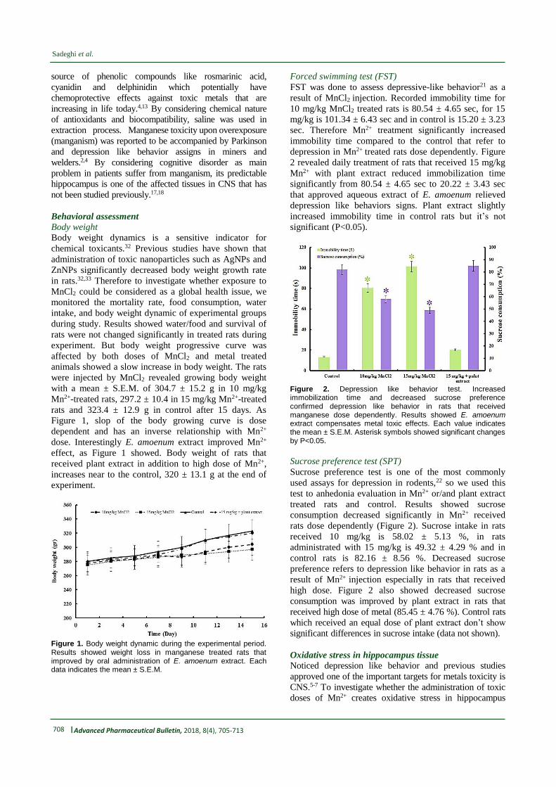
Figure 1, slop of the body growing curve is dose
dependent and has an inverse relationship with Mn2+
dose. Interestingly E. amoenum extract improved Mn2+
effect, as Figure 1 showed. Body weight of rats that
received plant extract in addition to high dose of Mn2+,
increases near to the control, 320 ± 13.1 g at the end of
experiment.
Figure 1. Body weight dynamic during the experimental period. Results showed weight loss in manganese treated rats that improved by oral administration of E. amoenum extract. Each data indicates the mean ± S.E.M.
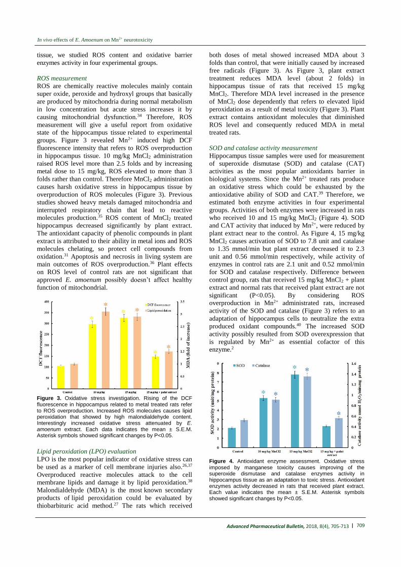
Forced swimming test (FST)
FST was done to assess depressive-like behavior21 as a
result of MnCl2 injection. Recorded immobility time for
10 mg/kg MnCl2 treated rats is 80.54 ± 4.65 sec, for 15
mg/kg is 101.34 ± 6.43 sec and in control is 15.20 ± 3.23
sec. Therefore Mn2+ treatment significantly increased
immobility time compared to the control that refer to
depression in Mn2+ treated rats dose dependently. Figure
2 revealed daily treatment of rats that received 15 mg/kg
Mn2+ with plant extract reduced immobilization time
significantly from 80.54 ± 4.65 sec to 20.22 ± 3.43 sec
that approved aqueous extract of E. amoenum relieved
depression like behaviors signs. Plant extract slightly
increased immobility time in control rats but it’s not
significant (P<0.05).
Figure 2. Depression like behavior test. Increased immobilization time and decreased sucrose preference confirmed depression like behavior in rats that received manganese dose dependently. Results showed E. amoenum extract compensates metal toxic effects. Each value indicates the mean ± S.E.M. Asterisk symbols showed significant changes by P<0.05.
Sucrose preference test (SPT)
Sucrose preference test is one of the most commonly
used assays for depression in rodents,22 so we used this
test to anhedonia evaluation in Mn2+ or/and plant extract
treated rats and control. Results showed sucrose
consumption decreased significantly in Mn2+ received
rats dose dependently (Figure 2). Sucrose intake in rats
received 10 mg/kg is 58.02 ± 5.13 %, in rats
administrated with 15 mg/kg is 49.32 ± 4.29 % and in
control rats is 82.16 ± 8.56 %. Decreased sucrose
preference refers to depression like behavior in rats as a
result of Mn2+ injection especially in rats that received
high dose. Figure 2 also showed decreased sucrose
consumption was improved by plant extract in rats that
received high dose of metal (85.45 ± 4.76 %). Control rats
which received an equal dose of plant extract don’t show
significant differences in sucrose intake (data not shown).
Oxidative stress in hippocampus tissue
Noticed depression like behavior and previous studies
approved one of the important targets for metals toxicity is
CNS.5-7 To investigate whether the administration of toxic
doses of Mn2+ creates oxidative stress in hippocampus
| 709
In vivo effects of E. Amoenum on Mn2+ neurotoxicity
Advanced Pharmaceutical Bulletin, 2018, 8(4), 705-713
tissue, we studied ROS content and oxidative barrier
enzymes activity in four experimental groups.
ROS measurement
ROS are chemically reactive molecules mainly contain
super oxide, peroxide and hydroxyl groups that basically
are produced by mitochondria during normal metabolism
in low concentration but acute stress increases it by
causing mitochondrial dysfunction.34 Therefore, ROS
measurement will give a useful report from oxidative
state of the hippocampus tissue related to experimental
groups. Figure 3 revealed Mn2+ induced high DCF
fluorescence intensity that refers to ROS overproduction
in hippocampus tissue. 10 mg/kg MnCl2 administration
raised ROS level more than 2.5 folds and by increasing
metal dose to 15 mg/kg, ROS elevated to more than 3
folds rather than control. Therefore MnCl2 administration
causes harsh oxidative stress in hippocampus tissue by
overproduction of ROS molecules (Figure 3). Previous
studies showed heavy metals damaged mitochondria and
interrupted respiratory chain that lead to reactive
molecules production.35 ROS content of MnCl2 treated
hippocampus decreased significantly by plant extract.
The antioxidant capacity of phenolic compounds in plant
extract is attributed to their ability in metal ions and ROS
molecules chelating, so protect cell compounds from
oxidation.31 Apoptosis and necrosis in living system are
main outcomes of ROS overproduction.36 Plant effects
on ROS level of control rats are not significant that
approved E. amoenum possibly doesn’t affect healthy
function of mitochondrial.
Figure 3. Oxidative stress investigation. Rising of the DCF fluorescence in hippocampus related to metal treated rats refer to ROS overproduction. Increased ROS molecules causes lipid peroxidation that showed by high malondialdehyde content. Interestingly increased oxidative stress attenuated by E. amoenum extract. Each data indicates the mean ± S.E.M. Asterisk symbols showed significant changes by P<0.05.
Lipid peroxidation (LPO) evaluation
LPO is the most popular indicator of oxidative stress can
be used as a marker of cell membrane injuries also.26,37
Overproduced reactive molecules attack to the cell
membrane lipids and damage it by lipid peroxidation.38
Malondialdehyde (MDA) is the most known secondary
products of lipid peroxidation could be evaluated by
thiobarbituric acid method.27 The rats which received
both doses of metal showed increased MDA about 3
folds than control, that were initially caused by increased
free radicals (Figure 3). As Figure 3, plant extract
treatment reduces MDA level (about 2 folds) in
hippocampus tissue of rats that received 15 mg/kg
MnCl2. Therefore MDA level increased in the presence
of MnCl2 dose dependently that refers to elevated lipid
peroxidation as a result of metal toxicity (Figure 3). Plant
extract contains antioxidant molecules that diminished
ROS level and consequently reduced MDA in metal
treated rats.
SOD and catalase activity measurement
Hippocampus tissue samples were used for measurement
of superoxide dismutase (SOD) and catalase (CAT)
activities as the most popular antioxidants barrier in
biological systems. Since the Mn2+ treated rats produce
an oxidative stress which could be exhausted by the
antioxidative ability of SOD and CAT.39 Therefore, we
estimated both enzyme activities in four experimental
groups. Activities of both enzymes were increased in rats
who received 10 and 15 mg/kg MnCl2 (Figure 4). SOD
and CAT activity that induced by Mn2+, were reduced by
plant extract near to the control. As Figure 4, 15 mg/kg
MnCl2 causes activation of SOD to 7.8 unit and catalase
to 1.35 mmol/min but plant extract decreased it to 2.3
unit and 0.56 mmol/min respectively, while activity of
enzymes in control rats are 2.1 unit and 0.52 mmol/min
for SOD and catalase respectively. Difference between
control group, rats that received 15 mg/kg MnCl2 + plant
extract and normal rats that received plant extract are not
significant (P<0.05). By considering ROS
overproduction in Mn2+ administrated rats, increased
activity of the SOD and catalase (Figure 3) refers to an
adaptation of hippocampus cells to neutralize the extra
produced oxidant compounds.40 The increased SOD
activity possibly resulted from SOD overexpression that
is regulated by Mn2+ as essential cofactor of this
enzyme.2
Figure 4. Antioxidant enzyme assessment. Oxidative stress imposed by manganese toxicity causes improving of the superoxide dismutase and catalase enzymes activity in hippocampus tissue as an adaptation to toxic stress. Antioxidant enzymes activity decreased in rats that received plant extract. Each value indicates the mean ± S.E.M. Asterisk symbols showed significant changes by P<0.05.

710 | Advanced Pharmaceutical Bulletin, 2018, 8(4), 705-713
Sadeghi et al.
Catecholamine content of hippocampus tissue
Catecholamines, including dopamine and
norepinephrine, are most the important
neurotransmitters that mediate a variety of functions in
CNS, such as motor control, cognition, emotion,
memory processing, and endocrine modulation.41
Dysfunctions in catecholamine neurotransmission are
related to some neuropsychiatric disorders specially
Parkinson disease and epilepsy.31 Similar
neuropsychiatric signs in Parkinson disease and
manganism possibly are caused by equal molecular
events.2 Therefore catecholamine content of
hippocampus tissue was compared between
experimental groups as follow: 10 mg/kg MnCl2,
142.43 ± 12.52 ng/mg protein; 15 mg/kg MnCl2, 91.45
± 4.52 ng/mg protein; 15 mg/kg MnCl2 + plant extract,
250.45 ± 12.34 ng/mg protein and control rats, 210.32 ±
10.23 ng/mg protein. Decreased catecholamine may be
caused by increased dopaminergic cell death in the
presence of metal ions.7 Diminished catecholamine was
returned near to (even more than) the control by plant
extraction treatment, while these kinds of
neurotransmitters have dual action (Neurotoxic and
neuroprotective) and according to previous
experiments, high doses of catecholamine induces
apoptosis in the neurons.42 Control rats that received
plant extract showed increase in catecholamine content
(224.41 ± 14.29 ng/mg protein) but it’s not significant
and does not accompanied with abnormal
neurobehaviours. Relieving effects of E. amoenum in
molecular level especially catecholamine rising, finally
lead to improved depression like behavior in rats
treated by toxic doses of metal as discussed above.
Caspase 9 and caspase 3 analysis Raised oxidative stress and reduced catecholamine
possibly cause cell death in metal treated hippocampus.
Catecholamine level of brain is important in healthy
function and survival of neurons and decreased
catecholamine lead to neurodegeneration in some
neurological disease.42 ROS overproduction was caused
by mitochondrial dysfunction or/and inefficient
antioxidant barrier that lead to mitochondrial-dependent
and –independent apoptosis with different molecular
mechanisms.43 Caspase 9 involves in mitochondrial-
independent and caspase 3 participates in mitochondrial-
dependent apoptosis.44,45 Our experiments revealed in
rats that received 10 mg/kg MnCl2 only caspase 9
increased significantly but in rats treated by 15 mg/kg
MnCl2 both of the caspase 3 and caspase 9 increased in
hippocampus (Figure 5). These results confirmed more
sensitivity of the mitochondria against metal toxicity.
Manganese overexposing well documented to result in a
disrupted Fe2+ homeostasis that lead to mitochondrial
dysfunction.44 As Figure 5, increased expression of the
caspase 3 and 9 that induced by 15 mg/kg of metal was
improved by oral administration of plant extract.
Figure 5. Immunoblotting studies. Up-regulation of caspase 3 and 9 during manganese intoxication refers to increased apoptosis in metal received rats. E. amoenum extraction significantly decreased neurodegeneration in hippocampus tissue. The intensity of bands was quantified by ImageJ software. The data were expressed as mean ± S.E.M of three independent experiments. Asterisk (*) was used to denote statistical significance (P<0.05).
Histological studies
The biological significance and toxicological
importance of any changes which are found between
tissue section in control and experimental groups have
been considered as biochemical results confirmation.
Therefore after the end of experimental time course, rats
were anesthetized and brain tissue separated from scalp.
Tissue samples were treated by formalin for fixation and
stained by hematoxilin-eosin method and then studied by
light microscope.46 Results showed presence of necrotic
and apoptotic cells in tissues were administrated by 10
and 15 mg/kg MnCl2 (Figure 6). Early apoptotic nuclei
have a condensed appearance that frequently seen in
MnCl2 administrated tissue especially in 15 mg/kg
MnCl2 received rats. Increased apoptosis in the metal
treated rats accompanied by decreased level of the
catecholamine may be due to catecholamine positive role
in cell survival or catecholamine producing cell death in
Mn2+ neurotoxicity. Histology results also were
confirmed by elevated level of caspase 3 and 9 in
intoxicated rats. As Figure 6, amounts of condensed
apoptotic and deformed necrotic cells reduced in tissues
related to rats received plant extract+15 mg/kg MnCl2
that accompanied by decreased caspases and improved
behavioral abnormalities also.

| 711
In vivo effects of E. Amoenum on Mn2+ neurotoxicity
Advanced Pharmaceutical Bulletin, 2018, 8(4), 705-713
Figure 6. Histological studies. Hematoxylin/eosin staining of hippocampus sections revealed presence of the apoptotic and necrotic cells in MnCl2 treated rat hippocampus rather than control. Result showed oral administration of E. amoenum extraction significantly decreased apoptotic and necrotic cells in hippocampus.
Conclusion
Pathophysiological signs of manganism in human and
animal models suggest hippocampus as a possible
affected tissue. Our biochemical results approved hash
oxidative damages in the presence of MnCl2 doses that
attenuated by plant extract. Increased expression of the
caspase 9 in low dose of Mn2+ revealed, metal toxicity
causes mitochondrial dysfunction at first and then
induces oxidative damages that lead to mitochondrial
independent apoptosis in the presence of high metal dose
(caspase 3 upregulation). Despite the prevalent use of E.
amoenum as an antidepressant, there are no
pharmacological data to support such effects. Our
molecular and biochemical studies confirmed E.
amoenum extract inhibited apoptosis from both described
pathways possibly by ROS molecules scavenging,
mitochondrial dysfunction improving and metal ions
trapping. All of the identified beneficial effects or
possibly uncharacterized mechanisms lead to decreased
depressive behaviors. Investigated therapeutic effects of
E. amoenum on Mn2+ neurotoxicity revealed this plant
could be considered in antidepressant drug design and as
supplement against all of the metals toxicity or oxidative
damages. By considering physiological and molecular
similarities between manganism and Parkinson disease
and also E. amoenum role in catecholamine
overproduction, this plant could be used as natural co-
treatment in associated diseases.
Ethical Issues
All the experimental works were approved by the Ethical
Committee of Isfahan University of Medical Science
(Isfahan, Iran) and conform to the European
Communities Council Directive of 24 November 1986
(86/609/EEC).
Conflict of Interest
All of the Authors have no conflict of interest to declare.
References
1. O’Neal SL, Zheng W. Manganese Toxicity Upon
Overexposure: a Decade in Review. Curr Environ
Health Rep 2015;2(3):315-28. doi: 10.1007/s40572-
015-0056-x
2. Kwakye GF, Paoliello MM, Mukhopadhyay S,
Bowman AB, Aschner M. Manganese-induced
parkinsonism and parkinson's disease: Shared and
distinguishable features. Int J Environ Res Public
Health 2015;12(7):7519-40. doi:
10.3390/ijerph120707519
3. Takeda A. Manganese action in brain function. Brain
Res Brain Res Rev 2003;41(1):79-87.
doi.org/10.1016/S0165-0173(02)00234-5

712 | Advanced Pharmaceutical Bulletin, 2018, 8(4), 705-713
Sadeghi et al.
4. Long Z, Jiang YM, Li XR, Fadel W, Xu J, Yeh CL, et
al. Vulnerability of welders to manganese exposure-
-a neuroimaging study. Neurotoxicology
2014;45:285-92. doi: 10.1016/j.neuro.2014.03.007
5. Oulhote Y, Mergler D, Barbeau B, Bellinger DC,
Bouffard T, Brodeur ME, et al. Neurobehavioral
function in school-age children exposed to
manganese in drinking water. Environ Health
Perspect 2014;122(12):1343-50. doi:
10.1289/ehp.1307918
6. Peres TV, Schettinger MR, Chen P, Carvalho F, Avila
DS, Bowman AB, et al. Manganese-induced
neurotoxicity: a review of its behavioral
consequences and neuroprotective strategies. BMC
Pharmacol Toxicol 2016;17(1):57. doi:
10.1186/s40360-016-0099-0
7. Yousefi Babadi V, Sadeghi L, Amraie E, Rezaei M,
Malekirad AA, Abarghouei Nejad M. Manganese
toxicity in the central nervous system: Decreeing of
catecholamine in rat’s brains. Health
2013;5(12):2146-9.
doi: 10.4236/health.2013.512292
8. Belyaeva EA, Sokolova TV, Emelyanova LV,
Zakharova IO. Mitochondrial electron transport
chain in heavy metal-induced neurotoxicity: Effects
of cadmium, mercury, and copper.
ScientificWorldJournal 2012;2012:136063. doi:
10.1100/2012/136063
9. Safaeian L, Haghjoo Javanmard S, Ghanadian M,
Seifabadi S. Cytoprotective and antioxidant effects
of echium amoenum anthocyanin-rich extract in
human endothelial cells (huvecs). Avicenna J
Phytomed 2015;5(2):157-66.
10. Ranjbar A, Khorami S, Safarabadi M, Shahmoradi A,
Malekirad AA, Vakilian K, et al. Antioxidant
activity of iranian echium amoenum fisch & c.A.
Mey flower decoction in humans: A cross-sectional
before/after clinical trial. Evid Based Complement
Alternat Med 2006;3(4):469-73. doi:
10.1093/ecam/nel031
11. Nadi F. Bioactive compound retention in Echium
amoenum Fisch. & C. A. Mey. petals: Effect of
fluidized bed drying conditions. Int J Food Prot
2017;20:2249-60. doi:
10.1080/10942912.2016.1233436
13. Rabbani M, Sajjadi SE, Vaseghi G, Jafarian A.
Anxiolytic effects of echium amoenum on the
elevated plus-maze model of anxiety in mice.
Fitoterapia 2004;75(5):457-64. doi:
10.1016/j.fitote.2004.04.004
14. Hosseini N, Abolhassani M. Immunomodulatory
properties of borage (Echium amoenum) on BALB/c
mice infected with Leishmania major. J Clin
Immunol 2011;31(3):465-71. doi: 10.1007/s10875-
010-9502-6
15. Abed A, Minaiyan M, Ghannadi A, Mahzouni P,
Babavalian MR. Effect of echium amoenum fisch.
Et mey a traditional iranian herbal remedy in an
experimental model of acute pancreatitis. ISRN
Gastroenterol 2012;2012:141548. doi:
10.5402/2012/141548
16. Sayyah M, Boostani H, Pakseresht S, Malaieri A.
Efficacy of aqueous extract of echium amoenum in
treatment of obsessive-compulsive disorder. Prog
Neuropsychopharmacol Biol Psychiatry
2009;33(8):1513-6. doi:
10.1016/j.pnpbp.2009.08.021
17. Munoz-Espada AC, Watkins BA. Cyanidin
attenuates PGE2 production and cyclooxygenase-2
expression in LNCaP human prostate cancer cells. J
Nutr Biochem 2006;17(9):589-96. doi:
10.1016/j.jnutbio.2005.10.007
18. Sarnyai Z, Sibille EL, Pavlides C, Fenster RJ,
McEwen BS, Toth M. Impaired hippocampal-
dependent learning and functional abnormalities in
the hippocampus in mice lacking serotonin(1A)
receptors. Proc Natl Acad Sci U S A
2000;97(26):14731-6. doi:
10.1073/pnas.97.26.14731
19. Robison G, Zakharova T, Fu S, Jiang W, Fulper R,
Barrea R, et al. X-ray fluorescence imaging of the
hippocampal formation after manganese exposure.
Metallomics 2013;5(11):1554-65. doi:
10.1039/c3mt00133d
20. Deepa P, Kannappan N. Comparative in vitro
antioxidant studies of aqueous solution of
formulated poly herbal formulation with marketed
preparation. Der Pharm Lett 2012;4(5):1515-7.
21. Yankelevitch-Yahav R, Franko M, Huly A, Doron R.
The forced swim test as a model of depressive-like
behavior. J Vis Exp 2015(97). doi: 10.3791/52587
22. Overstreet DH. Modeling depression in animal
models. Methods Mol Biol 2012;829:125-44. doi:
10.1007/978-1-61779-458-2_7
23. Keston AS, Brandt R. The fluorometric analysis of
ultramicro quantities of hydrogen peroxide. Anal
Biochem 1965;11:1-5.
24. LeBel CP, Ischiropoulos H, Bondy SC. Evaluation of
the probe 2',7'-dichlorofluorescin as an indicator of
reactive oxygen species formation and oxidative
stress. Chem Res Toxicol 1992;5(2):227-31.
25. Bradford MM. A rapid and sensitive method for the
quantitation of microgram quantities of protein
utilizing the principle of protein-dye binding. Anal
Biochem 1976;72:248-54.
26. Niki E. Lipid peroxidation products as oxidative
stress biomarkers. Biofactors 2008;34(2):171-80.
doi: 10.1002/biof.5520340208.
27. Buege JA, Aust SD. Microsomal lipid, Peroxidation.
In: Flesicher S, Packer L, editors. Methods in
Enzymology. New-York: Academic Press; 1978.
28. Oyanagui Y. Reevaluation of assay methods and
establishment of kit for superoxide dismutase
activity. Anal Biochem 1984;142(2):290-6.
12. Abolhassani M. Antibacterial effect of borage
(echium amoenum) on staphylococcus aureus. Braz
J Infect Dis 2004;8(5):382-5. doi:
10.1590/S1413-86702004000500008
| 713
In vivo effects of E. Amoenum on Mn2+ neurotoxicity
Advanced Pharmaceutical Bulletin, 2018, 8(4), 705-713
29. Mittal M, Flora SJ. Vitamin E supplementation
protects oxidative stress during arsenic and fluoride
antagonism in male mice. Drug Chem Toxicol
2007;30(3):263-81.
doi:10.1080/01480540701380075
30. Felice LJ, Felice JD, Kissinger PT. Determination of
catecholamines in rat brain parts by re- verse-phase
ion-pair liquid chromatography. J Neurochem
1978;31(6):1461-5.
31. Sadeghi L, Rizvanov AA, Salafutdinov, II,
Dabirmanesh B, Sayyah M, Fathollahi Y, et al.
Hippocampal asymmetry: Differences in the left and
right hippocampus proteome in the rat model of
temporal lobe epilepsy. J Proteomics 2017;154:22-
9. doi: 10.1016/j.jprot.2016.11.023
32. Jacquier M, Crauste F, Soulage CO, Soula HA. A
predictive model of the dynamics of body weight
and food intake in rats submitted to caloric
restrictions. PLoS One 2014;9(6):e100073. doi:
10.1371/journal.pone.0100073
33. Yin N, Yao X, Zhou Q, Faiola F, Jiang G. Vitamin E
attenuates silver nanoparticle-induced effects on
body weight and neurotoxicity in rats. Biochem
Biophys Res Commun 2015;458(2):405-10. doi:
10.1016/j.bbrc.2015.01.130
34. Gao L, Laude K, Cai H. Mitochondrial
pathophysiology, reactive oxygen species, and
cardiovascular diseases. Vet Clin North Am Small
Anim Pract 2008;38(1):137-55, vi. doi:
10.1016/j.cvsm.2007.10.004
35. Sharma B, Singh S, Siddiqi NJ. Biomedical
implications of heavy metals induced imbalances in
redox systems. Biomed Res Int 2014;2014:640754.
doi: 10.1155/2014/640754
36. Fu PP, Xia Q, Hwang HM, Ray PC, Yu H.
Mechanisms of nanotoxicity: generation of reactive
oxygen species. J Food Drug Anal 2014;22(1):64-
75. doi: 10.1016/j.jfda.2014.01.005
37. Barrera G. Oxidative stress and lipid peroxidation
products in cancer progression and therapy. ISRN
Oncol 2012;2012:137289. doi:
10.5402/2012/137289
38. Pernot F, Heinrich C, Barbier L, Peinnequin A,
Carpentier P, Dhote F, et al. Inflammatory changes
during epileptogenesis and spontaneous seizures in a
mouse model of mesiotemporal lobe epilepsy.
Epilepsia 2011;52(12):2315-25. doi:
10.1111/j.1528-1167.2011.03273.x
39. Zhan CD, Sindhu RK, Pang J, Ehdaie A, Vaziri ND.
Superoxide dismutase, catalase and glutathione
peroxidase in the spontaneously hypertensive rat
kidney: Effect of antioxidant-rich diet. J Hypertens
2004;22(10):2025-33.
40. Vermeij WP, Alia A, Backendorf C. Ros quenching
potential of the epidermal cornified cell envelope. J
Invest Dermatol 2011;131(7):1435-41. doi:
10.1038/jid.2010.433
41. Kobayashi K. Role of catecholamine signaling in
brain and nervous system functions: New insights
from mouse molecular genetic study. J Investig
Dermatol Symp Proc 2001;6(1):115-21. doi:
10.1046/j.0022-202x.2001.00011.x
42. Noh JS, Kim EY, Kang JS, Kim HR, Oh YJ, Gwag
BJ. Neurotoxic and neuroprotective actions of
catecholamines in cortical neurons. Exp Neurol
1999;159(1):217-24. doi: 10.1006/exnr.1999.7144
43. Tai YK, Chew KC, Tan BW, Lim KL, Soong TW.
Iron mitigates dmt1-mediated manganese
cytotoxicity via the ask1-jnk signaling axis:
Implications of iron supplementation for manganese
toxicity. Sci Rep 2016;6:21113. doi:
10.1038/srep21113
44. Wang C, Youle RJ. The role of mitochondria in
apoptosis. Annu Rev Genet 2009;43:95-118. doi:
10.1146/annurev-genet-102108-134850
45. Brentnall M, Rodriguez-Menocal L, De Guevara RL,
Cepero E, Boise LH. Caspase-9, caspase-3 and
caspase-7 have distinct roles during intrinsic
apoptosis. BMC Cell Biol 2013;14:32. doi:
10.1186/1471-2121-14-32
46. Dhandapani S, Subramanian VR, Rajagopal S,
Namasivayam N. Hypolipidemic effect of cuminum
cyminum l. On alloxan-induced diabetic rats.
Pharmacol Res 2002;46(3):251-5.
Related Documents