*Corresponding Author Address: Dr. Noorah S Al-Sowayan, Department of Biology, Faculty of Science, Qassim University, P. O. Box: (30230), Buraydah (51744), Saudi Arabia; E-mail: [email protected] World Journal of Pharmaceutical Sciences ISSN (Print): 2321-3310; ISSN (Online): 2321-3086 Published by Atom and Cell Publishers © All Rights Reserved Available online at: http://www.wjpsonline.org/ Original Article Physiological alteration of mitochondrial genome in relation to aging on male albino rats Noorah S. Al-Sowayan Department of Biology, Faculty of Science, Qassim University, P. O. Box: (30230), Buraydah (51744), Saudi Arabia Received: 16-01-2015 / Revised: 18-02-2015 / Accepted: 20-02-2015 Abstract There is no doubt that there is a great interest on aging and its causes from human especially women, this increase scientific researches on aging including this increase research, which detect mitochondrial DNA in relation to aging, where mitochondria is the house of energy in the living cell. Sixty male albino rats were divided into three equal groups 1 st group, young rats (6 months age) as control, 2 nd group, adult rats (12 months age) and 3 rd group, aged rats (21, months age). Rats were sacrificed and the brain and liver tissues were preserved in liquid nitrogen immediately for molecular biological analysis to see the different gene expression in the respiratory chain at different ages. Other samples were taken from the liver, brain and serum for the biochemical and physiological analysis to find out the effect of aging on glutathione peroxidase activity and concentration of reduced glutathione, total antioxidant capacity, nitric oxide and finally malondialdhyde. NADH dehydrogenase and cytochrome oxidase gene expression, showed a decreased activity in both liver and brain with aging. Glutathione peroxidase showed non-significant decrease in its activity in liver, but a significant decrease in brain in adult rats, and a significant decrease in both liver & brain in aged rats. Reduced glutathione showed non-significant decrease in its concentration in both liver & brain in adult rats but showed a significant decrease in aged rats. Serum total antioxidant capacity showed non-significant decrease in its concentration in adult rats but significantly decreased in aged rats. Liver and brain nitric oxide showed a significant increase in its concentration in both adult and aged rats. Liver malondialdhyde showed a significant increase in its concentration in both adult and aged rats, but in brain, showed non-significant increase in adult rats while, significantly increased in aged rats. All previous results in compared with young rats (control group). Keywords: Mitochondrial genome aging, male albino rats liver & brain, NADH dehydrogenase gene expression, cytochrome oxidase gene expression glutathione peroxidase, reduced glutathione, serum total antioxidant, nitric oxide, malondialdhyde. INTRODUCTION In cell biology a mitochondrion (plural mitochondrion) is a membrane-enclosed organelle found in most eukaryotic cells. These organelles range from 0.5 to 10 micrometers (μm) in diameter (1) . Several characteristics make mitochondria unique. The number of mitochondria in a cell varies widely by organism and tissue type. Many cells have only a single mitochondrion, whereas others can contain several thousand mitochondria (2) . Mitochondrial proteins vary depending on the tissue and the species. In humans, 615 distinct types of proteins have been identified from cardiac mitochondria (3) . Whereas, in Murinae (rats), 940 proteins encoded by distinct genes have been reported. The mitochondrial proteome is thought to be dynamically regulated (4) . Mitochondria are sometimes described as “cellular power plants” because they generate most of the cell’s supply of ATP, used as a source of chemical energy (5) . In addition to supplying cellular energy, mitochondria are involved in a range of other processes, such as signaling, cellular differentiation, cell death, as well as the control of cell cycle and cell growth (6) . Mitochondria have been implicated in several human diseases, including mitochondrial disorders (7) . Extant mammalian mtDNAs have retained only 13 polypeptide genes, all of which encode essential components of oxidative phosphorylation. MTDNA also encodes the 12S and 16S rRNA genes and 22 tRNA genes required for mitochondrial protein synthesis. The remaining mitochondrial oxidative phosphorylation proteins,

Welcome message from author
This document is posted to help you gain knowledge. Please leave a comment to let me know what you think about it! Share it to your friends and learn new things together.
Transcript
*Corresponding Author Address: Dr. Noorah S Al-Sowayan, Department of Biology, Faculty of Science, Qassim University, P. O. Box:
(30230), Buraydah (51744), Saudi Arabia; E-mail: [email protected]
World Journal of Pharmaceutical Sciences ISSN (Print): 2321-3310; ISSN (Online): 2321-3086
Published by Atom and Cell Publishers © All Rights Reserved
Available online at: http://www.wjpsonline.org/
Original Article
Physiological alteration of mitochondrial genome in relation to aging on male albino
rats
Noorah S. Al-Sowayan
Department of Biology, Faculty of Science, Qassim University, P. O. Box: (30230), Buraydah (51744),
Saudi Arabia
Received: 16-01-2015 / Revised: 18-02-2015 / Accepted: 20-02-2015
Abstract
There is no doubt that there is a great interest on aging and its causes from human especially women, this
increase scientific researches on aging including this increase research, which detect mitochondrial DNA in
relation to aging, where mitochondria is the house of energy in the living cell. Sixty male albino rats were
divided into three equal groups 1st group, young rats (6 months age) as control, 2
nd group, adult rats (12 months
age) and 3rd
group, aged rats (21, months age). Rats were sacrificed and the brain and liver tissues were
preserved in liquid nitrogen immediately for molecular biological analysis to see the different gene expression in
the respiratory chain at different ages. Other samples were taken from the liver, brain and serum for the
biochemical and physiological analysis to find out the effect of aging on glutathione peroxidase activity and
concentration of reduced glutathione, total antioxidant capacity, nitric oxide and finally malondialdhyde. NADH
dehydrogenase and cytochrome oxidase gene expression, showed a decreased activity in both liver and brain
with aging. Glutathione peroxidase showed non-significant decrease in its activity in liver, but a significant
decrease in brain in adult rats, and a significant decrease in both liver & brain in aged rats. Reduced glutathione
showed non-significant decrease in its concentration in both liver & brain in adult rats but showed a significant
decrease in aged rats. Serum total antioxidant capacity showed non-significant decrease in its concentration in
adult rats but significantly decreased in aged rats. Liver and brain nitric oxide showed a significant increase in
its concentration in both adult and aged rats. Liver malondialdhyde showed a significant increase in its
concentration in both adult and aged rats, but in brain, showed non-significant increase in adult rats while,
significantly increased in aged rats. All previous results in compared with young rats (control group).
Keywords: Mitochondrial genome aging, male albino rats liver & brain, NADH dehydrogenase gene
expression, cytochrome oxidase gene expression glutathione peroxidase, reduced glutathione, serum total
antioxidant, nitric oxide, malondialdhyde.
INTRODUCTION
In cell biology a mitochondrion (plural
mitochondrion) is a membrane-enclosed organelle
found in most eukaryotic cells. These organelles
range from 0.5 to 10 micrometers (µm) in diameter (1)
. Several characteristics make mitochondria
unique. The number of mitochondria in a cell
varies widely by organism and tissue type. Many
cells have only a single mitochondrion, whereas
others can contain several thousand mitochondria (2)
. Mitochondrial proteins vary depending on the
tissue and the species. In humans, 615 distinct
types of proteins have been identified from cardiac
mitochondria (3)
. Whereas, in Murinae (rats), 940
proteins encoded by distinct genes have been
reported. The mitochondrial proteome is thought to
be dynamically regulated (4)
. Mitochondria are
sometimes described as “cellular power plants”
because they generate most of the cell’s supply of
ATP, used as a source of chemical energy (5)
. In
addition to supplying cellular energy, mitochondria
are involved in a range of other processes, such as
signaling, cellular differentiation, cell death, as
well as the control of cell cycle and cell growth (6)
.
Mitochondria have been implicated in several
human diseases, including mitochondrial disorders
(7). Extant mammalian mtDNAs have retained only
13 polypeptide genes, all of which encode essential
components of oxidative phosphorylation.
MTDNA also encodes the 12S and 16S rRNA
genes and 22 tRNA genes required for
mitochondrial protein synthesis. The remaining
mitochondrial oxidative phosphorylation proteins,
Noorah, World J Pharm Sci 2015; 3(3): 413-425
414
the metabolic enzymes, the DNA and RNA
polymerases, the ribosomal proteins and the
mtDNA regulatory factors are all encoded by
nuclear genes, synthesized in the cytosol and then
imported into the organelle (8)
. The human
mitochondrial genome is 16.569 (bp) in length
closed, circular molecule. Mitochondrial DNA has
two strands, a guanine- rich heavy (H) strand and a
cytosine-rich light (L) strand. The heavy strand
contains 12 of 13 polypeptide –encoding genes, 14
of the 22 tRNA- encoding genes and all the coding
sequences are contiguous (9)
. The only non-conding
segment of mtDNA is the displacement loop (D-
loop), a region of 1121 bp that contains the origin
of replication of the H-strand (OH) and the
parameters for L and H –strand transcription. The
mtDNA is replicated from two origins. DNA
replication is initiated at OH using an RNA primer
generated from the L-strand transcript.
H-strand synthesis proceeds two –thirds of the way
around the mtDNA, displacing the parental H-
strand until it reaches the L-strand origin (OL),
situated in a cluster of five tRNA genes. Once
exposed on the displaced H-strand, OL folds a stem
–loop structure and L-strand synthesis is initiated
and proceeds beak along the H-strand template.
Conseq2uently, mtDNA replication is bidirectional
but asynchronous (10)
. The mammalian ova contain
about 100.000 molecules of mtDNA, while the
sperm contains of the order of 100-1500 mtDNA.
Sperm mitochondria enter the ova during
fertilization but they appear to be lost early in
embryogenesis, soon after fertilization, between the
two-cell and four cell stages (11)
. Mitochondria lack
an efficient DNA repair system. Moreover,
protective proteins such as histones are missing and
mtDNA is physically associated with the inner
mitochondrial membrane, where highly somatic
mutagenic oxidative phosphorylation(12)
. Ageing
(British English) or aging (American English) is
the accumulation of changes in an organism or
object over time (13)
. The cumulative accumulation
of these somatic mutations during life may cause a
bioenergic deficit leading to cell death, or apoptosis
and normal aging. In addition to the aging or
senescence process somatic mtDNA mutations may
be important for determining the onset and
progression of mtDNA disease (14)
. Aging is
associated with common diseases including,
Alzheimer’s, arthritis, heart attack, cancer,
Parkinson’s, heart disease, pneumonia, stroke,
osteoporosis and diabetes (15)
. Free radicals are
atoms or groups of atoms with an odd number of
electrons and can be formed when oxygen interacts
with certain molecules. Once formed, these highly
reactive radicals can start a chain of reaction, like
dominos (16)
. Free radicals divided into Reactive
Nitrogen Species (RNS) including Nitric Oxide
(NO) and peroxynitrite (ONOO-) and reactive
oxygen species (ROS), which include radical
superoxide (O2-) and hydroxyl radical (HO) or non
radical species such as hydrogen peroxide (H2O2) (17)
.
These compounds are toxic to both the invader and
the cell itself, so they are kept in compartments
inside the cell. This method is referred to as oxygen
– dependent into cellular killing of which there are
two types (18)
. The first type is the oxygen –
dependent production of a superoxide which is an
oxygen –rich bacteria – killing substance. The
superoxide is converted to hydrogen peroxide and
singlet oxygen by an enzyme called superoxide
dismutase. Superoxides also react with the
hydrogen peroxide to produce hydroxyl radicals
which assist in killing the invading microbe (19)
.
The second type involves the use of the enzyme
myeloperoxidase from neutrophil granules. When
granules fuse with a phagosome, myeloperoxidase
in released into the phagodysosome and this
enzyme uses hydrogen peroxide and chlorine to
create hypochlorite, a substance used in extremely
toxic to bacteria (19)
. The membrane – associated
NAD (P) H oxidase, cytochrome oxidase and
xanthine oxidase(20)
. No generation occurs through
specific nitric oxide synthase isozymes, including
mitochondrial nitric oxide synthase (mtNOS),
neuronal NOS (nNOS), endothelial NOS (eNOS)
and inducible NOS (iNOS)(21)
. Malondialdhyde
mainly exists in the enol form. MDA is generated
from reactive oxygen species (ROS) and as such is
assayed in vivo as a bio. merker of oxidative
stress(22)
. Malondialdhyde reacts with
deoxyadenosine and deoxyguanine in DNA,
forming DNA adducts, the primary one being
M1G1, which is mutagenic. The guandine group of
arginine residues condenses with NDA to give
aminopyrimidines (23)
. An ideal antioxidant should
be easily absorbed and counteract free radicals and
chelate redox metals at physiologically relevant
levels. It should also work in both aqueous and /or
membrane domains and have a positive effect on
gene expression.
Endogenous antioxidants ploy a crucial role in
maintaining optimal cellular Rumetions(24)
.
Enzymatic antioxidants include superoxide
dismutase, catalase and glutathione peroxidase(25)
.
Non-enzymatic antioxidants include vitamine E
and C, thiol antioxidants (glutathione thioredoxin
and lipoic acid), carotenoids, natural flavonoids,
mdatomin and other compounds (26)
. Superoxide
dismutase, one the most efficient intracellular
enzymatic antioxidants is superoxide dismutase
(SOD). SOD is the antioxidant enzyme that
catalyses the dismutation of O2- to O2 and to less
the reactive species H2O2 (27).
Catalase is present in
Noorah, World J Pharm Sci 2015; 3(3): 413-425
415
the peroxisome of aerobic cells and plays a major
role in the conversion of hydrogen peroxide to
water and molecular oxygen. Catalase has one of
the highest turnover rates for all enzymes, one
molecule of catalase can convert approximately 6
million molecules of hydrogen peroxide to water
and oxygen each minute(24)
. Glutathione peroxidase
(GPx) had five isoforms of selenium (Se)
dependent are found in humans. GPx1, is
ubiquitously expressed and a major scavenger for
H2O2 and lipid hydroperoxides. GPx2, is
epithelium –specific and highly expressed in the
gastrointestinal tract. GPx3 can uses thioredoxin
and glutaredoxin in addition to GSH as electron
donors to reduced a board range of hydroperoxide.
GPx4 is present in cytosolic, mitochondrial and
nuclear forms by alternative splicing, and is a
major enzyme preventing oxidation of membrane
phospholipids. GPx6 (newly discovered) is
localized preferentially in olfactory mucosa and
embryonic tissues (28)
. This study designed to
investigate the gene expression of some
mitochondrial respiratory chain complexes at
different ages in rats as NADH dehydrogenase
(complex I) cytochrome oxidase (complex IV),
moreover, determination the activity of glutathione
peroxidase enzyme. Also the concentration of
reduced glutathione total antioxidant capacity,
metric oxide and malondialdhyde were determined
at different ages.
MATERIAL AND METHODS
Experimental animals: Sixty male albino rats
were obtained from laboratory animal house unit,
College of pharmacy, King Soud University, Saudi
Arabia, divided into three equal groups, twenty of
each. They were kept for two weeks before starting
this study in laboratory environment for
accommodation. The animals were kept in suitable
environmental and nutritional conditions with free
access of fresh and clean tap water and a standard
commercial chow cake for 90 days. They were
divided into three equal each of 20 rats as follows,
group 1 as control or young animals (6 months
old), group 2 adult (12 months old) and group 3,
(24 months, old).
Sampling: After sacrificing one gram of brain and
liver were taken immediately and stored in liquid
nitrogen container till use for molecular analysis.
Biochemical measurement of glutathione
peroxidase (GPx), reduced glutathione (GSH),
Nitric oxide (NO) and malondialdhyde (MDA), the
tissues from liver and brain were taken and
homogenized to obtain their homogenate. Serum
samples were prepared to be used for total
antioxidant capacity measurement.
Sample preparation:-
Tissue sample preparation: Liver and brain were
homogenized in 48 volumes (per weight tissue) of
cold buffer (5 mM phosphate buffer, PH 7.0
containing 5 M EDTA and 1 mM 2. mercop to
ethanol) and centrifuged at 4000 rpm for 10-20
minutes at 2-8 oC, then the supernatant were
collected and frozen at -80 oC till use
(29).
Serum sample preparation: Blood was collected
without using an anticoagulant and was allowed to
clot for 30 min. at 25 oC, the centrifuged at 3000
rpm for 15 min. at 4 oC. The top yellow serum
layer was pipetted without disrupting the white
Buffy layer serum was frozen at -80 oC till use
(30).
Molecular determination:-
Gene expression: NADH dehydrogenase
(complex-1) quanitative Rt- PCR (31)
.
RNA extraction from tissues: Total TNA was
extracted with E.Z.N.A. spin column RNa
extraction kit (Oemga Biotech, Cairo Egypt).
Synthesis of first strand (cDNA): The synthesis
of first strand was performed by using (Qiagen
long range) RT-PCR kit (20). Mat. No. 1474.
PCR amplification: The PCR amplification was
occur using 2XPCR master Mix from (Qiagen long
range). PCR was carried out in a volume of 50 µ
containing of 1 µg of cDNA, 25 µl of 2X master
Mix (Qiagen), forward and reverse primer and
completed to 50 µl with water, nuclease –free. The
next table shows specific concentration of dNTP
and oligonucleatide primer, along with PCR
conditions for each gene tested. Oligonucteatide
primers were designed with Eugene version 2.2
software (Ambion). A primer pair for β-actin
(Ambion, Austin, TX) was used as an internal
control for a PCR analysis. All reactions were
preformed in 2720 thermocyeler (Applied
Biosystem) in which samples underwent a 5 min.
initial denaturing step to release DNA polymerase
activity (hot start PCR), followed by the number of
cycles indicated in table (1) for 30 sec. at 94 oC, at
the annealing temperature indicated in the same
table and 45 sec. at 72 oC. The final extension step
was 5- min at 72 oC
(31). PCR products were
separated on a 1.5% agarose gel in Tris acetate
EDTA buffer with 0.5 µg/ml. ethdium bromides.
The electrophoretic picture was taken by digital
camera 3.2 mega pixel and quantitative with gel
analyzer 2 software (31)
.
Biochemical analysis:
Determination of glutathione peroxidase (GPx)
activity: Glutathione peroxidase activity was
Noorah, World J Pharm Sci 2015; 3(3): 413-425
416
calorimetrically determined according to the
method adopted by paglia and valentine (29)
.
Determination of Reduced glutathione (GSH)
concentration: GSH was measured according to
the method of Butler et al. (32)
.
Total antioxidant capacity: Total antioxidant
capacity was determined according to the method
of Koraceiv et al., (30)
.
Determination of nitric oxide (NO)
concentration: Nitric oxide (NO) concentration
was determined according to the method described
by Moshage et al. (33)
.
Determination L-Malanaldhyde (MDA)
concentration: MDA were calorimetrically
according to the method adopted by Esterbauer et
al., (34)
.
Statistical analysis: The data were analyzed using
one way ANOVA to determine the statistical
significance of difference among groups (35)
.
RESULTS
Molecular determination:
β-actin, mRNA expression of rats liver and
brain:- The obtained results were statistically and
analysed in table (2) and illustrated in Figs. (1 &
2).
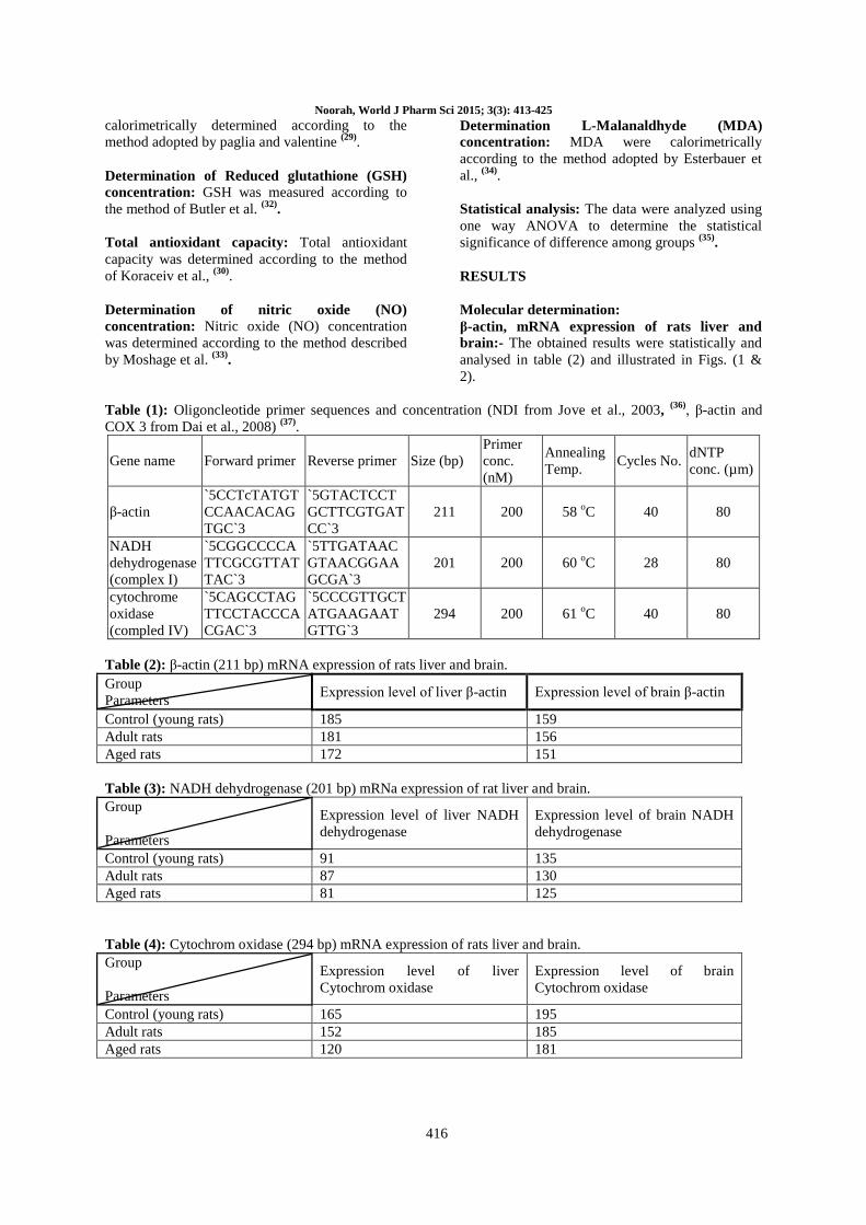
Table (1): Oligoncleotide primer sequences and concentration (NDI from Jove et al., 2003, (36)
, β-actin and
COX 3 from Dai et al., 2008) (37)
.
Gene name Forward primer Reverse primer Size (bp)
Primer
conc.
(nM)
Annealing
Temp. Cycles No.
dNTP
conc. (µm)
β-actin
`5CCTcTATGT
CCAACACAG
TGC`3
`5GTACTCCT
GCTTCGTGAT
CC`3
211 200 58 oC 40 80
NADH
dehydrogenase
(complex I)
`5CGGCCCCA
TTCGCGTTAT
TAC`3
`5TTGATAAC
GTAACGGAA
GCGA`3
201 200 60 oC 28 80
cytochrome
oxidase
(compled IV)
`5CAGCCTAG
TTCCTACCCA
CGAC`3
`5CCCGTTGCT
ATGAAGAAT
GTTG`3
294 200 61 oC 40 80
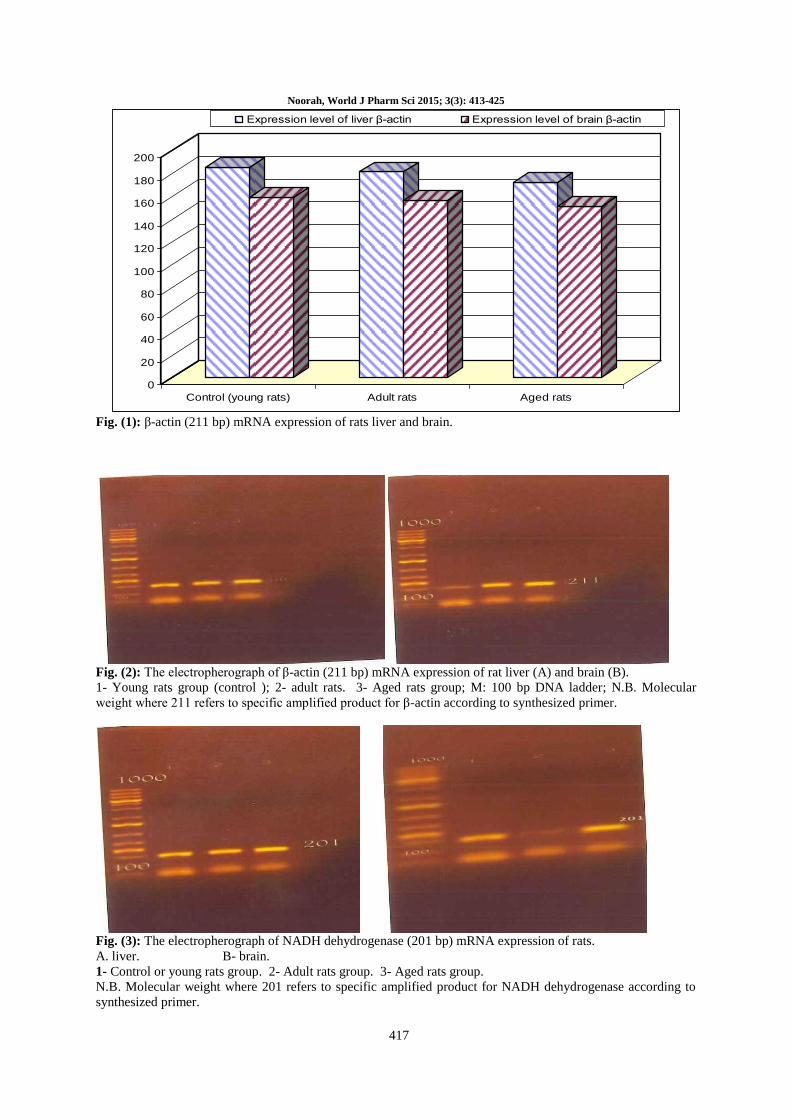
Table (2): β-actin (211 bp) mRNA expression of rats liver and brain.
Group
Parameters Expression level of liver β-actin Expression level of brain β-actin
Control (young rats) 185 159
Adult rats 181 156
Aged rats 172 151
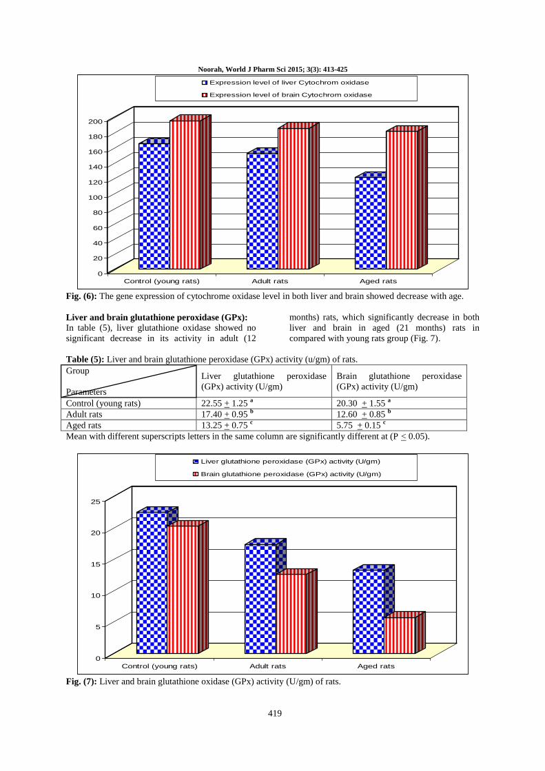
Table (3): NADH dehydrogenase (201 bp) mRNa expression of rat liver and brain.
Group
Parameters
Expression level of liver NADH
dehydrogenase
Expression level of brain NADH
dehydrogenase
Control (young rats) 91 135
Adult rats 87 130
Aged rats 81 125
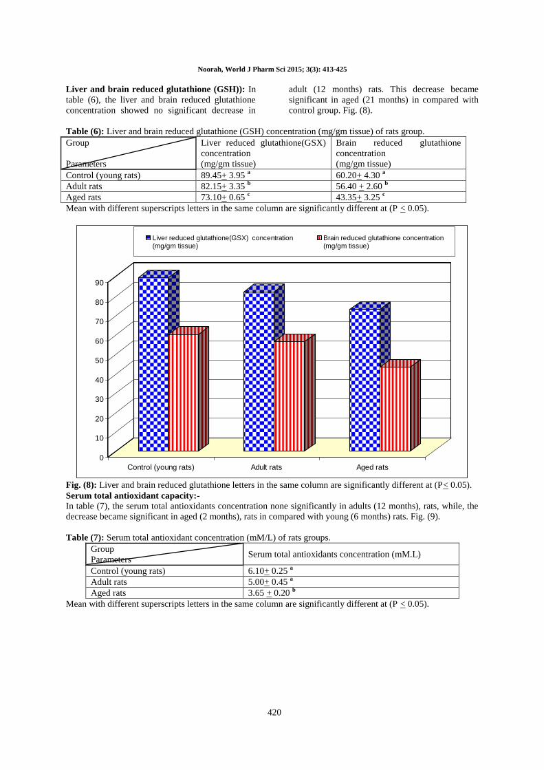
Table (4): Cytochrom oxidase (294 bp) mRNA expression of rats liver and brain.
Group
Parameters
Expression level of liver
Cytochrom oxidase
Expression level of brain
Cytochrom oxidase
Control (young rats) 165 195
Adult rats 152 185
Aged rats 120 181
Noorah, World J Pharm Sci 2015; 3(3): 413-425
417
0
20
40
60
80
100
120
140
160
180
200
Control (young rats) Adult rats Aged rats
Expression level of liver β-actin Expression level of brain β-actin
Fig. (1): β-actin (211 bp) mRNA expression of rats liver and brain.
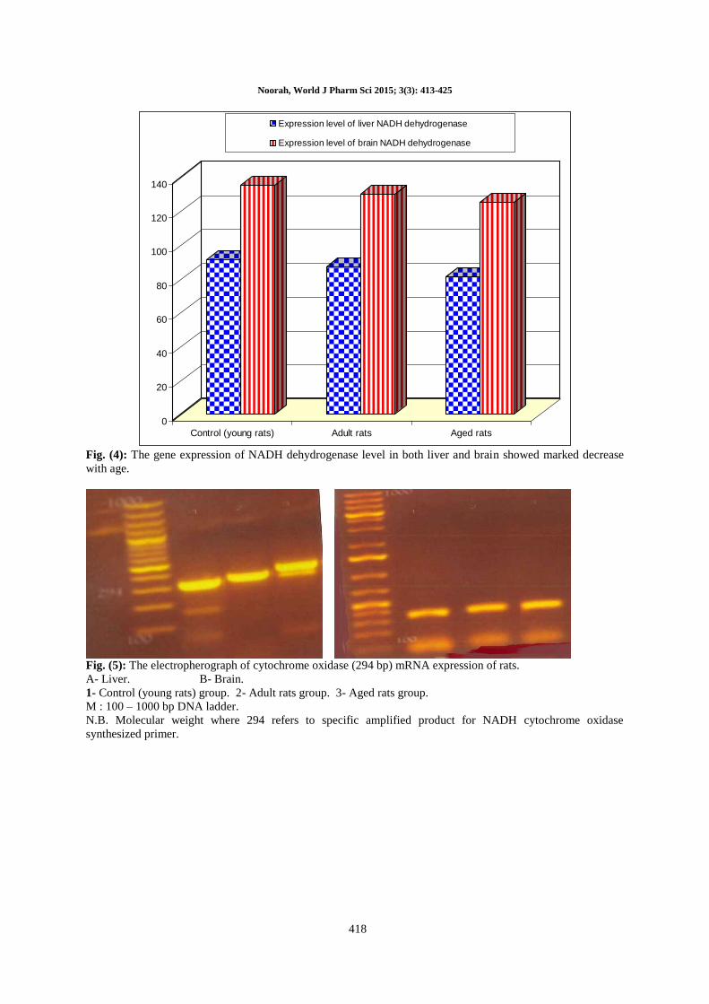
Fig. (2): The electropherograph of β-actin (211 bp) mRNA expression of rat liver (A) and brain (B).
1- Young rats group (control ); 2- adult rats. 3- Aged rats group; M: 100 bp DNA ladder; N.B. Molecular
weight where 211 refers to specific amplified product for β-actin according to synthesized primer.
Fig. (3): The electropherograph of NADH dehydrogenase (201 bp) mRNA expression of rats.
A. liver. B- brain.
1- Control or young rats group. 2- Adult rats group. 3- Aged rats group.
N.B. Molecular weight where 201 refers to specific amplified product for NADH dehydrogenase according to
synthesized primer.
Noorah, World J Pharm Sci 2015; 3(3): 413-425
418
0
20
40
60
80
100
120
140
Control (young rats) Adult rats Aged rats
Expression level of liver NADH dehydrogenase
Expression level of brain NADH dehydrogenase
Fig. (4): The gene expression of NADH dehydrogenase level in both liver and brain showed marked decrease
with age.
Fig. (5): The electropherograph of cytochrome oxidase (294 bp) mRNA expression of rats.
A- Liver. B- Brain.
1- Control (young rats) group. 2- Adult rats group. 3- Aged rats group.
M : 100 – 1000 bp DNA ladder.
N.B. Molecular weight where 294 refers to specific amplified product for NADH cytochrome oxidase
synthesized primer.
Noorah, World J Pharm Sci 2015; 3(3): 413-425
419
0
20
40
60
80
100
120
140
160
180
200
Control (young rats) Adult rats Aged rats
Expression level of liver Cytochrom oxidase
Expression level of brain Cytochrom oxidase
Fig. (6): The gene expression of cytochrome oxidase level in both liver and brain showed decrease with age.
Liver and brain glutathione peroxidase (GPx):
In table (5), liver glutathione oxidase showed no
significant decrease in its activity in adult (12
months) rats, which significantly decrease in both
liver and brain in aged (21 months) rats in
compared with young rats group (Fig. 7).
Table (5): Liver and brain glutathione peroxidase (GPx) activity (u/gm) of rats.
Group
Parameters
Liver glutathione peroxidase
(GPx) activity (U/gm)
Brain glutathione peroxidase
(GPx) activity (U/gm)
Control (young rats) 22.55 + 1.25 a 20.30 + 1.55
a
Adult rats 17.40 + 0.95 b 12.60 + 0.85
b
Aged rats 13.25 + 0.75 c 5.75 + 0.15
c
Mean with different superscripts letters in the same column are significantly different at (P < 0.05).
0
5
10
15
20
25
Control (young rats) Adult rats Aged rats
Liver glutathione peroxidase (GPx) activity (U/gm)
Brain glutathione peroxidase (GPx) activity (U/gm)
Fig. (7): Liver and brain glutathione oxidase (GPx) activity (U/gm) of rats.
Noorah, World J Pharm Sci 2015; 3(3): 413-425
420
Liver and brain reduced glutathione (GSH)): In
table (6), the liver and brain reduced glutathione
concentration showed no significant decrease in
adult (12 months) rats. This decrease became
significant in aged (21 months) in compared with
control group. Fig. (8).
Table (6): Liver and brain reduced glutathione (GSH) concentration (mg/gm tissue) of rats group.
Group
Parameters
Liver reduced glutathione(GSX)
concentration
(mg/gm tissue)
Brain reduced glutathione
concentration
(mg/gm tissue)
Control (young rats) 89.45+ 3.95 a 60.20+ 4.30
a
Adult rats 82.15+ 3.35 b 56.40 + 2.60
b
Aged rats 73.10+ 0.65 c 43.35+ 3.25
c
Mean with different superscripts letters in the same column are significantly different at (P < 0.05).
0
10
20
30
40
50
60
70
80
90
Control (young rats) Adult rats Aged rats
Liver reduced glutathione(GSX) concentration(mg/gm tissue)
Brain reduced glutathione concentration(mg/gm tissue)
Fig. (8): Liver and brain reduced glutathione letters in the same column are significantly different at (P< 0.05).
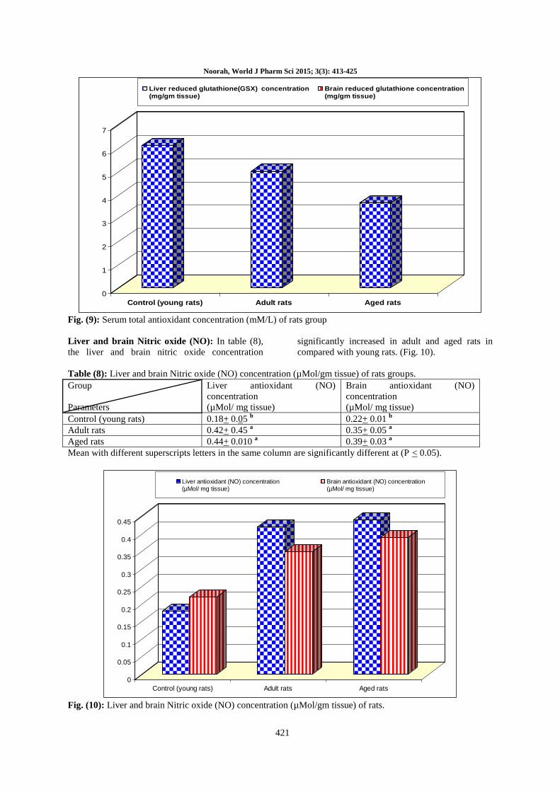
Serum total antioxidant capacity:-
In table (7), the serum total antioxidants concentration none significantly in adults (12 months), rats, while, the
decrease became significant in aged (2 months), rats in compared with young (6 months) rats. Fig. (9).
Table (7): Serum total antioxidant concentration (mM/L) of rats groups.
Group
Parameters Serum total antioxidants concentration (mM.L)
Control (young rats) 6.10+ 0.25 a
Adult rats 5.00+ 0.45 a
Aged rats 3.65 + 0.20 b
Mean with different superscripts letters in the same column are significantly different at (P < 0.05).
Noorah, World J Pharm Sci 2015; 3(3): 413-425
421
0
1
2
3
4
5
6
7
Control (young rats) Adult rats Aged rats
Liver reduced glutathione(GSX) concentration(mg/gm tissue)
Brain reduced glutathione concentration(mg/gm tissue)
Fig. (9): Serum total antioxidant concentration (mM/L) of rats group
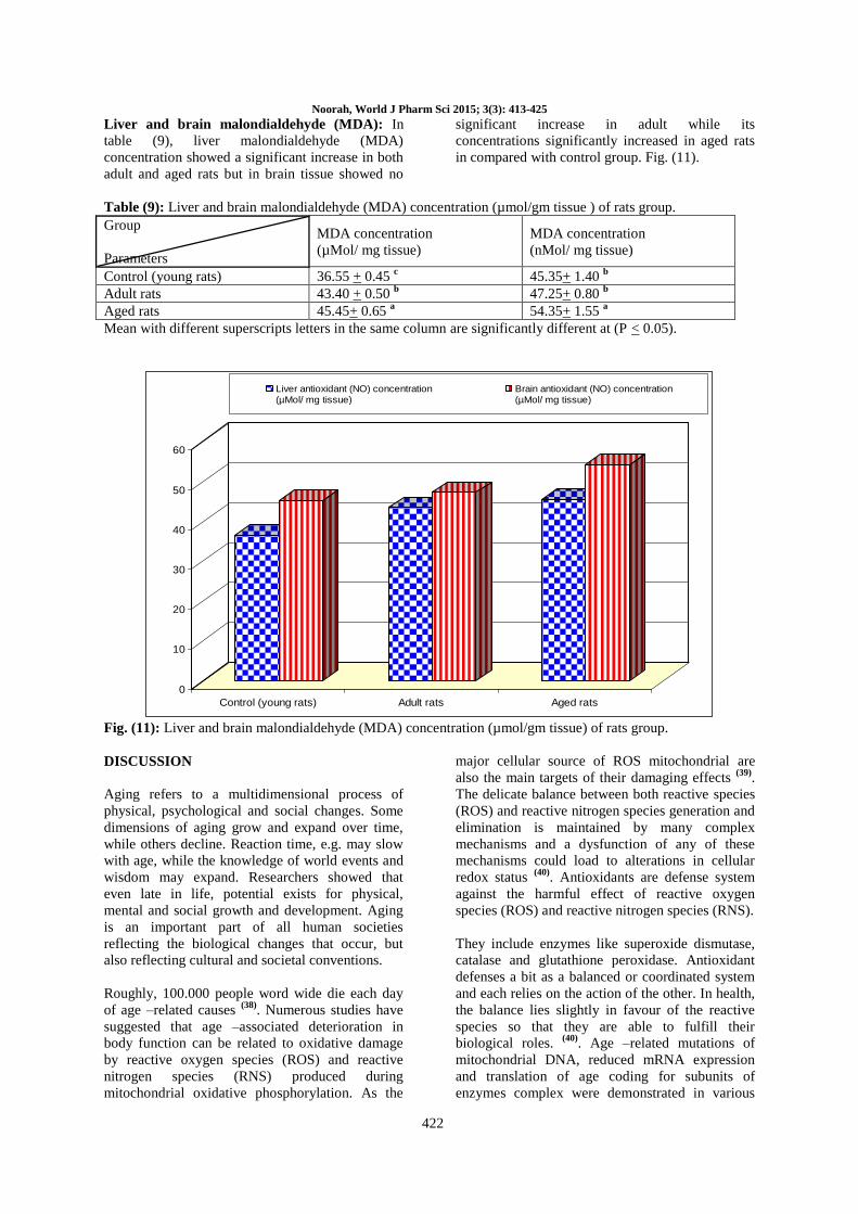
Liver and brain Nitric oxide (NO): In table (8),
the liver and brain nitric oxide concentration
significantly increased in adult and aged rats in
compared with young rats. (Fig. 10).
Table (8): Liver and brain Nitric oxide (NO) concentration (µMol/gm tissue) of rats groups.
Group
Parameters
Liver antioxidant (NO)
concentration
(µMol/ mg tissue)
Brain antioxidant (NO)
concentration
(µMol/ mg tissue)
Control (young rats) 0.18+ 0.05 b 0.22+ 0.01
b
Adult rats 0.42+ 0.45 a 0.35+ 0.05
a
Aged rats 0.44+ 0.010 a 0.39+ 0.03
a
Mean with different superscripts letters in the same column are significantly different at (P < 0.05).
0
0.05
0.1
0.15
0.2
0.25
0.3
0.35
0.4
0.45
Control (young rats) Adult rats Aged rats
Liver antioxidant (NO) concentration(µMol/ mg tissue)
Brain antioxidant (NO) concentration(µMol/ mg tissue)
Fig. (10): Liver and brain Nitric oxide (NO) concentration (µMol/gm tissue) of rats.
Noorah, World J Pharm Sci 2015; 3(3): 413-425
422
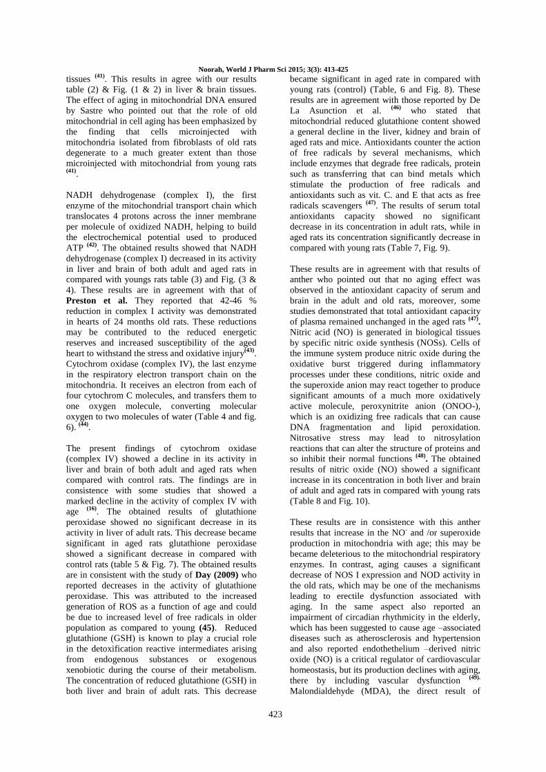
Liver and brain malondialdehyde (MDA): In
table (9), liver malondialdehyde (MDA)
concentration showed a significant increase in both
adult and aged rats but in brain tissue showed no
significant increase in adult while its
concentrations significantly increased in aged rats
in compared with control group. Fig. (11).
Table (9): Liver and brain malondialdehyde (MDA) concentration (µmol/gm tissue ) of rats group.
Group
Parameters
MDA concentration
(µMol/ mg tissue)
MDA concentration
(nMol/ mg tissue)
Control (young rats) 36.55 + 0.45 c 45.35+ 1.40
b
Adult rats 43.40 + 0.50 b 47.25+ 0.80
b
Aged rats 45.45+ 0.65 a 54.35+ 1.55
a
Mean with different superscripts letters in the same column are significantly different at (P < 0.05).
0
10
20
30
40
50
60
Control (young rats) Adult rats Aged rats
Liver antioxidant (NO) concentration(µMol/ mg tissue)
Brain antioxidant (NO) concentration(µMol/ mg tissue)
Fig. (11): Liver and brain malondialdehyde (MDA) concentration (µmol/gm tissue) of rats group.
DISCUSSION
Aging refers to a multidimensional process of
physical, psychological and social changes. Some
dimensions of aging grow and expand over time,
while others decline. Reaction time, e.g. may slow
with age, while the knowledge of world events and
wisdom may expand. Researchers showed that
even late in life, potential exists for physical,
mental and social growth and development. Aging
is an important part of all human societies
reflecting the biological changes that occur, but
also reflecting cultural and societal conventions.
Roughly, 100.000 people word wide die each day
of age –related causes (38)
. Numerous studies have
suggested that age –associated deterioration in
body function can be related to oxidative damage
by reactive oxygen species (ROS) and reactive
nitrogen species (RNS) produced during
mitochondrial oxidative phosphorylation. As the
major cellular source of ROS mitochondrial are
also the main targets of their damaging effects (39)
.
The delicate balance between both reactive species
(ROS) and reactive nitrogen species generation and
elimination is maintained by many complex
mechanisms and a dysfunction of any of these
mechanisms could load to alterations in cellular
redox status (40)
. Antioxidants are defense system
against the harmful effect of reactive oxygen
species (ROS) and reactive nitrogen species (RNS).
They include enzymes like superoxide dismutase,
catalase and glutathione peroxidase. Antioxidant
defenses a bit as a balanced or coordinated system
and each relies on the action of the other. In health,
the balance lies slightly in favour of the reactive
species so that they are able to fulfill their
biological roles. (40)
. Age –related mutations of
mitochondrial DNA, reduced mRNA expression
and translation of age coding for subunits of
enzymes complex were demonstrated in various
Noorah, World J Pharm Sci 2015; 3(3): 413-425
423
tissues (41)
. This results in agree with our results
table (2) & Fig. (1 & 2) in liver & brain tissues.
The effect of aging in mitochondrial DNA ensured
by Sastre who pointed out that the role of old
mitochondrial in cell aging has been emphasized by
the finding that cells microinjected with
mitochondria isolated from fibroblasts of old rats
degenerate to a much greater extent than those
microinjected with mitochondrial from young rats (41)
.
NADH dehydrogenase (complex I), the first
enzyme of the mitochondrial transport chain which
translocates 4 protons across the inner membrane
per molecule of oxidized NADH, helping to build
the electrochemical potential used to produced
ATP (42)
. The obtained results showed that NADH
dehydrogenase (complex I) decreased in its activity
in liver and brain of both adult and aged rats in
compared with youngs rats table (3) and Fig. (3 &
4). These results are in agreement with that of
Preston et al. They reported that 42-46 %
reduction in complex I activity was demonstrated
in hearts of 24 months old rats. These reductions
may be contributed to the reduced energetic
reserves and increased susceptibility of the aged
heart to withstand the stress and oxidative injury(43)
.
Cytochrom oxidase (complex IV), the last enzyme
in the respiratory electron transport chain on the
mitochondria. It receives an electron from each of
four cytochrom C molecules, and transfers them to
one oxygen molecule, converting molecular
oxygen to two molecules of water (Table 4 and fig.
6). (44)
.
The present findings of cytochrom oxidase
(complex IV) showed a decline in its activity in
liver and brain of both adult and aged rats when
compared with control rats. The findings are in
consistence with some studies that showed a
marked decline in the activity of complex IV with
age (16)
. The obtained results of glutathione
peroxidase showed no significant decrease in its
activity in liver of adult rats. This decrease became
significant in aged rats glutathione peroxidase
showed a significant decrease in compared with
control rats (table 5 & Fig. 7). The obtained results
are in consistent with the study of Day (2009) who
reported decreases in the activity of glutathione
peroxidase. This was attributed to the increased
generation of ROS as a function of age and could
be due to increased level of free radicals in older
population as compared to young (45). Reduced
glutathione (GSH) is known to play a crucial role
in the detoxification reactive intermediates arising
from endogenous substances or exogenous
xenobiotic during the course of their metabolism.
The concentration of reduced glutathione (GSH) in
both liver and brain of adult rats. This decrease
became significant in aged rate in compared with
young rats (control) (Table, 6 and Fig. 8). These
results are in agreement with those reported by De
La Asunction et al. (46)
who stated that
mitochondrial reduced glutathione content showed
a general decline in the liver, kidney and brain of
aged rats and mice. Antioxidants counter the action
of free radicals by several mechanisms, which
include enzymes that degrade free radicals, protein
such as transferring that can bind metals which
stimulate the production of free radicals and
antioxidants such as vit. C. and E that acts as free
radicals scavengers (47)
. The results of serum total
antioxidants capacity showed no significant
decrease in its concentration in adult rats, while in
aged rats its concentration significantly decrease in
compared with young rats (Table 7, Fig. 9).
These results are in agreement with that results of
anther who pointed out that no aging effect was
observed in the antioxidant capacity of serum and
brain in the adult and old rats, moreover, some
studies demonstrated that total antioxidant capacity
of plasma remained unchanged in the aged rats (47)
.
Nitric acid (NO) is generated in biological tissues
by specific nitric oxide synthesis (NOSs). Cells of
the immune system produce nitric oxide during the
oxidative burst triggered during inflammatory
processes under these conditions, nitric oxide and
the superoxide anion may react together to produce
significant amounts of a much more oxidatively
active molecule, peroxynitrite anion (ONOO-),
which is an oxidizing free radicals that can cause
DNA fragmentation and lipid peroxidation.
Nitrosative stress may lead to nitrosylation
reactions that can alter the structure of proteins and
so inhibit their normal functions (48)
. The obtained
results of nitric oxide (NO) showed a significant
increase in its concentration in both liver and brain
of adult and aged rats in compared with young rats
(Table 8 and Fig. 10).
These results are in consistence with this anther
results that increase in the NO- and /or superoxide
production in mitochondria with age; this may be
became deleterious to the mitochondrial respiratory
enzymes. In contrast, aging causes a significant
decrease of NOS I expression and NOD activity in
the old rats, which may be one of the mechanisms
leading to erectile dysfunction associated with
aging. In the same aspect also reported an
impairment of circadian rhythmicity in the elderly,
which has been suggested to cause age –associated
diseases such as atherosclerosis and hypertension
and also reported endothethelium –derived nitric
oxide (NO) is a critical regulator of cardiovascular
homeostasis, but its production declines with aging,
there by including vascular dysfunction (49).
Malondialdehyde (MDA), the direct result of
Noorah, World J Pharm Sci 2015; 3(3): 413-425
424
oxidative stress taken as biomarker of lipid
peroxidation of different tissues. The findings of
MDA in the present investigation showed a
significant increase in its concentration in liver of
both adult and aged rats in compared with young
rats.
This increase was non-significant in the brain of
adult rats and became significant in the brain of
aged rats in compared with young rats (Table, 9
and Fig. 11). This findings are in agreement with
the anther had reported in Shaal sheep the lowest
concentration of MDA, which is widely used as a
biomarker of lipid peroxidation, was also seen in
the youngest group. It seems that in the early
growth phase (50).
In contrast to the previous
findings, the age-related variation in oxidant status
and lipid damage and occluded that the
mitochondrial lipid peroxidation index was not
different in young and old mice (51)
.
CONCLUSION From this paper results, it can be concluded that
aging has a great harmful effect on mitochondrial
DNA, that can be detected from the obtained
results: decreases in the activity of NADH
dehydrogenase (complex I) and cytochrom oxidase
(complex IV) , which correlated with age, decease
in the activity of glutathione peroxidase and in
concentration of both reduced glutathione and total
antioxidant capacity and increase in the
concentration of nitric oxide and malondialdhyde
with age.
Recommendation: The molecular mechanism of
the antioxidants that enables the body to face the
free radicals harmful effects should be more
clarified. Increase the human awareness at the
beginning of the aging with the danger of free
radicals and how to decrease its side effects and
consequently decrease aging disease and change
the human life style, which is easier in youth and
aims to avoid the harmful effect of free radicals.
REFERENCES
1. Henze, Ki and Martin, W.: Evolutionary biology essence of mitochondria. Nature, (2003) , 426(6963):127-8.
2. Voet, D.; Voet, J.G. and Pratt, C.W.: Fundamentals of Biochemistry, 2nd Edition, John Wiley and sons, Inc. (2006) , pp.547. 3. Taylor, S.W.; Fahy, E.; Zhang, B.; Glenn, G.M.; Warnock, D.E.; Wiley, S.; Murphy, A.N.; Gaucher, S.P.; Capaldi, R.A.;
Gibson, B.W. and Ghosh, S.S.: Characterization of the human heart mitochondrial proteome. Nat Bio. Technol. (2003) , 21(3):
281-6. 4. Zhang, J.; Li, X.; Mueller, M.; Wang, Y.; Zong, C.; Deng, N.; Vondriska, T.M.; Liem, D.A.; Yong, J.; Korage, P.; Honda, H.;
Weiss, J.N.; Apweiler, R. and ping, P.: Systematic characterization of the murine mitochondrial proteome using functionally
validated cardiac mitochondria. Proteomics, (2008) , 8(8): 1564-1575. 5. Campbell, G.; Neil, A.; Brad Williamson; Robin, J. Heyden (2006): Biology: Exploring life. Bosteon, Massachusetts: Pearson
prentice Hall.
6. McBride, H.M.; Neuspiel, M. and Wasiak, S.: Mitochondria more than Just a power house. Curr. Boil. (2006) , 16(14): 551-8. 7. Gardner, A. and Boles, R.G.: Is mitochondrial psychiatry in the future? A Review. Curr. Psychiatry Reb view, (2005) ,1(3): 255-
271.
8. Wallace, D.C.: Mitochondrial DNA mutation, and bioenergic defects in aging and degenerative diseases. In: Rosenberg RN, Prusiner SB, Dimauro S, Barchi RL (eds). The molecular and genetic Basis of neurological Disease. Butter worth Heinmann-
Boston, (1997) , pp.237-269.
9. Zeviani, M.; Tiranti, V. and Piantadosi, C.: Reviews in molecular medicine-Mitochondrial disorders. Medicine . (1998) , 77:P59-72.
10. Clayton, D.A.: Replication of animal mitochondrial DNA. Cell , (1982) , 28: 693-705.
11. Manfredi, G.; Thyagarajan, D.; Papadopoulau, L.C.; Pallotti, F. and Schon, E.A.: The fate of human sperm –derived mtDNA in somatic cells. Am. . Hum. Genet, (1997) , 61:953-960.
12. Bogenhagen, D.F.: Repair of mtDNA in vertebrate. Am. J. Hum. Genet, (1999) , 64:1276-1281.
13. Bowen, R.I. and Atwood, C.S.: Living and dying for sex. Atheory of aging based on the nodulation of cell cycle signaling by reproductive hormones. Gerontology, (2004) ,50(5): 265-290.
14. Ozawa, O.A.: Mechanism of somatic mitochondrial DNA mutation associated with age and disease. Biochem. Biophys Acta,
(2014) , 1128:189-198. 15. Center for cardiovascular Education, Inc. Distrbuted under license. Parlay International: 5290 only licenses may copy or
distrubted this page, electronically or otherwise. For license information call – (2014) ,959-3855.
16. Stohs, J.A.: The role of free radicals in toxicity and disease. J. of Basic Clinnical physiology and pharmacology. (2013) ,116(5):907-932.
17. Hirota, H. and Komoto, I.: Redox regulation of cellular signaling. Cell signal., (2014) , 149: 59-68.
18. Fang, F.C.: Antimicrobial reactive oxygen and nitrogen species, concepts and controversies. Nat. Rev. microbial. (2004) , 2(10): 820-832.
19. Mayer, T.: Immunology-Chapter one; Innate (non-specific) Immunity. Microbiology and immunology on line textbook. USC
School of Medicine. Retrieved November. (2013): 42. 20. Trachootham, D.; Lu, W.; Ogasawara, M.A.; Nilsa, R.D. and Huang, P.: Redox regulation of cell survival. Antioxid Redox
Signal; (2008) , 8(10):1343-74.
21. Ghafourifar, P. and Cadenas, E.: Mitochondrial nitric oxide synthase. Trends pharmacol. Sci., (2005) , 26:190-195. 22. Farmer, E.E. and Davoine, C.: Reactive electrophile species. Curr. Opin. Plant. Boil., (2007) , 10(4): 380-6.
23. Rio Del, D.; Pellegani, N. and Stewart, A.J.: Imbalance of oxidants and antioxidant systems in subjects with cirrhosis. Nutr
Metab. Cardiovasc. Dis. (2012) , 85(1):915-32. 24. Rahman, K.: Studies on free radicals antioxidants and co-factors.clinical Intervention in Aging, (2011) , 22(4):321-338.
Noorah, World J Pharm Sci 2015; 3(3): 413-425
425
25. Mates, J.M.; Perez-Gomes, C.De Castro, Z.N.: Antioxidant enzymes and human diseases. Clin. Biochem., (1999) , 32:595-603. 26. Frei, B. and Call, M.R.: Can antioxidant vitamins materially reduce oxidative damage in humans? Free Rad. Biol. Med., (2014) ,
210(5):5099-5115.
27. Valko, M.; Rhodes, C.J.; Moncol, J.; Izakovic, M. nad Mazur, M.: Free radicals metals and antioxidant in oxidative stress –induced cancer. Chem. Biol. Interacts., (2006) , 160:1-40.
28. Brigellus-Flohe, R.: Glutathione peroxidases and redox-regulated transcription factors. Boil. Chem., (2006) ,387:1329-1335.
29. Paglia, D.E. and Valentine, W.N.: Studies on the quantative and qualitative characterization of erythrocyte glutathione peroxidase. Journal of laboratory and Clinical Medicine, (1967) ,70(1):158-169.
30. Koracevic, D.; Koracevic, G.; Djordjevic, V.; Andrejevic, S. and Cosic, V.: Methods for the measurement of antioxidant activity
in human fluids. J. Clin. Pathol., (2001) , 54: 356-361. 31. Meadus, W.G.: Semi-Quantitative Rt-PCR method to measure the in vivo effect of diatery conjucated linoleic acid in protein
muscle PPAR gene expression. Bio. Proced. On line, (2003) , 5(1):20-28.
32. Beutler, E.; Duron, O. and Kelly, M.B.: Improved method for the dtermination of blood glutathione. J. Lab. Clin. Med., (1963) , 61:881-892.
33. Moshage, H.; Kok, B. and Huizenga, J.R.: Nitrite and nitrate determination in plasma: A critical evaluation. Clin. Chem., (1995)
,41:892-896. 34. Esterbauer, H.; Cheeseman, K.H.; Danzani, M.U.; Poli, G. and Slater, T.F.: Separation and characterization of the aldhyde
products of ADP/FeZC+ stimulation lipid peroxidation in rat liver microsome. Biochem. J. (1982) , 208: 129-140.
35. Snedecor and Cochran: Statistical Medthiod, the Iowa State Univ. Press. Ames. Iowa, USA, (1967): 593. 36. Jove, M.; Joel Salla, Annaplanavila, Agatha Cabrero, Liliane Michlik, Walter Wahli, Juan C. Laguna and Manuel Vazquez
Carrera: Oligonucleatide primersquence. Puplished, JIR paper in press.Dol., (2003) ,11, 1194/M300208-JLR-200.
37. Dai, W.; Cheng, H.; Ren-Qiang, R.; Zongzhuang, Z. nad Shi, J.: Quantitative detection of the expression of mitochondrial cytochrom C oxidase subunits mRNA in the cerebral cortex after experimental traumatic brain injury. Trends Biochem. Sci.,
(2008) , 28:37-48.
38. Aubrey, D. J. and Grey, D.E.: Life span Extension Research nad Public Debate: Sociatal considerations: Studies in Ethics, Low and Technology, (2007) , 1(1) Article (5).
39. Drahota, Z.; Rauchova, H.; Nuskova, H. and Krivakova, P.: peroxidative damage to mitochondrial respiration is substrate –dependent –Physiol. Res. (2014) , 98:951-964.
40. Valko, M.; Leibfritz, D.; Moncol. J.; Cronin, M.D.; Mazur, M. and Telser, J.: Free radicals and antioxidants in normal
physiological functions and human disease. Int. J. Biochem. Cell Biol., (2007) ,39:44-84. 41. Sastre, J.: A ginkgo biloba extract (Eg b 761) prevents mitochondrial aging by protecting against oxidative stress. Free Rad. Biol.
Med., (1998) , 24:298-304.
42. Shannon, R.J.; Hirs, T.J. and Walker, J.: Bovine complex I, is a complex of 45 different subunits. J. Biol. Chem., (2014) , 35(125):9345-9354.
43. Jahangir, A.; Terzic, A.; Sagar, S. and Oberlin, S.: Aging –induced alteration in gene transcripts and functional activity of
mitochondrial oxidative phosphorylation complexes in the heart. Mech. Aging Dev. (2014) ,4/9: 324-336.
44. Yamasht, E.; Itoh, K.; Yaona, R. and Shinaza: Structural of metal site of oxidized bovine heart cytochrome oxidase. Science,
(2014) , 459:3126-35.
45. Day, B.J.: Catalase and glutathione peroxidase mimics. Biochem. Pharamacol., (2009) , 77:285-96. 46. De La Asunction, J.G.; Millan, A.; Pla, R.; Bkuseghini, L.; Esteras, A.; Pallardo, F.V.; Sastre, J.; Vina, J. and Faseb, J.:
Mitochondrial glutathione oxidation correlates with aged –associated oxidative damage to mitochondrial DNA. Comp. Biochem.
Physiol., (1996) ,10(2): 333-338. 47. Adiga, U.: Total antioxidant activity in old age. Biomedical Res., (2008) ,84:155-161.
48. Harrison, R.: Structure and function of xanthine oxidoreductase where are we now? Free Rad Biol. Med., (2002) , 33: 774-797.
49. Kunieda, T.; Minamina, T.; Miura, K.; Katsuno, T.; Tateno, K. and Komuro, L.: Reduced oxide causes Age –Associated Impairment of circadian Rhythmcity. Circ. Res., (2008) , 102(5): 607-614.
50. Amoli, J. and Baghbanzadeh, A.: Oxidative stress in Shaal sheep of different age groups. Turk. J. Vet. Anim. Sci., (2010) ,
34(4): 379-383. 51. Moncol, J.; Mazur, M.; Cronin, M. and Valls: Age related change antioxidant status and oxidative damage to lipids and DNA
mitochondria of rat liver. Process Biochem., (2013) ,40:903-908.
Related Documents