Physical and Genetic Mapping of the Human X Chromosome Centromere: Repression of Recombination Melanie M. Mahtani 1,3 and Huntington F. Willard 2,4 1 Department of Genetics, Stanford University School of Medicine, Stanford, California 94305-5120 USA; 2 Department of Genetics and Center for Human Genetics, Case Western Reserve University School of Medicine, Cleveland, Ohio 44106 USA Classical genetic studies in Drosophila and yeast have shown that chromosome centromeres have a cis-acting ability to repress meiotic exchange in adjacent DNA. To determine whether a similar phenomenon exists at human centromeres, we measured the rate of meiotic recombination across the centromere of the human X chromosome. We have constructed a long-range physical map of centromeric a-satellite DNA (DXZ1) by pulsed-field gel analysis, as well as detailed meiotic maps of the pericentromeric region of the X chromosome in the CEPH family panel. By comparing these two maps, we determined that, in the proximal region of the X chromosome, a genetic distance of 0.57 cM exists between markers that span the centromere and are separated by at least the average 3600 kb physical distance mapped across the DXZ1 array. Therefore, the rate of meiotic exchange across the X chromosome centromere is <1 cM/6300 kb (and perhaps as low as 1 cM/17,000 kb on the basis of other physical mapping data), at least eightfold lower than the average rate of female recombination on the X chromosome and one of the lowest rates of exchange yet observed in the human genome. Meiotic exchange is not distributed randomly along the length of eukaryotic chromosomes; indeed, much regional variation in recombination fre- quency has been observed. Perhaps the most con- spicuous departure from uniformity is the dramatic repression of exchange found near eukaryotic chro- mosome centromeres and some telomeres (Mather 1936, 1939). Repression of meiotic recombination adjacent to the centromere (the centromere effect) is most obvious on the Drosophila X chromosome in which the centric heterochromatin, comprising half of the chromosome’s cytogenetic length, barely contributes to its genetic length (Mather 1939; Rob- erts 1965); a similar centromere-associated repres- sion of recombination is found on the autosomes of Drosophila (Beadle 1932; Painter 1935; Thompson 1963). Some of this repression of exchange is caused by the large blocks of heterochromatin present at the centromeres of higher eukaryotes (Willard 1990; Murphy and Karpen 1995), because heterochroma- tin, at least in Drosophila, is a poor substrate for re- combination regardless of chromosomal location (Baker 1958). Because deletions of centric hetero- chromatin result in lowered levels of meiotic ex- change in centromere-adjacent euchromatin (Ya- mamoto and Miklos 1978), the presence alone of heterochromatin at Drosophila centromeres does not fully explain the centromere effect. Rather, the centromere seems to exert a suppression of recom- bination that spreads to adjacent DNA. Studies in yeast support a similar centromeric suppression of meiotic exchange in proximal chro- mosome regions, although this effect may be less pronounced. In both Saccharomyces cerevisiae and Schizosaccharomyces pombe, mitotic recombination is relatively more frequent than meiotic recombina- tion in the proximity of centromeres (Malone et al. 1980; Minet et al. 1980). Direct evidence for the centromere effect in yeast has come from studies of cloned centromeres, in which DNA adjacent to the 60-kb centromere domain of S. pombe has an appar- ently lowered rate of meiotic exchange (Nakaseko et al. 1986). Further, in S. cerevisiae, removal and distal displacement of a 600-bp fragment containing the centromere of chromosome III (CEN3) results in a three- to fivefold decrease in crossing over at the new site of integration (Lambie and Roeder 1986). Although the genetic maps of most other higher eukaryotes are less well characterized, there is evi- 3 Present address: Microcide Pharmaceuticals, Mountain View, California 94043 USA. 4 Corresponding author. E-MAIL [email protected]; FAX (216) 368-3030. RESEARCH 100 GENOME RESEARCH 8:100–110 ©1998 by Cold Spring Harbor Laboratory Press ISSN 1054-9803/98 $5.00; www.genome.org Cold Spring Harbor Laboratory Press on September 20, 2016 - Published by genome.cshlp.org Downloaded from

Welcome message from author
This document is posted to help you gain knowledge. Please leave a comment to let me know what you think about it! Share it to your friends and learn new things together.
Transcript
Physical and Genetic Mapping of the HumanX Chromosome Centromere: Repression
of RecombinationMelanie M. Mahtani1,3 and Huntington F. Willard2,4
1Department of Genetics, Stanford University School of Medicine, Stanford, California 94305-5120 USA;2Department of Genetics and Center for Human Genetics, Case Western Reserve University School of
Medicine, Cleveland, Ohio 44106 USA
Classical genetic studies in Drosophila and yeast have shown that chromosome centromeres have a cis-actingability to repress meiotic exchange in adjacent DNA. To determine whether a similar phenomenon exists athuman centromeres, we measured the rate of meiotic recombination across the centromere of the human Xchromosome. We have constructed a long-range physical map of centromeric a-satellite DNA (DXZ1) bypulsed-field gel analysis, as well as detailed meiotic maps of the pericentromeric region of the X chromosome inthe CEPH family panel. By comparing these two maps, we determined that, in the proximal region of the Xchromosome, a genetic distance of 0.57 cM exists between markers that span the centromere and are separatedby at least the average 3600 kb physical distance mapped across the DXZ1 array. Therefore, the rate of meioticexchange across the X chromosome centromere is <1 cM/6300 kb (and perhaps as low as 1 cM/17,000 kb onthe basis of other physical mapping data), at least eightfold lower than the average rate of female recombinationon the X chromosome and one of the lowest rates of exchange yet observed in the human genome.
Meiotic exchange is not distributed randomly alongthe length of eukaryotic chromosomes; indeed,much regional variation in recombination fre-quency has been observed. Perhaps the most con-spicuous departure from uniformity is the dramaticrepression of exchange found near eukaryotic chro-mosome centromeres and some telomeres (Mather1936, 1939). Repression of meiotic recombinationadjacent to the centromere (the centromere effect)is most obvious on the Drosophila X chromosome inwhich the centric heterochromatin, comprising halfof the chromosome’s cytogenetic length, barelycontributes to its genetic length (Mather 1939; Rob-erts 1965); a similar centromere-associated repres-sion of recombination is found on the autosomes ofDrosophila (Beadle 1932; Painter 1935; Thompson1963). Some of this repression of exchange is causedby the large blocks of heterochromatin present atthe centromeres of higher eukaryotes (Willard 1990;Murphy and Karpen 1995), because heterochroma-tin, at least in Drosophila, is a poor substrate for re-combination regardless of chromosomal location
(Baker 1958). Because deletions of centric hetero-chromatin result in lowered levels of meiotic ex-change in centromere-adjacent euchromatin (Ya-mamoto and Miklos 1978), the presence alone ofheterochromatin at Drosophila centromeres doesnot fully explain the centromere effect. Rather, thecentromere seems to exert a suppression of recom-bination that spreads to adjacent DNA.
Studies in yeast support a similar centromericsuppression of meiotic exchange in proximal chro-mosome regions, although this effect may be lesspronounced. In both Saccharomyces cerevisiae andSchizosaccharomyces pombe, mitotic recombinationis relatively more frequent than meiotic recombina-tion in the proximity of centromeres (Malone et al.1980; Minet et al. 1980). Direct evidence for thecentromere effect in yeast has come from studies ofcloned centromeres, in which DNA adjacent to the60-kb centromere domain of S. pombe has an appar-ently lowered rate of meiotic exchange (Nakaseko etal. 1986). Further, in S. cerevisiae, removal and distaldisplacement of a 600-bp fragment containing thecentromere of chromosome III (CEN3) results in athree- to fivefold decrease in crossing over at thenew site of integration (Lambie and Roeder 1986).Although the genetic maps of most other highereukaryotes are less well characterized, there is evi-
3Present address: Microcide Pharmaceuticals, Mountain View,California 94043 USA.4Corresponding author.E-MAIL [email protected]; FAX (216) 368-3030.
RESEARCH
100 GENOME RESEARCH 8:100–110 ©1998 by Cold Spring Harbor Laboratory Press ISSN 1054-9803/98 $5.00; www.genome.org
Cold Spring Harbor Laboratory Press on September 20, 2016 - Published by genome.cshlp.orgDownloaded from
dence in Neurospora, corn, tomato, and barley for acentromeric suppression of meiotic recombination(Charlesworth et al. 1986; van Daelen et al. 1993;Davis et al. 1994).
In humans, chiasma distributions predict thatcrossing over occurs more rarely near chromosomecentromeres than elsewhere (Hulten 1974). Until re-cently, the lack of detailed long-range physicalmaps spanning centromeres (and uncertainty re-garding the functional identification of a centro-mere itself) has precluded the definitive measure-ment of recombination rates at human centro-meres. Preliminary comparisons of physical withgenetic distances at or near the centromeres of sev-eral human chromosomes have resulted in variousestimates of pericentromeric recombination rates,some consistent (Carson and Simpson 1991; Lichteret al. 1993; Weeks et al. 1995) and others inconsis-tent (Jabs et al. 1991; Janson et al. 1991; Van Hul etal. 1993) with a suppression of centromeric meioticexchange. A recent study, which compares a yeastartificial chromosome (YAC) physical map with thecorresponding genetic map, suggests that recombi-nation may be reduced by an order of magnitudeacross the centromere of chromosome 10 (Jacksonet al. 1996). Data from recent radiation hybrid (RH)maps are also consistent with a repression of ex-change at human centromeres (James et al. 1994;Hudson et al. 1995). These data are difficult to in-terpret, however, because proximal chromosome re-gions may be preferentially retained in RH cell lines,at a frequency disproportionate to their physicalsize (D. Slonim and M.M. Mahtani, unpubl.).
Here, we use the tools of pulsed-field gel map-ping to provide an estimate of recombination acrossthe human X chromosome centromere relative to ameasured physical distance, using markers that areexclusively chromosome-specific. By comparinglong-range physical maps with their correspondinggenetic maps, we find that meiotic exchange is re-pressed at least 8-fold, and perhaps as much as 20-fold, at the centromere relative to the average ex-change rate on the human X chromosome.
RESULTS
Physical Map Across the Centromere: Rationale
To determine the physical distance across the cen-tromere, we constructed long-range restrictionmaps spanning the a-satellite array (DXZ1) at twovery disparately sized X chromosome centromeres.a satellite is a repetitive DNA family found at thecentromeres of all human chromosomes and has
been implicated in centromere function (Tyler-Smith and Willard 1993; Larin et al. 1994; Harring-ton et al. 1997). It is based on a ∼171-bp monomerunit that is tandemly arranged into long arrays com-prising up to several megabases of DNA (Willard1990). Different chromosomes and different copiesof the same chromosome carry often highly variablequantities of a satellite at their centromeres(Wevrick and Willard 1989; Mahtani and Willard1990; Oakey and Tyler-Smith 1990). On the X chro-mosome, extending results of an earlier study(Mahtani and Willard 1990), we find that theamount of a-satellite DNA ranges from 2200 to 3730kb at individual centromeres, with a mean of 3010kb (S.D. = 429; n = 49). One outlier (3.5 S.D. awayfrom the mean), however, was identified (Mahtaniand Willard 1990) with an unusually small DXZ1array size (∼1500 kb, cell line LT690; see below).Because several X chromosomes from CEPH indi-viduals were included in the size estimates, geneticdistances derived in CEPH meioses can be expectedto reflect recombination across array sizes of, on av-erage, ∼3010-kb arrays of a-satellite DNA.
Two cell lines, LT690 and AHA–11aB1, a mouse/human somatic cell hybrid segregating a human Xchromosome as its only human material, were cho-sen for long-range mapping on the basis of thesearray size measurements. AHA–11aB1 contains asingle X chromosome with an a satellite arraylength of ∼3000 kb, very close to the observed meanarray length for the X chromosome. In contrast, themale lymphoblast line LT690 has the smallest arraysize yet measured (Mahtani and Willard 1990). Bymaking two maps, we hoped to compare the struc-tural organization of the full-sized AHA–11aB1 arraywith the shorter LT690 array.
Structural Similarities Between the Two Arrays:Mapping with Common-Cutting Enzymes
The general features of the restriction site maps ofthe two X chromosome arrays were similar. Digestswith common-cutting enzymes were used to givetotal size estimates of the arrays, because by defini-tion, these enzymes digest away most nonrepetitiveflanking DNA from a-satellite-containing restrictionfragments (Wevrick and Willard 1989; Mahtani andWillard 1990; Oakey and Tyler-Smith 1990). Theseenzyme digests also allowed us to position restric-tion sites within the array. The derived long-rangerestriction maps of the two arrays are presented inFigure 1, and representative data for the LT690 arrayare shown in Figure 2.
DNAs were digested with ApaI, BglI, BglII, BstEII,
REPRESSION OF RECOMBINATION AT THE HUMAN X CENTROMERE
GENOME RESEARCH 101
Cold Spring Harbor Laboratory Press on September 20, 2016 - Published by genome.cshlp.orgDownloaded from
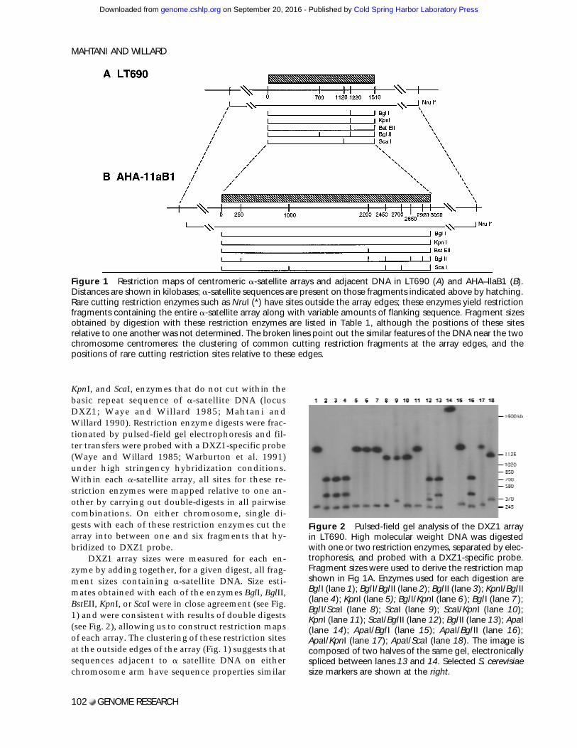
KpnI, and ScaI, enzymes that do not cut within thebasic repeat sequence of a-satellite DNA (locusDXZ1; Waye and Willard 1985; Mahtani andWillard 1990). Restriction enzyme digests were frac-tionated by pulsed-field gel electrophoresis and fil-ter transfers were probed with a DXZ1-specific probe(Waye and Willard 1985; Warburton et al. 1991)under high stringency hybridization conditions.Within each a-satellite array, all sites for these re-striction enzymes were mapped relative to one an-other by carrying out double-digests in all pairwisecombinations. On either chromosome, single di-gests with each of these restriction enzymes cut thearray into between one and six fragments that hy-bridized to DXZ1 probe.
DXZ1 array sizes were measured for each en-zyme by adding together, for a given digest, all frag-ment sizes containing a-satellite DNA. Size esti-mates obtained with each of the enzymes BglI, BglII,BstEII, KpnI, or ScaI were in close agreement (see Fig.1) and were consistent with results of double digests(see Fig. 2), allowing us to construct restriction mapsof each array. The clustering of these restriction sitesat the outside edges of the array (Fig. 1) suggests thatsequences adjacent to a satellite DNA on eitherchromosome arm have sequence properties similar
Figure 1 Restriction maps of centromeric a-satellite arrays and adjacent DNA in LT690 (A) and AHA–llaB1 (B).Distances are shown in kilobases; a-satellite sequences are present on those fragments indicated above by hatching.Rare cutting restriction enzymes such as NruI (*) have sites outside the array edges; these enzymes yield restrictionfragments containing the entire a-satellite array along with variable amounts of flanking sequence. Fragment sizesobtained by digestion with these restriction enzymes are listed in Table 1, although the positions of these sitesrelative to one another was not determined. The broken lines point out the similar features of the DNA near the twochromosome centromeres: the clustering of common cutting restriction fragments at the array edges, and thepositions of rare cutting restriction sites relative to these edges.
Figure 2 Pulsed-field gel analysis of the DXZ1 arrayin LT690. High molecular weight DNA was digestedwith one or two restriction enzymes, separated by elec-trophoresis, and probed with a DXZ1-specific probe.Fragment sizes were used to derive the restriction mapshown in Fig 1A. Enzymes used for each digestion areBgl I (lane 1); Bgl I/Bgl II (lane 2); Bgl II (lane 3); KpnI/Bgl II(lane 4); KpnI (lane 5); Bgl I/KpnI (lane 6); Bgl I (lane 7);Bgl I/ScaI (lane 8); ScaI (lane 9); ScaI/KpnI (lane 10);KpnI (lane 11); ScaI/Bgl II (lane 12); Bgl II (lane 13); ApaI(lane 14); ApaI/Bgl I (lane 15); ApaI/Bgl II (lane 16);ApaI/KpnI (lane 17); ApaI/ScaI (lane 18). The image iscomposed of two halves of the same gel, electronicallyspliced between lanes 13 and 14. Selected S. cerevisiaesize markers are shown at the right.
MAHTANI AND WILLARD
102 GENOME RESEARCH
Cold Spring Harbor Laboratory Press on September 20, 2016 - Published by genome.cshlp.orgDownloaded from
to typical single-copy DNA (Tyler-Smith andWillard 1993).
Digestion with ApaI, an enzyme that cuts rela-tively frequently in typical genomic DNA, releasedeach of the two arrays onto one large fragment ∼200kb larger than the predicted total array size (Table1). Therefore, whereas the edges of the DXZ1 arrayare characterized by a clustering of many common-cutting sites, this flanking DNA may not have atypical ApaI restriction site distribution. It is pos-sible that DXZ1 sequences may be flanked on one orboth sides by a small amount (∼200 kb or less) ofother repetitive sequences such as diverged a satel-lite (e.g., Cooper et al. 1993; Bayne et al. 1994) oranother family of repetitive DNA. In fact, at the cen-tromeres of chromosomes 7 (Wevrick et al. 1992), 8(Lin et al. 1993), Y (Cooper et al. 1993), and theacrocentric chromosomes (Trowell et al. 1993;Wohr et al. 1996), there is evidence that smallamounts of other tandemly repeated DNA familiesare located near a-satellite sequences.
Whereas the cluster of BglI, KpnI, BstEII, andBglII sites at position 1220 kb in LT690 (Fig. 1) mayidentify a small amount of DNA with typical restric-tion site composition at this position in the array(perhaps reflecting small amounts of interspersed,retroposed repetitive elements such as L1; e.g.,Wevrick et al. 1992), there is no evidence withineither LT690 or AHA–11aB1 for a large (>100 kb)amount of nonrepetitive DNA either interspersedwith or present as a large block in X chromosomea-satellite sequences. Such internal fragments con-
taining nonrepetitive (i.e., unique sequence) DNAshould not hybridize to the a-satellite probe andwould be expected to yield inconsistent size esti-mates for different enzyme digests. The consistentagreement between array length estimates derivedfrom multiple independent digests with different re-striction enzymes suggests that such unique se-quence DNA is largely absent.
Mapping the Arrays with Rare-Cutting Enzymes
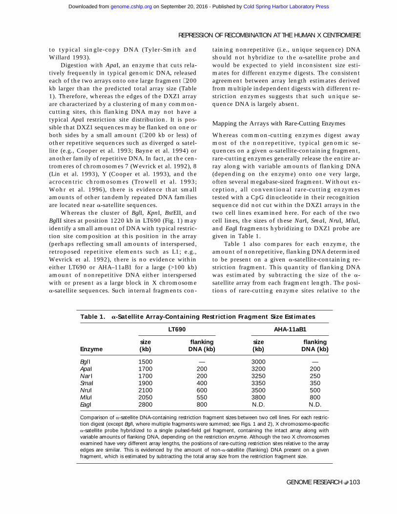
Whereas common-cutting enzymes digest awaymost of the nonrepetitive, typical genomic se-quences on a given a-satellite-containing fragment,rare-cutting enzymes generally release the entire ar-ray along with variable amounts of flanking DNA(depending on the enzyme) onto one very large,often several megabase-sized fragment. Without ex-ception, all conventional rare-cutting enzymestested with a CpG dinucleotide in their recognitionsequence did not cut within the DXZ1 arrays in thetwo cell lines examined here. For each of the twocell lines, the sizes of these NarI, SmaI, NruI, MluI,and EagI fragments hybridizing to DXZ1 probe aregiven in Table 1.
Table 1 also compares for each enzyme, theamount of nonrepetitive, flanking DNA determinedto be present on a given a-satellite-containing re-striction fragment. This quantity of flanking DNAwas estimated by subtracting the size of the a-satellite array from each fragment length. The posi-tions of rare-cutting enzyme sites relative to the
Table 1. a-Satellite Array-Containing Restriction Fragment Size Estimates
Enzyme
LT690 AHA-11aB1
size(kb)
flankingDNA (kb)
size(kb)
flankingDNA (kb)
Bgl I 1500 — 3000 —ApaI 1700 200 3200 200Nar I 1700 200 3250 250SmaI 1900 400 3350 350NruI 2100 600 3500 500MluI 2050 550 3800 800EagI 2800 800 N.D. N.D.
Comparison of a-satellite DNA-containing restriction fragment sizes between two cell lines. For each restric-tion digest (except BglI, where multiple fragments were summed; see Figs. 1 and 2), X chromosome-specifica-satellite probe hybridized to a single pulsed-field gel fragment, containing the intact array along withvariable amounts of flanking DNA, depending on the restriction enzyme. Although the two X chromosomesexamined have very different array lengths, the positions of rare-cutting restriction sites relative to the arrayedges are similar. This is evidenced by the amount of non-a-satellite (flanking) DNA present on a givenfragment, which is estimated by subtracting the total array size from the restriction fragment size.
REPRESSION OF RECOMBINATION AT THE HUMAN X CENTROMERE
GENOME RESEARCH 103
Cold Spring Harbor Laboratory Press on September 20, 2016 - Published by genome.cshlp.orgDownloaded from
edges of the a satellite array on the two X chromo-somes (in AHA–11aB1 and LT690) are strongly cor-related with one another. For example, in both celllines, the enzymes ApaI and NarI release fragments∼200-kb larger than the a satellite array; SmaI cuts∼400 kb outside the LT690 array and ∼350 kb out-side the AHA–11aB1 array. Similarly, in LT690, theNruI fragment is predicted to contain ∼600 kb ofsingle-copy sequence relative to ∼500 kb in AHA–11aB1. In one case, notably MluI, the correlationmay be less obvious because of the difficulty, par-ticularly in AHA–11aB1, of accurately sizing verylarge (>3000 kb) fragments.
On the X chromosome, with the exception ofthe small array in LT690, the sizes of a-satellite ar-rays vary continuously from 2200 to 3730 kb as aresult of array length polymorphism (e.g., Mahtaniand Willard 1990). If restriction sites in the DNAflanking the array are positioned at fixed, predict-able distances from the array edges, then, for a givenenzyme, the sizes of rare-cutting fragments shouldfollow the same distribution as that of the arraylengths itself. Under this hypothesis, the amount offlanking DNA on a restriction fragment that alsocontains the entire DXZ1 array should be identicalfor many different X chromosomes digested withthe same restriction enzyme. As is shown in Table 1,data from these two extensively mapped arrays sup-port this hypothesis. Thus, the variation in restric-tion fragment lengths caused by array size polymor-phism (Wevrick and Willard 1989; Mahtani andWillard 1990) may taper off close to the edges of thea-satellite array, as this DNA becomes typical eu-chromatic sequence. More data that precisely posi-tion single-copy markers relative to the array edgeswill help evaluate the validity of this hypothesis. Todate, in situ hybridization experiments have failedto identify large, variable blocks of other repetitiveDNA families adjacent to DXZ1 sequences (Miller etal. 1995; V. Powers and H.F. Willard, unpubl.), eventhough such sequences are readily detected at manyother centromeres. This also supports the idea thatthe a-satellite array measured here may lie close tothe unique, euchromatic sequences of the two chro-mosome arms. As suggested by the ApaI digests, iffamilies of other repetitive DNAs are present in thevicinity of the centromere of the X chromosome,then these sequences must exist in relatively small(<200-kb) blocks.
When restriction fragments containing a-satellite sequences were tested for cohybridizationwith probes from the proximal unique sequencesDXS422 (in Xp11.21) and DXS62 (in Xq11), wewere unable to detect any colocalization with filter
hybridization (M.M. Mahtani, data not shown). InLT690, because DXZ1 is found on a single NruI frag-ment that also contains ∼600 kb of non-a-satellitesequences, the DXS422 and DXS62 loci must lie atleast 2100 kb apart and thus 300–600 kb outside thea-satellite array.
Because the small LT690 DXZ1 array is almostexactly half the size of the mean array length fromthe population sampled, we considered the possibil-ity that the measured array might be present in du-plicate on that chromosome. If the LT690 arraywere itself tandemly duplicated in such a manner,however, then digests with restriction enzymes thatcut within the array or very close to its edges (i.e.,BglI, KpuI, BglII, etc.) would yield bands of doubleintensity. Whereas such doublets might not be eas-ily scored in LT690, they should be readily apparentin the DNA of the mother of LT690, who carriesboth an average-sized array and the small array.Comparisons of the a-satellite gel patterns from thetwo X chromosomes in the mother, however, re-vealed no obvious intensity differences betweenfragments corresponding to one or the other haplo-type (M.M. Mahtani, data not shown). Thus, it isunlikely that a simple tandem duplication of onlyDXZ1 is present in LT690, and we conclude that theactual DXZ1 array size is ∼1500 kb.
Genetic Distance Across the Centromere
Some information is lost when genetic distances arederived from linkage analysis, because only thosemeioses informative for the relevant markers areconsidered in the analysis. To examine each chro-mosome in the CEPH database (i.e., not only thoseinformative for a given pair of markers), instead weconstructed 27-locus meiotic maps of inherited Xchromosomes from the entire youngest generation(349 female meioses; see Methods) of the CEPH fam-ily panel. From this 27-locus marker set, on average10.1 markers were informative within a given fam-ily (range 5–15 loci per family). Because these loci allmap to a ∼20-cM region spanning the centromere(Nelson et al. 1995; Weeks et al. 1995), the averageintermarker distance is ∼2 cM.
For each of the meioses in the 40 family CEPHpanel, positions of recombination events were plot-ted relative to the inherited maternal X chromo-some (both by hand and with the CRIMAP programCHROMPIC) for the 27-marker loci. By use of distalmarkers to establish phase and to rule out doublecrossovers in flanking regions, chromosomes wereidentified that showed evidence for recombinationbetween DXS255 in Xp11.22 and DXS153 in Xq13
MAHTANI AND WILLARD
104 GENOME RESEARCH
Cold Spring Harbor Laboratory Press on September 20, 2016 - Published by genome.cshlp.orgDownloaded from
(∼15 Mb physical distance; Nelson et al. 1995; Na-garaja et al. 1997). In the data set of 349 meioses,only 26 crossovers occurred in this interval; nodouble crossovers were detected.
Crossing-over occurs rarely across the centro-mere, but when it does occur, it seems to cluster ina few families. Specifically, most families showed noevidence for crossing-over in the pericentromeric re-gion, whereas more pedigrees than expected(x2 = 7.2; P = 0.03) had two or three individualswith single crossovers occurring somewhere be-tween DXS255 and DXS153. This may reflect an un-derlying genetic (interfamilial) variation in recom-bination rates.
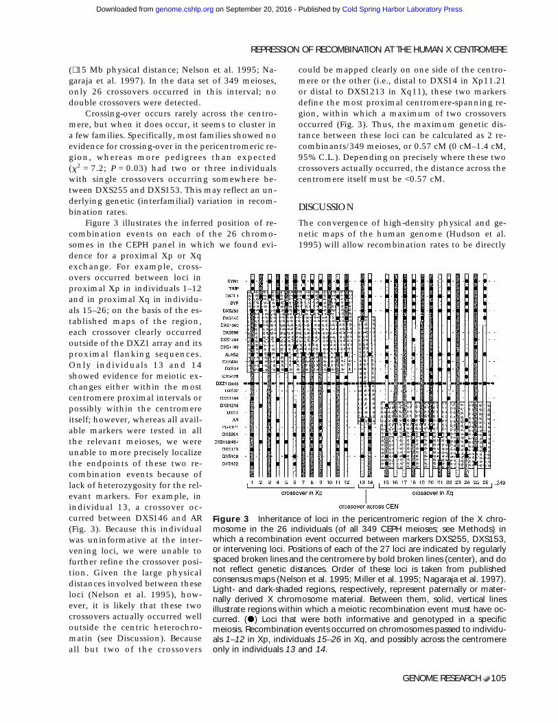
Figure 3 illustrates the inferred position of re-combination events on each of the 26 chromo-somes in the CEPH panel in which we found evi-dence for a proximal Xp or Xqexchange. For example, cross-overs occurred between loci inproximal Xp in individuals 1–12and in proximal Xq in individu-als 15–26; on the basis of the es-tablished maps of the region,each crossover clearly occurredoutside of the DXZ1 array and itsproximal flanking sequences.Only individuals 13 and 14showed evidence for meiotic ex-changes either within the mostcentromere proximal intervals orpossibly within the centromereitself; however, whereas all avail-able markers were tested in allthe relevant meioses, we wereunable to more precisely localizethe endpoints of these two re-combination events because oflack of heterozygosity for the rel-evant markers. For example, inindividual 13, a crossover oc-curred between DXS146 and AR(Fig. 3). Because this individualwas uninformative at the inter-vening loci, we were unable tofurther refine the crossover posi-tion. Given the large physicaldistances involved between theseloci (Nelson et al. 1995), how-ever, it is likely that these twocrossovers actually occurred welloutside the centric heterochro-matin (see Discussion). Becauseall but two of the crossovers
could be mapped clearly on one side of the centro-mere or the other (i.e., distal to DXS14 in Xp11.21or distal to DXS1213 in Xq11), these two markersdefine the most proximal centromere-spanning re-gion, within which a maximum of two crossoversoccurred (Fig. 3). Thus, the maximum genetic dis-tance between these loci can be calculated as 2 re-combinants/349 meioses, or 0.57 cM (0 cM–1.4 cM,95% C.L.). Depending on precisely where these twocrossovers actually occurred, the distance across thecentromere itself must be <0.57 cM.
DISCUSSION
The convergence of high-density physical and ge-netic maps of the human genome (Hudson et al.1995) will allow recombination rates to be directly
Figure 3 Inheritance of loci in the pericentromeric region of the X chro-mosome in the 26 individuals (of all 349 CEPH meioses; see Methods) inwhich a recombination event occurred between markers DXS255, DXS153,or intervening loci. Positions of each of the 27 loci are indicated by regularlyspaced broken lines and the centromere by bold broken lines (center), and donot reflect genetic distances. Order of these loci is taken from publishedconsensus maps (Nelson et al. 1995; Miller et al. 1995; Nagaraja et al. 1997).Light- and dark-shaded regions, respectively, represent paternally or mater-nally derived X chromosome material. Between them, solid, vertical linesillustrate regions within which a meiotic recombination event must have oc-curred. (d) Loci that were both informative and genotyped in a specificmeiosis. Recombination events occurred on chromosomes passed to individu-als 1–12 in Xp, individuals 15–26 in Xq, and possibly across the centromereonly in individuals 13 and 14.
REPRESSION OF RECOMBINATION AT THE HUMAN X CENTROMERE
GENOME RESEARCH 105
Cold Spring Harbor Laboratory Press on September 20, 2016 - Published by genome.cshlp.orgDownloaded from
compared with known physical distances at regularintervals along each of the human chromosomes.Preliminary comparisons suggest that the ratios ofphysical to genetic map lengths will vary greatlyrelative to the genomic average. This variation inrecombination frequency reflects differences withina chromosome, between chromosomes, and be-tween the sexes. For example, the genetic map is1.6-fold longer in females than males (Dib et al.1996), revealing a generally higher rate of exchangein female than male meiosis. Averaged over the ge-nome, the mean rate of exchange is 1 cM every 717and 1155 kb in females and males, respectively,with a similar range of variation for individual chro-mosomes (Morton 1991; Dib et al. 1996).
Such chromosome- and genome-averages, how-ever, fail to consider another level of map lengthvariation: the probable abundance of local hot andcold spots. In fact, when long-range (restriction orYAC contig) physical maps are compared with theircorresponding genetic maps, large regional varia-tions in crossing-over rates are observed (e.g., Abbset al. 1990; Allitto et al. 1991; Nagaraja et al. 1997).Unusually high or infrequent recombination ratesare thought to be dependent on proximity to localrecombination signals, changes in higher orderchromatin structure, or presence of chromosomalstructural elements such as centromeres or telo-meres.
In Drosophila (Dobzhansky 1930; Mather 1939),yeast (Clarke and Carbon 1980), and Neurospora(Davis et al. 1994), recombination at centromeres ismarkedly reduced relative to that found in otherchromosome regions. This repression of recombina-tion persists in Drosophila when adjacent hetero-chromatin is deleted (Yamamoto and Miklos 1978)and is generated de novo in yeast when a centro-mere is integrated into a new site on the chromo-some (Nakaseko et al. 1986). In humans, chiasmataare found infrequently near chromosome centro-meres (Hulten 1974; Hulten et al. 1982), but defini-tive studies of recombination rates across measuredphysical distances at human centromeres have onlybecome available recently.
Perhaps most relevant is a comparison of physi-cal distance across the centromere of chromosome10 with the genetic distance between two centro-mere-flanking microsatellite markers; this study re-ported a 10-fold repression of recombination (Jack-son et al. 1996), consistent with earlier studies onthis chromosome (Carson and Simpson 1991;Lichter et al. 1993). On the other hand, estimates ofrecombination rates at the pericentromeric regionsof chromosomes 11 (Janson et al. 1991) and 21 (Jabs
et al. 1991; Van Hul et al. 1993) seem less consistentwith a centromeric suppression of meiotic ex-change. These apparently conflicting data, however,may be reconciled by the fact that the chromosome11 map may be bounded by loci a significant dis-tance away from a-satellite sequences, because thephysical distance between the centromere and theloci studied is not known. In fact, radiation hybridmapping data on chromosome 11 suggest that themost proximal centromeric markers span a largephysical distance (in centiRads) but exhibit no re-combination (James et al. 1994). On chromosome21, the extensive homology between a-satellite ar-rays on chromosomes 21 and 13 (for review, seeTrowell et al. 1993) may generate ambiguous geno-types of the a-satellite polymorphisms within thesearrays, artificially inflating observed recombinationrates. Thus, when more definitive data are available,recombination frequencies at the centromeres ofthese and other chromosomes may be consistentwith the repressed rates of meiotic exchange foundon the X chromosome (as reported here; see alsoFain et al. 1995; Weeks et al. 1995) and chromo-some 10 (Carson and Simpson 1991; Lichter et al.1993; Jackson et al. 1996).
To determine the rate of exchange across the Xchromosome centromere and characterize the ex-tent of repression of meiotic exchange in humans,we measured the frequency of genetic exchangeacross the X chromosome centromere by comparinga long-range restriction map with detailed geneticmaps of the CEPH meioses. The physical map acrossthe centromere encompasses on average ∼3000 kbof a-satellite DNA and ∼600 kb of unique sequenceDNA, although still exclusive of either of the mostcentromere-proximal genetic markers, DXS422(ZXDA) or DXS62 (Miller et al. 1995; Nelson et al.1995). The maximum genetic distance across thisregion was evaluated by identifying positions of re-combination events relative to 27 loci in the peri-centromeric region of the X chromosome. At most,two chromosomes from a sample of 349 femalemeioses of the CEPH family panel were recombinedin the genetic intervals spanning or including thecentromere (Fig. 3). Therefore, the maximum ge-netic distance across the 3600-kb region is ∼0.57 cM,suggesting an upper limit on the rate of exchange inthis region of 1 cM/6300 kb (i.e., 0.57 cM/3600 kb).
This recombination frequency is much lowerthan that expected from a similar-sized interval else-where on the X chromosome. For example, assum-ing an average frequency of exchange of ∼1 cM/800kb (the genetic to physical distance ratio when av-eraged over the entire X chromosome; Dib et al.
MAHTANI AND WILLARD
106 GENOME RESEARCH
Cold Spring Harbor Laboratory Press on September 20, 2016 - Published by genome.cshlp.orgDownloaded from
1996; Nagaraja et al. 1997), a 3600-kb interval else-where on this chromosome would have an expectedmap length of 4.6 cM (2.4 cM–6.8 cM, 95% C.L.). Ina sample size similar to that examined here, over agenetic distance of 4.6 cM, one would expect to ob-serve about 16 crossovers. Our observation of only 2crossovers is well outside the predicted Poissonvariation around this mean (P = 1.4 2 1015), indi-cating a highly depressed rate of exchange at thecentromere relative to the chromosome average.This repression provides initial quantitation of amore general effect inferred on the proximal X chro-mosome, when genetic distances are compared withrough cytogenetic band sizes (Fain et al. 1995;Weeks et al. 1995) or to a YAC contig map (Nagarajaet al. 1997).
The calculated genetic distance of 0.57 cM is aconservative estimate of the extent of centromericrepression, because it reflects the genetic maplength of a region that includes, but is not limitedto, the ∼3600 kb mapped here. Therefore, the rate ofexchange reported here overestimates crossing overunless both recombination events fall within themost proximal 3600 kb, including the a-satellite ar-ray. In fact, this genetic distance of 0.57 cM may fallin any interval, or be evenly distributed, within thecentromere-spanning region. The consensus map ofthe X chromosome indicates that this region mayencompass up to 8–10 Mb of DNA, including thelength of the a-satellite array (Nelson et al. 1995).Once the gaps in the physical map of the X chro-mosome are closed, a definitive rate of exchangecan be calculated for the entire interval. Because it ispossible that the genetic distance of 0.57 cM may bedistributed over as much as 10,000 kb, the recom-bination rate across the centromere (including cen-tromere adjacent DNA), may be as low as 1 cM/17,000 kb. Nonetheless, this rate should be taken asan upper limit for the rate of crossing over at thecentromere itself, keeping in mind that it is formallypossible that, as in Drosophila (Baker 1958), recom-bination may not occur at all in proximal hetero-chromatin.
Regional variation in recombination rates onthe X chromosome is extensive. In fact, when long-range physical maps are compared with their ge-netic counterparts, different regions of the X chro-mosome have widely different recombination rates.For example, 1 cM corresponds to 190 kb in theDMD region (van Ommen et al. 1986; Abbs et al.1990), 340–800 kb near the end of the long arm[Xq27.2-qter (Kenwrick and Gitschier 1989; Poustkaet al. 1991)], 600 kb near the end of the short arm[Xp22.3-pter (Petit et al. 1990)], 640 kb in Xq26
(Little et al. 1992), and >5 Mb in Xq13.3-q21.3 (Na-garaja et al. 1997). Whereas recombination ratesmay be reported more frequently for regions of highrecombination, recombination rates approachingthat reported here for the centromere have not beendocumented for other regions on the X or, untilrecently (Jackson et al. 1996), for other regions ofthe genome.
METHODS
Physical Mapping
To generate restriction maps of the DXZ1 a-satellite array onthe X chromosome, we chose restriction enzymes that typi-cally cut mammalian DNA into small (<20 kb) fragments, butthat fail to cut within the basic 2.0-kb higher-order repeatsequence on the X chromosome (Waye and Willard 1985;Mahtani and Willard 1990). High molecular weight DNA wasprepared and digested as described (van Ommen and Verkerk1986; Wevrick and Willard 1989). When restriction bufferswere incompatible between two enzymes, double digests (ofDNA in agarose blocks) were done sequentially for 4 hr each,separated by a 1-hr rinse in 10 mM Tris-Cl; 10 mM EDTA. S.cervisiae and S. pombe chromosomes (Bio-Rad) were used ashigh molecular weight size standards. The gel-purified PCRproduct of X chromosome specific a-satellite primers X-3Aand X-4A (Warburton et al. 1991) or the plasmid pBamX7(Waye and Willard 1985) was used as a probe for DXZ1.Pulsed-field (CHEF) gel electrophoresis, Southern blotting,and hybridization were performed as described (Waye et al.1988; Wevrick and Willard 1989; Mahtani and Willard 1990).
LT690 is a lymphoblastoid cell line derived from a nor-mal Atlantic Canadian male shown previously to carry a par-ticularly small X chromosome a-satellite array (Mahtani andWillard 1990). AHA-11aB1 is a mouse/human somatic cellhybrid containing an X chromosome as its only human ma-terial (Willard 1983).
Genetic Mapping
DNA from the 40 family panel of the CEPH was used in ge-netic mapping studies. This panel contains 362 female meio-ses in the youngest generation of primarily three-generationfamilies; therefore, most of these were phase-known meioses.Exactly 349 of these individuals were informative for enoughmarkers in this region such that gaps of no greater than 6 cMexisted between adjacent informative loci; the remaining 13meioses were removed from the calculation of recombinationrates to eliminate the possibility of undetected double-crossovers in those regions.
Genotypic data from 27 loci in the pericentromeric re-gion of the X chromosome were compiled from a previouslypublished 18-point linkage map (Mahtani et al. 1991) andstudies of microsatellites in proximal Xq (Mahtani andWillard 1993), the ALAS2 gene (Cox et al. 1992), the ZXDAgene (locus DXS422; G. Greig and M.M. Mahtani, data notshown), and the CEPH database V7.1 to generate meioticmaps (Fain et al. 1995). All available individuals in CEPH weregenotyped for at least 18 of the loci (Mahtani et al. 1991).Those with pericentromeric crossovers (Fig. 3) were then
REPRESSION OF RECOMBINATION AT THE HUMAN X CENTROMERE
GENOME RESEARCH 107
Cold Spring Harbor Laboratory Press on September 20, 2016 - Published by genome.cshlp.orgDownloaded from
typed for all additional loci. The total of 27 loci span a geneticdistance of ∼20 cM in the pericentromeric region (Mahtani etal. 1991; Fain et al. 1995; Weeks et al. 1995), extending fromthe cytogenetic band Xp11.22 on the short arm, across thecentromere and into band Xq12 on the long arm (Miller et al.1995; Nelson et al. 1995). Genotypes for markers DXS7 inXp11.3 and PGK1 in Xq13.3, two flanking loci that map distalto the region examined here, were used to ensure consistencyand to eliminate genotyping errors from the dataset. Locusorder was determined as described previously (Mahtani et al.1991) and is consistent with consensus maps of the X chro-mosome (Wang et al. 1994; Fain et al. 1995; Nelson et al.1995; Weeks et al. 1995).
ACKNOWLEDGMENTSWe thank David Botstein, Rick Myers and Luca Cavalli-Sforzafor support; and David Botstein, Pat Brown, David Cox, EricLander, and Andrew Kirby for their interest and comments.This work was supported by National Institutes of Healthgrant HG00107 to H.F.W.
The publication costs of this article were defrayed in partby payment of page charges. This article must therefore behereby marked ‘‘advertisement’’ in accordance with 18 USCsection 1734 solely to indicate this fact.
REFERENCESAbbs, S., R.G. Roberts, C.G. Mathew, D.R. Bentley, and M.Bobrow. 1990. Accurate assessment of intragenicrecombination frequency within the Duchenne musculardystrophy gene. Genomics 7: 602–606.
Allitto, B.A., M.E. MacDonald, M. Bucan, J. Richards, D.Romano, W.L. Whaley, B. Falcone et al. 1991. Increasedrecombination adjacent to the Huntington disease-linkedD4S10 marker. Genomics 9: 104–112.
Baker, W.K. 1958. Crossing over in heterochromatin. Am.Nat. 92: 59–60.
Bayne, R.A.L., D. Broccoli, M.H. Taggart, E.J. Thomson, C.J.Farr, and H.J. Cooke. 1994. Sandwiching of a gene within12 kb of a functional telomere and a satellite does notresult in silencing. Hum. Mol. Genet. 3: 539–546.
Beadle, G.W. 1932. A possible influence of the spindle fibreon crossing-over in Drosophila. Proc. Natl. Acad. Sci.18: 160–165.
Carson, N.L. and N.E. Simpson. 1991. A physical map ofhuman chromosome 10 and a comparison with an existinggenetic map. Genomics 11: 379–388.
Charlesworth, B., C.H. Langley, and W. Stephan. 1986. Theevolution of restricted recombination and the accumulationof repeated DNA sequences. Genetics 112: 947–962.
Clarke, L. and J. Carbon. 1980. Isolation of thecentromere-linked CDC10 gene by complementation inyeast. Proc. Natl. Acad. Sci. 77: 2173–2177.
Cooper, K.F., R.B. Fisher, and C. Tyler-Smith. 1993.Structure of the sequences adjacent to the centromeric
alphoid DNA array on the human Y chromosome. J. Mol.Biol. 230: 787–799.
Cox, T.C., H.M. Kozman, W.H. Raskind, B.K. May, and J.C.Mulley. 1992. Identification of a highly polymorphicmarker within intron 7 of the ALAS2 gene and suggestionof at least two loci for X-linked sideroblastic anemia. Hum.Mol. Genet. 1: 639–641.
Davis, C.R., R.R. Kempainen, M.S. Srodes, and C.R.McClung. 1994. Correlation of the physical and geneticmaps of the centromeric region of the right arm of linkagegroup III of Neurospora crassa. Genetics 136: 1297–1306.
Dib, C., S. Faure, C. Flzames, D. Samson, N. Drouot, A.Vignal, P. Millasseasu, S. Marc, J. Hazan, E. Seboun et al.1996. A comprehensive genetic map of the human genomebased on 5,264 microsatellites. Nature 380: 152–154.
Dobzhansky, T. 1930. Translocations involving the thirdand the fourth chromosomes of Drosophila melanogaster.Genetics 15:1347–399.
Fain, P.R., E.N. Kort, P.F. Chance, K. Nguyen, D.F. Redd,M.J. Econs, and D.F. Barker. 1995. A 2D crossover-basedmap of the human X chromosome as a model for mapintegration. Nature Genet. 9: 261–266.
Harrington, J.J., G. Van Bokkelen, R.W. Mays, K.Gustashaw, and H.F. Willard. 1997. Formation of de novocentromeres and construction of first-generation humanartificial chromosomes. Nature Genet. 15: 345–355.
Hudson, T.B., L.D. Stein, S.S. Gerety, J. Ma, A.B. Castle, J.Silva, D.K. Slonim et al. 1995. An STS-based map of thehuman genome: A scaffold for large-scale sequencing.Science 270: 1893–2064.
Hulten, M. 1974. Chiasma distribution at diakinesis in thenormal human male. Hereditas 76: 55–78.
Hulten, M.A., R.W. Palmer, and D.A. Laurie. 1982. Chiasmaderived genetic maps and recombination fractions:Chromosome 1. Ann. Hum. Genet. 46: 167–175.
Jabs, E.W., A.C. Warren, E.W. Taylor, C.R. Colyer, D.A.Meyers, and S.E. Antonarakis. 1991. Alphoid DNApolymorphisms for chromosome 21 can be distinguishedfrom those of chromosome 13 using probes homologous toboth. Genomics 9: 141–146.
Jackson, M.S., C.G. See, L.M. Mulligan, and B.F. Lauffart.1996. A 9.75-Mb map across the centromere of humanchromosome 10. Genomics 33: 258–270.
James, M.R., C.W. Richard III, J.-J. Schott, C. Yousry, K.Clark, J. Bell, J.D. Terwilliger et al. 1994. A radiation hybridmap of 506 STS markers spanning human chromosome 11.Nature Genet. 8: 70–76.
Janson, M., C. Larsson, B. Werelius, C. Jones, T. Glaser, Y.Nakamura, P. Jones et al. 1991. Detailed physical map ofhuman chromosomal region 11q12-13 shows high meioticrecombination rate around the MEN1 locus. Proc. Natl.Acad. Sci. 88: 10609–10613.
MAHTANI AND WILLARD
108 GENOME RESEARCH
Cold Spring Harbor Laboratory Press on September 20, 2016 - Published by genome.cshlp.orgDownloaded from
Kenwrick, S. and J. Gitschier. 1989. A contiguous, 3-Mbphysical map of Xq28 extending from the colorblindnesslocus to DXS15. Am. J. Hum. Genet. 45: 873–882.
Lambie, E.J. and G.S. Roeder. 1986. Repression of meioticcrossing over by a centromere (CEN3) in Saccharomycescerevisiae. Genetics 114: 769–789.
Larin, Z., M.D. Fricker, and C. Tyler-Smith. 1994. De novoformation of several features of a centromere followingintroduction of a Y alphoid YAC into mammalian cells.Hum. Mol. Genet. 3: 689–695.
Lichter, J.B., M.J. Difilippantonio, A.J. Pakstis, P.J.Goodfellow, D.C. Ward, and K.K. Kidd. 1993. Physical andgenetic maps for chromosome 10. Genomics 16: 320–324.
Lin, C.C., R. Sasi, C. Lee, Y.S. Fan, and D. Court. 1993.Isolation and characterization of a novel tandemly repeatedDNA sequence in the centromeric region of humanchromosome 8. Chromosoma 102: 333–339.
Little, R.D., G. Pilia, S. Johnson, M. D’Urso, and D.Schlessinger. 1992. Yeast artificial chromosomes spanning 8megabases and 10-15 centimorgans of human cytogeneticband Xq26. Proc. Natl. Acad. Sci. 89: 177–181.
Mahtani, M.M., R.G. Lafreniere, T.A. Kruse, and H.F.Willard. 1991. An 18-locus linkage map of thepericentromeric region of the human X chromosome:Genetic framework for mapping X-linked disorders.Genomics 10: 849–857.
Mahtani, M.M. and H.F. Willard. 1990. Pulsed-field gelanalysis of alpha satellite DNA at the human Xchromosome centromere: High frequency polymorphismsand array size estimate. Genomics 7: 607–613.
———. 1993. A polymorphic X-linked tetranucleotiderepeat locus displaying a high rate of new mutation:Implications for mechanisms of mutation at short tandemrepeat loci. Hum. Mol. Genet. 2: 431–437.
Malone, R.E., J.E. Golin, and M.S. Esposito. 1980. Mitoticversus meiotic recombination in Saccharomyces cerevisiae.Curr. Genet. 1:1241–248.
Mather, K. 1936. The determination of position incrossing-over. I. Drosophila melanogaster. J. Genet.33: 207–235.
———. 1939. Crossing over and heterochromatin in the Xchromosome of Drosophila melanogaster. Genetics24: 413–435.
Miller, A.P., K. Gustashaw, D.J. Wolff, S.H. Rider, A.P.Monaco, B. Eble, D. Schlessinger, and H.F. Willard. 1995.Three genes that escape X chromosome inactivation areclustered within a 6 Mb YAC contig and STS map inXp11.21-p11.22. Hum. Mol. Genet. 4: 731–739.
Minet, M., A.-M. Grossenbacher-Grunder, and P. Thuriaux.1980. The origin of a centromere effect on mitotic
recombination: A study in the fission yeastSchizosaccharomyces pombe. Curr. Genet. 2: 53–60.
Morton, N.E. 1991. Parameters of the human genome. Proc.Natl. Acad. Sci. 88: 7474–7476.
Murphy, T.D. and G.H. Karpen. 1995. Localization ofcentromere function in a Drosophila minichromosome. Cell82: 599–609.
Nagaraja, R., S. MacMillan, J. Kere, C. Jones, S. Griffin, M.Schmatz, J. Terrell, M. Shomaker, X. Jermak, C. Hott et al.1997. X chromosome map at 75-kb STS resolution,revealing estimates of recombination and GC content.Genome Res. 7: 210–222.
Nakaseko, Y., Y. Adachi, S. Funahashi, O. Niwa, and M.Yanagida. 1986. Chromosome walking shows a highlyhomologous repetitive sequence present in all thecentromere regions of fission yeast. EMBO J. 5: 1011–1021.
Nelson, D., A. Ballabio, F. Cremers, A.P. Monaco, and D.Schlessinger. 1995. Report of the sixth internationalworkshop on X chromosome mapping. Cytogenet. CellGenet. 71: 308–336.
Oakey, R. and C. Tyler-Smith. 1990. Y chromosome DNAhaplotyping suggests that most European and Asian menare descended from one of two males. Genomics 7: 325–330.
Painter, T.S. 1935. The morphology of the thirdchromosome in the salivary gland of Drosophilamelanogaster and a new cytological map of this element.Genetics 20: 301–326.
Petit, C., J. Levillier, and J. Weissenbach. 1990. Long-rangerestriction map of the terminal part of the short arm of thehuman X chromosome. Proc. Natl. Acad. Sci. 87: 3680–3684.
Poustka, A., A. Dietrich, G. Langenstein, D. Toniolo, S.T.Warren , and H. Lehrach. 1991. Physical map of humanXq27-qter: Localizing the region of the fragile X mutation.Proc. Natl. Acad. Sci. 88: 8302–8306.
Roberts, P.A. 1965. Difference in the behaviour of eu- andhetero-chromatin: Crossing-over. Nature 205: 725–726.
Thompson, P.E. 1963. Centric pairing and crossing-over inDrosophila melanogaster. Genetics 48: 697–701.
Trowell, H.E., A. Nagy, B. Vissel, and K.H. Choo. 1993.Long-range analysis of the centromeric regions of humanchromosomes 13, 14 and 21: Identification of a narrowdomain containing two key centromeric DNA elements.Hum. Mol. Genet. 10: 1639–1649.
Tyler-Smith, C. and H.F. Willard. 1993. Mammalianchromosome structure. Curr. Opin. Genet. Dev. 3: 390–397.
van Daelen, R.A., F. Gerbens, F. van Ruissen, J. Aarts, J.Hontelez, and P. Zabel. 1993. Long-range physical maps oftwo loci (Aps-1 and GP79) flanking the root-knot nematoderesistance gene (Mi) near the centromere of tomatochromosome 6. Plant Mol. Biol. 23: 185–192.
REPRESSION OF RECOMBINATION AT THE HUMAN X CENTROMERE
GENOME RESEARCH 109
Cold Spring Harbor Laboratory Press on September 20, 2016 - Published by genome.cshlp.orgDownloaded from
Van Hul, W., G. Van Camp, L. Stuyver, J.M. Delabar, M.G.McInnis, A.C. Warren, S.E. Antonarakis et al. 1993. Acontiguous physical map of the pericentromeric region ofchromosome 21q between D21Z1 and D21S13E. Genomics15: 626–630.
van Ommen, G.J.B. and J.M.H. Verkerk. 1986. Restrictionanalysis of chromosomal DNA in a size range up to twomillion basepairs by pulsed field gradient electrophoresis. InHuman genetic disease: A practical approach (ed. K.E. Davies),pp. 113–133. IRL Press, Oxford, UK.
van Ommen, G.J.B., J.M.H. Verkerk, M.H. Hofker, A.P.Monaco, L.M. Kunkel, P. Ray, R. Worton et al. 1986. Aphysical map of 4 million bp around the Duchennemuscular dystrophy gene on the human X chromosome.Cell 47: 499–504.
Wang, L.H., S. Lawrence, B.J. Keats, and N.E. Morton. 1994.Integration of gene maps: Chromosome X. Genomics22: 590–604.
Warburton, P.E., G.M. Greig, T. Haaf, and H.F. Willard.1991. PCR amplification of chromosome-specific a satelliteDNA: Definition of centromeric STS markers andpolymorphic analysis. Genomics 11: 324–333.
Waye, J.S. and H.F. Willard. 1985. Chromosome-specificalpha satellite DNA: Nucleotide sequence analysis of the 2.0kb repeat from the human X chromosome. Nucleic AcidsRes. 13: 2731–2743.
Waye, J.S., A.R. Mitchell, and H.F. Willard. 1988.Organization and genomic distribution of ‘‘82H’’ alphasatellite DNA: Evidence for a low-copy or single-copyalphoid domain located on human chromosome 14. Hum.Genet. 78: 27–32.
Weeks, D.E., T.G. Nygaard, M. Neystat, L.D. Harby, andK.C. Wilhelmsen. 1995. A high resolution genetic linkagemap of the pericentromeric region of the human Xchromosome. Genomics 26: 39–46.
Wevrick, R. and H.F. Willard. 1989. Long-rangeorganization of tandem arrays of a satellite DNA at thecentromeres of human chromosomes: High-frequencyarray-length polymorphism and meiotic stability. Proc. Natl.Acad. Sci. 86: 9394–9398.
Wevrick, R., V.P. Willard, and H.F. Willard. 1992. Structureof DNA near long tandem arrays of a satellite DNA at thecentromere of human chromosome 7. Genomics14: 912–923.
Willard, H.F. 1990. Centromeres of mammalianchromosomes. Trends Genet. 12: 410–416.
———. 1993. Replication of human X chromosomes infibroblasts and somatic cell hybrids: Cytogenetic analysisand a molecular perspective. In Cytogenetics of themammalian X chromosome (ed. A.A. Sandberg), pp. 427–447.A.R. Liss, New York, NY.
Wohr, G., T. Fink, and G. Assum. 1996. A palindromicstructure in the pericentromeric region of various humanchromosomes. Genome Res. 6: 267–279.
Yamamoto, M. and G.L.G. Miklos. 1978. Genetic studies onheterochromatin in Drosophila melanogaster and theirimplications for the functions of satellite DNA. Chromosoma66: 71–98.
Received August 25, 1997; revised version accepted January 5,1998.
MAHTANI AND WILLARD
110 GENOME RESEARCH
Cold Spring Harbor Laboratory Press on September 20, 2016 - Published by genome.cshlp.orgDownloaded from
10.1101/gr.8.2.100Access the most recent version at doi:1998 8: 100-110 Genome Res.
Melanie M. Mahtani and Huntington F. Willard
Recombination Centromere: Repression ofPhysical and Genetic Mapping of the Human X Chromosome
References
http://genome.cshlp.org/content/8/2/100.full.html#ref-list-1
This article cites 60 articles, 23 of which can be accessed free at:
License
Commons Creative
http://creativecommons.org/licenses/by-nc/3.0/.described at
a Creative Commons License (Attribution-NonCommercial 3.0 Unported License), as ). After six months, it is available underhttp://genome.cshlp.org/site/misc/terms.xhtml
first six months after the full-issue publication date (see This article is distributed exclusively by Cold Spring Harbor Laboratory Press for the
ServiceEmail Alerting
click here.top right corner of the article or
Receive free email alerts when new articles cite this article - sign up in the box at the
http://genome.cshlp.org/subscriptionsgo to: Genome Research To subscribe to
Cold Spring Harbor Laboratory Press
Cold Spring Harbor Laboratory Press on September 20, 2016 - Published by genome.cshlp.orgDownloaded from
Related Documents