1 Running head: Pterin carbinolamine dehydratase family (COG2154) proteins Correspondence to: Andrew D. Hanson University of Florida Horticultural Sciences Department P.O. Box 110690 Gainesville, FL 32611-0690 Telephone: (352) 392 1928 ext 334 Fax: (352) 392 5653 e-mail: [email protected] Journal Research Area: Biochemical Processes and Macromolecular Structures Plant Physiology Preview. Published on February 1, 2008, as DOI:10.1104/pp.107.114090 Copyright 2008 by the American Society of Plant Biologists www.plantphysiol.org on November 8, 2016 - Published by www.plantphysiol.org Downloaded from Copyright © 2008 American Society of Plant Biologists. All rights reserved.

Welcome message from author
This document is posted to help you gain knowledge. Please leave a comment to let me know what you think about it! Share it to your friends and learn new things together.
Transcript
1
Running head: Pterin carbinolamine dehydratase family (COG2154) proteins
Correspondence to: Andrew D. Hanson
University of Florida
Horticultural Sciences Department
P.O. Box 110690
Gainesville, FL 32611-0690
Telephone: (352) 392 1928 ext 334
Fax: (352) 392 5653
e-mail: [email protected]
Journal Research Area: Biochemical Processes and Macromolecular Structures
Plant Physiology Preview. Published on February 1, 2008, as DOI:10.1104/pp.107.114090
Copyright 2008 by the American Society of Plant Biologists
www.plantphysiol.org on November 8, 2016 - Published by www.plantphysiol.orgDownloaded from Copyright © 2008 American Society of Plant Biologists. All rights reserved.
2
Phylogenomic and functional analysis of pterin-4a-carbinolamine
dehydratase family (COG2154) proteins in plants and microorganisms1
Valeria Naponelli2, Alexandre Noiriel2, Michael J. Ziemak, Stephen M. Beverley,
Lon-Fye Lye, Andrew M. Plume3, José Ramon Botella, Karen Loizeau, Stéphane Ravanel,
Fabrice Rébeillé, Valérie de Crécy-Lagard, and Andrew D. Hanson*
Horticultural Sciences (V.N., A.N., M.J.Z., A.D.H) and Microbiology and Cell Science (V.C.)
Departments, University of Florida, Gainesville, Florida 32611; Department of Molecular Micro-
biology, Washington University School of Medicine, St. Louis, Missouri 63110 (S.M.B., L.-
F.L); Department of Botany, University of Queensland, Brisbane, Queensland 4072, Australia
(A.M.P., J.R.B.); and Laboratoire de Physiologie Cellulaire Végétale, CNRS/CEA/INRA/Univ-
ersité Joseph Fourier, CEA-Grenoble, F-38054 Grenoble Cedex 9, France (K.L., S.R., F.R.)
www.plantphysiol.org on November 8, 2016 - Published by www.plantphysiol.orgDownloaded from Copyright © 2008 American Society of Plant Biologists. All rights reserved.

3
1This project was supported by the U.S. Department of Energy (DE-FG02-07ER64498 to V.C.
and A.D.H.), the National Institutes of Health (AI 21903 to S.M.B.), and by an endowment from
the C.V. Griffin, Sr. Foundation.
2These authors contributed equally to the paper.
3Present address: Elsevier Ltd., The Boulevard, Langford Lane, Kidlington, Oxford, OX5 1GB, United Kingdom.
*Corresponding author; e-mail [email protected]; fax 352-392-5653
The author responsible for distribution of materials integral to the findings presented in this art-
icle in accordance with the policy described in the Instructions for Authors (www.plantphys-
iol.org) is: Andrew D. Hanson ([email protected]).
www.plantphysiol.org on November 8, 2016 - Published by www.plantphysiol.orgDownloaded from Copyright © 2008 American Society of Plant Biologists. All rights reserved.
4
ABSTRACT
Pterin-4a-carbinolamine dehydratases (PCDs) recycle oxidized pterin cofactors generated by aro-
matic amino acid hydroxylases (AAHs). PCDs are known biochemically only from animals and
one bacterium, but PCD-like proteins (COG2154 in the COGs database) are encoded by many
plant and microbial genomes. Since these genomes often encode no AAH homologs, the annot-
ation of their COG2154 proteins as PCDs is questionable. Moreover, some COG2154 proteins
lack canonical residues that are catalytically important in mammalian PCDs. Diverse COG2154
proteins of plant, fungal, protistan, and prokaryotic origin were therefore tested for PCD activity
by functional complementation in Escherichia coli, and the plant proteins were localized using
green fluorescent protein fusions. Higher and lower plants proved to have two COG2154 prot-
eins, a mitochondrial one with PCD activity and a non-canonical, plastidial one without. Phylo-
genetic analysis indicated that the latter is unique to plants and arose from the former early in the
plant lineage. All ten microbial COG2154 proteins tested had PCD activity; six of these came
from genomes with no AAH, and six were non-canonical. The results suggested the motif [EDK-
H]-x(3)-H-[HN]-[PCS]-x(5,6)-[YWF]-x(9)-[HW]-x(8,15)-D as a signature for PCD activity.
Organisms having a functional PCD but no AAH partner include angiosperms, yeast, and various
prokaryotes. In these cases PCD presumably has another function. An ancillary role in molybdo-
pterin cofactor metabolism, hypothesized from phylogenomic evidence, was supported by de-
monstrating significantly lowered activities of two molybdoenzymes in Arabidopsis (Arabidop-
sis thaliana) PCD knockout mutants. Besides this role, we propose that partnerless PCDs support
the function of as yet unrecognized pterin-dependent enzymes.
www.plantphysiol.org on November 8, 2016 - Published by www.plantphysiol.orgDownloaded from Copyright © 2008 American Society of Plant Biologists. All rights reserved.
5
Pterin-4a-carbinolamine dehydratase (PCD; EC 4.2.1.96) is a small protein that mediates the first
of two reactions in the recycling of tetrahydropterins, the cofactors of aromatic amino hydroxyl-
ases (AAHs) (Thöny et al., 2000). PCD catalyzes the dehydration of the pterin 4a-carbinolamine
formed in the hydroxylase reaction, giving a quinonoid (q) dihydropterin that is then reduced to
the tetrahydro level by q-dihydropterin reductase (Fig. 1A). The dehydration reaction can also
occur spontaneously at a low rate (Thöny et al., 2000). PCDs are best known from mammals but
have also been cloned and functionally verified from fruit flies (Seong et al., 1998), apicomplex-
an protists (Wang et al., 2006), and the bacterium Pseudomonas aeruginosa (Zhao et al., 1994).
PCDs have no established metabolic role beyond supporting the function of AAHs by regenerat-
ing their cofactors, and this role is substantiated by genetic evidence (Thöny et al., 1998; Song et
al., 1999). In animals, PCD supports three different AAHs that act on Phe, Tyr, or Trp (Hufton et
al., 1995) while in P. aeruginosa and related bacteria PCD supports a single Phe hydroxylase
that is induced by Phe (Nakata et al., 1979; Song et al., 1999). Phe hydroxylases in animals and
P. aeruginosa serve a catabolic function, converting Phe to Tyr (Fig. 1B) that can then be degr-
aded via the homogentisate pathway (Gu et al., 1998; Arias-Barrau et al., 2004; Moran, 2005).
Crystal structures, sequences, and site-directed mutagenesis of animal and P. aeruginosa PCDs
point to a canonical catalytic motif, [DE]-x(3)-H-H-P-x(5)-[YW]-x(9)-H-x(8)-D (PROSITE syn-
tax; Hulo et al., 2006), in which the three His residues are particularly critical (Cronk et al.,
1996; Köster et al., 1996; Suck and Ficner, 1996; Köster et al., 1998; Rebrin et al., 1998). Crystal
structures also establish that mammalian PCD is a tetramer with a ‘dimers of dimers’ configurat-
ion whereas bacterial PCDs are dimers (Suck and Ficner, 1996; Rose et al., 2004). The tetramer-
ization relates to a second function of mammalian PCD as a transcriptional co-activator that
binds to, and enhances activity of, HNF1 transcription factors; this non-metabolic function is
termed DCoH (Dimerization Cofactor of HNF1) (Suck and Ficner, 1996). DCoH activity is
unrelated to PCD activity; the HNF1 and pterin binding sites are separate, and complex format-
ion with HNF1 does not affect PCD activity (Rhee et al., 1997). The DCoH function, like HNF1
proteins, is confined to mammals and other vertebrates, and is thought to have co-evolved with
tetramerization in the vertebrate lineage (Rose et al., 2004). Bacterial PCD has no role in trans-
cription (Song et al., 1999).
www.plantphysiol.org on November 8, 2016 - Published by www.plantphysiol.orgDownloaded from Copyright © 2008 American Society of Plant Biologists. All rights reserved.
6
This classical picture of PCD distribution and function, based on very few organisms, has been
challenged by the advent of hundreds of sequenced genomes. Inspection of these genomes indic-
ates that PCD-like sequences (COG2154 in the Clusters of Orthologous Groups database; Tatu-
sov et al., 2003) occur widely in plants, fungi, protists, archaea, and bacteria. However, such in-
spection also shows that COG2154 sequences in many plants and other organisms occur without
an AAH (Wang et al., 2006), and that in some of them the inferred catalytic motif is not con-
served. These observations cast doubt on whether all COG2154 proteins have PCD activity, ma-
king their annotation as PCDs (as is common in current databases) unwarranted without further
evidence. Another reason for caution in annotating COG2154 proteins is that known PCDs share
as little as ~20-30% amino acid identity (Suck and Ficner, 1996; Seong et al., 1998).
This situation led us to conduct a comprehensive comparative genomic (phylogenomic) analysis
of the COG2154 proteins of plants and microorganisms to assess their diversity and possible fun-
ction. This analysis guided experiments to test the PCD activity of representative COG2154 prot-
eins and to localize them in plants. The results established that plants have functional PCDs, and
strongly implied that PCDs have other metabolic roles besides supporting the function of AAHs.
RESULTS
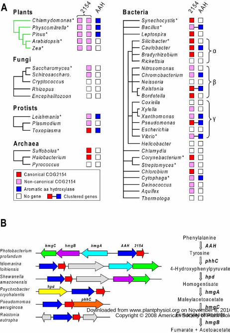
Phylogenomic Analysis of COG2154 in Plants and Microbes
We first systematically analyzed the distribution of COG2154 sequences in relation to that of
AAH sequences in plants, fungi, protists, and prokaryotes. COG2154 sequences were identified
using the NCBI Conserved Domain (CD) search tool (Marchler-Bauer et al., 2005). For prokary-
otes, the enzymes of the Tyr-catabolizing homogentisate pathway were included in the analysis
because of the known catabolic role of P. aeruginosa Phe hydroxylase and PCD (Gu et al.,
1998). The results are summarized in Figure 2 and given in detail in Supplemental Table S1. Five
points emerged from the data.
First, COG2154 sequences of two types were found in all plant taxa examined (chlorophyte alg-
ae, mosses, gymnosperms, and angiosperms). Second, a single COG2154 sequence was found in
all protists surveyed and in many but not all fungi, bacteria, and archaea. Third, COG2154 se-
quences are not accompanied by an AAH sequence in many organisms, including angiosperms,
fungi, and a diverse array of prokaryotes. Fourth, in bacteria with both COG2154 and AAH,
www.plantphysiol.org on November 8, 2016 - Published by www.plantphysiol.orgDownloaded from Copyright © 2008 American Society of Plant Biologists. All rights reserved.
7
these genes are often adjacent and sometimes clustered with homogentisate pathway genes. Fifth,
certain COG2154 sequences lack one or more of the canonical His residues or other putatively
crucial residues or show atypical spacing of the latter. These points are explored below in more
detail. For simplicity and clarity, hereafter we designate test organisms by their genus name only.
Plant COG2154 Sequences
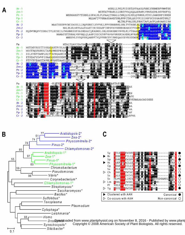
Both types of COG1254 sequences in plants have N-terminal extensions of ~60-100 amino acids
compared to Pseudomonas PCD but are otherwise clearly distinct from each other (Fig. 3A). The
first type (type 1) has an almost canonical catalytic motif, departing only in having an extra
residue between the H-H-P and [YW] (i.e., Y or W) elements. This type is typically predicted by
three algorithms (TargetP, Predotar, PSORT) to be targeted to mitochondria. The second type of
sequence (type 2) departs radically from the canon: most notably, the [YW] element is invariably
absent and, in all cases except Pinus, one or more of the His residues are missing. Moreover, a
key feature of type 2 sequences not shared with type 1 or with any other COG2154 sequences is
a conserved ~45-residue domain just upstream of the core COG2154 domain (Fig. 3A). Type 2
sequences are most often predicted to be plastidial.
Phylogenetic analysis of representative COG2154 sequences from plants and other organisms
placed plant type 1 and type 2 proteins in two clades that branched together, with high bootstrap
values, implying a shared evolutionary history (Fig. 3B). An exception was the Chlamydomonas
type 1 sequence, which did not cluster with the rest. Due to the diverged nature of COG2154
sequences, other branching patterns were poorly resolved, reflected by low bootstrap values. The
close relationship between plant type 1 and 2 sequences was corroborated by a comparison of
intron positions: type 1 and 2 genes were found to share two introns not found in other eukary-
otes (Fig. 3A). Chlamydomonas type 1 COG2154 was again unusual since it lacked both of these
introns. Taken together, the above data basically suggest that an archetypal type 2 sequence arose
early within the plant lineage from a type 1 sequence and acquired a novel domain.
Although all plant groups have COG2154 sequences, not all have AAH sequences. As indicated
in Figure 2A, Chlamydomonas, Physcomitrella, and Pinus have proteins very like animal and
bacterial AAHs (35-45% amino acid identity) but Arabidopsis and Zea lack proteins with any
detectable similarity to AAHs. Data from other plant genomes and ESTs (not shown) confirmed
the generality of this pattern: non-flowering plants have AAH sequences, angiosperms do not.
www.plantphysiol.org on November 8, 2016 - Published by www.plantphysiol.orgDownloaded from Copyright © 2008 American Society of Plant Biologists. All rights reserved.
8
Microorganism COG2154 Sequences
COG2154 sequences were found in all protists surveyed, in ascomycete fungi, and in diverse
groups of bacteria and archaea (Fig. 2A), generally as a single copy. Of 467 complete bacterial
and archaeal genomes analyzed, 211 genomes (45%) encoded COG2154 proteins and represent-
ed almost all the major taxa. Many of the bacteria lacking COG2154 proteins are obligate intra-
cellular organisms, such as Rickettsia, Coxiella, and Chlamydia (Fig. 2A) that have ceded
various metabolic functions to the host. Of the 211 genomes with COG2154, fewer than half (90)
have an AAH sequence, so that the ‘partnerless COG2154’ situation noted above for angiosp-
erms is very common among prokaryotes.
The 90 COG2154 genes that co-occur with AAH can reasonably be presumed a priori to encode
active PCDs. Moreover, as in Pseudomonas (Zhao et al., 1994), these COG2154 genes are often
adjacent to an AAH gene in an operonic structure, providing additional circumstantial evidence
for their functionality (Fig. 2B). Yet further support for this inference is the fairly frequent clust-
ering of COG2154 and AAH genes with one or more genes of the homogentisate pathway (Fig.
2B and Supplemental Table S1).
Given the strong probability of PCD activity that is implied by association evidence (i.e., co-
occurrence and clustering), it is noteworthy that microbial COG2154 proteins that are associated
in these ways with AAH quite often lack a canonical catalytic motif (Fig. 3C). This implies that
the current motif (based only on PCDs from animals and one bacterium) is too narrowly defined.
If so, it would follow that the various non-canonical microbial and plant COG2154 proteins that
have no AAH partner could also be active PCD enzymes.
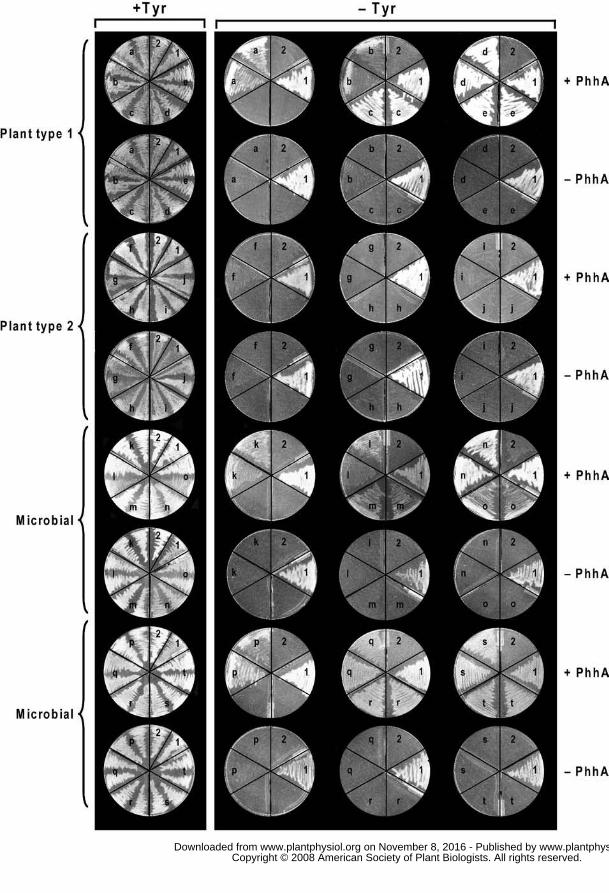
Tests of PCD Activity
The above issues led us to test a range of plant and prokaryote COG2154 proteins for PCD activ-
ity and, in the process, to re-examine the catalytic motif. We used a functional complementation
assay in Escherichia coli (Song et al., 1999; Wang et al., 2006) rather than in-vitro assays since
the latter involve artificial pterin-4a-carbinolamine substrates (which may not necessarily be att-
acked by all PCDs) or lack sensitivity due to a background rate of chemical dehydration (Citron
et al., 1992; Rebrin et al., 1995).
www.plantphysiol.org on November 8, 2016 - Published by www.plantphysiol.orgDownloaded from Copyright © 2008 American Society of Plant Biologists. All rights reserved.

9
The complementation assay uses an E. coli Tyr auxotroph. Since E. coli lacks both Phe hydrox-
ylase and PCD, but has q-dihydropterin reductase activity (Fig. 1A), Tyr prototrophy can be rest-
ored by co-expression of foreign Phe hydroxylase and PCD genes; neither gene alone suffices
(Zhao et al., 1994; Song et al., 1999). The Pseudomonas Phe hydroxylase gene (phhA) and sel-
ected COG2154 sequences were accordingly cloned into compatible plasmids and introduced
into the mutant. Transformants were then tested for growth in the presence or absence of Tyr
(Fig. 4). The COG2154 proteins chosen included plant types 1 and 2 from five major taxa (green
algae, mosses, gymnosperms, monocots and eudicots; Fig. 3A) and 10 microbial proteins that
were either non-canonical or from genomes lacking AAH, or both (Fig. 3C), these being potent-
ially the most informative. The microbial proteins were also selected for taxonomic diversity;
they represented fungi, protists, archaea, and five phyla of bacteria. The plant proteins were
truncated at the positions shown in Figure 3A to remove their N-terminal extensions.
Among the plant proteins, all type 1 sequences (whose catalytic motif is close to canonical) had
PCD activity whereas all the type 2 sequences lacked activity (Fig. 4, top four rows). All the
microbial proteins proved to be active (Fig. 4, bottom four rows). While we cannot exclude the
possibility that the truncation point used for the plant type 2 proteins caused loss of activity, we
consider it unlikely because this point lay well upstream of the core COG2154 domain (Fig. 3A).
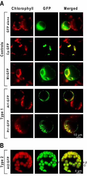
Subcellular Localization of Plant Proteins
To test whether the predicted organellar targeting sequences of type 1 and 2 proteins are funct-
ional in planta, selected COG2154 coding sequences were fused to green fluorescent protein
(GFP) and expressed in Arabidopsis. Subcellular localization in protoplasts was visualized by
epifluorescence or confocal laser scanning microscopy. The Arabidopsis and Pinus type 1 prot-
eins gave many small, punctate GFP signals that did not coincide with chloroplasts, indicating a
mitochondrial location (Fig. 5A). In contrast, the Arabidopsis type 2 protein typically gave a
strong GFP signal that precisely overlapped with chlorophyll fluorescence (Fig. 5B). Our results
for the Arabidopsis type 2 protein agree with a brief report, based on a yellow fluorescent protein
fusion, that this protein is chloroplastic (Valkai, 2004).
Essentiality Data and Functional Predictions
The puzzling occurrence of COG2154 proteins with PCD activity in genomes with no AAH led
us first to mine the microbial literature for data on COG2154 knockout mutants. Five reports
www.plantphysiol.org on November 8, 2016 - Published by www.plantphysiol.orgDownloaded from Copyright © 2008 American Society of Plant Biologists. All rights reserved.
10
were found, involving four organisms, of which three lack an AAH (Table I). In no case was the
COG2154 gene classified as essential, although in the fission yeast Schizosaccharomyces, which
has no AAH, deletants showed defective spore wall formation (Kakihara et al., 2003). In Pseudo-
monas, which has AAH, inactivating PCD abolished the ability to grow on Phe, as would be
expected (Song et al., 1999). Relative to these findings of non-essentiality, it should be noted that
PCD mutations in mammals give mild phenotypes. This may be partly due to non-enzymatic de-
hydration of the pterin 4a-carbinolamine (Thöny et al., 2000). The non-essentiality of PCD clear-
ly constrains our ability to hypothesize – or test – its functions in organisms that lack AAH.
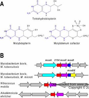
However, given that a pterin 4a-carbinolamine can be formed by chemical oxidation of a tetrahy-
dropterin (Moore et al., 2002), one hypothesis is that PCD facilitates – but is not essential for –
regeneration of chemically oxidized pterins. Besides AAH cofactors, another common type of
pterin is the molybdenum cofactor (Moco) and its precursors (e.g., molybdopterin). Moco occurs
in all life’s kingdoms, and is essential for about 40 enzymes of which four occur in plants (Schw-
arz and Mendel, 2006). It is very sensitive to oxidation (Rajagopalan and Johnson, 1992). Moco
has a ring structure that is electronically equivalent to a tetrahydropterin (Fig. 6A) and has some
chemical characteristics of tetrahydropterins as well as others due to the third ring (Enemark and
Garner, 1997; Nieter Burgmayer et al., 2004). Both substrate specificity studies (Rebrin et al.,
1995) and the crystal structure of PCD complexed with a substrate analog (Cronk et al., 1996) in-
dicate that PCD recognizes the pterin ring rather than its side chains. Thus, if Moco or its precur-
sors form 4a-carbinolamines, they could be PCD substrates. Furthermore, some genomic evid-
ence suggests a role for COG2154 proteins in Moco metabolism. Analysis of the genomic con-
text of COG2154 genes in bacteria with no AAH reveals one embedded in molybdopterin synth-
esis gene clusters in Mycobacterium species, and translationally coupled to moaC (Fig. 6B). A
COG2154 gene is also next to moaC in some γ-proteobacteria (Fig. 6B). Indeed, COG2154 sequ-
ences in GenBank are occasionally annotated as ‘putative molybdopterin biosynthesis protein’.
A Link Between Arabidopsis PCD and Moco Metabolism
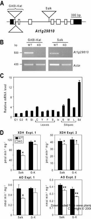
To explore a connection between PCD and Moco, two independent insertional mutants in the
Arabidopsis gene (At1g29810) encoding the type 1 protein were isolated and authenticated; the
mutants came from the Salk and GABI-Kat collections (Fig. 7, A and B). Both mutations were
knockouts, as judged by absence of detectable At1g29810 mRNA (Fig. 7B). The expression of
www.plantphysiol.org on November 8, 2016 - Published by www.plantphysiol.orgDownloaded from Copyright © 2008 American Society of Plant Biologists. All rights reserved.
11
the At1g29810 gene in wild-type plants was examined and found to be constitutive, with levels
(relative to actin) highest in seeds (Fig. 7C). Mutant homozygotes grew normally compared to
wild type in soil, or when cultured in vitro (not shown).
Plantlets grown in vitro were tested for the activities of the four molybdoenzymes, nitrate reduct-
ase, sulfite oxidase, xanthine dehydrogenase, and aldehyde oxidase. As these enzymes lose activ-
ity in plants with Moco synthesis defects (Schwarz and Mendel, 2006), we reasoned that there
should also be some activity loss if PCD facilitates salvage of 4a-carbinolamines formed by oxi-
dation of Moco or its precursors. Given the normal phenotype of the mutant plants, the activity
loss was predicted to be modest, mandating high precision (low variability) in measuring enzyme
activities. Pilot experiments showed that nitrate reductase and sulfite oxidase activities were low
and too variable (coefficient of variation among replicates up to ~40%) to allow detection of
subtle differences. We accordingly focused on xanthine dehydrogenase and aldehyde oxidase,
and developed sensitive fluorometric HPLC assays for both enzymes (see ‘Materials and Meth-
ods’ and Supplemental Figure S1). In two separate experiments, xanthine dehydrogenase activity
was significantly (P < 0.05) lower in both knockout lines than in the corresponding wild-type
controls, the activity loss being between 10 and 28% (Fig. 7D). In the same experiments, alde-
hyde oxidase activity was also consistently lower in the knockout lines (by 17 to 36%), the diff-
erences being statistically significant (P < 0.05 or < 0.01) in all but one case (Fig. 7D).
DISCUSSION
The results of in-vivo tests of COG2154 sequences from plants and diverse microorganisms (Fig.
4 and Wang et al., 2006) allow definition of a new catalytic motif that can be used as a signature
for PCD activity in any organism: [EDKH]-x(3)-H-[HN]-[PCS]-x(5,6)-[YWF]-x(9)-[HW]-x-
(8,15)-D. This motif, which is far broader than the one based solely on mutational and structural
studies of mammalian and Pseudomonas PCDs, can be used to more reliably annotate COG2154
genes from plants and other organisms. By assuming that all microbial COG2154 genes co-occ-
urring with AAH genes (Supplemental Table S1) encode active PCDs, the motif can be broaden-
ed slightly further to [EDKHQN]-x(3)-H-[HN]-[PCS]-x(5,6)-[YWFH]-x(9)-[HW]-x(8,15)-D.
Our study demonstrates that COG2154 genes specifying active PCDs occur in the absence of
AAH genes in many microorganisms and in angiosperms. While this surprising situation in ang-
www.plantphysiol.org on November 8, 2016 - Published by www.plantphysiol.orgDownloaded from Copyright © 2008 American Society of Plant Biologists. All rights reserved.
12
iosperms could be due to an evolutionarily recent loss of AAH that has not yet been followed by
loss of the redundant PCD, this explanation seems improbable in light of the prevalence of PCDs
in microorganisms with no AAH. It is more plausible to suppose that PCD has at least one other
function besides recycling the pterin cofactors of AAHs, and that this function is common to
plants, fungi, bacteria, and archaea. What could this function be?
We obtained evidence to support the intriguing scenario, suggested by phylogenomics, that PCD
has a role in the metabolism of Moco or its precursors, at least in Arabidopsis. Such a role would
necessarily be ancillary, not central to Moco formation, since (a) no genes are ‘missing’ from the
Moco biosynthesis pathway (Schwarz and Mendel, 2006), (b) various Moco-producing bacteria
(e.g., E. coli) lack PCD, (c) PCD mutants are viable in Arabidopsis and microorganisms, and (d)
ablating PCD activity reduced molybdoenzyme activity only modestly. One possibility is that
PCD helps salvage 4a-carbinolamines formed by oxidation of Moco biosynthesis intermediates
or Moco itself. As the first steps in Moco synthesis in plants may be mitochondrial (Schwarz and
Mendel, 2006), PCD – a mitochondrial enzyme – would have ready access to a 4a-carbinolamine
formed in the early part of the synthesis pathway. However, later biosynthesis steps – and all
four Moco-containing enzymes – are extramitochondrial in plants, so that a role for PCD at these
levels would require trafficking of 4a-carbinolamines and pterins in and out of mitochondria.
Although carbinolamine oxidation products of Moco precursors have not so far been detected
(Santamaria-Araujo et al., 2004), their formation is not chemically unreasonable. In this connect-
ion, it is worth noting that even for the best known pterin, tetrahydrobiopterin, whose chemical
oxidation has received considerable attention for 30 years, definitive evidence for carbinolamine
formation during H2O2-mediated oxidation emerged only quite recently (Moore et al., 2002).
Lastly, it should be noted that, while reduced cofactor activity is the simplest explanation for the
parallel activity reductions in two Moco enzymes in Arabidopsis PCD knockouts, our data do not
exclude the possibility that this is due to correlated reductions in apoprotein levels.
There are several other possible functions for PCDs. First, they might facilitate recycling of
chemically oxidized tetrahydropterins. However, this still leaves the question of why such hypo-
thetical tetrahydropterins would need to be recycled if there is no AAH. A second possibility –
that PCDs recycle chemically oxidized tetrahydrofolates (which have a tetrahydropterin ring) –
might seem attractive because one common folate, 5-methyltetrahydrofolate, is known to form a
www.plantphysiol.org on November 8, 2016 - Published by www.plantphysiol.orgDownloaded from Copyright © 2008 American Society of Plant Biologists. All rights reserved.
13
4a-hydroxy derivative upon oxidation (Gregory, 2007). However, this compound is not a likely
substrate for PCD because this enzyme’s action is considered to require a proton at the N5 posit-
ion (Fig. 1A) (Rebrin et al., 1998) and a methyl group replaces this proton in 5-methyltetrahydro-
folate. A closely related possibility – albeit only in some prokaryotes – is that PCD recycles ox-
idized tetrahydromethanopterin (a tetrahydrofolate analog confined to methanogens and methyl-
otrophs). Favoring this, PCD genes in the genomes of aerobic methylotrophic bacteria cluster
with genes of tetrahydromethanopterin synthesis and metabolism (Kalyuzhnaya et al., 2005).
A third possibility is that genomes with a PCD but no AAH have other pterin-dependent en-
zymes that generate 4a-carbinolamines. We view this as probable, since mammals are known to
have a pterin-dependent glyceryl ether monooxygenase (Taguchi and Armarego, 1998) that has
not been cloned and therefore cannot be recognized in genomes. Moreover, other pterin-depend-
ent monooxygenases have been reported from microorganisms, but again not cloned (Reddy and
Vaidyanathan, 1975; Bhat and Vaidyanathan, 1976). Another formal possibility is that PCD prot-
eins participate in transcriptional regulation, as in mammals. However, there is experimental evi-
dence against this in Pseudomonas (Song et al., 1999) and such a role is hard to reconcile with
the distribution pattern of COG2154 genes in bacteria, where some species of a genus may have
COG2154 and others not (e.g., Bacillus and Corynebacterium; Supplemental Table S1). Further-
more, a transcriptional role like that in mammals would not involve the PCD catalytic site (Rhee
et al., 1997), and so is inconsistent with the conservation of catalytic residues and of enzyme act-
ivity in COG2154 proteins that have no AAH partner. Lastly, neither plant type 1 nor type 2
proteins were apparently targeted to the nucleus, which a transcriptional role would require.
Our results show that plants, unlike other organisms surveyed, have two distinct types of COG-
2154 protein. Type 1 is canonical (according to the catalytic motif just defined), has PCD activ-
ity, and is located in mitochondria. Type 2 is unique to plants, lacks PCD activity, is localized in
plastids, and apparently arose from type 1 early in plant evolution. Apart from lacking the cataly-
tic motif, type 2 proteins have a characteristic subterminal domain that includes the motif [GE]-
[DN]-[FL]-G-A-R-D-P-x(3)-E-x(4)-F-G-[DE]K (Fig. 3A), which can be used for positive ident-
ification. That the type 2 protein is not a functional PCD, yet apparently evolved from the type 1
enzyme, implies that it has taken on a different role. The presence in type 2 proteins of the sub-
terminal domain fits with this idea. In an exploratory Arabidopsis study, overexpressing the type
www.plantphysiol.org on November 8, 2016 - Published by www.plantphysiol.orgDownloaded from Copyright © 2008 American Society of Plant Biologists. All rights reserved.
14
2 gene (At5g51110) had no apparent phenotypic effect, and its suppression by RNAi resulted in a
small (~10%) but significant reduction in leaf pigment content and a larger reduction (~30%) in
chloroplast number per mesophyll cell (Plume, 2002). The function of type 2 proteins is thus
unclear. We propose that they be annotated as ‘PCD-like protein’ until more is known.
For the plant mitochondrial PCD to participate in pterin recycling would require the existence of
a q-dihydropterin reductase (Fig. 1A), which – given the lability of dihydropterins – seems likely
also to be mitochondrial. q-Dihydropterin reductases in mammals, protists, and certain bacteria
belong to the large and diverse short chain dehydrogenase-reductase (SDR) family (Lye et al.,
2002; Wilquet et al., 2004). Although Arabidopsis has some 86 SDR proteins, only eight have
predicted mitochondrial targeting peptides, and none of the eight yet has a known function (Noir-
iel et al., 2007). One or more of these could thus well have q-dihydropterin reductase activity.
MATERIALS AND METHODS
Bioinformatics
Microbial genomes were analyzed using the SEED database and its tools (Overbeek et al., 2005)
at http://anno-3.nmpdr.org/anno/FIG/subsys.cgi. Results are available in full in the ‘Pterin carbin-
olamine dehydratase sub-system’ on the public SEED server at http://theseed.uchicago.edu/FIG/
and in summary form in Supplemental Table S1. Plant genomes and/or ESTs were sought using
BLAST algorithms and the NCBI (http://www.ncbi.nlm.nih.gov/), DOE Joint Genome Institute
(http://www.jgi.doe.gov/) and Chlamydomonas (http://www.chlamy.org/chlamydb.html) databas-
es. PCD-like sequences were identified using the Conserved Domain (CD) database and search
tool (http://www.ncbi.nlm.nih.gov/Structure/cdd/cdd.shtml), which includes COG and pfam
classifications as well as CD classifications. As all three classifications are equivalent in the case
of PCD-like proteins, we used the COG identifier (COG2154) in preference to the CD (cd00488)
or pfam (pfam01329) identifiers because the COG system is widely used in genome annotation.
Other resources were the integrated microbial genomes system (http://img.jgi.doe.gov/cgi-
bin/pub/main.cgi) and the COG database (http://www.ncbi.nlm.nih.gov/COG/). Sequence align-
ments (Fig. 3, A and C) were made using MultAlin (http://bioinfo.genopole-toulouse.prd.fr/mult-
alin/multalin.html). Phylogenetic analyses were made with MEGA 4 (Tamura et al., 2007).
Microbial COG2154 Genes
www.plantphysiol.org on November 8, 2016 - Published by www.plantphysiol.orgDownloaded from Copyright © 2008 American Society of Plant Biologists. All rights reserved.
15
Genomic DNA of Bacillus cereus strain NRS 248, Corynebacterium glutamicum strain 534,
Silicibacter pomeroyi strain DSS-3, Streptomyces avermitilis strain MA-4680, Synechocystis sp.
strain PCC 6803, and Cytophaga hutchinsonii strain NCIB 9469 was purchased from the Ameri-
can Type Culture Collection (Manassas, VA, [email protected]). For Sulfolobus solfataricus strain
DSM 1617 and Saccharomyces cerevisiae strain 971/6c (a gift from M.L. Agostini Carbone,
Università di Milano), DNA was extracted from cells; a S. cerevisiae colony was suspended in
0.5 mL water and microwaved for 3 min, and lyophilized S. solfataricus cells were suspended in
0.5 mL of water and boiled for 10 min. Extracts then were centrifuged for 10 min at 10,000g.
The COG2154 gene of Vibrio cholerae (GenBank NC_002506) was synthesized by GenScript
(Piscataway, NJ, www.genscript.com), adding 5´ NdeI and 3´ KpnI sites. The COG2154 coding
sequences of Synechocystis and S. cerevisiae were PCR-amplified using KOD HiFi polymerase
(Novagen, http://www.emdbiosciences.com) and primers harboring 5´ BamHI and 3´ KpnI sites.
Primer sequences for COG2154 constructs are given in Supplemental Table S2. The forward
primers contained a BamHI site, a stop codon in frame with LacZ, a Shine-Dalgarno sequence,
and an NdeI site that included the start codon. The amplicons were cloned between the BamHI
and KpnI sites of pBluescript SK-, and introduced into E. coli strain DH5α. The sequence-
verified construct containing the Synechocystis gene was the parent of all other COG2154
constructs except that from Leishmania major. COG2154 coding sequences were PCR-amplified
from genomic DNA using Taq (Invitrogen, www.invitrogen.com) or KOD HiFi polymerase;
DMSO 6% (v/v) was added to the reaction mixture in the case of C. glutamicum. The control
PCD gene phhB from Pseudomonas aeruginosa was amplified from plasmid pJZ9-4 (Song et al.,
1999). Amplicons were digested with NdeI and KpnI and then ligated into the Synechocystis con-
struct from which the COG2154 sequence had been excised with NdeI and KpnI. The L. major
strain FV1 COG2154 gene was amplified from genomic DNA using Phusion DNA polymerase
(New England BioLabs, www.neb.com), a forward primer containing an XbaI site, a stop codon
in frame with LacZ, a Shine-Dalgarno sequence, and a reverse primer containing an EcoRI site.
The amplicon was cloned into pGEM-T Easy (Promega, www.promega.com). The insert was
then excised with XbaI and EcoRI and cloned into the matching sites in pBluescript SK-. pBlue-
script SK- constructs were introduced into E. coli strain DH5α or DH10B and sequence-verified.
Plant COG2154 cDNAs
www.plantphysiol.org on November 8, 2016 - Published by www.plantphysiol.orgDownloaded from Copyright © 2008 American Society of Plant Biologists. All rights reserved.
16
For complementation tests, plant COG2154 type 1 and 2 cDNAs were truncated, using PCR to
replace their N-terminal putative targeting sequences by a start codon. Primers (Supplemental
Table S2) harbored NdeI and KpnI sites as above. Type 1 and 2 cDNAs were amplified from the
following ESTs: GenBank numbers BAC41856 and U13619 for Arabidopsis thaliana; BU647-
613 and BI999118 for Chlamydomonas reinhardtii; BJ203205 and BQ039368 for Physcomitr-
ella patens; DT635299 and CO168958 for Pinus taeda; DN218430 and EC883040 for Zea mays.
Functional Complementation Tests
Plasmid pJS11, consisting of the P. aeruginosa phhA cloned into pACYC177 (Song et al., 1999),
provided the Phe hydroxylase gene for complementation tests. The matching vector-only control
plasmid (pACYC∆Ap) was constructed by digesting pACYC177 with ScaI and FspI to delete an
internal fragment of the ampicillin-resistance gene (Ap), and religating. Electroporation was used
to simultaneously transform E. coli strain JP2255 (aroF363 pheA361 pheO352 tyrA382 thi-1
strR712 lacY1 xyl-15) (Zhao et al., 1994) with each COG2154 construct or pBluescript SK- and
pJS11 or pACYC∆Ap. Complementation tests were made by streaking transformed cells on
plates of minimal medium containing M9 salts (Sambrook et al., 1989), 0.1 mM CaCl2, 0.5 mM
MgSO4, trace elements (Sherman, 1991), 0.4% (w/v) glucose, thiamine (17 µg mL-1), IPTG (0.5
or 1 mM), ampicillin (100 µg mL-1), streptomycin (100 µg mL-1), kanamycin (100 µg mL-1), Phe
(50 µg mL-1), plus or minus tyrosine (54 µg mL-1). The agar concentration in plates was 15 g L-1.
Plates were incubated at room temperature (22°C) for 2-7 days.
Construction and Expression in Arabidopsis of GFP Fusion Proteins
Full length cDNAs of two plant type 1 COG2154 sequences (At1g29810 and its Pinus taeda
ortholog) were amplified using primers At1g29810-GFP-Fwd and At1g29810-GFP-Rev and
PtPCD-GFP-Fwd and PtPCD-GFP-Rev, respectively (Supplemental Table S3). The PCR prod-
ucts were digested with SalI and NcoI and cloned in-frame upstream of the GFP sequence in the
pTH2 plasmid (Niwa, 2003). Transient expression in protoplasts, controls for GFP targeting, and
epifluorescence microscopy were as previously described (Pinon et al., 2005).
The coding region of type 2 COG2154 sequence At5g51110 was cloned into pAVA393 in frame
with the GFP coding region under the control of the CaMV 35S promoter. The pAVA393 plas-
mid is based on pAVA319 (von Arnim et al., 1998) but contains the GFP5 variant (Siemering et
al., 1996). The CaMV 35S promoter-At5g51110-gfp5-35S terminator cassette from this vector
www.plantphysiol.org on November 8, 2016 - Published by www.plantphysiol.orgDownloaded from Copyright © 2008 American Society of Plant Biologists. All rights reserved.
17
was subcloned into the binary vector pSLJ755I5, a derivative of pRK290 (Jones et al., 1992).
Arabidopsis (ecotype Columbia) plants were transformed by the floral dip method (Clough and
Bent, 1998). Herbicide selection was performed by spraying seedlings growing in soil with 0.4%
glufosinate (Basta®). Protoplasts for confocal laser scanning microscopy were isolated from
leaves of single-copy, homozygous T3 plants by incubation at 30°C for 1 h in a solution
containing 400 mM D-mannitol, 8 mM CaCl2·2H2O, 5 mM MES-KOH (pH 5.6), 2% (w/v)
SERVA Onozuka cellulase R-10 and 0.5% (w/v) SERVA macerozyme R-10. Protoplasts were
analyzed using a Bio-Rad Radiance 2000 microscope with blue excitation (DAPI filter set) and
green (FITC filter set; GFP) or red (TRITC filter set; chlorophyll autofluorescence) emission.
Isolation of Arabidopsis At1g29810 mutants
Two T-DNA insertional mutant lines (ecotype Columbia) were identified: line 648F10 was from
the GABI-Kat collection, and line SALK_121474 from the Salk collection. Wild type or
homozygote mutant segregants from each line were identified by PCR screening using
At1g29810 gene-specific primers located 5´ and 3´ of the insertion site, and primers located on
the T-DNA (Supplemental Table S3). The insertion sites were confirmed by sequencing the amp-
licons obtained from mutant homozygotes. The kanamycin resistance gene was shown by PCR
with primers nptB and nptD (Supplemental Table S3) to co-segregate with the insertion, indic-
ating the absence of insertions at other loci. Line 648F10 was shown to have duplicate T-DNA
insertions at the same locus, in opposite orientation (Fig. 7A). Homozygous mutants and wild-
type segregants were selfed, and the progeny used for experiments. For RT-PCR experiments to
demonstrate absence of a functional At1g29810 mRNA in the mutant lines, RNA was prepared
from plantlets 4 weeks old and treated as described below. Primers used to analyze At1g29810
expression were located in the first and last exons. These primers and those for the Actin7 gene
(At5g09810) are given in Supplemental Table S3. For PCR, 30 ng of cDNA were used per react-
ion. After 5 min at 95°C, the reaction was carried out with 35 cycles of 95°C for 30 sec, 45°C for
30 sec and 72°C for 50 sec, and a final step at 72°C for 10 min.
RNA Isolation and Analysis
Tissues were ground in liquid N2. Total RNA from the following tissues was isolated using
RNeasy plant mini kits (Qiagen, www.qiagen.com): seedlings 3 or 7 days old, roots from hydro-
ponic culture, rosette leaves from plants at 16, 30, or 42 days of age, stems and cauline leaves
www.plantphysiol.org on November 8, 2016 - Published by www.plantphysiol.orgDownloaded from Copyright © 2008 American Society of Plant Biologists. All rights reserved.
18
from plants at 42 days of age, inflorescences, and siliques in early development. Total RNA from
siliques at mid or late (yellowing) stages of development, and from dry seeds was extracted using
a LiCl precipitation method (Vicient and Delseny, 1999). The samples were dissolved in ribo-
nuclease-free water and DNase-treated. For real-time PCR, 1 µg of RNA was reverse-transcribed
using a RevertAid™ first-strand cDNA synthesis kit (Fermentas, http://www.fermentas.com/)
with random hexamers. The mixture was then diluted fivefold with water. PCR primers for
At1g29810 and for the reference gene Actin7 (At5g09810) are given in Supplemental Table S3;
amplicon lengths were 69 and 71 bp, respectively. The At1g29810 amplicon spanned the third
and fourth introns. PCR was performed using the MyiQ™ version 2.0 Real-Time PCR Detection
System (Bio-Rad, www.bio-rad.com) using the SYBR® Green PCR master mix (Applied Bio-
systems, www.appliedbiosystems.com) in 25-µL mixtures containing 5 µL of the diluted cDNA
solution and 300 nM of each primer. After 10 min at 95°C, the PCR reaction was carried out with
40 cycles of 95°C for 15 sec and 60°C for 60 sec. Data were analyzed using MyiQ™ software.
Expression values were normalized to that of Actin7, calculating the difference between the
crossing threshold (CT) of At1g29810 and that of Actin7 for each sample (Livak and Schmittgen,
2001). The results presented are for cDNA pools from three independent RNA extractions, each
analyzed in triplicate. The identity of the PCR products was confirmed by sequencing.
Culture of Arabidopsis Plantlets for Molybdoenzyme Assays
Arabidopsis seeds were surface sterilized and germinated on plates containing agar and 0.33×
Murashige and Skoog (MS) medium. At 12 days, seedlings were transferred aseptically to 250-
mL flasks (seven per flask) containing 100 mL of 0.33× MS medium containing 10 g L-1 of Suc
and cultured for 17 days. Flasks were shaken at 80 rpm. Temperature was 23-28°C. Daylength
was 12 h; photosynthetic photon flux density was 80 µE m-2 s-1. Harvested plantlets from each
flask (constituting one replicate sample) were frozen in liquid N2 and stored at -80°C.
Extraction and Assay of Molybdoenzymes
Protein extracts for assay of xanthine dehydrogenase, aldehyde oxidase, and sulfite oxidase were
prepared as follows. Samples were ground in liquid N2 and the resulting powder was extracted in
ice-cold buffer (1:3, w/v) containing 100 mM K-phosphate, pH 7.5, 0.1 mM Na2MoO4, 1 mM
EDTA, 1 mM phenylmethylsulfonyl fluoride, 1 mM DTT, and 10% (w/v) polyvinylpolypyrrolid-
one. Extracts were centrifuged (30,000g, 20 min, 4°C), and supernatants were fractionated with
www.plantphysiol.org on November 8, 2016 - Published by www.plantphysiol.orgDownloaded from Copyright © 2008 American Society of Plant Biologists. All rights reserved.
19
ammonium sulfate (0-60% saturation), stirring for 30 min at 4°C. After centrifuging (40,000g, 25
min, 4°C), pellets were resuspended in 1-2 mL of 100 mM K-phosphate, pH 7.5, and 0.2-mL
aliquots were desalted on 1-mL Sephadex G25 spin columns equilibrated with 100 mM K-phos-
phate, pH 7.5 or, for sulfite oxidase assays, 20 mM Tris-acetate, pH 8.0, 0.1 mM EDTA.
Xanthine dehydrogenase was assayed with pterin as substrate (Mest et al., 1992; Montalbini,
1998). Pterin (Sigma, www.sigmaaldrich.com) and isoxanthopterin (Schircks, Jona, Switzerland,
www.schircks.com) were dissolved in 0.1 M NaOH and the solutions were titered spectrophoto-
metrically using extinction coefficients of 21,380 M-1cm-1 at 251 nm and 13,800 M-1cm-1 at 339
nm, respectively (Pfleiderer, 1985). Assays (50 µL) contained 20 µL of protein extract, 0.1 mM
pterin, and 1 mM 2,6-dichlorophenolindophenol in 100 mM K-phosphate, pH 7.5. Triplicate ass-
ays were run at 30°C for 0, 30, and 60 min and stopped by adding 5 µL of 1 N HCl. After centri-
fuging, a portion of the supernatant was injected onto a 4 µm, 250 × 4.6 mm Synergi Fusion RP
80 column (Phenomenex, http://www.phenomenex.com), which was eluted isocratically (1.5 mL
min-1) for 20 min using 20 mM glycine, pH 2.5, or 10 mM Na-phosphate, pH 6.0. The column
was washed for 5 min with a 0 to 50% acetonitrile gradient, then re-equilibrated in separation
buffer for 5 min. Peaks were quantified by fluorescence (350 nm excitation, 450 nm emission).
Aldehyde oxidase was assayed with indole-3-carboxaldehyde as substrate (Seo et al., 1998). Ind-
ole-3-carboxaldehyde and indole-3-carboxylic acid (Aldrich, www.sigmaaldrich.com) were dis-
solved in water. Assays (50 µL) contained 20 µL of protein extract and 0.17 mM indole-3-carb-
oxaldehyde in 100 mM K-phosphate, pH 7.5. Triplicate assays were run at 30°C for 0, 30 and 60
min and stopped, processed and analyzed by HPLC as above, using 10 mM Na-phosphate, pH
6.0, as eluant. The indole-3-carboxylic acid product was quantified by fluorescence (280 nm
excitation, 360 nm emission). Xanthine dehydrogenase and aldehyde oxidase activities were
measured in two separate experiments. Experiment 1 had three independent knockout and wild
type replicates. Experiment 2 had four to five such replicates, and knockout and wild type samp-
les were extracted and analyzed in pairs. Statistically significant differences were evaluated us-
ing one-tailed Student’s t-tests for independent (experiment 1) or paired (experiment 2) samples.
Sulfite oxidase assays (100 µL) contained 20-50 µL of desalted extract and 0.4 mM K3Fe(CN)6,
in 20 mM Tris-acetate, pH 8.0, containing 0.1 mM EDTA. Sodium sulfite (2 µL, 20 mM) was
added and the reaction was monitored spectrophotometrically by following reduction of ferri-
www.plantphysiol.org on November 8, 2016 - Published by www.plantphysiol.orgDownloaded from Copyright © 2008 American Society of Plant Biologists. All rights reserved.
20
cyanide (extinction coefficient 1,020 M-1cm-1) at 420 nm, correcting for the base-line rate in
absence of sulfite (Garrett and Rajagopalan, 1994; Eilers et al., 2001).
Nitrate reductase activity was extracted by grinding tissue (0.1-0.5 g) in a mortar with ice-cold
100 mM Hepes-KOH, pH 7.5, 2 mM EDTA, 7 mM cysteine (1:5, w/v). After centrifugation at
14,000g for 10 min at 4°C, 60-µL samples of the supernatant were immediately added to assay
mixtures (700 µL final volume) containing 100 mM Hepes-KOH, pH 7.5, 2 mM EDTA, 0.2 mM
NADH, and 5 mM KNO3 (Jonassen et al, 2007). Assays were run in triplicate at 22°C for up to
40 min and stopped by adding 700 µL of 1% sulfanilamide and 0.02% N-(naphthyl)-ethylene-di-
amine dihydrochloride in 1.5 M HCl. The color was allowed to develop for 30 min before
absorbance was measured at 540 nm.
Supplemental Data
The following materials are available in the online version of this article.
Supplemental Figure S1. Xanthine dehydrogenase and aldehyde oxidase assays by HPLC.
Supplemental Table S1. Distribution and clustering of microbial genes encoding COG2154.
Supplemental Table S2. Synthetic oligonucleotides used to amplify COG2154 sequences for
complementation tests.
Supplemental Table S3. Synthetic oligonucleotides used to construct GFP fusion constructs,
to screen segregating populations, and to study At1g29810 gene expression.
ACKNOWLEDGMENTS
We thank the following organizations for providing ESTs: the University of Leeds (UK), the
RIKEN Bio Resource Center (Tsukuba-shi, Japan), the Carnegie Institute (Stanford, CA), the
Schnable laboratory, Iowa State University (Ames, IA), the University of Georgia (Athens, GA),
the J. Craig Venter Institute (Rockville, MD), and the Arizona Genomics Institute (Tucson, AZ).
LITERATURE CITED
Arias-Barrau E, Olivera ER, Luengo JM, Fernandez C, Galan B, Garcia JL, Diaz E,
Minambres B (2004) The homogentisate pathway: a central catabolic pathway involved in
www.plantphysiol.org on November 8, 2016 - Published by www.plantphysiol.orgDownloaded from Copyright © 2008 American Society of Plant Biologists. All rights reserved.
21
the degradation of L-phenylalanine, L-tyrosine, and 3-hydroxyphenylacetate in Pseudomonas
putida. J Bacteriol 186: 5062-5077
Bhat SG, Vaidyanathan CS (1976) Involvement of 4-hydroxymandelic acid in the degradation
of mandelic acid by Pseudomonas convexa. J Bacteriol 127: 1108-1118
Citron BA, Davis MD, Milstien S, Gutierrez J, Mendel DB, Crabtree GR, Kaufman S.
(1992) Identity of 4a-carbinolamine dehydratase, a component of the phenylalanine hydroxyl-
ation system, and DCoH, a transregulator of homeodomain proteins. Proc Natl Acad Sci USA
89: 11891-11894
Clough SJ, Bent AF (1998) Floral dip: A simplified method for Agrobacterium-mediated trans-
formation of Arabidopsis thaliana. Plant J 16: 735-743
Cronk JD, Endrizzi JA, Alber T (1996) High-resolution structures of the bifunctional enzyme
and transcriptional coactivator DCoH and its complex with a product analogue. Protein Sci 5:
1963-1972
Eilers T, Schwarz G, Brinkmann H, Witt C, Richter T, Nieder J, Koch B, Hille R, Hänsch
R, Mendel RR (2001) Identification and biochemical characterization of Arabidopsis thal-
iana sulfite oxidase. J Biol Chem 276: 46989-46994
Enemark JH, Garner CD (1997) The coordination chemistry and function of the molybdenum
centres of the oxomolybdoenzymes. J Biol Inorg Chem 2: 817-822
Garrett RM, Rajagopalan (1994) Molecular cloning of rat liver sulfite oxidase. J Biol Chem
269: 272-276
Gregory JF 3rd (2007) Vitamins. In S Damodaran, K Parkin, OR Fennema, eds, Fennema’s
Food Chemistry, Ed 4. CRC Press, Boca Raton, pp 439-521
Gu W, Song J, Bonner CA, Xie G, Jensen RA (1998) PhhC is an essential aminotransferase
for aromatic amino acid catabolism in Pseudomonas aeruginosa. Microbiology 144: 3127-
3134
Guroff G, Rhoads CA (1969) Phenylalanine hydroxylation by Pseudomonas species (ATCC
11299a). Nature of the cofactor. J Biol Chem 244: 142-146
Hufton SE, Jennings IG, Cotton RGH (1995) Structure and function of the aromatic amino
acid hydroxylases. Biochem J 311: 353-366
Hulo N, Bairoch A, Bulliard V, Cerutti L, De Castro E, Langendijk-Genevaux PS, Pagni
M, Sigrist CJ (2006) The PROSITE database. Nucleic Acids Res 34: D227-D230
www.plantphysiol.org on November 8, 2016 - Published by www.plantphysiol.orgDownloaded from Copyright © 2008 American Society of Plant Biologists. All rights reserved.
22
Jonassen EM, Lea US, Lillo C (2007). HY5 and HYH are positive regulators of nitrate reduct-
ase in seedlings and rosette stage plants. Planta Oct 11 [Epub ahead of print]
Jones JD, Shlumukov L, Carland F, English J, Scofield SR, Bishop GJ, Harrison K (1992)
Effective vectors for transformation, expression of heterologous genes, and assaying trans-
poson excision in transgenic plants. Transgenic Res 1: 285-297
Kakihara Y, Nabeshima K, Hirata A, Nojima H (2003) Overlapping omt1+ and omt2+ genes
are required for spore wall maturation in Schizosaccharomyces pombe. Genes Cells 8: 547-
558
Kalyuzhnaya MG, Korotkova N, Crowther G, Marx CJ, Lidstrom ME, Chistoserdova L
(2005) Analysis of gene islands involved in methanopterin-linked C1 transfer reactions re-
veals new functions and provides evolutionary insights. J Bacteriol 187: 4607-4614
Köster S, Stier G, Ficner R, Hölzer M, Curtius HC, Suck D, Ghisla S (1996) Location of the
active site and proposed catalytic mechanism of pterin-4a-carbinolamine dehydratase. Eur J
Biochem 241: 858-864
Köster S, Stier G, Kubasch N, Curtius HC, Ghisla S (1998) Pterin-4a-carbinolamine dehydr-
atase from Pseudomonas aeruginosa: characterization, catalytic mechanism and comparison
to the human enzyme. Biol Chem 379: 1427-1432
Liberati NT, Urbach JM, Miyata S, Lee DG, Drenkard E, Wu G, Villanueva J, Wei T,
Ausubel FM (2006) An ordered, nonredundant library of Pseudomonas aeruginosa strain
PA14 transposon insertion mutants. Proc Natl Acad Sci USA 103: 2833-2838
Livak KJ, Schmittgen TD (2001) Analysis of relative gene expression data using real-time
quantitative PCR and the 2−∆∆CT method. Methods 25: 402-408
Lye LF, Cunningham ML, Beverley SM (2002) Characterization of quinonoid-dihydropterid-
ine reductase (QDPR) from the lower eukaryote Leishmania major. J Biol Chem 277: 38245-
38253
Marchler-Bauer A, Anderson JB, Cherukuri PF, DeWeese-Scott C, Geer LY, Gwadz M,
He S, Hurwitz DI, Jackson JD, Ke Z, et al (2005) CDD: a Conserved Domain Database for
protein classification. Nucleic Acids Res 33: D192-D196
Mest SJ, Kosted PJ, van Kuijk FJ (1992) 2,6-Dichlorophenolindophenol is a competitive in-
hibitor for xanthine oxidase and is therefore not usable as an electron acceptor in the fluoro-
metric assay. Free Radic Biol Med 12: 189-192
www.plantphysiol.org on November 8, 2016 - Published by www.plantphysiol.orgDownloaded from Copyright © 2008 American Society of Plant Biologists. All rights reserved.
23
Montalbini P (1998) Purification and some properties of xanthine dehydrogenase from wheat
leaves. Plant Sci 134: 89-102
Moore J, Wood JM, Schallreuter KU (2002) H2O2-mediated oxidation of tetrahydrobiopterin:
Fourier transform Raman investigations provide mechanistic implications for the enzymatic
utilization and recycling of this essential cofactor. J Raman Spectrosc 33: 610-617
Moran GR (2005) 4-Hydroxyphenylpyruvate dioxygenase. Arch Biochem Biophys 433: 117-
128
Nakata H, Yamauchi T, Fujisawa H (1979) Phenylalanine hydroxylase from Chromobacter-
ium violaceum. Purification and characterization. J Biol Chem 254: 1829-1833
Neiter Burgmayer SJ, Pearsall DL, Blaney SM, Moore EM, Sauk-Schubert C (2004) Redox
reactions of the pyranopterin system of the molybdenum cofactor. J Biol Inorg Chem 9: 59-66
Niwa Y (2003) A synthetic green fluorescent protein gene for plant biotechnology. Plant
Biotechnol 20: 1-11
Noiriel A, Naponelli V, Bozzo GG, Gregory JF 3rd, Hanson AD (2007) Folate salvage in
plants: pterin aldehyde reduction is mediated by multiple non-specific aldehyde reductases.
Plant J 51: 378-389
Overbeek R, Begley T, Butler RM, Choudhuri JV, Chuang HY, Cohoon M, de Crécy-Lag-
ard V, Diaz N, Disz T, Edwards R, et al (2005) The subsystems approach to genome annot-
ation and its use in the project to annotate 1000 genomes. Nucleic Acids Res 33: 5691-5702
Pfleiderer W (1985) Chemistry of naturally occurring pterins. In RL Blakley, SJ Benkovic, eds,
Folates and Pterins, Vol 2. Wiley, New York, pp 43-114
Pinon V, Ravanel S, Douce R, Alban C (2005) Biotin synthesis in plants. The first committed
step of the pathway is catalyzed by a cytosolic 7-keto-8-aminopelargonic acid synthase. Plant
Physiol 139: 1666-1676
Plume AM (2002) Functional characterisation of Arabidopsis DRGs: Clues from the DRG2
interactor PDL1. PhD Thesis, University of Queensland, Australia
Rajagopalan KV, Johnson JL (1992) The pterin molybdenum cofactors. J Biol Chem 267:
10199-10202
Rebrin I, Bailey SW, Boerth SR, Ardell MD, Ayling JE (1995) Catalytic characterization of
4a-hydroxytetrahydropterin dehydratase. Biochemistry 34: 5801-5810
www.plantphysiol.org on November 8, 2016 - Published by www.plantphysiol.orgDownloaded from Copyright © 2008 American Society of Plant Biologists. All rights reserved.
24
Rebrin I, Thony B, Bailey SW, Ayling JE (1998) Stereospecificity and catalytic function of
histidine residues in 4a-hydroxy-tetrahydropterin dehydratase/DCoH. Biochemistry 37:
11246-11254
Reddy CC, Vaidyanathan CS (1975) Purification, properties and induction of a specific benz-
oate-4-hydroxylase from Aspergillus niger (UBC 814). Biochim Biophys Acta 384: 46-57
Rhee KH, Stier G, Becker PB, Suck D, Sandaltzopoulos R (1997) The bifunctional protein
DCoH modulates interactions of the homeodomain transcription factor HNF1 with nucleic
acids. J Mol Biol 265: 20-29
Rose RB, Pullen KE, Bayle JH, Crabtree GR, Alber T (2004) Biochemical and structural
basis for partially redundant enzymatic and transcriptional functions of DCoH and DCoH2.
Biochemistry 43: 7345-7355
Sambrook J, Fritsch EF, Maniatis T (1989) Molecular Cloning: A Laboratory Manual, Ed 2.
Cold Spring Harbor Laboratory, Cold Spring Harbor, NY, p A3
Santamaria-Araujo JA, Fischer B, Otte T, Nimtz M, Mendel RR, Wray V, Schwarz G
(2004) The tetrahydropyranopterin structure of the sulfur-free and metal-free molybdenum
cofactor precursor. J Biol Chem 279: 15994-15999
Sassetti CM, Boyd DH, Rubin EJ (2003) Genes required for mycobacterial growth defined by
high density mutagenesis. Mol Microbiol 48: 77-84
Schwarz G, Mendel RR (2006) Molybdenum cofactor biosynthesis and molybdenum enzymes.
Annu Rev Plant Biol 57: 623-647
Seo M, Akaba S, Oritani T, Delarue M, Bellini C, Caboche M, Koshiba T (1998) Higher
activity of an aldehyde oxidase in the auxin-overproducing superroot1 mutant of Arabidopsis
thaliana. Plant Physiol 116: 687-693
Seong C, Jeong S, Park D, Yoon J, Oh Y, Yim J, Han K, Baek K (1998) Molecular character-
ization of the Drosophila melanogaster gene encoding the pterin 4alpha-carbinolamine de-
hydratase. Biochim Biophys Acta 1388: 273-278
Sherman F (1991) Getting started with yeast. Methods Enzymol 194: 3-21
Siemering KR, Golbik R, Sever R, Haseloff J (1996) Mutations that suppress the thermosensit-
ivity of green fluorescent protein. Curr Biol 6: 1653-1663
www.plantphysiol.org on November 8, 2016 - Published by www.plantphysiol.orgDownloaded from Copyright © 2008 American Society of Plant Biologists. All rights reserved.
25
Song J, Xia T, Jensen RA (1999) PhhB, a Pseudomonas aeruginosa homolog of mammalian
pterin 4a-carbinolamine dehydratase/DCoH, does not regulate expression of phenylalanine
hydroxylase at the transcriptional level. J Bacteriol 181: 2789-2796
Suck D, Ficner R (1996) Structure and function of PCD/DCoH, an enzyme with regulatory
properties. FEBS Lett 389: 35-39
Tamura K, Dudley J, Nei M, Kumar S (2007) MEGA4: Molecular Evolutionary Genetics
Analysis (MEGA) software version 4.0. Mol Biol Evol 24: 1596-1599
Taguchi H, Armarego WL (1998) Glyceryl-ether monooxygenase [EC 1.14.16.5]. A micro-
somal enzyme of ether lipid metabolism. Med Res Rev 18: 43-89
Tatusov RL, Fedorova ND, Jackson JD, Jacobs AR, Kiryutin B, Koonin EV, Krylov DM,
Mazumder R, Mekhedov SL, Nikolskaya, et al (2003) The COG database: an updated vers-
ion includes eukaryotes. BMC Bioinformatics 4: 41
Thöny B, Auerbach G, Blau N (2000) Tetrahydrobiopterin biosynthesis, regeneration and
functions. Biochem J 347: 1-16
Thöny B, Neuheiser F, Kierat L, Rolland MO, Guibaud P, Schluter T, Germann R,
Heidenreich RA, Duran M, de Klerk JB, et al (1998) Mutations in the pterin-4alpha-carbin-
olamine dehydratase (PCBD) gene cause a benign form of hyperphenylalaninemia. Hum
Genet 103: 162-167
Valkai I (2004) 28D, a new component of the phytochrome B signal transduction, in Arabidop-
sis thaliana. Acta Biol Szeged 48: 87
Vicient CM, Delseny M (1999) Isolation of total RNA from Arabidopsis thaliana seeds. Anal
Biochem 268: 412-413
von Arnim AG, Deng XW, Stacey MG (1998) Cloning vectors for the expression of green
fluorescent protein fusion proteins in transgenic plants. Gene 221: 35-43
Wang Q, Hauser V, Read M, Wang P, Hanson AD, Sims PF, Hyde JE (2006) Functional id-
entification of orthologous genes encoding pterin recycling activity in Plasmodium falciparum
and Toxoplasma gondii. Mol Biochem Parasitol 146: 109-112
White RH (1985) 7-Methylpterin and 7-methyllumizine: oxidative degradation products of 7-
methyl-substituted pteridines in methanogenic bacteria. J Bacteriol 162: 516-520
www.plantphysiol.org on November 8, 2016 - Published by www.plantphysiol.orgDownloaded from Copyright © 2008 American Society of Plant Biologists. All rights reserved.
26
Wilquet V, Van de Casteele M, Gigot D, Legrain C, Glansdorff N (2004) Dihydropteridine
reductase as an alternative to dihydrofolate reductase for synthesis of tetrahydrofolate in
Thermus thermophilus. J Bacteriol 186: 351-355
Zhao G, Xia T, Song J, Jensen RA (1994) Pseudomonas aeruginosa possesses homologues of
mammalian phenylalanine hydroxylase and 4 alpha-carbinolamine dehydratase/DCoH as part
of a three-component gene cluster. Proc Natl Acad Sci USA 91: 1366-1370
www.plantphysiol.org on November 8, 2016 - Published by www.plantphysiol.orgDownloaded from Copyright © 2008 American Society of Plant Biologists. All rights reserved.
27
FIGURE LEGENDS
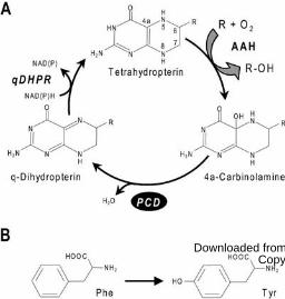
Figure 1. The role of PCD in regenerating the tetrahydropterin cofactor of AAHs. A, The cofac-
tor regeneration cycle. R = Phe, Tyr, or Trp. B, The Phe � Tyr hydroxylation reaction mediated
by Phe hydroxylase, a representative tetrahydropterin-dependent AAH. In AAH reactions, the
tetrahydropterin cofactor is oxidized to a 4a-carbinolamine. The carbinolamine is dehydrated via
the action of PCD to give a quinonoid (q) dihydropterin, which is then reduced to the tetrahydro
level by an NAD(P)H-linked q-dihydropterin reductase (qDHPR). The natural cofactor for
AAHs is tetrahydrobiopterin in mammals (Thony et al., 2000) and may be tetrahydromonapterin
in bacteria (Guroff and Rhoads, 1969).
Figure 2. Phylogenomic analysis of COG2154 proteins and AAH homologs in plants and micro-
organisms. A, Distribution of canonical and non-canonical COG2154 proteins and AAH homo-
logs. COG2154 sequences taken as canonical contain the motif [DE]-x(3)-H-H-P-x(5)-[YW]-
x(9)-H-x(8)-D. Major groups of plants and microorganisms are represented by single genera. For
plant groups, the genus is typical of all members of the group. For microorganism groups, the
genus has a distribution pattern that is common in the group but not necessarily universal. Gene
clustering in prokaryotes is symbolized by connecting lines. The major groups of proteobacteria
(α,β,γ) are indicated. Asterisks denote organisms whose COG2154 proteins were tested for PCD
activity in this study. Abbreviation: Schizosaccharo., Schizosaccharomyces. B, Clustering of
bacterial genes encoding COG2154 and AAH with each other and with genes of the homogent-
isate pathway of Tyr degradation. Matching colors correspond to orthologous genes; gray arrows
are unrelated genes. The steps in the homogentisate pathway and the corresponding genes are
shown on the right.
Figure 3. Primary structures and phylogeny of COG2154 proteins. A, Multiple sequence align-
ment of representative plant type 1 and type 2 COG2154 proteins. At, Arabidopsis thaliana; Zm,
Zea mays; Pt, Pinus taeda; Pp, Physcomitrella patens; Cr, Chlamydomonas reinhardtii. Pseudo-
monas aeruginosa (Pa) PCD is included for comparison, and defines the approximate extent of
the core COG2154 domain common to all sequences in the alignment. The domain unique to
type 2 proteins is in blue. Identical residues are shaded in black or dark blue, similar residues in
gray or light blue. Residues of the catalytic motif [DE]-x(3)-H-H-P-x(5)-[YW]-x(9)-H-x(8)-D
are shaded in red. Residues bolded and shaded in yellow were changed to an initiation codon in
www.plantphysiol.org on November 8, 2016 - Published by www.plantphysiol.orgDownloaded from Copyright © 2008 American Society of Plant Biologists. All rights reserved.
28
the constructs used in complementation assays. Dashes are gaps introduced to maximize align-
ment. Open triangles show positions of two introns present in type 1 and type 2 proteins from
Arabidopsis and Physcomitrella, and the type 2 protein from Chlamydomonas, but not in other
eukaryotic proteins. B, Unrooted neighbor-joining tree for COG2154 proteins from plants and
representative species of animals, protists, fungi, and prokaryotes. The sequences analyzed were
those tested in this study (asterisked) and those known or inferred from the literature to have
PCD activity. Bootstrap values are indicated only for nodes with >50% support. Evolutionary
distances are in units of the number of amino acid substitutions per site. C, Sequence alignment
of the catalytic motif region of selected non-plant COG2154 proteins from the phylogenetic ana-
lysis. Shading is as in part A. Sy, Synechocystis sp.; Bc, Bacillus cereus; Sp, Silicibacter pomer-
oyi; Vc, Vibrio cholerae; Cg, Corynebacterium glutamicum; Sa, Streptomyces avermitilis; Ch,
Cytophaga hutchinsonii; Ss, Sulfolobus solfataricus; Sc, Saccharomyces cerevisiae; Lm, Leish-
mania major; Pf, Plasmodium falciparum; Tg, Toxoplasma gondii.
Figure 4. Assay of PCD activity by functional complementation in E. coli. A tyrosine auxotroph
(strain JP2255) was transformed with pACYC177 containing Pseudomonas aeruginosa Phe hyd-
roxylase (phhA) or with pACYC177 (with the Apr gene inactivated) alone, plus pBluescript
alone or containing P. aeruginosa PCD (phhB) or COG2154 sequences from plants (a-j) or mic-
robes (k-t). The doubly transformed cells were plated on medium plus or minus Tyr. Each plate
included a positive control (sector 1, phhA + P. aeruginosa PCD) and a negative control (sector
2, phhA + pBluescript). Key to COG2154 genes: a, Arabidopsis thaliana 1 (At1g29810); b, Chla-
mydomonas reinhardtii 1; c, Physcomitrella patens 1; d, Pinus taeda 1; e, Zea mays 1; f, A. thal-
iana 2 (At5g51110); g, C. reinhardtii 2; h, P. patens 2; i, P. taeda 2; j, Z. mays 2; k, Corynebact-
erium glutamicum; l, Streptomyces avermitilis; m, Cytophaga hutchinsonii; n, Vibrio cholerae; o,
Bacillus cereus; p, Sulfolobus solfataricus; q, Silicibacter pomeroyi; r, Synechocystis; s, Sacch-
aromyces cerevisiae; t, Leishmania major.
Figure 5. Organellar targeting of plant types 1 and 2 COG2154 proteins fused to GFP. Chloro-
phyll autofluorescence (left-hand panels) and GFP fluorescence (middle panels) are merged in
the right-hand panels. A, Transient expression in Arabidopsis protoplasts of GFP fused to the
type 1 protein of Arabidopsis (At1g29810, At1, fourth row) or Pinus (Pt1, fifth row). The upper
three rows show controls: cytosolic expression of GFP alone (top row), chloroplastic expression
www.plantphysiol.org on November 8, 2016 - Published by www.plantphysiol.orgDownloaded from Copyright © 2008 American Society of Plant Biologists. All rights reserved.
29
of GFP fused to the transit peptide of the Rubisco small subunit (second row), and mitochondrial
expression of GFP fused to hydroxymethyldihydropterin pyrophosphokinase-dihydropteroate
synthase (third row). Fluorescence was observed with an epifluorescence microscope. B, Con-
focal laser scanning micrographs of mesophyll protoplasts isolated from leaves of transgenic
Arabidopsis plants expressing GFP fused to the type 2 protein of Arabidopsis (At5g51110, At2).
Figure 6. Structure of the molybdenum cofactor (Moco) and clustering of COG2154 genes with
Moco biosythesis genes in bacterial genomes. A, The structures of Moco and its precursor mol-
ybdopterin compared to that of a typical tetrahydropterin. The tetrahydropterin ring structure is
colored blue. The form of Moco shown occurs in xanthine dehydrogenase and aldehyde oxidase.
B, Clustering of COG2154 genes with up to four genes of Moco biosynthesis (moaA, moaC,
moaD, and a moaD-moaE fusion). Matching colors correspond to orthologous genes; gray
arrows are unrelated genes. Note that the genomes of Mycobacterium bovis and M. tuberculosis
each have two clusters, and that the genes flanking the COG2154-moaC pair in Nitrococcus
mobilis and Alkalilimnicola ehrlichei are different, so that only the pair is conserved.
Figure 7. Insertional knockout and expression of the At1g29810 gene, and the effect of knock-
outs on activities of Moco-dependent enzymes. A, The At1g29810 gene showing the positions of
the double T-DNA insert in the GABI-Kat and the single insert the Salk line. Introns are repres-
ented as solid lines and exons by boxes; 5´ and 3´ untranslated regions are in black. B, RT-PCR
validation that the insertions in both lines eliminate a functional At1g29810 message. Leaf RNA
was used as RT-PCR template. Note the absence of an At1g29810 amplicon in both mutant lines,
and the presence of the control (actin) amplicon. C, Expression of At1g29810 in organs of
Arabidopsis ecotype Columbia: G1 and G2, seedlings at 3 and 7 d of germination, respectively;
R, root; St, stem; C, cauline leaves; Y, F, S, young, fully expanded, and senescent rosette leaves;
In, inflorescence; E, M, L, siliques at early, mid, and late (yellowing) stages of development; Sd,
dry seeds. D, Activities of xanthine dehydrogenase (XDH) and aldehyde oxidase (AO) in in-vitro
cultured plantlets of homozygous Salk and GABI-Kat (G-K) knockout lines (KO) and the corres-
ponding wild type segregants (WT). The data are from two separate experiments, with three
(experiment 1) or four to five (experiment 2) independent replicates. Error bars are SE. Asterisks
indicate activities in the knockout that are significantly lower (*, P < 0.05; **, P < 0.01) than in
the corresponding wild type.
www.plantphysiol.org on November 8, 2016 - Published by www.plantphysiol.orgDownloaded from Copyright © 2008 American Society of Plant Biologists. All rights reserved.
30
Table I. Literature reports of COG2154 knockouts in microorganisms
Organism AAH Essential Phenotype References
Schizosaccharomyces pombe No No Defective spore walls Kakihara et al., 2003
Mycobacterium tuberculosis No No Sassetti et al., 2003
Methylobacterium extorquens No No Kalyuzhnaya et al., 2005
Pseudomonas aeruginosa Yes No Unable to grow on Phe Liberati et al., 2006; Song et al., 1999
w
ww
.plantphysiol.org on N
ovember 8, 2016 - P
ublished by w
ww
.plantphysiol.orgD
ownloaded from
C
opyright © 2008 A
merican S
ociety of Plant B
iologists. All rights reserved.
www.plantphysiol.org on November 8, 2016 - Published by www.plantphysiol.orgDownloaded from Copyright © 2008 American Society of Plant Biologists. All rights reserved.
www.plantphysiol.org on November 8, 2016 - Published by www.plantphysiol.orgDownloaded from Copyright © 2008 American Society of Plant Biologists. All rights reserved.
www.plantphysiol.org on November 8, 2016 - Published by www.plantphysiol.orgDownloaded from Copyright © 2008 American Society of Plant Biologists. All rights reserved.
www.plantphysiol.org on November 8, 2016 - Published by www.plantphysiol.orgDownloaded from Copyright © 2008 American Society of Plant Biologists. All rights reserved.
www.plantphysiol.org on November 8, 2016 - Published by www.plantphysiol.orgDownloaded from Copyright © 2008 American Society of Plant Biologists. All rights reserved.
www.plantphysiol.org on November 8, 2016 - Published by www.plantphysiol.orgDownloaded from Copyright © 2008 American Society of Plant Biologists. All rights reserved.
www.plantphysiol.org on November 8, 2016 - Published by www.plantphysiol.orgDownloaded from Copyright © 2008 American Society of Plant Biologists. All rights reserved.
Related Documents


![The Dehydratase ADT3 Affects ROS Homeostasis and … · The Dehydratase ADT3 Affects ROS Homeostasis and Cotyledon Development1[OPEN] Alessia Para, DurreShahwar Muhammad, Danielle](https://static.cupdf.com/doc/110x72/5cca391988c993570d8d8d21/the-dehydratase-adt3-affects-ros-homeostasis-and-the-dehydratase-adt3-affects.jpg)


