Illinois Wesleyan University Digital Commons @ IWU Digital Commons @ IWU Honors Projects Biology 4-18-1997 Phylogenetic Systematics of the Primate Genus Aotus Based on Phylogenetic Systematics of the Primate Genus Aotus Based on Hyoid Morphology Hyoid Morphology Melissa S. Immel '97 Illinois Wesleyan University Follow this and additional works at: https://digitalcommons.iwu.edu/bio_honproj Part of the Biology Commons Recommended Citation Immel '97, Melissa S., "Phylogenetic Systematics of the Primate Genus Aotus Based on Hyoid Morphology" (1997). Honors Projects. 14. https://digitalcommons.iwu.edu/bio_honproj/14 This Article is protected by copyright and/or related rights. It has been brought to you by Digital Commons @ IWU with permission from the rights-holder(s). You are free to use this material in any way that is permitted by the copyright and related rights legislation that applies to your use. For other uses you need to obtain permission from the rights-holder(s) directly, unless additional rights are indicated by a Creative Commons license in the record and/ or on the work itself. This material has been accepted for inclusion by faculty at Illinois Wesleyan University. For more information, please contact [email protected]. ©Copyright is owned by the author of this document.

Welcome message from author
This document is posted to help you gain knowledge. Please leave a comment to let me know what you think about it! Share it to your friends and learn new things together.
Transcript

Illinois Wesleyan University
Digital Commons @ IWU Digital Commons @ IWU
Honors Projects Biology
4-18-1997
Phylogenetic Systematics of the Primate Genus Aotus Based on Phylogenetic Systematics of the Primate Genus Aotus Based on
Hyoid Morphology Hyoid Morphology
Melissa S. Immel '97 Illinois Wesleyan University
Follow this and additional works at: https://digitalcommons.iwu.edu/bio_honproj
Part of the Biology Commons
Recommended Citation Immel '97, Melissa S., "Phylogenetic Systematics of the Primate Genus Aotus Based on Hyoid Morphology" (1997). Honors Projects. 14. https://digitalcommons.iwu.edu/bio_honproj/14
This Article is protected by copyright and/or related rights. It has been brought to you by Digital Commons @ IWU with permission from the rights-holder(s). You are free to use this material in any way that is permitted by the copyright and related rights legislation that applies to your use. For other uses you need to obtain permission from the rights-holder(s) directly, unless additional rights are indicated by a Creative Commons license in the record and/ or on the work itself. This material has been accepted for inclusion by faculty at Illinois Wesleyan University. For more information, please contact [email protected]. ©Copyright is owned by the author of this document.

on Hyoid Morphology
.ed
Abstract
Dissection of a specimen of the South American primate genus Aotus was
performed, focusing on the morphology of the hyoid apparatus and surrounding
musculature within the hyoid region (the ventral side of the throat). Data collected
from this dissection, along with data from published descriptions of primate genera
from both primate suborders Haplorhini and Strepsirhini, were analyzed using the
computer software program PAUP (Phylogenetic Analysis Using Parsimony). This
analysis generated four equally probable phylogenetic trees (cladograms) depicting
possible phylogenetic relationships of Aotus to other primates. While the
cladograms differ in minor detail, they all show that Aotus appears to be more
closely related to the more apomorphic suborder Haplorhini than to the
Strepsirhini, and that it is a relatively plesiomorphous member of the haplorhine
group. This suggests that the ancestor of the New World monkeys arrived in South
America at about the time when haplorhine primates were beginning to diverge in
the rest of the world, about 28 million years ago.

Introduction
Traditionally, primates have been divided into two taxa, the prosimians and
anthropoids, with the tarsier group included as prosimians. More recent
investigations have supported the hypothesis that tarsiers are more closely related
to the anthropoids. This arrangement was first proposed by Pocock (1918) and
supported by Goodman (1975) and Erickson (1996). With this change in grouping,
there is a consequent change in nomenclature. Therefore, the prosimian group
without the tarsiers is known as the suborder Strepsirhini and the anthropoids
along with the tarsiers comprise the suborder Haplorhini. Members of the suborder
Strepsirhini are considered relatively plesiomorphous and include such groups as
the lemurs, aye-ayes, and lorises (Goodman 1975). The suborder Haplorhini
includes the primate groups like the Old World monkeys (e.g. rhesus monkeys,
baboons) and the apes (Goodman 1975). New World (South American) monkeys
have traditionally been included with the haplorhines (Simpson 1945). However, it
is not known exactly where they fit in: closer to the anthropoids or to the tarsiers.
The origin of South American primates has long been a mystery to
primatologists, both where they came from (Africa and North America each
contained possible ancestors, Carroll 1988) and when they arrived in South America.
One way to examine these questions is to use the fossil record. The earliest fossil
primates found in South America have been classified as members of the suborder
Haplorhini that lived approximately 27-28 million years ago (mya), during the
Upper Oligocene epoch (Carroll 1988). The earliest haplorhine fossils from Africa
have been dated to the Middle Oligocene epoch, about 28-30 mya (Carroll 1988). This
suggests that New World monkeys arrived in South America at about the time
when haplorhines began to diverge from the more plesiomorphous strepsirhine
group in Africa. According to Carroll (1988), New World monkeys are a
monophyletic group. Therefore, all extant and extinct species are the result of a

3
asion and in this analysis it is assumed that findings for the representative
) are consistent with those for the rest of the South American taxa.
e genus Aotus contains species that are collectively referred to as "owl
." They are the only strictly nocturnal anthropoid group (not including
that exist (Ford 1994). There is some debate as to how many species of
xist (Nowak 1991); however, it is herein assumed that all Aotine species (if
more than one) are similar with regard to hyoid morphology.
The morphology of the primate hyoid region seems to be relatively stable,
ionarily (Griffiths pers. comm.). As a result, differences in this anatomical
among primate groups should indicate evolutionary divergence. The
xrrpose of this project was to describe the morphology of the Aotine hyoid region
compare it with that of other previously described primate groups in order to
ne the relationships between this South American primate genus and those
ather groups. This information may shed light on the questions of when and from
wl~cre New World monkeys originated.
Materials and Methods
The specimen of Aotus examined was that of a newborn male, preserved in
formalin and ethanol, from the Illinois Wesleyan University fluid-preserved
primate collection. The species of the specimen could not be determined with
crrlainty because the original color patterns in the fur were altered due to the long-
term formalin preservation. Dissection of the hyoid region was performed using a
ifa~usch & Lomb dissecting microscope, twin fiber optic light sources, and standard
rtricroscope dissecting technique. Pencil drawings were made of each muscle layer
exposed by the dissection. The drawings most relevant to the subsequent analysis
were inked and labeled for inclusion into this paper. Written descriptions were
rtlade of all hyoid muscles for comparison with similar descriptions of the hyoid
regions of other primates (lemurs, Hill 1953; aye-ayes, Hill 1953; tarsiers, Erickson
1996; rhesus monkeys, Geist 1961, Howell and Straus 1961, Huber 1961; and humans,
Williams et al. 1989) along with that of a closely related non-primate, the tree shrew
(Griffiths in manuscript). Variations in hyoid morphological traits from all of these
groups were assigned character state values in a MacClade data matrix table. The
computer program PAUP (Phylogenetic Analysis Using Parsimony, Swofford 1993)
was used to perform the analysis. Character state data from tree shrews, a group of
mammals considered to be related to the ancestor of primates, were designated as
the "outgroup" (a group of organisms that are considered equally dissimilar to each
of the other ingroups analyzed). PAUP calculated the amount of dissimilarity
between the organisms in question and translated that into phylogenetic trees
(cladograms). Cladograms are diagrams of theoretical evolutionary paths depicting
the relationships between organisms. The goal of this analysis is to find the most
parsimonious cladograms (those with the fewest number of evolutionary steps
involved) to explain the variation seen among these organisms.
Results
Branchiomeric Musculature
Extrahyoid Muscles
M. digastricus anterior (Figs. 1 & 2)
Origin: This muscle has a double origin from the digastric tendon and the
ventral surface of the basihyal (mostly from the tendon).
Insertion: Fibers insert onto the medial surface of the mandibular body from
a point approximately 1 mm posterior to the mandibular symphysis to a point 10
mm posterior to the symphysis.
Comments: This muscle is indirectly connected at its origin to the omohyoid
and the sternohyoid through fascia. There is no direct connection between the two

anterior digastrics except that they are connected to the basihyal. There is not a
typical arcuate tendon present. Instead there is what is herein referred to as a
vestigial arcuate tendon. This tendon is deep to the anterior digastrics and has
associated muscle fibers which pass perpendicularly to the sagittal plane connecting
the posterior digastrics. Some fibers run from the deep surface of the anterior
digastric into the vestigial arcuate tendon.
M. dig;astricus posterior (Figs. 1,2 & 3)
Origin: This band-like muscle originates from the paraoccipital bone just
posterior to the auditory bulla, immediately posterior to and in common with the
connective tissue fibers of the stylohyal ligament.
Insertion: Fibers run anteromedially to insert into the intermediate tendon
and the vestigial arcuate tendon.
Mylohyoid Group
Muscles of this group are innervated by branches of the mylohyoid nerve,
which is a branch of the mandibular nerve which, in turn, is a branch of the
trigeminal (fifth cranial) nerve.
M. mvlohyoideus (Figs. 2 & 3)
Origin: This muscle originates from the ventro-medial surface of the
mandibular body to a point approximately 10 mm posterior to the mandibular
symphysis.
Insertion: Most fibers run postero-medially. Those fibers that originate from
the mandibular symphysis to a point approximately 3.5 mm posterior to it run
perpendicular to the sagittal plane. Fibers insert into a midline connective tissue
6
phe and onto the ventral surface of the basihyal, deep to the origin of the anterior
This muscle is absent.
M. mandibulo-hyoideus
This muscle has been recently determined to be the same muscle as the
anterior digastric (Griffiths, in manuscript). The anterior digastric was previously
described in this paper.
Hyoid Constrictor Group
Muscles of this group are innervated by branches of the facial nerve (seventh
cranial nerve).
M. s&lohyoideus (Figs. 1 & 2)
Origin: This muscle originates from a short tendon which in turn takes
origin from the same paraoccipital connective tissue that gives rise to the stylohyal
ligament and the posterior digastric.
Insertion: Fibers run antero-medially to insert near the insertion of the
anterior digastrics and superficial to the insertion of the omohyoid onto the
basihy al.
Comments: This muscle does not insert onto the basihyal.
M. iugulohyoideus
This muscle is absent.
M. sphincter colli profundus
This muscle is absent.
Glossopharyngeal Group
Muscles of this group are innervated by branches of the glossopharyngeal
nerve (ninth cranial nerve).
M. stylopharyngeus (Figs, 3 & 4)
Origin: This muscle originates from the medial surface of the stylohyal
ligament at a point approximately opposite from the origin of the styloglossus.
Insertion: Fibers insert into the lateral pharynx just anterior to the
thyropharyngeus.
Comments: This muscle is extremely reduced in the specimen examined.
M. ceratohyoideus (Fig. 4)
Origin: From the anterior surface of the entire length of the thyrohyal.
Insertion: Fibers insert on the entire posterior surface of the two ossified
anterior cornua elements: the ceratohyal and epihyal.
Pharyngeal Constrictor Group
Muscles of this group are innervated by branches of the vagus (tenth cranial)
nerve.
M. hyopharyngeus (superior constrictor muscle - not illustrated)
Origin: This origin of this muscle is the pterygoid region of the inferior
surface of the skull and the fascia of the lateral styloglossus region.
8
Insertion: Fibers run dorsally and insert onto the dorsal midline raphe of the
(middle constrictor muscle - Fig. 4)
Origin: This muscle originates from the dorsal surface and tip of the
rohyal at a point deep to the lateral fibers of the hyoglossus.
Insertion: Fibers pass dorsally, overlapping the anterior part of the
eclcopharyngeus, to insert onto the dorsal pharyngeal midline and intermingle with
the fibers of the antimere.
M. cricopharyngeus (inferior constrictor muscle - not illustrated)
Origin: This muscle originates from the lateral cricoid cartilage and from the
posterior most cornu on the thyroid cartilage.
Insertion: Fibers insert into the dorsal pharyngeal midline and into the fibers
sf the antimere.
Myotomic Musculature
Lingual Group
Muscles of this group are innervated by branches of the hypoglossal (twelfth
cranial) nerve.
M. p;enioglossus (not illustrated)
$ Origin: This muscle originates from the posteromediaf surface of the 4 1 mandible, immediately lateral to the mandibular symphysis. I I f Insertion: Fibers pass posteriorly to two distinct insertions. One insertion is b
into the posterior one-third of the tongue (these fibers become part of the intrinsic

9
e musculature). Some fibers continue posteriorly to the other insertion, onto
nterior surface of the basihyal.
(Fig. 3)
Origin: This muscle originates from the lateral basihyal, lateral to the origin
he geniohyoid, and from the ventral thyrohyal.
Insertion: Fibers run antero-laterally and deep to the styloglossus to insert
into the lateral tongue.
Comments: The hyoglossus is a very broad, unbroken, fan-shaped muscle.
M. s ~ l o ~ l o s s u s (Figs. 3 & 4)
Origin: This muscle originates from the posterolateral surface of the
stylohyal ligament. The origin is about 4 5 mm anterior to the dorsal attachment of
the stylohyal ligament.
Insertion: Fibers are anterior and run parallel to the hyoglossus nerve to
insert along and into the lateral tongue surface.
Medial Ventral Cervical Group
Muscles of this group are innervated by the ansa cervicalis, a complex of
nerves originating in the anterior cervical region, except for the geniohyoid which is
innervated by the hypoglossus (twelfth cranial) nerve.
M. aeniohyoideus (Fig. 3)
Origin: From the medial surface of the mandible, lateral and posterior to the
mandibular symphysis.
Insertion: Fibers pass posteriorly to insert onto the anterior surface of the
basihy al.

10
Comments: The geniohyoid is a broad strap which covers the genioglossus
(Figs. 1 & 2)
Origin: The sternohyoid originates from the dorsal surface of the entire
width of the manubrium of the sternum.
Insertion: This muscle inserts onto the postero-ventral surface of the
basihyal.
Comments: The two antimeres of this muscle are fused at the midline. The
origin extends laterally along the manubrium out to the sternoclavicular
articulation. No fibers originate from the clavicle.
M. sternothvroideus (Figs. 1 & 2)
Origin: The sternothyroid originates from the dorsal surface of the
lateral-most part of the manubrium of the sternum.
Insertion: Fibers insert onto the lateral surface of the thyroid cartilage of the
larynx.
Comments: This muscle is fused at its origin to the lateral sternohyoid with
which it forms a continuous sheet across the posterior end of the trachea. As fibers
of these muscles pass anteriorly, the sternothyroid separates from the sternohyoid so
that the medial sternothyroid passes deep to the lateral sternohyoid (from a ventral
viewpoint).
M. omohyoideus (Figs. I& 2)
Origin: This muscle originates by a very short tendon from the midpoint of
the superior border of the scapula, just medial to the scapular notch.

Insertion: Fibers pass antero-medially, emerging from deep to the
sternomastoid and insert onto the basihyal lateral to the insertion of the
sternohyoid.
M. thyrohyoideus (Fj igs.
Origin: The thyrohyoid originates from the entire lateral surface of the
thyroid cartilage.
Insertion: This muscle inserts into the entire posterior surface of the
thyrohy al.
Comments: This muscle is very broad.
Hyoid A
The hyoid apparatus of Aotus is made up of several elements. In this
specimen, the basihyal, the largest component, has a roughly rectangular shape with
f wo proiections (cornua) that extend from it laterallv. The more anterior cornu
(termed the lesser cornu by human anatomists) has two fused bony constituents, the
more medial ceratohyal and the more lateral epihyal. These, in turn are attached to
the stylohyal ligament which extends laterally around the auditory bulla to the - paraoccipital region of the skull. The more posterior cornu originating from the
basihyal (the greater cornu of human anatomists) has only one bony element, the
thyrohyal, which is strongly fused to the basihyal.
Results and Discussion
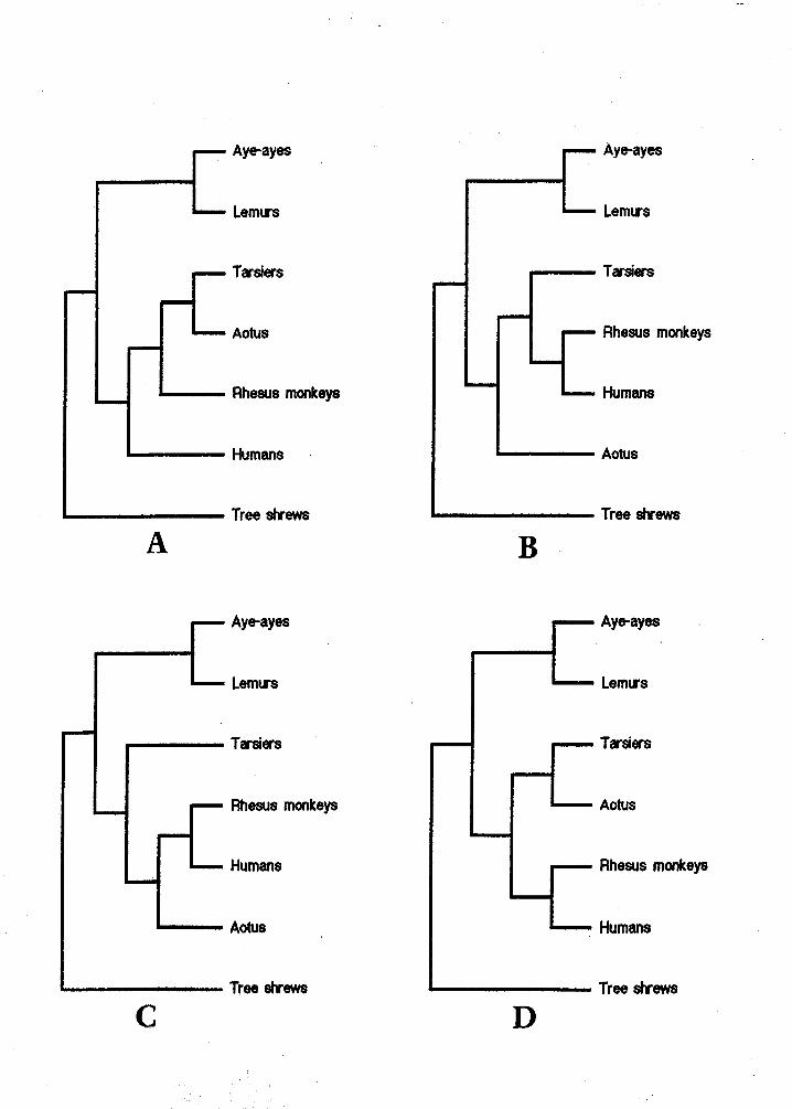
The data used for the PAUP analysis (collected from the literatu
dissection) are presented in Table 1. Four equally parsimo
A-D) were generated by PAUP delineating possi
primate evolutionary tre re. Differences

minor, but indicate the need for more data to help reduce the number of most
parsimonious cladograms down to one or two. However, all four cladograms show
three consistent groupings: the aye-ayes with the lemurs (supporting their
traditional placement in the taxonomic group Strepsirhini); the tarsiers with rhesus
monkeys, humans, and Aotus (supporting their classification in the haplorhine
group); and finally, the tree shrews (separate because they are the outgroup). The
consistent placement of the divergence of Aotus near that of tarsiers (a
plesiomorphic haplorhine) suggests that this particular New World monkey is also
a plesiomorphic member of the haplorhine group. This implies that the ancestor of
the New World monkeys arrived in South America at about the time that
haplorhine primates were beginning to diverge in other parts of the world (about 28
mya), providing independent confirmation of what the fossil record evidence seems
The question of "Where did South American monkeys come from?" can be
addressed by examining the details of the cladograms. However, there is not a
definitive answer. Figure 5A depicts Aotus and tarsiers as more closely related to
each other than to any of the other primate groups. This suggests that tarsiers and
Aotus share a common ancestor. It has been hypothesized that a fossil group of - tarsiers (family Omomyidae) was ancestral to New World monkeys based on
dentition comparison (Gazin 1958). Fossil evidence exists of these omomyid tarsiers
ving in North America around the time of the primate arrival in South America
(Carroll 1988). Given this fossil evidence and the degree of similarity between the
rsier and Aotine hyoid regions, these data support the hypothesis that the ancestor
f New World monkeys was a tarsier-like organism that migrated south from
orth America. Although the majority of tarsier fossils come from North America,
ere is one fossil tarsier (family Tarsiidae) found in northern Africa from this same
time period (Carroll 1988). Therefore, it is still possible that the ancestor to New
World monkeys may have been a tarsier group that came from Africa.
Figure 5B depicts Aotus as the earliest lineage of the haplorhine group
included in this study. This suggests that the ancestor of the New World monkeys
diverged from the strepsirhine lineage before any other group did. This implies that
the ancestor of New World monkeys must have diverged in Africa from other
primates before the tarsiers split off (and thus before they could appear in North
America). Therefore, according to Figure 5B it would appear that a very early
African origin for South American monkeys is probable.
Figure 5C is identical to 5B except that the evolutionary placements of tarsiers
and Aotus are switched relative to each other. Consequently, this implies that
tarsiers diverged from the Strepsirhini line before any other strepsirhine group. It
also depids a closer relationship between Aotus and the human-rhesus group than
between Aotus and tarsiers. Therefore, from this cladogram it would seem more
likely that the ancestral New World Monkey came from African haplorhines and
not the widespread tarsiers.
The final cladogram, Figure 5D, shows a close relationship between Aotus
and tarsiers as does Figure 5A. However, the difference is in the timing of the
human divergence from the other haplorhines. Therefore, it does not discount the
possibility of a tarsier origin for the South American primates.
Although the "When" question of origins can be addressed by my analysis,
some uncertainty remains surrounding the question of "From where?" The hyoid
ata can be interpreted to support either an African or North American origin for
outh American primate species. Further study is necessary to address this
ACKNOWLEDGMENTS
I would like to thank my hearing committee members: Dr. Bruce Criley, Dr.
Sheryl Soukup, and Dr. Martin Nickels, for their time and attention to this project.
In addition, I wish to extend my gratitude and appreciation toward Dr. Tom Griffiths
for his time, insight, and encouragement.
LITERATURE CITED
Carroll, R. L. 1988. Vertebrate paleontology and evolution. W. H. Freeman and
Company, New York, xiv + 632 pp.
Erickson, J. 1996. Hyoid morphology of Tarsius syrichta: Implications for
phylogeny. Unpublished Senior Honors Manuscript, Biology Department,
IWU, 32 pp.
Ford, S. 1994. Taxonomy and distribution of the owl monkey, In J. F. Baer, R. E.
Weller, and I. Kakoma (eds.): Aotus: The Owl Monkey. San Diego: Academic
Press. pp. 1-59.
Gazin, C. L. 1958. A review of the Middle and Upper Eocene primates of North
America. Smithso. Misc. Coll., 136: 1-112.
Geist, F. D. 1961. Nasal cavity, larynx, mouth, and pharynx, In C. G. Hartman and
W. L. Strauss, Jr (eds.): The Anatomy of the Rhesus Monkey (Macaca mulatta).
New York: Hafner Publ. Co. pp. 189-209.
oodman, M. 1975. Protein sequence and immunological specificity: their role in
phylogenetic studies of primates, In W. P. Luckett and F. S. Szalay (eds.):
Phylogeny of the Primates: A Multidisciplinary Approach. New York: Plenum
Press. pp. 219-248.
iffiths, T. In manuscript. Hyoid morphology of colugos and tree shrews
(Mammalia: Orders Dermoptera and Scandentia).
Hill, W. 0. C. 1953. Primates - Comparative Anatomy and Taxonomy I.
Strepsirhini. Edinburgh: Edinburgh University Press.
Howell, A. B. , and W. L. Straus Jr. 1961. The muscular system. In C. G. Hartman
and W. L. Strauss, Jr (eds.): The Anatomy of the Rhesus Monkey (Macaca
mulatta). NewYork: Hafner PubI. Co. pp. 75-89.
Huber, E. 1961. The facial musculature and its innervation. In C. G. Hartman and
W. L. Strauss, Jr (eds.): The Anatomy of the Rhesus Monkey (Macaca mulatta).
NewYork: Hafner Publ. Co. pp. 176-188.
Nowak, R. M. 1991. Walker's Mammals of the World: Fifth Edition. Baltimore:
The Johns Hopkins University Press. pp. 447-448.
Pocock, R. I. 1918. On the external characters of the lemurs and Tarsius. Proc. of
Zool. Soc. Lond., 1918: pp. 19-53.
Simpson, G. G. 1945. The principles of classification and a classification of mammals.
Bulletin of the American Museum of Natural History, 85: xvi + 350 pp.
Swofford, D. L. 1993. Phylogenetic Analysis Using Parsimony (PAUP) version 3.1.1
(distributed by Smithsonian Institute, Washington, D. C. 20560).
Williams, P. L., R. Warwick, M. Dyson, and L. H. Banniste 1989. Gray's Anatomy.
Edinburgh: Churchill Livingstone.
Table 1: Summary of character states in the MacClade matrix used by PAUP to generate the most parsimonious cladograms.

Aotus data 1 1 2 st!!lohyoid course gen~oqlossus
1 1 Aye-ayes superficial to digastric I strongly attached to basihyal
2 Lemurs superficial to digastric strongly attached to basihyal
3 Tarsiers deep to digastric reduced, attached to basihyal
4 Rhesus monkevs deep to digastric strongly attached to basihyal
5 Humans superficial and deep to digastr strongly attached to basihyal
6 Tree shrews superficial and deep to digastr no connection to basihyal
7 Aottls superficial to digastric reduced, attached to basihyal
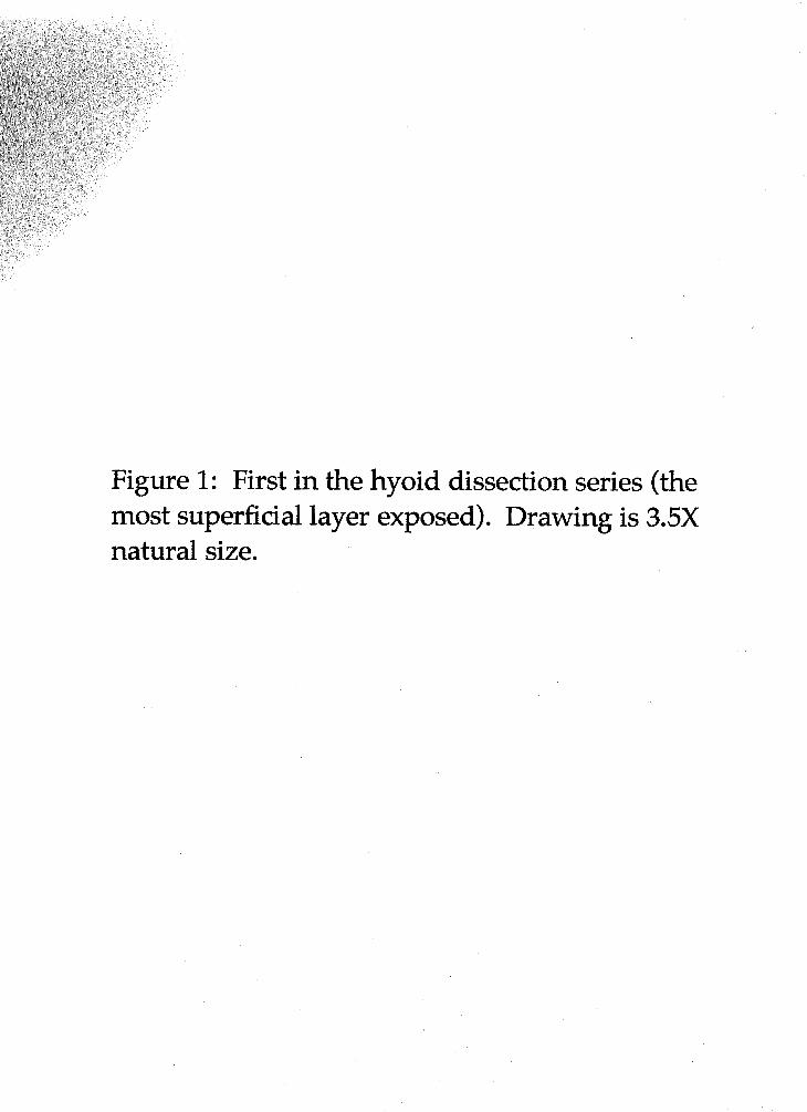
Figure 1: First in the hyoid dissection series (the most superficial layer exposed). Drawing is 3.5X natural size.
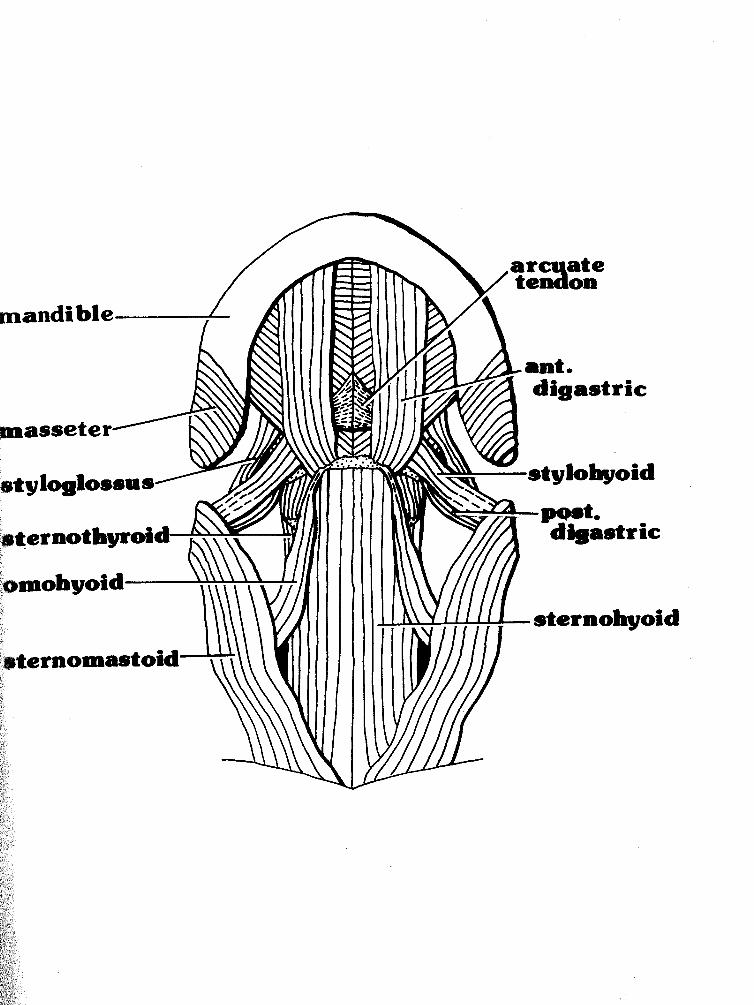

Figure 2: Second in the hyoid dissection series (both the superficial layer and the one exposed deep to it). Drawing is 3.5X natural size.
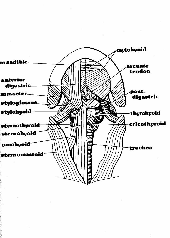

Figure 3: Third in the hyoid dissection series with a deeper layer exposed. Drawing is 3.5X natural size.

arcuate
sternomastoid

Figure 4: Fourth in the hyoid dissection series (the hyoid apparatus-the deepest layer dissected). Drawing is 3.5X natural size.
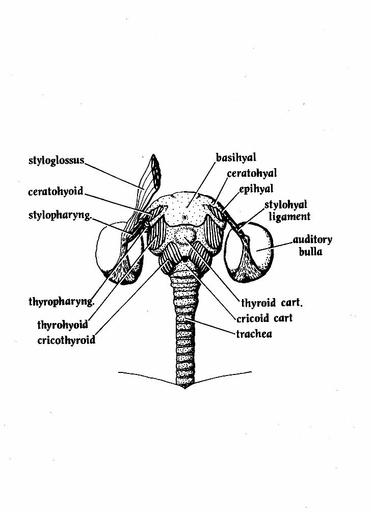
Figure 5, A-D: The four most parsimonious cladograms generated by the PAUP analysis (using the data in Table 1).
Related Documents