Phylogenetic relationships within the South American fish family Anostomidae (Teleostei, Ostariophysi, Characiformes) BRIAN L. SIDLAUSKAS 1–4 * and RICHARD P. VARI FLS 4 1 Committee on Evolutionary Biology & University of Chicago, 1025 E. 57th St., Culver Hall 402, Chicago, IL 60637, USA 2 Division of Fishes, The Field Museum, 1400 S. Lake Shore Drive, Chicago, IL 60605, USA 3 National Evolutionary Synthesis Center, 2024 W. Main St. A200, Durham, NC 27705, USA 4 Department of Vertebrate Zoology, MRC-159, National Museum of Natural History, PO Box 37012, Smithsonian Institution, Washington, DC 20013-7012, USA Received 17 September 2007; accepted for publication 17 September 2007 Analysis of a morphological dataset containing 152 parsimony-informative characters yielded the first phylogenetic reconstruction spanning the South American characiform family Anostomidae. The reconstruction included 46 ingroup species representing all anostomid genera and subgenera. Outgroup comparisons included members of the sister group to the Anostomidae (the Chilodontidae) as well as members of the families Curimatidae, Characidae, Citharinidae, Distichodontidae, Hemiodontidae, Parodontidae and Prochilodontidae. The results supported a clade containing Anostomus, Gnathodolus, Pseudanos, Sartor and Synaptolaemus (the subfamily Anostominae sensu Winterbottom) albeit with a somewhat different set of relationships among the species within these genera. Anostomus as previously recognized was found to be paraphyletic and is split herein into two monophyletic components, a restricted Anostomus and the new genus Petulanos gen. nov., described herein. Laemolyta appeared as sister to the clade containing Anostomus, Gnathodolus, Petulanos, Pseudanos, Sartor and Synapto- laemus. Rhytiodus and Schizodon together formed a well-supported clade that was, in turn, sister to the clade containing Anostomus, Gnathodolus, Laemolyta, Petulanos, Pseudanos, Sartor and Synaptolaemus. Anostomoides was sister to the clade formed by these nine genera. Leporinus as currently defined was not found to be monophyletic, although certain clades within that genus were supported, including the species with subterminal mouths in the former subgenus Hypomasticus which we recognize herein as a genus. Abramites nested in Leporinus, and Leporellus was found to be the most basal anostomid genus. The presence of cis- and trans-Andean species in Abramites, Leporellus, Leporinus and Schizodon, all relatively basal genera, suggests that much of the diversification of anostomid species pre-dates the uplift of the Andean Cordilleras circa 11.8 million years ago. Several important morphological shifts in anostomid evolution are illustrated and discussed, including instances of convergence and reversal. © 2008 The Linnean Society of London, Zoological Journal of the Linnean Society, 2008, 154, 70–210. ADDITIONAL KEYWORDS: Anostominae – biogeography – cladistics – headstanders – morphology – Neotropics – new genus – osteology – South America. INTRODUCTION Members of the Anostomidae inhabit myriad fresh- water environments in tropical and subtropical South America that range from the Río Atrato of north- western Colombia south to the central portions of Argentina. The greatest diversity at both the specific and the generic levels occurs east of the Andes Cor- dilleras where representatives of the family inhabit all major drainages from the Río Orinoco basin (Lasso et al., 2004b) to the Río de La Plata system (Garavello & Britski, 2003). The vast reaches of the Amazon and *Corresponding author. E-mail: [email protected] Zoological Journal of the Linnean Society, 2008, 154, 70–210. With 53 figures No claim to original US Government works. Journal compilation © 2008 The Linnean Society of London, Zoological Journal of the Linnean Society, 2008, 154, 70–210 70

Welcome message from author
This document is posted to help you gain knowledge. Please leave a comment to let me know what you think about it! Share it to your friends and learn new things together.
Transcript
Phylogenetic relationships within the South Americanfish family Anostomidae (Teleostei, Ostariophysi,Characiformes)
BRIAN L. SIDLAUSKAS1–4* and RICHARD P. VARI FLS4
1Committee on Evolutionary Biology & University of Chicago, 1025 E. 57th St., Culver Hall 402,Chicago, IL 60637, USA2Division of Fishes, The Field Museum, 1400 S. Lake Shore Drive, Chicago, IL 60605, USA3National Evolutionary Synthesis Center, 2024 W. Main St. A200, Durham, NC 27705, USA4Department of Vertebrate Zoology, MRC-159, National Museum of Natural History, PO Box 37012,Smithsonian Institution, Washington, DC 20013-7012, USA
Received 17 September 2007; accepted for publication 17 September 2007
Analysis of a morphological dataset containing 152 parsimony-informative characters yielded the first phylogeneticreconstruction spanning the South American characiform family Anostomidae. The reconstruction included 46ingroup species representing all anostomid genera and subgenera. Outgroup comparisons included members of thesister group to the Anostomidae (the Chilodontidae) as well as members of the families Curimatidae, Characidae,Citharinidae, Distichodontidae, Hemiodontidae, Parodontidae and Prochilodontidae. The results supported a cladecontaining Anostomus, Gnathodolus, Pseudanos, Sartor and Synaptolaemus (the subfamily Anostominae sensuWinterbottom) albeit with a somewhat different set of relationships among the species within these genera.Anostomus as previously recognized was found to be paraphyletic and is split herein into two monophyleticcomponents, a restricted Anostomus and the new genus Petulanos gen. nov., described herein. Laemolytaappeared as sister to the clade containing Anostomus, Gnathodolus, Petulanos, Pseudanos, Sartor and Synapto-laemus. Rhytiodus and Schizodon together formed a well-supported clade that was, in turn, sister to the cladecontaining Anostomus, Gnathodolus, Laemolyta, Petulanos, Pseudanos, Sartor and Synaptolaemus. Anostomoideswas sister to the clade formed by these nine genera. Leporinus as currently defined was not found to bemonophyletic, although certain clades within that genus were supported, including the species with subterminalmouths in the former subgenus Hypomasticus which we recognize herein as a genus. Abramites nested inLeporinus, and Leporellus was found to be the most basal anostomid genus. The presence of cis- and trans-Andeanspecies in Abramites, Leporellus, Leporinus and Schizodon, all relatively basal genera, suggests that much of thediversification of anostomid species pre-dates the uplift of the Andean Cordilleras circa 11.8 million years ago.Several important morphological shifts in anostomid evolution are illustrated and discussed, including instancesof convergence and reversal. © 2008 The Linnean Society of London, Zoological Journal of the Linnean Society,2008, 154, 70–210.
ADDITIONAL KEYWORDS: Anostominae – biogeography – cladistics – headstanders – morphology –Neotropics – new genus – osteology – South America.
INTRODUCTION
Members of the Anostomidae inhabit myriad fresh-water environments in tropical and subtropical SouthAmerica that range from the Río Atrato of north-
western Colombia south to the central portions ofArgentina. The greatest diversity at both the specificand the generic levels occurs east of the Andes Cor-dilleras where representatives of the family inhabitall major drainages from the Río Orinoco basin (Lassoet al., 2004b) to the Río de La Plata system (Garavello& Britski, 2003). The vast reaches of the Amazon and*Corresponding author. E-mail: [email protected]
Zoological Journal of the Linnean Society, 2008, 154, 70–210. With 53 figures
No claim to original US Government works.Journal compilation © 2008 The Linnean Society of London, Zoological Journal of the Linnean Society, 2008, 154, 70–210
70
the adjoining Río Orinoco basin are inhabited by thelargest number of genera and species, including someof the most modified members of the family. TheAtlantic versant of the Guayana shield in Guyana,Suriname and French Guiana also harbours manyspecies (Planquette, Keith & Le Bail, 1996), whilea less diverse fauna inhabits the hydrographicallyhighly-dissected trans-Andean portions of SouthAmerica. West of the Andean Cordilleras, anostomidsare present in the arch from Lago Maracaibo of theCaribbean Sea versant as far south as the Guayasbasin of Ecuador, which drains into the Pacific Ocean.Although few species inhabit the trans-Andeanregions, most that do occur there are endemic to verysmall regions. For example, Abramites eques and Lep-orinus muyscorum are known only from trans-AndeanColombia (Steindachner, 1900, 1902; Vari & Williams,1987) and Schizodon corti only from Lago Maracaiboin Venezuela (Vari & Raredon, 1991).
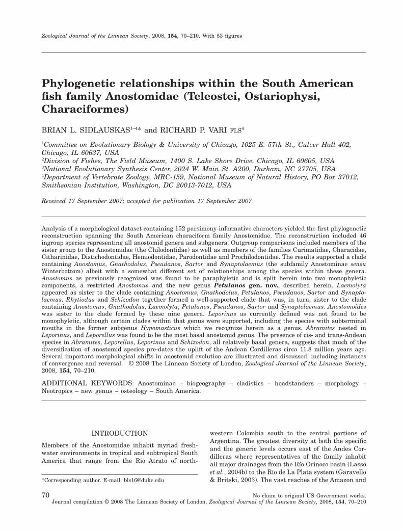
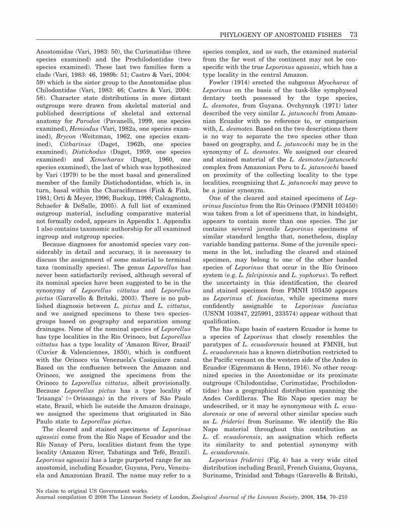
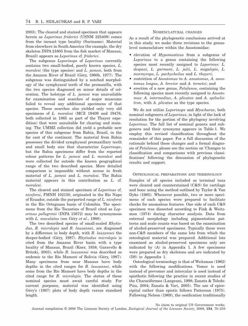
Overall body form in the Anostomidae ranges fromthe elongate, fusiform species of Rhytiodus (Fig. 1;Géry, 1977: 180; Santos, 1980: fig. 12) to the deep-bodied, laterally compressed species of Abramites(Fig. 2; Vari & Williams, 1987: fig. 3). Most anosto-mids, however, are moderately elongate species thatare rounded in cross-section. Despite their similarityin overall body form, members of the Anostomidaedemonstrate a wide range of jaw, tooth, neurocraniumand suspensorium morphologies (Sidlauskas, 2007).Mouth positions range from distinctly subterminalwithin Hypomasticus, particularly in Hypomasticusmormyrops (Steindachner, 1875: pl. 6) and Hypomas-ticus pachycheilus (Britski, 1976; Sidlauskas, 2007:fig. 2B) to supraterminal and posteriorly directed in
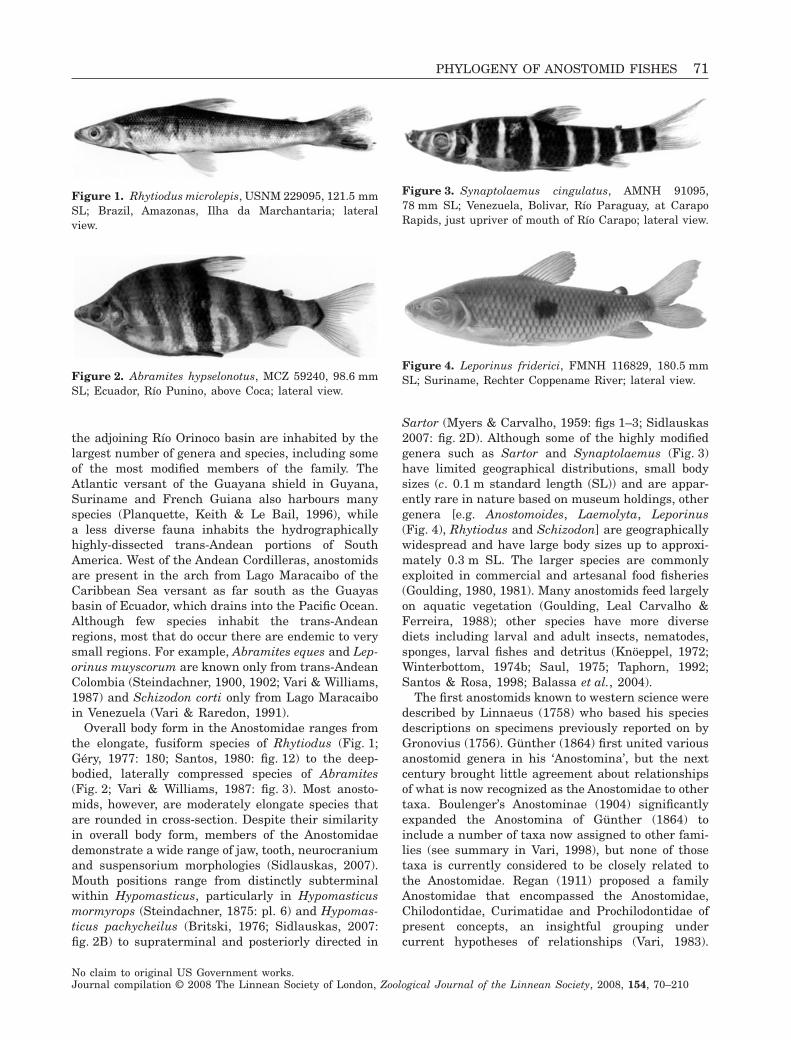
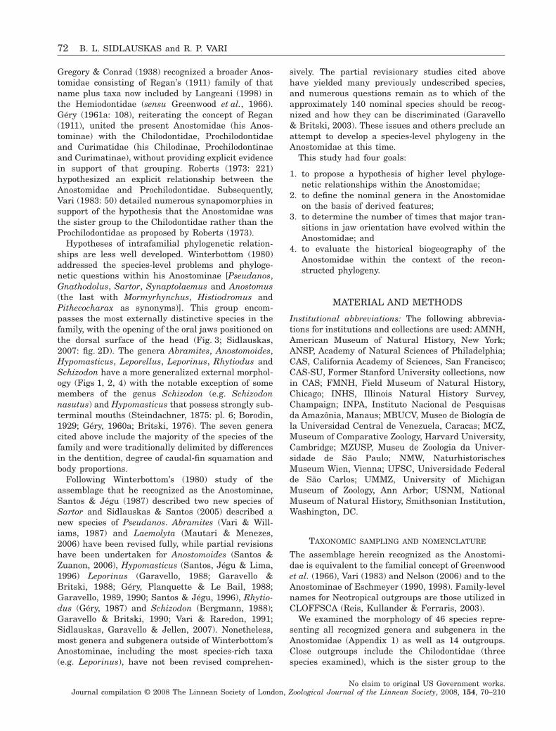
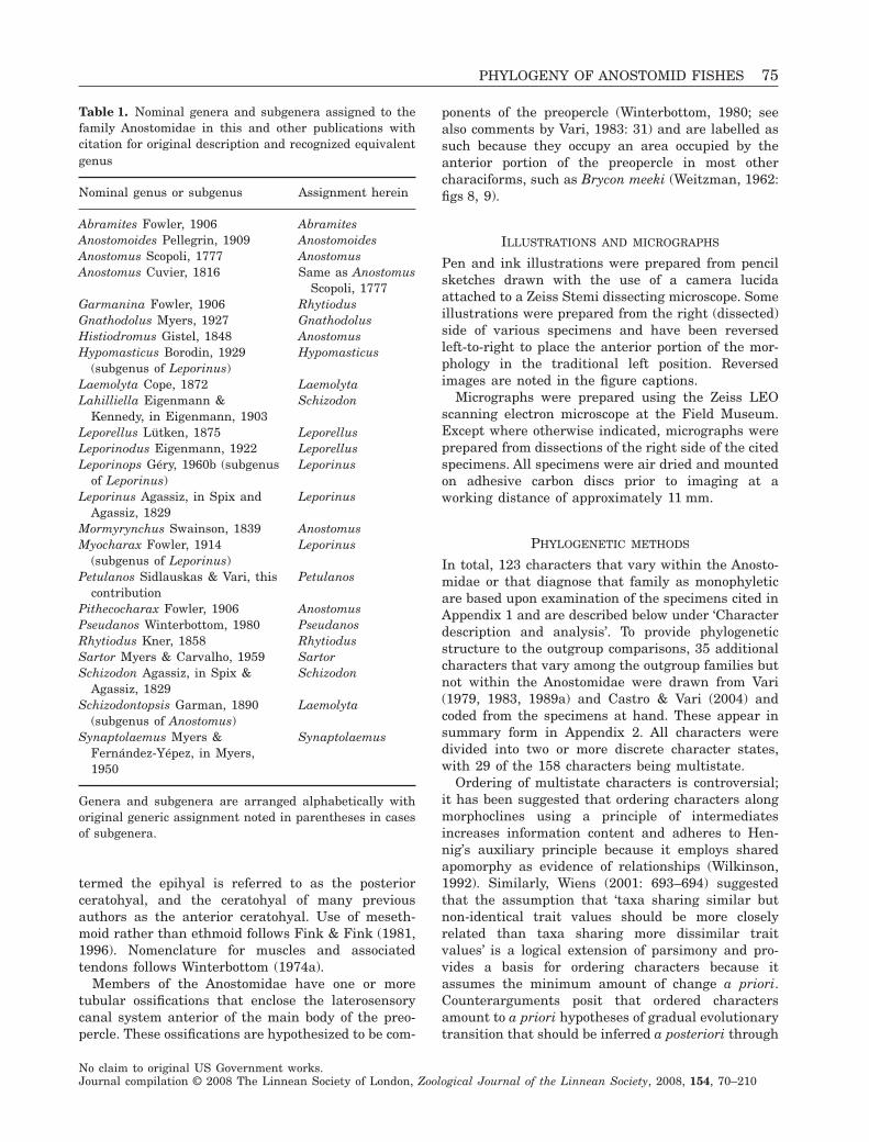
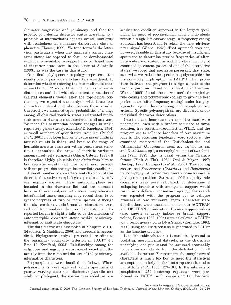
Sartor (Myers & Carvalho, 1959: figs 1–3; Sidlauskas2007: fig. 2D). Although some of the highly modifiedgenera such as Sartor and Synaptolaemus (Fig. 3)have limited geographical distributions, small bodysizes (c. 0.1 m standard length (SL)) and are appar-ently rare in nature based on museum holdings, othergenera [e.g. Anostomoides, Laemolyta, Leporinus(Fig. 4), Rhytiodus and Schizodon] are geographicallywidespread and have large body sizes up to approxi-mately 0.3 m SL. The larger species are commonlyexploited in commercial and artesanal food fisheries(Goulding, 1980, 1981). Many anostomids feed largelyon aquatic vegetation (Goulding, Leal Carvalho &Ferreira, 1988); other species have more diversediets including larval and adult insects, nematodes,sponges, larval fishes and detritus (Knöeppel, 1972;Winterbottom, 1974b; Saul, 1975; Taphorn, 1992;Santos & Rosa, 1998; Balassa et al., 2004).
The first anostomids known to western science weredescribed by Linnaeus (1758) who based his speciesdescriptions on specimens previously reported on byGronovius (1756). Günther (1864) first united variousanostomid genera in his ‘Anostomina’, but the nextcentury brought little agreement about relationshipsof what is now recognized as the Anostomidae to othertaxa. Boulenger’s Anostominae (1904) significantlyexpanded the Anostomina of Günther (1864) toinclude a number of taxa now assigned to other fami-lies (see summary in Vari, 1998), but none of thosetaxa is currently considered to be closely related tothe Anostomidae. Regan (1911) proposed a familyAnostomidae that encompassed the Anostomidae,Chilodontidae, Curimatidae and Prochilodontidae ofpresent concepts, an insightful grouping undercurrent hypotheses of relationships (Vari, 1983).
Figure 1. Rhytiodus microlepis, USNM 229095, 121.5 mmSL; Brazil, Amazonas, Ilha da Marchantaria; lateralview.
Figure 2. Abramites hypselonotus, MCZ 59240, 98.6 mmSL; Ecuador, Río Punino, above Coca; lateral view.
Figure 3. Synaptolaemus cingulatus, AMNH 91095,78 mm SL; Venezuela, Bolivar, Río Paraguay, at CarapoRapids, just upriver of mouth of Río Carapo; lateral view.
Figure 4. Leporinus friderici, FMNH 116829, 180.5 mmSL; Suriname, Rechter Coppename River; lateral view.
PHYLOGENY OF ANOSTOMID FISHES 71
No claim to original US Government works.Journal compilation © 2008 The Linnean Society of London, Zoological Journal of the Linnean Society, 2008, 154, 70–210
Gregory & Conrad (1938) recognized a broader Anos-tomidae consisting of Regan’s (1911) family of thatname plus taxa now included by Langeani (1998) inthe Hemiodontidae (sensu Greenwood et al., 1966).Géry (1961a: 108), reiterating the concept of Regan(1911), united the present Anostomidae (his Anos-tominae) with the Chilodontidae, Prochilodontidaeand Curimatidae (his Chilodinae, Prochilodontinaeand Curimatinae), without providing explicit evidencein support of that grouping. Roberts (1973: 221)hypothesized an explicit relationship between theAnostomidae and Prochilodontidae. Subsequently,Vari (1983: 50) detailed numerous synapomorphies insupport of the hypothesis that the Anostomidae wasthe sister group to the Chilodontidae rather than theProchilodontidae as proposed by Roberts (1973).
Hypotheses of intrafamilial phylogenetic relation-ships are less well developed. Winterbottom (1980)addressed the species-level problems and phyloge-netic questions within his Anostominae [Pseudanos,Gnathodolus, Sartor, Synaptolaemus and Anostomus(the last with Mormyrhynchus, Histiodromus andPithecocharax as synonyms)]. This group encom-passes the most externally distinctive species in thefamily, with the opening of the oral jaws positioned onthe dorsal surface of the head (Fig. 3; Sidlauskas,2007: fig. 2D). The genera Abramites, Anostomoides,Hypomasticus, Leporellus, Leporinus, Rhytiodus andSchizodon have a more generalized external morphol-ogy (Figs 1, 2, 4) with the notable exception of somemembers of the genus Schizodon (e.g. Schizodonnasutus) and Hypomasticus that possess strongly sub-terminal mouths (Steindachner, 1875: pl. 6; Borodin,1929; Géry, 1960a; Britski, 1976). The seven generacited above include the majority of the species of thefamily and were traditionally delimited by differencesin the dentition, degree of caudal-fin squamation andbody proportions.
Following Winterbottom’s (1980) study of theassemblage that he recognized as the Anostominae,Santos & Jégu (1987) described two new species ofSartor and Sidlauskas & Santos (2005) described anew species of Pseudanos. Abramites (Vari & Will-iams, 1987) and Laemolyta (Mautari & Menezes,2006) have been revised fully, while partial revisionshave been undertaken for Anostomoides (Santos &Zuanon, 2006), Hypomasticus (Santos, Jégu & Lima,1996) Leporinus (Garavello, 1988; Garavello &Britski, 1988; Géry, Planquette & Le Bail, 1988;Garavello, 1989, 1990; Santos & Jégu, 1996), Rhytio-dus (Géry, 1987) and Schizodon (Bergmann, 1988);Garavello & Britski, 1990; Vari & Raredon, 1991;Sidlauskas, Garavello & Jellen, 2007). Nonetheless,most genera and subgenera outside of Winterbottom’sAnostominae, including the most species-rich taxa(e.g. Leporinus), have not been revised comprehen-
sively. The partial revisionary studies cited abovehave yielded many previously undescribed species,and numerous questions remain as to which of theapproximately 140 nominal species should be recog-nized and how they can be discriminated (Garavello& Britski, 2003). These issues and others preclude anattempt to develop a species-level phylogeny in theAnostomidae at this time.
This study had four goals:
1. to propose a hypothesis of higher level phyloge-netic relationships within the Anostomidae;
2. to define the nominal genera in the Anostomidaeon the basis of derived features;
3. to determine the number of times that major tran-sitions in jaw orientation have evolved within theAnostomidae; and
4. to evaluate the historical biogeography of theAnostomidae within the context of the recon-structed phylogeny.
MATERIAL AND METHODS
Institutional abbreviations: The following abbrevia-tions for institutions and collections are used: AMNH,American Museum of Natural History, New York;ANSP, Academy of Natural Sciences of Philadelphia;CAS, California Academy of Sciences, San Francisco;CAS-SU, Former Stanford University collections, nowin CAS; FMNH, Field Museum of Natural History,Chicago; INHS, Illinois Natural History Survey,Champaign; INPA, Instituto Nacional de Pesquisasda Amazônia, Manaus; MBUCV, Museo de Biología dela Universidad Central de Venezuela, Caracas; MCZ,Museum of Comparative Zoology, Harvard University,Cambridge; MZUSP, Museu de Zoologia da Univer-sidade de São Paulo; NMW, NaturhistorischesMuseum Wien, Vienna; UFSC, Universidade Federalde São Carlos; UMMZ, University of MichiganMuseum of Zoology, Ann Arbor; USNM, NationalMuseum of Natural History, Smithsonian Institution,Washington, DC.
TAXONOMIC SAMPLING AND NOMENCLATURE
The assemblage herein recognized as the Anostomi-dae is equivalent to the familial concept of Greenwoodet al. (1966), Vari (1983) and Nelson (2006) and to theAnostominae of Eschmeyer (1990, 1998). Family-levelnames for Neotropical outgroups are those utilized inCLOFFSCA (Reis, Kullander & Ferraris, 2003).
We examined the morphology of 46 species repre-senting all recognized genera and subgenera in theAnostomidae (Appendix 1) as well as 14 outgroups.Close outgroups include the Chilodontidae (threespecies examined), which is the sister group to the
72 B. L. SIDLAUSKAS and R. P. VARI
No claim to original US Government works.Journal compilation © 2008 The Linnean Society of London, Zoological Journal of the Linnean Society, 2008, 154, 70–210
Anostomidae (Vari, 1983: 50), the Curimatidae (threespecies examined) and the Prochilodontidae (twospecies examined). These last two families form aclade (Vari, 1983: 46, 1989b: 51; Castro & Vari, 2004:59) which is the sister group to the Anostomidae plusChilodontidae (Vari, 1983: 46; Castro & Vari, 2004:58). Character state distributions in more distantoutgroups were drawn from skeletal material andpublished descriptions of skeletal and externalanatomy for Parodon (Pavanelli, 1999, one speciesexamined), Hemiodus (Vari, 1982a, one species exam-ined), Brycon (Weitzman, 1962, one species exam-ined), Citharinus (Daget, 1962b, one speciesexamined), Distichodus (Daget, 1959, one speciesexamined) and Xenocharax (Daget, 1960, onespecies examined), the last of which was hypothesizedby Vari (1979) to be the most basal and generalizedmember of the family Distichodontidae, which is, inturn, basal within the Characiformes (Fink & Fink,1981; Orti & Meyer, 1996; Buckup, 1998; Calcagnotto,Schaefer & DeSalle, 2005). A full list of examinedoutgroup material, including comparative materialnot formally coded, appears in Appendix 1. Appendix1 also contains taxonomic authorship for all examinedingroup and outgroup species.
Because diagnoses for anostomid species vary con-siderably in detail and accuracy, it is necessary todiscuss the assignment of some material to terminaltaxa (nominally species). The genus Leporellus hasnever been satisfactorily revised, although several ofits nominal species have been suggested to be in thesynonymy of Leporellus vittatus and Leporelluspictus (Garavello & Britski, 2003). There is no pub-lished diagnosis between L. pictus and L. vittatus,and we assigned specimens to these two species-groups based on geography and separation amongdrainages. None of the nominal species of Leporellushas type localities in the Río Orinoco, but Leporellusvittatus has a type locality of ‘Amazon River, Brazil’(Cuvier & Valenciennes, 1850), which is confluentwith the Orinoco via Venezuela’s Casiquiare canal.Based on the confluence between the Amazon andOrinoco, we assigned the specimens from theOrinoco to Leporellus vittatus, albeit provisionally.Because Leporellus pictus has a type locality of‘Irisanga’ (= Orissanga) in the rivers of São Paulostate, Brazil, which lie outside the Amazon drainage,we assigned the specimens that originated in SãoPaulo state to Leporellus pictus.
The cleared and stained specimens of Leporinusagassizi come from the Río Napo of Ecuador and theRío Nanay of Peru, localities distant from the typelocality (Amazon River, Tabatinga and Tefé, Brazil).Leporinus agassizi has a large purported range for ananostomid, including Ecuador, Guyana, Peru, Venezu-ela and Amazonian Brazil. The name may refer to a
species complex, and as such, the examined materialfrom the far west of the continent may not be con-specific with the true Leporinus agassizi, which has atype locality in the central Amazon.
Fowler (1914) erected the subgenus Myocharax ofLeporinus on the basis of the tusk-like symphysealdentary teeth possessed by the type species,L. desmotes, from Guyana. Ovchynnyk (1971) laterdescribed the very similar L. jatuncochi from Amazo-nian Ecuador with no reference to, or comparisonwith, L. desmotes. Based on the two descriptions thereis no way to separate the two species other thanbased on geography, and L. jatuncochi may be in thesynonymy of L. desmotes. We assigned our clearedand stained material of the L. desmotes/jatuncochicomplex from Amazonian Peru to L. jatuncochi basedon proximity of the collecting locality to the typelocalities, recognizing that L. jatuncochi may prove tobe a junior synonym.
One of the cleared and stained specimens of Lep-orinus fasciatus from the Río Orinoco (FMNH 103450)was taken from a lot of specimens that, in hindsight,appears to contain more than one species. The jarcontains several juvenile Leporinus specimens ofsimilar standard lengths that, nonetheless, displayvariable banding patterns. Some of the juvenile speci-mens in the lot, including the cleared and stainedspecimen, may belong to one of the other bandedspecies of Leporinus that occur in the Río Orinocosystem (e.g. L. falcipinnis and L. yophorus). To reflectthe uncertainty in this identification, the clearedand stained specimen from FMNH 103450 appearsas Leporinus cf. fasciatus, while specimens moreconfidently assignable to Leporinus fasciatus(USNM 103847, 225991, 233574) appear without thatqualification.
The Río Napo basin of eastern Ecuador is home toa species of Leporinus that closely resembles theparatypes of L. ecuadorensis housed at FMNH, butL. ecuadorensis has a known distribution restricted tothe Pacific versant on the western side of the Andes inEcuador (Eigenmann & Henn, 1916). No other recog-nized species in the Anostomidae or its proximateoutgroups (Chilodontidae, Curimatidae, Prochilodon-tidae) has a geographical distribution spanning theAndes Cordilleras. The Río Napo species may beundescribed, or it may be synonymous with L. ecua-dorensis or one of several other similar species suchas L. friderici from Suriname. We identify the RíoNapo material throughout this contribution asL. cf. ecuadorensis, an assignation which reflectsits similarity to and potential synonymy withL. ecuadorensis.
Leporinus friderici (Fig. 4) has a very wide citeddistribution including Brazil, French Guiana, Guyana,Suriname, Trinidad and Tobago (Garavello & Britski,
PHYLOGENY OF ANOSTOMID FISHES 73
No claim to original US Government works.Journal compilation © 2008 The Linnean Society of London, Zoological Journal of the Linnean Society, 2008, 154, 70–210
2003). The cleared and stained specimen that appearsherein as Leporinus friderici (USNM 225409) comesfrom the inexact type locality (Suriname). Materialfrom elsewhere in South America (for example, the dryskeleton INPA 21605 from the fish market of Manaus,Brazil) appears as Leporinus cf. friderici.
The subgenus Leporinops of Leporinus currentlycontains two small-bodied, poorly known species, L.moralesi (the type species) and L. jamesi, both fromthe Amazon River of Brazil (Géry, 1960b, 1977). Thesubgenus was distinguished by a notched morphol-ogy of the symphyseal teeth of the premaxilla, withthe two species diagnosed on minor details of col-oration. The holotype of L. jamesi was unavailablefor examination and searches of major museumsfailed to reveal any additional specimens of thatspecies. These searches also yielded only very oldspecimens of L. moralesi (MCZ 19409 and 19476,both collected in 1865 as part of the Thayer expe-dition) that were unsuitable for clearing and stain-ing. The UMMZ collection did yield a probable newspecies of this subgenus from Bahia, Brazil, in thefar east of the continent. The material from Bahiapossesses the divided symphyseal premaxillary teethand small body size that characterize Leporinops,but the Bahia specimens differ from the reportedcolour patterns for L. jamesi and L. moralesi andwere collected far outside the known geographicalrange of the two described species. More detailedcomparison is impossible without access to freshmaterial of L. jamesi and L. moralesi. The Bahiamaterial appears in this contribution as L. cf.moralesi.
The cleared and stained specimen of Leporinus cf.niceforoi, FMNH 102150, originated in the Río Napoof Ecuador, outside the purported range of L. niceforoiin the Río Orteguasa basin of Colombia. The speci-mens from the Rio Tocantins of Brazil cited as Lep-orinus pellegrinii (INPA 15672) may be synonymouswith L. maculatus (see Géry et al., 1988).
The two described species of small-scaled Rhytio-dus, R. microlepis and R. lauzannei, are diagnosedby a difference in body depth, with R. lauzannei thedeeper-bodied (Géry, 1987). Rhytiodus microlepis iscited from the Amazon River basin with a typelocality of Manaus, Brazil (Kner, 1858; Garavello &Britski, 2003), while R. lauzannei was described asendemic to the Río Mamoré of Bolivia (Géry, 1987).Many specimens from near Manaus have bodydepths in the cited ranges for R. lauzannei, whilesome from the Río Mamoré have body depths in thecited range for R. microlepis. The status of thesenominal species must await careful study. Forcurrent purposes, material was identified usingGéry’s (1987) plots of body depth versus standardlength.
NOMENCLATURAL CHANGES
As a result of the phylogenetic conclusions arrived atin this study, we make three revisions to the genus-level nomenclature within the Anostomidae:
• elevation of Hypomasticus from a subgenus ofLeporinus to a genus containing the followingspecies most recently assigned to Leporinus: L.despaxi, L. garmani, L. julii, L. megalepis, L.mormyrops, L. pachycheilus and L. thayeri;
• restriction of Anostomus to A. anostomus, A. anos-tomus longus, A. brevior and A. ternetzi; and
• erection of a new genus, Petulanos, containing thefollowing species most recently assigned to Anosto-mus: A. intermedius, A. plicatus and A. spiloclis-tron, with A. plicatus as the type species.
We do not utilize Leporinops and Myocharax, bothnominal subgenera of Leporinus, in light of the lack ofresolution for the portion of the phylogeny involvingLeporinus. The full list of nominal genera and sub-genera and their synonyms appears in Table 1. Weemploy this revised classification throughout theremainder of this paper. For a full discussion of therationale behind these changes and a formal diagno-sis of Petulanos, please see the section on ‘Changes toclassification and comparisons with previous classi-fications’ following the discussion of phylogeneticresults and support.
OSTEOLOGICAL PREPARATIONS AND TERMINOLOGY
Samples of all species included as terminal taxawere cleared and counterstained (C&S) for cartilageand bone using the method outlined by Taylor & VanDyke (1985). Whenever possible, two or more speci-mens of each species were prepared to facilitatechecks for anomalous features. One side of each C&Sspecimen was dissected according to Fink & Weitz-man (1974) during character analysis. Data fromexternal morphology including pigmentation pat-terns and scale counts was based on the examinationof alcohol-preserved specimens. Typically these werenon-C&S members of the same lots from which theosteological material was prepared. Additional lotsexamined as alcohol-preserved specimens only areindicated by (A) in Appendix 1. A few specimenswere prepared as dry skeletons and are indicated by(DS) in Appendix 1.
Osteological terminology is that of Weitzman (1962)with the following modifications. Vomer is usedinstead of prevomer and intercalar is used instead ofopisthotic following the practice in recent studies ofthe Characiformes (Langeani, 1998; Zanata & Toledo-Piza, 2004; Zanata & Vari, 2005). The use of epioc-cipital rather than epiotic follows Patterson (1975).Following Nelson (1969), the ossification traditionally
74 B. L. SIDLAUSKAS and R. P. VARI
No claim to original US Government works.Journal compilation © 2008 The Linnean Society of London, Zoological Journal of the Linnean Society, 2008, 154, 70–210
termed the epihyal is referred to as the posteriorceratohyal, and the ceratohyal of many previousauthors as the anterior ceratohyal. Use of meseth-moid rather than ethmoid follows Fink & Fink (1981,1996). Nomenclature for muscles and associatedtendons follows Winterbottom (1974a).
Members of the Anostomidae have one or moretubular ossifications that enclose the laterosensorycanal system anterior of the main body of the preo-percle. These ossifications are hypothesized to be com-
ponents of the preopercle (Winterbottom, 1980; seealso comments by Vari, 1983: 31) and are labelled assuch because they occupy an area occupied by theanterior portion of the preopercle in most othercharaciforms, such as Brycon meeki (Weitzman, 1962:figs 8, 9).
ILLUSTRATIONS AND MICROGRAPHS
Pen and ink illustrations were prepared from pencilsketches drawn with the use of a camera lucidaattached to a Zeiss Stemi dissecting microscope. Someillustrations were prepared from the right (dissected)side of various specimens and have been reversedleft-to-right to place the anterior portion of the mor-phology in the traditional left position. Reversedimages are noted in the figure captions.
Micrographs were prepared using the Zeiss LEOscanning electron microscope at the Field Museum.Except where otherwise indicated, micrographs wereprepared from dissections of the right side of the citedspecimens. All specimens were air dried and mountedon adhesive carbon discs prior to imaging at aworking distance of approximately 11 mm.
PHYLOGENETIC METHODS
In total, 123 characters that vary within the Anosto-midae or that diagnose that family as monophyleticare based upon examination of the specimens cited inAppendix 1 and are described below under ‘Characterdescription and analysis’. To provide phylogeneticstructure to the outgroup comparisons, 35 additionalcharacters that vary among the outgroup families butnot within the Anostomidae were drawn from Vari(1979, 1983, 1989a) and Castro & Vari (2004) andcoded from the specimens at hand. These appear insummary form in Appendix 2. All characters weredivided into two or more discrete character states,with 29 of the 158 characters being multistate.
Ordering of multistate characters is controversial;it has been suggested that ordering characters alongmorphoclines using a principle of intermediatesincreases information content and adheres to Hen-nig’s auxiliary principle because it employs sharedapomorphy as evidence of relationships (Wilkinson,1992). Similarly, Wiens (2001: 693–694) suggestedthat the assumption that ‘taxa sharing similar butnon-identical trait values should be more closelyrelated than taxa sharing more dissimilar traitvalues’ is a logical extension of parsimony and pro-vides a basis for ordering characters because itassumes the minimum amount of change a priori.Counterarguments posit that ordered charactersamount to a priori hypotheses of gradual evolutionarytransition that should be inferred a posteriori through
Table 1. Nominal genera and subgenera assigned to thefamily Anostomidae in this and other publications withcitation for original description and recognized equivalentgenus
Nominal genus or subgenus Assignment herein
Abramites Fowler, 1906 AbramitesAnostomoides Pellegrin, 1909 AnostomoidesAnostomus Scopoli, 1777 AnostomusAnostomus Cuvier, 1816 Same as Anostomus
Scopoli, 1777Garmanina Fowler, 1906 RhytiodusGnathodolus Myers, 1927 GnathodolusHistiodromus Gistel, 1848 AnostomusHypomasticus Borodin, 1929
(subgenus of Leporinus)Hypomasticus
Laemolyta Cope, 1872 LaemolytaLahilliella Eigenmann &
Kennedy, in Eigenmann, 1903Schizodon
Leporellus Lütken, 1875 LeporellusLeporinodus Eigenmann, 1922 LeporellusLeporinops Géry, 1960b (subgenus
of Leporinus)Leporinus
Leporinus Agassiz, in Spix andAgassiz, 1829
Leporinus
Mormyrynchus Swainson, 1839 AnostomusMyocharax Fowler, 1914
(subgenus of Leporinus)Leporinus
Petulanos Sidlauskas & Vari, thiscontribution
Petulanos
Pithecocharax Fowler, 1906 AnostomusPseudanos Winterbottom, 1980 PseudanosRhytiodus Kner, 1858 RhytiodusSartor Myers & Carvalho, 1959 SartorSchizodon Agassiz, in Spix &
Agassiz, 1829Schizodon
Schizodontopsis Garman, 1890(subgenus of Anostomus)
Laemolyta
Synaptolaemus Myers &Fernández-Yépez, in Myers,1950
Synaptolaemus
Genera and subgenera are arranged alphabetically withoriginal generic assignment noted in parentheses in casesof subgenera.
PHYLOGENY OF ANOSTOMID FISHES 75
No claim to original US Government works.Journal compilation © 2008 The Linnean Society of London, Zoological Journal of the Linnean Society, 2008, 154, 70–210
character congruence and parsimony, and that thepractice of ordering character states according to aprinciple of intermediates equates overall similaritywith relatedness in a manner dangerously close tophenetics (Hauser, 1992). We tend towards the latterview, particularly when only similarity among char-acter states (as opposed to fossil or developmentalevidence) is available to support a priori hypothesesof character state trees in the sense of Slowinski(1993), as was the case in this study.
Our final phylogenetic topology represents theresults of analysis with all characters unordered. Todetermine whether ordering the four multistate char-acters (17, 46, 72 and 77) that include clear interme-diate states and deal with size, extent or rotation ofskeletal elements would alter the topological con-clusions, we repeated the analysis with those fourcharacters ordered and also discuss those results.Conversely, we assumed equal probabilities of changeamong all observed meristic states and treated multi-state meristic characters as unordered in all analyses.We made this assumption because changes in singleregulatory genes (Leary, Allendorf & Knudsen, 1984)or small numbers of quantitative trait loci (Peichelet al., 2001) have been known to cause major shifts inmeristic counts in fishes, and because the range ofheritable meristic variation within populations some-times approaches the range of meristic variationamong closely related species (Ahn & Gibson, 1999). Itis therefore highly plausible that shifts from high tolow meristic counts and vice versa may proceedwithout progression through intermediate conditions.
A small number of characters and character statesdescribe distinctive morphologies possessed by onlyone ingroup species. These autapomorphies areincluded in the character list and are discussedbecause future analyses with more comprehensiveintrafamilial taxon sampling may reveal them to besynapomorphies of two or more species. Althoughthe six parsimony-uninformative characters wereexcluded from analysis, the overall consistency indexreported herein is slightly inflated by the inclusion ofautapomorphic character states within parsimony-informative multistate characters.
The data matrix was assembled in Mesquite v. 1.12(Maddison & Maddison, 2006) and appears in Appen-dix 3. Phylogenetic analysis proceeded according tothe parsimony optimality criterion in PAUP* 4.0Beta 10 (Swofford, 2003). Relationships among theoutgroups and ingroups were reconstructed simulta-neously from the combined dataset of 152 parsimony-informative characters.
Polymorphisms were handled as follows. Wherepolymorphisms were apparent among specimens ofgreatly varying sizes (i.e. distinctive juvenile andadult morphologies), the species was coded as pos-
sessing the condition apparent in the largest speci-mens. In cases of polymorphism among individualswithin a single life-history stage, a frequency codingapproach has been found to retain the most phyloge-netic signal (Wiens, 1995). That approach was not,however, feasible in this study because of insufficientspecimens to determine precise frequencies of alter-native observed states. Instead, if a clear majority ofexamined specimens possessed one of the alternativestates, we coded that species as possessing that state;otherwise we coded the species as polymorphic (themstaxa = polymorph option in PAUP*). That proce-dure instructs the program to assign a state to thetaxon a posteriori based on its position in the tree.Wiens (1995) found these two methods (majority-rule coding and polymorphic coding) to give the bestperformance (after frequency coding) under his phy-logenetic signal, bootstrapping and sampling-errorcriteria. Specific polymorphisms are discussed underindividual character descriptions.
One thousand heuristic searches of treespace wereundertaken, each with a random sequence of taxonaddition, tree bisection–reconnection (TBR), and theprogram set to collapse branches of zero maximumlength. The resulting topologies were rooted at theexamined members of the Distichodontidae andCitharinidae (Xenocharax spilurus, Citharinus sp.and Distichodus sp.), a monophyletic unit of two fami-lies (Vari, 1979) that is basal within the Characi-formes (Fink & Fink, 1981; Orti & Meyer, 1997;Buckup, 1998; Calcagnotto et al., 2005). This rootingconstrained Xenocharax, Citharinus and Distichodusto monophyly; all other taxa were unconstrained inphylogenetic position. Strict and 50% majority ruleconsensus trees were calculated. To determine ifcollapsing branches with ambiguous support wouldresult in a different consensus topology, the searchwas repeated with the program set to collapsebranches of zero minimum length. Character statedistributions were examined using both ACCTRANand DELTRAN optimization. Bremer support values(also known as decay indices or branch supportvalues, Bremer 1988, 1994) were calculated in PAUP*via a script generated in DNA Stacks (Eernisse, 1992,2000) using the strict consensus generated in PAUP*as the baseline topology.
It is debatable whether it is statistically sound tobootstrap morphological datasets, as the charactersunderlying analysis cannot be assumed reasonablyto be drawn randomly from the distribution of allavailable characters. Furthermore, the sample size ofcharacters is much too low to meet the statisticalassumptions underlying the bootstrap (see discussionin Kitching et al., 1998: 129–131) In the interests ofcompleteness 250 bootstrap replicates were per-formed in PAUP*, each comprising ten heuristic
76 B. L. SIDLAUSKAS and R. P. VARI
No claim to original US Government works.Journal compilation © 2008 The Linnean Society of London, Zoological Journal of the Linnean Society, 2008, 154, 70–210
searches with all characters unordered, TBR, randomaddition sequences and character resampling withreplacement across the entire dataset. To restrict thesearch to a reasonable length of time each individualbootstrap replicate was limited to 1000 000 trees.
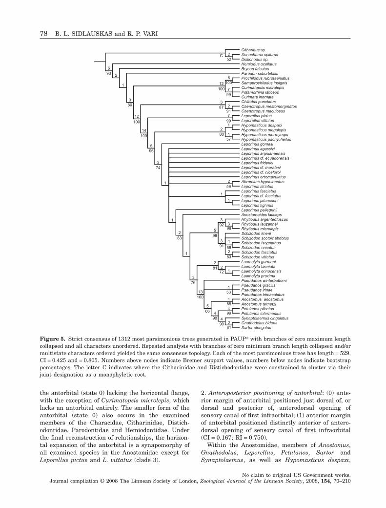
PHYLOGENETIC RESULTS
Analysis of the data matrix (Appendix 3) in PAUP*with branches of zero maximum length collapsed andmultistate characters unordered yielded 1312 mostparsimonious trees of length 529 steps in a singletree island, which was discovered in 991 of the 1000addition sequence replicates. The other nine replicatesfound shortest trees of 531 steps; results from theseruns were not retained. Each most parsimonious treehad a CI = 0.425 and RI = 0.805. The repeated analysiswith branches of zero minimum length collapsedyielded 240 most parsimonious trees in a single treeisland. Parallel analyses with multistate characters17, 46, 72 and 77 ordered obtained 1200 mostparsimonious trees (collapsing branches with zeromaximum length) or 240 most parsimonious trees(collapsing branches with zero minimum length),again in a single tree island discovered in 99% of allheuristic replicates. Each of the most parsimonioustrees resulting from analyses with multistate charac-ters ordered had length 531, CI = 0.424 and RI = 0.805.
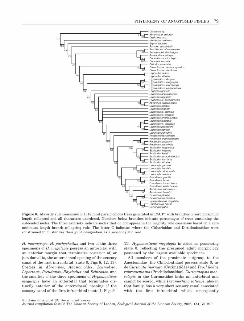
All four analyses discussed above yielded identicalstrict consensus topologies (Fig. 5). All four majorityrule consensus trees returned fundamentally similartopologies (Fig. 6), although the percentages of treescontaining nodes not present in the strict consensusvaried slightly among the four treatments. Threenodes that appear in the majority rule consensusesunder the zero maximum length branch collapsingrule are unresolved in the majority rule consensusesbased on the zero minimum length branch collapsingrule; these are indicated with asterisks in Figure 6.
In general the relative strengths of Bremer supportand bootstrap values, which appear above and belowthe nodes in the strict consensus (Fig. 5), respectively,were concordant across the phylogeny. For example,all nodes receiving Bremer values of 3 or higher havebootstrap values of 74 or more, while nodes withBremer values of 1 typically have bootstrap valuesbelow 60.
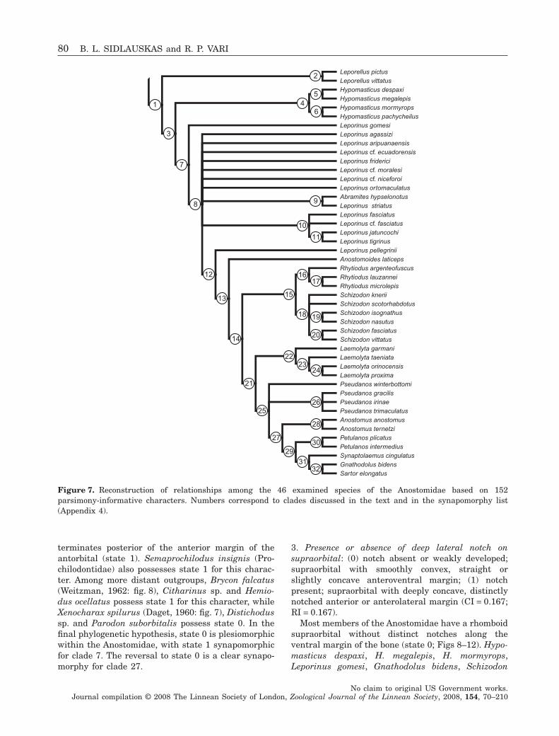
Overall, phylogenetic results were robust to changesin branch collapsing rule and ordering of characters.We consider the strict consensus topology and associ-ated support values returned by the unordered analy-sis with branches of maximum length zero collapsed(Fig. 5) to be the final hypothesis of relationshipsarrived at herein. The 32 ingroup clades in the finalphylogenetic hypothesis are numbered in Figure 7 andreferenced in the character descriptions and analyses
that follow. A complete list of synapomorphies for eachnumbered clade appears in Appendix 4.
CHARACTER DESCRIPTION AND ANALYSIS
Each of the 123 morphological characters describedbelow varied within the Anostomidae or was found tobe informative about the monophyly of the Anosto-midae. Discussions are arranged by discrete bodysystems in an overall anterior to posterior pattern.Each character description includes the name of thecharacter, descriptions of the character states, theconsistency and retention indices on the final phylo-genetic hypothesis (Fig. 5), a discussion of the distri-bution of states among the terminal taxa, and theoptimization of the character on the final phylogenywith reference to the numbered nodes in Figure 7. Insome instances, citations of figures from the literatureinvolve congeners rather than the exact outgroupspecies examined as skeletal preparations and codedin the data matrix. For example, we often cite Weitz-man’s (1962) illustrations of the skeleton of Bryconmeeki in our discussion of its very similar congener,B. falcatus, which we used as an outgroup.
ANTORBITAL, SUPRAORBITAL AND INFRAORBITALS
1. Presence or absence of horizontal flange of antor-bital: (0) antorbital small, vertically orientated andrunning posterior to nasal cavity with only slighthorizontal expansion along ventral margin; (1) antor-bital relatively large, with distinct horizontallydirected flange running ventral to nasal cavity; flatbony plate may join vertical and horizontal portions ofantorbital to varying degrees (CI = 1.000; RI = 1.000).
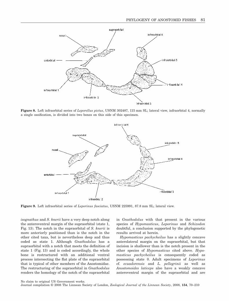
Within the Anostomidae, Leporellus pictus andL. vittatus uniquely possess a small antorbital in theshape of a vertically orientated bar which is positionedjust posterior of the lateral opening into the nasalcavity (state 0; Fig. 8). All other examined members ofthe Anostomidae have an antorbital with an additionalhorizontally directed flange running ventral to theaperture (state 1; Figs 9–13). That flange is absent inthe species of Leporellus. The morphology of the antor-bital varies greatly within the Anostomidae outside ofLeporellus, ranging from a thin, somewhat L-shapedossification in some species of Leporinus (e.g. L. fas-ciatus, Fig. 9) to a wide, plate-like ossification border-ing the entire ventral, lateral and posterior margins ofthe nasal cavity (e.g. Petulanos plicatus, Fig. 12).Other than for the presence or absence of the horizon-tal flange, much of this variation in antorbital mor-phology proved impossible to parse discretely.
All examined members of the proximate and secondoutgroups to the Anostomidae (Chilodontidae, Curi-matidae, Prochilodontidae) have the smaller form of
PHYLOGENY OF ANOSTOMID FISHES 77
No claim to original US Government works.Journal compilation © 2008 The Linnean Society of London, Zoological Journal of the Linnean Society, 2008, 154, 70–210
the antorbital (state 0) lacking the horizontal flange,with the exception of Curimatopsis microlepis, whichlacks an antorbital entirely. The smaller form of theantorbital (state 0) also occurs in the examinedmembers of the Characidae, Citharinidae, Distich-odontidae, Parodontidae and Hemiodontidae. Underthe final reconstruction of relationships, the horizon-tal expansion of the antorbital is a synapomorphy ofall examined species in the Anostomidae except forLeporellus pictus and L. vittatus (clade 3).
2. Anteroposterior positioning of antorbital: (0) ante-rior margin of antorbital positioned just dorsal of, ordorsal and posterior of, anterodorsal opening ofsensory canal of first infraorbital; (1) anterior marginof antorbital positioned distinctly anterior of antero-dorsal opening of sensory canal of first infraorbital(CI = 0.167; RI = 0.750).
Within the Anostomidae, members of Anostomus,Gnathodolus, Leporellus, Petulanos, Sartor andSynaptolaemus, as well as Hypomasticus despaxi,
Citharinus sp.Xenocharax spilurusDistichodus sp.Hemiodus ocellatusBrycon falcatusParodon suborbitalisProchilodus rubrotaeniatusSemaprochilodus insignisCurimatopsis microlepisPotamorhina laticepsCurimata inornataChilodus punctatusCaenotropus mestomorgmatosCaenotropus maculosusLeporellus pictusLeporellus vittatusHypomasticus despaxiHypomasticus megalepisHypomasticus mormyropsHypomasticus pachycheilusLeporinus gomesiLeporinus agassiziLeporinus aripuanaensisLeporinus cf. ecuadorensisLeporinus fridericiLeporinus cf. moralesiLeporinus cf. niceforoiLeporinus ortomaculatusAbramites hypselonotusLeporinus striatusLeporinus fasciatusLeporinus cf. fasciatusLeporinus jatuncochiLeporinus tigrinusLeporinus pellegriniiAnostomoides laticepsRhytiodus argenteofuscusRhytiodus lauzanneiRhytiodus microlepisSchizodon kneriiSchizodon scotorhabdotusSchizodon isognathusSchizodon nasutusSchizodon fasciatusSchizodon vittatusLaemolyta garmaniLaemolyta taeniataLaemolyta orinocensisLaemolyta proximaPseudanos winterbottomiPseudanos gracilisPseudanos irinaePseudanos trimaculatusAnostomus anostomusAnostomus ternetziPetulanos plicatusPetulanos intermediusSynaptolaemus cingulatusGnathodolus bidensSartor elongatus
56
88
99
8190
90
86
53100
76
8172
9299
98
91 56
83
63
74
96
100 8057
99
8791
99
100
100
80
100
93
C522
8
7
12
3
2
1
3
12
5
2
7
21
114
6
3
1
1
2
1
3
13
5
4
42
4
1
1
22
1
5
3
33
1
2
1
1
2
Figure 5. Strict consensus of 1312 most parsimonious trees generated in PAUP* with branches of zero maximum lengthcollapsed and all characters unordered. Repeated analysis with branches of zero minimum branch length collapsed and/ormultistate characters ordered yielded the same consensus topology. Each of the most parsimonious trees has length = 529,CI = 0.425 and = 0.805. Numbers above nodes indicate Bremer support values, numbers below nodes indicate bootstrappercentages. The letter C indicates where the Citharinidae and Distichodontidae were constrained to cluster via theirjoint designation as a monophyletic root.
78 B. L. SIDLAUSKAS and R. P. VARI
No claim to original US Government works.Journal compilation © 2008 The Linnean Society of London, Zoological Journal of the Linnean Society, 2008, 154, 70–210
H. mormyrops, H. pachycheilus and two of the threespecimens of H. megalepis possess an antorbital withan anterior margin that terminates posterior of, orjust dorsal to, the anterodorsal opening of the sensorycanal of the first infraorbital (state 0; Figs 8, 12, 13).Species in Abramites, Anostomoides, Laemolyta,Leporinus, Pseudanos, Rhytiodus and Schizodon andthe smallest of the three specimens of Hypomasticusmegalepis have an antorbital that terminates dis-tinctly anterior of the anterodorsal opening of thesensory canal of the first infraorbital (state 1; Figs 9–
11). Hypomasticus megalepis is coded as possessingstate 0, reflecting the presumed adult morphologypossessed by the largest available specimens.
All members of the proximate outgroup to theAnostomidae (the Chilodontidae) possess state 0, asdo Curimata inornata (Curimatidae) and Prochilodusrubrotaeniatus (Prochilodontidae). Curimatopsis mac-rolepis in the Curimatidae lacks an antorbital andcannot be scored, while Potamorhina laticeps, also inthat family, has a very short sensory canal associatedwith the first infraorbital which consequently
Citharinus sp.Xenocharax spilurusDistichodus sp.Hemiodus ocellatusBrycon falcatusParodon suborbitalisProchilodus rubrotaeniatusSemaprochilodus insignisPotamorhina laticepsCurimatopsis microlepisCurimata inornataChilodus punctatusCaenotropus mestomorgmatosCaenotropus maculosusLeporellus pictusLeporellus vittatusHypomasticus despaxiHypomasticus megalepisHypomasticus mormyropsHypomasticus pachycheilusLeporinus gomesiLeporinus aripuanaensisLeporinus agassiziLeporinus cf. ecuadorensisAbramites hypselonotusLeporinus striatusLeporinus fridericiLeporinus cf. moralesiLeporinus cf. niceforoiLeporinus ortomaculatusLeporinus fasciatusLeporinus cf. fasciatusLeporinus jatuncochiLeporinus tigrinusLeporinus pellegriniiAnostomoides laticepsRhytiodus argenteofuscusRhytiodus lauzanneiRhytiodus microlepisSchizodon isognathusSchizodon nasutusSchizodon kneriiSchizodon scotorhabdotusSchizodon fasciatusSchizodon vittatusLaemolyta garmaniLaemolyta taeniataLaemolyta orinocensisLaemolyta proximaPseudanos gracilisPseudanos irinaePseudanos trimaculatusPseudanos winterbottomiAnostomus anostomusAnostomus ternetziPetulanos plicatusPetulanos intermediusSynaptolaemus cingulatusGnathodolus bidensSartor elongatus
77
67
79
75
65
79
69
52
54*
*
*
100
100
100
100
100
100
100
100
100
100
100C
100100
100100
100
100
100
100
100100
100
100
100
100100
100
100
100
100100
100
100
100100
100
100
100
100
Figure 6. Majority rule consensus of 1312 most parsimonious trees generated in PAUP* with branches of zero maximumlength collapsed and all characters unordered. Numbers below branches indicate percentages of trees containing thesubtended nodes. The three asterisks indicate nodes that do not appear in the majority rule consensus based on a zerominimum length branch collapsing rule. The letter C indicates where the Citharinidae and Distichodontidae wereconstrained to cluster via their joint designation as a monophyletic root.
PHYLOGENY OF ANOSTOMID FISHES 79
No claim to original US Government works.Journal compilation © 2008 The Linnean Society of London, Zoological Journal of the Linnean Society, 2008, 154, 70–210
terminates posterior of the anterior margin of theantorbital (state 1). Semaprochilodus insignis (Pro-chilodontidae) also possesses state 1 for this charac-ter. Among more distant outgroups, Brycon falcatus(Weitzman, 1962: fig. 8), Citharinus sp. and Hemio-dus ocellatus possess state 1 for this character, whileXenocharax spilurus (Daget, 1960: fig. 7), Distichodussp. and Parodon suborbitalis possess state 0. In thefinal phylogenetic hypothesis, state 0 is plesiomorphicwithin the Anostomidae, with state 1 synapomorphicfor clade 7. The reversal to state 0 is a clear synapo-morphy for clade 27.
3. Presence or absence of deep lateral notch onsupraorbital: (0) notch absent or weakly developed;supraorbital with smoothly convex, straight orslightly concave anteroventral margin; (1) notchpresent; supraorbital with deeply concave, distinctlynotched anterior or anterolateral margin (CI = 0.167;RI = 0.167).
Most members of the Anostomidae have a rhomboidsupraorbital without distinct notches along theventral margin of the bone (state 0; Figs 8–12). Hypo-masticus despaxi, H. megalepis, H. mormyrops,Leporinus gomesi, Gnathodolus bidens, Schizodon
Leporellus pictusLeporellus vittatusHypomasticus despaxiHypomasticus megalepisHypomasticus mormyropsHypomasticus pachycheilusLeporinus gomesiLeporinus agassiziLeporinus aripuanaensisLeporinus cf. ecuadorensisLeporinus fridericiLeporinus cf. moralesiLeporinus cf. niceforoiLeporinus ortomaculatusAbramites hypselonotusLeporinus striatusLeporinus fasciatusLeporinus cf. fasciatusLeporinus jatuncochiLeporinus tigrinusLeporinus pellegriniiAnostomoides laticepsRhytiodus argenteofuscusRhytiodus lauzanneiRhytiodus microlepisSchizodon kneriiSchizodon scotorhabdotusSchizodon isognathusSchizodon nasutusSchizodon fasciatusSchizodon vittatusLaemolyta garmaniLaemolyta taeniataLaemolyta orinocensisLaemolyta proximaPseudanos winterbottomiPseudanos gracilisPseudanos irinaePseudanos trimaculatusAnostomus anostomusAnostomus ternetziPetulanos plicatusPetulanos intermediusSynaptolaemus cingulatusGnathodolus bidensSartor elongatus
32
1
2
3
45
6
7
8 9
10
11
12
13
14
15
1617
18 19
20
21
2223
24
2526
27
28
2930
31
Figure 7. Reconstruction of relationships among the 46 examined species of the Anostomidae based on 152parsimony-informative characters. Numbers correspond to clades discussed in the text and in the synapomorphy list(Appendix 4).
80 B. L. SIDLAUSKAS and R. P. VARI
No claim to original US Government works.Journal compilation © 2008 The Linnean Society of London, Zoological Journal of the Linnean Society, 2008, 154, 70–210
isognathus and S. knerii have a very deep notch alongthe anteroventral margin of the supraorbital (state 1,Fig. 13). The notch in the supraorbital of S. knerii ismore anteriorly positioned than is the notch in theother cited taxa, but is nevertheless deep and thuscoded as state 1. Although Gnathodolus has asupraorbital with a notch that meets the definition ofstate 1 (Fig. 13) and is coded accordingly, the wholebone is restructured with an additional ventralprocess intersecting the flat plate of the supraorbitalthat is typical of other members of the Anostomidae.The restructuring of the supraorbital in Gnathodolusrenders the homology of the notch of the supraorbital
in Gnathodolus with that present in the variousspecies of Hypomasticus, Leporinus and Schizodondoubtful, a conclusion supported by the phylogeneticresults arrived at herein.
Hypomasticus pachycheilus has a slightly concaveanterolateral margin on the supraorbital, but thatincision is shallower than is the notch present in theother species of Hypomasticus cited above. Hypo-masticus pachycheilus is consequently coded aspossessing state 0. Adult specimens of Leporinuscf. ecuadorensis and L. pellegrinii as well asAnostomoides laticeps also have a weakly concaveanteroventral margin of the supraorbital and are
Figure 8. Left infraorbital series of Leporellus pictus, USNM 302487, 115 mm SL; lateral view, infraorbital 4, normallya single ossification, is divided into two bones on this side of this specimen.
Figure 9. Left infraorbital series of Leporinus fasciatus, USNM 225991, 87.8 mm SL; lateral view.
PHYLOGENY OF ANOSTOMID FISHES 81
No claim to original US Government works.Journal compilation © 2008 The Linnean Society of London, Zoological Journal of the Linnean Society, 2008, 154, 70–210
coded as having state 0. All remaining members of theAnostomidae and all examined outgroup taxa lack anotch in the ventral margin of the supraorbital.
Gregory & Conrad (1938: 348) erroneously reportedthat the supraorbital was lacking in Leporinus. Thismistaken observation probably resulted from theirbasing their drawing and observation on a dry skel-eton that presumably had lost that ossification duringpreparation.
Optimization of this character on the final phylog-eny is largely ambiguous. The notched condition
clearly originated once in Gnathodolus, and the poly-tomy in clade 18 makes it impossible to know whethera notched antorbital evolved once or twice in Schiz-odon. Near the base of the phylogeny the notch eitherevolved in the common ancestor of clade 3 with sub-sequent loss in Hypomasticus pachycheilus and clade8, or originated independently in Hypomasticusmormyrops, Leporinus gomesi and clade 5.
4. Orientation of sensory canal of first infraorbital: (0)origin of sensory canal of first infraorbital in region
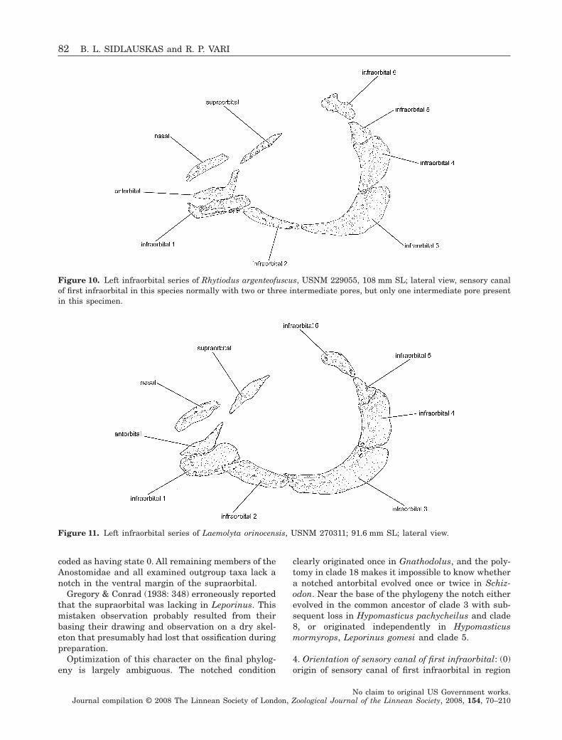
Figure 10. Left infraorbital series of Rhytiodus argenteofuscus, USNM 229055, 108 mm SL; lateral view, sensory canalof first infraorbital in this species normally with two or three intermediate pores, but only one intermediate pore presentin this specimen.
Figure 11. Left infraorbital series of Laemolyta orinocensis, USNM 270311; 91.6 mm SL; lateral view.
82 B. L. SIDLAUSKAS and R. P. VARI
No claim to original US Government works.Journal compilation © 2008 The Linnean Society of London, Zoological Journal of the Linnean Society, 2008, 154, 70–210
proximate to antorbital positioned anterior of distalterminus of sensory canal where that canal contactssensory canal of second infraorbital; (1) origin ofsensory canal of first infraorbital situated directlydorsal to, or posterodorsal to, distal terminus of canalof first infraorbital where that canal contacts canal ofsecond infraorbital (CI = 1.000; RI = 1.000).
In Abramites, Anostomoides, Hypomasticus,Laemolyta, Leporellus, Leporinus, Rhytiodus andSchizodon, the primary laterosensory canal segment
of the first infraorbital canal has a horizontal orien-tation, with the canal beginning near the antorbital,running ventrally for a short distance through theanterior portion of the first infraorbital and thencontinuing posteriorly to contact the anterior openingof the canal of the second infraorbital (Figs 8–11).In Anostomus, Gnathodolus, Petulanos, Pseudanos,Sartor and Synaptolaemus the canal runs from thepore proximate to the antorbital anteroventrallytowards the snout for at least a short distance before
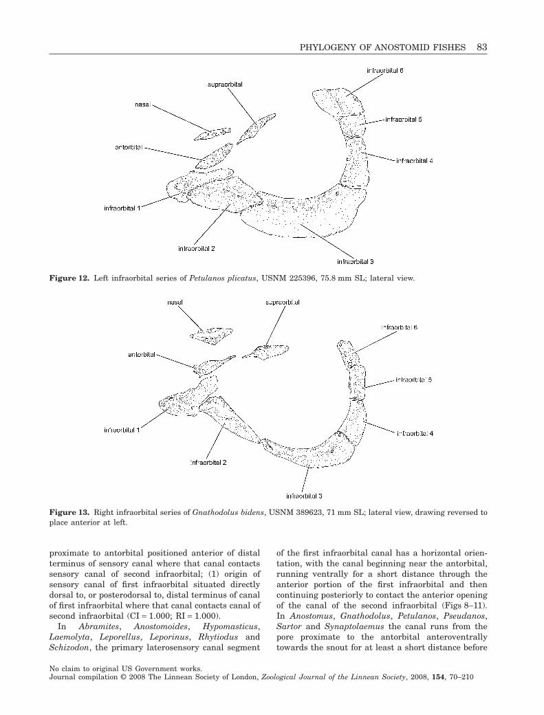
Figure 12. Left infraorbital series of Petulanos plicatus, USNM 225396, 75.8 mm SL; lateral view.
Figure 13. Right infraorbital series of Gnathodolus bidens, USNM 389623, 71 mm SL; lateral view, drawing reversed toplace anterior at left.
PHYLOGENY OF ANOSTOMID FISHES 83
No claim to original US Government works.Journal compilation © 2008 The Linnean Society of London, Zoological Journal of the Linnean Society, 2008, 154, 70–210
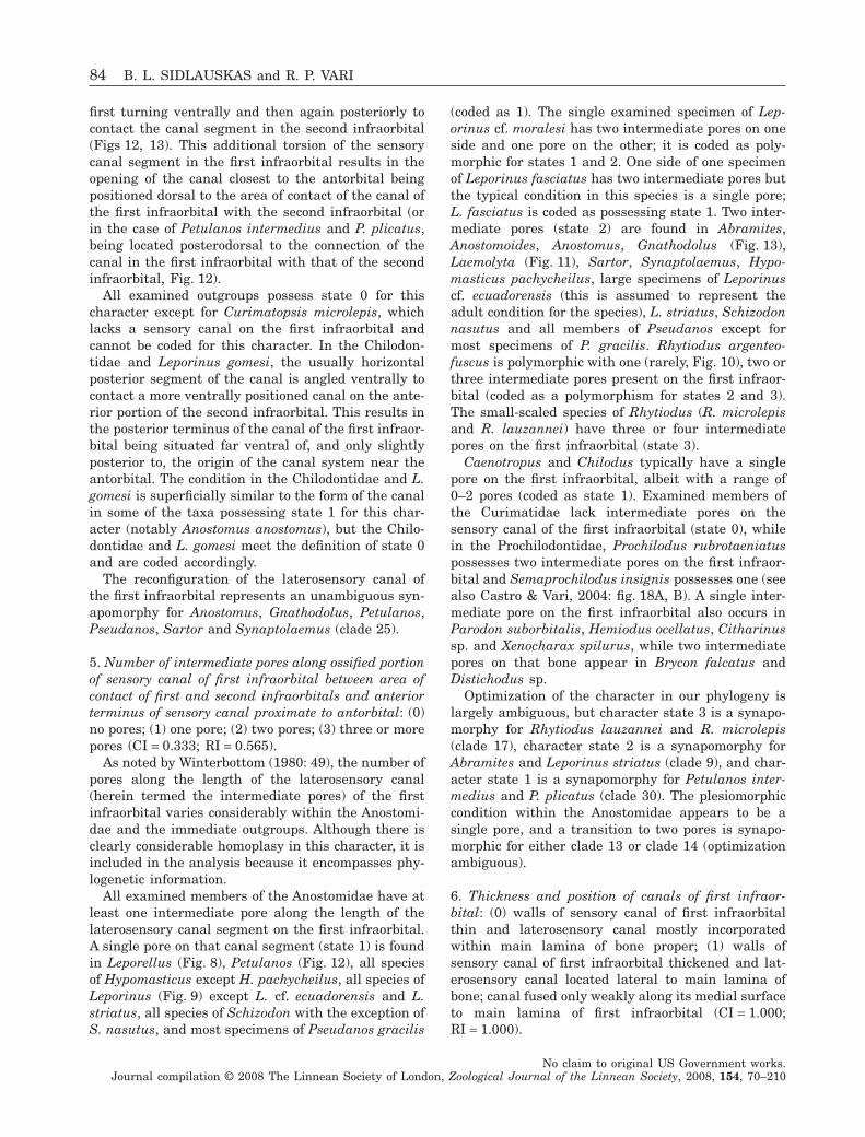
first turning ventrally and then again posteriorly tocontact the canal segment in the second infraorbital(Figs 12, 13). This additional torsion of the sensorycanal segment in the first infraorbital results in theopening of the canal closest to the antorbital beingpositioned dorsal to the area of contact of the canal ofthe first infraorbital with the second infraorbital (orin the case of Petulanos intermedius and P. plicatus,being located posterodorsal to the connection of thecanal in the first infraorbital with that of the secondinfraorbital, Fig. 12).
All examined outgroups possess state 0 for thischaracter except for Curimatopsis microlepis, whichlacks a sensory canal on the first infraorbital andcannot be coded for this character. In the Chilodon-tidae and Leporinus gomesi, the usually horizontalposterior segment of the canal is angled ventrally tocontact a more ventrally positioned canal on the ante-rior portion of the second infraorbital. This results inthe posterior terminus of the canal of the first infraor-bital being situated far ventral of, and only slightlyposterior to, the origin of the canal system near theantorbital. The condition in the Chilodontidae and L.gomesi is superficially similar to the form of the canalin some of the taxa possessing state 1 for this char-acter (notably Anostomus anostomus), but the Chilo-dontidae and L. gomesi meet the definition of state 0and are coded accordingly.
The reconfiguration of the laterosensory canal ofthe first infraorbital represents an unambiguous syn-apomorphy for Anostomus, Gnathodolus, Petulanos,Pseudanos, Sartor and Synaptolaemus (clade 25).
5. Number of intermediate pores along ossified portionof sensory canal of first infraorbital between area ofcontact of first and second infraorbitals and anteriorterminus of sensory canal proximate to antorbital: (0)no pores; (1) one pore; (2) two pores; (3) three or morepores (CI = 0.333; RI = 0.565).
As noted by Winterbottom (1980: 49), the number ofpores along the length of the laterosensory canal(herein termed the intermediate pores) of the firstinfraorbital varies considerably within the Anostomi-dae and the immediate outgroups. Although there isclearly considerable homoplasy in this character, it isincluded in the analysis because it encompasses phy-logenetic information.
All examined members of the Anostomidae have atleast one intermediate pore along the length of thelaterosensory canal segment on the first infraorbital.A single pore on that canal segment (state 1) is foundin Leporellus (Fig. 8), Petulanos (Fig. 12), all speciesof Hypomasticus except H. pachycheilus, all species ofLeporinus (Fig. 9) except L. cf. ecuadorensis and L.striatus, all species of Schizodon with the exception ofS. nasutus, and most specimens of Pseudanos gracilis
(coded as 1). The single examined specimen of Lep-orinus cf. moralesi has two intermediate pores on oneside and one pore on the other; it is coded as poly-morphic for states 1 and 2. One side of one specimenof Leporinus fasciatus has two intermediate pores butthe typical condition in this species is a single pore;L. fasciatus is coded as possessing state 1. Two inter-mediate pores (state 2) are found in Abramites,Anostomoides, Anostomus, Gnathodolus (Fig. 13),Laemolyta (Fig. 11), Sartor, Synaptolaemus, Hypo-masticus pachycheilus, large specimens of Leporinuscf. ecuadorensis (this is assumed to represent theadult condition for the species), L. striatus, Schizodonnasutus and all members of Pseudanos except formost specimens of P. gracilis. Rhytiodus argenteo-fuscus is polymorphic with one (rarely, Fig. 10), two orthree intermediate pores present on the first infraor-bital (coded as a polymorphism for states 2 and 3).The small-scaled species of Rhytiodus (R. microlepisand R. lauzannei) have three or four intermediatepores on the first infraorbital (state 3).
Caenotropus and Chilodus typically have a singlepore on the first infraorbital, albeit with a range of0–2 pores (coded as state 1). Examined members ofthe Curimatidae lack intermediate pores on thesensory canal of the first infraorbital (state 0), whilein the Prochilodontidae, Prochilodus rubrotaeniatuspossesses two intermediate pores on the first infraor-bital and Semaprochilodus insignis possesses one (seealso Castro & Vari, 2004: fig. 18A, B). A single inter-mediate pore on the first infraorbital also occurs inParodon suborbitalis, Hemiodus ocellatus, Citharinussp. and Xenocharax spilurus, while two intermediatepores on that bone appear in Brycon falcatus andDistichodus sp.
Optimization of the character in our phylogeny islargely ambiguous, but character state 3 is a synapo-morphy for Rhytiodus lauzannei and R. microlepis(clade 17), character state 2 is a synapomorphy forAbramites and Leporinus striatus (clade 9), and char-acter state 1 is a synapomorphy for Petulanos inter-medius and P. plicatus (clade 30). The plesiomorphiccondition within the Anostomidae appears to be asingle pore, and a transition to two pores is synapo-morphic for either clade 13 or clade 14 (optimizationambiguous).
6. Thickness and position of canals of first infraor-bital: (0) walls of sensory canal of first infraorbitalthin and laterosensory canal mostly incorporatedwithin main lamina of bone proper; (1) walls ofsensory canal of first infraorbital thickened and lat-erosensory canal located lateral to main lamina ofbone; canal fused only weakly along its medial surfaceto main lamina of first infraorbital (CI = 1.000;RI = 1.000).
84 B. L. SIDLAUSKAS and R. P. VARI
No claim to original US Government works.Journal compilation © 2008 The Linnean Society of London, Zoological Journal of the Linnean Society, 2008, 154, 70–210
The three examined species of Rhytiodus possess anunusual morphology of the sensory canal of the firstinfraorbital unique among the examined speciesinside and outside the Anostomidae. In Rhytiodus thewalls of this canal are thickened and overall the canalhas the appearance of a tube located lateral to, albeitfused medially to, the main lamina of the bone alongthe medial one-third of the canal (state 1; Fig. 10).
Members of several other anostomid genera(notably the species of Schizodon, many species ofLeporinus, and Laemolyta garmani) also have some-what thickened or raised sensory canals of the firstinfraorbital, but in none of these taxa are the condi-tions nearly as pronounced as the thickening andlateral displacement found in Rhytiodus. No exam-ined members of the outgroups possess a comparableraised and thickened condition of the canal. In thefinal hypothesis of relationships, this character opti-mizes as a synapomorphy for the species of Rhytiodus(clade 16).
7. Extent of development of dorsal lamina of secondinfraorbital: (0) lamina located dorsal to sensorycanal absent or poorly developed with maximumheight less than diameter of sensory canal; (1) laminalocated dorsal to sensory canal well developed withmaximum height of dorsal lamina greater than diam-eter of sensory canal along at least part of length ofcanal (CI = 0.200; RI = 0.500).
Most members of the Anostomidae have a highlydeveloped triangular or rounded lamina of boneextending dorsal of the ossified sensory canal of thesecond infraorbital (state 1, Figs 8, 9, 11–13). In Rhy-tiodus, Schizodon nasutus, S. fasciatus, S. scotorhab-dotus and S. vittatus, this lamina is alternativelyeither very narrow or entirely absent (state 0, Fig. 10).In Laemolyta taeniata and L. proxima, the dorsallamina is poorly developed but still present, with thewidth of the lamina greater than the diameter of thesensory canal for at least part of the length of the canal(coded as state 1). In Anostomoides laticeps, Laemolytagarmani, L. orinocensis, Schizodon isognathus andS. knerii, the dorsal lamina is present and wider thanthe canal, with the bulk of this process anteriorlypositioned and the lamina effectively absent along theposterior portion of the bone (coded as state 1). Thevarious species of Leporinus and Hypomasticuspossess a well-developed dorsal lamina on the secondinfraorbital but vary considerably in the form of thatlamina. In some cases (e.g. L. striatus) the greatestwidth of the lamina is anteriorly positioned while otherspecies of Leporinus have the lamina widest above themidpoint of the horizontal length of that bone. Despitethe variation in the form of the dorsal lamina on thesecond infraorbital, we found it impossible to parseunambiguously that variation into discrete character
states and elected to code only the presence or absenceof a dorsal lamina greater in width than the width ofthe associated sensory canal.
The form of the second infraorbital varies widelyamong the proximate outgroups to the Anostomidae.The dorsal flange is well developed in most membersof the Chilodontidae, e.g. Chilodus punctatus (Vari,Castro & Raredon, 1995: fig. 1B) albeit narrower inCaenotropus mestomorgmatos (Vari et al., 1995:fig. 1A), and narrow and anteriorly positioned but stillpresent in the examined members of the Prochilodon-tidae (Castro & Vari, 2004: fig. 18A–C) and Curima-tidae (Vari, 1991: fig. 9A, B). The lamina is alsopresent in the distant outgroups Brycon (Weitzman,1962: fig. 9), Hemiodus, Parodon and Distichodus, butabsent in Citharinus sp. and Xenocharax spilurus.
The plesiomorphic condition of the character in theAnostomidae appears to be state 1 (lamina well devel-oped). The reduction of the dorsal lamina of thesecond infraorbital is a synapomorphy for Rhytiodusplus Schizodon (clade 15), with a subsequent reversalto a well-developed lamina in Schizodon knerii andS. isognathus.
8. Number of intermediate pores along ossified portionof sensory canal of second infraorbital: (0) no pores;(1) one pore; (2) two pores; (3) three pores (CI = 0.333;RI = 0.630).
Anostomids have 1–3 intermediate pores presentalong the body of the sensory canal of the secondinfraorbital. All examined species of Abramites, Anos-tomus, Gnathodolus, Laemolyta, Pseudanos, Sartorand Synaptolaemus, along with Hypomasticus pachy-cheilus, Rhytiodus argenteofuscus, Leporinus striatusand Schizodon nasutus have only a single intermediatepore present along the length of the body of the sensorycanal of the second infraorbital (state 1; Figs 10, 11,13). Leporinus tigrinus has three intermediate poreson the canal of the second infraorbital (state 3). Theremaining species of Hypomasticus and Leporinus(Fig. 9) as well as Anostomoides, Leporellus (Fig. 8),Petulanos (Fig. 12), Schizodon knerii, S. fasciatus, S.scotorhabdotus and S. vittatus have two intermediatepores along the length of the sensory canal (state 2).Although some specimens of Rhytiodus microlepishave one intermediate pore on the second infraorbital,the majority have two pores and this species is codedas state 2. Rhytiodus lauzannei and Schizodon isog-nathus are polymorphic for states 2 and 3.
The examined proximate outgroups vary in thenumber of intermediate pores along the sensory canalof the second infraorbital. The first outgroup (Chilo-dontidae) is variable for the feature with one inter-mediate pore present in Chilodus punctatus (Variet al., 1995: fig. 1B) and two intermediate pores occur-ring in the species of Caenotropus (Vari et al., 1995:
PHYLOGENY OF ANOSTOMID FISHES 85
No claim to original US Government works.Journal compilation © 2008 The Linnean Society of London, Zoological Journal of the Linnean Society, 2008, 154, 70–210
fig. 1A). Members of the Curimatidae usually have asingle intermediate pore, although some species inthe family have a second pore (Vari, 1991: fig. 9) andwith the examined specimens of the basal Curimatop-sis macrolepis lacking an intermediate pore on thisbone. Semaprochilodus insignis in the Prochilodon-tidae has three intermediate pores, with two or occa-sionally three pores present in other species of thatgenus along with Prochilodus and Ichthyoelephas (seeCastro & Vari, 2004: fig. 18). Among more distantoutgroups, Parodon suborbitalis, Hemiodus ocellatus,Citharinus sp. and Xenocharax spilurus possess asingle intermediate pore on the second infraorbital,Distichodus sp. possesses two pores, and Brycon fal-catus lacks an intermediate pore on that bone.
In the final reconstruction character state 2 is ple-siomorphic within the Anostomidae. State 1 is a syna-pomorphy for clades 9 and 21 with a reversal to state2 synapomorphic for clade 30 (Petulanos).
9. Fusion of fourth and fifth infraorbitals: (0) unfused;(1) fused (CI = 0.333; RI = 0.200).
The typical condition of the fourth and fifth infraor-bitals in the Characiformes, including most membersof the Anostomidae, is for these elements to be presentas separate ossifications (state 0). These two bonesoccasionally fuse in anomalous individuals of speciesthat otherwise have the ossifications separate (seecondition in Pseudanos gracilis; Sidlauskas & Santos,2005: fig. 2A). In light of such intraspecific variation,we scored the fused condition as being present onlyfor those specimens in which the conjoined fourth andfifth infraorbitals were observed on both sides ofthat individual. Hypomasticus despaxi, H. megalepis,Leporinus aripuanaensis, L. ortomaculatus, L. pelle-grinii and L. tigrinus exhibit invariant fusion of thefourth and fifth infraorbitals (state 1). Laemolytaorinocensis is polymorphic for this character. One ofthe two examined cleared and stained specimens of L.orinocensis has a fusion of the fourth and fifth infraor-bitals on both sides of the head, whereas the otherspecimen retains unfused infraorbitals on both sides.The remaining examined species in the Anostomidaehave separate fourth and fifth infraorbitals.
The fourth and fifth infraorbitals are separate inproximate outgroups to the Anostomidae (Curima-tidae, see Vari, 1991: fig. 9; Chilodontidae, see Variet al., 1995: fig. 1; Prochilodontidae, see Castro &Vari, 2004: fig. 18). Autogenous fourth and fifthinfraorbitals are also present in the Parodontidae(Roberts, 1974: 61–62), Hemiodontidae [e.g. Hemio-dus ocellatus (Vari, 1982a)], the Citharinidae [e.g.Citharidium (Daget, 1962a: fig. 10) and Citharinus(Daget (1962b: fig. 7)] and the Distichodontidae [e.g.Paradistichodus (Daget, 1958: fig. 9) and Xenocharax(Daget, 1960: fig. 7)].
Under the final hypothesis of relationships, thefusion of the fourth and fifth infraorbitals is a syna-pomorphy for Hypomasticus despaxi and H. megalepis(clade 5). The relationship of Leporinus aripuanaensisto L. ortomaculatus is unresolved due to the polytomyin clade 8, but the fusion of the fourth and fifthinfraorbitals is potentially synapomorphic for thosetwo species. The independent fusions of the fourthand fifth infraorbitals in Leporinus tigrinus and L.pellegrinii are homoplasies with respect to the fusionsin Hypomasticus and in the other cited species ofLeporinus.
10. Relative height of fourth and fifth infraorbitals:(0) vertical extent of fourth and fifth infraorbitalsapproximately equal or fifth infraorbital longer alongvertical axis than fourth; (1) fourth infraorbital muchlonger along vertical axis than fifth infraorbital(CI = 0.333; RI = 0.917).
In the typical characiform condition the verticalextents of the fourth and fifth infraorbitals areapproximately equal and both contribute to the pos-terior margin of the orbit to the same approximatedegree (state 0). This condition, or a condition inwhich the fifth infraorbital is longer along the verticalaxis than is the fourth, occurs in all examined out-groups (e.g. Chilodontidae, Vari et al., 1995: fig. 1).
Like the outgroups, members of Hypomasticus,Leporellus and Leporinus possess a large fifth in-fraorbital. Leporellus has an abbreviated fourthinfraorbital that is slightly shorter in vertical extentthan the fifth infraorbital (state 0; Fig. 8, noting thatthe fourth infraorbital is anomalously separated intotwo ossifications in the illustrated specimen). Manyspecies of Leporinus possess fourth and fifth infraor-bitals of roughly equivalent vertical extents (state 0,Figs 9, 14). Several species in Hypomasticus and Lep-orinus cannot be coded for this character because ofthe fusion of the fourth and fifth infraorbitals (seecharacter 9). In Abramites, Anostomoides, Anostomus,Laemolyta, Petulanos, Pseudanos, Rhytiodus, Sartor,Schizodon and Synaptolaemus, the fourth infraorbitalis vertically distinctly more developed than the fifthand forms a much greater portion of the posteriormargin of the orbit (state 1, Figs 10–12). In Gna-thodolus the fourth and fifth infraorbitals are verynarrow relative to the condition in other anostomids,but of about equivalent heights (Fig. 13). Gnathodolusis thus coded as state 0 for this character.
State 1 for this character (fourth infraorbital muchlonger along vertical axis than fifth) is a synapomorphyfor either clade 12 or 13 in the final reconstruction. Theambiguity is due to Leporinus pellegrinii (a componentof clade 12 but not clade 13), which has fused fourthand fifth infraorbitals and cannot be coded for thischaracter. The occurrence of state 1 (fourth infraorbital
86 B. L. SIDLAUSKAS and R. P. VARI
No claim to original US Government works.Journal compilation © 2008 The Linnean Society of London, Zoological Journal of the Linnean Society, 2008, 154, 70–210
longer along vertical axis than is the fifth infraorbital)in Abramites is a homoplasy relative to its occurrencein either clade 12 or 13 and is possibly correlated withthe great increase in body depth in that taxon. Thedeeply nested Gnathodolus bidens has reverted to theplesiomorphic condition of fourth and fifth infraorbit-als of approximately equivalent heights.
11. Degree of development of flange of fifth infraorbitalposterior of sensory canal: (0) flange present alongentire margin of bone; (1) flange less extensive orentirely absent, with at least dorsal section of flangemissing (CI = 0.143; RI = 0.538).
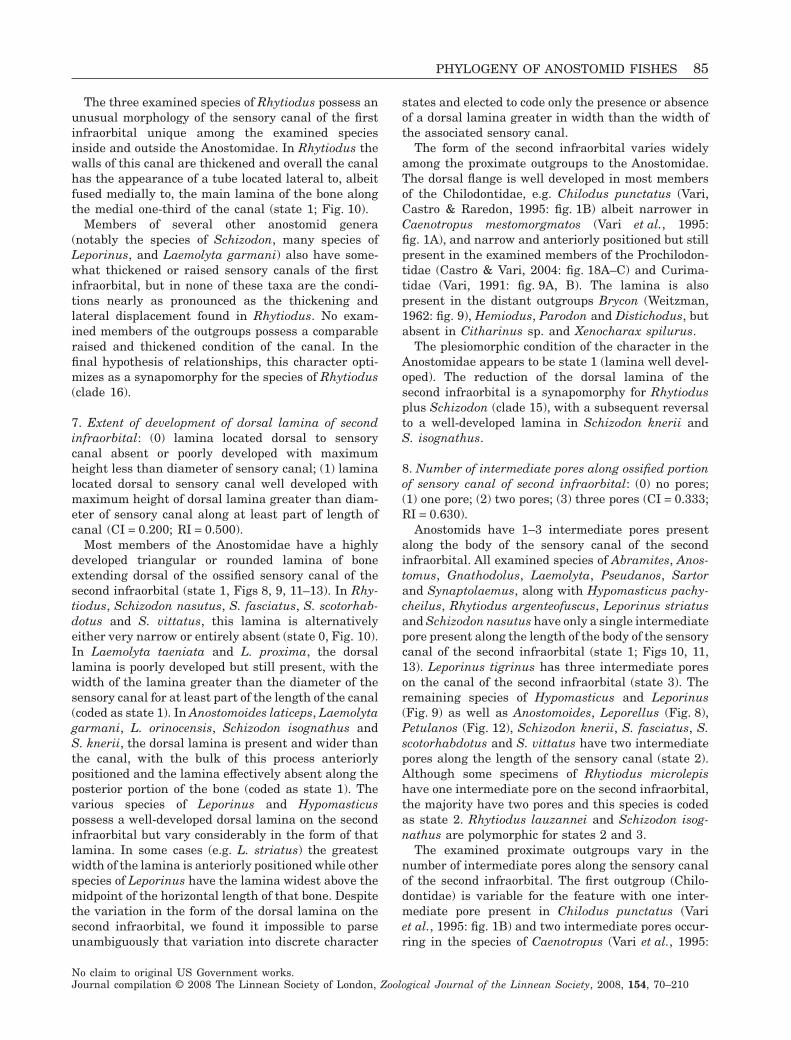
The fifth infraorbital in the Anostomidae commonlyhas the form of a flat plate with a dorsoventrallyaligned sensory canal running through the anteriorportion of the bone and with a wide ossified laminaextending posterior of the posterior margin of thesensory canal (Figs 8, 9, 12, 13; Winterbottom, 1980:figs 30, 32; Sidlauskas & Santos, 2005: fig. 2). Rhy-tiodus argenteofuscus and the species of Laemolytalack the dorsal portion of this posterior lamina. Theposterodorsal limit of the sensory canal consequentlyforms the posterodorsal margin of the fifth infraor-bital (state 1; Figs 10, 11). In Rhytiodus lauzanneiand R. microlepis the entire posterior lamina of thefifth infraorbital is missing, with the posterior marginof that ossification completely delimited by the pos-terior wall of the sensory canal (also coded as state 1).
Among the proximate outgroups to the Anostomi-dae, the posterior lamina on the fifth infraorbital isabsent in the Chilodontidae in which that ossifica-tion is reduced to a tubular ossification (Vari et al.,1995: fig. 1), but present and fully developed in theProchilodontidae (Castro & Vari, 2004: fig. 18).Within the Curimatidae a well-developed posteriorlamina is absent in Curimatopsis microlepis, presentin Potamorhina (Vari, 1989a: fig. 35B) and greatlyreduced or absent in more derived components ofthat family including Curimata inornata, Stein-dachnerina (Vari, 1991: fig. 9), Psectrogaster (Vari,1989a: fig. 34A) and Curimatella (Vari, 1989a:fig. 34C). Among more distant outgroups, the laminais present in the Parodontidae (Roberts, 1974: figs61, 62), Hemiodus ocellatus, Xenocharax spilurus(Daget, 1960: fig. 7) and Brycon (Weitzman, 1962:fig. 8). In light of that information and that factthat both Laemolyta and Rhytiodus appear in arelatively derived position in the final hypothesis ofrelationships for the Anostomidae, the presence ofa complete posterior lamina on the fifth infraorbitalis most parsimoniously hypothesized to be the ple-siomorphic condition for the clade consisting ofthe Anostomidae, Chilodontidae, Curimatidae andProchilodontidae, with a reduction of the laminahaving arisen independently in the Chilodontidae,and some components of the Curimatidae andAnostomidae.
Figure 14. Dorsal portions of left infraorbital series, opercular series and pectoral girdle of Leporinus agassizi, FMNH102219, 98.6 mm SL; lateral view.
PHYLOGENY OF ANOSTOMID FISHES 87
No claim to original US Government works.Journal compilation © 2008 The Linnean Society of London, Zoological Journal of the Linnean Society, 2008, 154, 70–210
The final phylogenetic hypothesis suggests that thereduction or elimination of the posterior lamina of thefifth infraorbital arose separately in Laemolyta (clade22) and Rhytiodus (clade 16). Despite the globalhomoplasy, character state 1 is synapomorphic foreach of these clades considered individually.
12. Branching and poring pattern of sensory canals insixth infraorbital: (0) canal tripartite or with form ofangled tube with medial pore situated at angle oftube; (1) canal straight and lacking medial pore; (2)canal with five or more components (CI = 0.364;RI = 0.462).
The typical characiform condition for the sixthinfraorbital is a flat plate bearing a tripartite sensorycanal (state 0), as in Xenocharax (Daget, 1960: fig. 7)and other members of the Distichodontidae (Vari,1979: 296–301). Most anostomids share this tripartitecondition of the canal [Abramites (Fig. 15), Anosto-moides, Gnathodolus (Fig. 13), Leporellus (Fig. 8),Rhytiodus (Fig. 10), Schizodon, Synaptolaemus,Laemolyta garmani, one of two specimens of L. tae-niata, and most species of Hypomasticus and Lepori-nus (Figs 9, 14)] or a slightly modified condition inwhich the posterodorsal branch of the canal is signifi-cantly truncated and represented only by the poresituated at the angle along the primary canal[Laemolyta proxima, L. orinocensis (Fig. 11) and theother specimen of L. taeniata, also coded as state 0]. InAnostomus, Petulanos, Pseudanos, Sartor, Hypomasti-
cus despaxi and most specimens of H. mormyrops oneof the dorsal branches of the sensory canal and theassociated pore are absent and the laterosensory canalin the sixth infraorbital has the form of a simple,dorsoventrally inclined, unbranched tube (Fig. 12; seediscussion in Winterbottom, 1980: 49). Although mostspecimens of H. mormyrops have this unbranchedmorphology, one has tripartite canals on both sixthinfraorbitals. One specimen of L. fasciatus and the soleexamined specimens of L. gomesi and L striatus differbetween contralateral sides in the presence of the thirdbranch of the canal in question, indicating apparentlatent intraspecific developmental variability.
A reduction to an effectively single tube with amedial pore within the sixth infraorbital is commonto both Caenotropus and Chilodus of the Chilodon-tidae (state 0, Vari et al., 1995: fig. 1) as well asCurimatopsis microlepis within the Curimatidae,whereas a tripartite canal (state 0) or an elabora-tion of that system to five or more branches (state2) is common to Potamorhina, Curimata and Psec-trogaster within the Curimatidae (Vari, 1989a: 39,fig. 36A–C). The remaining genera in that familyhave the laterosensory system in the sixth infraor-bital reduced to a single canal (Vari, 1989a:fig. 36D). Similarly, Prochilodus and Semaprochilo-dus in the Prochilodontidae have a canal with threeor more branches in the sixth infraorbital (Castro &Vari, 2004: fig. 18A, B). Among the more distant out-groups, Parodon suborbitalis has an unbranched
Figure 15. Dorsal portions of left infraorbital series, opercular series and pectoral girdle of Abramites hypselonotus,FMNH 97711, 76.0 mm SL; lateral view, fusion of infraorbital 6 and dorsal suprapreopercle in this specimen not typicalof the species.
88 B. L. SIDLAUSKAS and R. P. VARI
No claim to original US Government works.Journal compilation © 2008 The Linnean Society of London, Zoological Journal of the Linnean Society, 2008, 154, 70–210
canal of the sixth infraorbital while Hemiodus ocel-latus, Brycon falcatus, Citharinus sp., Xenocharaxspilurus and Distichodus sp. possess the tripartitecondition.
In the final phylogenetic reconstruction, the tripar-tite condition of the canal of the sixth infraorbital isplesiomorphic with respect to the Anostomidae andthe unbranched condition is a synapomorphy for clade25. Reversion to the tripartite condition occurs inSynaptolaemus and Gnathodolus within clade 25.The occurrence of the unbranched condition of thesixth infraorbital canal in Hypomasticus despaxi,H. mormyrops, Leporinus gomesi and L. striatus arehomoplasies under this reconstruction.
13. Overlap of anteroventral process of sphenotic bysixth infraorbital: (0) anteroventral portion of sixthinfraorbital expanded and overlapping anteroventralprocess of sphenotic, with anterior margin of sixthinfraorbital reaching or surpassing anterior border ofventral process of sphenotic that forms posterodorsalportion of the bony orbit; (1) anteroventral portion ofsixth infraorbital not greatly expanded and overlap-ping anteroventral process of sphenotic (CI = 0.333;RI = 0.538).
In most members of the Anostomidae the sixthinfraorbital covers the dilatator fossa, a dorsallyconcave, posteroventrally aligned indentation situ-ated on the dorsolateral portion of the neurocraniumand formed by adjoining sections of the sphenotic,frontal and pterotic bones. In general, the sixthinfraorbital in members of the Anostomidae is circum-scribed by the sphenotic, frontal and pterotic bonesand fails to overlap significantly any of these ossifi-cations (state 1, e.g. Leporinus agassizi, Fig. 14). InAbramites (Fig. 15), Anostomus anostomus, Leporel-lus, some specimens of Hypomasticus mormyrops andone side of the specimen of Leporinus striatus, thesixth infraorbital overlaps the sphenotic and contrib-utes to the rim of the orbit (state 0). The overallfrequency of such overlap is impossible to determinein H. mormyrops and L. striatus; those two speciesare coded as polymorphic for this character.
Within the Chilodontidae, the first outgroup to theAnostomidae, the sixth infraorbital demonstrates twodifferent conditions, being either a flat plate-like bonewith an enclosed sensory canal (Chilodus) or limitedto an ossified tube surrounding the segment of thecanal (Caenotropus). In Chilodus the plate-like sixthinfraorbital extends further anteriorly and passeslateral to the ventral process of the sphenotic. As aconsequence the anteroventral margin of the sixthinfraorbital in that genus forms a portion of theposterodorsal margin of the bony orbit (state 0). Theoverlap of the sphenotic by the sixth infraorbital isalso observed in Curimata inornata, Prochilodus
rubrotaeniatus, Parodon suborbitalis, Hemiodus ocel-latus, Brycon falcatus, Xenocharax spilurus and Dis-tichodus sp. The overlap is absent in Curimatopsismicrolepis, Potamorhina laticeps, Semaprochilodusinsignis and Citharinus sp.
Within the Anostomidae, the occurrence of theoverlap of the sphenotic by the sixth infraorbital inLeporellus reflects the retention of the plesiomorphicmorphology of the ossification under the arrived-atphylogenetic hypothesis. The loss of that overlap(state 1) is a synapomorphy for clade 3, containing theremainder of the Anostomidae. Although Abramites,Leporinus striatus and Hypomasticus mormyrops arerelatively basal components of the Anostomidae, theirpossession of state 0 for this character is homoplasticwith respect to the similar condition in Leporellus andthe outgroups. If an examination of a larger series ofL. striatus reveals that state 0 is typical for thatspecies (currently coded as polymorphic), then rever-sal to state 0 would then optimize as a synapomorphyof clade 9, linking L. striatus to Abramites. Anostomusanostomus, which shares a sixth infraorbital thatoverlaps the sphenotic, possesses many other derivedfeatures that nest it within the clade consisting ofAnostomus, Gnathodolus, Petulanos, Pseudanos,Sartor and Synaptolaemus (clade 25). Due to thephylogenetic separation between Anostomus anosto-mus, on the one hand, and the basal genera in theAnostomidae and Chilodus in the Chilodontidae, onthe other, the overlap of the ventral process of thesphenotic by the sixth infraorbital in Anostomusanostomus is clearly homoplastic with respect to thesimilar condition in those taxa.
14. Posterior extent of sixth infraorbital and fusionwith suprapreopercle: (0) sixth infraorbital not poste-riorly expanded and entirely separate from supra-preopercle; (1) sixth infraorbital posteriorly expandedand fused with suprapreopercle (CI = 0.500;RI = 0.857).
In the species of Anostomus, Petulanos and Pseu-danos the sixth infraorbital is expanded posteriorlyalong the ventral margin of the pterotic to extendover a portion of the dilatator fossa that is plesiomor-phically covered solely by soft tissue in most characi-forms. In the three taxa cited above, the sixthinfraorbital encloses a portion of the sensory canalleading from the preopercle into the posterior pore ofthe pterotic [state 1, for condition in Pseudanos seeSidlauskas & Santos (2005: fig. 2)] and is apparentlyfused with the upper of the two components of thesuprapreopercle that are present in many anosto-mids. Leporinus striatus has a posteriorly expandedsixth infraorbital that contacts the fleshy sensorycanal leading into the posterior pore on the pteroticbut does not fuse with a suprapreopercle; L. striatus
PHYLOGENY OF ANOSTOMID FISHES 89
No claim to original US Government works.Journal compilation © 2008 The Linnean Society of London, Zoological Journal of the Linnean Society, 2008, 154, 70–210
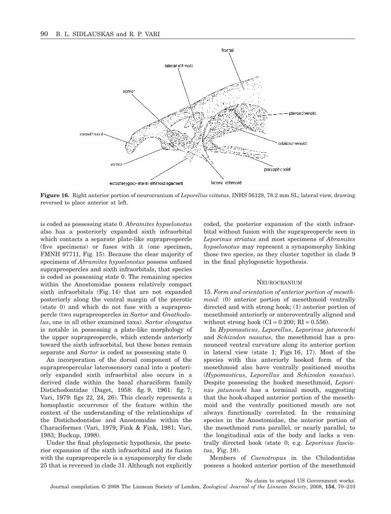
is coded as possessing state 0. Abramites hypselonotusalso has a posteriorly expanded sixth infraorbitalwhich contacts a separate plate-like suprapreopercle(five specimens) or fuses with it (one specimen,FMNH 97711, Fig. 15). Because the clear majority ofspecimens of Abramites hypselonotus possess unfusedsuprapreopercles and sixth infraorbitals, that speciesis coded as possessing state 0. The remaining specieswithin the Anostomidae possess relatively compactsixth infraorbitals (Fig. 14) that are not expandedposteriorly along the ventral margin of the pterotic(state 0) and which do not fuse with a suprapreo-percle (two suprapreopercles in Sartor and Gnathodo-lus, one in all other examined taxa). Sartor elongatusis notable in possessing a plate-like morphology ofthe upper suprapreopercle, which extends anteriorlytoward the sixth infraorbital, but these bones remainseparate and Sartor is coded as possessing state 0.
An incorporation of the dorsal component of thesuprapreopercular laterosensory canal into a posteri-orly expanded sixth infraorbital also occurs in aderived clade within the basal characiform familyDistichodontidae (Daget, 1958: fig. 9, 1961: fig. 7;Vari, 1979: figs 22, 24, 26). This clearly represents ahomoplastic occurrence of the feature within thecontext of the understanding of the relationships ofthe Distichodontidae and Anostomidae within theCharaciformes (Vari, 1979; Fink & Fink, 1981; Vari,1983; Buckup, 1998).
Under the final phylogenetic hypothesis, the poste-rior expansion of the sixth infraorbital and its fusionwith the suprapreopercle is a synapomorphy for clade25 that is reversed in clade 31. Although not explicitly
coded, the posterior expansion of the sixth infraor-bital without fusion with the suprapreopercle seen inLeporinus striatus and most specimens of Abramiteshypselonotus may represent a synapomorphy linkingthose two species, as they cluster together in clade 9in the final phylogenetic hypothesis.
NEUROCRANIUM
15. Form and orientation of anterior portion of meseth-moid: (0) anterior portion of mesethmoid ventrallydirected and with strong hook; (1) anterior portion ofmesethmoid anteriorly or anteroventrally aligned andwithout strong hook (CI = 0.200; RI = 0.556).
In Hypomasticus, Leporellus, Leporinus jatuncochiand Schizodon nasutus, the mesethmoid has a pro-nounced ventral curvature along its anterior portionin lateral view (state 1; Figs 16, 17). Most of thespecies with this anteriorly hooked form of themesethmoid also have ventrally positioned mouths(Hypomasticus, Leporellus and Schizodon nasutus).Despite possessing the hooked mesethmoid, Lepori-nus jatuncochi has a terminal mouth, suggestingthat the hook-shaped anterior portion of the meseth-moid and the ventrally positioned mouth are notalways functionally correlated. In the remainingspecies in the Anostomidae, the anterior portion ofthe mesethmoid runs parallel, or nearly parallel, tothe longitudinal axis of the body and lacks a ven-trally directed hook (state 0; e.g. Leporinus fascia-tus, Fig. 18).
Members of Caenotropus in the Chilodontidaepossess a hooked anterior portion of the mesethmoid
Figure 16. Right anterior portion of neurocranium of Leporellus vittatus, INHS 56128, 78.2 mm SL; lateral view, drawingreversed to place anterior at left.
90 B. L. SIDLAUSKAS and R. P. VARI
No claim to original US Government works.Journal compilation © 2008 The Linnean Society of London, Zoological Journal of the Linnean Society, 2008, 154, 70–210
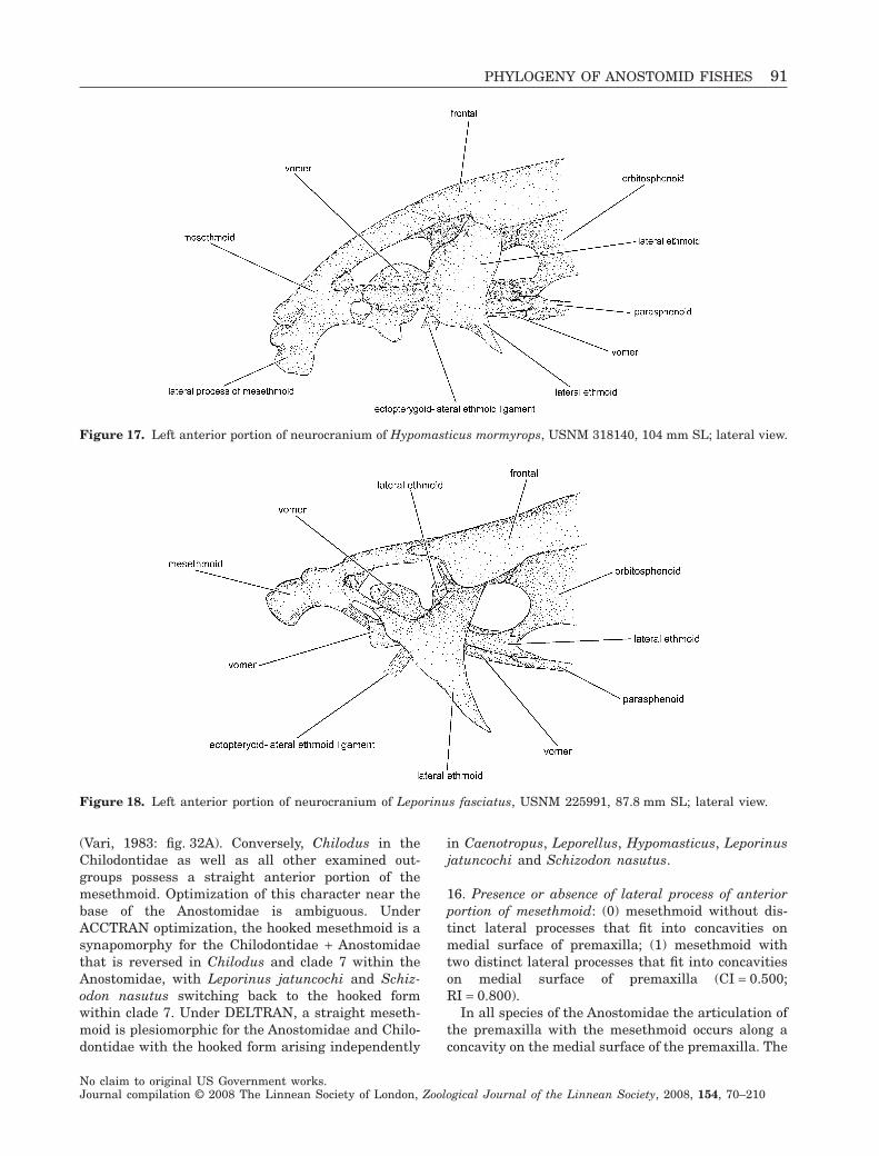
(Vari, 1983: fig. 32A). Conversely, Chilodus in theChilodontidae as well as all other examined out-groups possess a straight anterior portion of themesethmoid. Optimization of this character near thebase of the Anostomidae is ambiguous. UnderACCTRAN optimization, the hooked mesethmoid is asynapomorphy for the Chilodontidae + Anostomidaethat is reversed in Chilodus and clade 7 within theAnostomidae, with Leporinus jatuncochi and Schiz-odon nasutus switching back to the hooked formwithin clade 7. Under DELTRAN, a straight meseth-moid is plesiomorphic for the Anostomidae and Chilo-dontidae with the hooked form arising independently
in Caenotropus, Leporellus, Hypomasticus, Leporinusjatuncochi and Schizodon nasutus.
16. Presence or absence of lateral process of anteriorportion of mesethmoid: (0) mesethmoid without dis-tinct lateral processes that fit into concavities onmedial surface of premaxilla; (1) mesethmoid withtwo distinct lateral processes that fit into concavitieson medial surface of premaxilla (CI = 0.500;RI = 0.800).
In all species of the Anostomidae the articulation ofthe premaxilla with the mesethmoid occurs along aconcavity on the medial surface of the premaxilla. The
Figure 17. Left anterior portion of neurocranium of Hypomasticus mormyrops, USNM 318140, 104 mm SL; lateral view.
Figure 18. Left anterior portion of neurocranium of Leporinus fasciatus, USNM 225991, 87.8 mm SL; lateral view.
PHYLOGENY OF ANOSTOMID FISHES 91
No claim to original US Government works.Journal compilation © 2008 The Linnean Society of London, Zoological Journal of the Linnean Society, 2008, 154, 70–210
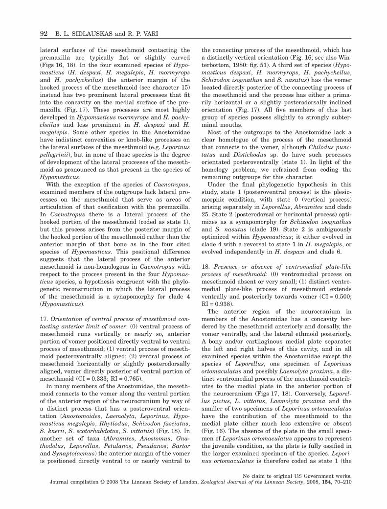
lateral surfaces of the mesethmoid contacting thepremaxilla are typically flat or slightly curved(Figs 16, 18). In the four examined species of Hypo-masticus (H. despaxi, H. megalepis, H. mormyropsand H. pachycheilus) the anterior margin of thehooked process of the mesethmoid (see character 15)instead has two prominent lateral processes that fitinto the concavity on the medial surface of the pre-maxilla (Fig. 17). These processes are most highlydeveloped in Hypomasticus mormyrops and H. pachy-cheilus and less prominent in H. despaxi and H.megalepis. Some other species in the Anostomidaehave indistinct convexities or knob-like processes onthe lateral surfaces of the mesethmoid (e.g. Leporinuspellegrinii), but in none of those species is the degreeof development of the lateral processes of the meseth-moid as pronounced as that present in the species ofHypomasticus.
With the exception of the species of Caenotropus,examined members of the outgroups lack lateral pro-cesses on the mesethmoid that serve as areas ofarticulation of that ossification with the premaxilla.In Caenotropus there is a lateral process of thehooked portion of the mesethmoid (coded as state 1),but this process arises from the posterior margin ofthe hooked portion of the mesethmoid rather than theanterior margin of that bone as in the four citedspecies of Hypomasticus. This positional differencesuggests that the lateral process of the anteriormesethmoid is non-homologous in Caenotropus withrespect to the process present in the four Hypomas-ticus species, a hypothesis congruent with the phylo-genetic reconstruction in which the lateral processof the mesethmoid is a synapomorphy for clade 4(Hypomasticus).
17. Orientation of ventral process of mesethmoid con-tacting anterior limit of vomer: (0) ventral process ofmesethmoid runs vertically or nearly so, anteriorportion of vomer positioned directly ventral to ventralprocess of mesethmoid; (1) ventral process of meseth-moid posteroventrally aligned; (2) ventral process ofmesethmoid horizontally or slightly posterodorsallyaligned, vomer directly posterior of ventral portion ofmesethmoid (CI = 0.333; RI = 0.765).
In many members of the Anostomidae, the meseth-moid connects to the vomer along the ventral portionof the anterior region of the neurocranium by way ofa distinct process that has a posteroventral orien-tation (Anostomoides, Laemolyta, Leporinus, Hypo-masticus megalepis, Rhytiodus, Schizodon fasciatus,S. knerii, S. scotorhabdotus, S. vittatus) (Fig. 18). Inanother set of taxa (Abramites, Anostomus, Gna-thodolus, Leporellus, Petulanos, Pseudanos, Sartorand Synaptolaemus) the anterior margin of the vomeris positioned directly ventral to or nearly ventral to
the connecting process of the mesethmoid, which hasa distinctly vertical orientation (Fig. 16; see also Win-terbottom, 1980: fig. 51). A third set of species (Hypo-masticus despaxi, H. mormyrops, H. pachycheilus,Schizodon isognathus and S. nasutus) has the vomerlocated directly posterior of the connecting process ofthe mesethmoid and the process has either a prima-rily horizontal or a slightly posterodorsally inclinedorientation (Fig. 17). All five members of this lastgroup of species possess slightly to strongly subter-minal mouths.
Most of the outgroups to the Anostomidae lack aclear homologue of the process of the mesethmoidthat connects to the vomer, although Chilodus punc-tatus and Distichodus sp. do have such processesorientated posteroventrally (state 1). In light of thehomology problem, we refrained from coding theremaining outgroups for this character.
Under the final phylogenetic hypothesis in thisstudy, state 1 (posteroventral process) is the plesio-morphic condition, with state 0 (vertical process)arising separately in Leporellus, Abramites and clade25. State 2 (posterodorsal or horizontal process) opti-mizes as a synapomorphy for Schizodon isognathusand S. nasutus (clade 19). State 2 is ambiguouslyoptimized within Hypomasticus; it either evolved inclade 4 with a reversal to state 1 in H. megalepis, orevolved independently in H. despaxi and clade 6.
18. Presence or absence of ventromedial plate-likeprocess of mesethmoid: (0) ventromedial process onmesethmoid absent or very small; (1) distinct ventro-medial plate-like process of mesethmoid extendsventrally and posteriorly towards vomer (CI = 0.500;RI = 0.938).
The anterior region of the neurocranium inmembers of the Anostomidae has a concavity bor-dered by the mesethmoid anteriorly and dorsally, thevomer ventrally, and the lateral ethmoid posteriorly.A bony and/or cartilaginous medial plate separatesthe left and right halves of this cavity, and in allexamined species within the Anostomidae except thespecies of Leporellus, one specimen of Leporinusortomaculatus and possibly Laemolyta proxima, a dis-tinct ventromedial process of the mesethmoid contrib-utes to the medial plate in the anterior portion ofthe neurocranium (Figs 17, 18). Conversely, Leporel-lus pictus, L. vittatus, Laemolyta proxima and thesmaller of two specimens of Leporinus ortomaculatushave the contribution of the mesethmoid to themedial plate either much less extensive or absent(Fig. 16). The absence of the plate in the small speci-men of Leporinus ortomaculatus appears to representthe juvenile condition, as the plate is fully ossified inthe larger examined specimen of the species. Lepori-nus ortomaculatus is therefore coded as state 1 (the
92 B. L. SIDLAUSKAS and R. P. VARI
No claim to original US Government works.Journal compilation © 2008 The Linnean Society of London, Zoological Journal of the Linnean Society, 2008, 154, 70–210
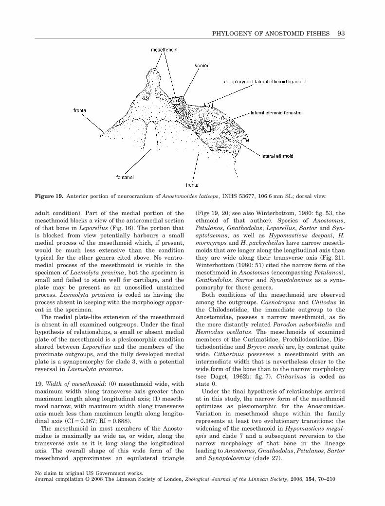
adult condition). Part of the medial portion of themesethmoid blocks a view of the anteromedial sectionof that bone in Leporellus (Fig. 16). The portion thatis blocked from view potentially harbours a smallmedial process of the mesethmoid which, if present,would be much less extensive than the conditiontypical for the other genera cited above. No ventro-medial process of the mesethmoid is visible in thespecimen of Laemolyta proxima, but the specimen issmall and failed to stain well for cartilage, and theplate may be present as an unossified unstainedprocess. Laemolyta proxima is coded as having theprocess absent in keeping with the morphology appar-ent in the specimen.
The medial plate-like extension of the mesethmoidis absent in all examined outgroups. Under the finalhypothesis of relationships, a small or absent medialplate of the mesethmoid is a plesiomorphic conditionshared between Leporellus and the members of theproximate outgroups, and the fully developed medialplate is a synapomorphy for clade 3, with a potentialreversal in Laemolyta proxima.
19. Width of mesethmoid: (0) mesethmoid wide, withmaximum width along transverse axis greater thanmaximum length along longitudinal axis; (1) meseth-moid narrow, with maximum width along transverseaxis much less than maximum length along longitu-dinal axis (CI = 0.167; RI = 0.688).
The mesethmoid in most members of the Anosto-midae is maximally as wide as, or wider, along thetransverse axis as it is long along the longitudinalaxis. The overall shape of this wide form of themesethmoid approximates an equilateral triangle
(Figs 19, 20; see also Winterbottom, 1980: fig. 53, theethmoid of that author). Species of Anostomus,Petulanos, Gnathodolus, Leporellus, Sartor and Syn-aptolaemus, as well as Hypomasticus despaxi, H.mormyrops and H. pachycheilus have narrow meseth-moids that are longer along the longitudinal axis thanthey are wide along their transverse axis (Fig. 21).Winterbottom (1980: 51) cited the narrow form of themesethmoid in Anostomus (encompassing Petulanos),Gnathodolus, Sartor and Synaptolaemus as a syna-pomorphy for those genera.
Both conditions of the mesethmoid are observedamong the outgroups. Caenotropus and Chilodus inthe Chilodontidae, the immediate outgroup to theAnostomidae, possess a narrow mesethmoid, as dothe more distantly related Parodon suborbitalis andHemiodus ocellatus. The mesethmoids of examinedmembers of the Curimatidae, Prochilodontidae, Dis-tichodontidae and Brycon meeki are, by contrast quitewide. Citharinus possesses a mesethmoid with anintermediate width that is nevertheless closer to thewide form of the bone than to the narrow morphology(see Daget, 1962b: fig. 7). Citharinus is coded asstate 0.
Under the final hypothesis of relationships arrivedat in this study, the narrow form of the mesethmoidoptimizes as plesiomorphic for the Anostomidae.Variation in mesethmoid shape within the familyrepresents at least two evolutionary transitions: thewidening of the mesethmoid in Hypomasticus megal-epis and clade 7 and a subsequent reversion to thenarrow morphology of that bone in the lineageleading to Anostomus, Gnathodolus, Petulanos, Sartorand Synaptolaemus (clade 27).
Figure 19. Anterior portion of neurocranium of Anostomoides laticeps, INHS 53677, 106.6 mm SL; dorsal view.
PHYLOGENY OF ANOSTOMID FISHES 93
No claim to original US Government works.Journal compilation © 2008 The Linnean Society of London, Zoological Journal of the Linnean Society, 2008, 154, 70–210
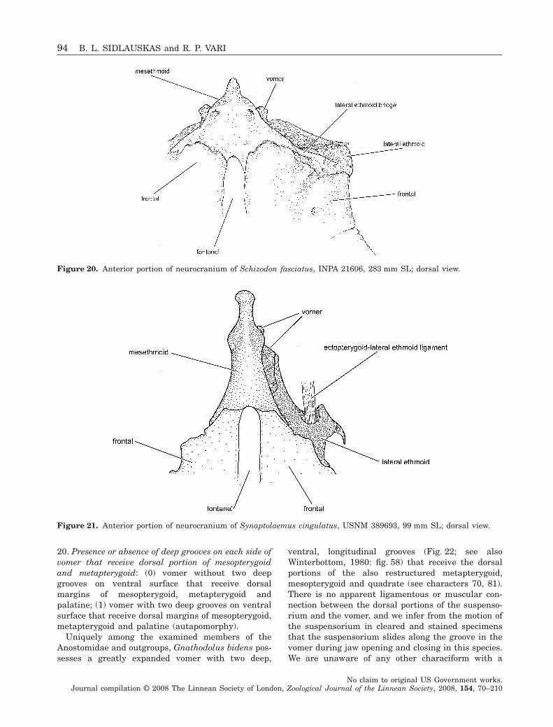
20. Presence or absence of deep grooves on each side ofvomer that receive dorsal portion of mesopterygoidand metapterygoid: (0) vomer without two deepgrooves on ventral surface that receive dorsalmargins of mesopterygoid, metapterygoid andpalatine; (1) vomer with two deep grooves on ventralsurface that receive dorsal margins of mesopterygoid,metapterygoid and palatine (autapomorphy).
Uniquely among the examined members of theAnostomidae and outgroups, Gnathodolus bidens pos-sesses a greatly expanded vomer with two deep,
ventral, longitudinal grooves (Fig. 22; see alsoWinterbottom, 1980: fig. 58) that receive the dorsalportions of the also restructured metapterygoid,mesopterygoid and quadrate (see characters 70, 81).There is no apparent ligamentous or muscular con-nection between the dorsal portions of the suspenso-rium and the vomer, and we infer from the motion ofthe suspensorium in cleared and stained specimensthat the suspensorium slides along the groove in thevomer during jaw opening and closing in this species.We are unaware of any other characiform with a
Figure 20. Anterior portion of neurocranium of Schizodon fasciatus, INPA 21606, 283 mm SL; dorsal view.
Figure 21. Anterior portion of neurocranium of Synaptolaemus cingulatus, USNM 389693, 99 mm SL; dorsal view.
94 B. L. SIDLAUSKAS and R. P. VARI
No claim to original US Government works.Journal compilation © 2008 The Linnean Society of London, Zoological Journal of the Linnean Society, 2008, 154, 70–210
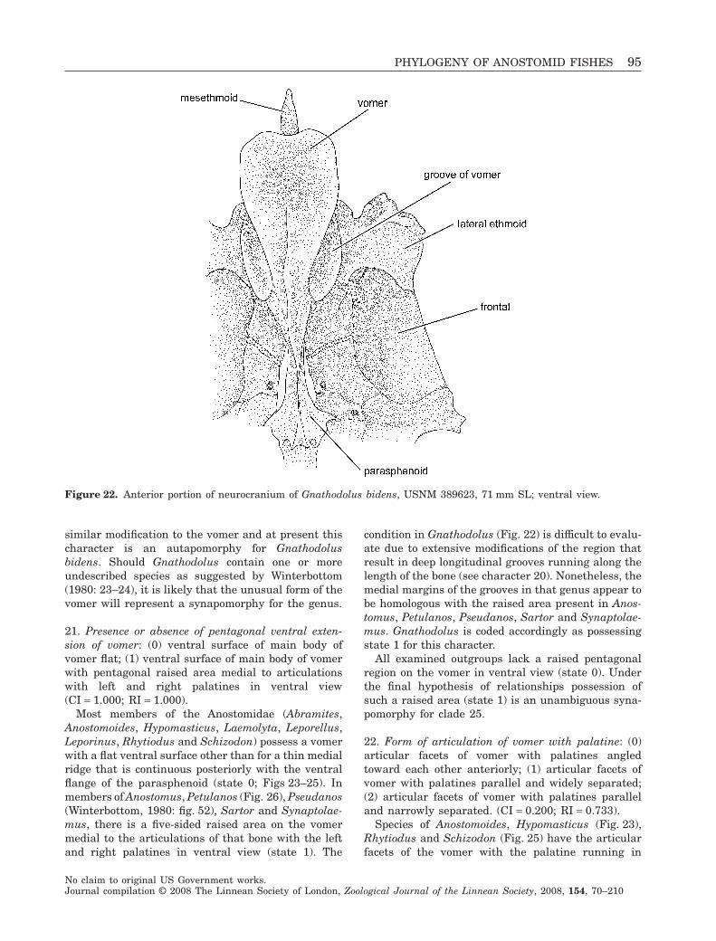
similar modification to the vomer and at present thischaracter is an autapomorphy for Gnathodolusbidens. Should Gnathodolus contain one or moreundescribed species as suggested by Winterbottom(1980: 23–24), it is likely that the unusual form of thevomer will represent a synapomorphy for the genus.
21. Presence or absence of pentagonal ventral exten-sion of vomer: (0) ventral surface of main body ofvomer flat; (1) ventral surface of main body of vomerwith pentagonal raised area medial to articulationswith left and right palatines in ventral view(CI = 1.000; RI = 1.000).
Most members of the Anostomidae (Abramites,Anostomoides, Hypomasticus, Laemolyta, Leporellus,Leporinus, Rhytiodus and Schizodon) possess a vomerwith a flat ventral surface other than for a thin medialridge that is continuous posteriorly with the ventralflange of the parasphenoid (state 0; Figs 23–25). Inmembers of Anostomus, Petulanos (Fig. 26), Pseudanos(Winterbottom, 1980: fig. 52), Sartor and Synaptolae-mus, there is a five-sided raised area on the vomermedial to the articulations of that bone with the leftand right palatines in ventral view (state 1). The
condition in Gnathodolus (Fig. 22) is difficult to evalu-ate due to extensive modifications of the region thatresult in deep longitudinal grooves running along thelength of the bone (see character 20). Nonetheless, themedial margins of the grooves in that genus appear tobe homologous with the raised area present in Anos-tomus, Petulanos, Pseudanos, Sartor and Synaptolae-mus. Gnathodolus is coded accordingly as possessingstate 1 for this character.
All examined outgroups lack a raised pentagonalregion on the vomer in ventral view (state 0). Underthe final hypothesis of relationships possession ofsuch a raised area (state 1) is an unambiguous syna-pomorphy for clade 25.
22. Form of articulation of vomer with palatine: (0)articular facets of vomer with palatines angledtoward each other anteriorly; (1) articular facets ofvomer with palatines parallel and widely separated;(2) articular facets of vomer with palatines paralleland narrowly separated. (CI = 0.200; RI = 0.733).
Species of Anostomoides, Hypomasticus (Fig. 23),Rhytiodus and Schizodon (Fig. 25) have the articularfacets of the vomer with the palatine running in
Figure 22. Anterior portion of neurocranium of Gnathodolus bidens, USNM 389623, 71 mm SL; ventral view.
PHYLOGENY OF ANOSTOMID FISHES 95
No claim to original US Government works.Journal compilation © 2008 The Linnean Society of London, Zoological Journal of the Linnean Society, 2008, 154, 70–210
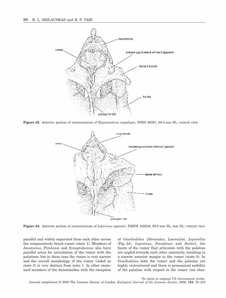
parallel and widely separated from each other acrossthe comparatively broad vomer (state 1). Members ofAnostomus, Petulanos and Synaptolaemus also haveparallel areas for articulation of the vomer with thepalatines, but in these taxa the vomer is very narrowand the overall morphology of the vomer (coded asstate 2) is very distinct from state 1. In other exam-ined members of the Anostomidae with the exception
of Gnathodolus [Abramites, Laemolyta, Leporellus(Fig. 24), Leporinus, Pseudanos and Sartor], thefacets of the vomer that articulate with the palatineare angled towards each other anteriorly, resulting ina narrow anterior margin to the vomer (state 0). InGnathodolus both the vomer and the palatine arehighly restructured and there is pronounced mobilityof the palatine with respect to the vomer (see char-
Figure 23. Anterior portion of neurocranium of Hypomasticus megalepis, INHS 49387, 68.4 mm SL; ventral view.
Figure 24. Anterior portion of neurocranium of Leporinus agassizi, FMNH 102219; 98.6 mm SL, mm SL; ventral view.
96 B. L. SIDLAUSKAS and R. P. VARI
No claim to original US Government works.Journal compilation © 2008 The Linnean Society of London, Zoological Journal of the Linnean Society, 2008, 154, 70–210
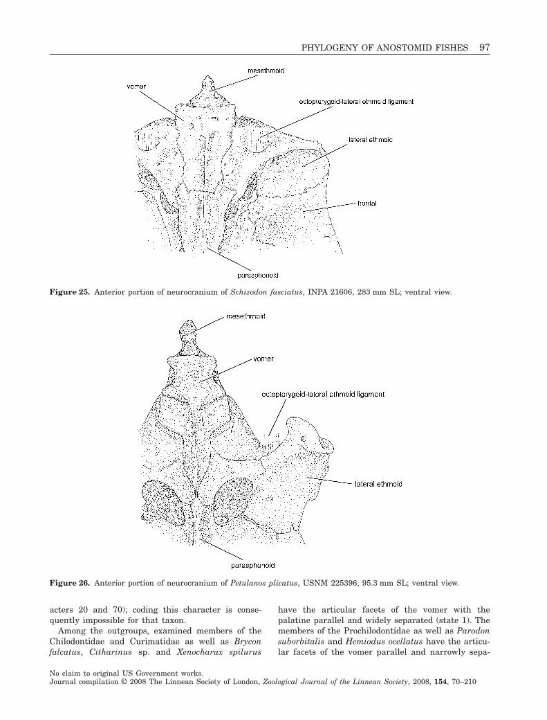
acters 20 and 70); coding this character is conse-quently impossible for that taxon.
Among the outgroups, examined members of theChilodontidae and Curimatidae as well as Bryconfalcatus, Citharinus sp. and Xenocharax spilurus
have the articular facets of the vomer with thepalatine parallel and widely separated (state 1). Themembers of the Prochilodontidae as well as Parodonsuborbitalis and Hemiodus ocellatus have the articu-lar facets of the vomer parallel and narrowly sepa-
Figure 25. Anterior portion of neurocranium of Schizodon fasciatus, INPA 21606, 283 mm SL; ventral view.
Figure 26. Anterior portion of neurocranium of Petulanos plicatus, USNM 225396, 95.3 mm SL; ventral view.
PHYLOGENY OF ANOSTOMID FISHES 97
No claim to original US Government works.Journal compilation © 2008 The Linnean Society of London, Zoological Journal of the Linnean Society, 2008, 154, 70–210
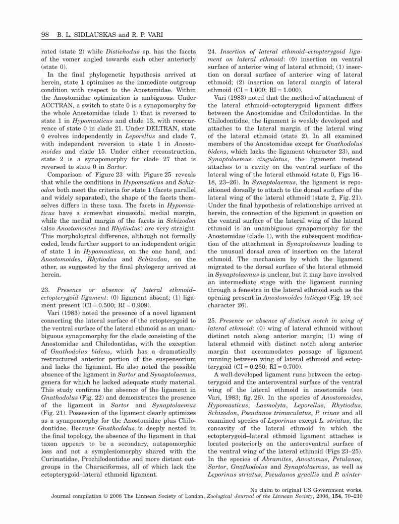
rated (state 2) while Distichodus sp. has the facetsof the vomer angled towards each other anteriorly(state 0).
In the final phylogenetic hypothesis arrived atherein, state 1 optimizes as the immediate outgroupcondition with respect to the Anostomidae. Withinthe Anostomidae optimization is ambiguous. UnderACCTRAN, a switch to state 0 is a synapomorphy forthe whole Anostomidae (clade 1) that is reversed tostate 1 in Hypomasticus and clade 13, with reoccur-rence of state 0 in clade 21. Under DELTRAN, state0 evolves independently in Leporellus and clade 7,with independent reversion to state 1 in Anosto-moides and clade 15. Under either reconstruction,state 2 is a synapomorphy for clade 27 that isreversed to state 0 in Sartor.
Comparison of Figure 23 with Figure 25 revealsthat while the conditions in Hypomasticus and Schiz-odon both meet the criteria for state 1 (facets paralleland widely separated), the shape of the facets them-selves differs in these taxa. The facets in Hypomas-ticus have a somewhat sinusoidal medial margin,while the medial margin of the facets in Schizodon(also Anostomoides and Rhytiodus) are very straight.This morphological difference, although not formallycoded, lends further support to an independent originof state 1 in Hypomasticus, on the one hand, andAnostomoides, Rhytiodus and Schizodon, on theother, as suggested by the final phylogeny arrived atherein.
23. Presence or absence of lateral ethmoid–ectopterygoid ligament: (0) ligament absent; (1) liga-ment present (CI = 0.500; RI = 0.909).
Vari (1983) noted the presence of a novel ligamentconnecting the lateral surface of the ectopterygoid tothe ventral surface of the lateral ethmoid as an unam-biguous synapomorphy for the clade consisting of theAnostomidae and Chilodontidae, with the exceptionof Gnathodolus bidens, which has a dramaticallyrestructured anterior portion of the suspensoriumand lacks the ligament. He also noted the possibleabsence of the ligament in Sartor and Synaptolaemus,genera for which he lacked adequate study material.This study confirms the absence of the ligament inGnathodolus (Fig. 22) and demonstrates the presenceof the ligament in Sartor and Synaptolaemus(Fig. 21). Possession of the ligament clearly optimizesas a synapomorphy for the Anostomidae plus Chilo-dontidae. Because Gnathodolus is deeply nested inthe final topology, the absence of the ligament in thattaxon appears to be a secondary, autapomorphicloss and not a symplesiomorphy shared with theCurimatidae, Prochilodontidae and more distant out-groups in the Characiformes, all of which lack theectopterygoid–lateral ethmoid ligament.
24. Insertion of lateral ethmoid–ectopterygoid liga-ment on lateral ethmoid: (0) insertion on ventralsurface of anterior wing of lateral ethmoid; (1) inser-tion on dorsal surface of anterior wing of lateralethmoid; (2) insertion on lateral margin of lateralethmoid (CI = 1.000; RI = 1.000).
Vari (1983) noted that the method of attachment ofthe lateral ethmoid–ectopterygoid ligament differsbetween the Anostomidae and Chilodontidae. In theChilodontidae, the ligament is weakly developed andattaches to the lateral margin of the lateral wingof the lateral ethmoid (state 2). In all examinedmembers of the Anostomidae except for Gnathodolusbidens, which lacks the ligament (character 23), andSynaptolaemus cingulatus, the ligament insteadattaches to a cavity on the ventral surface of thelateral wing of the lateral ethmoid (state 0, Figs 16–18, 23–26). In Synaptolaemus, the ligament is repo-sitioned dorsally to attach to the dorsal surface of thelateral wing of the lateral ethmoid (state 2, Fig. 21).Under the final hypothesis of relationships arrived atherein, the connection of the ligament in question onthe ventral surface of the lateral wing of the lateralethmoid is an unambiguous synapomorphy for theAnostomidae (clade 1), with the subsequent modifica-tion of the attachment in Synaptolaemus leading tothe unusual dorsal area of insertion on the lateralethmoid. The mechanism by which the ligamentmigrated to the dorsal surface of the lateral ethmoidin Synaptolaemus is unclear, but it may have involvedan intermediate stage with the ligament runningthrough a fenestra in the lateral ethmoid such as theopening present in Anostomoides laticeps (Fig. 19, seecharacter 26).
25. Presence or absence of distinct notch in wing oflateral ethmoid: (0) wing of lateral ethmoid withoutdistinct notch along anterior margin; (1) wing oflateral ethmoid with distinct notch along anteriormargin that accommodates passage of ligamentrunning between wing of lateral ethmoid and ectop-terygoid (CI = 0.250; RI = 0.700).
A well-developed ligament runs between the ectop-terygoid and the anteroventral surface of the ventralwing of the lateral ethmoid in anostomids (seeVari, 1983; fig. 26). In the species of Anostomoides,Hypomasticus, Laemolyta, Leporellus, Rhytiodus,Schizodon, Pseudanos trimaculatus, P. irinae and allexamined species of Leporinus except L. striatus, theconcavity of the lateral ethmoid in which theectopterygoid–lateral ethmoid ligament attaches islocated posteriorly on the anteroventral surface ofthe ventral wing of the lateral ethmoid (Figs 23–25).In the species of Abramites, Anostomus, Petulanos,Sartor, Gnathodolus and Synaptolaemus, as well asLeporinus striatus, Pseudanos gracilis and P. winter-
98 B. L. SIDLAUSKAS and R. P. VARI
No claim to original US Government works.Journal compilation © 2008 The Linnean Society of London, Zoological Journal of the Linnean Society, 2008, 154, 70–210
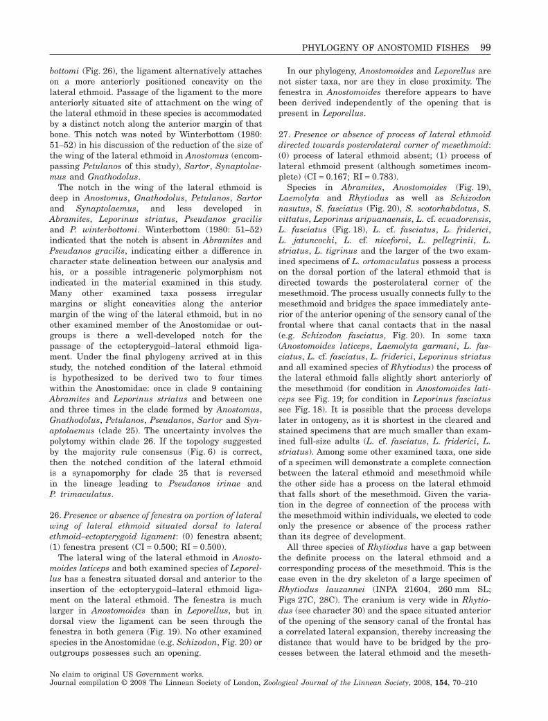
bottomi (Fig. 26), the ligament alternatively attacheson a more anteriorly positioned concavity on thelateral ethmoid. Passage of the ligament to the moreanteriorly situated site of attachment on the wing ofthe lateral ethmoid in these species is accommodatedby a distinct notch along the anterior margin of thatbone. This notch was noted by Winterbottom (1980:51–52) in his discussion of the reduction of the size ofthe wing of the lateral ethmoid in Anostomus (encom-passing Petulanos of this study), Sartor, Synaptolae-mus and Gnathodolus.
The notch in the wing of the lateral ethmoid isdeep in Anostomus, Gnathodolus, Petulanos, Sartorand Synaptolaemus, and less developed inAbramites, Leporinus striatus, Pseudanos gracilisand P. winterbottomi. Winterbottom (1980: 51–52)indicated that the notch is absent in Abramites andPseudanos gracilis, indicating either a difference incharacter state delineation between our analysis andhis, or a possible intrageneric polymorphism notindicated in the material examined in this study.Many other examined taxa possess irregularmargins or slight concavities along the anteriormargin of the wing of the lateral ethmoid, but in noother examined member of the Anostomidae or out-groups is there a well-developed notch for thepassage of the ectopterygoid–lateral ethmoid liga-ment. Under the final phylogeny arrived at in thisstudy, the notched condition of the lateral ethmoidis hypothesized to be derived two to four timeswithin the Anostomidae: once in clade 9 containingAbramites and Leporinus striatus and between oneand three times in the clade formed by Anostomus,Gnathodolus, Petulanos, Pseudanos, Sartor and Syn-aptolaemus (clade 25). The uncertainty involves thepolytomy within clade 26. If the topology suggestedby the majority rule consensus (Fig. 6) is correct,then the notched condition of the lateral ethmoidis a synapomorphy for clade 25 that is reversedin the lineage leading to Pseudanos irinae andP. trimaculatus.
26. Presence or absence of fenestra on portion of lateralwing of lateral ethmoid situated dorsal to lateralethmoid–ectopterygoid ligament: (0) fenestra absent;(1) fenestra present (CI = 0.500; RI = 0.500).
The lateral wing of the lateral ethmoid in Anosto-moides laticeps and both examined species of Leporel-lus has a fenestra situated dorsal and anterior to theinsertion of the ectopterygoid–lateral ethmoid liga-ment on the lateral ethmoid. The fenestra is muchlarger in Anostomoides than in Leporellus, but indorsal view the ligament can be seen through thefenestra in both genera (Fig. 19). No other examinedspecies in the Anostomidae (e.g. Schizodon, Fig. 20) oroutgroups possesses such an opening.
In our phylogeny, Anostomoides and Leporellus arenot sister taxa, nor are they in close proximity. Thefenestra in Anostomoides therefore appears to havebeen derived independently of the opening that ispresent in Leporellus.
27. Presence or absence of process of lateral ethmoiddirected towards posterolateral corner of mesethmoid:(0) process of lateral ethmoid absent; (1) process oflateral ethmoid present (although sometimes incom-plete) (CI = 0.167; RI = 0.783).
Species in Abramites, Anostomoides (Fig. 19),Laemolyta and Rhytiodus as well as Schizodonnasutus, S. fasciatus (Fig. 20), S. scotorhabdotus, S.vittatus, Leporinus aripuanaensis, L. cf. ecuadorensis,L. fasciatus (Fig. 18), L. cf. fasciatus, L. friderici,L. jatuncochi, L. cf. niceforoi, L. pellegrinii, L.striatus, L. tigrinus and the larger of the two exam-ined specimens of L. ortomaculatus possess a processon the dorsal portion of the lateral ethmoid that isdirected towards the posterolateral corner of themesethmoid. The process usually connects fully to themesethmoid and bridges the space immediately ante-rior of the anterior opening of the sensory canal of thefrontal where that canal contacts that in the nasal(e.g. Schizodon fasciatus, Fig. 20). In some taxa(Anostomoides laticeps, Laemolyta garmani, L. fas-ciatus, L. cf. fasciatus, L. friderici, Leporinus striatusand all examined species of Rhytiodus) the process ofthe lateral ethmoid falls slightly short anteriorly ofthe mesethmoid (for condition in Anostomoides lati-ceps see Fig. 19; for condition in Leporinus fasciatussee Fig. 18). It is possible that the process developslater in ontogeny, as it is shortest in the cleared andstained specimens that are much smaller than exam-ined full-size adults (L. cf. fasciatus, L. friderici, L.striatus). Among some other examined taxa, one sideof a specimen will demonstrate a complete connectionbetween the lateral ethmoid and mesethmoid whilethe other side has a process on the lateral ethmoidthat falls short of the mesethmoid. Given the varia-tion in the degree of connection of the process withthe mesethmoid within individuals, we elected to codeonly the presence or absence of the process ratherthan its degree of development.
All three species of Rhytiodus have a gap betweenthe definite process on the lateral ethmoid and acorresponding process of the mesethmoid. This is thecase even in the dry skeleton of a large specimen ofRhytiodus lauzannei (INPA 21604, 260 mm SL;Figs 27C, 28C). The cranium is very wide in Rhytio-dus (see character 30) and the space situated anteriorof the opening of the sensory canal of the frontal hasa correlated lateral expansion, thereby increasing thedistance that would have to be bridged by the pro-cesses between the lateral ethmoid and the meseth-
PHYLOGENY OF ANOSTOMID FISHES 99
No claim to original US Government works.Journal compilation © 2008 The Linnean Society of London, Zoological Journal of the Linnean Society, 2008, 154, 70–210
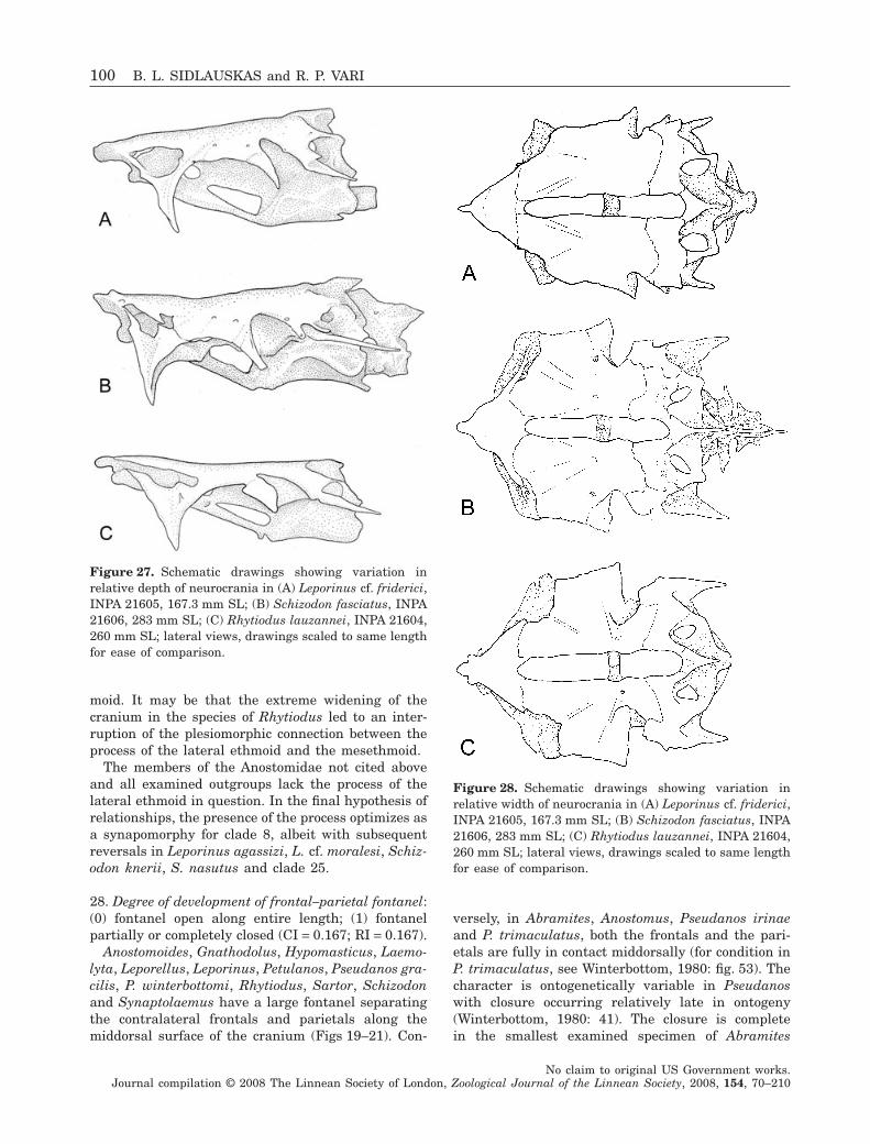
moid. It may be that the extreme widening of thecranium in the species of Rhytiodus led to an inter-ruption of the plesiomorphic connection between theprocess of the lateral ethmoid and the mesethmoid.
The members of the Anostomidae not cited aboveand all examined outgroups lack the process of thelateral ethmoid in question. In the final hypothesis ofrelationships, the presence of the process optimizes asa synapomorphy for clade 8, albeit with subsequentreversals in Leporinus agassizi, L. cf. moralesi, Schiz-odon knerii, S. nasutus and clade 25.
28. Degree of development of frontal–parietal fontanel:(0) fontanel open along entire length; (1) fontanelpartially or completely closed (CI = 0.167; RI = 0.167).
Anostomoides, Gnathodolus, Hypomasticus, Laemo-lyta, Leporellus, Leporinus, Petulanos, Pseudanos gra-cilis, P. winterbottomi, Rhytiodus, Sartor, Schizodonand Synaptolaemus have a large fontanel separatingthe contralateral frontals and parietals along themiddorsal surface of the cranium (Figs 19–21). Con-
versely, in Abramites, Anostomus, Pseudanos irinaeand P. trimaculatus, both the frontals and the pari-etals are fully in contact middorsally (for condition inP. trimaculatus, see Winterbottom, 1980: fig. 53). Thecharacter is ontogenetically variable in Pseudanoswith closure occurring relatively late in ontogeny(Winterbottom, 1980: 41). The closure is completein the smallest examined specimen of Abramites
Figure 27. Schematic drawings showing variation inrelative depth of neurocrania in (A) Leporinus cf. friderici,INPA 21605, 167.3 mm SL; (B) Schizodon fasciatus, INPA21606, 283 mm SL; (C) Rhytiodus lauzannei, INPA 21604,260 mm SL; lateral views, drawings scaled to same lengthfor ease of comparison.
Figure 28. Schematic drawings showing variation inrelative width of neurocrania in (A) Leporinus cf. friderici,INPA 21605, 167.3 mm SL; (B) Schizodon fasciatus, INPA21606, 283 mm SL; (C) Rhytiodus lauzannei, INPA 21604,260 mm SL; lateral views, drawings scaled to same lengthfor ease of comparison.
100 B. L. SIDLAUSKAS and R. P. VARI
No claim to original US Government works.Journal compilation © 2008 The Linnean Society of London, Zoological Journal of the Linnean Society, 2008, 154, 70–210
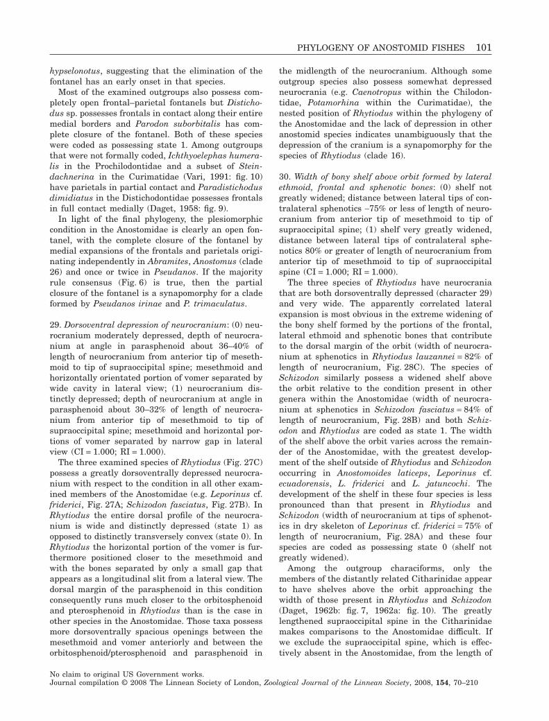
hypselonotus, suggesting that the elimination of thefontanel has an early onset in that species.
Most of the examined outgroups also possess com-pletely open frontal–parietal fontanels but Disticho-dus sp. possesses frontals in contact along their entiremedial borders and Parodon suborbitalis has com-plete closure of the fontanel. Both of these specieswere coded as possessing state 1. Among outgroupsthat were not formally coded, Ichthyoelephas humera-lis in the Prochilodontidae and a subset of Stein-dachnerina in the Curimatidae (Vari, 1991: fig. 10)have parietals in partial contact and Paradistichodusdimidiatus in the Distichodontidae possesses frontalsin full contact medially (Daget, 1958: fig. 9).
In light of the final phylogeny, the plesiomorphiccondition in the Anostomidae is clearly an open fon-tanel, with the complete closure of the fontanel bymedial expansions of the frontals and parietals origi-nating independently in Abramites, Anostomus (clade26) and once or twice in Pseudanos. If the majorityrule consensus (Fig. 6) is true, then the partialclosure of the fontanel is a synapomorphy for a cladeformed by Pseudanos irinae and P. trimaculatus.
29. Dorsoventral depression of neurocranium: (0) neu-rocranium moderately depressed, depth of neurocra-nium at angle in parasphenoid about 36–40% oflength of neurocranium from anterior tip of meseth-moid to tip of supraoccipital spine; mesethmoid andhorizontally orientated portion of vomer separated bywide cavity in lateral view; (1) neurocranium dis-tinctly depressed; depth of neurocranium at angle inparasphenoid about 30–32% of length of neurocra-nium from anterior tip of mesethmoid to tip ofsupraoccipital spine; mesethmoid and horizontal por-tions of vomer separated by narrow gap in lateralview (CI = 1.000; RI = 1.000).
The three examined species of Rhytiodus (Fig. 27C)possess a greatly dorsoventrally depressed neurocra-nium with respect to the condition in all other exam-ined members of the Anostomidae (e.g. Leporinus cf.friderici, Fig. 27A; Schizodon fasciatus, Fig. 27B). InRhytiodus the entire dorsal profile of the neurocra-nium is wide and distinctly depressed (state 1) asopposed to distinctly transversely convex (state 0). InRhytiodus the horizontal portion of the vomer is fur-thermore positioned closer to the mesethmoid andwith the bones separated by only a small gap thatappears as a longitudinal slit from a lateral view. Thedorsal margin of the parasphenoid in this conditionconsequently runs much closer to the orbitosphenoidand pterosphenoid in Rhytiodus than is the case inother species in the Anostomidae. Those taxa possessmore dorsoventrally spacious openings between themesethmoid and vomer anteriorly and between theorbitosphenoid/pterosphenoid and parasphenoid in
the midlength of the neurocranium. Although someoutgroup species also possess somewhat depressedneurocrania (e.g. Caenotropus within the Chilodon-tidae, Potamorhina within the Curimatidae), thenested position of Rhytiodus within the phylogeny ofthe Anostomidae and the lack of depression in otheranostomid species indicates unambiguously that thedepression of the cranium is a synapomorphy for thespecies of Rhytiodus (clade 16).
30. Width of bony shelf above orbit formed by lateralethmoid, frontal and sphenotic bones: (0) shelf notgreatly widened; distance between lateral tips of con-tralateral sphenotics ~75% or less of length of neuro-cranium from anterior tip of mesethmoid to tip ofsupraoccipital spine; (1) shelf very greatly widened,distance between lateral tips of contralateral sphe-notics 80% or greater of length of neurocranium fromanterior tip of mesethmoid to tip of supraoccipitalspine (CI = 1.000; RI = 1.000).
The three species of Rhytiodus have neurocraniathat are both dorsoventrally depressed (character 29)and very wide. The apparently correlated lateralexpansion is most obvious in the extreme widening ofthe bony shelf formed by the portions of the frontal,lateral ethmoid and sphenotic bones that contributeto the dorsal margin of the orbit (width of neurocra-nium at sphenotics in Rhytiodus lauzannei = 82% oflength of neurocranium, Fig. 28C). The species ofSchizodon similarly possess a widened shelf abovethe orbit relative to the condition present in othergenera within the Anostomidae (width of neurocra-nium at sphenotics in Schizodon fasciatus = 84% oflength of neurocranium, Fig. 28B) and both Schiz-odon and Rhytiodus are coded as state 1. The widthof the shelf above the orbit varies across the remain-der of the Anostomidae, with the greatest develop-ment of the shelf outside of Rhytiodus and Schizodonoccurring in Anostomoides laticeps, Leporinus cf.ecuadorensis, L. friderici and L. jatuncochi. Thedevelopment of the shelf in these four species is lesspronounced than that present in Rhytiodus andSchizodon (width of neurocranium at tips of sphenot-ics in dry skeleton of Leporinus cf. friderici = 75% oflength of neurocranium, Fig. 28A) and these fourspecies are coded as possessing state 0 (shelf notgreatly widened).
Among the outgroup characiforms, only themembers of the distantly related Citharinidae appearto have shelves above the orbit approaching thewidth of those present in Rhytiodus and Schizodon(Daget, 1962b: fig. 7, 1962a: fig. 10). The greatlylengthened supraoccipital spine in the Citharinidaemakes comparisons to the Anostomidae difficult. Ifwe exclude the supraoccipital spine, which is effec-tively absent in the Anostomidae, from the length of
PHYLOGENY OF ANOSTOMID FISHES 101
No claim to original US Government works.Journal compilation © 2008 The Linnean Society of London, Zoological Journal of the Linnean Society, 2008, 154, 70–210
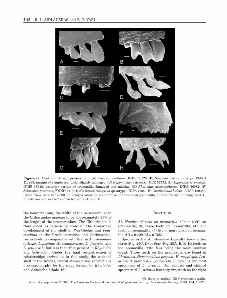
the neurocranium, the width of the neurocranium inthe Citharinidae appears to be approximately 75% ofthe length of the neurocranium. The Citharinidae isthus coded as possessing state 0. The transversedevelopment of the shelf in Prochilodus and Pota-morhina in the Prochilodontidae and Curimatidae,respectively, is comparable with that in Anostomoideslaticeps, Leporinus cf. ecuadorensis, L. friderici andL. jatuncochi but less than that present in Rhytiodusand Schizodon. Under the final reconstruction ofrelationships arrived at in this study, the widenedshelf of the frontal, lateral ethmoid and sphenotic isa synapomorphy for the clade formed by Rhytiodusand Schizodon (clade 15).
DENTITION
31. Number of teeth on premaxilla: (0) no teeth onpremaxilla; (3) three teeth on premaxilla; (4) fourteeth on premaxilla; (5) five or more teeth on premax-illa (CI = 0.429 RI = 0.765).
Species in the Anostomidae typically have eitherthree (Fig. 29C, D) or four (Fig. 29A, B, E–H) teeth onthe premaxilla, with four being the most commoncount. Three teeth on the premaxilla are found inAbramites, Hypomasticus despaxi, H. megalepis, Lep-orinus cf. moralesi, L. jatuncochi, L. tigrinus and mostspecimens of L. striatus. Our cleared and stainedspecimen of L. striatus has only two teeth on the right
Figure 29. Dentition of right premaxilla in (A) Leporellus vittatus, INHS 56128; (B) Hypomasticus mormyrops, FMNH112963, margin of symphyseal tooth slightly damaged; (C) Hypomasticus despaxi, MCZ 56552; (D) Leporinus jatuncochi;INHS 38940, posterior portion of premaxilla damaged and missing; (E) Rhytiodus argenteofuscus, INHS 66082; (F)Schizodon fasciatus, FMNH 111351; (G) Sartor elongatus (paratype), INPA 1168; (H) Gnathodolus bidens, ANSP 159389;lateral view, scale bar = 200 mm, images rotated to standardize orientation of premaxilla, anterior to right of image in A–C,to bottom-right in D–F and to bottom in G and H.
102 B. L. SIDLAUSKAS and R. P. VARI
No claim to original US Government works.Journal compilation © 2008 The Linnean Society of London, Zoological Journal of the Linnean Society, 2008, 154, 70–210
side of the upper jaw and only two incoming replace-ment teeth on that side, but the left side of thatspecimen has three teeth, as do all other whole indi-viduals from the same lot. We code L. striatus ashaving three premaxillary teeth, which appears to bethe typical count.
The number of teeth on the premaxilla among theclose outgroups to the Anostomidae ranges in theChilodontidae from five in Caenotropus labyrinthicusand Chilodus punctatus to seven or eight in Cae-notropus mestomorgmatos. Species in the Prochilo-dontidae can possess up to several hundred tinyteeth in multiple tooth rows, with Roberts (1973:217) reporting 600 teeth across the entire ‘oral disk’in a 97.2-mm SL specimen of Ichthyoelephashumeralis. There are 27–47 teeth present in theinner row of the upper jaw and 20–29 teeth presentin the lower jaw of that species with its sisterspecies, I. longirostris, having, respectively, 50–76and 35–70 teeth in those series (Castro & Vari, 2004:73). The species of the Curimatidae only have teethas larvae and very small juveniles and informationon the number of teeth at such early life stages isavailable for only few species of that family. Géry(1977: 231) illustrated approximately 12 teeth on thepremaxilla in an unidentified species of curimatidand we have also observed about that number ofteeth in larval curimatids. Members of the Curima-tidae are coded as state 0 for this character, reflect-ing their edentulous adult condition. Among moredistant outgroups, Parodon suborbitalis possessesfour teeth on each premaxilla, while Brycon, Hemio-dus, Citharinus, Distichodus and Xenocharax possessseven or more (all coded as state 5).
A reduction to fewer than five teeth on the premax-illa optimizes as a synapomorphy for the Anostomidae(clade 1). Based on the final reconstruction of rela-tionships arrived at herein, a subsequent reduction tothree teeth on the premaxilla appears to have evolvedthree or four times in the Anostomidae, once in clade5 (Hypomasticus despaxi and H. megalepis), once inclade 11 (Leporinus jatuncochi and L. tigrinus) andeither once or twice within clade 8. A polytomy withinthat clade makes it impossible to determine whetherthe loss of the fourth tooth in Leporinus cf. moralesiand in clade 9 (Abramites + Leporinus striatus) rep-resents one or two evolutionary events.
32. Number of cusps on symphyseal tooth of premax-illa: (1) tooth with only single cusp; (2) tooth with twocusps; (3) tooth with three cusps; (4) tooth withfour cusps; (5) tooth with five cusps (CI = 0.222;RI = 0.588).
Homologies among the tooth cusps are not readilyapparent as a consequence of the considerable varia-tion among the species in the Anostomidae in the
number and degree of development of the tooth cuspson the premaxillary teeth (Figs 29, 30). We elected tocode only the number of cusps present on the sym-physeal tooth without attempting to determine apriori a more detailed pattern of cusp gain and/orloss. Cusps on the premaxillary teeth are definedbroadly as being portions of the cutting or bitingsurface of the tooth with either pointed or roundedmargins. By this definition, a continuously roundedtooth with no distinct point of inflection was consid-ered to have a single cusp. Where multiple cusps wereconsidered to be present, the cusps were distin-guished from each other by a separation via a distinctincision or notch in the margin of the tooth and/or bya distinct thickening of the medial surface of the toothat the point of greatest development of each cusp(Fig. 30). Because cusps vary in their degree of devel-opment even within a species and because tooth wearcan obscure cusps, we coded even small or faint cuspsas being present for purposes of consistency. Somesmall cusps near the anterior or posterior margin of
Figure 30. Right premaxilla and associated dentition in(A) Leporinus cf. ecuadorensis, FMNH 102198; (B) Lepori-nus cf. moralesi, UMMZ 216435; medial view, scalebar = 200 mm.
PHYLOGENY OF ANOSTOMID FISHES 103
No claim to original US Government works.Journal compilation © 2008 The Linnean Society of London, Zoological Journal of the Linnean Society, 2008, 154, 70–210
teeth are visible only in medial view, particularly inthe various species of Leporinus; as a consequence,some species are coded as possessing tooth cusps thatare not apparent in the accompanying SEM images ofthe lateral tooth surfaces.
By the definition utilized above, all species in theAnostomidae possess at least one cusp on the sym-physeal tooth of the premaxilla, with most speciespossessing at least two and some species bearing asmany as four cusps on this tooth. Species possessinga unicuspid symphyseal tooth of the premaxilla areHypomasticus megalepis, H. mormyrops (Fig. 29B,note that the edge of the tooth is slightly chipped), H.pachycheilus, Leporinus aripuanaensis and L. gomesi.Taxa possessing two cusps on the symphyseal tooth ofthe premaxilla include Abramites hypselonotus, Anos-tomoides laticeps, Hypomasticus despaxi (Fig. 29C,unequivocal determination difficult due to damage toteeth), Gnathodolus (Fig. 29H), Leporellus (Fig. 29A),Leporinus agassizi, L. cf. ecuadorensis (Fig. 30A), L.fasciatus, L. cf. fasciatus, L. striatus and Petulanos.Some of the species cited above possess only weaksecond cusps (Leporinus fasciatus, L. cf. fasciatus,L. striatus) whereas Abramites, Anostomoides, Gna-thodolus, Leporinus cf. ecuadorensis and Petulanospossess very clearly bicuspid dentition. Species pos-sessing three cusps on the symphyseal tooth of thepremaxilla are Anostomus anostomus, A. ternetzi,Laemolyta garmani, Leporinus friderici, L. jatuncochi(Fig. 29D), L. cf. moralesi (Fig. 30B), L. cf. niceforoi,L. ortomaculatus, L. pellegrinii, L. tigrinus, Rhytio-dus lauzannei, R. microlepis, Sartor (Fig. 29G), Syn-aptolaemus and all four species of Pseudanos. Specieswith four cusps on the symphyseal premaxillarytooth include Laemolyta orinocensis, L. proxima,L. taeniata, Rhytiodus argenteofuscus (Fig. 29E, cuspsrather poorly developed in this species) and all exam-ined species of Schizodon (Fig. 29F).
Proximate outgroups to the Anostomidae possessunicuspid teeth without a definite cutting edge. Cae-notropus and Chilodus in the Chilodontidae possesssmall rod-like teeth (coded as having one cusp) whilespecies in the Curimatidae possess unicuspid, cone-shaped teeth as larvae and are edentulous as adults(coded herein as missing data). Members of theProchilodontidae possess distally rounded, spatulateteeth (Castro & Vari, 2004: fig. 7) that are coded asunicuspid in light of the overall form of the distalmargin. Among more distant outgroups, members ofthe Citharinidae and Distichodontidae possess bicus-pid dentition at least at some point in their ontogeny(Vari, 1979: 275), while the premaxillary teeth ofBrycon are tricuspid (Weitzman, 1962: 32, fig. 34).Hemiodus ocellatus and Parodon suborbitalis havefive cusps on each symphyseal premaxillary tooth(coded as state 5). Based on the unicuspid condition in
the proximate outgroups, the various multicuspidforms of dentition observed in the Anostomidae arejudged to be apomorphic.
Several genera in the Anostomidae have unusualcusping patterns that may represent synapomorphiesfor small groups of species but that are not codedexplicitly herein. For example, in Leporellus(Fig. 29A) the premaxillary teeth have an unusualquasi-multicuspid morphology wherein the cuspsform a single unbroken distal edge with a wavymargin as opposed to the more discretely pointedcusps that occur elsewhere in the family. The cuspingof the premaxillary teeth is particularly well devel-oped in all examined species of Schizodon (Fig. 29F),whereas Gnathodolus possesses a markedly forkedbicuspid or tricuspid morphology to the upper teeth(Fig. 29H). Hypomasticus despaxi possesses small,broad and widely spaced premaxillary teeth unlikethose in any other species in the dataset (Fig. 29C).The cusping of the premaxillary dentition of variousother anostomid species also appears to be distinctiveat the alpha taxonomic level, but coding that exten-sive series of autapomorphies would be uninformativeas to supraspecific relationships.
Perhaps unsurprisingly, the final hypothesis ofrelationships indicates a high level of lability in theevolution of this character, with 18 state transitionsimplied on the strict consensus (14 on the majorityrule consensus). The presence of two cusps on thesymphyseal tooth of the premaxilla optimizes as asynapomorphy for Leporellus, clade 9 containingAbramites and Leporinus striatus, and Petulanos.State 2 also occurs homoplastically in Anostomoides,Hypomasticus despaxi, Gnathodolus, Leporinus agas-sizi and Leporinus cf. ecuadorensis, although themajority rule consensus (Fig. 6) suggests that thepossession of two cusps on this tooth may be a syna-pomorphy for a clade containing Abramites, Lepori-nus cf. ecuadorensis and L. striatus and also for aclade containing Leporinus fasciatus and L. cf. fascia-tus. The presence of three cusps is a synapomorphyfor clade 8 (albeit with significant degrees of reversaland/or modification at less inclusive levels). Posses-sion of four cusps is a synapomorphy for clade 23(Laemolyta orinocensis, L. proxima and L. taeniata).State 4 (four cusps) is also either a synapomorphy forclade 15 (Rhytiodus + Schizodon) that was reversed inclade 17 (Rhytiodus microlepis and R. lauzannei) orarose independently in R. argenteofuscus and Schiz-odon (clade 18).
33. Number of cusps on second and third teeth ofpremaxilla: (1) each tooth with single cusp; (2) eachtooth with two cusps; (3) each tooth with three cusps;(4) each tooth with four cusps; (5) each tooth with fivecusps (CI = 0.429; RI = 0.692).
104 B. L. SIDLAUSKAS and R. P. VARI
No claim to original US Government works.Journal compilation © 2008 The Linnean Society of London, Zoological Journal of the Linnean Society, 2008, 154, 70–210
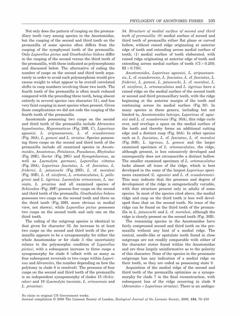
Not only does the pattern of cusping on the premax-illary teeth vary among species in the Anostomidae,but the cusping of the second and third tooth on thepremaxilla of some species often differs from thecusping of the symphyseal tooth of the premaxilla.Only Leporellus pictus and Gnathodolus bidens differin the cusping of the second versus the third teeth ofthe premaxilla, with these indicated as polymorphismsand discussed below. The alternative of coding thenumber of cusps on the second and third teeth sepa-rately in order to avoid such polymorphism would giveexcess weight to what appear to be overall correlatedshifts in cusp numbers involving those two teeth. Thefourth tooth of the premaxilla is often much reducedcompared with the preceding three teeth, being absententirely in several species (see character 31), and hasvery faint cusping in most species when present. Giventhose complications we did not code the cusping of thefourth tooth of the premaxilla.
Anostomids possessing two cusps on the secondand third teeth of the premaxilla include Abramiteshypselonotus, Hypomasticus (Fig. 29B, C), Leporinusagassizi, L. aripuanaensis, L. cf. ecuadorensis(Fig. 30A), L. gomesi and L. striatus. Species possess-ing three cusps on the second and third teeth of thepremaxilla include all examined species in Anosto-moides, Anostomus, Petulanos, Pseudanos, Rhytiodus(Fig. 29E), Sartor (Fig. 29G) and Synaptolaemus, aswell as Laemolyta garmani, Leporellus vittatus(Fig. 29A), Leporinus fasciatus, L. cf. fasciatus, L.friderici, L. jatuncochi (Fig. 29D), L. cf. moralesi(Fig. 30B), L. cf. niceforoi, L. ortomaculatus, L. pelle-grinii and L. tigrinus. Laemolyta orinocensis, L. tae-niata, L. proxima and all examined species ofSchizodon (Fig. 29F) possess four cusps on the secondand third teeth of the premaxilla. Gnathodolus bidenspossesses two cusps on the second tooth and three onthe third tooth (Fig. 29H, more obvious in medialview, not shown), while Leporellus pictus possessestwo cusps on the second tooth and only one on thethird tooth.
The coding of the outgroup species is identical tothat given for character 32. An increase to at leasttwo cusps on the second and third teeth of the pre-maxilla appears to be a synapomorphy for either thewhole Anostomidae or for clade 3 (the uncertaintyrelates to the polymorphic condition of Leporelluspictus), with a subsequent increase to three cusps asynapomorphy for clade 8 (albeit with as many asfour subsequent reversals to two cusps within Lepori-nus and Abramites, the number depending on how thepolytomy in clade 8 is resolved). The presence of fourcusps on the second and third teeth of the premaxillais an independent synapomorphy of clades 15 (Schiz-odon) and 19 (Laemolyta taeniata, L. orinocensis andL. proxima).
34. Structure of medial surface of second and thirdteeth of premaxilla: (0) medial surface of second andthird teeth of premaxilla either flat plane or curvedhollow, without raised ridge originating at anterioredge of tooth and extending across medial surface oftooth; (1) medial surface of tooth elaborated, withraised ridge originating at anterior edge of tooth andextending across medial surface of tooth (CI = 0.250;RI = 0.750).
Anostomoides, Leporinus agassizi, L. aripuanaen-sis, L. cf. ecuadorensis, L. fasciatus, L. cf. fasciatus, L.friderici, L. gomesi, L. jatuncochi, L. cf. moralesi, L.cf. niceforoi, L. ortomaculatus and L. tigrinus have araised ridge on the medial surface of the second toothor second and third premaxillary teeth, with the ridgebeginning at the anterior margin of the tooth andcontinuing across its medial surface (Fig. 30). Inmany species in these genera, including but notlimited to, Anostomoides laticeps, Leporinus cf. agas-sizi and L. cf. ecuadorensis (Fig. 30A), this ridge curlsover, and overlaps a space on the medial surface ofthe tooth and thereby forms an additional cuttingedge and a distinct cusp (Fig. 30A). In other speciessuch as L. fasciatus, L. cf. fasciatus, L. cf. moralesi(Fig. 30B), L. tigrinus, L. gomesi and the largerexamined specimen of L. ortomaculatus, the ridge,although present, is less extensively developed andconsequently does not circumscribe a distinct hollow.The smaller examined specimen of L. ortomaculatuslacks almost all trace of the ridge, which is bestdeveloped in the some of the largest Leporinus speci-mens examined (L. agassizi and L. cf. ecuadorensis).This may indicate that the presence and degree ofdevelopment of the ridge is ontogenetically variable,with that structure present only in adults of somespecies. In most of the species cited above, the medialridge and cusp on the third tooth is less well devel-oped than that on the second tooth. No trace of theridge can be found on the third tooth of the premax-illa in L. jatuncochi and L. cf. moralesi, although theridge is clearly present on the second tooth (Fig. 30B).
The remaining species in the Anostomidae havefairly compressed second and third teeth on the pre-maxilla without any hint of a medial ridge. Theconical, needle-like or spatulate teeth found in closeoutgroups are not readily comparable with either ofthe character states found within the Anostomidaeand are thus largely uninformative as to the polarityof this character. None of the species in the proximateoutgroups has any indication of a medial ridge ontheir tooth, so they are coded as possessing state 0.
Acquisition of the medial ridge of the second andthird teeth of the premaxilla optimizes as a synapo-morphy for clade 7 in the final reconstruction, withsubsequent loss of the ridge occurring in clade 9(Abramites + Leporinus striatus). There is an ambigu-
PHYLOGENY OF ANOSTOMID FISHES 105
No claim to original US Government works.Journal compilation © 2008 The Linnean Society of London, Zoological Journal of the Linnean Society, 2008, 154, 70–210
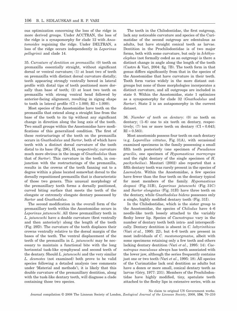
ous optimization concerning the loss of the ridge inmore derived groups. Under ACCTRAN, the loss ofthe ridge is a synapomorphy for clade 12 with Anos-tomoides regaining the ridge. Under DELTRAN, aloss of the ridge occurs independently in Leporinuspellegrinii and clade 14.
35. Curvature of dentition on premaxilla: (0) teeth onpremaxilla essentially straight, without significantdorsal or ventral curvature; (1) at least two of teethon premaxilla with distinct dorsal curvature distally;teeth appearing strongly ventrally bowed in lateralprofile with distal tips of teeth positioned more dor-sally than base of teeth; (2) at least two teeth onpremaxilla with strong ventral bend followed byanterior-facing alignment, resulting in zigzag shapeto tooth in lateral profile (CI = 1.000; RI = 1.000).
Most species of the Anostomidae have teeth on thepremaxilla that extend along a straight line from thebase of the tooth to its tip without any significantchange in direction along the long axis of the tooth.Two small groups within the Anostomidae have modi-fications of this generalized condition. The first ofthese restructurings of the teeth on the premaxillaoccurs in Gnathodolus and Sartor, both of which haveteeth with a distinct dorsal curvature of the toothdistal to its base (Fig. 29G, H, respectively; curvaturemuch more obvious in the image of Gnathodolus thanthat of Sartor). This curvature in the tooth, in con-junction with the restructurings of the premaxilla,results in the crowns of the teeth fanning out to adegree within a plane located somewhat dorsal to thedorsally repositioned premaxilla that is characteristicof those two genera. This unusual morphology ofthe premaxillary teeth forms a dorsally positioned,curved biting surface that meets the teeth of theelongate or extremely elongate dentary possessed bySartor and Gnathodolus.
The second modification in the overall form of thepremaxillary teeth within the Anostomidae occurs inLeporinus jatuncochi. All three premaxillary teeth inL. jatuncochi have a double curvature (first ventrallyand then anteriorly) along the length of the tooth(Fig. 29D). The curvature of the teeth displaces theircrowns ventrally relative to the dorsal margin of thebases of the teeth. The ventral displacement of theteeth of the premaxilla in L. jatuncochi may be nec-essary to maintain a functional bite with the longhorizontal tusk-like symphyseal and second teeth ofthe dentary. Should L. jatuncochi and the very similarL. desmotes (not examined) both prove to be validspecies following a detailed analysis (see commentsunder ‘Material and methods’), it is likely that thisdouble curvature of the premaxillary dentition, alongwith the tusk-like dentary teeth, will diagnose a cladecontaining those two species.
The teeth in the Chilodontidae, the first outgroup,lack any noticeable curvature and species of the Curi-matidae of the second outgroup are edentulous asadults, but have straight conical teeth as larvae.Dentition in the Prochilodontidae is of two majorforms, both with some curvature, but only in Ichthyo-elephas (not formally coded as an outgroup) is there adistinct change in angle along the length of the tooth(Castro & Vari, 2004: fig. 7B). The tooth form in thatgenus differs significantly from that in the species ofthe Anostomidae that have curvature in their teeth.Tooth form varies widely in the more distant out-groups but none of those morphologies incorporates adistinct curvature, and all outgroups are included instate 0. Within the Anostomidae, state 1 optimizesas a synapomorphy for clade 32 (Gnathodolus andSartor). State 2 is an autapomorphy in the currentdataset.
36. Number of teeth on dentary: (0) no teeth ondentary; (1–6) one to six teeth on dentary, respec-tively; (7) ten or more teeth on dentary (CI = 0.643;RI = 0.583).
Most anostomids possess four teeth on each dentary(e.g. Leporellus vittatus, Fig. 31A), with occasionalexamined specimens in the family possessing a smallfifth tooth posteriorly (one specimen of Pseudanosgracilis, one specimen of Hypomasticus mormyropsand the right dentary of the single specimen of H.pachycheilus). Mautari (2002) also reported that afifth dentary tooth was rarely present in the species ofLaemolyta. Within the Anostomidae, a few specieshave fewer than the four teeth on the dentary typicalfor most members of the family. Hypomasticusdespaxi (Fig. 31B), Leporinus jatuncochi (Fig. 31C)and Sartor elongatus (Fig. 31H) have three teeth onthe dentary, while Gnathodolus bidens possesses onlya single, highly modified dentary tooth (Fig. 31I).
In the Chilodontidae, which is the sister group tothe Anostomidae, the species of Chilodus have 4–6needle-like teeth loosely attached to the variablyfleshy lower lip. Species of Caenotropus vary in thenumber of dentary teeth both intra- and interspecifi-cally. Dentary dentition is absent in C. labyrinthicus(Vari et al., 1995: 22), but 4–6 teeth are present inmost individuals of C. mestomorgmatos, albeit withsome specimens retaining only a few teeth and otherslacking dentary dentition (Vari et al., 1995: 14). Cae-notropus maculosus always has teeth associated withthe lower jaw, although the series frequently containsjust one or two teeth (Vari et al., 1995: 18). All speciesof the Curimatidae lack oral dentition as adults buthave a dozen or more small, conical dentary teeth aslarvae (Géry, 1977: 231). Members of the Prochilodon-tidae have highly modified, tiny, spatulate teethattached to the fleshy lips in extensive series, with as
106 B. L. SIDLAUSKAS and R. P. VARI
No claim to original US Government works.Journal compilation © 2008 The Linnean Society of London, Zoological Journal of the Linnean Society, 2008, 154, 70–210
many as 76 teeth present on the inner row alone(Roberts, 1973: fig. 7; Castro & Vari, 2004: 22). Theplesiomorphic condition for the Anostomidae appearsto have been the possession of four teeth on thedentary, with the reduced numbers of such dentitionin a few members of the family considered to bederived.
37. Overall shape of dentary teeth: (0) dentary teethspade- or chisel-shaped, tapering to pointed or bluntdistal margin with or without additional cusping;teeth not extremely elongate and resembling awl;(1) dentary teeth elongate with symphyseal toothextremely elongate and recurved, overall shape remi-niscent of awl or very narrow chisel, distal tips maybe elaborated with cusps; (2) dentary teeth squat,much wider than high with distinct lateral bend; (3)dentary teeth small, spatulate and affixed to lips;(4) dentary teeth tiny, conical or rod-like in shape; (5)dentary teeth crown-like with 3–5 jagged cusps; (6)dentary teeth in form of large hooks (CI = 0.857;RI = 0.875).
Anostomids possess a greater array of dentarytooth forms relative to the intrafamilial variation
typical within other families in the Characiformes,with the symphyseal tooth demonstrating the great-est range of morphologies of all the teeth on thedentary. The dentary teeth typically have spade- orchisel-shaped morphologies, with variations on thisbasic form ranging from fairly elongate and pointed(e.g. the symphyseal teeth of Leporinus cf. fasciatus,Fig. 31D; Synaptolaemus, Fig. 31G), to squat andblunt (e.g. Anostomoides laticeps, Fig. 32C), to narrowand distinctly chisel-like (e.g. Pseudanos trimacula-tus, Fig. 32F), to thin and incisiform (e.g. Laemolytataeniata, Fig. 31E). Exclusive of the cusping and theform of the posterior lamina (see characters 38–44)the variation among the general tooth forms dis-cussed above is continuous. Two additional dentarytooth morphologies seen within the Anostomidae arealternatively so distinctive that they merit recogni-tion with distinct character states.
Leporinus jatuncochi, Gnathodolus bidens andSartor elongatus possess very elongate, recurvedtusk- or awl-like teeth (state 1; Fig. 31C, H, I). Fourother examined species also possess elongate mor-phologies of the symphyseal tooth of the dentary[Synaptolaemus cingulatus (Fig. 31G), L. fasciatus,
Figure 31. Dentition of right dentary in (A) Leporellus vittatus, INHS 56128; (B) Hypomasticus despaxi, MCZ 56552; (C)Leporinus jatuncochi, INHS 38940; (D) Leporinus cf. fasciatus, FMNH 103450; (E) Laemolyta taeniata, INHS 61513; (F)Petulanos intermedius, INPA 15184; (G) Synaptolaemus cingulatus, FMNH 103455; (H) Sartor elongatus, INPA 1168; (I)Gnathodolus bidens, ANSP 159389; lateral view, scale bar = 200 mm, images rotated to standardize orientation of dentary,anterior to top of image in A and B, to right in C and D, to bottom right in E–G, and to bottom in H and I.
PHYLOGENY OF ANOSTOMID FISHES 107
No claim to original US Government works.Journal compilation © 2008 The Linnean Society of London, Zoological Journal of the Linnean Society, 2008, 154, 70–210
Leporinus cf. fasciatus (Fig. 32D), and L. tigrinus].The elongation of the teeth in those four taxa is,however, less pronounced than that present in Sartor,Gnathodolus and Leporinus jatuncochi. The variationin symphyseal tooth size is continuous between L.fasciatus, L. cf. fasciatus, L. tigrinus, Synaptolaemusand the other species of Leporinus. As a consequenceall of these species are coded as possessing state 0.
Hypomasticus despaxi possesses a squat, widenedtooth form in which the distal portion of each toothdisplays a pronounced anterolateral curvature (state2; Fig. 31B). As far as we are aware this tooth form isunique to this species among characiforms, althoughwhen more species are examined this tooth morphol-ogy may be found to be diagnostic of a small cladewithin Hypomasticus. The function of this unusualtooth form is unknown.
Looking to the conditions in the proximate out-groups, the species of the Prochilodontidae possessvariably bowed, spatulate teeth (Castro & Vari, 2004:fig. 7). The Curimatidae lack teeth as adults (codedas missing data) but members of that family have
conical teeth as larvae (Géry, 1977: 231; Vari, 1983:fig. 8) while the species of the Chilodontidae thatpossess lower-jaw dentition have small rod-like, uni-cuspid teeth (state 4). All of these conditions areabsent within the Anostomidae. Likewise, the numer-ous small rod-like bicuspid teeth of various morpholo-gies found in basal members of the Citharinidae andDistichodontidae (state 4; see Vari, 1979: figs 2–5),and the strongly pointed and crown-like tri- to pen-tacuspid tooth morphology found in Brycon (state 5;Weitzman, 1962: figs 4, 8) and the hooked teeth ofParodon (state 6) lack direct homologues with thespade- or chisel-shaped morphologies of the dentaryteeth seen in the Anostomidae. Hemiodus ocellatusalso lacks teeth on the dentary (coded as missingdata). Possession of spade- or chisel-shaped dentaryteeth optimizes as a synapomorphy for the Anostomi-dae in the final reconstruction, with subsequent modi-fications of the dentition into distinctive tooth formsoccurring in Hypomasticus despaxi, Leporinus jatun-cochi, Sartor and Gnathodolus. The elongate toothmorphology appears to have evolved twice in the
Figure 32. Dentition of right dentary in (A) Leporinus cf. moralesi, UMMZ 216435; (B) Abramites hypselonotus, FMNH106574; (C) Anostomoides laticeps, INHS53677, posterior (fourth) tooth dislodged from socket; (D) Rhytiodus argenteo-fuscus, INHS 66082; (E) Schizodon fasciatus, FMNH 11135; (F) Pseudanos trimaculatus, FMNH 102122; medial view,scale bar = 200 mm, images rotated to standardize orientation of dentary, anterior to left in A, B, D and E, to bottom leftin C and to bottom in F.
108 B. L. SIDLAUSKAS and R. P. VARI
No claim to original US Government works.Journal compilation © 2008 The Linnean Society of London, Zoological Journal of the Linnean Society, 2008, 154, 70–210
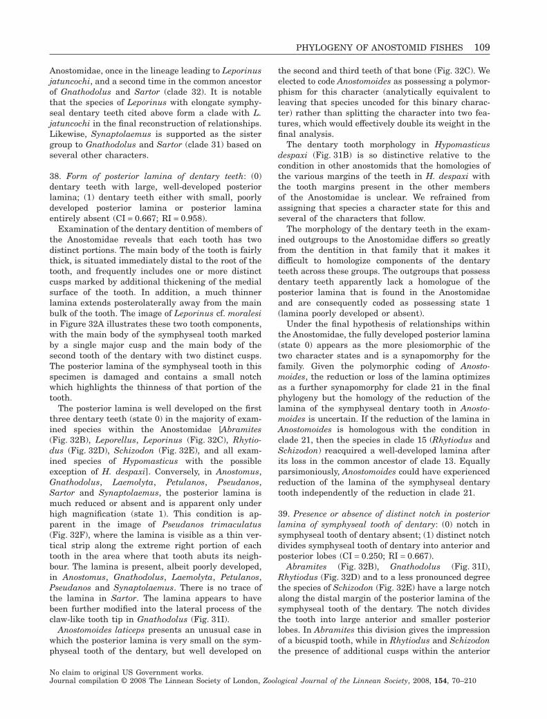
Anostomidae, once in the lineage leading to Leporinusjatuncochi, and a second time in the common ancestorof Gnathodolus and Sartor (clade 32). It is notablethat the species of Leporinus with elongate symphy-seal dentary teeth cited above form a clade with L.jatuncochi in the final reconstruction of relationships.Likewise, Synaptolaemus is supported as the sistergroup to Gnathodolus and Sartor (clade 31) based onseveral other characters.
38. Form of posterior lamina of dentary teeth: (0)dentary teeth with large, well-developed posteriorlamina; (1) dentary teeth either with small, poorlydeveloped posterior lamina or posterior laminaentirely absent (CI = 0.667; RI = 0.958).
Examination of the dentary dentition of members ofthe Anostomidae reveals that each tooth has twodistinct portions. The main body of the tooth is fairlythick, is situated immediately distal to the root of thetooth, and frequently includes one or more distinctcusps marked by additional thickening of the medialsurface of the tooth. In addition, a much thinnerlamina extends posterolaterally away from the mainbulk of the tooth. The image of Leporinus cf. moralesiin Figure 32A illustrates these two tooth components,with the main body of the symphyseal tooth markedby a single major cusp and the main body of thesecond tooth of the dentary with two distinct cusps.The posterior lamina of the symphyseal tooth in thisspecimen is damaged and contains a small notchwhich highlights the thinness of that portion of thetooth.
The posterior lamina is well developed on the firstthree dentary teeth (state 0) in the majority of exam-ined species within the Anostomidae [Abramites(Fig. 32B), Leporellus, Leporinus (Fig. 32C), Rhytio-dus (Fig. 32D), Schizodon (Fig. 32E), and all exam-ined species of Hypomasticus with the possibleexception of H. despaxi]. Conversely, in Anostomus,Gnathodolus, Laemolyta, Petulanos, Pseudanos,Sartor and Synaptolaemus, the posterior lamina ismuch reduced or absent and is apparent only underhigh magnification (state 1). This condition is ap-parent in the image of Pseudanos trimaculatus(Fig. 32F), where the lamina is visible as a thin ver-tical strip along the extreme right portion of eachtooth in the area where that tooth abuts its neigh-bour. The lamina is present, albeit poorly developed,in Anostomus, Gnathodolus, Laemolyta, Petulanos,Pseudanos and Synaptolaemus. There is no trace ofthe lamina in Sartor. The lamina appears to havebeen further modified into the lateral process of theclaw-like tooth tip in Gnathodolus (Fig. 31I).
Anostomoides laticeps presents an unusual case inwhich the posterior lamina is very small on the sym-physeal tooth of the dentary, but well developed on
the second and third teeth of that bone (Fig. 32C). Weelected to code Anostomoides as possessing a polymor-phism for this character (analytically equivalent toleaving that species uncoded for this binary charac-ter) rather than splitting the character into two fea-tures, which would effectively double its weight in thefinal analysis.
The dentary tooth morphology in Hypomasticusdespaxi (Fig. 31B) is so distinctive relative to thecondition in other anostomids that the homologies ofthe various margins of the teeth in H. despaxi withthe tooth margins present in the other membersof the Anostomidae is unclear. We refrained fromassigning that species a character state for this andseveral of the characters that follow.
The morphology of the dentary teeth in the exam-ined outgroups to the Anostomidae differs so greatlyfrom the dentition in that family that it makes itdifficult to homologize components of the dentaryteeth across these groups. The outgroups that possessdentary teeth apparently lack a homologue of theposterior lamina that is found in the Anostomidaeand are consequently coded as possessing state 1(lamina poorly developed or absent).
Under the final hypothesis of relationships withinthe Anostomidae, the fully developed posterior lamina(state 0) appears as the more plesiomorphic of thetwo character states and is a synapomorphy for thefamily. Given the polymorphic coding of Anosto-moides, the reduction or loss of the lamina optimizesas a further synapomorphy for clade 21 in the finalphylogeny but the homology of the reduction of thelamina of the symphyseal dentary tooth in Anosto-moides is uncertain. If the reduction of the lamina inAnostomoides is homologous with the condition inclade 21, then the species in clade 15 (Rhytiodus andSchizodon) reacquired a well-developed lamina afterits loss in the common ancestor of clade 13. Equallyparsimoniously, Anostomoides could have experiencedreduction of the lamina of the symphyseal dentarytooth independently of the reduction in clade 21.
39. Presence or absence of distinct notch in posteriorlamina of symphyseal tooth of dentary: (0) notch insymphyseal tooth of dentary absent; (1) distinct notchdivides symphyseal tooth of dentary into anterior andposterior lobes (CI = 0.250; RI = 0.667).
Abramites (Fig. 32B), Gnathodolus (Fig. 31I),Rhytiodus (Fig. 32D) and to a less pronounced degreethe species of Schizodon (Fig. 32E) have a large notchalong the distal margin of the posterior lamina of thesymphyseal tooth of the dentary. The notch dividesthe tooth into large anterior and smaller posteriorlobes. In Abramites this division gives the impressionof a bicuspid tooth, while in Rhytiodus and Schizodonthe presence of additional cusps within the anterior
PHYLOGENY OF ANOSTOMID FISHES 109
No claim to original US Government works.Journal compilation © 2008 The Linnean Society of London, Zoological Journal of the Linnean Society, 2008, 154, 70–210
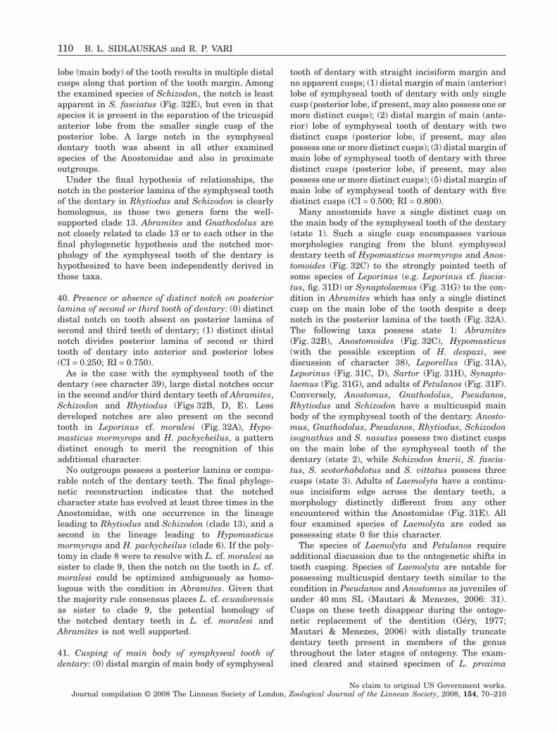
lobe (main body) of the tooth results in multiple distalcusps along that portion of the tooth margin. Amongthe examined species of Schizodon, the notch is leastapparent in S. fasciatus (Fig. 32E), but even in thatspecies it is present in the separation of the tricuspidanterior lobe from the smaller single cusp of theposterior lobe. A large notch in the symphysealdentary tooth was absent in all other examinedspecies of the Anostomidae and also in proximateoutgroups.
Under the final hypothesis of relationships, thenotch in the posterior lamina of the symphyseal toothof the dentary in Rhytiodus and Schizodon is clearlyhomologous, as those two genera form the well-supported clade 13. Abramites and Gnathodolus arenot closely related to clade 13 or to each other in thefinal phylogenetic hypothesis and the notched mor-phology of the symphyseal tooth of the dentary ishypothesized to have been independently derived inthose taxa.
40. Presence or absence of distinct notch on posteriorlamina of second or third tooth of dentary: (0) distinctdistal notch on tooth absent on posterior lamina ofsecond and third teeth of dentary; (1) distinct distalnotch divides posterior lamina of second or thirdtooth of dentary into anterior and posterior lobes(CI = 0.250; RI = 0.750).
As is the case with the symphyseal tooth of thedentary (see character 39), large distal notches occurin the second and/or third dentary teeth of Abramites,Schizodon and Rhytiodus (Figs 32B, D, E). Lessdeveloped notches are also present on the secondtooth in Leporinus cf. moralesi (Fig. 32A), Hypo-masticus mormyrops and H. pachycheilus, a patterndistinct enough to merit the recognition of thisadditional character.
No outgroups possess a posterior lamina or compa-rable notch of the dentary teeth. The final phyloge-netic reconstruction indicates that the notchedcharacter state has evolved at least three times in theAnostomidae, with one occurrence in the lineageleading to Rhytiodus and Schizodon (clade 13), and asecond in the lineage leading to Hypomasticusmormyrops and H. pachycheilus (clade 6). If the poly-tomy in clade 8 were to resolve with L. cf. moralesi assister to clade 9, then the notch on the tooth in L. cf.moralesi could be optimized ambiguously as homo-logous with the condition in Abramites. Given thatthe majority rule consensus places L. cf. ecuadorensisas sister to clade 9, the potential homology ofthe notched dentary teeth in L. cf. moralesi andAbramites is not well supported.
41. Cusping of main body of symphyseal tooth ofdentary: (0) distal margin of main body of symphyseal
tooth of dentary with straight incisiform margin andno apparent cusps; (1) distal margin of main (anterior)lobe of symphyseal tooth of dentary with only singlecusp (posterior lobe, if present, may also possess one ormore distinct cusps); (2) distal margin of main (ante-rior) lobe of symphyseal tooth of dentary with twodistinct cusps (posterior lobe, if present, may alsopossess one or more distinct cusps); (3) distal margin ofmain lobe of symphyseal tooth of dentary with threedistinct cusps (posterior lobe, if present, may alsopossess one or more distinct cusps); (5) distal margin ofmain lobe of symphyseal tooth of dentary with fivedistinct cusps (CI = 0.500; RI = 0.800).
Many anostomids have a single distinct cusp onthe main body of the symphyseal tooth of the dentary(state 1). Such a single cusp encompasses variousmorphologies ranging from the blunt symphysealdentary teeth of Hypomasticus mormyrops and Anos-tomoides (Fig. 32C) to the strongly pointed teeth ofsome species of Leporinus (e.g. Leporinus cf. fascia-tus, fig. 31D) or Synaptolaemus (Fig. 31G) to the con-dition in Abramites which has only a single distinctcusp on the main lobe of the tooth despite a deepnotch in the posterior lamina of the tooth (Fig. 32A).The following taxa possess state 1: Abramites(Fig. 32B), Anostomoides (Fig. 32C), Hypomasticus(with the possible exception of H. despaxi, seediscussion of character 38), Leporellus (Fig. 31A),Leporinus (Fig. 31C, D), Sartor (Fig. 31H), Synapto-laemus (Fig. 31G), and adults of Petulanos (Fig. 31F).Conversely, Anostomus, Gnathodolus, Pseudanos,Rhytiodus and Schizodon have a multicuspid mainbody of the symphyseal tooth of the dentary. Anosto-mus, Gnathodolus, Pseudanos, Rhytiodus, Schizodonisognathus and S. nasutus possess two distinct cuspson the main lobe of the symphyseal tooth of thedentary (state 2), while Schizodon knerii, S. fascia-tus, S. scotorhabdotus and S. vittatus possess threecusps (state 3). Adults of Laemolyta have a continu-ous incisiform edge across the dentary teeth, amorphology distinctly different from any otherencountered within the Anostomidae (Fig. 31E). Allfour examined species of Laemolyta are coded aspossessing state 0 for this character.
The species of Laemolyta and Petulanos requireadditional discussion due to the ontogenetic shifts intooth cusping. Species of Laemolyta are notable forpossessing multicuspid dentary teeth similar to thecondition in Pseudanos and Anostomus as juveniles ofunder 40 mm SL (Mautari & Menezes, 2006: 31).Cusps on these teeth disappear during the ontoge-netic replacement of the dentition (Géry, 1977;Mautari & Menezes, 2006) with distally truncatedentary teeth present in members of the genusthroughout the later stages of ontogeny. The exam-ined cleared and stained specimen of L. proxima
110 B. L. SIDLAUSKAS and R. P. VARI
No claim to original US Government works.Journal compilation © 2008 The Linnean Society of London, Zoological Journal of the Linnean Society, 2008, 154, 70–210
retains cusping on all four dentary teeth contrary tothe lack of such cusping in the other examined con-geners of similar size. Such cusping is a juvenilecharacter in light of the lack of that feature in adultsof the species (Mautari & Menezes, 2006). We conse-quently coded L. proxima as state 0 in keeping withthe uncusped dentary teeth in adults of that species.This observation may be evidence that L. proximaretains cusps on its dentary teeth to a greater bodysize than do its congeners.
In the final phylogenetic hypothesis arrived atherein, Laemolyta is nested deeply within a clade oftaxa with strongly multicuspid dentary teeth. Thatevidence, combined with the observed ontogeneticprogression, reveals that the truncate, incisiformtooth morphology in Laemolyta is derived from aplesiomorphically multicuspid adult tooth.
Petulanos intermedius and P. plicatus also exhibitan ontogenetic reduction in the degree of cusping ofthe symphyseal tooth, having bicuspid teeth in juve-niles smaller than about 60 mm SL and unicuspidteeth in adults (Winterbottom, 1980: 15, 55). Inkeeping with the observed adult morphology(Fig. 31F), these two taxa are coded as possessingstate 1 (unicuspid teeth), but this condition is clearlyderived from a bicuspid precursor. In this light andgiven the final phylogeny, the unicuspid symphysealdentition of Sartor (Fig. 31H) and Synaptolaemus(Fig. 31G), which together with Gnathodolus form thesister clade to Petulanos, can also be understood as aderived character state modified from a bicuspid pre-cursor and as such non-homologous with the unicus-pid teeth present in some species of Leporinus.
The teeth of the Chilodontidae and Prochilodon-tidae in the proximate outgroups to the Anostomidaeall have only a single cusp. A single cusp is alsopresent in the larvae of the Curimatidae, themembers of which lose all dentition ontogenetically(Vari, 1983: 8). Parodon suborbitalis has unicuspiddentary teeth and Hemiodus ocellatus lacks dentaryteeth. Members of the Citharinidae and basalmembers of the Distichodontidae possess bicuspidteeth, and Brycon falcatus possesses pentacuspidteeth (coded as the autapomorphic state 5).
In light of the final phylogenetic hypothesis, thepresence of two cusps on the symphyseal tooth of thedentary (state 2) is a synapomorphy for clade 14, witha further modification to the incisiform, uncuspedcondition a synapomorphy for Laemolyta (clade 22).The possession of a unicuspid symphyseal dentarytooth (state 1) is a synapomorphy for clade 29, whichis reversed in Gnathodolus. State 3 (tricuspid mainbody of symphyseal dentary teeth) optimizes as asynapomorphy for Schizodon that is reversed to thebicuspid condition in clade 19 (Schizodon isognathus+ S. nasutus). We note that the potential resolution of
the polytomy within Schizodon such that clade 19 isbasal within the genus would reverse the polarity ofthat state transition.
42. Cusping of main body of second tooth of dentary:(0) distal margin of main body of second tooth ofdentary with straight incisiform margin and noapparent cusps; (1) distal margin of main lobe ofsecond tooth of dentary with only single cusp (poste-rior lobe, if present, may also possess one or moredistinct cusps); (2) distal margin of main lobe ofsecond tooth of dentary with two distinct cusps (pos-terior lobe, if present, may also possess one or moredistinct cusps); (3) distal margin of main lobe ofsecond tooth of dentary with three distinct cusps(posterior lobe, if present, may also possess one ormore distinct cusps) (CI = 0.333; RI = 0.727).
Most species in the Anostomidae have similarcusping patterns on the three dentary teeth locatedclosest to the symphysis. Nonetheless, sufficientspecies in the family differ in the characteristiccusping patterns of the symphyseal, second and thirdteeth of the dentary as to merit a separate coding foreach of these teeth. Species not discussed below arecoded as for character 41. Synaptolaemus has a tri-cuspid second dentary tooth (Fig. 31G; state 3), whichcontrasts with the unicuspid morphology of the sym-physeal dentary tooth in that taxon. Leporinus cf.moralesi (Fig. 32A) has a bicuspid main lobe of thesecond dentary tooth despite lacking a second cusp onthe symphyseal tooth. Gnathodolus lacks the secondtooth of the dentary and consequently cannot becoded for this character.
In the final phylogeny, this character yields thesynapomorphies of equal inclusiveness and supportsthe same clades as does character 41. The cuspingpatterns observed in Leporinus cf. moralesi and Syn-aptolaemus are autapomorphic in the context of theanalysed set of taxa, but may prove to be synapomor-phies of Synaptolaemus or a portion of Leporinuswhen additional species of each genus are added tothe analysis.
43. Cusping of main body of third tooth of dentary: (0)distal margin of main body of third tooth of dentarywith straight incisiform margin and no apparentcusps; (1) distal margin of main lobe of third tooth ofdentary with only single cusp (posterior lobe, ifpresent, may also possess one or more distinct cusps);(2) distal margin of main lobe of third tooth of dentarywith two distinct cusps (posterior lobe, if present, mayalso possess one or more distinct cusps); (3) distalmargin of main lobe of third tooth of dentary withthree distinct cusps (posterior lobe, if present, mayalso possess one or more distinct cusps) (CI = 0.429;RI = 0.810).
PHYLOGENY OF ANOSTOMID FISHES 111
No claim to original US Government works.Journal compilation © 2008 The Linnean Society of London, Zoological Journal of the Linnean Society, 2008, 154, 70–210
The species of Petulanos have tricuspid main lobesof the third tooth of the dentary (state 3) as opposed tothe unicuspid adult dentition of the symphyseal andsecond teeth on that bone. The specimen of Petulanosintermedius that appears in the micrograph (Fig. 31F)has an anomalously rounded (worn?) third dentarytooth whereas the contralateral tooth on the left side ofthat individual is clearly tricuspid. This difference mayrepresent different levels of wear if the species ofthe Anostomidae undergo the simultaneous completereplacement of the teeth on one side of the jaw such asoccurs in various groups in the Characiformes(Roberts, 1967; Berkovitz, 1975; Berkovitz & Shellis,1978). In support of that conjecture, several examinedalcohol specimens of Petulanos intermedius (INPA15184) are missing all but the symphyseal tooth in oneside of the lower jaw. Leporinus cf. moralesi (Fig. 32A)has a unicuspid main body of the third tooth of thedentary (state 1) that contrasts with the distinctlybicuspid main lobe of the second tooth of the dentary inthat taxon. Rhytiodus argenteofuscus has only a singlecusp on the main lobe of the third tooth of the dentary(Fig. 32D), while Pseudanos (Fig. 32F), Anostomus,Schizodon nasutus and S. isognathus have three cuspson the main lobe of the third tooth of the dentarycontrary to the two cusps that they possess on thesymphyseal and second teeth of that bone. The thirdteeth of the dentary of Synaptolaemus (Fig. 31G) andSartor (Fig. 31H) are reduced in size (note that the tinythird tooth in the figured specimen of Sartor has adamaged tip and is hardly visible), but are apparentlyunicuspid and coded as such (state 1). Taxa not citedabove are coded as for character 42.
In the final hypothesis of relationships, state 1 isplesiomorphic within the Anostomidae. The posses-sion of two cusps on the third tooth of the dentary isa synapomorphy linking Rhytiodus lauzannei andR. microlepis (clade 17). The presence of an incisiformthird tooth (state 0) is a synapomorphy for Laemolyta.Optimization of character state 3 varies betweenACCTRAN and DELTRAN reconstructions. UnderACCTRAN, state 3 evolved in the common ancestor ofclade 14 with subsequent shifts in cusping in Rhytio-dus argenteofuscus, clade 17, Laemolyta and clade 31.Under DELTRAN, state 1 is the plesiomorphic condi-tion for clade 14 and the tricuspid third tooth of thedentary (state 3) evolved independently in Schizodonand clade 25, with additional state changes inLaemolyta, and clades 17 and 31.
44. Presence or absence of cusps on posterior lobes ofdentary teeth: (0) posterior lobes of dentary teethabsent, or if posterior lobes present, no distinct cusppresent on posterior lobes of tooth; (1) posterior lobesof dentary teeth with one or more distinct cusps(CI = 1.000; RI = 1.000).
Most anostomids lack a posterior lobe of thedentary teeth delimited by a distinct notch along theposterior lamina of the tooth (see characters 39 and40). Among those members of the family with a pos-terior lobe, only the species of Rhytiodus and Schiz-odon possess one or more distinct cusps on the distalmargin of that posterior lobe (Fig. 32D, E). Mostspecies in these genera possess a single cusp on thislobe, but Schizodon isognathus has two cusps. Noneof the examined outgroups possesses a posteriorlamina, let alone a notch or posterior lobe of thedentary teeth; consequently, we did not code the out-groups for this character. The possession of a distinctposterior cusp on the dentary teeth is a synapomor-phy for the clade containing Rhytiodus and Schizodon(clade 15) under the final phylogenetic hypothesis.
UPPER JAW
45. Form of ascending process of premaxilla: (0) eitherabsent or slightly to moderately developed andslender; (1) large, well developed and distinctly trian-gular overall (CI = 0.500; RI = 0.917).
In all members of the Anostomidae as well as in theoutgroup Parodon suborbitalis, the premaxilla is avery robust ossification with an enlarged ascendingprocess that progressively expands from its tiptowards the region where it joins the main body of thebone (state 1, Figs 33–37). The ascending process ofthe premaxilla has a resultant overall triangular formin lateral view and is less distinct from the main bodyof the premaxilla than in outgroups (see also com-ments in Vari, 1983: 10), which typically have anascending process of the premaxilla that is notablynarrower than, and quite distinct from, the main bodyof that bone (state 0). Curimata inornata and Disti-chodus sp. have deep, ventrally expanded premaxillaebut no distinct ascending process and are coded as 0,while an apparent process in Xenocharax spilurus ismedial in position and presumably non-homologouswith that in the Anostomidae (also coded as 0). In thefinal reconstruction, the triangular form of theascending process of the premaxilla is a synapomor-phy for the whole Anostomidae (clade 1). Parodonsuborbitalis is not the sister to the Anostomidae (Vari,1983; results herein), and the triangular ascendingprocess of the premaxilla in that species is ahomoplasy relative to the presence of that feature inthe family.
46. Orientation of premaxilla: (0) vertically or antero-dorsally inclined, with dorsal point of premaxillasituated immediately dorsal to, or anterior of, verticalthrough ventral point of premaxilla; premaxillaryteeth directed ventrally or posteroventrally; (1) poster-odorsally inclined, with dorsal portion of premaxilla
112 B. L. SIDLAUSKAS and R. P. VARI
No claim to original US Government works.Journal compilation © 2008 The Linnean Society of London, Zoological Journal of the Linnean Society, 2008, 154, 70–210
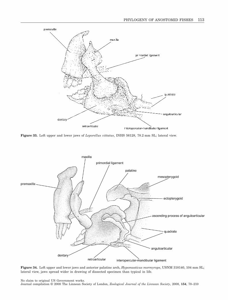
Figure 33. Left upper and lower jaws of Leporellus vittatus, INHS 56128, 78.2 mm SL; lateral view.
Figure 34. Left upper and lower jaws and anterior palatine arch, Hypomasticus mormyrops, USNM 318140, 104 mm SL;lateral view, jaws spread wider in drawing of dissected specimen than typical in life.
PHYLOGENY OF ANOSTOMID FISHES 113
No claim to original US Government works.Journal compilation © 2008 The Linnean Society of London, Zoological Journal of the Linnean Society, 2008, 154, 70–210
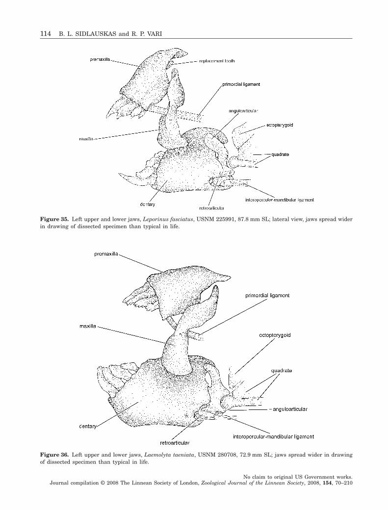
Figure 35. Left upper and lower jaws, Leporinus fasciatus, USNM 225991, 87.8 mm SL; lateral view, jaws spread widerin drawing of dissected specimen than typical in life.
Figure 36. Left upper and lower jaws, Laemolyta taeniata, USNM 280708, 72.9 mm SL; jaws spread wider in drawingof dissected specimen than typical in life.
114 B. L. SIDLAUSKAS and R. P. VARI
No claim to original US Government works.Journal compilation © 2008 The Linnean Society of London, Zoological Journal of the Linnean Society, 2008, 154, 70–210
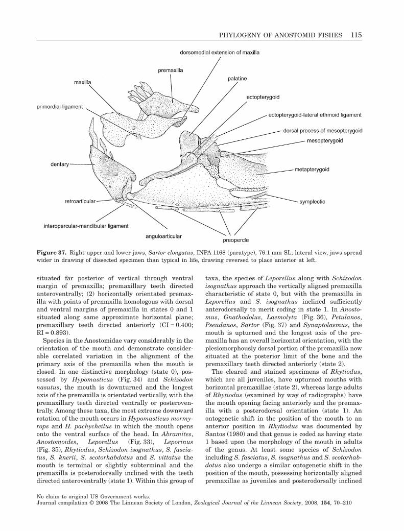
situated far posterior of vertical through ventralmargin of premaxilla; premaxillary teeth directedanteroventrally; (2) horizontally orientated premax-illa with points of premaxilla homologous with dorsaland ventral margins of premaxilla in states 0 and 1situated along same approximate horizontal plane;premaxillary teeth directed anteriorly (CI = 0.400;RI = 0.893).
Species in the Anostomidae vary considerably in theorientation of the mouth and demonstrate consider-able correlated variation in the alignment of theprimary axis of the premaxilla when the mouth isclosed. In one distinctive morphology (state 0), pos-sessed by Hypomasticus (Fig. 34) and Schizodonnasutus, the mouth is downturned and the longestaxis of the premaxilla is orientated vertically, with thepremaxillary teeth directed ventrally or posteroven-trally. Among these taxa, the most extreme downwardrotation of the mouth occurs in Hypomasticus mormy-rops and H. pachycheilus in which the mouth opensonto the ventral surface of the head. In Abramites,Anostomoides, Leporellus (Fig. 33), Leporinus(Fig. 35), Rhytiodus, Schizodon isognathus, S. fascia-tus, S. knerii, S. scotorhabdotus and S. vittatus themouth is terminal or slightly subterminal and thepremaxilla is posterodorsally inclined with the teethdirected anteroventrally (state 1). Within this group of
taxa, the species of Leporellus along with Schizodonisognathus approach the vertically aligned premaxillacharacteristic of state 0, but with the premaxilla inLeporellus and S. isognathus inclined sufficientlyanterodorsally to merit coding in state 1. In Anosto-mus, Gnathodolus, Laemolyta (Fig. 36), Petulanos,Pseudanos, Sartor (Fig. 37) and Synaptolaemus, themouth is upturned and the longest axis of the pre-maxilla has an overall horizontal orientation, with theplesiomorphously dorsal portion of the premaxilla nowsituated at the posterior limit of the bone and thepremaxillary teeth directed anteriorly (state 2).
The cleared and stained specimens of Rhytiodus,which are all juveniles, have upturned mouths withhorizontal premaxillae (state 2), whereas large adultsof Rhytiodus (examined by way of radiographs) havethe mouth opening facing anteriorly and the premax-illa with a posterodorsal orientation (state 1). Anontogenetic shift in the position of the mouth to ananterior position in Rhytiodus was documented bySantos (1980) and that genus is coded as having state1 based upon the morphology of the mouth in adultsof the genus. At least some species of Schizodonincluding S. fasciatus, S. isognathus and S. scotorhab-dotus also undergo a similar ontogenetic shift in theposition of the mouth, possessing horizontally alignedpremaxillae as juveniles and posterodorsally inclined
Figure 37. Right upper and lower jaws, Sartor elongatus, INPA 1168 (paratype), 76.1 mm SL; lateral view, jaws spreadwider in drawing of dissected specimen than typical in life, drawing reversed to place anterior at left.
PHYLOGENY OF ANOSTOMID FISHES 115
No claim to original US Government works.Journal compilation © 2008 The Linnean Society of London, Zoological Journal of the Linnean Society, 2008, 154, 70–210
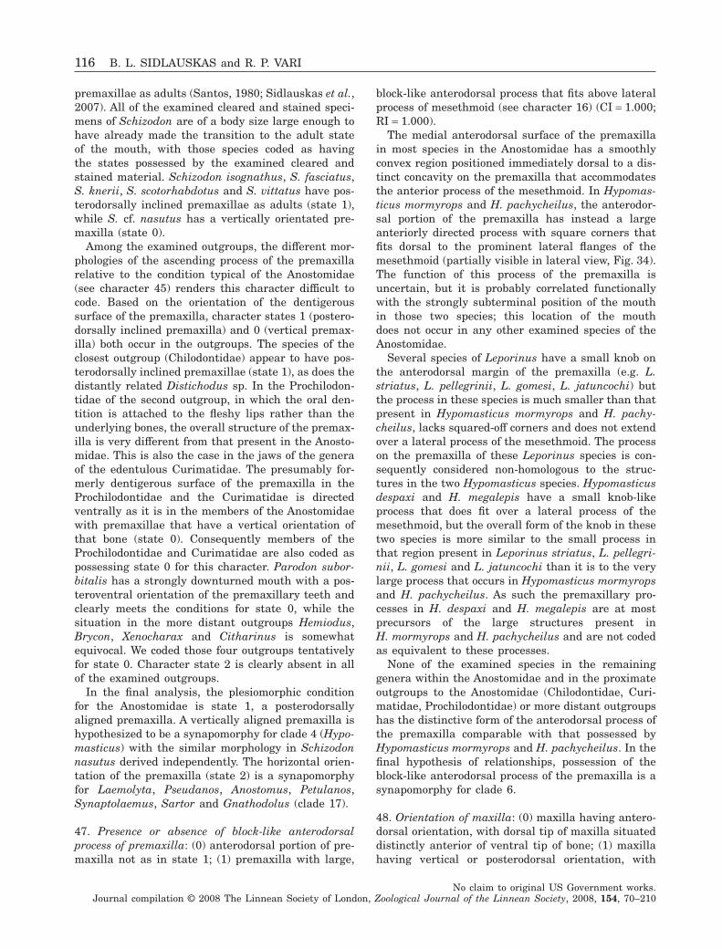
premaxillae as adults (Santos, 1980; Sidlauskas et al.,2007). All of the examined cleared and stained speci-mens of Schizodon are of a body size large enough tohave already made the transition to the adult stateof the mouth, with those species coded as havingthe states possessed by the examined cleared andstained material. Schizodon isognathus, S. fasciatus,S. knerii, S. scotorhabdotus and S. vittatus have pos-terodorsally inclined premaxillae as adults (state 1),while S. cf. nasutus has a vertically orientated pre-maxilla (state 0).
Among the examined outgroups, the different mor-phologies of the ascending process of the premaxillarelative to the condition typical of the Anostomidae(see character 45) renders this character difficult tocode. Based on the orientation of the dentigeroussurface of the premaxilla, character states 1 (postero-dorsally inclined premaxilla) and 0 (vertical premax-illa) both occur in the outgroups. The species of theclosest outgroup (Chilodontidae) appear to have pos-terodorsally inclined premaxillae (state 1), as does thedistantly related Distichodus sp. In the Prochilodon-tidae of the second outgroup, in which the oral den-tition is attached to the fleshy lips rather than theunderlying bones, the overall structure of the premax-illa is very different from that present in the Anosto-midae. This is also the case in the jaws of the generaof the edentulous Curimatidae. The presumably for-merly dentigerous surface of the premaxilla in theProchilodontidae and the Curimatidae is directedventrally as it is in the members of the Anostomidaewith premaxillae that have a vertical orientation ofthat bone (state 0). Consequently members of theProchilodontidae and Curimatidae are also coded aspossessing state 0 for this character. Parodon subor-bitalis has a strongly downturned mouth with a pos-teroventral orientation of the premaxillary teeth andclearly meets the conditions for state 0, while thesituation in the more distant outgroups Hemiodus,Brycon, Xenocharax and Citharinus is somewhatequivocal. We coded those four outgroups tentativelyfor state 0. Character state 2 is clearly absent in allof the examined outgroups.
In the final analysis, the plesiomorphic conditionfor the Anostomidae is state 1, a posterodorsallyaligned premaxilla. A vertically aligned premaxilla ishypothesized to be a synapomorphy for clade 4 (Hypo-masticus) with the similar morphology in Schizodonnasutus derived independently. The horizontal orien-tation of the premaxilla (state 2) is a synapomorphyfor Laemolyta, Pseudanos, Anostomus, Petulanos,Synaptolaemus, Sartor and Gnathodolus (clade 17).
47. Presence or absence of block-like anterodorsalprocess of premaxilla: (0) anterodorsal portion of pre-maxilla not as in state 1; (1) premaxilla with large,
block-like anterodorsal process that fits above lateralprocess of mesethmoid (see character 16) (CI = 1.000;RI = 1.000).
The medial anterodorsal surface of the premaxillain most species in the Anostomidae has a smoothlyconvex region positioned immediately dorsal to a dis-tinct concavity on the premaxilla that accommodatesthe anterior process of the mesethmoid. In Hypomas-ticus mormyrops and H. pachycheilus, the anterodor-sal portion of the premaxilla has instead a largeanteriorly directed process with square corners thatfits dorsal to the prominent lateral flanges of themesethmoid (partially visible in lateral view, Fig. 34).The function of this process of the premaxilla isuncertain, but it is probably correlated functionallywith the strongly subterminal position of the mouthin those two species; this location of the mouthdoes not occur in any other examined species of theAnostomidae.
Several species of Leporinus have a small knob onthe anterodorsal margin of the premaxilla (e.g. L.striatus, L. pellegrinii, L. gomesi, L. jatuncochi) butthe process in these species is much smaller than thatpresent in Hypomasticus mormyrops and H. pachy-cheilus, lacks squared-off corners and does not extendover a lateral process of the mesethmoid. The processon the premaxilla of these Leporinus species is con-sequently considered non-homologous to the struc-tures in the two Hypomasticus species. Hypomasticusdespaxi and H. megalepis have a small knob-likeprocess that does fit over a lateral process of themesethmoid, but the overall form of the knob in thesetwo species is more similar to the small process inthat region present in Leporinus striatus, L. pellegri-nii, L. gomesi and L. jatuncochi than it is to the verylarge process that occurs in Hypomasticus mormyropsand H. pachycheilus. As such the premaxillary pro-cesses in H. despaxi and H. megalepis are at mostprecursors of the large structures present inH. mormyrops and H. pachycheilus and are not codedas equivalent to these processes.
None of the examined species in the remaininggenera within the Anostomidae and in the proximateoutgroups to the Anostomidae (Chilodontidae, Curi-matidae, Prochilodontidae) or more distant outgroupshas the distinctive form of the anterodorsal process ofthe premaxilla comparable with that possessed byHypomasticus mormyrops and H. pachycheilus. In thefinal hypothesis of relationships, possession of theblock-like anterodorsal process of the premaxilla is asynapomorphy for clade 6.
48. Orientation of maxilla: (0) maxilla having antero-dorsal orientation, with dorsal tip of maxilla situateddistinctly anterior of ventral tip of bone; (1) maxillahaving vertical or posterodorsal orientation, with
116 B. L. SIDLAUSKAS and R. P. VARI
No claim to original US Government works.Journal compilation © 2008 The Linnean Society of London, Zoological Journal of the Linnean Society, 2008, 154, 70–210
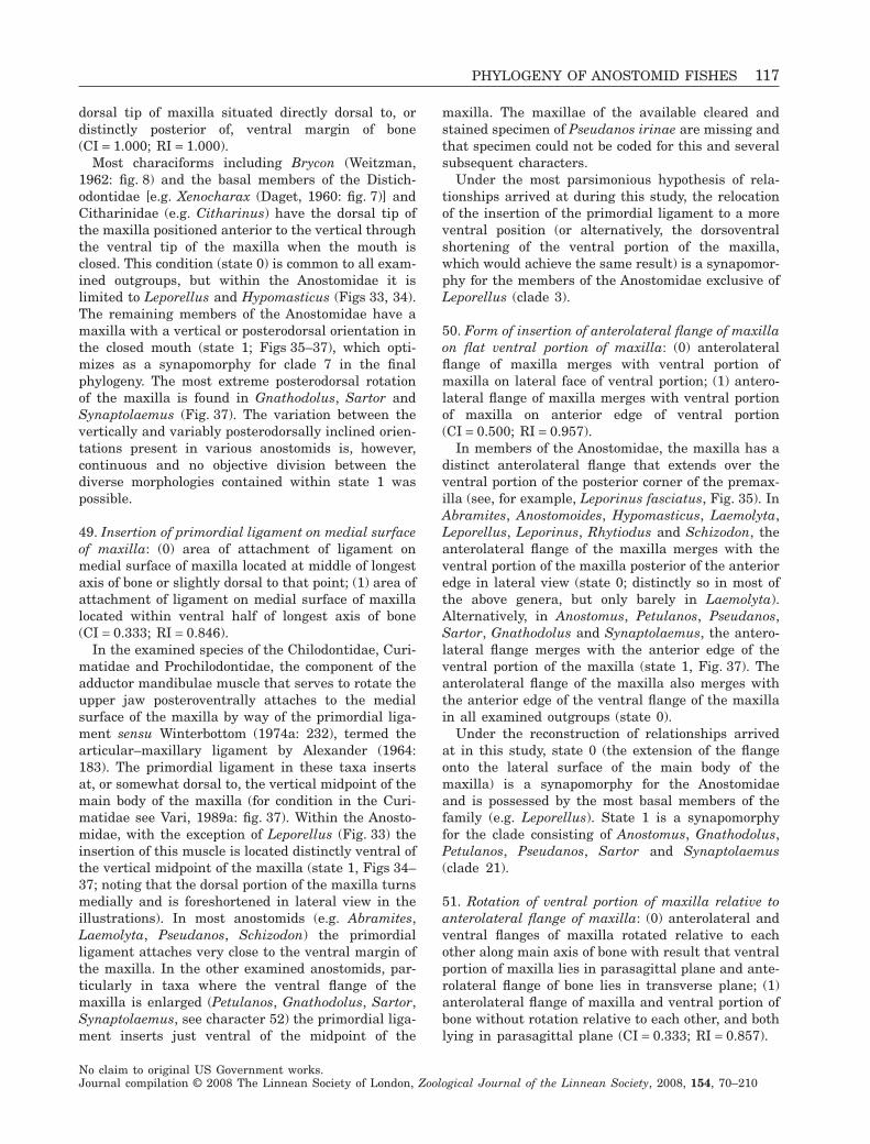
dorsal tip of maxilla situated directly dorsal to, ordistinctly posterior of, ventral margin of bone(CI = 1.000; RI = 1.000).
Most characiforms including Brycon (Weitzman,1962: fig. 8) and the basal members of the Distich-odontidae [e.g. Xenocharax (Daget, 1960: fig. 7)] andCitharinidae (e.g. Citharinus) have the dorsal tip ofthe maxilla positioned anterior to the vertical throughthe ventral tip of the maxilla when the mouth isclosed. This condition (state 0) is common to all exam-ined outgroups, but within the Anostomidae it islimited to Leporellus and Hypomasticus (Figs 33, 34).The remaining members of the Anostomidae have amaxilla with a vertical or posterodorsal orientation inthe closed mouth (state 1; Figs 35–37), which opti-mizes as a synapomorphy for clade 7 in the finalphylogeny. The most extreme posterodorsal rotationof the maxilla is found in Gnathodolus, Sartor andSynaptolaemus (Fig. 37). The variation between thevertically and variably posterodorsally inclined orien-tations present in various anostomids is, however,continuous and no objective division between thediverse morphologies contained within state 1 waspossible.
49. Insertion of primordial ligament on medial surfaceof maxilla: (0) area of attachment of ligament onmedial surface of maxilla located at middle of longestaxis of bone or slightly dorsal to that point; (1) area ofattachment of ligament on medial surface of maxillalocated within ventral half of longest axis of bone(CI = 0.333; RI = 0.846).
In the examined species of the Chilodontidae, Curi-matidae and Prochilodontidae, the component of theadductor mandibulae muscle that serves to rotate theupper jaw posteroventrally attaches to the medialsurface of the maxilla by way of the primordial liga-ment sensu Winterbottom (1974a: 232), termed thearticular–maxillary ligament by Alexander (1964:183). The primordial ligament in these taxa insertsat, or somewhat dorsal to, the vertical midpoint of themain body of the maxilla (for condition in the Curi-matidae see Vari, 1989a: fig. 37). Within the Anosto-midae, with the exception of Leporellus (Fig. 33) theinsertion of this muscle is located distinctly ventral ofthe vertical midpoint of the maxilla (state 1, Figs 34–37; noting that the dorsal portion of the maxilla turnsmedially and is foreshortened in lateral view in theillustrations). In most anostomids (e.g. Abramites,Laemolyta, Pseudanos, Schizodon) the primordialligament attaches very close to the ventral margin ofthe maxilla. In the other examined anostomids, par-ticularly in taxa where the ventral flange of themaxilla is enlarged (Petulanos, Gnathodolus, Sartor,Synaptolaemus, see character 52) the primordial liga-ment inserts just ventral of the midpoint of the
maxilla. The maxillae of the available cleared andstained specimen of Pseudanos irinae are missing andthat specimen could not be coded for this and severalsubsequent characters.
Under the most parsimonious hypothesis of rela-tionships arrived at during this study, the relocationof the insertion of the primordial ligament to a moreventral position (or alternatively, the dorsoventralshortening of the ventral portion of the maxilla,which would achieve the same result) is a synapomor-phy for the members of the Anostomidae exclusive ofLeporellus (clade 3).
50. Form of insertion of anterolateral flange of maxillaon flat ventral portion of maxilla: (0) anterolateralflange of maxilla merges with ventral portion ofmaxilla on lateral face of ventral portion; (1) antero-lateral flange of maxilla merges with ventral portionof maxilla on anterior edge of ventral portion(CI = 0.500; RI = 0.957).
In members of the Anostomidae, the maxilla has adistinct anterolateral flange that extends over theventral portion of the posterior corner of the premax-illa (see, for example, Leporinus fasciatus, Fig. 35). InAbramites, Anostomoides, Hypomasticus, Laemolyta,Leporellus, Leporinus, Rhytiodus and Schizodon, theanterolateral flange of the maxilla merges with theventral portion of the maxilla posterior of the anterioredge in lateral view (state 0; distinctly so in most ofthe above genera, but only barely in Laemolyta).Alternatively, in Anostomus, Petulanos, Pseudanos,Sartor, Gnathodolus and Synaptolaemus, the antero-lateral flange merges with the anterior edge of theventral portion of the maxilla (state 1, Fig. 37). Theanterolateral flange of the maxilla also merges withthe anterior edge of the ventral flange of the maxillain all examined outgroups (state 0).
Under the reconstruction of relationships arrivedat in this study, state 0 (the extension of the flangeonto the lateral surface of the main body of themaxilla) is a synapomorphy for the Anostomidaeand is possessed by the most basal members of thefamily (e.g. Leporellus). State 1 is a synapomorphyfor the clade consisting of Anostomus, Gnathodolus,Petulanos, Pseudanos, Sartor and Synaptolaemus(clade 21).
51. Rotation of ventral portion of maxilla relative toanterolateral flange of maxilla: (0) anterolateral andventral flanges of maxilla rotated relative to eachother along main axis of bone with result that ventralportion of maxilla lies in parasagittal plane and ante-rolateral flange of bone lies in transverse plane; (1)anterolateral flange of maxilla and ventral portion ofbone without rotation relative to each other, and bothlying in parasagittal plane (CI = 0.333; RI = 0.857).
PHYLOGENY OF ANOSTOMID FISHES 117
No claim to original US Government works.Journal compilation © 2008 The Linnean Society of London, Zoological Journal of the Linnean Society, 2008, 154, 70–210
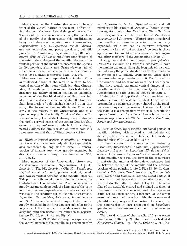
Most species in the Anostomidae have an obvioustwist of the ventral portion of the maxilla (Figs 33–36) relative to the anterolateral flange of the maxilla.The extent of this torsion varies among the membersof the family that demonstrate this modification,being well developed in Abramites, Anostomoides,Hypomasticus (Fig. 34), Leporinus (Fig. 35), Rhytio-dus and Schizodon, and poorly developed, but stillpresent, in Anostomus, Leporellus (Fig. 33), Lae-molyta, (Fig. 36) Petulanos and Pseudanos. Torsion ofthe anterolateral flange of the maxilla relative to theventral portion of the maxilla is absent in the speciesin Gnathodolus, Sartor and Synaptolaemus, all ofwhich have these two components of the maxillajoined into a single continuous plate (Fig. 37).
Most examined outgroups also lack torsion of theanterolateral flange of the maxilla relative to theventral portion of that bone (Chilodontidae, Charac-idae, Curimatidae, Citharinidae, Distichodontidae),although the highly modified maxilla in examinedmembers of the Prochilodontidae exhibits consider-able torsion and is coded as having state 0. Given thefinal hypothesis of relationships arrived at in thisstudy, the torsion of the maxilla (state 0) evolvedearly in the history of the Anostomidae and is asynapomorphy for the family. Torsion of the maxillawas secondarily lost (state 1) during the evolution ofthe highly derived species of the genera Gnathodolus,Sartor and Synaptolaemus, which form a deeplynested clade in the family (clade 31) under both thisreconstruction and that of Winterbottom (1980).
52. Width of ventral portion of maxilla: (0) ventralportion of maxilla narrow, only slightly expanded inaxis transverse to long axis of bone; (1) ventralportion of maxilla very wide, greatly expanded indirection transverse to long axis of bone (CI = 0.250;RI = 0.824).
Most members of the Anostomidae [Abramites,Anostomoides, Anostomus, Hypomasticus (Fig. 34),Laemolyta (Fig. 36), Leporinus (Fig. 35), Pseudanos,Rhytiodus and Schizodon] possess relatively smalland narrow ventral portions of the maxilla (state 0).This portion of the maxilla in the close outgroups, theChilodontidae, Curimatidae and Prochilodontidae, isgreatly expanded along both the long axis of the boneand the direction perpendicular to that axis (state 1)relative to the condition typical for the Anostomidae.Leporellus, Gnathodolus, Petulanos, Synaptolaemusand Sartor have the ventral flange of the maxillagreatly expanded in the direction perpendicular to thelong axis of the maxilla in a mode similar to theoutgroup condition (state 1; for condition in Leporel-lus see Fig. 33, for Sartor see Fig. 37).
Winterbottom (1980) cited a triangular expansion ofthe ventral portion of the maxilla as a synapomorphy
for Gnathodolus, Sartor, Synaptolaemus and allmembers of his concept of Anostomus (herein encom-passing Anostomus plus Petulanos). We differ fromhis interpretation of the maxillae of Anostomusanostomus and A. ternetzi. Winterbottom consideredthe maxillae in those two species to be ventrallyexpanded, while we see no objective differencebetween the form of that portion of the bone in thosespecies and the condition in Pseudanos, Laemolytaand other members of the Anostomidae (state 0).
Among more distant outgroups, Brycon falcatus,Hemiodus ocellatus and Parodon suborbitalis havethe maxilla expanded along the long axis of the bone,but not along the perpendicular axis (for the conditionin Brycon see Weitzman, 1962: fig. 8). These threetaxa are coded as possessing state 0. Members of theCitharinidae and basal members of the Distichodon-tidae have greatly expanded ventral flanges of themaxilla relative to the condition typical of theAnostomidae and are coded as possessing state 1.
Under the final hypothesis of relationships pre-sented herein, the widened ventral flange of thepremaxilla is a symplesiomorphy shared by the proxi-mate outgroups and Leporellus. The narrow form ofthe maxilla is a synapomorphy for clade 3, while therepeated evolution of a widened flange is, in turn, asynapomorphy for clade 20 (Gnathodolus, Petulanos,Sartor and Synaptolaemus).
53. Form of dorsal tip of maxilla: (0) dorsal portion ofmaxilla rod-like, with tapered or pointed tip; (1)dorsal portion of maxilla in form of flat, mediallydirected plate (CI = 0.333; RI = 0.667).
In most species in the Anostomidae, includingAbramites, Anostomoides, Anostomus, Hypomasticus,Laemolyta, Leporellus, Leporinus, Rhytiodus, Schiz-odon and Pseudanos trimaculatus the dorsal portionof the maxilla has a rod-like form in the area whereit contacts the anterior of the pair of cartilages thatlie between the tip of the maxilla and the anteriorportions of the palatine (state 0, Figs 33–36). In Gna-thodolus, Petulanos, Pseudanos gracilis, P. winterbot-tomi, Sartor and Synaptolaemus the dorsal portion ofthe maxilla that approaches the palatine is alterna-tively distinctly flattened (state 1, Fig. 37). The max-illae of the available cleared and stained specimen ofPseudanos irinae are missing and that specimencould not be coded for this character. Of the sevenexamined anostomid species with the compressedplate-like morphology of this portion of the maxilla,the compression is least pronounced in Pseudanosgracilis and P. winterbottomi and most pronounced inSartor.
The dorsal portion of the maxilla of Brycon meeki(Weitzman, 1962: fig. 2), the basal distichodontidXenocharax (Daget, 1960: fig. 7), Citharinus sp., Dis-
118 B. L. SIDLAUSKAS and R. P. VARI
No claim to original US Government works.Journal compilation © 2008 The Linnean Society of London, Zoological Journal of the Linnean Society, 2008, 154, 70–210
tichodus sp., Parodon suborbitalis, Hemiodus ocella-tus and most other members of the Characiformeshas a distinct rod-like form with a tapered dorsaltip (state 0). Members of the Curimatidae have thedorsal portion of the otherwise plate-like maxillaextended into a long rod (Vari, 1982b: fig. 11; 1989a:fig. 43). A similar rod-like dorsal portion of themaxilla, albeit a very short one, is found in thespecies of the Chilodontidae. In the Prochilodontidae,the anterior portion of the maxilla, which is homolo-gous to the dorsal portion of the maxilla in othercharaciforms, is also extended into a long rod andcoded as state 0.
Although state 0 is clearly plesiomorphic, the opti-mization of the gain of state 1 on the final phylogenyis ambiguous due to the polytomies in the region ofthe phylogeny involving Pseudanos and the missingdata for Pseudanos irinae. Under ACCTRAN theflattened dorsal region of the premaxilla evolved inthe common ancestor of Anostomus, Gnathodolus,Petulanos, Pseudanos, Sartor and Synaptolaemus(clade 25) with subsequent reversions in Pseudanostrimaculatus, the species of Anostomus (clade 28)and possibly Pseudanos irinae. Under DELTRAN thecompressed morphology of the dorsal portion of themaxilla represents a synapomorphy for clade 29,which is composed of Gnathodolus, Petulanos, Sartorand Synaptolaemus, with additional homoplasticgains within Pseudanos.
54. Form of contact of dorsal portion of maxilla withanterolateral and posterior portions of maxilla: (0)anterolateral and posterior portions of maxilla (ifpresent) merge without interruption into dorsomedialprocess of maxilla that contacts cartilage of palatine;dorsal portion of maxilla not as in state 1; (1) ante-rolateral and posterior portions of maxilla merge toform flat plate orientated perpendicular to process ofmaxilla that contacts cartilage of palatine (CI = 1.000;RI = 1.000).
In most species of the Anostomidae, as well as in allexamined outgroups, any flanges or bony plates asso-ciated with the dorsal portions of the maxilla gradesmoothly into the dorsal process of the maxilla in thearea where that bone contacts the cartilages associ-ated with the palatine (e.g. Laemolyta, Fig. 36). InGnathodolus and Sartor, these flanges alternativelyform a broad, flat plate that is orientated perpendicu-lar to the dorsal process of the maxilla that contactsthe cartilages associated with the palatine (for condi-tion in Sartor, see Fig. 37). Leporellus (Fig. 33) has amorphology of its maxilla with the dorsal processexpanded into a broad plate which is superficiallysimilar to that present in Gnathodolus and Sartor. InLeporellus this plate grades smoothly into the dorso-medial portions of the maxilla instead of being orien-
tated at right angles to that region as is the case inGnathodolus and Sartor. No other modification of thisportion of the maxilla in any anostomid or outgroupapproaches the morphology of that bone found inGnathodolus and Sartor, which is a synapomorphylinking those two genera (clade 30).
LOWER JAW
55. Width of ascending process of anguloarticular: (0)ascending process of anguloarticular narrow, notforming wide vertical ridge perpendicular to dentary;(1) ascending process of anguloarticular very wide,forming pronounced vertical ridge perpendicular todentary (CI = 0.500; RI = 0.889).
In most members of the Anostomidae and all exam-ined outgroups, the ascending process of the angu-loarticular is a thin, narrow process of bone, closelyassociated with, and in the same plane as, the pos-terior portions of the dentary (e.g. Leporellus, Fig. 33;see also Figs 35–37). Two groups within the Anosto-midae (Schizodon and Hypomasticus) instead havethe ascending process of the anguloarticular greatlythickened and widened, forming a raised verticalridge projecting laterally from the posterolateralsurface of the dentary (for condition in Hypomasticussee Fig. 34; for Schizodon see Figs 38, 39). A fewspecies of Leporinus (most notably L. ortomaculatus,L. pellegrinii and L. striatus) have a knob-like processnear the dorsal margin of the ascending process of theanguloarticular with the ascending processes slightlythickened overall relative to those present in theother species of Leporinus. The width of the ascendingprocess of the anguloarticular in these three species ismuch less pronounced than it is in Schizodon orHypomasticus. Leporinus ortomaculatus, L. pellegri-nii and L. striatus are consequently coded as possess-ing state 0 (narrow process) for this character.
Under the final hypothesis of relationships arrivedat under this study, the widened form of the angu-loarticular evolved twice within the Anostomidae,once in a common ancestor of Hypomasticus (clade 4)and once in the lineage leading to Schizodon (clade18).
56. Presence or absence of trough-like form of dorsalportion of ascending process of anguloarticular: (0)ascending process of anguloarticular not as in state 1;(1) dorsal portion of ascending process of anguloar-ticular in form of deep trough for passage of thick A1component of adductor muscle that inserts on medialsurface of maxilla by way of primordial ligament(CI = 0.500; RI = 0.750).
The morphology of the ascending process of theanguloarticular in the set of species with subterminalmouths within the Anostomidae is particularly strik-
PHYLOGENY OF ANOSTOMID FISHES 119
No claim to original US Government works.Journal compilation © 2008 The Linnean Society of London, Zoological Journal of the Linnean Society, 2008, 154, 70–210
ing, even allowing for the notable variation in theform of this process across the family. In all examinedspecies of Hypomasticus and also Schizodon nasutus(all of which possess the widened form of the ascend-ing process, see character 55), the dorsal portion ofthe ascending process of the anguloarticular is modi-fied into a deep trough through which a section of theanterior portion of the A1 portion of the adductormandibulae muscle passes before continuing furtheranteriorly via the primordial ligament to an attach-ment on the medial surface of the maxilla (for con-dition in Hypomasticus mormyrops see Fig. 34; forSchizodon nasutus see Fig. 39). After passing anteri-orly through the trough, the muscle and associated
ligament turn sharply ventrally to the insertion of theligament on the medial surface of the maxilla. InSchizodon nasutus the trough is primarily a notch inthe widened dorsal margin of the anguloarticularwith limited anterior expansion (Fig. 39), while inHypomasticus the notch is extended into an anteriorlyelongate trough (Fig. 34). No other examined speciesin either the Anostomidae or any outgroup has thetrough-like morphology of the ascending process ofthe angular possessed by the five cited species,although a slight depression in the widened ascend-ing process of the anguloarticular occurs in S. isog-nathus. Schizodon isognathus is the reconstructedsister species to S. nasutus (clade 19), and the slight
Figure 38. Right lower jaw, Schizodon fasciatus, USNM 280739, 103.2 mm SL; lateral view, drawing reversed to placeanterior at left.
Figure 39. Left lower jaw, Schizodon nasutus, USNM 326944, 111 mm SL; lateral view.
120 B. L. SIDLAUSKAS and R. P. VARI
No claim to original US Government works.Journal compilation © 2008 The Linnean Society of London, Zoological Journal of the Linnean Society, 2008, 154, 70–210
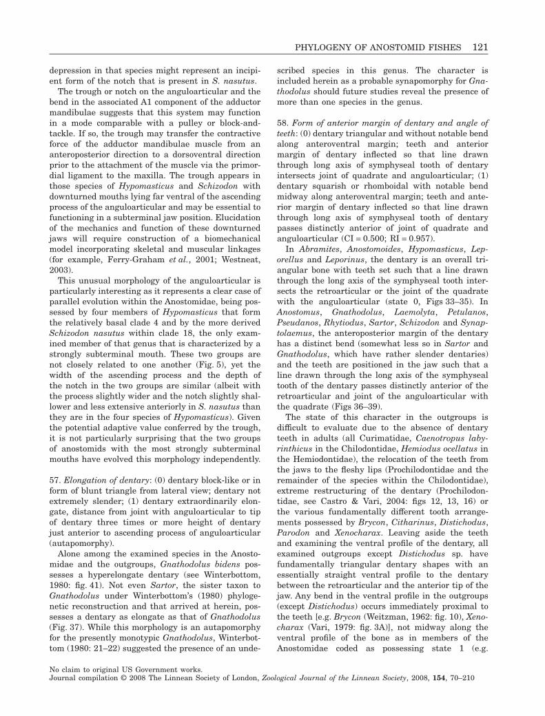
depression in that species might represent an incipi-ent form of the notch that is present in S. nasutus.
The trough or notch on the anguloarticular and thebend in the associated A1 component of the adductormandibulae suggests that this system may functionin a mode comparable with a pulley or block-and-tackle. If so, the trough may transfer the contractiveforce of the adductor mandibulae muscle from ananteroposterior direction to a dorsoventral directionprior to the attachment of the muscle via the primor-dial ligament to the maxilla. The trough appears inthose species of Hypomasticus and Schizodon withdownturned mouths lying far ventral of the ascendingprocess of the anguloarticular and may be essential tofunctioning in a subterminal jaw position. Elucidationof the mechanics and function of these downturnedjaws will require construction of a biomechanicalmodel incorporating skeletal and muscular linkages(for example, Ferry-Graham et al., 2001; Westneat,2003).
This unusual morphology of the anguloarticular isparticularly interesting as it represents a clear case ofparallel evolution within the Anostomidae, being pos-sessed by four members of Hypomasticus that formthe relatively basal clade 4 and by the more derivedSchizodon nasutus within clade 18, the only exam-ined member of that genus that is characterized by astrongly subterminal mouth. These two groups arenot closely related to one another (Fig. 5), yet thewidth of the ascending process and the depth ofthe notch in the two groups are similar (albeit withthe process slightly wider and the notch slightly shal-lower and less extensive anteriorly in S. nasutus thanthey are in the four species of Hypomasticus). Giventhe potential adaptive value conferred by the trough,it is not particularly surprising that the two groupsof anostomids with the most strongly subterminalmouths have evolved this morphology independently.
57. Elongation of dentary: (0) dentary block-like or inform of blunt triangle from lateral view; dentary notextremely slender; (1) dentary extraordinarily elon-gate, distance from joint with anguloarticular to tipof dentary three times or more height of dentaryjust anterior to ascending process of anguloarticular(autapomorphy).
Alone among the examined species in the Anosto-midae and the outgroups, Gnathodolus bidens pos-sesses a hyperelongate dentary (see Winterbottom,1980: fig. 41). Not even Sartor, the sister taxon toGnathodolus under Winterbottom’s (1980) phyloge-netic reconstruction and that arrived at herein, pos-sesses a dentary as elongate as that of Gnathodolus(Fig. 37). While this morphology is an autapomorphyfor the presently monotypic Gnathodolus, Winterbot-tom (1980: 21–22) suggested the presence of an unde-
scribed species in this genus. The character isincluded herein as a probable synapomorphy for Gna-thodolus should future studies reveal the presence ofmore than one species in the genus.
58. Form of anterior margin of dentary and angle ofteeth: (0) dentary triangular and without notable bendalong anteroventral margin; teeth and anteriormargin of dentary inflected so that line drawnthrough long axis of symphyseal tooth of dentaryintersects joint of quadrate and anguloarticular; (1)dentary squarish or rhomboidal with notable bendmidway along anteroventral margin; teeth and ante-rior margin of dentary inflected so that line drawnthrough long axis of symphyseal tooth of dentarypasses distinctly anterior of joint of quadrate andanguloarticular (CI = 0.500; RI = 0.957).
In Abramites, Anostomoides, Hypomasticus, Lep-orellus and Leporinus, the dentary is an overall tri-angular bone with teeth set such that a line drawnthrough the long axis of the symphyseal tooth inter-sects the retroarticular or the joint of the quadratewith the anguloarticular (state 0, Figs 33–35). InAnostomus, Gnathodolus, Laemolyta, Petulanos,Pseudanos, Rhytiodus, Sartor, Schizodon and Synap-tolaemus, the anteroposterior margin of the dentaryhas a distinct bend (somewhat less so in Sartor andGnathodolus, which have rather slender dentaries)and the teeth are positioned in the jaw such that aline drawn through the long axis of the symphysealtooth of the dentary passes distinctly anterior of theretroarticular and joint of the anguloarticular withthe quadrate (Figs 36–39).
The state of this character in the outgroups isdifficult to evaluate due to the absence of dentaryteeth in adults (all Curimatidae, Caenotropus laby-rinthicus in the Chilodontidae, Hemiodus ocellatus inthe Hemiodontidae), the relocation of the teeth fromthe jaws to the fleshy lips (Prochilodontidae and theremainder of the species within the Chilodontidae),extreme restructuring of the dentary (Prochilodon-tidae, see Castro & Vari, 2004: figs 12, 13, 16) orthe various fundamentally different tooth arrange-ments possessed by Brycon, Citharinus, Distichodus,Parodon and Xenocharax. Leaving aside the teethand examining the ventral profile of the dentary, allexamined outgroups except Distichodus sp. havefundamentally triangular dentary shapes with anessentially straight ventral profile to the dentarybetween the retroarticular and the anterior tip of thejaw. Any bend in the ventral profile in the outgroups(except Distichodus) occurs immediately proximal tothe teeth [e.g. Brycon (Weitzman, 1962: fig. 10), Xeno-charax (Vari, 1979: fig. 3A)], not midway along theventral profile of the bone as in members of theAnostomidae coded as possessing state 1 (e.g.
PHYLOGENY OF ANOSTOMID FISHES 121
No claim to original US Government works.Journal compilation © 2008 The Linnean Society of London, Zoological Journal of the Linnean Society, 2008, 154, 70–210
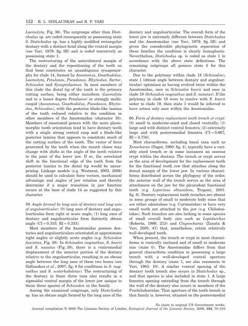
Laemolyta, Fig. 36). The outgroups other than Disti-chodus sp. are coded consequently as possessing state0. Distichodus sp. has a highly modified rectangulardentary with a distinct bend along the ventral margin(see Vari, 1979: fig. 3B) and is coded tentatively aspossessing state 1.
The restructuring of the anterolateral margin ofthe dentary and the repositioning of the teeth onthat bone constitutes an unambiguous synapomor-phy for clade 14, formed by Anostomus, Gnathodolus,Laemolyta, Petulanos, Pseudanos, Rhytiodus, Sartor,Schizodon and Synaptolaemus. In most members ofthis clade the distal tip of the tooth is the primarycutting surface, being either incisiform (Laemolytaand to a lesser degree Petulanos) or strongly multi-cuspid (Anostomus, Gnathodolus, Pseudanos, Rhytio-dus, Schizodon), with the posterior blade-like laminaof the tooth reduced relative to the condition inother members of the Anostomidae (character 38).Members of anostomid genera with the more plesio-morphic tooth orientation tend to have dentary teethwith a single strong central cusp and a blade-likeposterior lamina that appears to constitute much ofthe cutting surface of the tooth. The vector of forcegenerated by the teeth when the mouth closes maychange with shifts in the angle of the teeth relativeto the joint of the lower jaw. If so, the correlatedshift in the functional edge of the tooth from theposterior lamina to the distal tip would be unsur-prising. Linkage models (e.g. Westneat, 2003, 2006)should be used to calculate force vectors, mechanicaladvantage and angles of jaw rotation and therebydetermine if a major transition in jaw functionoccurs at the base of clade 14 as suggested by thischaracter.
59. Angle formed by long axis of dentary and long axisof anguloarticular: (0) long axes of dentary and angu-loarticular form right or acute angle; (1) long axes ofdentary and anguloarticular form distinctly obtuseangle (CI = 0.333; RI = 0.333).
Most members of the Anostomidae possess den-taries and anguloarticulars orientated at approximateright angles or slightly acute angles (e.g. Schizodonfasciatus, Fig. 38). In Schizodon isognathus, S. kneriiand S. nasutus (Fig. 39), there is a ventromedialdisplacement of the anterior portion of the dentaryrelative to the anguloarticular, resulting in an obtuseangle between the long axes of these two bones (seeSidlauskas et al., 2007: fig. 8 for conditions in S. isog-nathus and S. scotorhabdotus). The restructuring ofthe dentary in these three taxa also results in asigmoidal ventral margin of the lower jaw unique tothese three species of Schizodon in the family.
Among the examined outgroups, only Distichodussp. has an obtuse angle formed by the long axes of the
dentary and anguloarticular. The overall form of thelower jaw is extremely different between Distichodusand the Anostomidae (see Vari, 1979: fig. 3B) andgiven the considerable phylogenetic separation ofthese families the condition is clearly homoplastic.Nevertheless, Distichodus sp. is coded as state 1 inaccordance with the above state definitions. Theremaining outgroups all possess state 0 for thischaracter.
Due to the polytomy within clade 18 (Schizodon),state 1 (obtuse angle between dentary and anguloar-ticular) optimizes as having evolved twice within theAnostomidae, once in Schizodon knerii and once inclade 19 (Schizodon isognathus and S. nasutus). If thepolytomy in clade 18 were to resolve with S. kneriisister to clade 19, then state 1 would be inferred tohave arisen only once within the Anostomidae.
60. Form of dentary replacement tooth trench or crypt:(0) small to moderate-sized and closed ventrally; (1)large and with distinct ventral fenestra; (2) extremelylarge and with posteromedial fenestra (CI = 0.667;RI = 0.750).
Most characiforms, including basal taxa such asXenocharax (Daget, 1960: fig. 4), typically have a vari-ably sized trench or in some instances an osseouscrypt within the dentary. The trench or crypt servesas the area of development for the replacement teethfor the functional tooth row that attaches along thedorsal margin of the lower jaw. In various characi-forms distributed across the phylogeny of the order,the anterior wall of the trench serves as the area ofattachment on the jaw for the pleurodont functionalteeth (e.g. Leporinus obtusidens, Trapani, 2001:fig. 6). Dentary replacement tooth trenches are absentin some groups of small to moderate body sizes thatare either edentulous (e.g. Curimatidae) or have verysmall teeth not attached to the jaw (e.g. Chilodon-tidae). Such trenches are also lacking in some speciesof small overall body size such as Lepidarchus(Roberts, 1966: 213) and Clupeocharax (Zanata &Vari, 2005: 47) that, nonetheless, retain relativelywell-developed teeth.
When present, the trench or crypt in most characi-forms is ventrally enclosed and of small to moderatesize (state 0). The Anostomidae differs from thatgeneral characiform condition in possessing a largetrench with a well-developed ventral aperturethrough the dentary (state 1, see also comments inVari, 1983: 10). A similar ventral opening of thedentary tooth trench also occurs in Distichodus sp.,and that species is also included in state 1. A largefenestra opening extending from the trench throughthe wall of the dentary also occurs in members of theProchilodontidae. That aperture of the tooth trench inthat family is, however, situated on the posteromedial
122 B. L. SIDLAUSKAS and R. P. VARI
No claim to original US Government works.Journal compilation © 2008 The Linnean Society of London, Zoological Journal of the Linnean Society, 2008, 154, 70–210
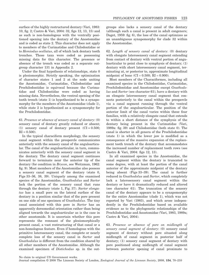
surface of the highly restructured dentary (Vari, 1983:10, fig. 2; Castro & Vari, 2004: 32, figs 12, 13, 15) andas such is non-homologous with the ventrally posi-tioned opening into the dentary of the Anostomidaeand is coded as state 2. This character does not applyto members of the Curimatidae and Chilodontidae orto Hemiodus ocellatus, all of which lack dentary toothtrenches. Those taxa were coded as possessingmissing data for this character. The presence orabsence of the trench was coded as a separate out-group character 131 in Appendix 2.
Under the final hypothesis of relationships, state 0is plesiomorphic. Strictly speaking, the optimizationof character states 1 and 2 at the node unitingthe Anostomidae, Curimatidae, Chilodontidae andProchilodontidae is equivocal because the Curima-tidae and Chilodontidae were coded as havingmissing data. Nevertheless, the ventral opening of thetooth trench (state 1) is hypothesized to be a synapo-morphy for the members of the Anostomidae (clade 1),while state 2 is hypothesized as a synapomorphy forthe Prochilodontidae.
61. Presence or absence of sensory canal of dentary: (0)sensory canal of dentary greatly reduced or absent;(1) sensory canal of dentary present (CI = 0.500;RI = 0.500).
In the typical characiform morphology, the sensorycanal segment within the preopercle communicatesanteriorly with the sensory canal of the anguloarticu-lar. The canal of the anguloarticular, in turn, commu-nicates anteriorly with the sensory canal segment inthe dentary. The dentary canal segment continuesforward to terminate near the anterior tip of thedentary (for condition in Brycon, see Weitzman, 1962:fig. 9). Most members of the Anostomidae also possessa sensory canal segment of the dentary (state 0,Figs 33–36, 38, 39). Uniquely among the examinedspecies of the Anostomidae, Gnathodolus and Sartorlack the portion of the sensory canal that runsthrough the dentary (state 1, Fig. 37). Sartor elonga-tus has a small pore on the lateral surface of thedentary in a position similar that of the pore presenton one side of one specimen of Gnathodolus. The tinycanal associated with this pore in Sartor has anapparently dorsomedial orientation rather than beingaligned towards the anguloarticular as is the case inother anostomids. It is uncertain whether this porerepresents the remnant of the plesiomorphicallypresent canal, a vast restructuring of that canal or anon-homologous feature. Even if homologous with theprimitive laterosensory canal, the complete or nearlycomplete loss of the sensory canal in Sartor andGnathodolus is different from the condition shared byall other members of the Anostomidae. Although theexamined specimen of Distichodus sp. in the out-
groups also lacks a sensory canal of the dentary(although such a canal is present in adult congeners;Daget, 1959: fig. 6), the loss of the canal optimizes asan unambiguous synapomorphy for clade 32 withinthe Anostomidae.
62. Length of sensory canal of dentary: (0) dentarywith elongate laterosensory canal segment extendingfrom contact of dentary with ventral portion of angu-loarticular to point close to symphysis of dentary; (1)dentary with short laterosensory canal segment, ter-minating at, or posterior to, approximate longitudinalmidpoint of bone (CI = 0.500; RI = 0.900).
Most members of the Characiformes, including allexamined species in the Chilodontidae, Curimatidae,Prochilodontidae and Anostomidae except Gnathodo-lus and Sartor (see character 61), have a dentary withan elongate laterosensory canal segment that con-nects posteriorly to the canal within the preoperclevia a canal segment running through the ventralportion of the anguloarticular. The position of theanterior limit of the canal varies within these fourfamilies, with a relatively elongate canal that extendsto within a short distance of the symphysis of thedentary being present in the Curimatidae (Vari,1989a: fig. 39) and Chilodontidae (both state 0). Thecanal is shorter in all genera of the Prochilodontidae(state 1) in which the lower jaw is modified as aconsequence of the massive expansion of the replace-ment tooth trench of the dentary that accommodatesthe increased number of replacement tooth rows (seeCastro & Vari, 2004: figs 12, 13).
In all examined species in the Anostomidae, thecanal segment within the dentary is truncated tosome degree, with at least the portion of the canalanterior of the approximate midpoint of the dentarybeing absent (Figs 33–39). The canal is furtherreduced in Gnathodolus and Sartor, which completelylack a laterosensory canal segment within thedentary or have it dramatically reduced and altered(see character 61). The truncation of the sensorycanal of the dentary appears to be a synapomorphyfor the entire Anostomidae (clade 1), which was notreported by Vari (1983), and which arose indepen-dently in the Prochilodontidae based on availableevidence as to the phylogenetic relationships of theProchilodontidae and Anostomidae (Vari, 1983, 1989a;Castro & Vari, 2004).
63. Presence or absence of pore on midlength ofsensory canal segment of dentary: (0) sensory canalsegment of dentary without pore situated alongmidlength of canal segment in posterior half ofdentary; (1) sensory canal segment of dentary withpore positioned along midlength of canal segmentbetween posterior opening of canal proximate to
PHYLOGENY OF ANOSTOMID FISHES 123
No claim to original US Government works.Journal compilation © 2008 The Linnean Society of London, Zoological Journal of the Linnean Society, 2008, 154, 70–210
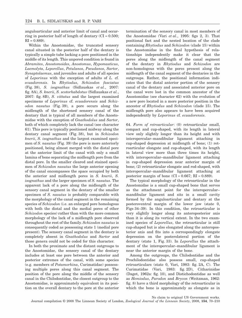
anguloarticular and anterior limit of canal and occur-ring in posterior half of length of dentary (CI = 0.500;RI = 0.889).
Within the Anostomidae, the truncated sensorycanal situated in the posterior half of the dentary istypically a simple tube lacking a pore positioned in themiddle of its length. This unpored condition is found inAbramites, Anostomoides, Anostomus, Hypomasticus,Laemolyta, Leporellus, Petulanos, Pseudanos, Sartor,Synaptolaemus, and juveniles and adults of all speciesof Leporinus with the exception of adults of L. cf.ecuadorensis. In Rhytiodus, Schizodon fasciatus(Fig. 38), S. isognathus (Sidlauskas et al., 2007:fig. 8A), S. knerii, S. scotorhabdotus (Sidlauskas et al.,2007: fig. 8B), S. vittatus and the largest examinedspecimens of Leporinus cf. ecuadorensis and Schiz-odon nasutus (Fig. 39), a pore occurs along themidlength of the shortened sensory canal of thedentary that is typical of all members of the Anosto-midae with the exception of Gnathodolus and Sartor,both of which completely lack the canal (see character61). This pore is typically positioned midway along thedentary canal segment (Fig. 38), but in Schizodonknerii, S. isognathus and the largest examined speci-men of S. nasutus (Fig. 39) the pore is more anteriorlypositioned, being almost merged with the distal poreat the anterior limit of the canal with only a smalllamina of bone separating the midlength pore from thedistal pore. In the smaller cleared and stained speci-men of Schizodon nasutus the large anterior openingof the canal encompasses the space occupied by boththe anterior and midlength pores in S. knerii, S.isognathus and the larger specimen of S. nasutus. Theapparent lack of a pore along the midlength of thesensory canal segment in the dentary of the smallerspecimen of S. nasutus is probably comparable withthe morphology of the canal segment in the remainingspecies of Schizodon (i.e. an enlarged pore homologouswith both the distal and the medial pores of otherSchizodon species) rather than with the more commonmorphology of the lack of a midlength pore observedthroughout the rest of the family. Schizodon nasutus isconsequently coded as possessing state 1 (medial porepresent). The sensory canal segment in the dentary iscompletely absent in Gnathodolus and Sartor andthose genera could not be coded for this character.
In both the proximate and the distant outgroups tothe Anostomidae, the sensory canal of the dentaryincludes at least one pore between the anterior andposterior extremes of the canal, with some species(e.g. members of Potamorhina, Curimatidae) possess-ing multiple pores along this canal segment. Theposition of the pore along the middle of the sensorycanal in the Chilodontidae, the closest outgroup to theAnostomidae, is approximately equivalent in its posi-tion on the overall dentary to the pore at the anterior
termination of the sensory canal in most members ofthe Anostomidae (Vari et al., 1995: figs 2, 3). Thatpositional fact and the nested location of the cladecontaining Rhytiodus and Schizodon (clade 15) withinthe Anostomidae in the final hypothesis of rela-tionships independently make it clear that thepores along the midlength of the canal segmentof the dentary in Rhytiodus and Schizodon arenon-homologous with the pores present along themidlength of the canal segment of the dentaries in theoutgroups. Rather, the positional information indi-cates that the distal anterior portion of the sensorycanal of the dentary and associated anterior pore onthe canal were lost in the common ancestor of theAnostomidae (see character 62) with the evolution ofa new pore located in a more posterior position in theancestor of Rhytiodus and Schizodon (clade 15). Themidlength pore also appears to have been acquiredindependently by Leporinus cf. ecuadorensis.
64. Form of retroarticular: (0) retroarticular small,compact and cup-shaped, with its length in lateralview only slightly longer than its height and withinteropercular–mandibular ligament attaching incup-shaped depression at midlength of bone; (1) ret-roarticular elongate and cup-shaped, with its lengthin lateral view more than three times its height,with interopercular–mandibular ligament attachingin cup-shaped depression near anterior margin ofbone; (2) retroarticular elongate and rod-shaped, withinteropercular–mandibular ligament attaching atposterior margin of bone (CI = 0.667; RI = 0.889).
The typical morphology of the retroarticular in theAnostomidae is a small cup-shaped bone that servesas the attachment point for the interopercular–mandibular ligament and sits in a small cavityformed by the anguloarticular and dentary at theposteroventral margin of the lower jaw (state 0,Figs 34–39). In this condition, the retroarticular isvery slightly longer along its anteroposterior axisthan it is along its vertical extent. In the two exam-ined species of Leporellus, the retroarticular is stillcup-shaped but is also elongated along the anteropos-terior axis and fits into a correspondingly elongatedepression on the posterolateral portion of thedentary (state 1, Fig. 33). In Leporellus the attach-ment of the interopercular–mandibular ligament isnear the anterior margin of the bone.
Among the outgroups, the Chilodontidae and theProchilodontidae also possess small, cup-shapedretroarticulars (state 0, Vari, 1983: fig. 2A, C). TheCurimatidae (Vari, 1983: fig. 2D), Citharinidae(Daget, 1962a: fig. 10), and Distichodontidae as wellas Hemiodus, Parodon and Brycon (Weitzman, 1962:fig. 8) have a third morphology of the retroarticular inwhich the bone is approximately as elongate as in
124 B. L. SIDLAUSKAS and R. P. VARI
No claim to original US Government works.Journal compilation © 2008 The Linnean Society of London, Zoological Journal of the Linnean Society, 2008, 154, 70–210
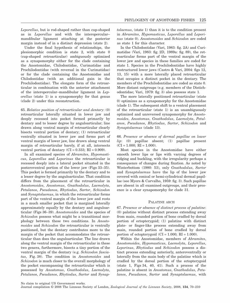
Leporellus, but is rod-shaped rather than cup-shapedas in Leporellus and with the interopercular–mandibular ligament attaching at the posteriormargin instead of in a distinct depression (state 2).
Under the final hypothesis of relationships, theplesiomorphic condition is state 2, with state 0(cup-shaped retroarticular) ambiguously optimizedas a synapomorphy either for the clade containingthe Anostomidae, Chilodontidae, Curimatidae andProchilodontidae (with reversal in the Curimatidae)or for the clade containing the Anostomidae andChilodontidae (with an additional gain in theProchilodontidae). The elongate form of the retroar-ticular in combination with the anterior attachmentof the interopercular–mandibular ligament in Lep-orellus (state 1) is a synapomorphy for that genus(clade 2) under this reconstruction.
65. Relative position of retroarticular and dentary: (0)retroarticular laterally situated in lower jaw anddeeply recessed into pocket formed primarily bydentary and to lesser degree by anguloarticular; linedrawn along ventral margin of retroarticular clearlybisects ventral portion of dentary; (1) retroarticularventrally situated in lower jaw and forms part ofventral margin of lower jaw, line drawn along ventralmargin of retroarticular barely, if at all, intersectsventral portion of dentary (CI = 0.333; RI = 0.909).
In all examined species of Abramites, Hypomasti-cus, Leporellus and Leporinus the retroarticular isrecessed deeply into a lateral pocket situated in theposteroventral portion of the lower jaw (Figs 33–35).This pocket is formed primarily by the dentary and toa lesser degree by the anguloarticular. That conditiondiffers from the placement of the retroarticular inAnostomoides, Anostomus, Gnathodolus, Laemolyta,Petulanos, Pseudanos, Rhytiodus, Sartor, Schizodonand Synaptolaemus, in which the retroarticular formspart of the ventral margin of the lower jaw and restsin a much smaller pocket that is margined laterallyapproximately equally by the dentary and anguloar-ticular (Figs 36–39). Anostomoides and the species ofSchizodon possess what might be a transitional mor-phology between these two conditions. In Anosto-moides and Schizodon the retroarticular is ventrallypositioned, but the dentary contributes more to themargin of the pocket that accommodates the retroar-ticular than does the anguloarticular. The line drawnalong the ventral margin of the retroarticular in thesetwo genera, furthermore, bisects a tiny portion of theventral margin of the dentary (e.g. Schizodon fascia-tus, Fig. 38). The condition in Anostomoides andSchizodon is much closer to the overall morphology ofthe pocket encompassing the retroarticular which ispossessed by Anostomus, Gnathodolus, Laemolyta,Petulanos, Pseudanos, Rhytiodus, Sartor and Synap-
tolaemus, (state 1) than it is to the condition presentin Abramites, Hypomasticus, Leporellus and Lepori-nus (state 0). Anostomoides and Schizodon are codedas state 1 for this character.
In the Chilodontidae (Vari, 1983: fig. 2A) and Curi-matidae (Vari, 1983: fig. 2D, 1989a: fig. 89), the ret-roarticular forms part of the ventral margin of thelower jaw and species in these families are coded forstate 1. Species in the Prochilodontidae have highlyrestructured lower jaws (Castro & Vari, 2004: figs 12,13, 15) with a more laterally placed retroarticularthat occupies a distinct pocket in the dentary. Themembers of the Prochilodontidae are coded as state 0.More distant outgroups (e.g. members of the Distich-odontidae; Vari, 1979: fig. 3) also possess state 1.
The more laterally positioned retroarticular (state0) optimizes as a synapomorphy for the Anostomidae(clade 1). The subsequent shift to a ventral placementof the retroarticular (state 1) is an unambiguouslyoptimized and unreversed synapomorphy for Anosto-moides, Anostomus, Gnathodolus, Laemolyta, Petul-anos, Pseudanos, Rhytiodus, Sartor, Schizodon andSynaptolaemus (clade 13).
66. Presence or absence of dermal papillae on lowerlip: (0) papillae absent; (1) papillae present(CI = 1.000; RI = 1.000).
Most species in the Anostomidae have eithersmooth lower lips or lips with varying degrees ofridging and buckling, with the irregularity perhaps aconsequence of changes during fixation. As noted byWinterbottom (1980: 53), only Gnathodolus, Sartorand Synaptolaemus have the lip of the lower jawcovered with conical or hemi-cylindrical dermal papil-lae (see Myers & Carvalho, 1959: fig. 3). Such papillaeare absent in all examined outgroups, and their pres-ence is a clear synapomorphy for clade 31.
PALATINE ARCH
67. Presence or absence of distinct process of palatine:(0) palatine without distinct process extending awayfrom main, rounded portion of bone cradled by dorsalportion of ectopterygoid; (1) palatine with distinctplate- or finger-like process extending away frommain, rounded portion of bone cradled by dorsalportion of ectopterygoid (CI = 1.000; RI = 1.000).
Within the Anostomidae, members of Abramites,Anostomoides, Hypomasticus, Laemolyta, Leporellus,Leporinus, Rhytiodus and Schizodon possess a dis-tinct process extending anteriorly, anteroventrally orlaterally from the main body of the palatine which iscradled by the dorsal portion of the ectopterygoid(state 1, Figs 34, 40, 41). Such a process of thepalatine is absent in Anostomus, Gnathodolus, Petu-lanos, Pseudanos, Sartor and Synaptolaemus, with
PHYLOGENY OF ANOSTOMID FISHES 125
No claim to original US Government works.Journal compilation © 2008 The Linnean Society of London, Zoological Journal of the Linnean Society, 2008, 154, 70–210
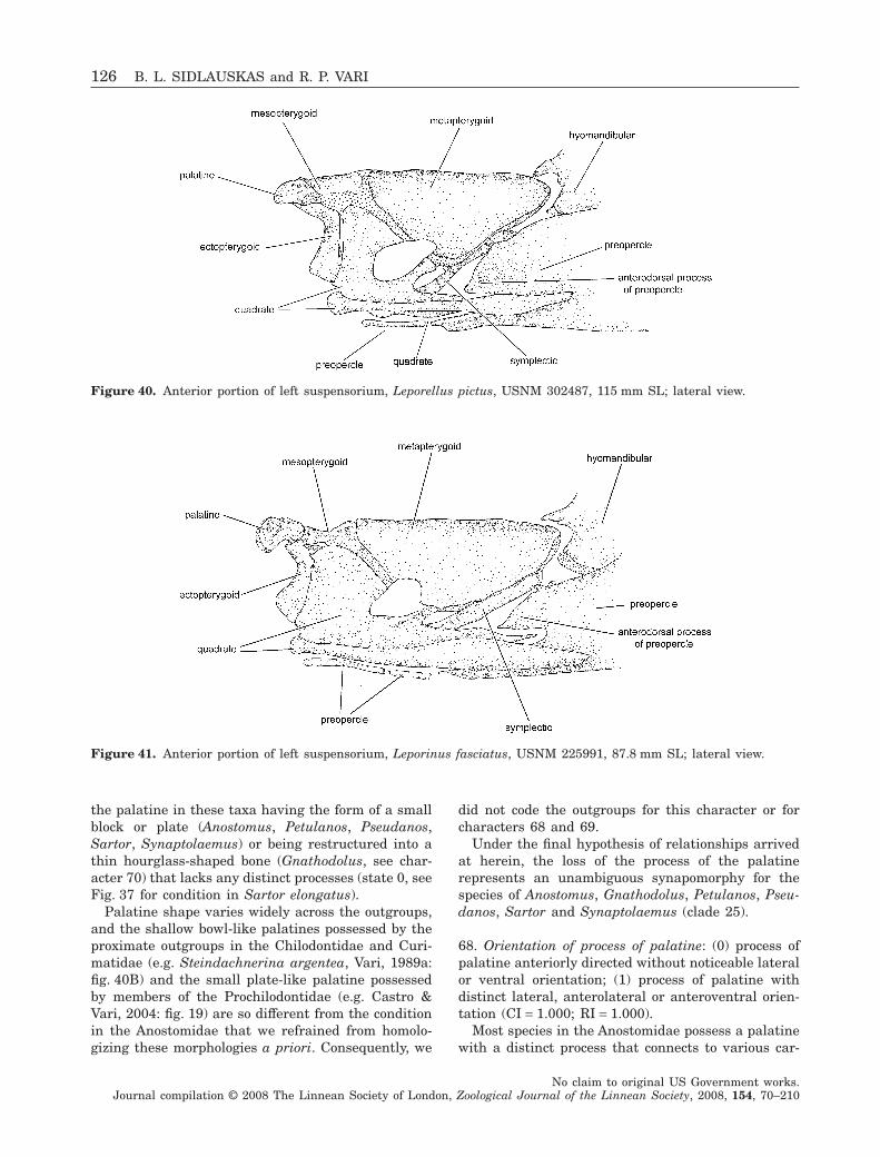
the palatine in these taxa having the form of a smallblock or plate (Anostomus, Petulanos, Pseudanos,Sartor, Synaptolaemus) or being restructured into athin hourglass-shaped bone (Gnathodolus, see char-acter 70) that lacks any distinct processes (state 0, seeFig. 37 for condition in Sartor elongatus).
Palatine shape varies widely across the outgroups,and the shallow bowl-like palatines possessed by theproximate outgroups in the Chilodontidae and Curi-matidae (e.g. Steindachnerina argentea, Vari, 1989a:fig. 40B) and the small plate-like palatine possessedby members of the Prochilodontidae (e.g. Castro &Vari, 2004: fig. 19) are so different from the conditionin the Anostomidae that we refrained from homolo-gizing these morphologies a priori. Consequently, we
did not code the outgroups for this character or forcharacters 68 and 69.
Under the final hypothesis of relationships arrivedat herein, the loss of the process of the palatinerepresents an unambiguous synapomorphy for thespecies of Anostomus, Gnathodolus, Petulanos, Pseu-danos, Sartor and Synaptolaemus (clade 25).
68. Orientation of process of palatine: (0) process ofpalatine anteriorly directed without noticeable lateralor ventral orientation; (1) process of palatine withdistinct lateral, anterolateral or anteroventral orien-tation (CI = 1.000; RI = 1.000).
Most species in the Anostomidae possess a palatinewith a distinct process that connects to various car-
Figure 40. Anterior portion of left suspensorium, Leporellus pictus, USNM 302487, 115 mm SL; lateral view.
Figure 41. Anterior portion of left suspensorium, Leporinus fasciatus, USNM 225991, 87.8 mm SL; lateral view.
126 B. L. SIDLAUSKAS and R. P. VARI
No claim to original US Government works.Journal compilation © 2008 The Linnean Society of London, Zoological Journal of the Linnean Society, 2008, 154, 70–210
tilages and soft tissues proximate to the maxilla andmesethmoid (character 67). The morphology of thatlateral process varies considerably across the Anosto-midae. In many species in the family (Anostomoides,Laemolyta, Leporinus agassizi, L. cf. niceforoi, L. cf.ecuadorensis, L. fasciatus, L. cf. fasciatus, L. friderici,L. gomesi, L. ortomaculatus, L. pellegrinii, L. striatus,Rhytiodus and Schizodon) this process of the palatinehas a primarily lateral orientation (e.g. Leporinusfasciatus, Fig. 41), while in others (Abramites, Hypo-masticus, Leporinus jatuncochi and L. tigrinus), theprocess of the palatine is directed anterolaterally,anteroventrally or anteroventrolaterally with onlyslight deviation of the body of the process from theparasagittal plane (e.g. Hypomasticus mormyrops,Fig. 34). The species of Hypomasticus as well as Lep-orinus jatuncochi have the process notably ventrallyslanted (Fig. 34). Unfortunately all the variation inAbramites, Anostomoides, Hypomasticus, Laemolyta,Leporinus, Rhytiodus and Schizodon forms a con-tinuum, and it proved impossible to parse the range ofmorphologies among these genera into discrete char-acter states. All these taxa are coded as state 1.
The two examined species of Leporellus have aprocess of the palatine that is aligned fully anteriorlywithout deviation from the parasagittal plane(Fig. 40). This is a unique condition within the Anos-tomidae and is coded as state 0. The species of Anos-tomus, Gnathodolus, Petulanos, Pseudanos, Sartorand Synaptolaemus lack any indication of a process ofthe palatine and have overall morphologies of thatbone that are highly modified relative to the form ofthe ossification in other anostomids and characiforms.Those five genera cannot be coded for this character.
Proximate outgroups to the Anostomidae all havesignificant modifications of the form of the palatinerelative to the condition present in many groupswithin the Characiformes, a situation that compli-cates comparisons between the palatine morphologiespresent in the Anostomidae and the outgroup species.The most common morphology of the palatine amongthe outgroup species is an anteriorly and anterolat-erally directed plate or spoon-shaped bone that under-lies a well-developed cartilage. None of the species inthe Anostomidae possesses a palatine morphologysimilar to that condition and none of the outgroupswas formally coded for this character. The anteriorand anterolateral orientation of the palatines in theChilodontidae and Curimatidae (Vari, 1989a: fig. 40)does, however, evoke the primarily anteriorly directedprocess of the palatine that occurs in Leporellus(Fig. 40), Abramites and some species of Leporinus.
Because we could not code the outgroups for thischaracter and because Leporellus appears as the mostbasal anostomoid, the state reconstruction for thischaracter at the base of the anostomid phylogeny is
ambiguous. A palatine with a directly anteriorly ori-entated process may be a synapomorphy for the twoexamined species of Leporellus (clade 2). Alternatively,the angled palatine characteristic of the remainder ofthe Anostomidae may be a synapomorphy for clade 3.It is equally possible that either of these conditionsrepresents the primitive condition for the Anostomi-dae, or that both are derived relative to the verydistinctive morphologies possessed by the outgroups.
Nevertheless, there is a trend in increasingly morederived members of the Anostomidae for the processof the palatine to shift from having a slight antero-lateral orientation (e.g. Hypomasticus mormyrops,Fig. 34) through a distinctly anterolateral orientation(e.g. Leporinus fasciatus, Fig. 41) to an almost fullylateral orientation in Schizodon, Laemolyta and Rhy-tiodus. In that light, the fully anterior orientation ofthe palatine in Leporellus (Fig. 40) could also be seenas the starting point of a trend towards increasinglateralization of the process in the Anostomidae priorto the loss of the process in the modified palatinecharacteristic of the species in clade 25.
69. Length of process of palatine: (0) process ofpalatine moderately to weakly developed, length ofprocess about 1.5 times width or less; (1) processof palatine very long, length of process approximatelytwo or more times width of process (CI = 1.000;RI = 1.000).
The length of the process of the palatine variesconsiderably within the Anostomidae, being short inLeporellus (Fig. 40), moderate in Abramites, manyspecies of Leporinus (e.g. L. fasciatus, Fig. 41), Hypo-masticus (e.g. H. mormyrops, Fig. 34) and Laemolyta,and slightly longer (length approximately 1.5 timeswidth) in Leporinus cf. ecuadorensis, L. friderici,Anostomoides, Rhytiodus, Schizodon isognathus, S.knerii, S. nasutus and S. scotorhabdotus. Variation iscontinuous across all of the above conditions, whichare all coded consequently as state 0. In Schizodonfasciatus and S. vittatus the length of the processreaches its extreme within the Anostomidae, with thelength of the process approximately twice its width(state 1). Palatine morphologies vary considerablyin the outgroups (see discussion in characters 68 and69) and the outgroups were not formally coded forthis character. Nevertheless, no examined outgroupspecies possess a palatine with a long laterallydirected process comparable with that present in S.fasciatus and S. vittatus. The extreme elongation ofthe process is a synapomorphy for these two speciesunder the final hypothesis of relationships (clade 20).
70. Shape of palatine: (0) main portion of palatine inform of block or plate; (1) palatine in shape of dors-oventrally elongate hourglass, with triangular dorsal
PHYLOGENY OF ANOSTOMID FISHES 127
No claim to original US Government works.Journal compilation © 2008 The Linnean Society of London, Zoological Journal of the Linnean Society, 2008, 154, 70–210
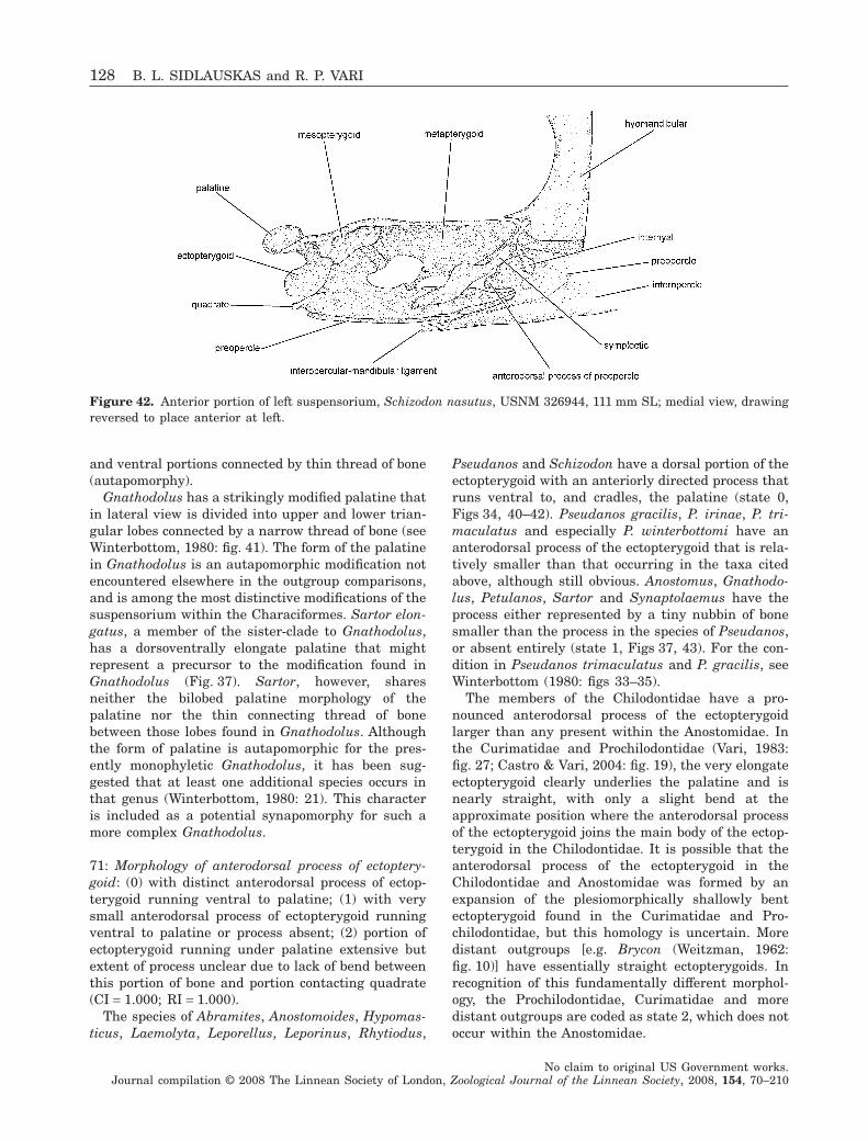
and ventral portions connected by thin thread of bone(autapomorphy).
Gnathodolus has a strikingly modified palatine thatin lateral view is divided into upper and lower trian-gular lobes connected by a narrow thread of bone (seeWinterbottom, 1980: fig. 41). The form of the palatinein Gnathodolus is an autapomorphic modification notencountered elsewhere in the outgroup comparisons,and is among the most distinctive modifications of thesuspensorium within the Characiformes. Sartor elon-gatus, a member of the sister-clade to Gnathodolus,has a dorsoventrally elongate palatine that mightrepresent a precursor to the modification found inGnathodolus (Fig. 37). Sartor, however, sharesneither the bilobed palatine morphology of thepalatine nor the thin connecting thread of bonebetween those lobes found in Gnathodolus. Althoughthe form of palatine is autapomorphic for the pres-ently monophyletic Gnathodolus, it has been sug-gested that at least one additional species occurs inthat genus (Winterbottom, 1980: 21). This characteris included as a potential synapomorphy for such amore complex Gnathodolus.
71: Morphology of anterodorsal process of ectoptery-goid: (0) with distinct anterodorsal process of ectop-terygoid running ventral to palatine; (1) with verysmall anterodorsal process of ectopterygoid runningventral to palatine or process absent; (2) portion ofectopterygoid running under palatine extensive butextent of process unclear due to lack of bend betweenthis portion of bone and portion contacting quadrate(CI = 1.000; RI = 1.000).
The species of Abramites, Anostomoides, Hypomas-ticus, Laemolyta, Leporellus, Leporinus, Rhytiodus,
Pseudanos and Schizodon have a dorsal portion of theectopterygoid with an anteriorly directed process thatruns ventral to, and cradles, the palatine (state 0,Figs 34, 40–42). Pseudanos gracilis, P. irinae, P. tri-maculatus and especially P. winterbottomi have ananterodorsal process of the ectopterygoid that is rela-tively smaller than that occurring in the taxa citedabove, although still obvious. Anostomus, Gnathodo-lus, Petulanos, Sartor and Synaptolaemus have theprocess either represented by a tiny nubbin of bonesmaller than the process in the species of Pseudanos,or absent entirely (state 1, Figs 37, 43). For the con-dition in Pseudanos trimaculatus and P. gracilis, seeWinterbottom (1980: figs 33–35).
The members of the Chilodontidae have a pro-nounced anterodorsal process of the ectopterygoidlarger than any present within the Anostomidae. Inthe Curimatidae and Prochilodontidae (Vari, 1983:fig. 27; Castro & Vari, 2004: fig. 19), the very elongateectopterygoid clearly underlies the palatine and isnearly straight, with only a slight bend at theapproximate position where the anterodorsal processof the ectopterygoid joins the main body of the ectop-terygoid in the Chilodontidae. It is possible that theanterodorsal process of the ectopterygoid in theChilodontidae and Anostomidae was formed by anexpansion of the plesiomorphically shallowly bentectopterygoid found in the Curimatidae and Pro-chilodontidae, but this homology is uncertain. Moredistant outgroups [e.g. Brycon (Weitzman, 1962:fig. 10)] have essentially straight ectopterygoids. Inrecognition of this fundamentally different morphol-ogy, the Prochilodontidae, Curimatidae and moredistant outgroups are coded as state 2, which does notoccur within the Anostomidae.
Figure 42. Anterior portion of left suspensorium, Schizodon nasutus, USNM 326944, 111 mm SL; medial view, drawingreversed to place anterior at left.
128 B. L. SIDLAUSKAS and R. P. VARI
No claim to original US Government works.Journal compilation © 2008 The Linnean Society of London, Zoological Journal of the Linnean Society, 2008, 154, 70–210
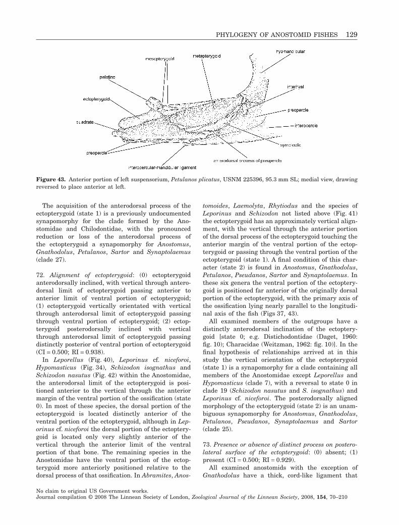
The acquisition of the anterodorsal process of theectopterygoid (state 1) is a previously undocumentedsynapomorphy for the clade formed by the Ano-stomidae and Chilodontidae, with the pronouncedreduction or loss of the anterodorsal process ofthe ectopterygoid a synapomorphy for Anostomus,Gnathodolus, Petulanos, Sartor and Synaptolaemus(clade 27).
72. Alignment of ectopterygoid: (0) ectopterygoidanterodorsally inclined, with vertical through antero-dorsal limit of ectopterygoid passing anterior toanterior limit of ventral portion of ectopterygoid;(1) ectopterygoid vertically orientated with verticalthrough anterodorsal limit of ectopterygoid passingthrough ventral portion of ectopterygoid; (2) ectop-terygoid posterodorsally inclined with verticalthrough anterodorsal limit of ectopterygoid passingdistinctly posterior of ventral portion of ectopterygoid(CI = 0.500; RI = 0.938).
In Leporellus (Fig. 40), Leporinus cf. niceforoi,Hypomasticus (Fig. 34), Schizodon isognathus andSchizodon nasutus (Fig. 42) within the Anostomidae,the anterodorsal limit of the ectopterygoid is posi-tioned anterior to the vertical through the anteriormargin of the ventral portion of the ossification (state0). In most of these species, the dorsal portion of theectopterygoid is located distinctly anterior of theventral portion of the ectopterygoid, although in Lep-orinus cf. niceforoi the dorsal portion of the ectoptery-goid is located only very slightly anterior of thevertical through the anterior limit of the ventralportion of that bone. The remaining species in theAnostomidae have the ventral portion of the ectop-terygoid more anteriorly positioned relative to thedorsal process of that ossification. In Abramites, Anos-
tomoides, Laemolyta, Rhytiodus and the species ofLeporinus and Schizodon not listed above (Fig. 41)the ectopterygoid has an approximately vertical align-ment, with the vertical through the anterior portionof the dorsal process of the ectopterygoid touching theanterior margin of the ventral portion of the ectop-terygoid or passing through the ventral portion of theectopterygoid (state 1). A final condition of this char-acter (state 2) is found in Anostomus, Gnathodolus,Petulanos, Pseudanos, Sartor and Synaptolaemus. Inthese six genera the ventral portion of the ectoptery-goid is positioned far anterior of the originally dorsalportion of the ectopterygoid, with the primary axis ofthe ossification lying nearly parallel to the longitudi-nal axis of the fish (Figs 37, 43).
All examined members of the outgroups have adistinctly anterodorsal inclination of the ectoptery-goid [state 0; e.g. Distichodontidae (Daget, 1960:fig. 10); Characidae (Weitzman, 1962: fig. 10)]. In thefinal hypothesis of relationships arrived at in thisstudy the vertical orientation of the ectopterygoid(state 1) is a synapomorphy for a clade containing allmembers of the Anostomidae except Leporellus andHypomasticus (clade 7), with a reversal to state 0 inclade 19 (Schizodon nasutus and S. isognathus) andLeporinus cf. niceforoi. The posterodorsally alignedmorphology of the ectopterygoid (state 2) is an unam-biguous synapomorphy for Anostomus, Gnathodolus,Petulanos, Pseudanos, Synaptolaemus and Sartor(clade 25).
73. Presence or absence of distinct process on postero-lateral surface of the ectopterygoid: (0) absent; (1)present (CI = 0.500; RI = 0.929).
All examined anostomids with the exception ofGnathodolus have a thick, cord-like ligament that
Figure 43. Anterior portion of left suspensorium, Petulanos plicatus, USNM 225396, 95.3 mm SL; medial view, drawingreversed to place anterior at left.
PHYLOGENY OF ANOSTOMID FISHES 129
No claim to original US Government works.Journal compilation © 2008 The Linnean Society of London, Zoological Journal of the Linnean Society, 2008, 154, 70–210
connects the posterodorsal portion of the ectoptery-goid to the lateral ethmoid (see character 23). Theattachment area of this ligament onto the ectoptery-goid has the form of a distinct process on the poste-rolateral surface of that ossification (Vari, 1983:fig. 26). This elaboration of the ectopterygoid has notbeen encountered in examined outgroups. Both theligament and the process on the ectopterygoid arelacking in Gnathodolus, with these losses apparentlycorrelated with the dramatic alternations of a numberof components of the suspensorium in that genus (seecharacters 20, 57, 70). Winterbottom (1980: 50) indi-cated that a dorsal process of the ectopterygoid ispresent in species of Anostomus (including Petulanos)and Pseudanos but absent in Gnathodolus, Sartorand Synaptolaemus. Assuming that Winterbottom’sdorsal process corresponds to the structure describedby Vari (1983), we differ from Winterbottom’s inter-pretation of the character, as our examined specimensof Sartor and Synaptolaemus possess the process inquestion (Fig. 37). In the final hypothesis of relation-ships, possession of this process on the ectopterygoidis a synapomorphy for the entire Anostomidae (clade1) that is reversed in Gnathodolus.
74. Posteriorly directed dorsolateral process ofmesopterygoid: (0) absent; (1) present (CI = 0.333;RI = 0.500).
The examined species of Anostomus, Petulanos andSartor possess a posteriorly directed process on thelateral surface of the mesopterygoid that is absentin all other examined species of the Anostomidae(Fig. 37). In the dissected cleared and stained speci-mens of these genera, a small amount of connectivetissue remains attached to the medial surface of thisprocess of the mesopterygoid, suggesting that it mayserve as an attachment point for a muscle or ligament.The species of Anostomus have the process of themesopterygoid situated medial to the well-developedligament connecting the ectopterygoid to the lateralethmoid in most members of the Anostomidae (char-acter 23) and is difficult to see the process withoutextensive dissection. In Sartor the process is similarlypositioned but more highly developed, forming alateral ridge that abuts the lateral surface of thedorsally elongate palatine (Fig. 37). No trace of theprocess of the mesopterygoid is present in Gnathodolusor Synaptolaemus. The mesopterygoid of Gnathodolusis modified and elongated dorsoventrally relative tothe typical anostomoid condition (Winterbottom, 1980:fig. 41), and it is possible that the dorsolateral processin Gnathodolus has become incorporated into the restof the mesopterygoid as it expanded dorsally. Alterna-tively, it may have been lost.
Species of Pseudanos have a small bump on themesopterygoid in the same position as the posteriorly
directed process which is present in that region inAnostomus, Petulanos and Sartor. While Pseudanos iscoded as state 0 (process absent) in light of its lack ofa distinct process, this bump on the mesopterygoid ofPseudanos may, nonetheless, represent a rudimentaryform of the more highly developed process whichoccurs in Anostomus, Petulanos and Sartor. If thathomology is correct, this character would be appli-cable at a higher level of inclusiveness. No otherexamined species, including all outgroups, possessesa dorsolateral process of the mesopterygoid.
Optimization of this character on the final phylog-eny is ambiguous. Either the mesopterygoid processin question evolved once in clade 27 and was lostindependently in Synaptolaemus and Gnathodolus, orit evolved three times in total, once each in Anosto-mus (clade 28), Petulanos (clade 30) and Sartor.
75. Association of quadrate with mesopterygoid: (0)mesopterygoid with well-developed ventral triangularprocess that significantly overlaps ossified portion ofmedial surface of quadrate; (1) mesopterygoid lackingventral process overlapping quadrate or process, ifpresent, much smaller than in state 0 and not sig-nificantly overlapping ossified portion of medialsurface of quadrate (CI = 0.250; RI = 0.857).
In Abramites, Hypomasticus, Leporellus, Leporinus,Rhytiodus and Schizodon the mesopterygoid has atriangular, ventral process that overlaps the medialsurface of the quadrate (e.g. Schizodon nasutus,Fig. 42). Anostomoides, Anostomus, Gnathodolus,Laemolyta, Petulanos, Pseudanos, Sartor and Synap-tolaemus either lack this triangular process or have itmuch smaller in size relative to the condition presentin the above cited species (e.g. Petulanos plicatus,Fig. 43). A lack of contact of the ventral margin of themesopterygoid with the quadrate characterizes Anos-tomoides, Gnathodolus, Petulanos, Pseudanos, Sartorand Synaptolaemus, with all these taxa coded as state1. In Anostomus ternetzi the ventral margin of themesopterygoid barely contacts the ossified dorsalmargin of the quadrate. Synaptolaemus, Laemolytaand Anostomus anostomus have the small ventralflange of the mesopterygoid approaching or contactingthe cartilaginous dorsal portion of the quadrate. Syn-aptolaemus, Laemolyta, Anostomus anostomus and A.ternetzi are all coded as possessing state 1 (ventralflange of mesopterygoid small or absent).
Species in the Chilodontidae (the immediate out-group of the Anostomidae) possess a large ventralflange of the mesopterygoid that overlaps the quad-rate in a mode similar to that in Leporinus and theother taxa possessing a ventral flange of the mesop-terygoid (see Vari et al., 1995: fig. 4 for illustration ofthe condition in the Chilodontidae). Among the com-ponents of the second outgroup to the Anostomidae,
130 B. L. SIDLAUSKAS and R. P. VARI
No claim to original US Government works.Journal compilation © 2008 The Linnean Society of London, Zoological Journal of the Linnean Society, 2008, 154, 70–210
an overlap of the quadrate by the mesopterygoid isabsent in the Prochilodontidae (Vari, 1983: fig. 28).Members of the Curimatidae appear to lack such anoverlap of these two elements, although the situationin that family is frequently difficult to evaluate due tothe expansion of the various cartilages locatedthroughout the region of the palatine and adjoiningstructures (e.g. Potamorhina latior, Vari, 1983:fig. 27). Among more distant outgroups, the overlap ofthe mesopterygoid and quadrate is clearly present inBrycon (Weitzman, 1962: fig. 10), Hemiodus ocellatusand the distichodontids Xenocharax spilurus, Disti-chodus sp. and Paradistichodus dimidiatus. Nooverlap of these two elements is apparent in Parodonsuborbitalis, and the condition in Citharinus sp. wasdifficult to evaluate due to the small size and poorstaining of the specimen (coded as missing data).
Optimization of this character on the final phylog-eny is ambiguous. The plesiomorphic condition withinthe Anostomidae optimizes as state 0 (overlap ofquadrate by mesopterygoid). Either the overlapbetween the quadrate and mesopterygoid was lost ina common ancestor of Anostomoides, Anostomus, Gna-thodolus, Laemolyta, Petulanos, Pseudanos, Rhytio-dus, Sartor, Schizodon and Synaptolaemus (clade 13)and re-evolved in the lineage leading to Rhytiodusand Schizodon (clade 15), or the loss occurred twice,once in Anostomoides and once in the lineage leadingto Anostomus, Gnathodolus, Laemolyta, Petulanos,Pseudanos, Sartor and Synaptolaemus (clade 21).
76. Form of association of posterodorsal and posteriorprocesses of quadrate: (0) processes separated withlength of separate portion of posterodorsal processapproximately twice width of process or greater; (1)processes entirely or almost entirely joined by thinintervening plate of bone with length of separateportion of posterodorsal process much less than twicewidth of process (CI = 0.250; RI = 0.667).
The posterior and dorsal regions of the quadrate ofmost groups in the Characiformes have three mainprocesses: a dorsal process that articulates along itsanterior margin with the ectopterygoid and dorsallywith the mesopterygoid, a posterodorsal process thatforms the ventral margin of the metapterygoid–quadrate fenestra, and a posterior process thatextends ventral to the symplectic (for condition inBrycon, see Weitzman, 1962: fig. 10). In the examinedproximate outgroups to the Anostomidae along withLeporellus in that family (Fig. 40), the posterodorsalprocess of the quadrate is separated posteriorly fromthe posterior process of the quadrate. There is ananteriorly directed indentation between the two pro-cesses and the length of the posterodorsal process istwo or more times the width of the posterodorsalprocess. The indentation is less developed in the
species of the Anostomidae other than Leporellus inwhich all, or almost all, of the plesiomorphically openspace between the posterodorsal and posterior pro-cesses of the quadrate is variably filled by a thin bonyplate (see also Roberts, 1973). Most of the specieswith that condition have the space completely filled,with only the position of the cartilage joining thequadrate and metapterygoid and the position of thesymplectic serving to delimit the position of the pos-terodorsal process of the quadrate (Figs 37, 42). Asmall notch in the bony plate is present in Hypomas-ticus despaxi, H. megalepis, H. mormyrops, Leporinusagassizi, L. cf. ecuadorensis, L. friderici, L. pellegrinii,L. cf. moralesi, L. jatuncochi, L. fasciatus (Fig. 41), L.cf. fasciatus, L. gomesi, L. ortomaculatus, L. tigrinus,Petulanos plicatus (Fig. 43) and Synaptolaemuscingulatus (Fig. 44). All anostomid species exceptLeporellus are coded as possessing state 1 for thischaracter.
In the outgroups, the notch between the postero-dorsal and posterior processes of the quadrate isclearly present (state 0) in Brycon falcatus, Parodonsuborbitalis, Xenocharax spilurus and members of theChilodontidae and Prochilodontidae (for the conditionin Prochilodontidae see Vari, 1983: fig. 28). The notchis absent in Citharinus sp., Distichodus sp. andHemiodus ocellatus (state 1). Our examined speci-mens of the Curimatidae have the notch present butvery small (coded as state 1); however, some membersof the Curimatidae have the notch well developed(e.g. Potamorhina latior, Vari, 1983: fig. 27). Underthe final hypothesis of the relationships, the loss of adeep notch between the posterodorsal and posteriorportions of the quadrate (state 1) is an unambiguousand unreversed synapomorphy for the Anostomidaeexclusive of Leporellus (clade 3).
77. Posterior extent of lateral shelf of quadrate: (0)lateral shelf of quadrate elongate, extending posteri-orly past posterior margin of symplectic to terminateventral to hyomandibular; (1) lateral shelf of quad-rate moderate in length, extending posteriorly pastmidpoint of the symplectic but not beyond posteriorlimit of symplectic; (2) lateral shelf of quadrate short,extending posteriorly to point short of midpoint ofsymplectic (CI = 0.333; RI = 0.789).
Species of the Anostomidae have a distinct, later-ally positioned, teardrop-shaped shelf of the quadratethat serves as an area of attachment for the ventralportions of the adductor mandibulae muscles (Win-terbottom, 1980: fig. 35; Vari, 1983: fig. 29). Such aprocess also appears in the Chilodontidae, Prochilo-dontidae and in Distichodus sp. (presence or absencecoded as character 132, see Appendix 2). Members ofthe Anostomidae vary widely in the degree of devel-opment of this process of the quadrate. In Abramites,
PHYLOGENY OF ANOSTOMID FISHES 131
No claim to original US Government works.Journal compilation © 2008 The Linnean Society of London, Zoological Journal of the Linnean Society, 2008, 154, 70–210
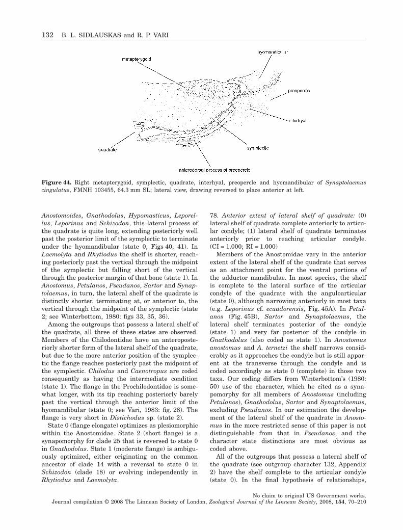
Anostomoides, Gnathodolus, Hypomasticus, Leporel-lus, Leporinus and Schizodon, this lateral process ofthe quadrate is quite long, extending posteriorly wellpast the posterior limit of the symplectic to terminateunder the hyomandibular (state 0, Figs 40, 41). InLaemolyta and Rhytiodus the shelf is shorter, reach-ing posteriorly past the vertical through the midpointof the symplectic but falling short of the verticalthrough the posterior margin of that bone (state 1). InAnostomus, Petulanos, Pseudanos, Sartor and Synap-tolaemus, in turn, the lateral shelf of the quadrate isdistinctly shorter, terminating at, or anterior to, thevertical through the midpoint of the symplectic (state2; see Winterbottom, 1980: figs 33, 35, 36).
Among the outgroups that possess a lateral shelf ofthe quadrate, all three of these states are observed.Members of the Chilodontidae have an anteroposte-riorly shorter form of the lateral shelf of the quadrate,but due to the more anterior position of the symplec-tic the flange reaches posteriorly past the midpoint ofthe symplectic. Chilodus and Caenotropus are codedconsequently as having the intermediate condition(state 1). The flange in the Prochilodontidae is some-what longer, with its tip reaching posteriorly barelypast the vertical through the anterior limit of thehyomandibular (state 0; see Vari, 1983: fig. 28). Theflange is very short in Distichodus sp. (state 2).
State 0 (flange elongate) optimizes as plesiomorphicwithin the Anostomidae. State 2 (short flange) is asynapomorphy for clade 25 that is reversed to state 0in Gnathodolus. State 1 (moderate flange) is ambigu-ously optimized, either originating on the commonancestor of clade 14 with a reversal to state 0 inSchizodon (clade 18) or evolving independently inRhytiodus and Laemolyta.
78. Anterior extent of lateral shelf of quadrate: (0)lateral shelf of quadrate complete anteriorly to articu-lar condyle; (1) lateral shelf of quadrate terminatesanteriorly prior to reaching articular condyle.(CI = 1.000; RI = 1.000)
Members of the Anostomidae vary in the anteriorextent of the lateral shelf of the quadrate that servesas an attachment point for the ventral portions ofthe adductor mandibulae. In most species, the shelfis complete to the lateral surface of the articularcondyle of the quadrate with the anguloarticular(state 0), although narrowing anteriorly in most taxa(e.g. Leporinus cf. ecuadorensis, Fig. 45A). In Petul-anos (Fig. 45B), Sartor and Synaptolaemus, thelateral shelf terminates posterior of the condyle(state 1) and very far posterior of the condyle inGnathodolus (also coded as state 1). In Anostomusanostomus and A. ternetzi the shelf narrows consid-erably as it approaches the condyle but is still appar-ent at the transverse through the condyle and iscoded accordingly as state 0 (complete) in those twotaxa. Our coding differs from Winterbottom’s (1980:50) use of the character, which he cited as a syna-pomorphy for all members of Anostomus (includingPetulanos), Gnathodolus, Sartor and Synaptolaemus,excluding Pseudanos. In our estimation the develop-ment of the lateral shelf of the quadrate in Anosto-mus in the more restricted sense of this paper is notdistinguishable from that in Pseudanos, and thecharacter state distinctions are most obvious ascoded above.
All of the outgroups that possess a lateral shelf ofthe quadrate (see outgroup character 132, Appendix2) have the shelf complete to the articular condyle(state 0). In the final hypothesis of relationships,
Figure 44. Right metapterygoid, symplectic, quadrate, interhyal, preopercle and hyomandibular of Synaptolaemuscingulatus, FMNH 103455, 64.3 mm SL; lateral view, drawing reversed to place anterior at left.
132 B. L. SIDLAUSKAS and R. P. VARI
No claim to original US Government works.Journal compilation © 2008 The Linnean Society of London, Zoological Journal of the Linnean Society, 2008, 154, 70–210
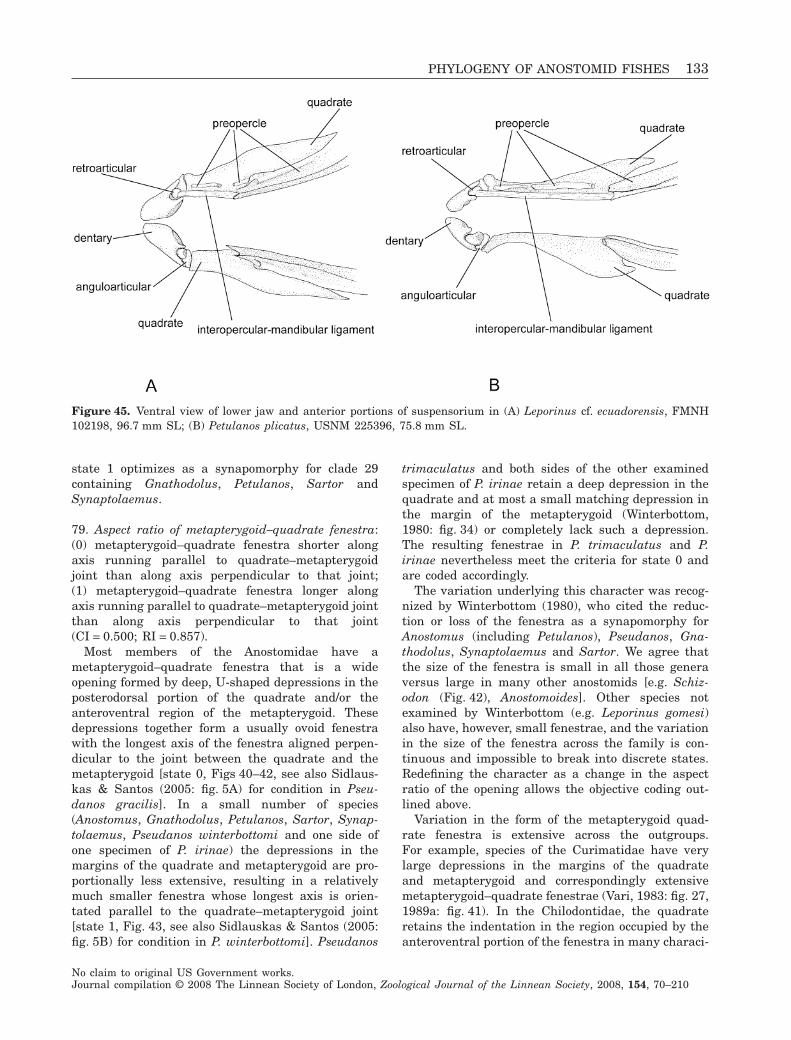
state 1 optimizes as a synapomorphy for clade 29containing Gnathodolus, Petulanos, Sartor andSynaptolaemus.
79. Aspect ratio of metapterygoid–quadrate fenestra:(0) metapterygoid–quadrate fenestra shorter alongaxis running parallel to quadrate–metapterygoidjoint than along axis perpendicular to that joint;(1) metapterygoid–quadrate fenestra longer alongaxis running parallel to quadrate–metapterygoid jointthan along axis perpendicular to that joint(CI = 0.500; RI = 0.857).
Most members of the Anostomidae have ametapterygoid–quadrate fenestra that is a wideopening formed by deep, U-shaped depressions in theposterodorsal portion of the quadrate and/or theanteroventral region of the metapterygoid. Thesedepressions together form a usually ovoid fenestrawith the longest axis of the fenestra aligned perpen-dicular to the joint between the quadrate and themetapterygoid [state 0, Figs 40–42, see also Sidlaus-kas & Santos (2005: fig. 5A) for condition in Pseu-danos gracilis]. In a small number of species(Anostomus, Gnathodolus, Petulanos, Sartor, Synap-tolaemus, Pseudanos winterbottomi and one side ofone specimen of P. irinae) the depressions in themargins of the quadrate and metapterygoid are pro-portionally less extensive, resulting in a relativelymuch smaller fenestra whose longest axis is orien-tated parallel to the quadrate–metapterygoid joint[state 1, Fig. 43, see also Sidlauskas & Santos (2005:fig. 5B) for condition in P. winterbottomi]. Pseudanos
trimaculatus and both sides of the other examinedspecimen of P. irinae retain a deep depression in thequadrate and at most a small matching depression inthe margin of the metapterygoid (Winterbottom,1980: fig. 34) or completely lack such a depression.The resulting fenestrae in P. trimaculatus and P.irinae nevertheless meet the criteria for state 0 andare coded accordingly.
The variation underlying this character was recog-nized by Winterbottom (1980), who cited the reduc-tion or loss of the fenestra as a synapomorphy forAnostomus (including Petulanos), Pseudanos, Gna-thodolus, Synaptolaemus and Sartor. We agree thatthe size of the fenestra is small in all those generaversus large in many other anostomids [e.g. Schiz-odon (Fig. 42), Anostomoides]. Other species notexamined by Winterbottom (e.g. Leporinus gomesi)also have, however, small fenestrae, and the variationin the size of the fenestra across the family is con-tinuous and impossible to break into discrete states.Redefining the character as a change in the aspectratio of the opening allows the objective coding out-lined above.
Variation in the form of the metapterygoid quad-rate fenestra is extensive across the outgroups.For example, species of the Curimatidae have verylarge depressions in the margins of the quadrateand metapterygoid and correspondingly extensivemetapterygoid–quadrate fenestrae (Vari, 1983: fig. 27,1989a: fig. 41). In the Chilodontidae, the quadrateretains the indentation in the region occupied by theanteroventral portion of the fenestra in many characi-
Figure 45. Ventral view of lower jaw and anterior portions of suspensorium in (A) Leporinus cf. ecuadorensis, FMNH102198, 96.7 mm SL; (B) Petulanos plicatus, USNM 225396, 75.8 mm SL.
PHYLOGENY OF ANOSTOMID FISHES 133
No claim to original US Government works.Journal compilation © 2008 The Linnean Society of London, Zoological Journal of the Linnean Society, 2008, 154, 70–210
forms [e.g. Brycon meeki (Weitzman, 1962: fig. 10)].The metapterygoid in the Chilodontidae has a dis-tinctive anterior process that largely (Chilodus) orcompletely (Caenotropus) fills the indentation in theposterior portion of the quadrate (Vari et al., 1995:fig. 4). Members of the Prochilodontidae haveincreased mobility within the suspensorium (Vari,1983) and an associated change in the overall form ofthe metapterygoid which now has a relativelystraight margin in the area proximate to the quadrate(Roberts, 1973: fig. 11; Vari, 1983: fig. 28). It is impos-sible to determine whether the lack of the componentof the fenestra formed by the metapterygoid in theProchilodontidae represents the lack of the indenta-tion or the loss of the processes of the metapterygoidthat extend towards the quadrate in those characi-forms with a fully developed fenestra. Because ofthe high degree of variability across the outgroupsand the difficulty in homologizing those conditionswith the morphologies observed within the Anosto-midae, we elected to not code the outgroups for thischaracter.
Optimization of this character on the final phylog-eny is unclear because of the lack of complete reso-lution for the species of Pseudanos. Assuming thepolytomy in clade 25 to be hard, either the shift in theaspect ratio of the metapterygoid–quadrate fenestraoccurred independently in Pseudanos winterbottomiand clade 27, or the shift is a synapomorphy for clade25 that was reversed in clade 27. If, instead, thepolytomy is resolved with Pseudanos paraphyletic assuggested by the majority rule consensus (Fig. 6),then the shift in aspect ratio of the metapterygoid–quadrate fenestra is a synapomorphy for a cladecontaining Anostomus, Gnathodolus, Petulanos, Pseu-danos winterbottomi, Sartor and Synaptolaemus.
80. Length of cartilage body spanning ventral sectionof metapterygoid–quadrate fenestra: (0) short andplug-like; metapterygoid and quadrate barely sepa-rated by cartilage; (1) long and rod-like; metaptery-goid and quadrate clearly separated by thin bridge ofcartilage (CI = 0.250; RI = 0.824).
Members of the Anostomidae demonstrate two dis-tinct morphologies of the cartilage that bridges thegap along the ventral portion of the metapterygoid–quadrate fenestra between the ossified proximateportions of the quadrate and metapterygoid. InAbramites, Hypomasticus, Leporellus, Leporinusagassizi, L. aripuanaensis, L. cf. ecuadorensis, L. fas-ciatus, L. friderici, L. gomesi, L. cf. moralesi, L. cf.niceforoi, L. ortomaculatus, L. striatus and Schizodonvittatus, the cartilage that bridges the very shortgap between the quadrate and metapterygoid has aplug-like morphology (Figs 40, 41). In Anostomoides,Anostomus, Laemolyta, Leporinus cf. fasciatus, L.
jatuncochi, L. pellegrinii, L. tigrinus, Petulanos, Pseu-danos, Rhytiodus, Sartor, Synaptolaemus and thespecies of Schizodon not cited above, the cartilagebridges a much wider gap and has a rod-like mor-phology (Figs 42, 43; see also Sidlauskas & Santos,2005: fig. 5)
As discussed under character 79, themetapterygoid/quadrate region is significantlyrestructured in the outgroups relative to the Anosto-midae, with an accompanying high level of variationin the apparent length of the cartilage of interest.Both Chilodus and Caenotropus in the Chilodontidaehave a considerable gap between the ossified postero-dorsal process of the quadrate and the proximateportion of the metapterygoid, with this gap bridged byan elongate cartilage (see Vari et al., 1995: fig. 4) TheProchilodontidae and Curimatidae, the second out-group, alternatively have short cartilages that cap theprocess of the quadrate within the relatively smallgap between the quadrate and the metapterygoid (seeVari, 1989a: fig. 41). There is a large gap between thequadrate and metapterygoid in Brycon meeki (Weitz-man, 1962: fig. 10) with the space filled by an elon-gate cartilage. Xenocharax, the basal member of theDistichodontidae, similarly has an elongate cartilagein the region. Overall, the outgroup condition relativeto the Anostomidae is ambiguous and we elected toleave all the outgroups uncoded for this character.
Based on the basal position under the final hypoth-esis of relationships of Hypomasticus and Leporellus,both of which have the plug-like morphology of thecartilage in question, it appears that state 0 is ple-siomorphic within the Anostomidae and the rod-likemorphology (state 1) is derived. Optimization of thischaracter on the strict consensus tree suggests thatstate 1 evolved two or three times. Under ACCTRANstate 1 evolves once in clade 10 and once in clade 12,with a reversal in Leporinus fasciatus. UnderDELTRAN state 1 evolves in Leporinus cf. fasciatus,and clades 11 and 12. Because these groups are allcomponents of the large polytomy in clade 8, futureanalysis may reveal the rod-like cartilage to be asynapomorphy linking two or all three clades. Indeed,if the majority rule consensus (Fig. 6) is correct, state1 is a synapomorphy linking clades 10 and 12 withLeporinus pellegrinii, with subsequent reversal inLeporinus fasciatus. Regardless of the choice of opti-mization, Schizodon vittatus is nested within clade12; the possession of state 0 by that taxon is also areversal.
81. Faceting of metapterygoid: (0) metapterygoid withtwo distinct facets; vertical sheet of bone dorsal toquadrate and symplectic and horizontal or mediallycurved dorsal portion that contacts mesopterygoid; (1)metapterygoid with three distinct facets; vertical
134 B. L. SIDLAUSKAS and R. P. VARI
No claim to original US Government works.Journal compilation © 2008 The Linnean Society of London, Zoological Journal of the Linnean Society, 2008, 154, 70–210
sheet dorsal to quadrate and symplectic, horizontal ormedially curved portion that contacts mesopterygoid,and inclined plane proximate to hyomandibular thatmeets other facets along well-defined margins(CI = 1.000, RI = 1.000).
The metapterygoid of members of the Anostomidaehas a primarily vertical orientation, with the dorsalportion curving at least slightly medially to form ahorizontal facet that meets the dorsal portions of themesopterygoid. Members of Abramites, Anostomoides,Hypomasticus, Laemolyta, Leporellus, Leporinus,Rhytiodus and Schizodon have just the vertical andhorizontal facets on the bone (state 0, Figs 40, 41).Species in Anostomus, Petulanos, Pseudanos, Sartorand Synaptolaemus have an inclined third facetproximate to the hyomandibular that meets the othertwo facets along well-defined margins that are visiblein lateral view (state 1, Fig. 44). Gnathodolus has arestructured metapterygoid that appears to retainonly the posterior portions of the bone. The verticaland inclined facets are clearly present in Gnathodo-lus, but the horizontal facet proximate to the mesop-terygoid appears to have been lost. Gnathodolus istentatively coded as possessing state 1 for this char-acter based on the presence of the inclined facet.
The condition in Laemolyta, although coded forstate 0, may in truth represent a transitional condi-tion. In species of Laemolyta the horizontal facet ofthe metapterygoid slopes laterally anterior to thehyomandibular to create an inclined area where thehorizontal facet grades into the vertical facet. Onlythe lack of a clear division between the inclinedportion of the bone and the other two facets excludesLaemolyta from state 1.
This character was noticed by Winterbottom (1980:50), who discussed a medial slope of the metapterygoidas a synapomorphy for Anostomus (including Petul-anos), Gnathodolus, Pseudanos, Sartor and Synapto-laemus. Our examination of specimens from across thefamily reveals that all anostomids have at least aslight medial slope of the metapterygoid, particularlyin the region proximate to the mesopterygoid (what wecall our horizontal facet). Winterbottom’s medial slopemost probably corresponds to our concept of the addi-tional inclined facet of the metapterygoid.
The typical outgroup condition for the metaptery-goid (possessed by all examined outgroups exceptParodon) is a more or less vertical sheet of bone witha slight medial bend at the dorsal extreme and iscoded as state 0. Parodon suborbitalis has a drasti-cally restructured and abbreviated metapterygoid. Wewere unable to determine whether it possesses aninclined facet of the metapterygoid and coded thatspecies as possessing missing data.
Under the final hypothesis of relationships the pos-session of an additional inclined facet of the metap-
terygoid is a synapomorphy for clade 25 containingAnostomus, Gnathodolus, Petulanos, Pseudanos,Sartor and Synaptolaemus. Laemolyta is recon-structed as the sister to clade 25, suggesting that thesloped morphology of the metapterygoid in that genusas described above is indeed an intermediate or pre-cursor to the inclined metapterygoid facet character-istic of the species in clade 25.
82. Presence or absence of lateral shelf on metaptery-goid: (0) lateral surface of metapterygoid withoutwell-developed lateral shelf; (1) lateral surface ofmetapterygoid with well-developed lateral shelf(autapomorphy).
Uniquely among the examined members of theAnostomidae and outgroups, Sartor elongatus pos-sesses a well-developed lateral shelf on the metap-terygoid (anterior portion visible in Fig. 37), resultingin a T-shaped bone in cross-section. Although thecharacter is an autapomorphy in the current dataset,there are two other described species of Sartor thatwere unavailable for inclusion in this analysis. Whenthese are examined osteologically, it may be foundthat the lateral shelf is a synapomorphy for thegenus.
SYMPLECTIC, HYOMANDIBULAR AND
OPERCULAR SERIES
83. Length of interopercular–mandibular ligament: (0)ligament very short, anterior tip of interopercle inclose proximity to posterior of retroarticular; (1) liga-ment elongate, interopercle notably separated fromretroarticular (CI = 0.500; RI = 0.909).
The interopercular–mandibular ligament in theCharaciformes runs from the anterior tip of theinteropercle to the retroarticular, typically bridging avery small gap between these elements in the areajust ventral to the anterior portion of the quadrate[see Brycon meeki (Weitzman, 1962: fig. 10) and Xeno-charax (Daget (1960: fig. 7)]. The condition in Bryconmeeki and Xenocharax is shared by Hemiodus ocella-tus, Parodon suborbitalis, Distichodus sp., Citharinussp. and all examined members of the Chilodontidaeand Curimatidae, although Chilodus punctatus pos-sesses a ligament of slightly greater length than thatpossessed by the remaining outgroups. In all exam-ined members of the Anostomidae and Prochilo-dontidae, the gap between the interopercle andretroarticular is large and as a consequence theinteropercular–mandibular ligament is more elongatethan in the typical characiform condition (Fig. 45),spanning a length comparable with, or greater than,the diameter of the eye in some of the more derivedmembers of the Anostomidae (Anostomus, Gnathodo-lus, Petulanos, Pseudanos, Sartor, Synaptolaemus).
PHYLOGENY OF ANOSTOMID FISHES 135
No claim to original US Government works.Journal compilation © 2008 The Linnean Society of London, Zoological Journal of the Linnean Society, 2008, 154, 70–210
Given that the Anostomidae and Prochilodontidaeare not sister taxa (Fig. 5, see also Vari, 1983: fig. 41;Castro & Vari, 2004: fig. 26), the elongation of theinteropercular–mandibular ligament in the Ano-stomidae is a homoplasy relative to the conditionin the Prochilodontidae. The elongation of theinteropercular–mandibular ligament optimizes as aclear synapomorphy for the Anostomidae which wasnot previously enumerated by Vari (1983), althoughthat author did discuss at some length the ossifiedsensory tubes of the preopercular canal that are asso-ciated with the expanded ligament in the Anostomi-dae and Prochilodontidae (Vari, 1983: 31–32, 52). Thepresence or absence of those tubes is coded in out-group character 134 (Appendix 2).
84. Presence or absence of large shelf on lateral surfaceof preopercle that serves as origin for portion ofadductor mandibulae muscle: (0) absent; (1) present(CI = 0.500; RI = 0.909).
The adductor musculature associated with theupper jaw in the Anostomidae arises from a broadregion on the lateral surface of the quadrate andpreopercle. This area of attachment is, in part, formedby a lateral shelf that arises from the main body ofthe ventral arm of the preopercle and which laterallycontacts a posteriorly directed process of the postero-lateral portion of the quadrate (see character 77 andoutgroup character 132, Appendix 2). The preopercu-lar shelf continues posteriorly as a gradually dimin-ishing ridge beyond the posterior terminus of thequadrate. Such a modification of the preopercle isabsent in proximate outgroups to the Anostomidaeexcept for the members of the Prochilodontidae,which possess a very small shelf on the preopercle. Alateral shelf on the preopercle, albeit of somewhatdifferent form, is also present in some more distantlyrelated characiforms including a subgroup of the Dis-tichodontidae that is not incorporated into the out-group analysis in this study (see Vari, 1979: fig. 18).Under the final hypothesis of relationships the pos-session of a lateral shelf on the preopercle optimizesas a synapomorphy for the entire Anostomidae(clade 1).
85. Form of anterodorsal process of preopercle: (0)anterodorsal process of preopercle that extendsbetween symplectic and quadrate well developed withbroad triangular or rhomboidal form; (1) anterodorsalprocess of preopercle with thin, needle-like form; (2)anterodorsal process of preopercle absent entirely(CI = 0.667; RI = 0.952).
All examined members of the Anostomidae andProchilodontidae possess an anterodorsal process ofthe preopercle that extends into the space betweenthe symplectic and the dorsal border of the quadrate.
This process is absent in examined members of theChilodontidae, Curimatidae, Characidae, Citharin-idae and Distichodontidae (state 2). In the Anostomi-dae, two distinct morphologies of this process wereobserved. In the more common morphology shared byAbramites, Anostomoides, Hypomasticus, Laemolyta,Leporellus, Leporinus, Rhytiodus and Schizodon, theprocess is large and either triangular or rhomboidal(state 0, Figs 40–42). A similar morphology is presentin members of the Prochilodontidae (Vari, 1983:fig. 28). In Anostomus, Gnathodolus, Petulanos, Pseu-danos, Sartor and Synaptolaemus, the process ispresent but much smaller, being represented only bya very thin, anteriorly directed extension from themain body of the preopercle (state 1, e.g. Petulanosplicatus, Fig. 43).
The presence of the process optimizes as a syn-apomorphy for the Anostomidae (clade 1) and thereduction of the process from the triangular tothe needle-like morphology is a synapomorphy for theclade consisting of Anostomus, Gnathodolus, Petul-anos, Pseudanos, Sartor and Synaptolaemus (clade25). The Prochilodontidae is not sister to the Anosto-midae (Fig. 5; Vari, 1983, 1989a; Castro & Vari, 2004),and the presence of the anterodorsal process of thepreopercle in these two families is a homoplasy.
86. Form of anterior margin of ascending arm ofpreopercle: (0) anterior margin of ascending arm ofpreopercle without distinct triangular process over-lapping joint between interhyal, hyomandibular,metapterygoid and symplectic in lateral view; (1)anterior margin of ascending arm of preopercle withdistinct triangular process overlapping joint betweeninterhyal, hyomandibular, metapterygoid and sym-plectic in lateral view (CI = 0.500; RI = 0.667).
In Gnathodolus, Synaptolaemus and Sartor, theanterior margin of the ascending arm of the preo-percle has a distinctly pointed triangular process thatoverlaps the joint of the interhyal, hyomandibular,metapterygoid and symplectic in lateral view [state 1,Fig. 44; see also Winterbottom (1980: figs 39, 41)].One side of one of the examined specimens ofPseudanos gracilis (FMNH 103454) also has such aprocess, but that species is coded as lacking theprocess in keeping with the typical morphology of thepreopercle for the species. No other examined speciesin the Anostomidae possess such a process, althoughsome species of Leporinus (L. gomesi, L. ortomacula-tus, L. striatus) have a somewhat widened androunded morphology of this region of the preoperclethat, however, lacks a distinct pointed process. Inter-estingly, Chilodus punctatus in the Chilodontidae (butnot the examined species of Caenotropus) also has atriangular process on the anterior margin of theascending arm of the preopercle. No other examined
136 B. L. SIDLAUSKAS and R. P. VARI
No claim to original US Government works.Journal compilation © 2008 The Linnean Society of London, Zoological Journal of the Linnean Society, 2008, 154, 70–210
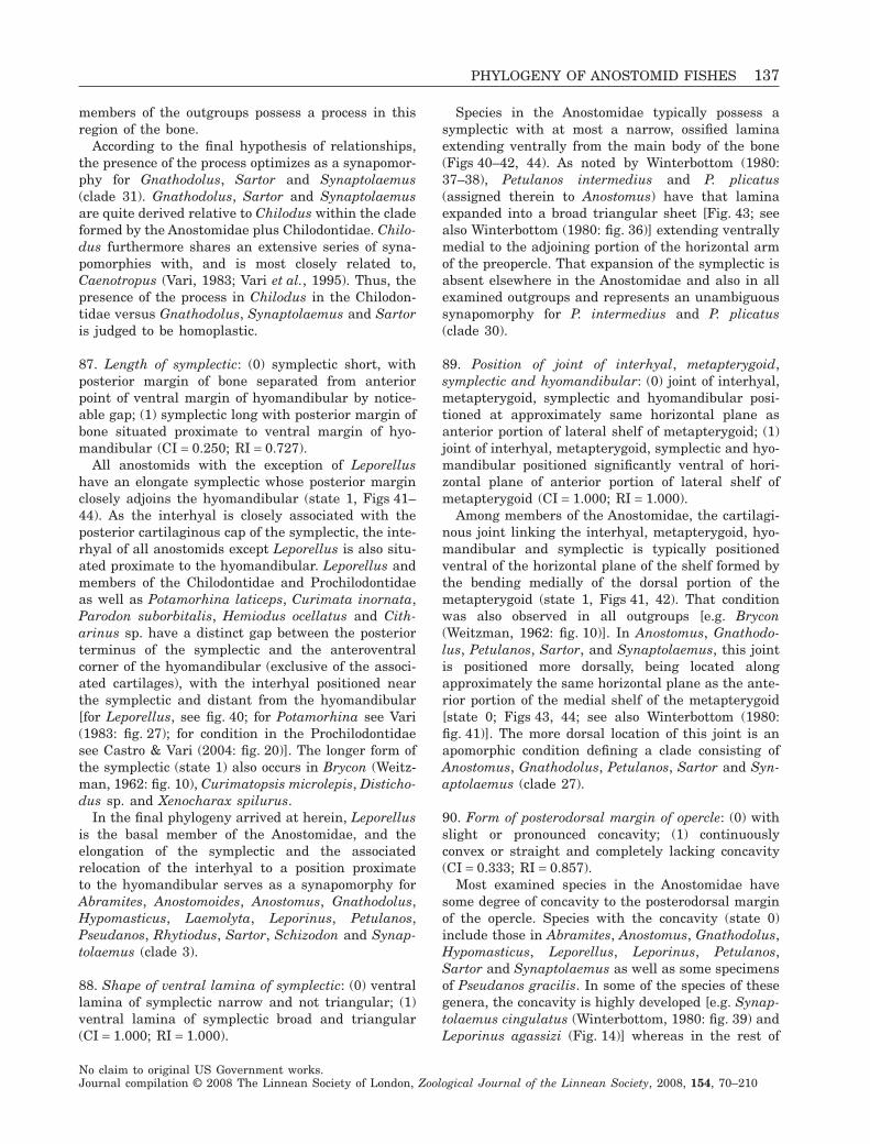
members of the outgroups possess a process in thisregion of the bone.
According to the final hypothesis of relationships,the presence of the process optimizes as a synapomor-phy for Gnathodolus, Sartor and Synaptolaemus(clade 31). Gnathodolus, Sartor and Synaptolaemusare quite derived relative to Chilodus within the cladeformed by the Anostomidae plus Chilodontidae. Chilo-dus furthermore shares an extensive series of syna-pomorphies with, and is most closely related to,Caenotropus (Vari, 1983; Vari et al., 1995). Thus, thepresence of the process in Chilodus in the Chilodon-tidae versus Gnathodolus, Synaptolaemus and Sartoris judged to be homoplastic.
87. Length of symplectic: (0) symplectic short, withposterior margin of bone separated from anteriorpoint of ventral margin of hyomandibular by notice-able gap; (1) symplectic long with posterior margin ofbone situated proximate to ventral margin of hyo-mandibular (CI = 0.250; RI = 0.727).
All anostomids with the exception of Leporellushave an elongate symplectic whose posterior marginclosely adjoins the hyomandibular (state 1, Figs 41–44). As the interhyal is closely associated with theposterior cartilaginous cap of the symplectic, the inte-rhyal of all anostomids except Leporellus is also situ-ated proximate to the hyomandibular. Leporellus andmembers of the Chilodontidae and Prochilodontidaeas well as Potamorhina laticeps, Curimata inornata,Parodon suborbitalis, Hemiodus ocellatus and Cith-arinus sp. have a distinct gap between the posteriorterminus of the symplectic and the anteroventralcorner of the hyomandibular (exclusive of the associ-ated cartilages), with the interhyal positioned nearthe symplectic and distant from the hyomandibular[for Leporellus, see fig. 40; for Potamorhina see Vari(1983: fig. 27); for condition in the Prochilodontidaesee Castro & Vari (2004: fig. 20)]. The longer form ofthe symplectic (state 1) also occurs in Brycon (Weitz-man, 1962: fig. 10), Curimatopsis microlepis, Disticho-dus sp. and Xenocharax spilurus.
In the final phylogeny arrived at herein, Leporellusis the basal member of the Anostomidae, and theelongation of the symplectic and the associatedrelocation of the interhyal to a position proximateto the hyomandibular serves as a synapomorphy forAbramites, Anostomoides, Anostomus, Gnathodolus,Hypomasticus, Laemolyta, Leporinus, Petulanos,Pseudanos, Rhytiodus, Sartor, Schizodon and Synap-tolaemus (clade 3).
88. Shape of ventral lamina of symplectic: (0) ventrallamina of symplectic narrow and not triangular; (1)ventral lamina of symplectic broad and triangular(CI = 1.000; RI = 1.000).
Species in the Anostomidae typically possess asymplectic with at most a narrow, ossified laminaextending ventrally from the main body of the bone(Figs 40–42, 44). As noted by Winterbottom (1980:37–38), Petulanos intermedius and P. plicatus(assigned therein to Anostomus) have that laminaexpanded into a broad triangular sheet [Fig. 43; seealso Winterbottom (1980: fig. 36)] extending ventrallymedial to the adjoining portion of the horizontal armof the preopercle. That expansion of the symplectic isabsent elsewhere in the Anostomidae and also in allexamined outgroups and represents an unambiguoussynapomorphy for P. intermedius and P. plicatus(clade 30).
89. Position of joint of interhyal, metapterygoid,symplectic and hyomandibular: (0) joint of interhyal,metapterygoid, symplectic and hyomandibular posi-tioned at approximately same horizontal plane asanterior portion of lateral shelf of metapterygoid; (1)joint of interhyal, metapterygoid, symplectic and hyo-mandibular positioned significantly ventral of hori-zontal plane of anterior portion of lateral shelf ofmetapterygoid (CI = 1.000; RI = 1.000).
Among members of the Anostomidae, the cartilagi-nous joint linking the interhyal, metapterygoid, hyo-mandibular and symplectic is typically positionedventral of the horizontal plane of the shelf formed bythe bending medially of the dorsal portion of themetapterygoid (state 1, Figs 41, 42). That conditionwas also observed in all outgroups [e.g. Brycon(Weitzman, 1962: fig. 10)]. In Anostomus, Gnathodo-lus, Petulanos, Sartor, and Synaptolaemus, this jointis positioned more dorsally, being located alongapproximately the same horizontal plane as the ante-rior portion of the medial shelf of the metapterygoid[state 0; Figs 43, 44; see also Winterbottom (1980:fig. 41)]. The more dorsal location of this joint is anapomorphic condition defining a clade consisting ofAnostomus, Gnathodolus, Petulanos, Sartor and Syn-aptolaemus (clade 27).
90. Form of posterodorsal margin of opercle: (0) withslight or pronounced concavity; (1) continuouslyconvex or straight and completely lacking concavity(CI = 0.333; RI = 0.857).
Most examined species in the Anostomidae havesome degree of concavity to the posterodorsal marginof the opercle. Species with the concavity (state 0)include those in Abramites, Anostomus, Gnathodolus,Hypomasticus, Leporellus, Leporinus, Petulanos,Sartor and Synaptolaemus as well as some specimensof Pseudanos gracilis. In some of the species of thesegenera, the concavity is highly developed [e.g. Synap-tolaemus cingulatus (Winterbottom, 1980: fig. 39) andLeporinus agassizi (Fig. 14)] whereas in the rest of
PHYLOGENY OF ANOSTOMID FISHES 137
No claim to original US Government works.Journal compilation © 2008 The Linnean Society of London, Zoological Journal of the Linnean Society, 2008, 154, 70–210
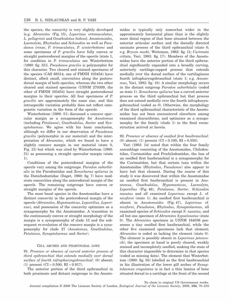
the species, the concavity is very slightly developed[e.g. Abramites (Fig. 15), Leporinus ortomaculatus,L. pellegrinii and Gnathodolus bidens]. Anostomoides,Laemolyta, Rhytiodus and Schizodon as well as Pseu-danos irinae, P. trimaculatus, P. winterbottomi andsome specimens of P. gracilis have fully convex orstraight posterodorsal margins of the opercle [state 1,for condition in P. trimaculatus see Winterbottom(1980: fig. 33)]. Pseudanos gracilis is polymorphic forthis character. Two cleared and stained specimens ofthe species (CAS 68314, one of FMNH 103454) havedistinct, albeit small, convexities along the postero-dorsal margin of both opercles, whereas the two othercleared and stained specimens (USNM 270309, theother of FMNH 103454) have straight posterodorsalmargins to their opercles. All four specimens of P.gracilis are approximately the same size, and thisintraspecific variation probably does not reflect onto-genetic variation in the form of the opercle.
Winterbottom (1980: 51) discussed a concave oper-cular margin as a synapomorphy for Anostomus(including Petulanos), Gnathodolus, Sartor and Syn-aptolaemus. We agree with his basic conclusion,although we differ in our observation of Pseudanosgracilis (polymorphic in our material) and the inter-pretation of Abramites, which we found to have aslightly concave margin in our material (state 0,Fig. 15) but which was cited by Winterbottom (1980:51) as possessing a straight opercular margin (state1).
Conditions of the posterodorsal margins of theopercle vary among the outgroups. Parodon suborbit-alis in the Parodontidae and Xenocharax spilurus inthe Distichodontidae (Daget, 1960: fig. 7) have mod-erate concavities along the anterodorsal margin of theopercle. The remaining outgroups have convex orstraight margins of the opercle.
The most basal species in the Anostomidae have adistinct concavity in the posterodorsal margin of theopercle (Abramites, Hypomasticus, Leporellus, Lepori-nus), and possession of the concavity optimizes as asynapomorphy for the Anostomidae. A transition tothe continuously convex or straight morphology of themargin is a synapomorphy of clade 13 and the sub-sequent re-evolution of the concave margin is a syna-pomorphy for clade 27 (Anostomus, Gnathodolus,Petulanos, Synaptolaemus and Sartor).
GILL ARCHES AND PHARYNGEAL JAWS
91. Presence or absence of curved anterior process ofthird epibranchial that extends medially over dorsalsurface of fourth infrapharyngobranchial: (0) absent;(1) present (CI = 0.500; RI = 0.917).
The anterior portion of the third epibranchial inboth proximate and distant outgroups to the Anosto-
midae is typically only somewhat wider in theapproximately horizontal plane than is the slightlymore distal region of that bone situated between theanterior articular surface and the dorsally directeduncinate process of the third epibranchial (state 0;e.g. Brycon meeki, Weitzman, 1962: fig. 11; Curimatavittata, Vari, 1983: fig. 17). Members of the Anosto-midae have the anterior portion of the third epibran-chial significantly expanded into a broadly curving,anteriorly cartilage-capped process that extendsmedially over the dorsal surface of the cartilaginousfourth infrapharyngobranchial (state 1; e.g. Anosto-mus, Vari, 1983: fig. 18). A similar morphology occursin the distant outgroup Parodon suborbitalis (codedas state 1). Xenocharax spilurus has a curved anteriorprocess on the third epibranchial but that processdoes not extend medially over the fourth infrapharyn-gobranchial (coded as 0). Otherwise, the morphologyof the third epibranchial characteristic of the Anosto-midae has not been encountered elsewhere amongexamined characiforms, and optimizes as a synapo-morphy for the family (clade 1) in the final recon-struction arrived at herein.
92. Presence or absence of ossified first basibranchial:(0) absent; (1) present (CI = 0.100; RI = 0.550).
Vari (1983: 14) noted that within the four familyassemblage consisting of the Anostomidae, Chilodon-tidae, Curimatidae and Prochilodontidae, the loss ofan ossified first basibranchial is a synapomorphy forthe Curimatidae, but that certain taxa within theAnostomidae (Rhytiodus, Pseudanos) also appear tohave lost that element. During the course of thisstudy it was discovered that within the Anostomidaean ossified first basibranchial is present in Ano-stomus, Gnathodolus, Hypomasticus, Laemolyta,Leporellus (Fig. 46), Petulanos, Sartor, Schizodonnasutus and all examined Leporinus except L. cf.niceforoi (state 1). An ossified first basibranchial isabsent in Anostomoides (Fig. 47), Leporinus cf.niceforoi, Pseudanos, Rhytiodus, Synaptolaemus, allexamined species of Schizodon except S. nasutus, andall but one specimen of Abramites hypselonotus (state0). The Abramites specimen in USNM 164036 pos-sesses a tiny ossified first basibranchial, while theother five examined specimens lack that element;Abramites is coded as lacking the element (state 0).The element is possibly absent in Leporinus jatunco-chi; the specimen at hand is poorly cleared, weaklystained and incompletely ossified, making the state ofthis character impossible to determine in that species(coded as missing data). The element that Winterbot-tom (1980: fig. 50) labelled as the first basibranchialin his illustration of the lower gill arches of Synap-tolaemus cingulatus is in fact a thin lamina of bonesituated dorsal to a cartilage at the front of the second
138 B. L. SIDLAUSKAS and R. P. VARI
No claim to original US Government works.Journal compilation © 2008 The Linnean Society of London, Zoological Journal of the Linnean Society, 2008, 154, 70–210
basibranchial and fused to that element in our mate-rial of the specimen. This bony plate appears to be aportion of the second basibranchial, with the firstbasibranchial absent in Synaptolaemus.
Taxa within the Anostomidae that possess an ossi-fied first basibranchial frequently have that elementreduced to tiny ball of bone (e.g. Leporellus, Fig. 46)relative to the condition in the first outgroup, theChilodontidae, in which the first basibranchial is acomparatively massive rod-like ossification (Vari,1983: fig. 9) similar to that present in many outgroupcharaciforms including Brycon (Vari, 1983: figs 3, 4)and the basal distichodontid Xenocharax. Among theother outgroups, members of the Prochilodontidae aswell as Distichodus sp. and Hemiodus ocellatus alsopossess ossified first basibranchials, while Citharinussp. and Parodon suborbitalis appear to be lacking thisossification.
In light of the final phylogeny for the Anostomidaearrived at herein, the first basibranchial is plesiomor-phically present within the family. The subsequentevolution of the character is extraordinarily homoplas-tic, necessitating no fewer than seven character statetransitions within the Anostomidae. Losses of theossification occur in Leporinus cf. niceforoi, Abramitesand clade 13. Within clade 13, ossification of thiselement re-evolves in Schizodon nasutus, Laemolytaand clade 27. Within clade 27, Synaptolaemus, in turn,has lost the first basibranchial.
In the Anostomidae the first basibranchial is smalleven when present and it is possible that the featureis ontogenetically variable. If the bone appears laterin the development of some species that lack the bone
Figure 46. Leporellus vittatus, INHS 56128, 78.2 mm SL: A, dorsal view of ventral portion of gill arches; B, close up ofbasibranchials 1 and 2.
Figure 47. Basibranchial 2 of Anostomoides laticeps,INHS 53677, 106.6 mm SL; ventral view.
PHYLOGENY OF ANOSTOMID FISHES 139
No claim to original US Government works.Journal compilation © 2008 The Linnean Society of London, Zoological Journal of the Linnean Society, 2008, 154, 70–210
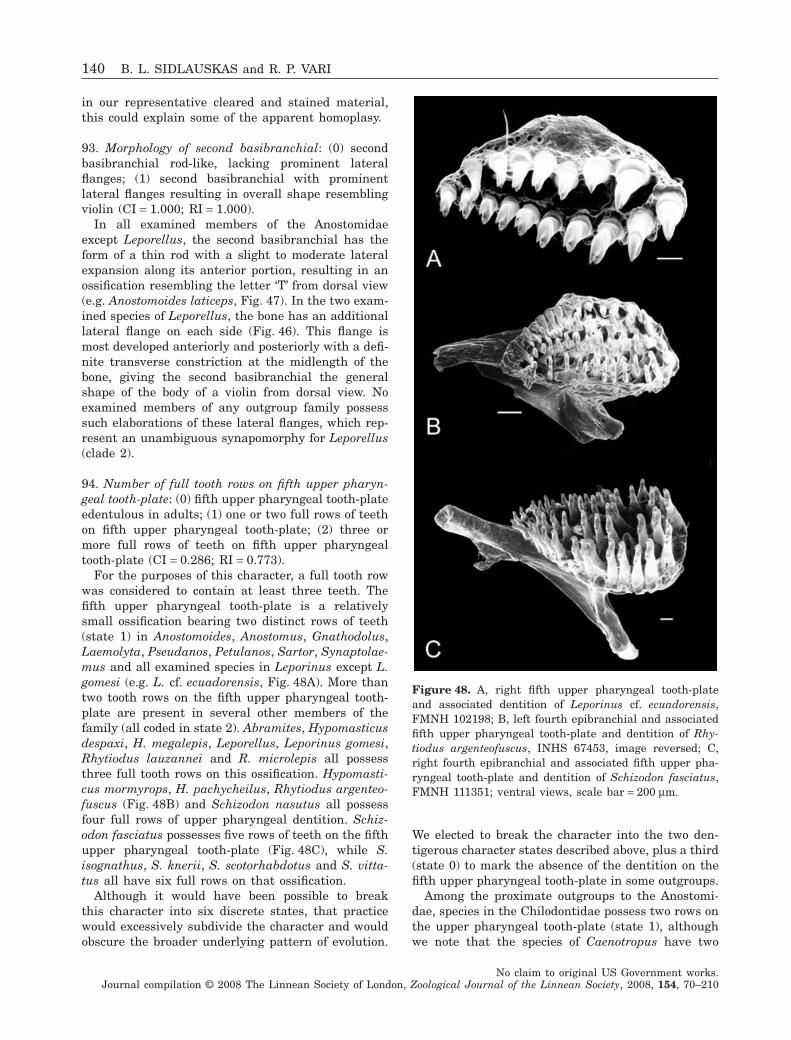
in our representative cleared and stained material,this could explain some of the apparent homoplasy.
93. Morphology of second basibranchial: (0) secondbasibranchial rod-like, lacking prominent lateralflanges; (1) second basibranchial with prominentlateral flanges resulting in overall shape resemblingviolin (CI = 1.000; RI = 1.000).
In all examined members of the Anostomidaeexcept Leporellus, the second basibranchial has theform of a thin rod with a slight to moderate lateralexpansion along its anterior portion, resulting in anossification resembling the letter ‘T’ from dorsal view(e.g. Anostomoides laticeps, Fig. 47). In the two exam-ined species of Leporellus, the bone has an additionallateral flange on each side (Fig. 46). This flange ismost developed anteriorly and posteriorly with a defi-nite transverse constriction at the midlength of thebone, giving the second basibranchial the generalshape of the body of a violin from dorsal view. Noexamined members of any outgroup family possesssuch elaborations of these lateral flanges, which rep-resent an unambiguous synapomorphy for Leporellus(clade 2).
94. Number of full tooth rows on fifth upper pharyn-geal tooth-plate: (0) fifth upper pharyngeal tooth-plateedentulous in adults; (1) one or two full rows of teethon fifth upper pharyngeal tooth-plate; (2) three ormore full rows of teeth on fifth upper pharyngealtooth-plate (CI = 0.286; RI = 0.773).
For the purposes of this character, a full tooth rowwas considered to contain at least three teeth. Thefifth upper pharyngeal tooth-plate is a relativelysmall ossification bearing two distinct rows of teeth(state 1) in Anostomoides, Anostomus, Gnathodolus,Laemolyta, Pseudanos, Petulanos, Sartor, Synaptolae-mus and all examined species in Leporinus except L.gomesi (e.g. L. cf. ecuadorensis, Fig. 48A). More thantwo tooth rows on the fifth upper pharyngeal tooth-plate are present in several other members of thefamily (all coded in state 2). Abramites, Hypomasticusdespaxi, H. megalepis, Leporellus, Leporinus gomesi,Rhytiodus lauzannei and R. microlepis all possessthree full tooth rows on this ossification. Hypomasti-cus mormyrops, H. pachycheilus, Rhytiodus argenteo-fuscus (Fig. 48B) and Schizodon nasutus all possessfour full rows of upper pharyngeal dentition. Schiz-odon fasciatus possesses five rows of teeth on the fifthupper pharyngeal tooth-plate (Fig. 48C), while S.isognathus, S. knerii, S. scotorhabdotus and S. vitta-tus all have six full rows on that ossification.
Although it would have been possible to breakthis character into six discrete states, that practicewould excessively subdivide the character and wouldobscure the broader underlying pattern of evolution.
We elected to break the character into the two den-tigerous character states described above, plus a third(state 0) to mark the absence of the dentition on thefifth upper pharyngeal tooth-plate in some outgroups.
Among the proximate outgroups to the Anostomi-dae, species in the Chilodontidae possess two rows onthe upper pharyngeal tooth-plate (state 1), althoughwe note that the species of Caenotropus have two
Figure 48. A, right fifth upper pharyngeal tooth-plateand associated dentition of Leporinus cf. ecuadorensis,FMNH 102198; B, left fourth epibranchial and associatedfifth upper pharyngeal tooth-plate and dentition of Rhy-tiodus argenteofuscus, INHS 67453, image reversed; C,right fourth epibranchial and associated fifth upper pha-ryngeal tooth-plate and dentition of Schizodon fasciatus,FMNH 111351; ventral views, scale bar = 200 mm.
140 B. L. SIDLAUSKAS and R. P. VARI
No claim to original US Government works.Journal compilation © 2008 The Linnean Society of London, Zoological Journal of the Linnean Society, 2008, 154, 70–210
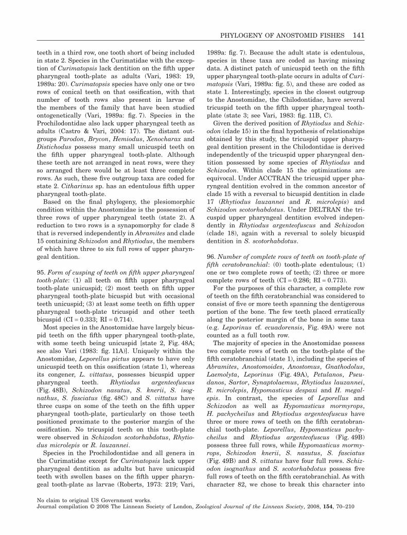
teeth in a third row, one tooth short of being includedin state 2. Species in the Curimatidae with the excep-tion of Curimatopsis lack dentition on the fifth upperpharyngeal tooth-plate as adults (Vari, 1983: 19,1989a: 20). Curimatopsis species have only one or tworows of conical teeth on that ossification, with thatnumber of tooth rows also present in larvae ofthe members of the family that have been studiedontogenetically (Vari, 1989a: fig. 7). Species in theProchilodontidae also lack upper pharyngeal teeth asadults (Castro & Vari, 2004: 17). The distant out-groups Parodon, Brycon, Hemiodus, Xenocharax andDistichodus possess many small unicuspid teeth onthe fifth upper pharyngeal tooth-plate. Althoughthese teeth are not arranged in neat rows, were theyso arranged there would be at least three completerows. As such, these five outgroup taxa are coded forstate 2. Citharinus sp. has an edentulous fifth upperpharyngeal tooth-plate.
Based on the final phylogeny, the plesiomorphiccondition within the Anostomidae is the possession ofthree rows of upper pharyngeal teeth (state 2). Areduction to two rows is a synapomorphy for clade 8that is reversed independently in Abramites and clade15 containing Schizodon and Rhytiodus, the membersof which have three to six full rows of upper pharyn-geal dentition.
95. Form of cusping of teeth on fifth upper pharyngealtooth-plate: (1) all teeth on fifth upper pharyngealtooth-plate unicuspid; (2) most teeth on fifth upperpharyngeal tooth-plate bicuspid but with occasionalteeth unicuspid; (3) at least some teeth on fifth upperpharyngeal tooth-plate tricuspid and other teethbicuspid (CI = 0.333; RI = 0.714).
Most species in the Anostomidae have largely bicus-pid teeth on the fifth upper pharyngeal tooth-plate,with some teeth being unicuspid [state 2, Fig. 48A;see also Vari (1983: fig. 11A)]. Uniquely within theAnostomidae, Leporellus pictus appears to have onlyunicuspid teeth on this ossification (state 1), whereasits congener, L. vittatus, possesses bicuspid upperpharyngeal teeth. Rhytiodus argenteofuscus(Fig. 48B), Schizodon nasutus, S. knerii, S. isog-nathus, S. fasciatus (fig. 48C) and S. vittatus havethree cusps on some of the teeth on the fifth upperpharyngeal tooth-plate, particularly on those teethpositioned proximate to the posterior margin of theossification. No tricuspid teeth on this tooth-platewere observed in Schizodon scotorhabdotus, Rhytio-dus microlepis or R. lauzannei.
Species in the Prochilodontidae and all genera inthe Curimatidae except for Curimatopsis lack upperpharyngeal dentition as adults but have unicuspidteeth with swollen bases on the fifth upper pharyn-geal tooth-plate as larvae (Roberts, 1973: 219; Vari,
1989a: fig. 7). Because the adult state is edentulous,species in these taxa are coded as having missingdata. A distinct patch of unicuspid teeth on the fifthupper pharyngeal tooth-plate occurs in adults of Curi-matopsis (Vari, 1989a: fig. 5), and these are coded asstate 1. Interestingly, species in the closest outgroupto the Anostomidae, the Chilodontidae, have severaltricuspid teeth on the fifth upper pharyngeal tooth-plate (state 3; see Vari, 1983: fig. 11B, C).
Given the derived position of Rhytiodus and Schiz-odon (clade 15) in the final hypothesis of relationshipsobtained by this study, the tricuspid upper pharyn-geal dentition present in the Chilodontidae is derivedindependently of the tricuspid upper pharyngeal den-tition possessed by some species of Rhytiodus andSchizodon. Within clade 15 the optimizations areequivocal. Under ACCTRAN the tricuspid upper pha-ryngeal dentition evolved in the common ancestor ofclade 15 with a reversal to bicuspid dentition in clade17 (Rhytiodus lauzannei and R. microlepis) andSchizodon scotorhabdotus. Under DELTRAN the tri-cuspid upper pharyngeal dentition evolved indepen-dently in Rhytiodus argenteofuscus and Schizodon(clade 18), again with a reversal to solely bicuspiddentition in S. scotorhabdotus.
96. Number of complete rows of teeth on tooth-plate offifth ceratobranchial: (0) tooth-plate edentulous; (1)one or two complete rows of teeth; (2) three or morecomplete rows of teeth (CI = 0.286; RI = 0.773).
For the purposes of this character, a complete rowof teeth on the fifth ceratobranchial was considered toconsist of five or more teeth spanning the dentigerousportion of the bone. The few teeth placed erraticallyalong the posterior margin of the bone in some taxa(e.g. Leporinus cf. ecuadorensis, Fig. 49A) were notcounted as a full tooth row.
The majority of species in the Anostomidae possesstwo complete rows of teeth on the tooth-plate of thefifth ceratobranchial (state 1), including the species ofAbramites, Anostomoides, Anostomus, Gnathodolus,Laemolyta, Leporinus (Fig. 49A), Petulanos, Pseu-danos, Sartor, Synaptolaemus, Rhytiodus lauzannei,R. microlepis, Hypomasticus despaxi and H. megal-epis. In contrast, the species of Leporellus andSchizodon as well as Hypomasticus mormyrops,H. pachycheilus and Rhytiodus argenteofuscus havethree or more rows of teeth on the fifth ceratobran-chial tooth-plate. Leporellus, Hypomasticus pachy-cheilus and Rhytiodus argenteofuscus (Fig. 49B)possess three full rows, while Hypomasticus mormy-rops, Schizodon knerii, S. nasutus, S. fasciatus(Fig. 49B) and S. vittatus have four full rows. Schiz-odon isognathus and S. scotorhabdotus possess fivefull rows of teeth on the fifth ceratobranchial. As withcharacter 82, we chose to break this character into
PHYLOGENY OF ANOSTOMID FISHES 141
No claim to original US Government works.Journal compilation © 2008 The Linnean Society of London, Zoological Journal of the Linnean Society, 2008, 154, 70–210
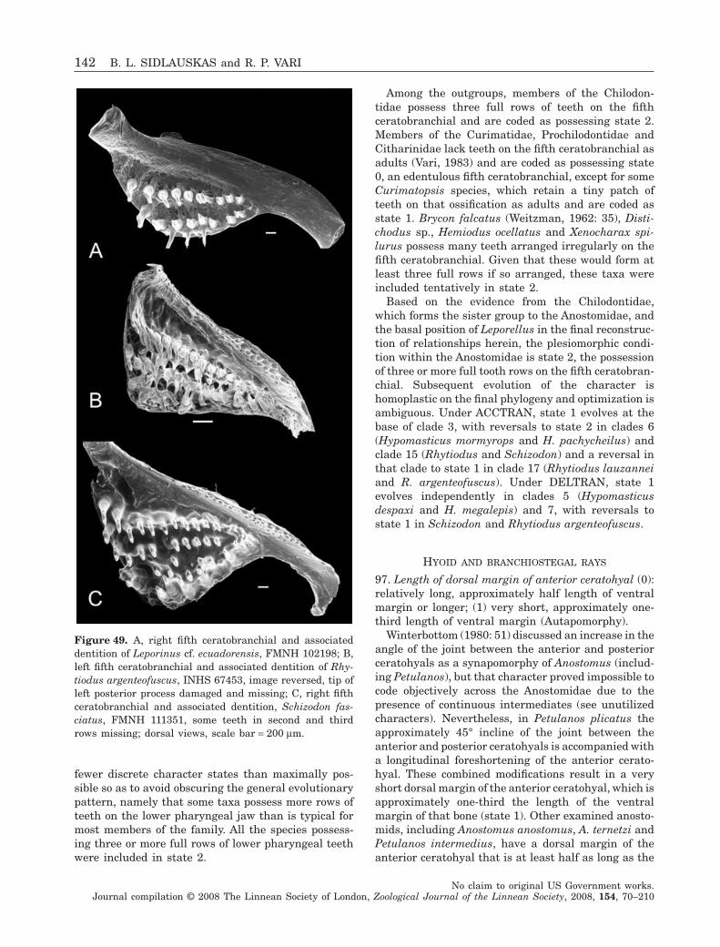
fewer discrete character states than maximally pos-sible so as to avoid obscuring the general evolutionarypattern, namely that some taxa possess more rows ofteeth on the lower pharyngeal jaw than is typical formost members of the family. All the species possess-ing three or more full rows of lower pharyngeal teethwere included in state 2.
Among the outgroups, members of the Chilodon-tidae possess three full rows of teeth on the fifthceratobranchial and are coded as possessing state 2.Members of the Curimatidae, Prochilodontidae andCitharinidae lack teeth on the fifth ceratobranchial asadults (Vari, 1983) and are coded as possessing state0, an edentulous fifth ceratobranchial, except for someCurimatopsis species, which retain a tiny patch ofteeth on that ossification as adults and are coded asstate 1. Brycon falcatus (Weitzman, 1962: 35), Disti-chodus sp., Hemiodus ocellatus and Xenocharax spi-lurus possess many teeth arranged irregularly on thefifth ceratobranchial. Given that these would form atleast three full rows if so arranged, these taxa wereincluded tentatively in state 2.
Based on the evidence from the Chilodontidae,which forms the sister group to the Anostomidae, andthe basal position of Leporellus in the final reconstruc-tion of relationships herein, the plesiomorphic condi-tion within the Anostomidae is state 2, the possessionof three or more full tooth rows on the fifth ceratobran-chial. Subsequent evolution of the character ishomoplastic on the final phylogeny and optimization isambiguous. Under ACCTRAN, state 1 evolves at thebase of clade 3, with reversals to state 2 in clades 6(Hypomasticus mormyrops and H. pachycheilus) andclade 15 (Rhytiodus and Schizodon) and a reversal inthat clade to state 1 in clade 17 (Rhytiodus lauzanneiand R. argenteofuscus). Under DELTRAN, state 1evolves independently in clades 5 (Hypomasticusdespaxi and H. megalepis) and 7, with reversals tostate 1 in Schizodon and Rhytiodus argenteofuscus.
HYOID AND BRANCHIOSTEGAL RAYS
97. Length of dorsal margin of anterior ceratohyal (0):relatively long, approximately half length of ventralmargin or longer; (1) very short, approximately one-third length of ventral margin (Autapomorphy).
Winterbottom (1980: 51) discussed an increase in theangle of the joint between the anterior and posteriorceratohyals as a synapomorphy of Anostomus (includ-ing Petulanos), but that character proved impossible tocode objectively across the Anostomidae due to thepresence of continuous intermediates (see unutilizedcharacters). Nevertheless, in Petulanos plicatus theapproximately 45° incline of the joint between theanterior and posterior ceratohyals is accompanied witha longitudinal foreshortening of the anterior cerato-hyal. These combined modifications result in a veryshort dorsal margin of the anterior ceratohyal, which isapproximately one-third the length of the ventralmargin of that bone (state 1). Other examined anosto-mids, including Anostomus anostomus, A. ternetzi andPetulanos intermedius, have a dorsal margin of theanterior ceratohyal that is at least half as long as the
Figure 49. A, right fifth ceratobranchial and associateddentition of Leporinus cf. ecuadorensis, FMNH 102198; B,left fifth ceratobranchial and associated dentition of Rhy-tiodus argenteofuscus, INHS 67453, image reversed, tip ofleft posterior process damaged and missing; C, right fifthceratobranchial and associated dentition, Schizodon fas-ciatus, FMNH 111351, some teeth in second and thirdrows missing; dorsal views, scale bar = 200 mm.
142 B. L. SIDLAUSKAS and R. P. VARI
No claim to original US Government works.Journal compilation © 2008 The Linnean Society of London, Zoological Journal of the Linnean Society, 2008, 154, 70–210
ventral margin, and many species have the twomargins approximately of equal length. Although thedorsally shorter morphology of the anterior ceratohyalin Petulanos plicatus is unique among the examinedanostomids and outgroups, it is shared with P. spilo-clistron (Winterbottom, 1980: fig. 46A) and would pre-sumably optimize as a synapomorphy for those twotaxa in a future comprehensive analysis.
98. Presence or absence of inflection point in jointbetween anterior and posterior ceratohyals and corre-sponding process of anterior ceratohyal runningventral to posterior ceratohyal: (0) absent; (1) present(CI = 1.000, RI = 1.000).
The two examined species of Leporellus possess adistinct inflection in the angle of the joint between theanterior and posterior ceratohyals. The inflection ofthe joint produces a thin process of the anteriorceratohyal that runs ventral to the posterior cerato-hyal. Many species in the Anostomidae have a steepangle to the joint between the ceratohyals [e.g. Petu-lanos spiloclistron (Winterbottom, 1980: fig. 46A, theepihyal and ceratohyal of that author) and Synapto-laemus cingulatus (Winterbottom, 1980: fig. 47)] butno others have a strong point of inflection in the jointor a similar ventral process. Inflection of this jointand the presence of a ventral process of the anteriorceratohyal were not observed in the outgroups, andpossession of the inflection and process is a synapo-morphy for Leporellus in the final phylogeneticreconstruction.
99. Height of, and angle between, dorsal and ventralmargins of urohyal: (0) urohyal low, dorsal andventral margins meet at an angle equal to or lessthan 45°; (1) urohyal high, dorsal and ventralmargins meet at an angle greater than 45°(CI = 0.500, RI = 0.667).
Winterbottom (1980: 55, Fig. 46B) cited a high(deep) urohyal as a synapomorphy for Petulanos inter-medius, P. plicatus and P. spiloclistron. We agree withthat observation; these three species have a very highurohyal with the dorsal and ventral margins meetingat approximately a 60° angle. We also note thatSynaptolaemus and Gnathodolus possess an urohyalalmost as high as that in the Petulanos species, withmargins meeting at approximately 50–55° angle.Under the definition of character states above, Gna-thodolus, Petulanos and Synaptolaemus are all codedas possessing state 1. The remaining species in theAnostomidae have shallower urohyals with the dorsaland ventral margins meeting along a 45° angle or less(state 0). Abramites, Leporellus, Sartor and Leporinusgomesi, L. jatuncochi, L. pellegrinii and L. tigrinushave somewhat deep urohyals with margins meetingalong an angle of approximately 40–45°. Species in
Anostomus, Anostomoides, Hypomasticus, Pseudanos,Rhytiodus and Schizodon as well as the members ofLeporinus not cited above have even shallower uro-hyals, typically with margins meeting at approxi-mately a 30° angle. Examined members of theoutgroups all have urohyals with dorsal and ventralmargins meeting at less than a 45° angle [e.g. Brycon(Weitzman, 1962: fig. 11B)]. Under the final hypoth-esis of relationships arrived at herein, possession of ahigh urohyal (state 1) optimizes as a synapomorphyfor clade 29 (Gnathodolus, Petulanos, Sartor and Syn-aptolaemus) that is reversed in Sartor.
100. Number of branchiostegal rays: (0) three; (1) four(CI = 0.250; RI = 0.400).
Many groups in the Characiformes have four pairsof branchiostegal rays (Roberts, 1969: 422) albeit withvariation from that number in various taxa. All exam-ined outgroups in this study possess four branchioste-gal rays, and that number of rays is also presentin most species within the Anostomidae. Abramiteshypselonotus, Anostomoides laticeps, Anostomus ter-netzi, Pseudanos gracilis, P. irinae and P. trimacula-tus lack the medial-most pair of branchiostegal rays,leaving a total of three pairs of rays (Winterbottom,1980: fig. 42). Anostomus anostomus and Pseudanoswinterbottomi are reported to be polymorphic for thischaracter, with most specimens possessing four pairsof branchiostegal rays (Winterbottom, 1980: 11; Sid-lauskas & Santos, 2005: 114) but occasional speci-mens possessing three pairs of rays. In accordancewith the state possessed by the majority of specimens(four rays), Pseudanos winterbottomi and Anostomusanostomus are coded as possessing state 1 for thischaracter.
Under the final phylogenetic hypothesis arrived atherein, possession of four branchiostegal rays isclearly the plesiomorphic condition within the Anos-tomidae. A reduction to three rays appears to haveoccurred four times in the family: once in Abramites,once in Anostomoides, once in Anostomus ternetzi andonce in clade 26 (Pseudanos gracilis, P. irinae and P.trimaculatus). The concentration of species with onlythree branchial pairs of rays in Pseudanos and Anos-tomus, and the known polymorphism in Pseudanoswinterbottomi and Anostomus anostomus, whichappear to be relatively basal members of the cladecontaining Anostomus, Gnathodolus, Petulanos, Pseu-danos, Sartor and Synaptolaemus (clade 25) suggeststhat the common ancestor of that clade may also havebeen polymorphic for this character.
WEBERIAN APPARATUS AND SUPRANEURALS
101. Presence or absence of anteriorly directed processof fourth pleural rib: (0) absent; (1) present(CI = 0.500; RI = 0.750).
PHYLOGENY OF ANOSTOMID FISHES 143
No claim to original US Government works.Journal compilation © 2008 The Linnean Society of London, Zoological Journal of the Linnean Society, 2008, 154, 70–210
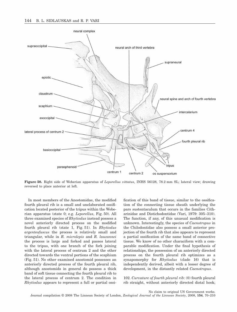
In most members of the Anostomidae, the modifiedfourth pleural rib is a small and unelaborated ossifi-cation located posterior of the tripus within the Webe-rian apparatus (state 0; e.g. Leporellus, Fig. 50). Allthree examined species of Rhytiodus instead possess anovel anteriorly directed process on the modifiedfourth pleural rib (state 1, Fig. 51). In Rhytiodusargenteofuscus the process is relatively small andtriangular, while in R. microlepis and R. lauzanneithe process is large and forked and passes lateralto the tripus, with one branch of the fork joiningwith the lateral process of centrum 2 and the otherdirected towards the ventral portions of the scaphium(Fig. 51). No other examined anostomid possesses ananteriorly directed process of the fourth pleural rib,although anostomids in general do possess a thickband of soft tissue connecting the fourth pleural rib tothe lateral process of centrum 2. The condition inRhytiodus appears to represent a full or partial ossi-
fication of this band of tissue, similar to the ossifica-tion of the connecting tissue sheath underlying thepars sustentaculum that occurs in the families Cith-arinidae and Distichodontidae (Vari, 1979: 305–310).The function, if any, of this unusual modification isunknown. Interestingly, the species of Caenotropus inthe Chilodontidae also possess a small anterior pro-jection of the fourth rib that also appears to representa partial ossification of the same band of connectivetissue. We know of no other characiform with a com-parable modification. Under the final hypothesis ofrelationships, the possession of an anteriorly directedprocess on the fourth pleural rib optimizes as asynapomorphy for Rhytiodus (clade 16) that isindependently derived, albeit with a lesser degree ofdevelopment, in the distantly related Caenotropus.
102. Curvature of fourth pleural rib: (0) fourth pleuralrib straight, without anteriorly directed distal hook;
Figure 50. Right side of Weberian apparatus of Leporellus vittatus, INHS 56128, 78.2 mm SL; lateral view; drawingreversed to place anterior at left.
144 B. L. SIDLAUSKAS and R. P. VARI
No claim to original US Government works.Journal compilation © 2008 The Linnean Society of London, Zoological Journal of the Linnean Society, 2008, 154, 70–210
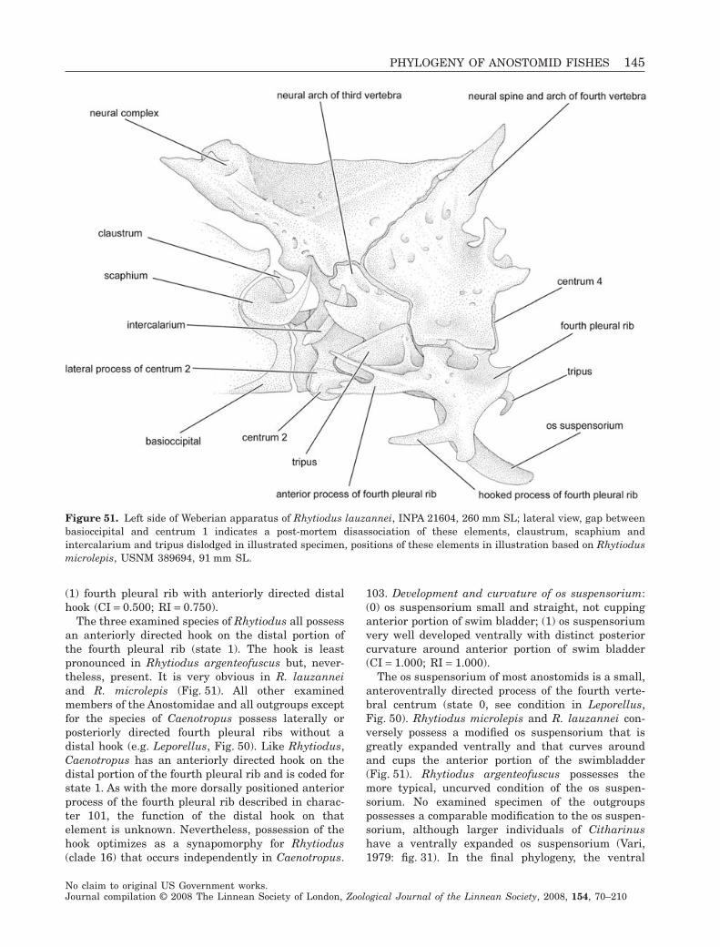
(1) fourth pleural rib with anteriorly directed distalhook (CI = 0.500; RI = 0.750).
The three examined species of Rhytiodus all possessan anteriorly directed hook on the distal portion ofthe fourth pleural rib (state 1). The hook is leastpronounced in Rhytiodus argenteofuscus but, never-theless, present. It is very obvious in R. lauzanneiand R. microlepis (Fig. 51). All other examinedmembers of the Anostomidae and all outgroups exceptfor the species of Caenotropus possess laterally orposteriorly directed fourth pleural ribs without adistal hook (e.g. Leporellus, Fig. 50). Like Rhytiodus,Caenotropus has an anteriorly directed hook on thedistal portion of the fourth pleural rib and is coded forstate 1. As with the more dorsally positioned anteriorprocess of the fourth pleural rib described in charac-ter 101, the function of the distal hook on thatelement is unknown. Nevertheless, possession of thehook optimizes as a synapomorphy for Rhytiodus(clade 16) that occurs independently in Caenotropus.
103. Development and curvature of os suspensorium:(0) os suspensorium small and straight, not cuppinganterior portion of swim bladder; (1) os suspensoriumvery well developed ventrally with distinct posteriorcurvature around anterior portion of swim bladder(CI = 1.000; RI = 1.000).
The os suspensorium of most anostomids is a small,anteroventrally directed process of the fourth verte-bral centrum (state 0, see condition in Leporellus,Fig. 50). Rhytiodus microlepis and R. lauzannei con-versely possess a modified os suspensorium that isgreatly expanded ventrally and that curves aroundand cups the anterior portion of the swimbladder(Fig. 51). Rhytiodus argenteofuscus possesses themore typical, uncurved condition of the os suspen-sorium. No examined specimen of the outgroupspossesses a comparable modification to the os suspen-sorium, although larger individuals of Citharinushave a ventrally expanded os suspensorium (Vari,1979: fig. 31). In the final phylogeny, the ventral
Figure 51. Left side of Weberian apparatus of Rhytiodus lauzannei, INPA 21604, 260 mm SL; lateral view, gap betweenbasioccipital and centrum 1 indicates a post-mortem disassociation of these elements, claustrum, scaphium andintercalarium and tripus dislodged in illustrated specimen, positions of these elements in illustration based on Rhytiodusmicrolepis, USNM 389694, 91 mm SL.
PHYLOGENY OF ANOSTOMID FISHES 145
No claim to original US Government works.Journal compilation © 2008 The Linnean Society of London, Zoological Journal of the Linnean Society, 2008, 154, 70–210
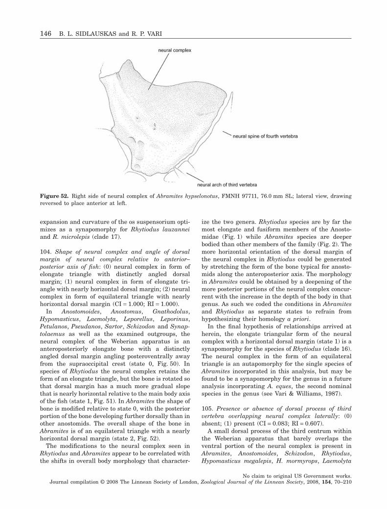
expansion and curvature of the os suspensorium opti-mizes as a synapomorphy for Rhytiodus lauzanneiand R. microlepis (clade 17).
104. Shape of neural complex and angle of dorsalmargin of neural complex relative to anterior–posterior axis of fish: (0) neural complex in form ofelongate triangle with distinctly angled dorsalmargin; (1) neural complex in form of elongate tri-angle with nearly horizontal dorsal margin; (2) neuralcomplex in form of equilateral triangle with nearlyhorizontal dorsal margin (CI = 1.000; RI = 1.000).
In Anostomoides, Anostomus, Gnathodolus,Hypomasticus, Laemolyta, Leporellus, Leporinus,Petulanos, Pseudanos, Sartor, Schizodon and Synap-tolaemus as well as the examined outgroups, theneural complex of the Weberian apparatus is ananteroposteriorly elongate bone with a distinctlyangled dorsal margin angling posteroventrally awayfrom the supraoccipital crest (state 0, Fig. 50). Inspecies of Rhytiodus the neural complex retains theform of an elongate triangle, but the bone is rotated sothat dorsal margin has a much more gradual slopethat is nearly horizontal relative to the main body axisof the fish (state 1, Fig. 51). In Abramites the shape ofbone is modified relative to state 0, with the posteriorportion of the bone developing further dorsally than inother anostomids. The overall shape of the bone inAbramites is of an equilateral triangle with a nearlyhorizontal dorsal margin (state 2, Fig. 52).
The modifications to the neural complex seen inRhytiodus and Abramites appear to be correlated withthe shifts in overall body morphology that character-
ize the two genera. Rhytiodus species are by far themost elongate and fusiform members of the Anosto-midae (Fig. 1) while Abramites species are deeperbodied than other members of the family (Fig. 2). Themore horizontal orientation of the dorsal margin ofthe neural complex in Rhytiodus could be generatedby stretching the form of the bone typical for anosto-mids along the anteroposterior axis. The morphologyin Abramites could be obtained by a deepening of themore posterior portions of the neural complex concur-rent with the increase in the depth of the body in thatgenus. As such we coded the conditions in Abramitesand Rhytiodus as separate states to refrain fromhypothesizing their homology a priori.
In the final hypothesis of relationships arrived atherein, the elongate triangular form of the neuralcomplex with a horizontal dorsal margin (state 1) is asynapomorphy for the species of Rhytiodus (clade 16).The neural complex in the form of an equilateraltriangle is an autapomorphy for the single species ofAbramites incorporated in this analysis, but may befound to be a synapomorphy for the genus in a futureanalysis incorporating A. eques, the second nominalspecies in the genus (see Vari & Williams, 1987).
105. Presence or absence of dorsal process of thirdvertebra overlapping neural complex laterally: (0)absent; (1) present (CI = 0.083; RI = 0.607).
A small dorsal process of the third centrum withinthe Weberian apparatus that barely overlaps theventral portion of the neural complex is present inAbramites, Anostomoides, Schizodon, Rhytiodus,Hypomasticus megalepis, H. mormyrops, Laemolyta
Figure 52. Right side of neural complex of Abramites hypselonotus, FMNH 97711, 76.0 mm SL; lateral view, drawingreversed to place anterior at left.
146 B. L. SIDLAUSKAS and R. P. VARI
No claim to original US Government works.Journal compilation © 2008 The Linnean Society of London, Zoological Journal of the Linnean Society, 2008, 154, 70–210
garmani, L. orinocensis, L. proxima, Leporinus agas-sizi, L. aripuanaensis, L. cf. ecuadorensis, L. fascia-tus, L. cf. fasciatus, L. friderici, L. gomesi, L.jatuncochi, L. cf. niceforoi and L. ortomaculatus. Theprocess is particularly well developed in the species ofRhytiodus and Schizodon (Figs 51, 52). The process isabsent in the species of Anostomus, Gnathodolus,Leporellus, Petulanos, Pseudanos, Sartor and Synap-tolaemus along with Laemolyta proxima, Hypomasti-cus despaxi, H. pachycheilus, Leporinus cf. moralesi,L. pellegrinii, L. striatus and L. tigrinus (Fig. 50).Among the outgroups such a process is present inBrycon falcatus, Hemiodus ocellatus and Prochilodusrubrotaeniatus. It is absent in the remaining exam-ined outgroups.
Although the evolution of this character on thefinal phylogenetic hypothesis arrived at herein ishomoplastic and ambiguously optimized within theAnostomidae, the character does appear to index con-siderable synapomorphy. The absence of the process(state 0) is the plesiomorphic condition present inLeporellus and the proximate outgroups. UnderACCTRAN, possession of the process (state 1) is asynapomorphy for clade 3 that is reversed in Hypo-masticus despaxi, H. pachycheilus, Leporinus cf.moralesi, L. striatus, Laemolyta proxima and clade 25containing Anostomus, Gnathodolus, Petulanos, Pseu-danos, Sartor and Synaptolaemus. Under DELTRAN,state 1 evolves independently in Hypomasticus mega-lepis, H. mormyrops and clade 7, again with reversalsto state 0 in Leporinus cf. moralesi, L. striatus,Laemolyta proxima and clade 25.
106. Presence or absence of supraneural dorsal toWeberian apparatus: (0) supraneural absent; (1)supraneural present (CI = 0.375; RI = 0.800).
In some species of the Anostomidae, a thin bone lieswithin the midsagittal plane dorsal to the posterodor-sal portion of the Weberian apparatus (e.g. Leporel-lus, Fig. 50). Under the terminology for the bones ofthe Weberian apparatus suggested by Weitzman(1962), the bone in question, when present, is posi-tioned posterior to the neural complex and slightlyanterior to the neural spine of the fourth vertebrae.The positioning of this bone just anterior to a neuralspine along the midsagittal plane suggests that theossification is serially homologous with the supraneu-ral series.
A supraneural positioned above the Weberian appa-ratus is present in Anostomus, Gnathodolus, Hypo-masticus, Leporellus, Petulanos, Synaptolaemus, mostspecies of Leporinus and most specimens of Pseu-danos. No supraneural lies above the Weberian appa-ratus in Abramites, Anostomoides, Laemolyta,Rhytiodus, Sartor, Schizodon or Leporinus striatus.Two species, Leporinus cf. ecuadorensis and Pseu-
danos irinae, are polymorphic for this character. Inthe case of Leporinus cf. ecuadorensis, seven clearedand stained specimens were examined, with thesupraneural missing in the largest and two of thesmallest examined individuals, present as a tiny car-tilaginous ball or rod in two other small specimens,and fully present, although small, in two specimens ofintermediate size. Of the two examined specimens ofPseudanos irinae, one has a well-developed supraneu-ral positioned dorsal to the Weberian apparatus whilethe bone is absent in the second individual. Bothspecimens had been previously extensively dissectedand it is possible that the accessory supraneural waslost in the second specimen of P. irinae in the course ofthose dissections. In neither case does either stateoccur in a clear majority of specimens and both arecoded as possessing a polymorphism for this character.
Among the examined outgroups, Brycon falcatus,Citharinus sp., Distichodus sp., Hemiodus ocellatusand Xenocharax spilurus possess a supraneural abovethe Weberian apparatus (state 1). Parodon suborbit-alis as well as all examined members of the proximateoutgroups (Chilodontidae, Curimatidae, Prochilodon-tidae) lack a supraneural or supraneural-like elementlocated dorsal to the Weberian apparatus. Our speci-men of Potamorhina laticeps (Curimatidae) doespossess an odd finger-like dorsal extension from theanterior dorsal margin of the neural complex. We areunsure of the homology of this element (it may simplybe an anomaly of the specimen) but based on its moreanterior position it is probably not homologous withthe supraneural above the Weberian apparatus pos-sessed by some members of the Anostomidae. Pota-morhina laticeps is coded as possessing state 0.
The supraneural dorsal to the Weberian apparatuswas gained and lost several times in the evolution ofthe Anostomidae. Presence of the supraneural inquestion optimizes as a synapomorphy for the Anos-tomidae (clade 1) with subsequent loss occurring inclade 9 (Abramites and Leporinus striatus) and clade13. Within clade 13 the ossification is reacquired inclade 25 and again lost in that clade in Sartor.
RIBS
107. Presence or absence of two or more intercostalligaments uniting in total four or more of anterior fullribs: (0) absent; (1) present (CI = 1.000; RI = 1.000)
As discussed in Vari (1983), anostomids have dis-tinct, anterodorsally aligned intercostal ligamentsthat join the mid and upper sections of the anterior-most pleural ribs. The ventral-most of these liga-ments arises from the posterior margin of the first rib,extends posterodorsally to attachments on the medialmargins of the second and third ribs and terminatesposteriorly at an attachment on the anterior border of
PHYLOGENY OF ANOSTOMID FISHES 147
No claim to original US Government works.Journal compilation © 2008 The Linnean Society of London, Zoological Journal of the Linnean Society, 2008, 154, 70–210
the fourth rib. A second more dorsally positionedligament extends between the first and third ribs,with an intervening attachment to the medial surfaceof the second rib. The described pattern of intercostalligaments is the least extensive in the family, withsome anostomids having increased numbers of liga-ments and in some instances a larger number ofinvolved ribs. Although comparable intercostal liga-ments also occur in the Chilodontidae (Vari, 1983: 41,fig. 36), the bands in the latter family are limited toonly the anterior-most three pleural ribs. Intercostalligaments were not observed among the outgroupswith the exception of the Chilodontidae. In our finalreconstruction, possession of two or more intercostalligaments uniting at least four ribs optimizes as asynapomorphy for the entire Anostomidae (clade 1).
DORSAL FIN
108. Modal number of branched dorsal-fin rays: (0)eight or nine branched dorsal-fin rays; (1) tenbranched dorsal-fin rays; (2) 13 or more brancheddorsal-fin rays (CI = 0.667; RI = 0.750).
Characiforms demonstrate a considerable range inthe number of branched dorsal-fin rays, with basalgroups in the order (Citharinidae, Distichodontidae)having high numbers of rays. Within the Citharin-idae, species of Citharinus possesses 11–15 branchedrays (Daget, 1962b: 84) and Citharidium 13–15branched rays (Daget, 1962a: 506). Within the Disti-chodontidae, Xenocharax possesses 14–18 branchedrays (Daget, 1960: 32), Paradistichodus 12–15branched rays (Daget, 1958: 1362) and Ichthyborus11–14 branched rays (Daget, 1967: 140). The majorityof characiforms, by contrast, have lower numbers ofbranched dorsal-fin rays. In their discussion of asubgroup of the Characidae, Malabarba & Weitzman(2003: 82) noted that eight or nine branched dorsal-finrays were typical for that group. This condition wasobserved in the outgroups in Brycon (Weitzman, 1962:fig. 16), Hemiodus ocellatus (Vari, 1982a), Curimatop-sis microlepis (Vari, 1982b), Curimata inornata (Vari,1989b) and all examined members of the Chilodon-tidae. In contrast, all members of the Anostomidaepossess (other than in a few individuals) a tenth setof pterygiophores and a tenth branched dorsal-fin ray(state 1). Among the examined outgroups tenbranched dorsal-fin rays also occur in Parodonsuborbitalis (Pavanelli, 1999) and Semaprochilodusinsignis (Castro & Vari, 2004). Potamorhina laticepsand Prochilodus rubrotaeniatus are polymorphic forthis character, with nine or ten branched dorsal-finrays present in Potamorhina laticeps (Vari, 1984) andseven to ten branched dorsal-fin rays present inProchilodus rubrotaeniatus (Castro & Vari, 2004).
Although constant within the Anostomidae, thischaracter is ambiguously optimized among the out-
groups in our final phylogenetic hypothesis. UnderDELTRAN the possession of ten branched dorsal finrays (state 1) is a previously undocumented synapo-morphy for the Anostomidae (clade 1) with indepen-dent evolution of character state 1 in elements ofthe Curimatidae, Prochilodontidae and Parodontidae.Under ACCTRAN state 1 evolves in the commonancestor of the Anostomidae, Curimatidae, Chilodon-tidae, Parodontidae and Prochilodontidae with rever-sal to state 0 in the Chilodontidae and elements of theProchilodontidae and Curimatidae.
PECTORAL GIRDLE
109. Development of dorsomedial flange of mesocora-coid: (0) dorsomedial flange of mesocoracoid weaklydeveloped or absent; (1) dorsomedial flange ofmesocoracoid very strongly developed (CI = 0.500;RI = 0.833).
All examined members of the Anostomidae exhibita distinct twist to the mesocoracoid, with the plesio-morphically anteroventral margin of the bone whichjoins the coracoid rotating to become the posterodor-sal margin of the bone where it joins the dorsalmargin of the cleithrum. The medial ridge or flangecreated by this torsion is typically indistinct andsmall in most anostomids, but in the species of Schiz-odon and most examined specimens of Abramiteshypselonotus, that flange is very well developed,obscuring much of the lateral portion of the bone inmedial view (state 1, Fig. 53). The development of theflange varies within Abramites and is independent ofthe size of the specimen, but the flange is noticeablein all but one examined specimen and very welldeveloped in most individuals. Abramites is coded aspossessing the flange.
Examined members of the outgroups haveuntwisted or weakly twisted mesocoracoids [e.g.Brycon (Weitzman, 1962: fig. 19)] and lack the pro-nounced medial flange of the mesocoracoid possessedby the species of Schizodon and some specimens ofAbramites. The degree of torsion of the mesocoracoidappears to be increased in all members of the Anos-tomidae relative to the outgroup condition and is apossible synapomorphy for the family. That charac-ter, however, is difficult to define objectively and isnot proposed herein formally as a defining charac-teristic for the Anostomidae. More importantly, theincrease in the development of the dorsomedialflange of the mesocoracoid in Schizodon is a syna-pomorphy for the genus (clade 18). Abramites is notthe sister group to Schizodon under the finalhypothesis of relationships, and the increase in thesize of the flange in some specimens of Abramites isa homoplasy relative to similar condition of thatstructure in Schizodon.
148 B. L. SIDLAUSKAS and R. P. VARI
No claim to original US Government works.Journal compilation © 2008 The Linnean Society of London, Zoological Journal of the Linnean Society, 2008, 154, 70–210
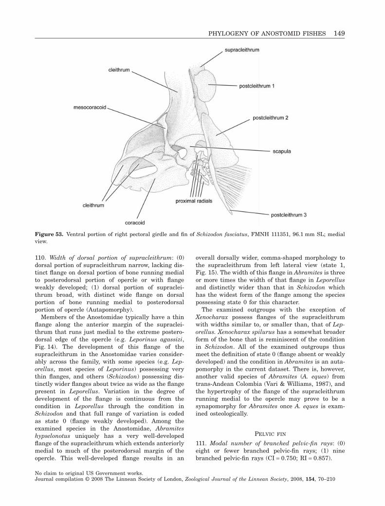
110. Width of dorsal portion of supracleithrum: (0)dorsal portion of supracleithrum narrow, lacking dis-tinct flange on dorsal portion of bone running medialto posterodorsal portion of opercle or with flangeweakly developed; (1) dorsal portion of supraclei-thrum broad, with distinct wide flange on dorsalportion of bone running medial to posterodorsalportion of opercle (Autapomorphy).
Members of the Anostomidae typically have a thinflange along the anterior margin of the supraclei-thrum that runs just medial to the extreme postero-dorsal edge of the opercle (e.g. Leporinus agassizi,Fig. 14). The development of this flange of thesupracleithrum in the Anostomidae varies consider-ably across the family, with some species (e.g. Lep-orellus, most species of Leporinus) possessing verythin flanges, and others (Schizodon) possessing dis-tinctly wider flanges about twice as wide as the flangepresent in Leporellus. Variation in the degree ofdevelopment of the flange is continuous from thecondition in Leporellus through the condition inSchizodon and that full range of variation is codedas state 0 (flange weakly developed). Among theexamined species in the Anostomidae, Abramiteshypselonotus uniquely has a very well-developedflange of the supracleithrum which extends anteriorlymedial to much of the posterodorsal margin of theopercle. This well-developed flange results in an
overall dorsally wider, comma-shaped morphology tothe supracleithrum from left lateral view (state 1,Fig. 15). The width of this flange in Abramites is threeor more times the width of that flange in Leporellusand distinctly wider than that in Schizodon whichhas the widest form of the flange among the speciespossessing state 0 for this character.
The examined outgroups with the exception ofXenocharax possess flanges of the supracleithrumwith widths similar to, or smaller than, that of Lep-orellus. Xenocharax spilurus has a somewhat broaderform of the bone that is reminiscent of the conditionin Schizodon. All of the examined outgroups thusmeet the definition of state 0 (flange absent or weaklydeveloped) and the condition in Abramites is an auta-pomorphy in the current dataset. There is, however,another valid species of Abramites (A. eques) fromtrans-Andean Colombia (Vari & Williams, 1987), andthe hypertrophy of the flange of the supracleithrumrunning medial to the opercle may prove to be asynapomorphy for Abramites once A. eques is exam-ined osteologically.
PELVIC FIN
111. Modal number of branched pelvic-fin rays: (0)eight or fewer branched pelvic-fin rays; (1) ninebranched pelvic-fin rays (CI = 0.750; RI = 0.857).
Figure 53. Ventral portion of right pectoral girdle and fin of Schizodon fasciatus, FMNH 111351, 96.1 mm SL; medialview.
PHYLOGENY OF ANOSTOMID FISHES 149
No claim to original US Government works.Journal compilation © 2008 The Linnean Society of London, Zoological Journal of the Linnean Society, 2008, 154, 70–210
A count of one unbranched and eight branchedpelvic-fin rays (state 0, nine total elements) iscommon among members of the Characiformes. Aninth branched pelvic-fin ray (state 1, ten total ele-ments) is present in a subset of the Anostomidaeconsisting of Leporinus jatuncochi, L. cf. fasciatus, L.fasciatus and L. tigrinus. Although not representedamong the cleared and stained material, examinedmaterial of Leporinus falcipinnis from the RioOrinoco (FMNH 104000), which displays a colourpattern similar to that of L. fasciatus, also possessesa ninth branched pelvic-fin ray. Among the proximateexamined outgroups, six to eight branched rays occurin Caenotropus (Vari et al., 1995) and eight in Chilo-dus (Isbrücker & Nijssen, 1988; Vari & Ortega, 1997).Turning to the second outgroup, there are seven tonine branched pelvic-fin rays in the Prochilodontidae(Castro & Vari, 2004), with basal genera in the Curi-matidae (Curimatopsis and Potamorhina) having sixor seven and eight or nine branched pelvic-fin rays,respectively (Vari, 1982b, 1984). A range of six toeight branched pelvic-fin rays is general for all othergenera in the Curimatidae with the exception of asubset of species within Curimata including C. inor-nata (Vari, 1989b) that have nine branched rays insome individuals. Potamorhina latior and Curimatainornata are therefore polymorphic for this character.Among more distant outgroups, Parodon suborbitalis(Pavanelli, 1999) and Brycon falcatus both possesseight branched pelvic-fin rays (state 0), while Hemio-dus ocellatus and examined members of the Citharin-idae and Distichodontidae possess nine branched rays(state 1).
In our final reconstruction, the plesiomorphic con-dition within the Anostomidae optimizes as the pos-session of eight or fewer branched pelvic-fin rays,with the possession of a modal count of nine branchedpelvic-fin rays a synapomorphy for the group of Lep-orinus species cited above (clade 10). The possessionof nine branched pelvic-fin rays in Potamorhinalatior, a subset of Curimata and the examinedmembers of the Citharinidae, Distichodontidae andHemiodontidae in the outgroups is clearly homoplas-tic with respect to the similar condition in somespecies of Leporinus under the present concepts ofrelationships of these taxa (Vari, 1979, 1983, 1989a, b,and results therein).
ANAL FIN
112. Modal number of branched anal-fin rays: (0)seven or fewer branched anal-fin rays; (1) eightbranched anal-fin rays; (2) nine or more branchedanal-fin rays (CI = 0.500; RI = 0.444).
Nearly every member of the Anostomidae possesseseight branched anal-fin rays, preceded by one to three
(usually two) unbranched anal-fin rays. Speciesof Abramites have higher branched anal-fin raycounts. Abramites hypselonotus possesses 10–12, andAbramites eques from the Río Magdalena of Colombiapossesses 13 or 14 branched anal-fin rays (Vari &Williams, 1987: 92). Anostomoides atrianalis report-edly possesses ten branched anal-fin rays (Santos& Zuanon, 2006: 67). Schizodon scotorhabdotusuniquely among the examined Anostomidae possessesonly seven branched anal-fin rays.
The branched anal-fin ray count varies widelyamong the outgroups. Members of the nearest out-group (family Chilodontidae) possess between sevenand ten branched-anal fin rays (Isbrücker & Nijssen,1988; Vari et al., 1995; Vari & Ortega, 1997).Members of the Curimatidae (e.g. Potamorhina latior)possess as many as 12 or as few as seven (e.g. Curi-matella alburna) rays (Vari, 1984, 1992). Distantlyrelated members of the outgroups in the Characi-formes have variable numbers of branched anal-finrays, with seven branched rays most common inParodon suborbitalis (Pavanelli, 1999), 9–10 rayspresent in Hemiodus ocellatus (Vari, 1982a), 10–13rays present in Xenocharax (Vari, 2007), 10–12 inDistichodus (Daget, 1959), 28 or more in Bryconmeeki (Weitzman, 1962) and 20–31 in the variousspecies of Citharinus (Daget, 1962b).
The conditions in Abramites hypselonotus andSchizodon scotorhabdotus are autapomorphies withinthe Anostomidae in the current matrix, but the twospecies of Abramites clearly share a derived increasedin the number of branched anal-fin rays; a featurethat will probably optimize as a synapomorphy for thegenus in a future study. The decreased number ofrays in S. scotorhabdotus may be found to be a syna-pomorphy of a small group of species in that genusonce more taxa are examined.
CAUDAL FIN
113. Form of connection of primary rays of caudal fin:(0) first four to six principal rays of upper lobe ofcaudal fin and last four to six rays of lower lobe ofcaudal fin distinctly separated from each other exceptfor membranous interconnections in adults; rays notthickened or conjoined into rigid plate-like structurein adults; (1) first four to six principal rays of upperlobe of caudal fin and last four to six rays of lower lobeof caudal fin not separated but rather with marginsin contact in adults; rays thickened and conjoinedinto rigid plate-like structure in adults (CI = 1.000;RI = 1.000).
Adults of the examined species of Schizodon andRhytiodus possess a remarkable morphology of thecaudal fin in which the dorsal four to six principalrays of the dorsal lobe of the fin and the ventral four
150 B. L. SIDLAUSKAS and R. P. VARI
No claim to original US Government works.Journal compilation © 2008 The Linnean Society of London, Zoological Journal of the Linnean Society, 2008, 154, 70–210
to six rays of the ventral lobe of the fin are thickenedand conjoined into rigid, plate-like structures (seecondition in Schizodon scotorhabdotus, Sidlauskaset al., 2007: fig. 2) This condition appears to developlate in ontogeny given that the examined cleared andstained specimens of Rhytiodus, which are distinctlysmaller than adults, have these caudal fin rays eitherseparate (R. argenteofuscus, R. microlepis) or onlypartially conjoined (R. lauzannei). The specimens ofSchizodon fasciatus, S. nasutus and S. vittatus andthe smaller specimens of S. knerii demonstrate only apartial connectivity of the rays, with complete con-nectivity present in the cleared and stained speci-mens and dry skeletons of S. scotorhabdotus and thelarger cleared and stained mature adults of S. isog-nathus and S. knerii (Sidlauskas et al., 2007). Theexamination of large-size alcohol-preserved speci-mens of all of these species of Rhytiodus and Schiz-odon leaves no doubt that thickened and tightlyconnected caudal-fin rays are present in adults of allof the examined species. No other examined species inthe Anostomidae or any outgroups possesses the dis-tinct union of the caudal-fin rays comparable withthat found in adults of Schizodon and Rhytiodus.
In his description of Schizodon jacuiensis, Berg-mann (1988) noted that S. jacuiensis lacks the tightconnection of the caudal-fin rays that is found in theother species of Schizodon. It is difficult to extractmuch information about the caudal-fin ray morphol-ogy from the photograph of the holotype of S. jacuien-sis included in that publication, but that specimen issurely an adult given its size (234.4 mm SL) and thethickening and tight connection of the caudal-fin raysappears to be either absent or much less pronouncedin S. jacuiensis contrary to the condition in its con-geners. If that is the situation, two scenarios exist.The thickening and conjoining of the caudal-fin raysmay represent a synapomorphy for Rhytiodus and foronly part of Schizodon, implying that Rhytiodus maybe derived from within Schizodon. Alternatively, thethickening and conjoining of the caudal-fin rays couldrepresent a synapomorphy for Schizodon and Rhytio-dus that was later lost or secondarily reduced inS. jacuiensis or a more encompassing lineage withinSchizodon.
It is also noteworthy that Schizodon isognathusand S. knerii display a lesser degree of thickeningof the caudal-fin rays than that occurring in someother congeners, although both species have greatlyincreased ramification of the caudal fin and demon-strate conjoined caudal-fin rays late in ontogeny (Sid-lauskas et al., 2007). The ontogenetic onset of thethickening of the caudal-fin rays into a conjoinedplate also varies within the genus, with onset rela-tively early in ontogeny in S. scotorhabdotus andcomparatively later in S. isognathus and S. knerii. A
comprehensive examination of all species of Schiz-odon may reveal that state 1 of this character sub-sumes two distinct conditions: the late onset ofeventual moderate thickening and interconnection ofthe caudal-fin rays such as the condition present inS. isognathus, and the early onset of the eventualextensive thickening and tight interconnection of thecaudal-fin rays that is typical of S. scotorhabdotus.Regardless of whether that is the case, state 1 is asynapomorphy for clade 15 in this reconstruction.
SCALES
114. Squamation of caudal fin: (0) lobes of caudal finfree of scales; (1) lobes of caudal fin covered withscales (CI = 1.000; RI = 1.000).
Among the examined members of the Anostomidae,only the species in Leporellus possess a dense cover-ing of scales over the major portion of both caudal-finlobes (state 1). In most other species in the family thelast transverse scale row overlaps the bases of thecaudal-fin rays, but the majority of the surface ofthe caudal fin is free of scales (state 0). The unscaledcondition of the lobes of the caudal fin is foundthroughout the examined outgroups. Although notcoded formally as outgroups in this study, we notethat members of Curimatella in the Curimatidaepossess scaled caudal fins (Vari, 1992: fig. 20).
A host of characters indicate that the Anostomidaeand Curimatidae are not closest relatives (Vari, 1983,1989a; and results herein) and furthermore Curima-tella is distinctly nested within the phylogeny of theCurimatidae (Vari, 1989a). The evolution of scaledlobes of the caudal fin in Leporellus and Curimatellais judged to be homoplastic, with the presence of thatfeature in Leporellus a synapomorphy for that genus(clade 2).
115. Number of lateral line scales: (0) 64 or fewerscales in complete lateral-line series, including scalesoverlapping the hypural plate; (1) 76 or more scales incomplete lateral-line series, including scales overlap-ping hypural plate (CI = 0.667; RI = 0.500).
The number of scales along the lateral line, whichis a function of the relative size of the scales, variesconsiderably among the examined members of theAnostomidae. The range extends from as few as 32scales in Leporinus gomesi to as many as 92 scales inRhytiodus microlepis (Géry, 1987: 372). Much of thisvariation is impossible to parse into discrete charac-ter states, but within the Anostomidae there is a largebreak between the 76 and 89 lateral-line scales foundin Rhytiodus lauzannei (state 1; Géry, 1987: 372) andthe next most numerous lateral-line scale countswithin the family [48–56 scales in Rhytiodus argen-teofuscus (Géry, 1987: 372) and 49–58 in Laemolyta
PHYLOGENY OF ANOSTOMID FISHES 151
No claim to original US Government works.Journal compilation © 2008 The Linnean Society of London, Zoological Journal of the Linnean Society, 2008, 154, 70–210
fernandezi (Mautari & Menezes, 2006: 32)]. Exami-nation of the proximate outgroups reveals that allspecies (with the exception of species in the curimatidgenus Potamorhina) possess 64 or fewer scales in thelateral line (state 0). Potamorhina, by contrast,possess 85–110 lateral-line scales, but that genus isnested within the Curimatidae (Vari, 1989a), withthat family, in turn, most closely related to theProchilodontidae (Vari, 1983, 1989a). Parodon subor-bitalis has a lateral-line series composed of relativelylarge and therefore fewer scales (state 0) while Xeno-charax spilurus and Hemiodus ocellatus have muchsmaller and numerous scales [state 1; 63–75 in Xeno-charax spilurus (Vari, 2007); 88–96 in Hemiodus ocel-latus (Vari, 1982a)]. Species of Distichodus andCitharinus vary greatly in lateral-line scale counts(Daget, 1959, 1962b), but we cannot determine theoriginal scale counts of Distichodus sp. and Cithari-nus sp. from the cleared and stained material athand, and the remainders of the lots from which thosespecimens were prepared were unavailable. Disticho-dus sp. and Citharinus sp. were therefore coded aspossessing missing data for this character and forpigmentation characters 116–123 below.
Under the reconstruction in this study Rhytiodus isdeeply nested within the Anostomidae, and thatfamily, in turn, is most closely related to the Chilo-dontidae (Vari, 1983). The increase in the number oflateral-line scales is judged to be a synapomorphy forR. lauzannei and R. microlepis (clade 17), and thesmall scales characteristic of Potamorhina, Hemiodusand Xenocharax are homoplastic with respect to thesmall scales in the two cited species of Rhytiodus.
PIGMENTATION
116. Vertical bands encircling body: (0) body withoutvertical bands of at least two scales width completelyencircling body; (1) body with vertical bands of atleast two scales width, at least some of which com-pletely encircle body (CI = 0.375; RI = 0.600).
Several species in the Anostomidae possess a seriesof wide transverse bands encircling their bodies,although with those bands becoming much fainter onthe abdominal regions. Such examined banded speciesinclude Abramites hypselonotus (Fig. 2), Leporinusjatuncochi, L. cf. fasciatus, L. fasciatus, L. tigrinus andSynaptolaemus cingulatus (Fig. 3). Petulanos plicatuspossesses very thin, irregular, vertical bands that areonly one scale wide (Winterbottom, 1980: fig. 15), apigmentation pattern that is notably different from,and presumably non-homologous with, the pattern ofwide bands seen in the six species cited above. Petul-anos plicatus is consequently not included in state 1.Leporinus pellegrinii possesses a series of alternatingthick and thin dark vertical bands (see character 117),
but these bands are incomplete ventrally and, thus,assigned to state 0. Colour pattern varies considerablyamong the examined outgroups but none possessescomplete transverse bands encircling the body.Alcohol-preserved material was unavailable for Cith-arinus sp. and Distichodus sp., and those two taxawere coded as possessing missing data for characters116–123, all dealing with pigmentation.
Our final phylogenetic reconstruction suggeststhat the banded pattern evolved independently inAbramites, Synaptolaemus and in clade 10 withinLeporinus, consisting of L. jatuncochi, L. cf. fasciatus,L. fasciatus and L. tigrinus. Although skeletal mate-rial was not available for Leporinus falcipinnis, thattaxon possesses the banded colour pattern and extrabranched pelvic-fin ray (see character 111) that opti-mize as synapomorphies for clade 10. Consequently,it is likely that L. falcipinnis is also a member ofthat clade.
117. Possession and form of dark vertical blotches onlateral surface of body: (0) lateral surface of bodywithout dark vertical blotches; (1) lateral surface ofbody with two to five dark, vertical blotches intersect-ing the lateral-line scale row and continuing dorsaland ventral to the lateral-line scale row (CI = 0.250;RI = 0.700).
Several species in the Anostomidae possess a seriesof two to five (usually three or four) dark verticalblotches along the lateral surface of body (e.g. Schiz-odon scotorhabdotus, Sidlauskas et al., 2007: fig. 1).In general, the first blotch is located immediatelyposterior to the opercle, the second ventral to thedorsal fin, the third midway between the bases of thedorsal and adipose fins, and the fourth above the analfin. This blotched colour pattern differs from thebanding pattern described in character 116 in thatthe dark coloration of the blotches fades out bothdorsally and ventrally and does not encircle the body.The dark coloration, furthermore, does not have theform of pigmentation patches with well-definedmargins as is the case in the bands in character 116,state 1. The blotched colour pattern differs from thepattern of lateral-line spots described in character118 in that the blotches extend significantly aboveand below the lateral-line scale row and frequentlyhave indistinct margins, whereas the spots in state 1of character 118 are mostly confined to the horizontalrow of scales containing the lateral line and havewell-defined margins. The blotched pattern is particu-larly noticeable in some species of Schizodon (e.g. S.fasciatus, S. vittatus and S. scotorhabdotus) but alsoappears, albeit indistinctly, in Anostomoides laticeps,Laemolyta fernandezi (Mautari & Menezes, 2006: figs6, 8, 9), L. orinocensis, L. proxima, Schizodon knerii(Sidlauskas et al., 2007: fig. 6B) and the species of
152 B. L. SIDLAUSKAS and R. P. VARI
No claim to original US Government works.Journal compilation © 2008 The Linnean Society of London, Zoological Journal of the Linnean Society, 2008, 154, 70–210
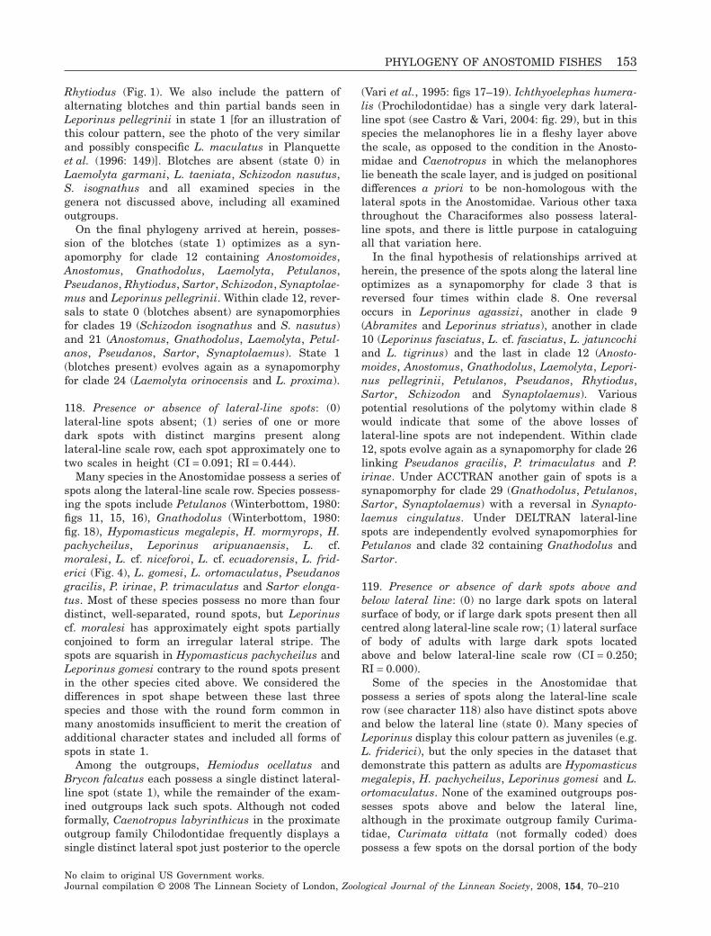
Rhytiodus (Fig. 1). We also include the pattern ofalternating blotches and thin partial bands seen inLeporinus pellegrinii in state 1 [for an illustration ofthis colour pattern, see the photo of the very similarand possibly conspecific L. maculatus in Planquetteet al. (1996: 149)]. Blotches are absent (state 0) inLaemolyta garmani, L. taeniata, Schizodon nasutus,S. isognathus and all examined species in thegenera not discussed above, including all examinedoutgroups.
On the final phylogeny arrived at herein, posses-sion of the blotches (state 1) optimizes as a syn-apomorphy for clade 12 containing Anostomoides,Anostomus, Gnathodolus, Laemolyta, Petulanos,Pseudanos, Rhytiodus, Sartor, Schizodon, Synaptolae-mus and Leporinus pellegrinii. Within clade 12, rever-sals to state 0 (blotches absent) are synapomorphiesfor clades 19 (Schizodon isognathus and S. nasutus)and 21 (Anostomus, Gnathodolus, Laemolyta, Petul-anos, Pseudanos, Sartor, Synaptolaemus). State 1(blotches present) evolves again as a synapomorphyfor clade 24 (Laemolyta orinocensis and L. proxima).
118. Presence or absence of lateral-line spots: (0)lateral-line spots absent; (1) series of one or moredark spots with distinct margins present alonglateral-line scale row, each spot approximately one totwo scales in height (CI = 0.091; RI = 0.444).
Many species in the Anostomidae possess a series ofspots along the lateral-line scale row. Species possess-ing the spots include Petulanos (Winterbottom, 1980:figs 11, 15, 16), Gnathodolus (Winterbottom, 1980:fig. 18), Hypomasticus megalepis, H. mormyrops, H.pachycheilus, Leporinus aripuanaensis, L. cf.moralesi, L. cf. niceforoi, L. cf. ecuadorensis, L. frid-erici (Fig. 4), L. gomesi, L. ortomaculatus, Pseudanosgracilis, P. irinae, P. trimaculatus and Sartor elonga-tus. Most of these species possess no more than fourdistinct, well-separated, round spots, but Leporinuscf. moralesi has approximately eight spots partiallyconjoined to form an irregular lateral stripe. Thespots are squarish in Hypomasticus pachycheilus andLeporinus gomesi contrary to the round spots presentin the other species cited above. We considered thedifferences in spot shape between these last threespecies and those with the round form common inmany anostomids insufficient to merit the creation ofadditional character states and included all forms ofspots in state 1.
Among the outgroups, Hemiodus ocellatus andBrycon falcatus each possess a single distinct lateral-line spot (state 1), while the remainder of the exam-ined outgroups lack such spots. Although not codedformally, Caenotropus labyrinthicus in the proximateoutgroup family Chilodontidae frequently displays asingle distinct lateral spot just posterior to the opercle
(Vari et al., 1995: figs 17–19). Ichthyoelephas humera-lis (Prochilodontidae) has a single very dark lateral-line spot (see Castro & Vari, 2004: fig. 29), but in thisspecies the melanophores lie in a fleshy layer abovethe scale, as opposed to the condition in the Anosto-midae and Caenotropus in which the melanophoreslie beneath the scale layer, and is judged on positionaldifferences a priori to be non-homologous with thelateral spots in the Anostomidae. Various other taxathroughout the Characiformes also possess lateral-line spots, and there is little purpose in cataloguingall that variation here.
In the final hypothesis of relationships arrived atherein, the presence of the spots along the lateral lineoptimizes as a synapomorphy for clade 3 that isreversed four times within clade 8. One reversaloccurs in Leporinus agassizi, another in clade 9(Abramites and Leporinus striatus), another in clade10 (Leporinus fasciatus, L. cf. fasciatus, L. jatuncochiand L. tigrinus) and the last in clade 12 (Anosto-moides, Anostomus, Gnathodolus, Laemolyta, Lepori-nus pellegrinii, Petulanos, Pseudanos, Rhytiodus,Sartor, Schizodon and Synaptolaemus). Variouspotential resolutions of the polytomy within clade 8would indicate that some of the above losses oflateral-line spots are not independent. Within clade12, spots evolve again as a synapomorphy for clade 26linking Pseudanos gracilis, P. trimaculatus and P.irinae. Under ACCTRAN another gain of spots is asynapomorphy for clade 29 (Gnathodolus, Petulanos,Sartor, Synaptolaemus) with a reversal in Synapto-laemus cingulatus. Under DELTRAN lateral-linespots are independently evolved synapomorphies forPetulanos and clade 32 containing Gnathodolus andSartor.
119. Presence or absence of dark spots above andbelow lateral line: (0) no large dark spots on lateralsurface of body, or if large dark spots present then allcentred along lateral-line scale row; (1) lateral surfaceof body of adults with large dark spots locatedabove and below lateral-line scale row (CI = 0.250;RI = 0.000).
Some of the species in the Anostomidae thatpossess a series of spots along the lateral-line scalerow (see character 118) also have distinct spots aboveand below the lateral line (state 0). Many species ofLeporinus display this colour pattern as juveniles (e.g.L. friderici), but the only species in the dataset thatdemonstrate this pattern as adults are Hypomasticusmegalepis, H. pachycheilus, Leporinus gomesi and L.ortomaculatus. None of the examined outgroups pos-sesses spots above and below the lateral line,although in the proximate outgroup family Curima-tidae, Curimata vittata (not formally coded) doespossess a few spots on the dorsal portion of the body
PHYLOGENY OF ANOSTOMID FISHES 153
No claim to original US Government works.Journal compilation © 2008 The Linnean Society of London, Zoological Journal of the Linnean Society, 2008, 154, 70–210
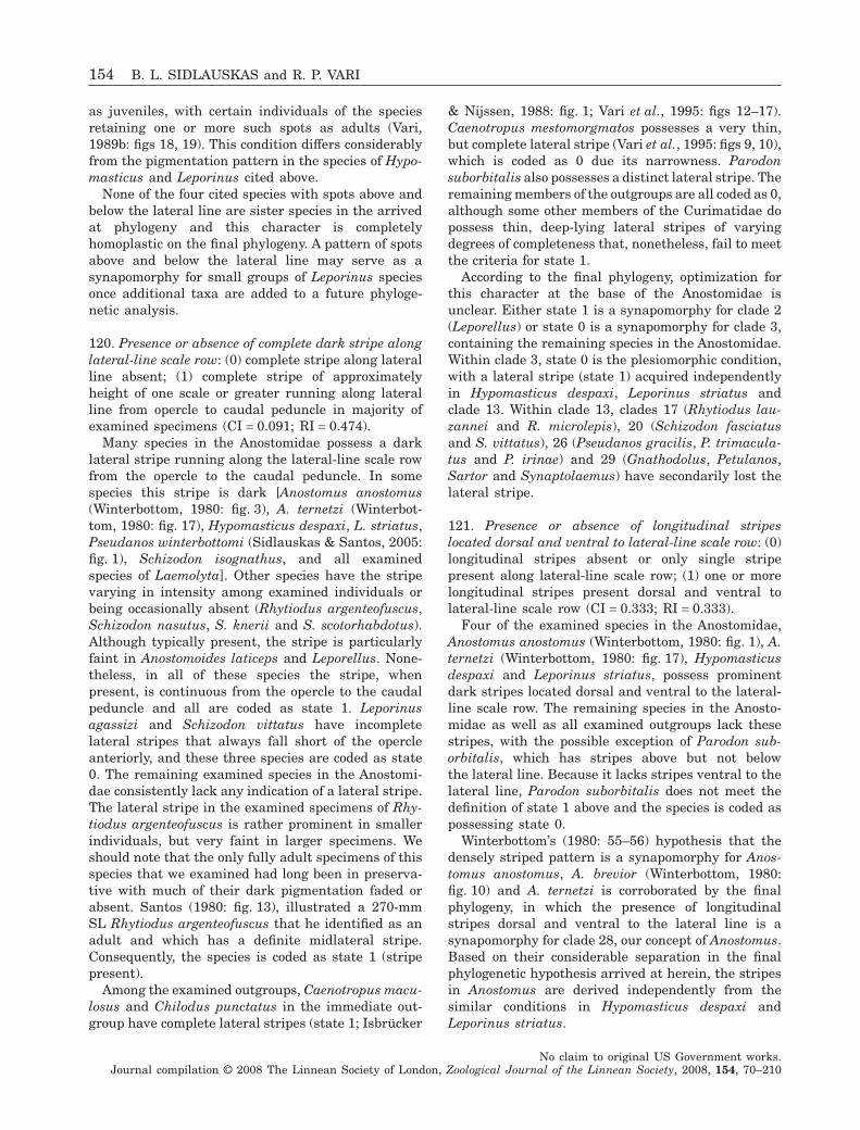
as juveniles, with certain individuals of the speciesretaining one or more such spots as adults (Vari,1989b: figs 18, 19). This condition differs considerablyfrom the pigmentation pattern in the species of Hypo-masticus and Leporinus cited above.
None of the four cited species with spots above andbelow the lateral line are sister species in the arrivedat phylogeny and this character is completelyhomoplastic on the final phylogeny. A pattern of spotsabove and below the lateral line may serve as asynapomorphy for small groups of Leporinus speciesonce additional taxa are added to a future phyloge-netic analysis.
120. Presence or absence of complete dark stripe alonglateral-line scale row: (0) complete stripe along lateralline absent; (1) complete stripe of approximatelyheight of one scale or greater running along lateralline from opercle to caudal peduncle in majority ofexamined specimens (CI = 0.091; RI = 0.474).
Many species in the Anostomidae possess a darklateral stripe running along the lateral-line scale rowfrom the opercle to the caudal peduncle. In somespecies this stripe is dark [Anostomus anostomus(Winterbottom, 1980: fig. 3), A. ternetzi (Winterbot-tom, 1980: fig. 17), Hypomasticus despaxi, L. striatus,Pseudanos winterbottomi (Sidlauskas & Santos, 2005:fig. 1), Schizodon isognathus, and all examinedspecies of Laemolyta]. Other species have the stripevarying in intensity among examined individuals orbeing occasionally absent (Rhytiodus argenteofuscus,Schizodon nasutus, S. knerii and S. scotorhabdotus).Although typically present, the stripe is particularlyfaint in Anostomoides laticeps and Leporellus. None-theless, in all of these species the stripe, whenpresent, is continuous from the opercle to the caudalpeduncle and all are coded as state 1. Leporinusagassizi and Schizodon vittatus have incompletelateral stripes that always fall short of the opercleanteriorly, and these three species are coded as state0. The remaining examined species in the Anostomi-dae consistently lack any indication of a lateral stripe.The lateral stripe in the examined specimens of Rhy-tiodus argenteofuscus is rather prominent in smallerindividuals, but very faint in larger specimens. Weshould note that the only fully adult specimens of thisspecies that we examined had long been in preserva-tive with much of their dark pigmentation faded orabsent. Santos (1980: fig. 13), illustrated a 270-mmSL Rhytiodus argenteofuscus that he identified as anadult and which has a definite midlateral stripe.Consequently, the species is coded as state 1 (stripepresent).
Among the examined outgroups, Caenotropus macu-losus and Chilodus punctatus in the immediate out-group have complete lateral stripes (state 1; Isbrücker
& Nijssen, 1988: fig. 1; Vari et al., 1995: figs 12–17).Caenotropus mestomorgmatos possesses a very thin,but complete lateral stripe (Vari et al., 1995: figs 9, 10),which is coded as 0 due its narrowness. Parodonsuborbitalis also possesses a distinct lateral stripe. Theremaining members of the outgroups are all coded as 0,although some other members of the Curimatidae dopossess thin, deep-lying lateral stripes of varyingdegrees of completeness that, nonetheless, fail to meetthe criteria for state 1.
According to the final phylogeny, optimization forthis character at the base of the Anostomidae isunclear. Either state 1 is a synapomorphy for clade 2(Leporellus) or state 0 is a synapomorphy for clade 3,containing the remaining species in the Anostomidae.Within clade 3, state 0 is the plesiomorphic condition,with a lateral stripe (state 1) acquired independentlyin Hypomasticus despaxi, Leporinus striatus andclade 13. Within clade 13, clades 17 (Rhytiodus lau-zannei and R. microlepis), 20 (Schizodon fasciatusand S. vittatus), 26 (Pseudanos gracilis, P. trimacula-tus and P. irinae) and 29 (Gnathodolus, Petulanos,Sartor and Synaptolaemus) have secondarily lost thelateral stripe.
121. Presence or absence of longitudinal stripeslocated dorsal and ventral to lateral-line scale row: (0)longitudinal stripes absent or only single stripepresent along lateral-line scale row; (1) one or morelongitudinal stripes present dorsal and ventral tolateral-line scale row (CI = 0.333; RI = 0.333).
Four of the examined species in the Anostomidae,Anostomus anostomus (Winterbottom, 1980: fig. 1), A.ternetzi (Winterbottom, 1980: fig. 17), Hypomasticusdespaxi and Leporinus striatus, possess prominentdark stripes located dorsal and ventral to the lateral-line scale row. The remaining species in the Anosto-midae as well as all examined outgroups lack thesestripes, with the possible exception of Parodon sub-orbitalis, which has stripes above but not belowthe lateral line. Because it lacks stripes ventral to thelateral line, Parodon suborbitalis does not meet thedefinition of state 1 above and the species is coded aspossessing state 0.
Winterbottom’s (1980: 55–56) hypothesis that thedensely striped pattern is a synapomorphy for Anos-tomus anostomus, A. brevior (Winterbottom, 1980:fig. 10) and A. ternetzi is corroborated by the finalphylogeny, in which the presence of longitudinalstripes dorsal and ventral to the lateral line is asynapomorphy for clade 28, our concept of Anostomus.Based on their considerable separation in the finalphylogenetic hypothesis arrived at herein, the stripesin Anostomus are derived independently from thesimilar conditions in Hypomasticus despaxi andLeporinus striatus.
154 B. L. SIDLAUSKAS and R. P. VARI
No claim to original US Government works.Journal compilation © 2008 The Linnean Society of London, Zoological Journal of the Linnean Society, 2008, 154, 70–210
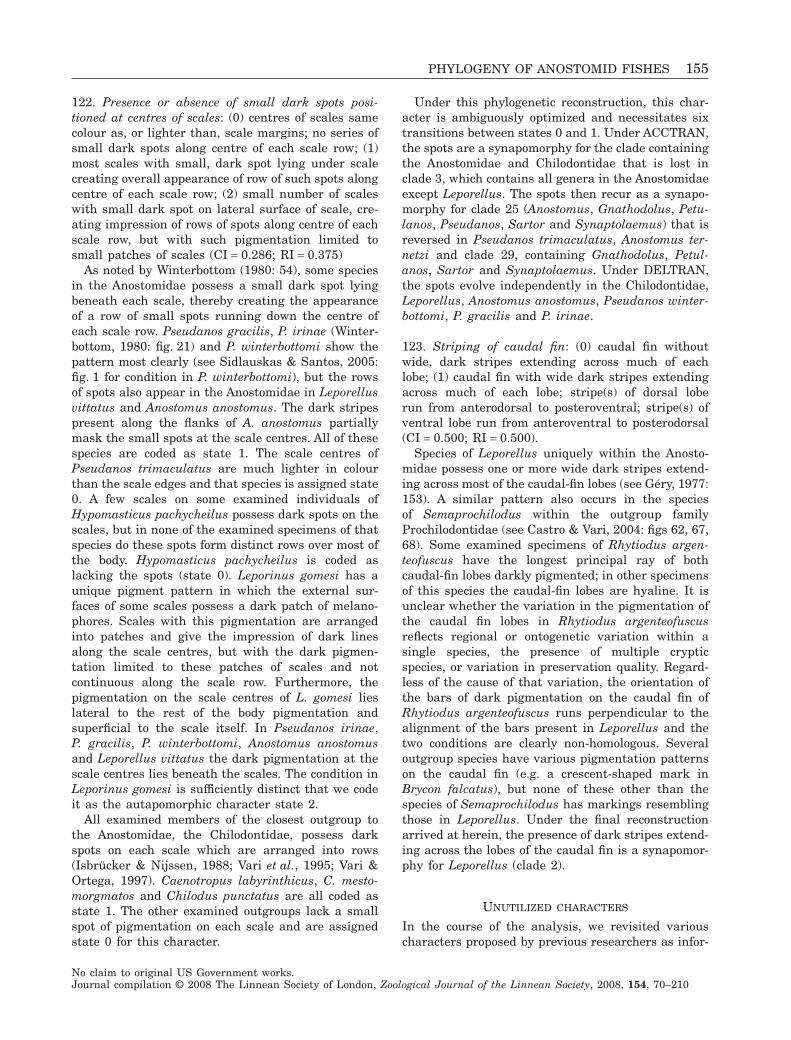
122. Presence or absence of small dark spots posi-tioned at centres of scales: (0) centres of scales samecolour as, or lighter than, scale margins; no series ofsmall dark spots along centre of each scale row; (1)most scales with small, dark spot lying under scalecreating overall appearance of row of such spots alongcentre of each scale row; (2) small number of scaleswith small dark spot on lateral surface of scale, cre-ating impression of rows of spots along centre of eachscale row, but with such pigmentation limited tosmall patches of scales (CI = 0.286; RI = 0.375)
As noted by Winterbottom (1980: 54), some speciesin the Anostomidae possess a small dark spot lyingbeneath each scale, thereby creating the appearanceof a row of small spots running down the centre ofeach scale row. Pseudanos gracilis, P. irinae (Winter-bottom, 1980: fig. 21) and P. winterbottomi show thepattern most clearly (see Sidlauskas & Santos, 2005:fig. 1 for condition in P. winterbottomi), but the rowsof spots also appear in the Anostomidae in Leporellusvittatus and Anostomus anostomus. The dark stripespresent along the flanks of A. anostomus partiallymask the small spots at the scale centres. All of thesespecies are coded as state 1. The scale centres ofPseudanos trimaculatus are much lighter in colourthan the scale edges and that species is assigned state0. A few scales on some examined individuals ofHypomasticus pachycheilus possess dark spots on thescales, but in none of the examined specimens of thatspecies do these spots form distinct rows over most ofthe body. Hypomasticus pachycheilus is coded aslacking the spots (state 0). Leporinus gomesi has aunique pigment pattern in which the external sur-faces of some scales possess a dark patch of melano-phores. Scales with this pigmentation are arrangedinto patches and give the impression of dark linesalong the scale centres, but with the dark pigmen-tation limited to these patches of scales and notcontinuous along the scale row. Furthermore, thepigmentation on the scale centres of L. gomesi lieslateral to the rest of the body pigmentation andsuperficial to the scale itself. In Pseudanos irinae,P. gracilis, P. winterbottomi, Anostomus anostomusand Leporellus vittatus the dark pigmentation at thescale centres lies beneath the scales. The condition inLeporinus gomesi is sufficiently distinct that we codeit as the autapomorphic character state 2.
All examined members of the closest outgroup tothe Anostomidae, the Chilodontidae, possess darkspots on each scale which are arranged into rows(Isbrücker & Nijssen, 1988; Vari et al., 1995; Vari &Ortega, 1997). Caenotropus labyrinthicus, C. mesto-morgmatos and Chilodus punctatus are all coded asstate 1. The other examined outgroups lack a smallspot of pigmentation on each scale and are assignedstate 0 for this character.
Under this phylogenetic reconstruction, this char-acter is ambiguously optimized and necessitates sixtransitions between states 0 and 1. Under ACCTRAN,the spots are a synapomorphy for the clade containingthe Anostomidae and Chilodontidae that is lost inclade 3, which contains all genera in the Anostomidaeexcept Leporellus. The spots then recur as a synapo-morphy for clade 25 (Anostomus, Gnathodolus, Petu-lanos, Pseudanos, Sartor and Synaptolaemus) that isreversed in Pseudanos trimaculatus, Anostomus ter-netzi and clade 29, containing Gnathodolus, Petul-anos, Sartor and Synaptolaemus. Under DELTRAN,the spots evolve independently in the Chilodontidae,Leporellus, Anostomus anostomus, Pseudanos winter-bottomi, P. gracilis and P. irinae.
123. Striping of caudal fin: (0) caudal fin withoutwide, dark stripes extending across much of eachlobe; (1) caudal fin with wide dark stripes extendingacross much of each lobe; stripe(s) of dorsal loberun from anterodorsal to posteroventral; stripe(s) ofventral lobe run from anteroventral to posterodorsal(CI = 0.500; RI = 0.500).
Species of Leporellus uniquely within the Anosto-midae possess one or more wide dark stripes extend-ing across most of the caudal-fin lobes (see Géry, 1977:153). A similar pattern also occurs in the speciesof Semaprochilodus within the outgroup familyProchilodontidae (see Castro & Vari, 2004: figs 62, 67,68). Some examined specimens of Rhytiodus argen-teofuscus have the longest principal ray of bothcaudal-fin lobes darkly pigmented; in other specimensof this species the caudal-fin lobes are hyaline. It isunclear whether the variation in the pigmentation ofthe caudal fin lobes in Rhytiodus argenteofuscusreflects regional or ontogenetic variation within asingle species, the presence of multiple crypticspecies, or variation in preservation quality. Regard-less of the cause of that variation, the orientation ofthe bars of dark pigmentation on the caudal fin ofRhytiodus argenteofuscus runs perpendicular to thealignment of the bars present in Leporellus and thetwo conditions are clearly non-homologous. Severaloutgroup species have various pigmentation patternson the caudal fin (e.g. a crescent-shaped mark inBrycon falcatus), but none of these other than thespecies of Semaprochilodus has markings resemblingthose in Leporellus. Under the final reconstructionarrived at herein, the presence of dark stripes extend-ing across the lobes of the caudal fin is a synapomor-phy for Leporellus (clade 2).
UNUTILIZED CHARACTERS
In the course of the analysis, we revisited variouscharacters proposed by previous researchers as infor-
PHYLOGENY OF ANOSTOMID FISHES 155
No claim to original US Government works.Journal compilation © 2008 The Linnean Society of London, Zoological Journal of the Linnean Society, 2008, 154, 70–210
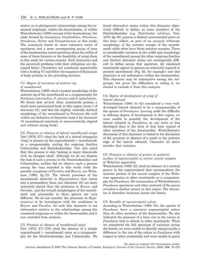
mative as to phylogenetic relationships among the ex-amined outgroups, within the Anostomidae, or withinWinterbottom’s (1980) concept of the Anostominae, theclade formed by Anostomus, Gnathodolus, Petulanos,Pseudanos, Sartor and Synaptolaemus in this study.The reanalysis based on more extensive series ofspecimens and a more encompassing group of taxaof the Anostomidae raised questions about the utility ofsome of these features or the feasibility of using themin this study for various reasons. Such characters andthe perceived problems with their utilization are dis-cussed below. Characters are numbered sequentiallywith a leading ‘U’ and follow the sequence of discussionof body systems in the preceding sections.
U1. Degree of curvature of anterior tipof mesethmoidWinterbottom (1980) cited a hooked morphology of theanterior tip of the mesethmoid as a synapomorphy forPetulanos intermedius, P. plicatus and P. spiloclistron.We found that several other anostomids possess amuch more pronounced hook in this region (state 1 ofcharacter 15), and that the slightly ventrally inclinedmorphology of the mesethmoid in Petulanos fits wellwithin our definition of character state 0 for character15 (mesethmoid anteriorly or anteroventrally alignedand without strong hook).
U2. Presence or absence of lateral mesethmoid wingsVari (1979: 277) cited the lack of a lateral triangular‘wing’ or process on the supraethmoid (= mesethmoid)as a synapomorphy uniting the outgroup familiesCitharinidae and Distichodontidae. Vari also notedthat this process is also ‘lacking in many characoidswith an elongate skull’. Although we do not disputethe lack of such a process in the Distichodontidae andCitharinidae, neither did we observe such a processamong the taxa included in this study [with thepossible exceptions of Parodon and Brycon, see Weitz-man (1962: fig. 2)]. The lateral processes of themesethmoid observed in Hypomasticus that insertinto a premaxillary fossa (see character 16) are moreanteriorly placed than the processes in Brycon andParodon, and the overall morphologies of the meseth-moid and premaxilla in these genera are verydifferent. We do not consider the processes in Hypo-masticus to be homologous with the conditions inBrycon and Parodon. As such this character is notinformative relative to the relationships among theexamined outgroups or within the Anostomidae and itwas excluded from analysis.
U3. Presence or absence of mesethmoid spineVari (1979: 277–278) cited the absence of a simplesupraethmoid (= mesethmoid) spine as a synapomor-phy for the Distichodontidae and Citharinidae. We
found alternative states within this character objec-tively difficult to define, as some members of theDistichodontidae (e.g. Distichodus niloticus, Vari,1979: fig. 6C) possess a distinct anteromedial point onthis bone (albeit, as part of an unusual trifurcatemorphology of the anterior margin of the meseth-moid) while other have blunt anterior margins. Thereis considerable variation in the width and morphologyof the mesethmoid among the other outgroup familiesand distinct character states are consequently diffi-cult to define across that spectrum. All examinedanostomids appear to possess an anteriorly tapered orpointed mesethmoid (Fig. 28, for example) and thischaracter is not informative within the Anostomidae.This character may be informative among the out-groups, but given the difficulties in coding it, weelected to exclude it from this analysis.
U4. Degree of development of wing oflateral ethmoidWinterbottom (1980: 51–52) considered a ‘very well-developed lateral ethmoid’ to be a synapomorphy ofthe species of Pseudanos. Leaving aside the difficultyin defining degree of development in this region, wewere unable to quantify the development of thelateral ethmoid in Pseudanos as any more or lessdeveloped than is the form of that bone typical ofother members of the Anostomidae. Winterbottom’sdiscussion of this character is linked to his discussionof the presence or absence of a notch in the anterioredge of the lateral ethmoid. Character 25 aboveencodes that variation.
U5. Presence or absence of groove in posteriorsurface of supraoccipital to receive neural complexof Weberian apparatusWinterbottom (1980: 52) cited an absence of a verticalgroove in the supraoccipital that accommodates theanterior portion of the neural complex of the Webe-rian apparatus in other anostomids as a synapomor-phy for Pseudanos. Re-examination of Winterbottom’sPseudanos specimens and other material of the genusrevealed a shallow groove in this region. The charac-ter is therefore invariant across the family.
U6. Breadth of supraoccipital sulcusAccording to Winterbottom (1980: 52), the species ofPseudanos have a narrower supraoccipital sulcusthan do other members of the Anostomidae. He alsoindicated the presence of a bony rim to the sulcus inPseudanos that is absent in other anostomids. Whenwe considered the full spectrum of variation acrossthe family, we were unable to identify unequivocally adifference in the rim of the sulcus in Pseudanos withrespect to other anostomids and were unable to parse
156 B. L. SIDLAUSKAS and R. P. VARI
No claim to original US Government works.Journal compilation © 2008 The Linnean Society of London, Zoological Journal of the Linnean Society, 2008, 154, 70–210
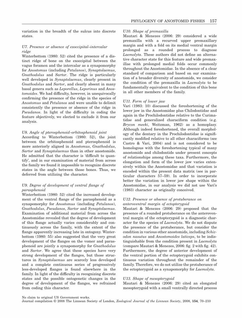
variation in the breadth of the sulcus into discretestates.
U7. Presence or absence of exoccipital–intercalarridgeWinterbottom (1980: 52) cited the presence of a dis-tinct ridge of bone on the exoccipital between thevagus foramen and the intercalar as a synapomorphyfor Anostomus (including Petulanos), Synaptolaemus,Gnathodolus and Sartor. The ridge is particularlywell developed in Synaptolaemus, clearly present inGnathodolus and Sartor, and clearly absent in manybasal genera such as Leporellus, Leporinus and Anos-tomoides. We had difficulty, however, in unequivocallyconfirming the presence of the ridge in the species ofAnostomus and Petulanos and were unable to delimitconsistently the presence or absence of the ridge inPseudanos. In light of the difficulty in coding thefeature objectively, we elected to exclude it from ouranalysis.
U8. Angle of pterosphenoid–orbitosphenoid jointAccording to Winterbottom (1980: 52), the jointbetween the orbitosphenoid and pterosphenoid ismore anteriorly aligned in Anostomus, Gnathodolus,Sartor and Synaptolaemus than in other anostomids.He admitted that the character is ‘difficult to quan-tify’, and in our examination of material from acrossthe family we found it impossible to recognize discretestates in the angle between those bones. Thus, wedeferred from utilizing the character.
U9. Degree of development of ventral flange ofparasphenoidWinterbottom (1980: 52) cited the increased develop-ment of the ventral flange of the parasphenoid as asynapomorphy for Anostomus (including Petulanos),Gnathodolus, Pseudanos, Sartor and Synaptolaemus.Examination of additional material from across theAnostomidae revealed that the degree of developmentof this flange actually varies considerably and con-tinuously across the family, with the extent of theflange apparently increasing late in ontogeny. Winter-bottom (1980: 55) also suggested that the very greatdevelopment of the flanges on the vomer and paras-phenoid are jointly a synapomorphy for Gnathodolusand Sartor. We agree that these species have verystrong development of the flanges, but those struc-tures in Synaptolaemus are scarcely less developedand a complete continuous series of progressivelyless-developed flanges is found elsewhere in thefamily. In light of the difficulty in recognizing discretestates and the possible ontogenetic changes in thedegree of development of the flanges, we refrainedfrom coding this character.
U10. Shape of premaxillaMautari & Menezes (2006: 29) considered a widepremaxilla with a recurved upper premaxillarymargin and with a fold on its medial ventral marginprolonged as a rounded process to diagnoseLaemolyta. These authors did not define an alterna-tive character state for this feature and wide premax-illae with prolonged medial folds occur commonlythroughout the Anostomidae. In the absence of a clearstandard of comparison and based on our examina-tion of a broader diversity of anostomids, we considerthe condition of the premaxilla in Laemolyta to befundamentally equivalent to the condition of this bonein all other members of the family.
U11. Form of lower jawVari (1983: 10) discussed the foreshortening of thelower jaw in the Anostomidae plus Chilodontidae andagain in the Prochilodontidae relative to the Curima-tidae and generalized characiform condition (e.g.Brycon meeki, Weitzman, 1962) as a homoplasy.Although indeed foreshortened, the overall morphol-ogy of the dentary in the Prochilodontidae is signifi-cantly modified relative to all other characiforms (seeCastro & Vari, 2004) and is not considered to behomologous with the foreshortening typical of manyanostomids and chilodontids under present conceptsof relationships among these taxa. Furthermore, theelongation and form of the lower jaw varies exten-sively within the Anostomidae and that variation isencoded within the present data matrix (see in par-ticular characters 57–59). In order to incorporatebetter the variation in lower jaw shape within theAnostomidae, in our analysis we did not use Vari’s(1983) character as originally conceived.
U12. Presence or absence of protuberance onanteroventral margin of ectopterygoidMautari & Menezes (2006: 29) proposed that thepresence of a rounded protuberance on the anteroven-tral margin of the ectopterygoid is a diagnostic char-acter for the species of Laemolyta. We do not disputethe presence of the protuberance, but consider thecondition in various other anostomids, including Schiz-odon nasutus and Anostomoides laticeps, to be indis-tinguishable from the condition present in Laemolyta(compare Mautari & Menezes, 2006: fig. 2 with fig. 42).Furthermore, the degree of anterior development ofthe ventral portion of the ectopterygoid exhibits con-tinuous variation throughout the remainder of thefamily. Therefore, we do not utilize the protuberance ofthe ectopterygoid as a synapomorphy for Laemolyta.
U13. Shape of mesopterygoidMautari & Menezes (2006: 29) cited an elongatedmesopterygoid with a small ventrally directed process
PHYLOGENY OF ANOSTOMID FISHES 157
No claim to original US Government works.Journal compilation © 2008 The Linnean Society of London, Zoological Journal of the Linnean Society, 2008, 154, 70–210
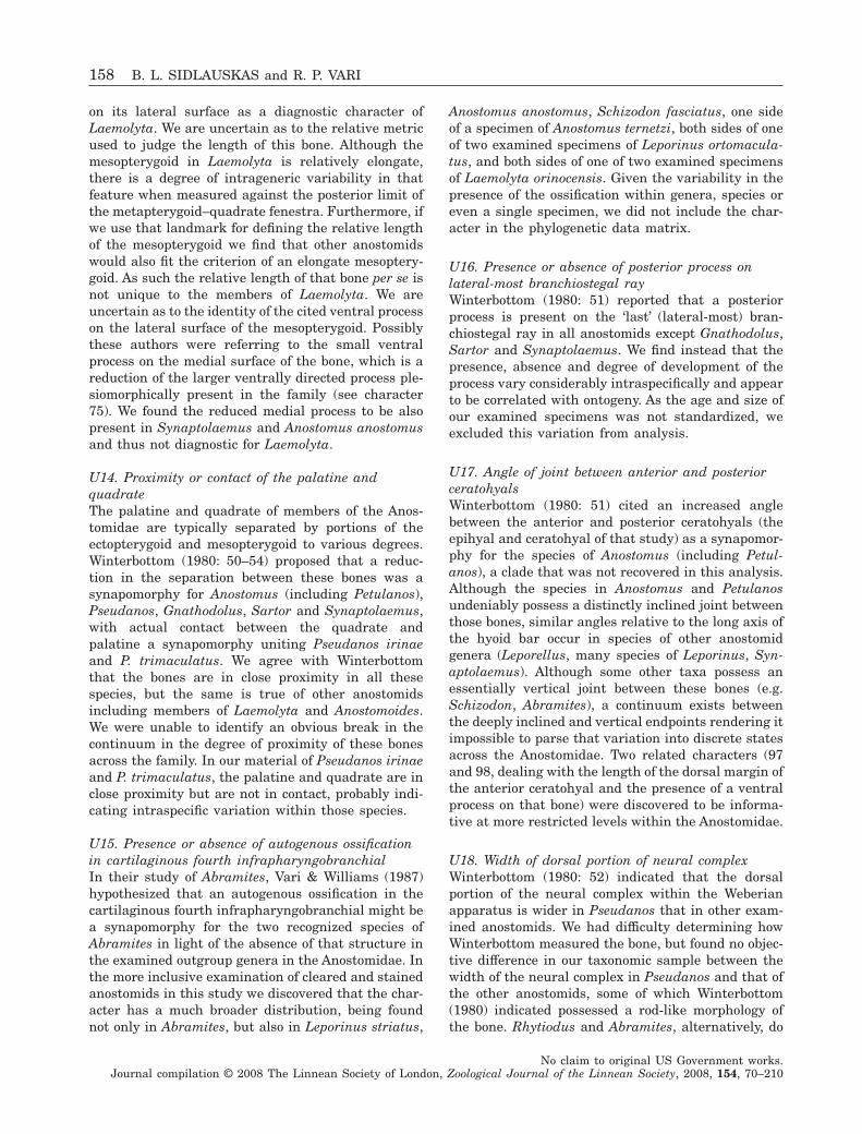
on its lateral surface as a diagnostic character ofLaemolyta. We are uncertain as to the relative metricused to judge the length of this bone. Although themesopterygoid in Laemolyta is relatively elongate,there is a degree of intrageneric variability in thatfeature when measured against the posterior limit ofthe metapterygoid–quadrate fenestra. Furthermore, ifwe use that landmark for defining the relative lengthof the mesopterygoid we find that other anostomidswould also fit the criterion of an elongate mesoptery-goid. As such the relative length of that bone per se isnot unique to the members of Laemolyta. We areuncertain as to the identity of the cited ventral processon the lateral surface of the mesopterygoid. Possiblythese authors were referring to the small ventralprocess on the medial surface of the bone, which is areduction of the larger ventrally directed process ple-siomorphically present in the family (see character75). We found the reduced medial process to be alsopresent in Synaptolaemus and Anostomus anostomusand thus not diagnostic for Laemolyta.
U14. Proximity or contact of the palatine andquadrateThe palatine and quadrate of members of the Anos-tomidae are typically separated by portions of theectopterygoid and mesopterygoid to various degrees.Winterbottom (1980: 50–54) proposed that a reduc-tion in the separation between these bones was asynapomorphy for Anostomus (including Petulanos),Pseudanos, Gnathodolus, Sartor and Synaptolaemus,with actual contact between the quadrate andpalatine a synapomorphy uniting Pseudanos irinaeand P. trimaculatus. We agree with Winterbottomthat the bones are in close proximity in all thesespecies, but the same is true of other anostomidsincluding members of Laemolyta and Anostomoides.We were unable to identify an obvious break in thecontinuum in the degree of proximity of these bonesacross the family. In our material of Pseudanos irinaeand P. trimaculatus, the palatine and quadrate are inclose proximity but are not in contact, probably indi-cating intraspecific variation within those species.
U15. Presence or absence of autogenous ossificationin cartilaginous fourth infrapharyngobranchialIn their study of Abramites, Vari & Williams (1987)hypothesized that an autogenous ossification in thecartilaginous fourth infrapharyngobranchial might bea synapomorphy for the two recognized species ofAbramites in light of the absence of that structure inthe examined outgroup genera in the Anostomidae. Inthe more inclusive examination of cleared and stainedanostomids in this study we discovered that the char-acter has a much broader distribution, being foundnot only in Abramites, but also in Leporinus striatus,
Anostomus anostomus, Schizodon fasciatus, one sideof a specimen of Anostomus ternetzi, both sides of oneof two examined specimens of Leporinus ortomacula-tus, and both sides of one of two examined specimensof Laemolyta orinocensis. Given the variability in thepresence of the ossification within genera, species oreven a single specimen, we did not include the char-acter in the phylogenetic data matrix.
U16. Presence or absence of posterior process onlateral-most branchiostegal rayWinterbottom (1980: 51) reported that a posteriorprocess is present on the ‘last’ (lateral-most) bran-chiostegal ray in all anostomids except Gnathodolus,Sartor and Synaptolaemus. We find instead that thepresence, absence and degree of development of theprocess vary considerably intraspecifically and appearto be correlated with ontogeny. As the age and size ofour examined specimens was not standardized, weexcluded this variation from analysis.
U17. Angle of joint between anterior and posteriorceratohyalsWinterbottom (1980: 51) cited an increased anglebetween the anterior and posterior ceratohyals (theepihyal and ceratohyal of that study) as a synapomor-phy for the species of Anostomus (including Petul-anos), a clade that was not recovered in this analysis.Although the species in Anostomus and Petulanosundeniably possess a distinctly inclined joint betweenthose bones, similar angles relative to the long axis ofthe hyoid bar occur in species of other anostomidgenera (Leporellus, many species of Leporinus, Syn-aptolaemus). Although some other taxa possess anessentially vertical joint between these bones (e.g.Schizodon, Abramites), a continuum exists betweenthe deeply inclined and vertical endpoints rendering itimpossible to parse that variation into discrete statesacross the Anostomidae. Two related characters (97and 98, dealing with the length of the dorsal margin ofthe anterior ceratohyal and the presence of a ventralprocess on that bone) were discovered to be informa-tive at more restricted levels within the Anostomidae.
U18. Width of dorsal portion of neural complexWinterbottom (1980: 52) indicated that the dorsalportion of the neural complex within the Weberianapparatus is wider in Pseudanos that in other exam-ined anostomids. We had difficulty determining howWinterbottom measured the bone, but found no objec-tive difference in our taxonomic sample between thewidth of the neural complex in Pseudanos and that ofthe other anostomids, some of which Winterbottom(1980) indicated possessed a rod-like morphology ofthe bone. Rhytiodus and Abramites, alternatively, do
158 B. L. SIDLAUSKAS and R. P. VARI
No claim to original US Government works.Journal compilation © 2008 The Linnean Society of London, Zoological Journal of the Linnean Society, 2008, 154, 70–210
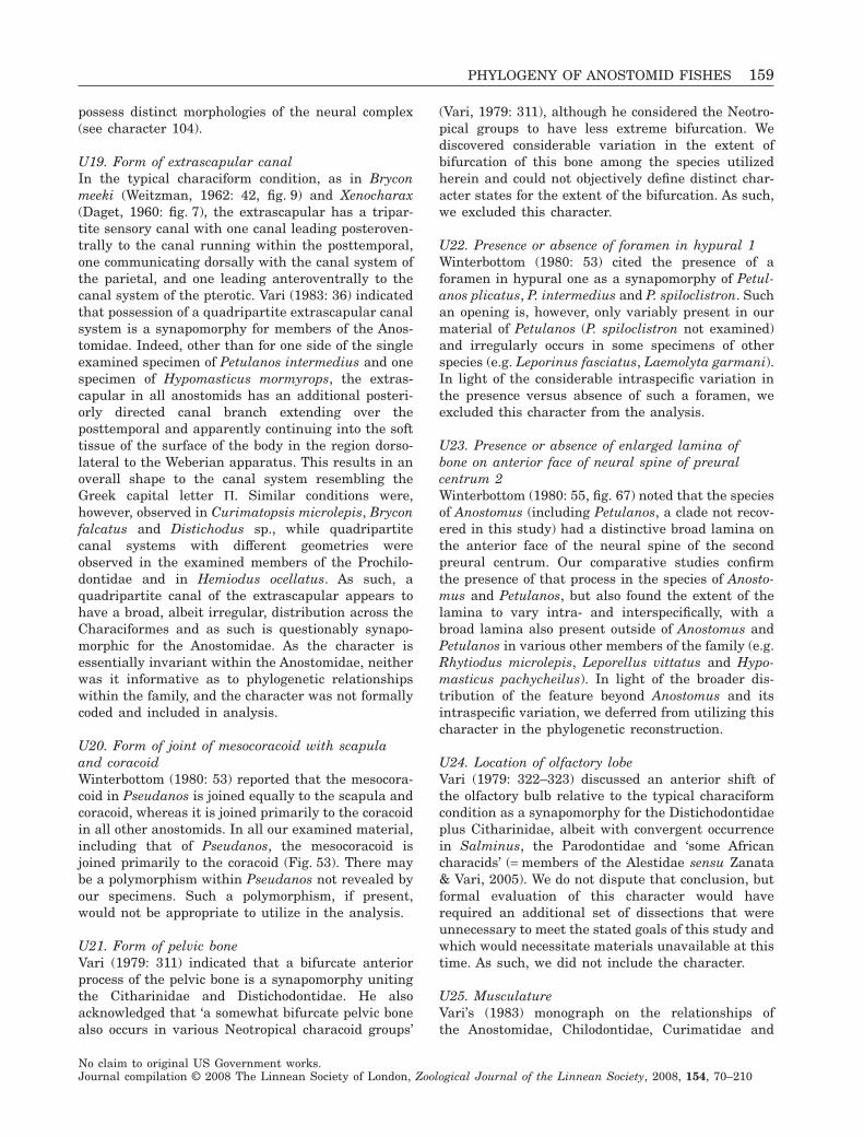
possess distinct morphologies of the neural complex(see character 104).
U19. Form of extrascapular canalIn the typical characiform condition, as in Bryconmeeki (Weitzman, 1962: 42, fig. 9) and Xenocharax(Daget, 1960: fig. 7), the extrascapular has a tripar-tite sensory canal with one canal leading posteroven-trally to the canal running within the posttemporal,one communicating dorsally with the canal system ofthe parietal, and one leading anteroventrally to thecanal system of the pterotic. Vari (1983: 36) indicatedthat possession of a quadripartite extrascapular canalsystem is a synapomorphy for members of the Anos-tomidae. Indeed, other than for one side of the singleexamined specimen of Petulanos intermedius and onespecimen of Hypomasticus mormyrops, the extras-capular in all anostomids has an additional posteri-orly directed canal branch extending over theposttemporal and apparently continuing into the softtissue of the surface of the body in the region dorso-lateral to the Weberian apparatus. This results in anoverall shape to the canal system resembling theGreek capital letter P. Similar conditions were,however, observed in Curimatopsis microlepis, Bryconfalcatus and Distichodus sp., while quadripartitecanal systems with different geometries wereobserved in the examined members of the Prochilo-dontidae and in Hemiodus ocellatus. As such, aquadripartite canal of the extrascapular appears tohave a broad, albeit irregular, distribution across theCharaciformes and as such is questionably synapo-morphic for the Anostomidae. As the character isessentially invariant within the Anostomidae, neitherwas it informative as to phylogenetic relationshipswithin the family, and the character was not formallycoded and included in analysis.
U20. Form of joint of mesocoracoid with scapulaand coracoidWinterbottom (1980: 53) reported that the mesocora-coid in Pseudanos is joined equally to the scapula andcoracoid, whereas it is joined primarily to the coracoidin all other anostomids. In all our examined material,including that of Pseudanos, the mesocoracoid isjoined primarily to the coracoid (Fig. 53). There maybe a polymorphism within Pseudanos not revealed byour specimens. Such a polymorphism, if present,would not be appropriate to utilize in the analysis.
U21. Form of pelvic boneVari (1979: 311) indicated that a bifurcate anteriorprocess of the pelvic bone is a synapomorphy unitingthe Citharinidae and Distichodontidae. He alsoacknowledged that ‘a somewhat bifurcate pelvic bonealso occurs in various Neotropical characoid groups’
(Vari, 1979: 311), although he considered the Neotro-pical groups to have less extreme bifurcation. Wediscovered considerable variation in the extent ofbifurcation of this bone among the species utilizedherein and could not objectively define distinct char-acter states for the extent of the bifurcation. As such,we excluded this character.
U22. Presence or absence of foramen in hypural 1Winterbottom (1980: 53) cited the presence of aforamen in hypural one as a synapomorphy of Petul-anos plicatus, P. intermedius and P. spiloclistron. Suchan opening is, however, only variably present in ourmaterial of Petulanos (P. spiloclistron not examined)and irregularly occurs in some specimens of otherspecies (e.g. Leporinus fasciatus, Laemolyta garmani).In light of the considerable intraspecific variation inthe presence versus absence of such a foramen, weexcluded this character from the analysis.
U23. Presence or absence of enlarged lamina ofbone on anterior face of neural spine of preuralcentrum 2Winterbottom (1980: 55, fig. 67) noted that the speciesof Anostomus (including Petulanos, a clade not recov-ered in this study) had a distinctive broad lamina onthe anterior face of the neural spine of the secondpreural centrum. Our comparative studies confirmthe presence of that process in the species of Anosto-mus and Petulanos, but also found the extent of thelamina to vary intra- and interspecifically, with abroad lamina also present outside of Anostomus andPetulanos in various other members of the family (e.g.Rhytiodus microlepis, Leporellus vittatus and Hypo-masticus pachycheilus). In light of the broader dis-tribution of the feature beyond Anostomus and itsintraspecific variation, we deferred from utilizing thischaracter in the phylogenetic reconstruction.
U24. Location of olfactory lobeVari (1979: 322–323) discussed an anterior shift ofthe olfactory bulb relative to the typical characiformcondition as a synapomorphy for the Distichodontidaeplus Citharinidae, albeit with convergent occurrencein Salminus, the Parodontidae and ‘some Africancharacids’ (= members of the Alestidae sensu Zanata& Vari, 2005). We do not dispute that conclusion, butformal evaluation of this character would haverequired an additional set of dissections that wereunnecessary to meet the stated goals of this study andwhich would necessitate materials unavailable at thistime. As such, we did not include the character.
U25. MusculatureVari’s (1983) monograph on the relationships ofthe Anostomidae, Chilodontidae, Curimatidae and
PHYLOGENY OF ANOSTOMID FISHES 159
No claim to original US Government works.Journal compilation © 2008 The Linnean Society of London, Zoological Journal of the Linnean Society, 2008, 154, 70–210
Prochilodontidae included several characters drawnfrom the musculature of the head. For example, hecited the longitudinally expanded attachment of thehyohyoidei abductores on the urohyal as a synapo-morphy for the four family assemblage (Vari, 1983:47), and the absence of the Aw portion of the adductormandibulae as a synapomorphy for the Anostomidae(Vari, 1983: 51). We do not dispute any of thesecharacters, but evaluating them in this study wouldhave required a parallel set of specimens for musculardissections. Many of the species examined herein arerare (e.g. Sartor elongatus, known only from the typeseries) and additional specimens of others of theexamined species were not consistently available. Asa consequence, we were unable to include Vari’s(1983) muscular characters in the present analysis.
DISCUSSION
Overall, this study obtained excellent phylogeneticresolution and the final hypothesis of relationshipspresented in Figure 5 represents a major advance inthe understanding of the evolutionary history of theAnostomidae. With over 120 morphological charactersdiscovered to vary informatively within the Anosto-midae and with many nodes in the final phylogenypossessing Bremer support values of three or higher,this analysis demonstrated that, contrary to thearguments of Scotland, Olmstead & Bennett (2003),many informative morphological characters remainunmined and a robust phylogenetic hypothesis canstill be formulated on morphological grounds. There isevery reason to believe that is also the situation withmany other groups of South American freshwaterfishes that have not been the subject of a morphology-based phylogenetic analysis, and indeed, many othergroups of fishes worldwide.
The list of synapomorphic character state transi-tions obtained by mapping character states onto thefinal phylogeny (Appendix 4) revealed that majorshifts in jaw position within the Anostomidaeoccurred nine times. Jaw position for the family isplesiomorphically subterminal (Leporellus, Hypomas-ticus), rather than the terminal position morecommon in characiforms. The superterminal or supe-rior jaws possessed by taxa such as Gnathodolus andSartor were revealed to be derived products of manysuccessive morphological shifts in the oral jaws, theanterior portions of the suspensorium and neurocra-nium and undoubtedly the associated soft tissuesystems. The reconstructed transitions in jaw evolu-tion are elaborated upon in a subsequent section.
Much of the discovered morphological variationwithin the Anostomidae was in the suspensorium (24characters), oral jaws and associated soft tissues (20characters), oral dentition (14 characters), and ante-
rior portions of the neurocranium (16 characters) andwas largely associated with the nine major shifts injaw morphology. Other highly variable systemsincluded the infraorbitals (14 characters) and integu-ment (scales and pigmentation, ten characters). Someportions of anatomy were found to be apparentlymore morphologically conservative within the Anos-tomidae, including the fins and fin girdles (six char-acters), Weberian apparatus (six characters) andhyoid arch (three characters). Although the pharyn-geal arches and associated dentition and musculatureare highly modified in the Anostomidae and its sisterclade, the Chilodontidae, relative to other characi-forms (see discussion in Vari, 1983: 18–21), thosesystems are relatively invariant within the Anostomi-dae (only six characters discovered herein).
The distribution of morphological variation amonganatomical systems in the Anostomidae differed strik-ingly from the distribution of variation in the closeoutgroups. For example, the Chilodontidae haveexperienced numerous morphological shifts withinthe pectoral girdle (Vari et al., 1995), and much ofthe phylogenetic resolution within the Curimatidaeresulted from informative variation in the pharyngealarches (Vari, 1989a), a system that is nearly invariantwithin the Anostomidae except for shifts in the degreeof development of, and dentition on, the dentigeroustooth-plates. The diverse patterns of phylogeneticallyinformative body systems exemplify the difficulty, orindeed, impossibility of predicting where the greatestconcentration of phylogenetically informative charac-ters will lie a priori. Morphological phylogeneticanalyses should clearly investigate as many anatomi-cal systems as possible.
While homoplasy certainly existed in the dataset(CI for each most parsimonious tree = 0.425) and con-tributed to the lack of resolution at certain levels,particularly within Leporinus, most of the similarityin the dataset could be interpreted as synapomorphy(RI for each most parsimonious tree = 0.805). Charac-ters with high levels of homoplasy (CI < 0.400)occurred in all major anatomical systems, but so didcharacters that optimized perfectly (CI = 1.000). Nev-ertheless, some systems interestingly demonstratedgreater levels of homoplasy than did others. Many ofthe characters with high levels of homoplasy withinthe Anostomidae dealt with tooth morphology andcusping (characters 32, 36, 39), infraorbital morphol-ogy (characters 3, 5, 8, 9, 12), presence or absence ofelements in the basibranchial series (character 92),branchiostegal series (character 100) or the bonessurrounding the Weberian ossicles (character 105) andpigmentation (characters 116, 118, 119, 120, 121, 122).Alternatively, characters involving the neurocranium,suspensorium, oral jaws and opercular series tendedto have much lower levels of homoplasy within the
160 B. L. SIDLAUSKAS and R. P. VARI
No claim to original US Government works.Journal compilation © 2008 The Linnean Society of London, Zoological Journal of the Linnean Society, 2008, 154, 70–210
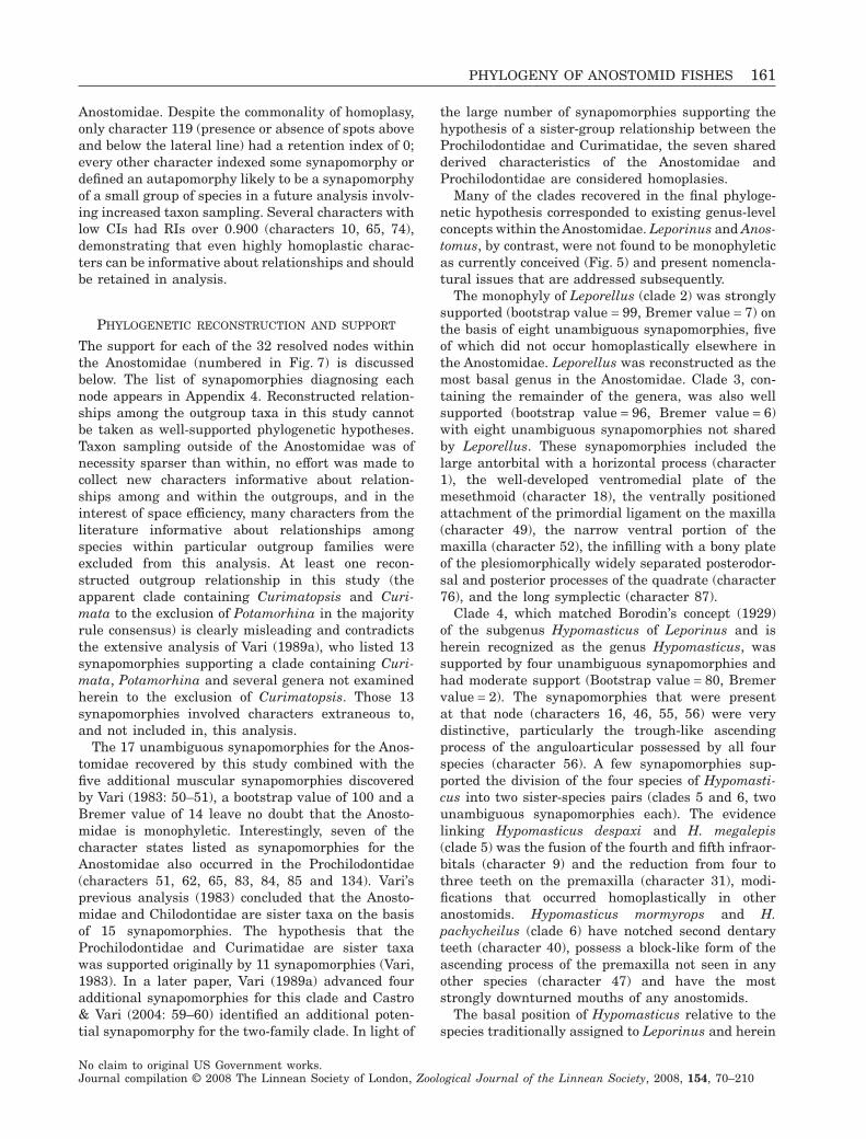
Anostomidae. Despite the commonality of homoplasy,only character 119 (presence or absence of spots aboveand below the lateral line) had a retention index of 0;every other character indexed some synapomorphy ordefined an autapomorphy likely to be a synapomorphyof a small group of species in a future analysis involv-ing increased taxon sampling. Several characters withlow CIs had RIs over 0.900 (characters 10, 65, 74),demonstrating that even highly homoplastic charac-ters can be informative about relationships and shouldbe retained in analysis.
PHYLOGENETIC RECONSTRUCTION AND SUPPORT
The support for each of the 32 resolved nodes withinthe Anostomidae (numbered in Fig. 7) is discussedbelow. The list of synapomorphies diagnosing eachnode appears in Appendix 4. Reconstructed relation-ships among the outgroup taxa in this study cannotbe taken as well-supported phylogenetic hypotheses.Taxon sampling outside of the Anostomidae was ofnecessity sparser than within, no effort was made tocollect new characters informative about relation-ships among and within the outgroups, and in theinterest of space efficiency, many characters from theliterature informative about relationships amongspecies within particular outgroup families wereexcluded from this analysis. At least one recon-structed outgroup relationship in this study (theapparent clade containing Curimatopsis and Curi-mata to the exclusion of Potamorhina in the majorityrule consensus) is clearly misleading and contradictsthe extensive analysis of Vari (1989a), who listed 13synapomorphies supporting a clade containing Curi-mata, Potamorhina and several genera not examinedherein to the exclusion of Curimatopsis. Those 13synapomorphies involved characters extraneous to,and not included in, this analysis.
The 17 unambiguous synapomorphies for the Anos-tomidae recovered by this study combined with thefive additional muscular synapomorphies discoveredby Vari (1983: 50–51), a bootstrap value of 100 and aBremer value of 14 leave no doubt that the Anosto-midae is monophyletic. Interestingly, seven of thecharacter states listed as synapomorphies for theAnostomidae also occurred in the Prochilodontidae(characters 51, 62, 65, 83, 84, 85 and 134). Vari’sprevious analysis (1983) concluded that the Anosto-midae and Chilodontidae are sister taxa on the basisof 15 synapomorphies. The hypothesis that theProchilodontidae and Curimatidae are sister taxawas supported originally by 11 synapomorphies (Vari,1983). In a later paper, Vari (1989a) advanced fouradditional synapomorphies for this clade and Castro& Vari (2004: 59–60) identified an additional poten-tial synapomorphy for the two-family clade. In light of
the large number of synapomorphies supporting thehypothesis of a sister-group relationship between theProchilodontidae and Curimatidae, the seven sharedderived characteristics of the Anostomidae andProchilodontidae are considered homoplasies.
Many of the clades recovered in the final phyloge-netic hypothesis corresponded to existing genus-levelconcepts within the Anostomidae. Leporinus and Anos-tomus, by contrast, were not found to be monophyleticas currently conceived (Fig. 5) and present nomencla-tural issues that are addressed subsequently.
The monophyly of Leporellus (clade 2) was stronglysupported (bootstrap value = 99, Bremer value = 7) onthe basis of eight unambiguous synapomorphies, fiveof which did not occur homoplastically elsewhere inthe Anostomidae. Leporellus was reconstructed as themost basal genus in the Anostomidae. Clade 3, con-taining the remainder of the genera, was also wellsupported (bootstrap value = 96, Bremer value = 6)with eight unambiguous synapomorphies not sharedby Leporellus. These synapomorphies included thelarge antorbital with a horizontal process (character1), the well-developed ventromedial plate of themesethmoid (character 18), the ventrally positionedattachment of the primordial ligament on the maxilla(character 49), the narrow ventral portion of themaxilla (character 52), the infilling with a bony plateof the plesiomorphically widely separated posterodor-sal and posterior processes of the quadrate (character76), and the long symplectic (character 87).
Clade 4, which matched Borodin’s concept (1929)of the subgenus Hypomasticus of Leporinus and isherein recognized as the genus Hypomasticus, wassupported by four unambiguous synapomorphies andhad moderate support (Bootstrap value = 80, Bremervalue = 2). The synapomorphies that were presentat that node (characters 16, 46, 55, 56) were verydistinctive, particularly the trough-like ascendingprocess of the anguloarticular possessed by all fourspecies (character 56). A few synapomorphies sup-ported the division of the four species of Hypomasti-cus into two sister-species pairs (clades 5 and 6, twounambiguous synapomorphies each). The evidencelinking Hypomasticus despaxi and H. megalepis(clade 5) was the fusion of the fourth and fifth infraor-bitals (character 9) and the reduction from four tothree teeth on the premaxilla (character 31), modi-fications that occurred homoplastically in otheranostomids. Hypomasticus mormyrops and H.pachycheilus (clade 6) have notched second dentaryteeth (character 40), possess a block-like form of theascending process of the premaxilla not seen in anyother species (character 47) and have the moststrongly downturned mouths of any anostomids.
The basal position of Hypomasticus relative to thespecies traditionally assigned to Leporinus and herein
PHYLOGENY OF ANOSTOMID FISHES 161
No claim to original US Government works.Journal compilation © 2008 The Linnean Society of London, Zoological Journal of the Linnean Society, 2008, 154, 70–210
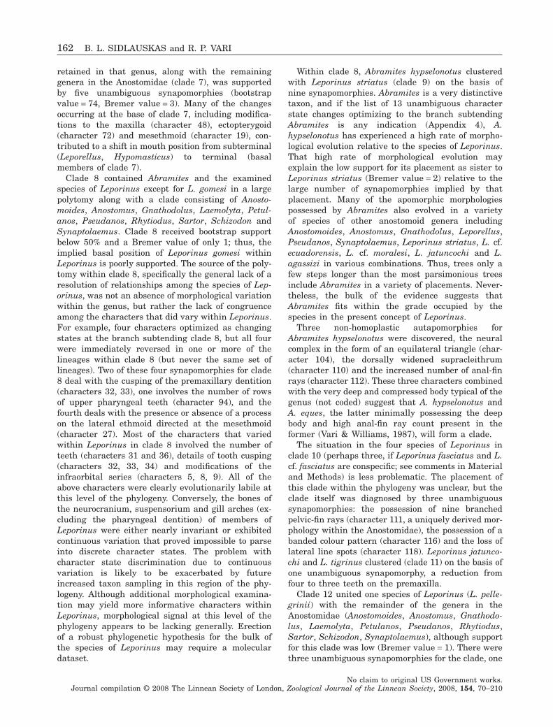
retained in that genus, along with the remaininggenera in the Anostomidae (clade 7), was supportedby five unambiguous synapomorphies (bootstrapvalue = 74, Bremer value = 3). Many of the changesoccurring at the base of clade 7, including modifica-tions to the maxilla (character 48), ectopterygoid(character 72) and mesethmoid (character 19), con-tributed to a shift in mouth position from subterminal(Leporellus, Hypomasticus) to terminal (basalmembers of clade 7).
Clade 8 contained Abramites and the examinedspecies of Leporinus except for L. gomesi in a largepolytomy along with a clade consisting of Anosto-moides, Anostomus, Gnathodolus, Laemolyta, Petul-anos, Pseudanos, Rhytiodus, Sartor, Schizodon andSynaptolaemus. Clade 8 received bootstrap supportbelow 50% and a Bremer value of only 1; thus, theimplied basal position of Leporinus gomesi withinLeporinus is poorly supported. The source of the poly-tomy within clade 8, specifically the general lack of aresolution of relationships among the species of Lep-orinus, was not an absence of morphological variationwithin the genus, but rather the lack of congruenceamong the characters that did vary within Leporinus.For example, four characters optimized as changingstates at the branch subtending clade 8, but all fourwere immediately reversed in one or more of thelineages within clade 8 (but never the same set oflineages). Two of these four synapomorphies for clade8 deal with the cusping of the premaxillary dentition(characters 32, 33), one involves the number of rowsof upper pharyngeal teeth (character 94), and thefourth deals with the presence or absence of a processon the lateral ethmoid directed at the mesethmoid(character 27). Most of the characters that variedwithin Leporinus in clade 8 involved the number ofteeth (characters 31 and 36), details of tooth cusping(characters 32, 33, 34) and modifications of theinfraorbital series (characters 5, 8, 9). All of theabove characters were clearly evolutionarily labile atthis level of the phylogeny. Conversely, the bones ofthe neurocranium, suspensorium and gill arches (ex-cluding the pharyngeal dentition) of members ofLeporinus were either nearly invariant or exhibitedcontinuous variation that proved impossible to parseinto discrete character states. The problem withcharacter state discrimination due to continuousvariation is likely to be exacerbated by futureincreased taxon sampling in this region of the phy-logeny. Although additional morphological examina-tion may yield more informative characters withinLeporinus, morphological signal at this level of thephylogeny appears to be lacking generally. Erectionof a robust phylogenetic hypothesis for the bulk ofthe species of Leporinus may require a moleculardataset.
Within clade 8, Abramites hypselonotus clusteredwith Leporinus striatus (clade 9) on the basis ofnine synapomorphies. Abramites is a very distinctivetaxon, and if the list of 13 unambiguous characterstate changes optimizing to the branch subtendingAbramites is any indication (Appendix 4), A.hypselonotus has experienced a high rate of morpho-logical evolution relative to the species of Leporinus.That high rate of morphological evolution mayexplain the low support for its placement as sister toLeporinus striatus (Bremer value = 2) relative to thelarge number of synapomorphies implied by thatplacement. Many of the apomorphic morphologiespossessed by Abramites also evolved in a varietyof species of other anostomoid genera includingAnostomoides, Anostomus, Gnathodolus, Leporellus,Pseudanos, Synaptolaemus, Leporinus striatus, L. cf.ecuadorensis, L. cf. moralesi, L. jatuncochi and L.agassizi in various combinations. Thus, trees only afew steps longer than the most parsimonious treesinclude Abramites in a variety of placements. Never-theless, the bulk of the evidence suggests thatAbramites fits within the grade occupied by thespecies in the present concept of Leporinus.
Three non-homoplastic autapomorphies forAbramites hypselonotus were discovered, the neuralcomplex in the form of an equilateral triangle (char-acter 104), the dorsally widened supracleithrum(character 110) and the increased number of anal-finrays (character 112). These three characters combinedwith the very deep and compressed body typical of thegenus (not coded) suggest that A. hypselonotus andA. eques, the latter minimally possessing the deepbody and high anal-fin ray count present in theformer (Vari & Williams, 1987), will form a clade.
The situation in the four species of Leporinus inclade 10 (perhaps three, if Leporinus fasciatus and L.cf. fasciatus are conspecific; see comments in Materialand Methods) is less problematic. The placement ofthis clade within the phylogeny was unclear, but theclade itself was diagnosed by three unambiguoussynapomorphies: the possession of nine branchedpelvic-fin rays (character 111, a uniquely derived mor-phology within the Anostomidae), the possession of abanded colour pattern (character 116) and the loss oflateral line spots (character 118). Leporinus jatunco-chi and L. tigrinus clustered (clade 11) on the basis ofone unambiguous synapomorphy, a reduction fromfour to three teeth on the premaxilla.
Clade 12 united one species of Leporinus (L. pelle-grinii) with the remainder of the genera in theAnostomidae (Anostomoides, Anostomus, Gnathodo-lus, Laemolyta, Petulanos, Pseudanos, Rhytiodus,Sartor, Schizodon, Synaptolaemus), although supportfor this clade was low (Bremer value = 1). There werethree unambiguous synapomorphies for the clade, one
162 B. L. SIDLAUSKAS and R. P. VARI
No claim to original US Government works.Journal compilation © 2008 The Linnean Society of London, Zoological Journal of the Linnean Society, 2008, 154, 70–210
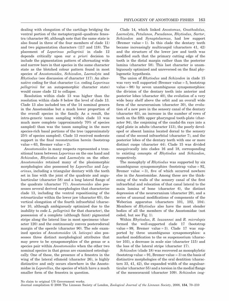
dealing with elongation of the cartilage bridging theventral portion of the metapterygoid–quadrate fenes-tra (character 80, although note that the same state isalso found in three of the four members of clade 11)and two pigmentation characters (117 and 118). Theplacement of Leporinus pellegrinii in clade 12depends critically upon our a priori decision toinclude the pigmentation pattern of alternating wideand narrow bars in that species in the same characterstate as the blotched colour pattern found in mostspecies of Anostomoides, Schizodon, Laemolyta andRhytiodus (see discussion of character 117). An alter-native coding for that character (i.e. coding Leporinuspellegrinii for an autapomorphic character state)would cause clade 12 to collapse.
Resolution within clade 13 was higher than theresolution within clade 8 below the level of clade 13.Clade 13 also included ten of the 14 nominal generain the Anostomidae but a much lower proportion ofthe overall species in the family. As a result, theintra-generic taxon sampling within clade 13 wasmuch more complete (approximately 70% of speciessampled) than was the taxon sampling in the morespecies-rich basal portions of the tree (approximately25% of species sampled). Clade 13 received moderatesupport in the final reconstruction herein (bootstrapvalue = 63, Bremer value = 2).
Anostomoides in many respects represented a tran-sitional taxon between Leporinus on the one hand andSchizodon, Rhytiodus and Laemolyta on the other.Anostomoides retained many of the plesiomorphicmorphologies also possessed by Leporellus and Lep-orinus, including a triangular dentary with the teethset in line with the joint of the quadrate and angu-loarticular (character 58) and a long lateral flange ofthe quadrate (character 77). Anostomoides also pos-sesses several derived morphologies that characterizeclade 13, including the ventral repositioning of theretroarticular within the lower jaw (character 65), thevertical elongation of the fourth infraorbital (charac-ter 10, although ambiguously optimized due to theinability to code L. pellegrinii for that character), thepossession of a complete (although faint) pigmentedstripe along the lateral line in most specimens (char-acter 120) and the continuously convex posterodorsalmargin of the opercle (character 90). The sole exam-ined species of Anostomoides (A. laticeps) also pos-sesses three distinct morphological attributes thatmay prove to be synapomorphies of the genus or aspecies pair within Anostomoides when the other twonominal species in that genus are examined osteologi-cally. One of these, the presence of a fenestra in thewing of the lateral ethmoid (character 26), is highlydistinctive and only occurs elsewhere in the Anosto-midae in Leporellus, the species of which have a muchsmaller form of the fenestra in question.
Clade 14, which linked Anostomus, Gnathodolus,Laemolyta, Petulanos, Pseudanos, Rhytiodus, Sartor,Schizodon and Synaptolaemus, had low support(Bremer value = 1). In this clade the dentary teethbecame increasingly multicuspid (characters 41, 42)and the structure of the lower jaw and teeth wasmodified such that the primary cutting edge of thetooth is the distal margin rather than the posteriorlamina (character 58). This last character is unam-biguously optimized and unreversed on the final phy-logenetic hypothesis.
The union of Rhytiodus and Schizodon in clade 15was very well supported (Bremer value = 5, bootstrapvalue = 98) by seven unambiguous synapomorphies:the division of the dentary teeth into anterior andposterior lobes (character 40), the evolution of a verywide bony shelf above the orbit and an overall wideform of the neurocranium (character 30), the evolu-tion of a new pore in the sensory canal of the dentary(character 63), an increase in the number of rows ofteeth on the fifth upper pharyngeal tooth-plate (char-acter 94), the conjoining of the caudal-fin rays into arigid plate in adults (character 113), the poorly devel-oped or absent lamina located dorsal to the sensorycanal of the second infraorbital (character 7), and theposterior lobes of the dentary teeth with one or moredistinct cusps (character 44). Clade 15 was dividedunequivocally into clades 16 and 18, correspondingto existing concepts of Rhytiodus and Schizodon,respectively.
The monophyly of Rhytiodus was supported by sixunambiguous synapomorphies (bootstrap value = 92,Bremer value = 3), five of which occurred nowhereelse in the Anostomidae. Among these are the thick-ening of the walls of the sensory canal of the firstinfraorbital and relocation of that canal lateral to themain lamina of bone (character 6), the distinctdepression of the neurocranium (character 29), and aseries of unusual modifications of components of theWeberian apparatus (characters 101, 102, 104).Members of Rhytiodus also have the most slenderbodies of all the members of the Anostomidae (notcoded, but see Fig. 1).
Within Rhytiodus, R. lauzannei and R. microlepisformed the well-supported clade 17 (bootstrapvalue = 99, Bremer value = 3). Clade 17 was sup-ported by three unambiguous synapomorphies: amarked modification to the os suspensorium (charac-ter 103), a decrease in scale size (character 115) andthe loss of the lateral stripe (character 17).
Schizodon (clade 18) was recovered as monophyletic(bootstrap value = 91, Bremer value = 3) on the basis ofdistinctive morphologies of the oral dentition (charac-ters 33, 41, 42), the expanded width of the anguloar-ticular (character 55) and a torsion in the medial flangeof the mesocoracoid (character 109). Schizodon isog-
PHYLOGENY OF ANOSTOMID FISHES 163
No claim to original US Government works.Journal compilation © 2008 The Linnean Society of London, Zoological Journal of the Linnean Society, 2008, 154, 70–210
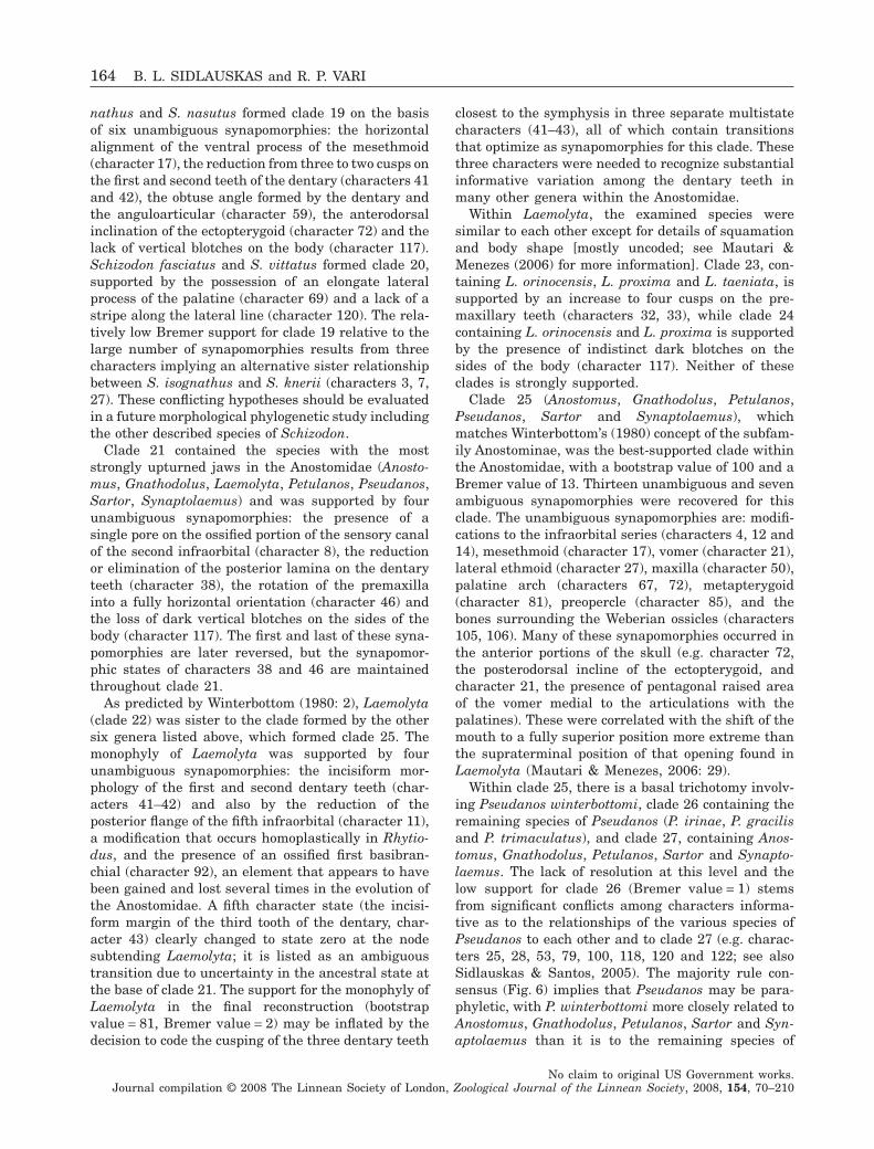
nathus and S. nasutus formed clade 19 on the basisof six unambiguous synapomorphies: the horizontalalignment of the ventral process of the mesethmoid(character 17), the reduction from three to two cusps onthe first and second teeth of the dentary (characters 41and 42), the obtuse angle formed by the dentary andthe anguloarticular (character 59), the anterodorsalinclination of the ectopterygoid (character 72) and thelack of vertical blotches on the body (character 117).Schizodon fasciatus and S. vittatus formed clade 20,supported by the possession of an elongate lateralprocess of the palatine (character 69) and a lack of astripe along the lateral line (character 120). The rela-tively low Bremer support for clade 19 relative to thelarge number of synapomorphies results from threecharacters implying an alternative sister relationshipbetween S. isognathus and S. knerii (characters 3, 7,27). These conflicting hypotheses should be evaluatedin a future morphological phylogenetic study includingthe other described species of Schizodon.
Clade 21 contained the species with the moststrongly upturned jaws in the Anostomidae (Anosto-mus, Gnathodolus, Laemolyta, Petulanos, Pseudanos,Sartor, Synaptolaemus) and was supported by fourunambiguous synapomorphies: the presence of asingle pore on the ossified portion of the sensory canalof the second infraorbital (character 8), the reductionor elimination of the posterior lamina on the dentaryteeth (character 38), the rotation of the premaxillainto a fully horizontal orientation (character 46) andthe loss of dark vertical blotches on the sides of thebody (character 117). The first and last of these syna-pomorphies are later reversed, but the synapomor-phic states of characters 38 and 46 are maintainedthroughout clade 21.
As predicted by Winterbottom (1980: 2), Laemolyta(clade 22) was sister to the clade formed by the othersix genera listed above, which formed clade 25. Themonophyly of Laemolyta was supported by fourunambiguous synapomorphies: the incisiform mor-phology of the first and second dentary teeth (char-acters 41–42) and also by the reduction of theposterior flange of the fifth infraorbital (character 11),a modification that occurs homoplastically in Rhytio-dus, and the presence of an ossified first basibran-chial (character 92), an element that appears to havebeen gained and lost several times in the evolution ofthe Anostomidae. A fifth character state (the incisi-form margin of the third tooth of the dentary, char-acter 43) clearly changed to state zero at the nodesubtending Laemolyta; it is listed as an ambiguoustransition due to uncertainty in the ancestral state atthe base of clade 21. The support for the monophyly ofLaemolyta in the final reconstruction (bootstrapvalue = 81, Bremer value = 2) may be inflated by thedecision to code the cusping of the three dentary teeth
closest to the symphysis in three separate multistatecharacters (41–43), all of which contain transitionsthat optimize as synapomorphies for this clade. Thesethree characters were needed to recognize substantialinformative variation among the dentary teeth inmany other genera within the Anostomidae.
Within Laemolyta, the examined species weresimilar to each other except for details of squamationand body shape [mostly uncoded; see Mautari &Menezes (2006) for more information]. Clade 23, con-taining L. orinocensis, L. proxima and L. taeniata, issupported by an increase to four cusps on the pre-maxillary teeth (characters 32, 33), while clade 24containing L. orinocensis and L. proxima is supportedby the presence of indistinct dark blotches on thesides of the body (character 117). Neither of theseclades is strongly supported.
Clade 25 (Anostomus, Gnathodolus, Petulanos,Pseudanos, Sartor and Synaptolaemus), whichmatches Winterbottom’s (1980) concept of the subfam-ily Anostominae, was the best-supported clade withinthe Anostomidae, with a bootstrap value of 100 and aBremer value of 13. Thirteen unambiguous and sevenambiguous synapomorphies were recovered for thisclade. The unambiguous synapomorphies are: modifi-cations to the infraorbital series (characters 4, 12 and14), mesethmoid (character 17), vomer (character 21),lateral ethmoid (character 27), maxilla (character 50),palatine arch (characters 67, 72), metapterygoid(character 81), preopercle (character 85), and thebones surrounding the Weberian ossicles (characters105, 106). Many of these synapomorphies occurred inthe anterior portions of the skull (e.g. character 72,the posterodorsal incline of the ectopterygoid, andcharacter 21, the presence of pentagonal raised areaof the vomer medial to the articulations with thepalatines). These were correlated with the shift of themouth to a fully superior position more extreme thanthe supraterminal position of that opening found inLaemolyta (Mautari & Menezes, 2006: 29).
Within clade 25, there is a basal trichotomy involv-ing Pseudanos winterbottomi, clade 26 containing theremaining species of Pseudanos (P. irinae, P. gracilisand P. trimaculatus), and clade 27, containing Anos-tomus, Gnathodolus, Petulanos, Sartor and Synapto-laemus. The lack of resolution at this level and thelow support for clade 26 (Bremer value = 1) stemsfrom significant conflicts among characters informa-tive as to the relationships of the various species ofPseudanos to each other and to clade 27 (e.g. charac-ters 25, 28, 53, 79, 100, 118, 120 and 122; see alsoSidlauskas & Santos, 2005). The majority rule con-sensus (Fig. 6) implies that Pseudanos may be para-phyletic, with P. winterbottomi more closely related toAnostomus, Gnathodolus, Petulanos, Sartor and Syn-aptolaemus than it is to the remaining species of
164 B. L. SIDLAUSKAS and R. P. VARI
No claim to original US Government works.Journal compilation © 2008 The Linnean Society of London, Zoological Journal of the Linnean Society, 2008, 154, 70–210
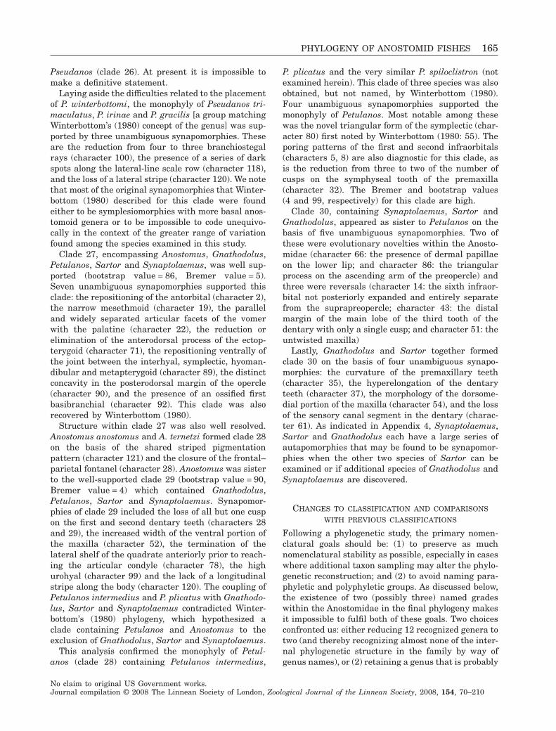
Pseudanos (clade 26). At present it is impossible tomake a definitive statement.
Laying aside the difficulties related to the placementof P. winterbottomi, the monophyly of Pseudanos tri-maculatus, P. irinae and P. gracilis [a group matchingWinterbottom’s (1980) concept of the genus] was sup-ported by three unambiguous synapomorphies. Theseare the reduction from four to three branchiostegalrays (character 100), the presence of a series of darkspots along the lateral-line scale row (character 118),and the loss of a lateral stripe (character 120). We notethat most of the original synapomorphies that Winter-bottom (1980) described for this clade were foundeither to be symplesiomorphies with more basal anos-tomoid genera or to be impossible to code unequivo-cally in the context of the greater range of variationfound among the species examined in this study.
Clade 27, encompassing Anostomus, Gnathodolus,Petulanos, Sartor and Synaptolaemus, was well sup-ported (bootstrap value = 86, Bremer value = 5).Seven unambiguous synapomorphies supported thisclade: the repositioning of the antorbital (character 2),the narrow mesethmoid (character 19), the paralleland widely separated articular facets of the vomerwith the palatine (character 22), the reduction orelimination of the anterodorsal process of the ectop-terygoid (character 71), the repositioning ventrally ofthe joint between the interhyal, symplectic, hyoman-dibular and metapterygoid (character 89), the distinctconcavity in the posterodorsal margin of the opercle(character 90), and the presence of an ossified firstbasibranchial (character 92). This clade was alsorecovered by Winterbottom (1980).
Structure within clade 27 was also well resolved.Anostomus anostomus and A. ternetzi formed clade 28on the basis of the shared striped pigmentationpattern (character 121) and the closure of the frontal–parietal fontanel (character 28). Anostomus was sisterto the well-supported clade 29 (bootstrap value = 90,Bremer value = 4) which contained Gnathodolus,Petulanos, Sartor and Synaptolaemus. Synapomor-phies of clade 29 included the loss of all but one cuspon the first and second dentary teeth (characters 28and 29), the increased width of the ventral portion ofthe maxilla (character 52), the termination of thelateral shelf of the quadrate anteriorly prior to reach-ing the articular condyle (character 78), the highurohyal (character 99) and the lack of a longitudinalstripe along the body (character 120). The coupling ofPetulanos intermedius and P. plicatus with Gnathodo-lus, Sartor and Synaptolaemus contradicted Winter-bottom’s (1980) phylogeny, which hypothesized aclade containing Petulanos and Anostomus to theexclusion of Gnathodolus, Sartor and Synaptolaemus.
This analysis confirmed the monophyly of Petul-anos (clade 28) containing Petulanos intermedius,
P. plicatus and the very similar P. spiloclistron (notexamined herein). This clade of three species was alsoobtained, but not named, by Winterbottom (1980).Four unambiguous synapomorphies supported themonophyly of Petulanos. Most notable among thesewas the novel triangular form of the symplectic (char-acter 80) first noted by Winterbottom (1980: 55). Theporing patterns of the first and second infraorbitals(characters 5, 8) are also diagnostic for this clade, asis the reduction from three to two of the number ofcusps on the symphyseal tooth of the premaxilla(character 32). The Bremer and bootstrap values(4 and 99, respectively) for this clade are high.
Clade 30, containing Synaptolaemus, Sartor andGnathodolus, appeared as sister to Petulanos on thebasis of five unambiguous synapomorphies. Two ofthese were evolutionary novelties within the Anosto-midae (character 66: the presence of dermal papillaeon the lower lip; and character 86: the triangularprocess on the ascending arm of the preopercle) andthree were reversals (character 14: the sixth infraor-bital not posteriorly expanded and entirely separatefrom the suprapreopercle; character 43: the distalmargin of the main lobe of the third tooth of thedentary with only a single cusp; and character 51: theuntwisted maxilla)
Lastly, Gnathodolus and Sartor together formedclade 30 on the basis of four unambiguous synapo-morphies: the curvature of the premaxillary teeth(character 35), the hyperelongation of the dentaryteeth (character 37), the morphology of the dorsome-dial portion of the maxilla (character 54), and the lossof the sensory canal segment in the dentary (charac-ter 61). As indicated in Appendix 4, Synaptolaemus,Sartor and Gnathodolus each have a large series ofautapomorphies that may be found to be synapomor-phies when the other two species of Sartor can beexamined or if additional species of Gnathodolus andSynaptolaemus are discovered.
CHANGES TO CLASSIFICATION AND COMPARISONS
WITH PREVIOUS CLASSIFICATIONS
Following a phylogenetic study, the primary nomen-clatural goals should be: (1) to preserve as muchnomenclatural stability as possible, especially in caseswhere additional taxon sampling may alter the phylo-genetic reconstruction; and (2) to avoid naming para-phyletic and polyphyletic groups. As discussed below,the existence of two (possibly three) named gradeswithin the Anostomidae in the final phylogeny makesit impossible to fulfil both of these goals. Two choicesconfronted us: either reducing 12 recognized genera totwo (and thereby recognizing almost none of the inter-nal phylogenetic structure in the family by way ofgenus names), or (2) retaining a genus that is probably
PHYLOGENY OF ANOSTOMID FISHES 165
No claim to original US Government works.Journal compilation © 2008 The Linnean Society of London, Zoological Journal of the Linnean Society, 2008, 154, 70–210
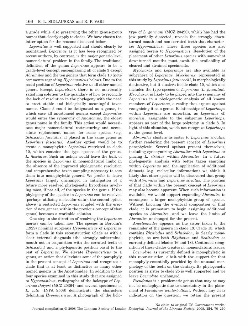
a grade while also preserving the other genus-groupnames that clearly apply to clades. We have chosen thelatter option for the reasons discussed below.
Leporellus is well supported and should clearly bemaintained. Leporinus as it has been recognized byrecent authors, by contrast, is the major generic-levelnomenclatural problem in the family. The traditionaldefinition of the genus Leporinus appears to be agrade-level concept encompassing all of clade 3 exceptAbramites and the ten genera that form clade 13 (notecomments regarding Hypomasticus below). Due to thebasal position of Leporinus relative to all other namedgenera (except Leporellus), there is no universallysatisfying solution to the quandary of how to reconcilethe lack of resolution in the phylogeny with the needto erect stable and biologically meaningful taxonnames. Clade 3 could be designated as a genus, inwhich case all anostomoid genera except Leporelluswould enter the synonymy of Anostomus, the oldestgenus name in the family. This action would necessi-tate major nomenclatural restructuring and neces-sitate replacement names for some species (e.g.Schizodon fasciatus, if placed in the same genus asLeporinus fasciatus). Another option would be tocreate a monophyletic Leporinus restricted to clade10, which contains the type species of the genus,L. fasciatus. Such an action would leave the bulk ofthe species in Leporinus in nomenclatural limbo inthe absence of the improved phylogenetic resolutionand comprehensive taxon sampling necessary to sortthem into monophyletic genera. We prefer to leaveLeporinus largely unchanged in anticipation of afuture more resolved phylogenetic hypothesis involv-ing most, if not all, of the species in the genus. If thephylogeny of the species in Leporinus can be resolved(perhaps utilizing molecular data), the second optionabove (a restricted Leporinus coupled with the erec-tion of new genera within the previous concept of thatgenus) becomes a workable solution.
One step in the direction of resolving the Leporinusmorass can be taken now. The species in Borodin’s(1929) nominal subgenus Hypomasticus of Leporinusform a clade in this reconstruction (clade 4) with aclear external diagnosis (the strongly subterminalmouth not in conjunction with the serrated teeth ofSchizodon) and a phylogenetic position basal to therest of Leporinus. We elevate Hypomasticus to agenus, an action that alleviates some of the paraphylyin the present concept of Leporinus and recognizes aclade that is at least as distinctive as many othernamed genera in the Anostomidae. In addition to thefour species examined in this study that are assignedto Hypomasticus, radiographs of the holotype of Lep-orinus thayeri (MCZ 20364) and several specimens ofL. julii (INPA 9508) demonstrate the charactersdelimiting Hypomasticus. A photograph of the holo-
type of L. garmani (MCZ 20420), which has had thejaw partially dissected, reveals the strongly down-turned mouth and non-serrated teeth that character-ize Hypomasticus. These three species are alsoassigned herein to Hypomasticus. Resolution of theplacement of other Leporinus species with somewhatdownturned mouths must await the availability ofcleared and strained speciments.
Myocharax and Leporinops are also available assubgenera of Leporinus. Myocharax, represented inthis study by Leporinus jatuncochi, is morphologicallydistinctive, but it clusters inside clade 10, which alsoincludes the type species of Leporinus (L. fasciatus).Myocharax is likely to be placed into the synonymy ofLeporinus in a phylogenetic analysis of all knownmembers of Leporinus, a reality that argues againstrecognizing it as a genus. Relationships of Leporinopswithin Leporinus are uncertain, as Leporinus cf.moralesi, assignable to the subgenus Leporinops,appears as part of the large polytomy in clade 8. Inlight of this situation, we do not recognize Leporinopsat the genus level.
Abramites clusters as sister to Leporinus striatus,further rendering the present concept of Leporinusparaphyletic. Several options present themselves,including synonymizing Abramites with Leporinus orplacing L. striatus within Abramites. In a futurephylogenetic analysis with better taxon samplingwithin Leporinus and the application of additionaldatasets (e.g. molecular information) we think itlikely that other species will be discovered that groupwith Abramites and Leporinus striatus. The positionof that clade within the present concept of Leporinusmay also become apparent. When such information isavailable, we would support expanding Abramites toencompass a larger monophyletic group of species.Without knowing the eventual composition of thatclade, it is premature to begin assigning additionalspecies to Abramites, and we leave the limits ofAbramites unchanged for the present.
Anostomoides appears as the sister taxon to theremainder of the genera in clade 13. Clade 15, whichcontains Rhytiodus and Schizodon, is clearly mono-phyletic, as are both Rhytiodus and Schizodon ascurrently defined (clades 16 and 18). Continued recog-nition of these clades creates no nomenclatural issues.
Laemolyta as currently defined is monophyletic inthis reconstruction, albeit with the support for thatmonophyly essentially provided by the unusual mor-phology of the teeth on the dentary. Its phylogeneticposition as sister to clade 25 is well supported and weleave Laemolyta unchanged.
Pseudanos is a problematic genus that may or maynot be monophyletic due to uncertainty in the place-ment of Pseudanos winterbottomi. Without any clearindication on the question, we retain the present
166 B. L. SIDLAUSKAS and R. P. VARI
No claim to original US Government works.Journal compilation © 2008 The Linnean Society of London, Zoological Journal of the Linnean Society, 2008, 154, 70–210
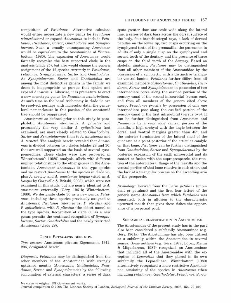
composition of Pseudanos. Alternative solutionswould either necessitate a new genus for Pseudanoswinterbottomi or expand Anostomus to include Petu-lanos, Pseudanos, Sartor, Gnathodolus and Synapto-laemus. Such a broadly encompassing Anostomuswould be equivalent to the Anostominae of Winter-bottom (1980). The expansion of Anostomus wouldformally recognize the best supported clade in theanalysis (clade 25), but also would change the genericassignment of the 12 species currently in Pseudanos,Petulanos, Synaptolaemus, Sartor and Gnathodolus.As Synaptolaemus, Sartor and Gnathodolus areamong the most distinctive genera in the family, wedeem it inappropriate to pursue that option andexpand Anostomus. Likewise, it is premature to erecta new monotypic genus for Pseudanos winterbottomi.At such time as the basal trichotomy in clade 25 canbe resolved, perhaps with molecular data, the genus-level nomenclature in this portion of the anostomidtree should be reappraised.
Anostomus as defined prior to this study is para-phyletic. Anostomus intermedius, A. plicatus andpresumably the very similar A. spiloclistron (notexamined) are more closely related to Gnathodolus,Sartor and Synaptolaemus than to A. anostomus andA. ternetzi. The analysis herein revealed that Anosto-mus is divided between two clades (clades 28 and 30)that are well supported on the basis of several syna-pomorphies. These clades were also recovered inWinterbottom’s (1980) analysis, albeit with differentimplied relationships to the other genera in the Anos-tomidae. Anostomus anostomus is the type speciesand we restrict Anostomus to the species in clade 28,plus A. brevior and A. anostomus longus (cited as A.longus by Garavello & Britski, 2003), which were notexamined in this study, but are nearly identical to A.anostomus externally (Géry, 1961b; Winterbottom,1980). We designate clade 30 as a new genus, Petul-anos, including three species previously assigned toAnostomus: Petulanos intermedius, P. plicatus andP. spiloclistron with P. plicatus (the oldest name) asthe type species. Recognition of clade 30 as a newgenus permits the continued recognition of Synapto-laemus, Sartor, Gnathodolus and the newly restrictedAnostomus (clade 28).
GENUS PETULANOS GEN. NOV.Type species: Anostomus plicatus Eigenmann, 1912:296, designated herein
Diagnosis: Petulanos may be distinguished from theother members of the Anostomidae with stronglyupturned mouths (Anostomus, Gnathodolus, Pseu-danos, Sartor and Synaptolaemus) by the followingcombination of external characters: a series of dark
spots greater than one scale wide along the lateralline, a series of dark bars across the dorsal surface ofthe body, four branchiostegal rays, a lack of dermalpapillae on the lower lip, two cusps occurring on thesymphyseal tooth of the premaxilla, the possession inadults of only a single cusp on the symphyseal andsecond tooth of the dentary, and the presence of threecusps on the third tooth of the dentary. Based onskeletal anatomy, Petulanos may be distinguishedfrom all other members of the Anostomidae by thepossession of a symplectic with a distinctive triangu-lar ventral lamina. Petulanos further differs from allexamined members of Anostomus, Gnathodolus, Pseu-danos, Sartor and Synaptolaemus in possession of twointermediate pores along the ossified portion of thesensory canal of the second infraorbital (versus one),and from all members of the genera cited aboveexcept Pseudanos gracilis by possession of only oneintermediate pore along the ossified portion of thesensory canal of the first infraorbital (versus two). Itcan be further distinguished from Anostomus andPseudanos by a very wide ventral portion of themaxilla, a high urohyal with the angle between thedorsal and ventral margins greater than 45°, andthe anterior termination of the lateral shelf of thequadrate at a point posterior of the articular condyleon that bone. Petulanos can be further distinguishedfrom Gnathodolus, Sartor and Synaptolaemus by theposterior expansion of the sixth infraorbital and itscontact or fusion with the suprapreopercle, the rota-tion of the anterolateral flange of the maxilla and theventral portion of that bone relative to each other, andthe lack of a triangular process on the ascending armof the preopercle.
Etymology: Derived from the Latin petulans (impu-dent or petulant) and the first four letters of thegeneric name Anostomus from which Petulanos wasseparated; both in allusion to the characteristicupturned mouth that gives these fishes the appear-ance of a perpetual pout.
SUBFAMILIAL CLASSIFICATION IN ANOSTOMIDAE
The Anostomidae of the present study has in the pastalso been considered a subfamily Anostominae (e.g.Géry, 1961a). The Anostominae has also been utilizedas a subfamily within the Anostomidae in severalsenses. Some authors (e.g. Géry, 1977; López, Menni& Miquelarena, 1987) recognized an Anostominaethat included all of the Anostomidae with the ex-ception of Leporellus that they placed in its ownsubfamily, the Leporellinae. Winterbottom (1980)alternatively recognized a more restrictive Anostomi-nae consisting of the species in Anostomus (thenincluding Petulanos), Gnathodolus, Pseudanos, Sartor
PHYLOGENY OF ANOSTOMID FISHES 167
No claim to original US Government works.Journal compilation © 2008 The Linnean Society of London, Zoological Journal of the Linnean Society, 2008, 154, 70–210
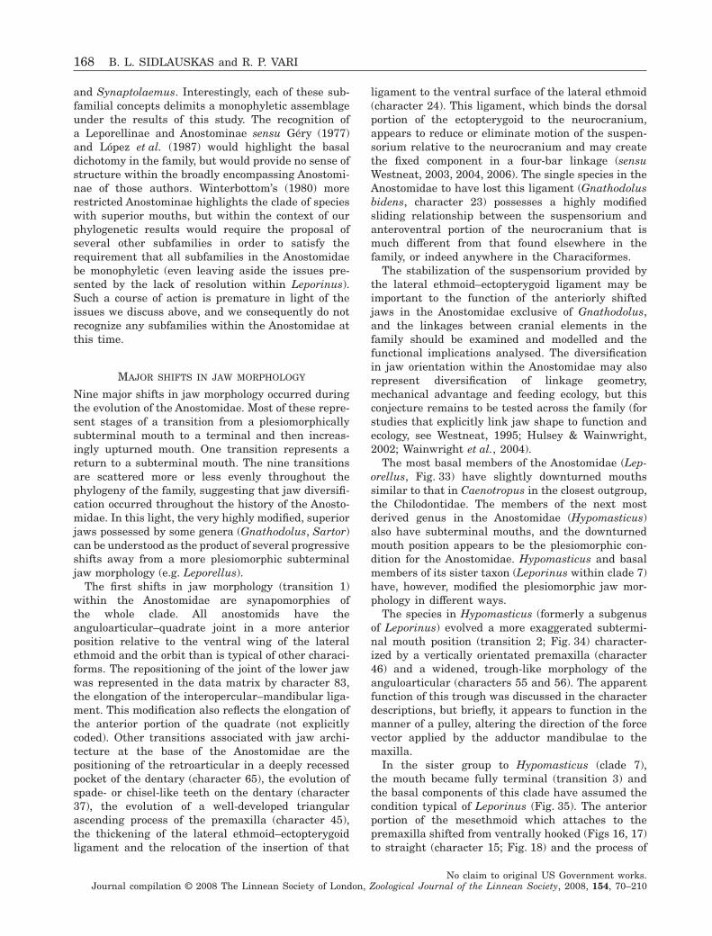
and Synaptolaemus. Interestingly, each of these sub-familial concepts delimits a monophyletic assemblageunder the results of this study. The recognition ofa Leporellinae and Anostominae sensu Géry (1977)and López et al. (1987) would highlight the basaldichotomy in the family, but would provide no sense ofstructure within the broadly encompassing Anostomi-nae of those authors. Winterbottom’s (1980) morerestricted Anostominae highlights the clade of specieswith superior mouths, but within the context of ourphylogenetic results would require the proposal ofseveral other subfamilies in order to satisfy therequirement that all subfamilies in the Anostomidaebe monophyletic (even leaving aside the issues pre-sented by the lack of resolution within Leporinus).Such a course of action is premature in light of theissues we discuss above, and we consequently do notrecognize any subfamilies within the Anostomidae atthis time.
MAJOR SHIFTS IN JAW MORPHOLOGY
Nine major shifts in jaw morphology occurred duringthe evolution of the Anostomidae. Most of these repre-sent stages of a transition from a plesiomorphicallysubterminal mouth to a terminal and then increas-ingly upturned mouth. One transition represents areturn to a subterminal mouth. The nine transitionsare scattered more or less evenly throughout thephylogeny of the family, suggesting that jaw diversifi-cation occurred throughout the history of the Anosto-midae. In this light, the very highly modified, superiorjaws possessed by some genera (Gnathodolus, Sartor)can be understood as the product of several progressiveshifts away from a more plesiomorphic subterminaljaw morphology (e.g. Leporellus).
The first shifts in jaw morphology (transition 1)within the Anostomidae are synapomorphies ofthe whole clade. All anostomids have theanguloarticular–quadrate joint in a more anteriorposition relative to the ventral wing of the lateralethmoid and the orbit than is typical of other characi-forms. The repositioning of the joint of the lower jawwas represented in the data matrix by character 83,the elongation of the interopercular–mandibular liga-ment. This modification also reflects the elongation ofthe anterior portion of the quadrate (not explicitlycoded). Other transitions associated with jaw archi-tecture at the base of the Anostomidae are thepositioning of the retroarticular in a deeply recessedpocket of the dentary (character 65), the evolution ofspade- or chisel-like teeth on the dentary (character37), the evolution of a well-developed triangularascending process of the premaxilla (character 45),the thickening of the lateral ethmoid–ectopterygoidligament and the relocation of the insertion of that
ligament to the ventral surface of the lateral ethmoid(character 24). This ligament, which binds the dorsalportion of the ectopterygoid to the neurocranium,appears to reduce or eliminate motion of the suspen-sorium relative to the neurocranium and may createthe fixed component in a four-bar linkage (sensuWestneat, 2003, 2004, 2006). The single species in theAnostomidae to have lost this ligament (Gnathodolusbidens, character 23) possesses a highly modifiedsliding relationship between the suspensorium andanteroventral portion of the neurocranium that ismuch different from that found elsewhere in thefamily, or indeed anywhere in the Characiformes.
The stabilization of the suspensorium provided bythe lateral ethmoid–ectopterygoid ligament may beimportant to the function of the anteriorly shiftedjaws in the Anostomidae exclusive of Gnathodolus,and the linkages between cranial elements in thefamily should be examined and modelled and thefunctional implications analysed. The diversificationin jaw orientation within the Anostomidae may alsorepresent diversification of linkage geometry,mechanical advantage and feeding ecology, but thisconjecture remains to be tested across the family (forstudies that explicitly link jaw shape to function andecology, see Westneat, 1995; Hulsey & Wainwright,2002; Wainwright et al., 2004).
The most basal members of the Anostomidae (Lep-orellus, Fig. 33) have slightly downturned mouthssimilar to that in Caenotropus in the closest outgroup,the Chilodontidae. The members of the next mostderived genus in the Anostomidae (Hypomasticus)also have subterminal mouths, and the downturnedmouth position appears to be the plesiomorphic con-dition for the Anostomidae. Hypomasticus and basalmembers of its sister taxon (Leporinus within clade 7)have, however, modified the plesiomorphic jaw mor-phology in different ways.
The species in Hypomasticus (formerly a subgenusof Leporinus) evolved a more exaggerated subtermi-nal mouth position (transition 2; Fig. 34) character-ized by a vertically orientated premaxilla (character46) and a widened, trough-like morphology of theanguloarticular (characters 55 and 56). The apparentfunction of this trough was discussed in the characterdescriptions, but briefly, it appears to function in themanner of a pulley, altering the direction of the forcevector applied by the adductor mandibulae to themaxilla.
In the sister group to Hypomasticus (clade 7),the mouth became fully terminal (transition 3) andthe basal components of this clade have assumed thecondition typical of Leporinus (Fig. 35). The anteriorportion of the mesethmoid which attaches to thepremaxilla shifted from ventrally hooked (Figs 16, 17)to straight (character 15; Fig. 18) and the process of
168 B. L. SIDLAUSKAS and R. P. VARI
No claim to original US Government works.Journal compilation © 2008 The Linnean Society of London, Zoological Journal of the Linnean Society, 2008, 154, 70–210
the mesethmoid connecting to the vomer becameposteroventrally aligned (character 17; Fig. 18). Themaxilla acquired a vertical or posterodorsal orienta-tion (character 48; Fig. 35) and the ectopterygoidassumed a vertical orientation (character 72; Fig. 41)as the joint of the lower jaw with the quadratebecame more anteriorly positioned than was the casein Leporellus, Hypomasticus and the proximate out-group, the Chilodontidae.
The next major shift in jaw morphology (transition4) occurred in clades 13 and 14, all members of whichpossess upturned mouths at some point in ontogeny.Rhytiodus and Schizodon have supraterminal mouthsonly as small juveniles and larvae and have terminalor subterminal mouths as adults. Anostomoides hasonly a slightly supraterminal mouth in the adultcondition (juveniles of the genus not examined), whileLaemolyta, Pseudanos, Anostomus, Petulanos, Synap-tolaemus, Sartor and Gnathodolus have supratermi-nal or fully superior mouths apparently throughoutontogeny (small juveniles of Synaptolaemus, Sartorand Gnathodolus were not examined). Transition 4involved a ventral shift in the position of the retroar-ticular in clade 13 (character 65; compare Figs 35,36). In the slightly more derived clade 14, the func-tional edge of the lower teeth shifted from the poste-rior lamina to the distal margin of the tooth (compareFig. 32A–C with Fig. 32E, F), and the orientation ofthe teeth in the dentary relative to that bone alsochanged (compare Fig. 35 with Figs 36, 38; see dis-cussion of character 58). The shift in orientation mayhave changed the angle of force applied by the teeth.A biomechanical model [e.g. that of Westneat (2003),or a modification thereof] should be applied to testthis conjecture and fully characterize the transition injaw function in clades 13 and 14.
The jaw of Schizodon nasutus (Fig. 39) within clade13 displays the most remarkable instance of conver-gence in the Anostomidae (transition 5). This speciesevolved a strongly subterminal mouth similar to,but independent of, the condition in Hypomasticus(Fig. 34). Schizodon nasutus also independentlyacquired three of the four skeletal morphologies thatdiagnose Hypomasticus: the transversely widenedanguloarticular (character 55, also shared with allspecies of Schizodon), the trough-like ascendingprocess of the anguloarticular (character 56) and thevertically aligned premaxilla (character 46). The con-vergence between Hypomasticus and Schizodonnasutus appears to be an excellent example of twolineages evolving the same solution to a comparablemechanical problem, effective functioning of ventrallyrotated oral jaws. It has not, however, been demon-strated that the subterminal jaws of Schizodonnasutus and Hypomasticus are convergent function-ally as well as morphologically.
The next major shift in jaw evolution in the Anos-tomidae (transition 6) occurred in clade 21, all themembers of which have fully upturned jaws through-out ontogeny. Transition 6 is characterized by a fullyhorizontal orientation of the premaxilla (character 46;Figs 36, 37) and the reduction or elimination of theposterior lamina of the dentary teeth (compareFig. 32E with 31E or 32F). The ontogenetic shift inmouth position in Rhytiodus and Schizodon (see dis-cussion in Sidlauskas et al., 2007), which togetherform the sister to clade 21, suggests that the stronglyupturned mouths of the taxa in clade 21 may haveresulted from heterochrony (paedomorphic fixation ofthe juvenile state in adults). The conjecture of het-erochrony should be investigated further by compari-son of ontogenetic series of skeletal preparations.
The six genera in clade 25 have the most stronglyupturned jaws in the Anostomidae, and indeed proxi-mate outgroup families, with the mouth opening com-pletely on the dorsal surface of the head. Severalmorphological synapomorphies characterize this shift(transition 7). The ventral process of the mesethmoidruns vertically or nearly so (character 17), the ventralsurface of the vomer developed a pentagonal raisedarea that contacts the restructured palatine (charac-ters 21 and 67), and the ectopterygoid became almosttotally posterodorsally inclined (character 66; Fig. 54)as the joint of the lower jaw with the quadrate movedever more anteriorly. Somewhere near the base of thisclade, a notch also developed in the lateral ethmoid toaccommodate the now nearly horizontal alignment ofthe ectopterygoid–lateral ethmoid ligament (charac-ter 25; Fig. 26). The level at which this notch of thelateral ethmoid is a synapomorphy is uncertain due tothe lack of resolution concerning Pseudanos.
The penultimate shift in jaw orientation in theAnostomidae occurred in the lineage leading to Sartorand Gnathodolus (transition 8). These two generahave the mouth so strongly upturned (backwardsfacing in Sartor) that the quadrate–anguloarticularjoint is relocated to anterior of the opening of themouth (Fig. 37). The maxilla also became plate-likewith a distinctive medial process (character 54), thedentary teeth became elongate and recurved (charac-ter 37; Figs 31H, I, 37), and the premaxillary teethbecame bowed (character 35; Fig. 29G, H).
The final major shift in jaw morphology in thefamily (transition 9) occurred in the derived genusGnathodolus, in which the dentary was reduced to aslender cylinder bearing only a single tooth (charac-ters 33 and 57; Fig. 31I), the ligamentous connectionof the lateral ethmoid to the ectopterygoid that char-acterizes all other anostomids was lost (character 23),the palatine assumed the shape of an elongate hour-glass (character 70) and the vomer developed a deepgroove on each side into which the dorsal portions of
PHYLOGENY OF ANOSTOMID FISHES 169
No claim to original US Government works.Journal compilation © 2008 The Linnean Society of London, Zoological Journal of the Linnean Society, 2008, 154, 70–210
the mesopterygoid and metapterygoid fit (character20, Fig. 22; see also Winterbottom, 1980: fig. 58).Functionality of the jaws in Gnathodolus appears torequire that the dorsal portion of the suspensoriumslide along grooves in the ventral portion of the neu-rocranium. That would be impossible if the lateralethmoid–ectopterygoid ligament were retained. If so,the linkages within the skull of Gnathodolus maydiffer considerably from those found in other anosto-mids and may require construction of a new biome-chanical model.
PHYLOGENETIC BIOGEOGRAPHY
All 14 genera in the Anostomidae comprising approxi-mately 140 species are present in the vast cis-Andeanmid-elevation and lowland portions of tropical andtemperate South America. Anostomids inhabit myriadriver systems from the Caribbean Sea versant drain-ages of northern Venezuela (Lasso et al., 2004a)through the Río Orinoco (Lasso et al., 2004b) and RioAmazonas (Garavello & Britski, 2003) basins south tothe Río de La Plata system (Britski, Silimon & Lopes,1999; López, Miquelarena & Menni, 2003; Menni,2004). Anostomids are also diverse in the Atlanticversant rivers of the continent from the Guianas(Lowe-McConnell, 1964; Planquette et al., 1996) souththrough the coastal rivers of eastern Brazil (Britski,Sato & Rosa, 1984; Malabarba, 1989). Many feweranostomid genera and species inhabit the less exten-sive but topographically complex trans-Andean regionof South America. Only six species in four genera arenow recognized in the arch from the Lago Maracaibobasin of northwestern Venezuela (Schizodon, Vari &Raredon, 1991) through the Caribbean and Pacificslope rivers of northern and western Colombia(Abramites, Leporellus, Leporinus, Dahl, 1971;Mojica-C, 1999) to the Río Guayas basin of southeast-ern Ecuador (Leporinus, Eigenmann & Henn, 1916;Barriga, 1991).
Incomplete understanding of anostomid speciesdiversity and geographical distribution at the specieslevel preclude a fine-grained analysis of their phylo-genetic biogeography. This problematic situation isexemplified by the continuing publications describingnew species within the family and/or refining con-cepts of species limits and geographical ranges forpreviously described forms (e.g. Sidlauskas & Santos,2005; Sidlauskas et al., 2007). Although the phyloge-netic reconstruction arrived at herein included only asubset of the species and yielded incomplete resolu-tion, particularly within Leporinus (Fig. 5), it never-theless allows estimates of minimum ages for severalmajor intrafamilial cladogenic events.
An interesting pattern is apparent when the distri-bution of genera of the Anostomidae to the two sides
of the Andean Cordilleras is evaluated within thecontext of the arrived at phylogeny (Fig. 5). Thisanalysis finds that those genera in the family withrepresentatives on both sides of that mountain chainare all restricted to the somewhat more basal compo-nents of the phylogeny. These are Abramites with onespecies (A. hypselonotus) broadly distributed in cis-Andean regions and its congener (A. eques) endemicto the Río Magdalena system (Vari & Williams, 1987;Mojica, 1999), Leporellus with several nominalspecies occurring east of the Andes and one formidentified as L. vittatus reported from the RíoMagdalena basin (Dahl, 1971; Maldonaldo-Ocampoet al., 2005), Schizodon with representatives broadlydistributed east of the Andes (Sidlauskas et al., 2007,and references therein) and one species, S. corti,endemic to the Lago Maracaibo basin of northernVenezuela (Vari & Raredon, 1991; Lasso et al., 2004a)and finally Leporinus, a genus with over 90 recog-nized species (Garavello & Britski, 2003), only threeof which are reported to occur in river systems to thewest of the Cordilleras. These are L. ecuadorensisfrom the Río Guayas basin of Ecuador (Eigenmann &Henn, 1916; Barriga, 1991), L. muyscorum from theRío Magdalena, Río Atrato, Río Ranchería and RíoSinú basins of northern Colombia (Mojica-C, 1999;Mojica et al., 2006), and L. striatus, reported fromriver systems of northern and western Colombia(Dahl, 1971; Mojica-C, 1999).
Although some species of the Anostomidae live inmid-level, upland river systems (e.g. Leporellus in theRío Magdalena system, Maldonaldo-Ocampo et al.,2005), most occur in lower elevation settings and nomembers of the family are inhabitants of the precipi-tous drainage systems of the higher regions of theAndean Cordilleras. As such, the uplift of the AndeanCordilleras probably served as a vicariant event orsequential events dividing the ancestral componentsof Abramites, Leporellus, Leporinus and Schizodoninto cis- and trans-Andean components.
Schizodon (clade 18) is the most deeply nested ofthe clades within the Anostomidae that includes cis-and trans-Andean components. Schizodon corti, anendemic to the trans-Andean Lago Maracaibo basin(Vari & Raredon, 1991; Lasso et al., 2004a) and thesole member of the genus found in a trans-Andeandrainage, was not incorporated into the phylogeneticanalysis of this study. Schizodon corti shares,however, the externally obvious attributes of its con-geners and it is reasonable to assume that S. corti isa component of clade 18. It follows directly that cladesarising at progressively more inclusive nodes withinthe phylogeny of the Anostomidae below clade 18 (theancestors of clades 1, 3, 7, 8, 12, 13, 14 and 15; Fig. 5)evolved prior to this isolation of components of thatfamily to each side of the uplifting Cordilleras.
170 B. L. SIDLAUSKAS and R. P. VARI
No claim to original US Government works.Journal compilation © 2008 The Linnean Society of London, Zoological Journal of the Linnean Society, 2008, 154, 70–210
Furthermore, the ancestors of the three genera withmembers to the two sides of the Andes (Abramites,Leporellus, Leporinus) evolved prior to the uplift andthe same applies to Anostomoides and Hypomasticus,genera limited to river basins east of the Andes.Given that clade 15 (containing Schizodon andRhytiodus) is the sister to clade 21 (Laemolyta,Pseudanos, Anostomus, Petulanos, Synaptolaemus,Gnathodolus, and Sartor), stem members of clade 21must also have evolved by the final uplift of theCordilleras.
The specific geological event associated with finalvicariance into cis- and trans-Andean components ofthe Anostomidae cannot be identified in so far as it isimpossible to determine the degree of uplift thatresulted in the ultimate division of that component ofthe ichthyofauna into eastern and western subunits.Nonetheless, the minimum age for such vicariancewould be the closure of the north-flowing Maracaibo–Falcon outlet of the Río Orinoco. That closureoccurred approximately 8 Mya during the final majoruplift sequence of the northern portions of the AndeanCordilleras (Hoorn, 1993; Hoorn et al., 1995; Lund-berg et al., 1998). Evidence from the phylogeny whencorrelated with geological information is, however,further informative in terms of older likely vicarianceevents for the Anostomidae.
Schizodon corti, which is endemic to the Lago Mara-caibo basin, is the sole trans-Andean species of theAnostomidae in that basin (Lasso et al., 2004a). Theother trans-Andean species of the Anostomidae(species of Abramites, Leporellus and Leporinus)inhabit river systems that lie west of the central andwestern Andean Cordilleras in the portions of SouthAmerica from the Río Guayas to the Río Ranchería.These mountain ranges underwent uplift prior to theclosure of the Maracaibo–Falcon outlet with theresultant isolation of Lago Maracaibo from what isnow the Río Orinoco basin. The most recent and thuspertinent of the major uplift events in that region thatis relevant to the question of a minimum date for thedivision of the anostomid faunas east and west ofthe Central and Western Andean Cordilleras involvedthe Sierra de Perija, the mountain range between theLago Maracaibo basin and the river systems thatdrain regions to the west of those uplands. This upliftevent of that portion of the northeastern Andes tookplace approximately 11.8 Mya as evidenced by thewestward shifts of the palaeocurrents within thepresent valley of the Río Magdalena system (Hoornet al., 1995; Lundberg et al., 1998). The uplift of theSierra de Perija 11.8 Mya provides a minimum age forthe Anostomidae and for the ancestors of Abramites,Leporellus, Hypomasticus and Leporinus (clades 1, 2,3, 7, 8, 9; Fig. 5). Although Abramites and Leporinusform a largely unresolved polytomy in our final phy-
logeny (Fig. 5), that lack of resolution does not bear onthe hypothesis of the age of these genera as thephylogenetic hypothesis clusters all members of thosegenera at clade 8. As such the ancestor of the subunitof the Anostomidae at that level of the phylogeny alsowas present prior to 11.8 Mya.
When we consider the degree of morphologicaldiversity present between the Anostomidae and itsclose relatives, the Chilodontidae, Curimatidae andProchilodontidae, the latter two of which also havecis- and trans-Andean components (Vari, 1988; Castro& Vari, 2004) and the fact that a series of cladogenicevents within the Anostomidae (the basal splitswithin clades 1, 3, 7 and 8) preceded the vicariantevents of 11.8 Mya, it seems very likely that the originof the family and its basal clades most likely signifi-cantly pre-dated that time. Evidence from the fossilrecord of the Anostomidae and its close relatives isinformative on this question.
Fossils of the Anostomidae have been rarely cited inthe literature. It is uncertain whether this situationrepresents the rarity of such material in depositswithin South America that include remains of fresh-water taxa or the fact that fossils of the family remainunrecognized among samples of fossil fishes fromsuch strata. Two reports of fossil anostomids arethat of Roberts (1975) for remains from the Cuencabasin of southern Ecuador at approximately 19 Mya(Roberts, 1975: 261), and the more recent citation byLundberg (1997: 73) based on material from the LaVenta formation in Colombia from between 13.5 and11.5 million years old (Guerrero, 1997: 41). Both ofthese samples are limited to teeth identified ashaving originated with a species of Leporinus (jawand pharyngeal teeth in the Ecuadorian samples anda single premaxillary tooth in the case of the La Ventamaterial). The age of these deposits minimally con-firms the existence of species of Leporinus at the timeof the uplift of the Sierra de Perija 11.8 Mya, aminimum age for the genus independently postulatedabove on the basis of distributional data evaluated ina geological context. The oldest possible age for thefossils (19 Myr for the Cuenca material) would indi-cate that the family and clades including Leporinussignificantly pre-date that time period.
Fossils of close relatives of the Anostomidae withinthe Characiformes are also very limited and onlyone is informative as to the minimum age of themore inclusive clade including that family plus itsclose relatives, the Chilodontidae, Curimatidae, andProchilodontidae. Malabarba (1996) demonstratedthat a fossil fish described by Travassos & Santos(1955) as Curimata mosesi from Oligocene deposits ofthe Tremembé basin of eastern Brazil was assignableto the curimatid genus Cyphocharax, a component ofa terminal polytomy deeply nested within the phylog-
PHYLOGENY OF ANOSTOMID FISHES 171
No claim to original US Government works.Journal compilation © 2008 The Linnean Society of London, Zoological Journal of the Linnean Society, 2008, 154, 70–210
eny of the Curimatidae (Vari, 1989a). The depositsfrom which the specimens of Cyphocharax mosesioriginated are at least 22.5 million years old (Mala-barba, 1998; Reis, 1998) and the ancestors of theclade formed by the Curimatidae along with theProchilodontidae and of that clade plus the Anosto-midae and Chilodontidae therefore pre-date thattime. Although not directly informative as to the ageof the Anostomidae, this older date of 22.5 Myr is,nonetheless, congruent with the likely diversificationof the Anostomidae well in advance of the upliftepisodes of the Andean Cordilleras cited above aspertinent to likely vicariance events within thefamily. That date also distinctly pre-dates theminimum age of 11.5–13.5 Myr for the Anostomidaeindicated by the limited fossil evidence for thatfamily. The numerous cladogenic events between thedivergence of the anostomid–chilodontid clade andthe ancestor of Cyphocharax in conjunction with thepronounced degree of morphological divergences ineach of those lineages are presumably indicative of alengthy intervening time period, but it is impossibleto determine whether the approximately 10 Myrbetween those events would accommodate that levelof divergence.
Although not directly informative on that issue,evidence does indicate that the Characiformesevolved long in advance of even the 22.5-Myr date.The Characiformes as a whole long pre-dates theearliest fossils assignable to the Anostomidae as evi-denced by both Old World fossil characiforms of thegenera Alestes, Brycinus and Bryconaethiops in thefamily Alestidae (Zanata & Vari, 2005) that extendback approximately 49–54.8 Myr. It is also notewor-thy that several instances of trans-Atlantic relation-ships within the Characiformes (see summary inZanata & Vari, 2005: 120) would extend the African–South American drift vicariance at several phyloge-netic levels within the Characiformes backapproximately 90–112 Mya. There was thus a lengthytime period to accommodate the accumulation of evo-lutionary novelties between the origin of the Characi-formes and the earliest fossils known at the level ofthe clade formed by the Anostomidae, Chilodontidae,Curimatidae and Prochilodontidae or of Leporinus inthe Anostomidae.
ACKNOWLEDGEMENTS
This paper benefited from the comments and criti-cisms of the first author’s thesis advisors Barry Cher-noff and Mark Westneat, thesis committee membersMichael Foote, Shannon Hackett and Leigh VanValen, and colleagues Jim Cooper, Doug Eernisse,Eric Hilton, Samantha Hopkins, Christian Kam-merer, Aaron Rice, C. Tristan Stayton, Philip Willink
and K. Rebecca Thomas. We are greatly indebted tothe following individuals and institutions for the loanand exchange of species, information and diverseother type of assistance: John Lundberg, Mark Sabajand Wasila Dahdul (ANSP), William Eschmeyer,David Catania and Jon Fong (CAS), Mary AnneRogers and Kevin Swagel (FMNH), Michael Retzer(INHS), Lucía Rapp Py-Daniel and Geraldo Mendesdos Santos (INPA), Francisco Provenzano (MBUCV),Karsten Hartel (MCZ), Mário de Pinna and OsvaldoOyakawa (MZUSP), Helmut Wellendorf (NMW),Julío Cesar Garavello (UFSC), and William Fink andDouglas Nelson (UMMZ). Drawings of osteologicalfeatures were prepared by Tamara L. Clark andlabelled by Sandra J. Raredon (USNM). JenniferJellen provided technical assistance at FMNH andLisa F. Palmer, Jeffrey M. Clayton and in particularSandra J. Raredon provided technical assistanceat USNM. Betty Strack (FMNH) helped preparethe scanning electron micrographs, and Sandra J.Raredon (USNM) digitally edited and cleaned themicrographs. Research associated with this study wassupported in part by EPA STAR Graduate Fellowship915987, NSF Doctoral Dissertation ImprovementGrant #DEB0412364, the National EvolutionarySynthesis Center (NSF EF-0423641), The FieldMuseum’s Lester Armour Graduate Fellowship, theHerbert R. and Evelyn Axelrod Chair in SystematicIchthyology in the Division of Fishes of the NationalMuseum of Natural History, Smithsonian Institution,The University of Chicago’s Hinds Fund and a BöhlkeAward from the Academy of Natural Sciences ofPhiladelphia. The Field Museum graciously providedseveral days of free beam time on its Zeiss LEOscanning electron microscope.
REFERENCES
Ahn D, Gibson G. 1999. Axial variation in the threespinestickleback: genetic and environmental factors. Evolution &Development 1: 100–112.
Alexander RM. 1964. Adaptation in the skulls and cranialmuscles of South American characinoid fishes. ZoologicalJournal of the Linnean Society 45: 169–190.
Balassa GC, Fugi R, Hahn NS, Galina AB. 2004. Dieta deespécies de Anostomidae (Teleostei, Characiformes) na áreade influência do reservatório de Manso, Mato Grosso, Brasil.Iheringia, Série Zoologia, Porto Alegre 94: 77–82.
Barriga R. 1991. Peces de agua dulce. Politecnia. Organooficial de la Escuela Politecnia Nacional, Quito 16: 89–162.
Bergmann LAC. 1988. Schizodon jacuiensis sp. n., um novoanostomídeo do sul do Brasil e redescrição de Schizodonkneri (Steindachner, 1875) e S. platae (Garman, 1890).(Pisces, Characiformes, Anostomidae). Comunicações doMuseu de Ciências da PUCRS, Série Zoologia 1: 13–28.
Berkovitz B. 1975. Observations on tooth replacement inpiranhas (Characidae). Archives of Oral Biology 20: 53–57.
172 B. L. SIDLAUSKAS and R. P. VARI
No claim to original US Government works.Journal compilation © 2008 The Linnean Society of London, Zoological Journal of the Linnean Society, 2008, 154, 70–210
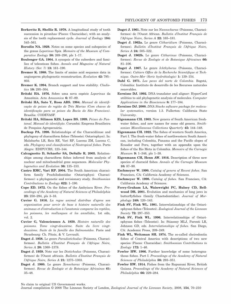
Berkovitz B, Shellis R. 1978. A longitudinal study of toothsuccession in piranhas (Pisces: Characidae), with an analy-sis of the tooth replacement cycle. Journal of Zoology 184:545–561.
Borodin NA. 1929. Notes on some species and subspecies ofthe genus Leporinus Spix. Memoirs of the Museum of Com-parative Zoology 50: 269–290, pls 1–17.
Boulenger GA. 1904. A synopsis of the suborders and fami-lies of teleostean fishes. Annals and Magazine of NaturalHistory (Ser. 7) 13: 161–190.
Bremer K. 1988. The limits of amino acid sequence data inangiosperm phylogenetic reconstruction. Evolution 42: 795–803.
Bremer K. 1994. Branch support and tree stability. Cladis-tics 10: 295–304.
Britski HA. 1976. Sobre uma nova espécie Leporinus daAmazônia. Acta Amazonica 6: 87–89.
Britski HA, Sato Y, Rosa ABS. 1984. Manual de identifi-cação de peixes do região de Três Marias (Com chaves deidentificação para os peixes da Bacia do São Francisco).Brasilia: CODEVASF.
Britski HA, Silimon KZS, Lopes BS. 1999. Peixes do Pan-tanal. Manual de identifição. Corumbá: Empresa Brasilieirade Pesquisa Agropecuária.
Buckup PA. 1998. Relationships of the Characidiinae andphylogeny of characiform fishes (Teleostei: Ostariophysi). In:Malabarba LR, Reis RE, Vari RP, Lucena ZM, Lucena C,eds. Phylogeny and classification of Neotropical fishes. PortoAlegre: EDIPUCRS, 123–144.
Calcagnotto D, Schaefer SA, DeSalle R. 2005. Relation-ships among characiform fishes inferred from analysis ofnuclear and mitochondrial gene sequences. Molecular Phy-logenetics and Evolution 36: 135–153.
Castro RMC, Vari RP. 2004. The South American characi-form family Prochilodontidae (Ostariophysi: Characi-formes): a phylogenetic and revisionary study. SmithsonianContributions to Zoology 622: 1–190.
Cope ED. 1872. On the fishes of the Ambyiacu River. Pro-ceedings of the Academy of Natural Sciences of Philadelphia23: 250–294, pls 3–16.
Cuvier G. 1816. La regne animal distribue d’apres sonorganisation pour servir de base à histoire naturelle desanimaux et d’introduction à l’anatomie comparee les reptiles,les poissons, les mollusques et les annélides, 1st edn,vol. 2.
Cuvier G, Valenciennes A. 1850. Histoire naturelle despoissons. Tome vingt-deuxième. Suite du livre vingt-deuxième. Suite de la famille des Salmonoïdes. Paris andStrasbourg: Ch. Pitois, & V. Levrault.
Daget J. 1958. Le genre Paradistichodus (Poissons, Characi-formes). Bulletin d’Institut Français de l’Afrique Noire,Series A 20: 1360–1378.
Daget J. 1959. Note sur les Distichodus (Poissons, Characi-formes) de l’Ouest africain. Bulletin d’Institut Français del’Afrique Noire, Series A 21: 1275–1303.
Daget J. 1960. Le genre Xenocharax (Poissons, Characi-formes). Revue de Zoologie et de Botanique Africaines 61:35–48.
Daget J. 1961. Note sur les Nannocharax (Poissons, Characi-formes) de l’Ouest African. Bulletin d’Institut Français del’Afrique Noire, Series A 22: 165–181.
Daget J. 1962a. Le genre Citharidium (Poissons, Characi-formes). Bulletin d’Institut Français de l’Afrique Noire,Series A 24: 505–522.
Daget J. 1962b. Le genre Citharinus (Poissons, Characi-formes). Revue de Zoologie et de Botanique Africaines 66:81–106.
Daget J. 1967. Le genre Ichthyborus (Poissons, Characi-formes). Cahiers Office de la Recherche Scientifique et Tech-nique. Outre-Mer (Serie hydrobiologie) 1: 139–154.
Dahl G. 1971. Los peces del norte de Colombia. Bogotá,Colombia: Instituto de desarrollo de les Recursos naturalesrenovables.
Eernisse DJ. 1992. DNA translator and aligner: HyperCardutilities to aid phylogenetic analysis of molecules. ComputerApplications in the Biosciences 8: 177–184.
Eernisse DJ. 2000. DNA Stacks software package for molecu-lar systematics, version 1.3. Fullerton: California StateUniversity.
Eigenmann CH. 1903. New genera of South American fresh-water fishes, and new names for some old genera. Smith-sonian Miscellaneous Collections Quarterly 45: 144–148.
Eigenmann CH. 1922. The fishes of western South America,Part I. The fresh-water fishes of northwestern South Ameri-can, including Colombia, Panama and the Pacific slopes ofEcuador and Peru, together with an appendix upon thefishes of the Rio Meta in Colombia. Memoirs of the CarnegieMuseum 9: 1–346, pls 1–38.
Eigenmann CH, Henn AW. 1916. Description of three newspecies of characid fishes. Annals of the Carnegie Museum10: 87–90.
Eschmeyer W. 1990. Catalog of genera of Recent fishes. SanFrancisco, CA: California Academy of Sciences.
Eschmeyer W. 1998. Catalog of fishes. San Francisco, CA:California Academy of Sciences.
Ferry-Graham LA, Wainwright PC, Hulsey CD, Bell-wood DR. 2001. Evolution and mechanics of long jaws inbutterflyfishes (family Chaetodontidae). Journal of Mor-phology 248: 120–143.
Fink SV, Fink WL. 1981. Interrelationships of the Ostari-ophysan fishes (Teleostei). Zoological Journal of the LinneanSociety 72: 297–353.
Fink SV, Fink WL. 1996. Interrelationships of Ostari-ophysan fishes (Teleostei). In: Stiassny MLJ, Parenti LR,Johnson GD, eds. Interrelationships of fishes. San Diego,CA: Academic Press, 209–249.
Fink WL, Weitzman SH. 1974. The so-called cheirodontinfishes of Central America with descriptions of two newspecies (Pisces: Characidae). Smithsonian Contributions toZoology 172: 1–46.
Fowler HW. 1906. Further knowledge of some heterogna-thous fishes. Part I. Proceedings of the Academy of NaturalSciences of Philadelphia 58: 293–351.
Fowler HW. 1914. Fishes from the Rupununi River, BritishGuiana. Proceedings of the Academy of Natural Sciences ofPhiladelphia 66: 229–284.
PHYLOGENY OF ANOSTOMID FISHES 173
No claim to original US Government works.Journal compilation © 2008 The Linnean Society of London, Zoological Journal of the Linnean Society, 2008, 154, 70–210
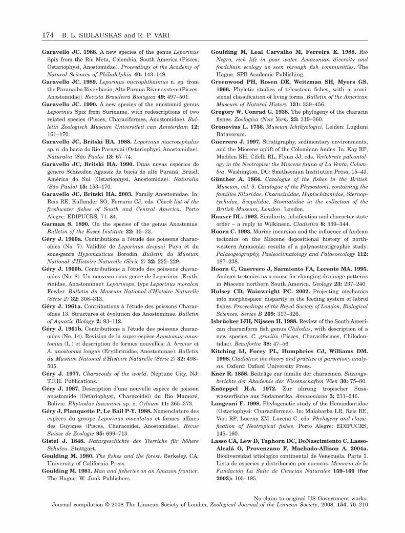
Garavello JC. 1988. A new species of the genus LeporinusSpix from the Río Meta, Colombia, South America (Pisces,Ostariophysi, Anostomidae). Proceedings of the Academy ofNatural Sciences of Philadelphia 40: 143–149.
Garavello JC. 1989. Leporinus microphthalmus n. sp. fromthe Paranaiba River basin, Alto Parana River system (Pisces:Anostomidae). Revista Brasileira Biologica 49: 497–501.
Garavello JC. 1990. A new species of the anostomid genusLeporinus Spix from Suriname, with redescriptions of tworelated species (Pisces, Characiformes, Anostomidae). Bul-letin Zoologisch Museum Universiteit van Amsterdam 12:161–170.
Garavello JC, Britski HA. 1988. Leporinus macrocephalussp. n. da bacia do Rio Paraguai (Ostariophysi, Anostomidae).Naturalia (São Paolo) 13: 67–74.
Garavello JC, Britski HA. 1990. Duas novas espécies dogênero Schizodon Agassiz da bacia do alto Paraná, Brasil,América do Sul (Ostariophysi, Anostomidae). Naturalia(São Paulo) 15: 153–170.
Garavello JC, Britski HA. 2003. Family Anostomidae. In:Reis RE, Kullander SO, Ferraris CJ, eds. Check list of thefreshwater fishes of South and Central America. PortoAlegre: EDIPUCRS, 71–84.
Garman S. 1890. On the species of the genus Anostomus.Bulletin of the Essex Institute 22: 15–23.
Géry J. 1960a. Contributions a l’étude des poissons charac-oïdes (No. 7). Validité de Leporinus despaxi Puyo et dusous-genre Hypomasticus Borodin. Bulletin du MuséumNational d’Histoire Naturelle (Série 2) 32: 222–229.
Géry J. 1960b. Contributions a l’étude des poissons charac-oïdes (No. 8). Un nouveau sous-genre de Leporinus (Eryth-rinidae, Anostominae): Leporinops, type Leporinus moralesiFowler. Bulletin du Muséum National d’Histoire Naturelle(Série 2) 32: 308–313.
Géry J. 1961a. Contributions à l’étude des poissons Charac-oïdes 13. Structures et évolution des Anostominae. Bulletinof Aquatic Biology 2: 93–112.
Géry J. 1961b. Contributions a l’étude des poissons charac-oïdes (No. 14). Revision de la super-espèce Anostomus anos-tomus (L.) et description de formes nouvelles: A. brevior etA. anostomus longus (Erythrinidae, Anostominae). Bulletindu Muséum National d’Histoire Naturelle (Série 2) 32: 498–505.
Géry J. 1977. Characoids of the world. Neptune City, NJ:T.F.H. Publications.
Géry J. 1987. Description d’une nouvelle espèce de poissonanostomidé (Ostariophysi, Characoidei) du Rio Mamoré,Bolivie. Rhytiodus lauzannei sp. n. Cybium 11: 365–373.
Géry J, Planquette P, Le Bail P-Y. 1988. Nomenclature desespèces du groupe Leporinus maculatus et formes affinesdes Guyanes (Pisces, Characoidei, Anostomidae). RevueSuisse de Zoologie 95: 699–713.
Gistel J. 1848. Naturgeschichte des Tierrichs für höhereSchuleu. Stuttgart.
Goulding M. 1980. The fishes and the forest. Berkeley, CA:University of California Press.
Goulding M. 1981. Man and fisheries on an Amazon frontier.The Hague: W. Junk Publishers.
Goulding M, Leal Carvalho M, Ferreira E. 1988. RioNegro, rich life in poor water. Amazonian diversity andfoodchain ecology as seen through fish communities. TheHague: SPB Academic Publishing.
Greenwood PH, Rosen DE, Weitzman SH, Myers GS.1966. Phyletic studies of teleostean fishes, with a provi-sional classification of living forms. Bulletin of the AmericanMuseum of Natural History 131: 339–456.
Gregory W, Conrad G. 1938. The phylogeny of the characinfishes. Zoologica (New York) 23: 319–360.
Gronovius L. 1756. Museum Ichthyologici. Leiden: LugduniBatavorum.
Guerrero J. 1997. Stratigraphy, sedimentary environments,and the Miocene uplift of the Colombian Andes. In: Kay RF,Madden RH, Cifelli RL, Flynn JJ, eds. Vertebrate paleontol-ogy in the Neotropics: the Miocene fauna of La Venta, Colom-bia. Washington, DC: Smithsonian Institution Press, 15–43.
Günther A. 1864. Catalogue of the fishes in the BritishMuseum, vol. 5. Catalogue of the Physostomi, containing thefamilies Siluridae, Characinidae, Haplochitonidae, Sternop-tychidae, Scopelidae, Stomiatidae in the collection of theBritish Museum, London. London.
Hauser DL. 1992. Similarity, falsification and character stateorder – a reply to Wilkinson. Cladistics 8: 339–344.
Hoorn C. 1993. Marine incursion and the influence of Andeantectonics on the Miocene depositional history of north-western Amazonia: results of a palynostratigraphic study.Palaeogeography, Paeleoclimatology and Palaeoecology 112:187–238.
Hoorn C, Guerrero J, Sarmiento FA, Lorente MA. 1995.Andean tectonics as a cause for changing drainage patternsin Miocene northern South America. Geology 23: 237–240.
Hulsey CD, Wainwright PC. 2002. Projecting mechanicsinto morphospace: disparity in the feeding system of labridfishes. Proceedings of the Royal Society of London, BiologicalSciences, Series B 269: 317–326.
Isbrücker IJH, Nijssen H. 1988. Review of the South Ameri-can characiform fish genus Chilodus, with description of anew species, C. gracilis (Pisces, Characiformes, Chilodon-tidae). Beaufortia 38: 47–56.
Kitching IJ, Forey PL, Humphries CJ, Williams DM.1998. Cladistics: the theory and practice of parsimony analy-sis. Oxford: Oxford University Press.
Kner R. 1858. Beiträge zur familie der characinen. Sitzungs-berichte der Akademie der Wissenschaften Wien 30: 75–80.
Knöeppel H-A. 1972. Zur ahrung tropischer Süss-wasserfische aus Südamerika. Amazoniana 3: 231–246.
Langeani F. 1998. Phylogenetic study of the Hemiodontidae(Ostariophysi: Characiformes). In: Malabarba LR, Reis RE,Vari RP, Lucena ZM, Lucena C, eds. Phylogeny and classi-fication of Neotropical fishes. Porto Alegre: EDIPUCRS,145–160.
Lasso CA, Lew D, Taphorn DC, DoNascimiento C, Lasso-Alcalá O, Provenzano F, Machado-Allison A. 2004a.Biodiversidad ictiologico continental de Venezuela. Parte 1.Lista de especies y distribución por cuencas. Memoria de laFundación La Salle de Ciencias Naturales 159–160 (for2003): 105–195.
174 B. L. SIDLAUSKAS and R. P. VARI
No claim to original US Government works.Journal compilation © 2008 The Linnean Society of London, Zoological Journal of the Linnean Society, 2008, 154, 70–210
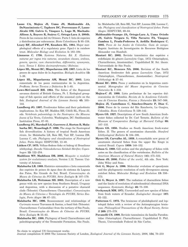
Lasso CA, Mojica JI, Usma JS, Maldonaldo JA,DoNascimiento C, Taphorn DC, Provenzano F, Lasso-Alcalá OM, Galvis G, Vásquez L, Lugo M, Machado-Allison A, Royero R, Suárez C, Ortega-Lara A. 2004b.Peces de las cuencas del río Orinoco. Parte I: lista de especiesy distribucíon por subcuencas. Biota Colombiana 5: 95–158.
Leary RF, Allendorf FW, Knudsen KL. 1984. Major mor-phological effects of a regulatory gene: Pgm1-t in rainbowtrout. Molecular Biology and Evolution 1: 183–194.
Linnaeus C. 1758. Systema Naturae, Ed. X. (Systemanaturae per regna tria naturae, secundum classes, ordines,genera, species, cum characteribus, differentiis, synonymis,locis. Tomus I. Editio decimareformata.) Holmiae.
López HL, Menni RC, Miquelarena AM. 1987. Lista de lospesces de agua dulce de la Argentina. Biología Acuática 12:1–36.
López HL, Miquelarena AM, Menni RC. 2003. Listacomentada de los peces continentales de la Argentina.ProBiota. Series Técnica y Didáctica 5: 1–85.
Lowe-McConnell RH. 1964. The fishes of the Rupununisavanna district of British Guiana, Pt. I. Ecological group-ings of fish species and effects of the seasonal cycle on thefish. Zoological Journal of the Linnean Society 45: 103–144.
Lundberg JG. 1997. Freshwater fishes and their paleobioticimplications. In: Kay RF, Madden RH, Cifelli RL, Flynn JJ,eds. Vertebrate paleontology in the Neotropics: the Miocenefauna of La Venta, Colombia. Washington, DC: SmithsonianInstitution Press, 67–91.
Lundberg JG, Marshall LG, Guerrero J, Horton B, Mala-barba MC, Wesselingh F. 1998. The stage for Neotropicalfish diversification: A history of tropical South Americanrivers. In: Malabarba LR, Reis RE, Vari RP, Lucena ZM,Lucena C, eds. Phylogeny and classification of Neotropicalfishes. Porto Alegre: EDIPUCRS, 13–48.
Lütken CF. 1875. Velhas-flodens fiske et bidrag til Brasiliensichthyologi. Danske Vidensksbernes Selskab Skrifter, Copen-hagen 12: 122–254.
Maddison WP, Maddison DR. 2006. Mesquite: a modularsystem for evolutionary analysis, Version 1.12. Tuscon: Uni-versity of Arizona.
Malabarba LR. 1989. Histórico sistemático e lista comentadadas espécies de peixes de água doce do sistema da Lagunados Patos, Rio Grande do Sul, Brasil. Comunicações doMuseu de Ciências da PUCRS, Série Zoologia 2: 107–179.
Malabarba LR, Weitzman SH. 2003. Description of a newgenus with six new species from Southern Brazil, Uruguayand Argentina, with a discussion of a putative characidclade (Teleostei: Characiformes: Characidae). Comunicaçõesdo Museu de Ciências e Tecnologia da PUCRS, Séries Zoo-logia, Porto Alegre 16: 67–151.
Malabarba MC. 1996. Reassessment and relationships ofCurimata mosesi Travassos & Santos, a fossil fish (Teleostei:Characiformes: Curimatidae) from the tertiary of São Paulo,Brazil. Comunicações do Museu de Ciências da PUCRS,Série Zoologia 9: 55–63.
Malabarba MC. 1998. Phylogeny of fossil Characiformes andpaleobiogeography of the Tremembé Formation, São Paulo.
In: Malabarba LR, Reis RE, Vari RP, Lucena ZM, Lucena C,eds. Phylogeny and classification of Neotropical fishes. PortoAlegre: EDIPUCRS, 69–84.
Maldonaldo-Ocampo JA, Ortega-Lara A, Usma OviedoJS, Galvis Vergara G, Villa Navarro FA, VásquezGamboa L, Prada-Pedreros S, Ardilla Rodríquez C.2005. Peces de los Andes de Colombia. Guía de campo.Bogotá: Instituto de Investigación de Recursos BiológicosAlexander von Humbolt.
Mautari KC. 2002. Revisão taxonômica das espécies eredefinição do gênero Laemolyta Cope, 1872 (Ostariophysi,Characiformes, Anostomidae). Unpublished M. Sci. thesis.Universidade de São Paulo.
Mautari KC, Menezes NA. 2006. Revision of the SouthAmerican freshwater fish genus Laemolyta Cope, 1872(Ostariophysi, Characiformes, Anostomidae). NeotropicalIchthyology 4: 27–44.
Menni RC. 2004. Peces y ambientes en la Argentina conti-nental. Monografias del Museo Argentina de CienciasNaturales 5: 1–316.
Mojica-C JI. 1999. Lista preliminar de las especies dul-ceacuícolas de Colombia. Revista de la Academia Colombi-ana de Ciencias Exactas, Fisicas y Naturales 23: 547–566.
Mojica JI, Castellanos C, Sánchez-Duarte P, Diaz C.2006. Peces de la cuenca del Río Ranchería, La Guajira,Colombia. Biota Colombiana 7: 129–142.
Myers GS. 1927. Descriptions of new South American fresh-water fishes collected by Dr. Carl Ternetz. Bulletin of theMuseum of Comparative Zoology at Harvard College 68:107–135.
Myers GS. 1950. Studies on South American fresh-waterfishes. II. The genera of anostomine characids. StanfordIchthyological Bulletin 3: 184–198.
Myers GS, Carvalho AL. 1959. A remarkable new genus ofanostomin characid fishes from the upper Rio Xingú incentral Brazil. Copeia 1959: 148–152.
Nelson G. 1969. Gill arches and the phylogeny of fishes withnotes on the classification of the vertebrates. Bulletin of theAmerican Museum of Natural History 141: 475–522.
Nelson JS. 2006. Fishes of the world, 4th edn. New York:John Wiley and Sons.
Orti G, Meyer A. 1996. Molecular evolution of ependyminand the phylogenetic resolution of early divergences amongeutelost fishes. Molecular Biology and Evolution 13: 556–573.
Orti G, Meyer A. 1997. The radiation of characiform fishesand the limits of resolution of mitochondrial ribosomal DNAsequences. Systematic Biology 46: 75–100.
Ovchynnyk MM. 1971. Unrecorded and new species of fishesfrom fresh waters of Ecuador. Zoologischer Anzeiger 187:82–122.
Patterson C. 1975. The braincase of pholidophorid and lep-tolepid fishes with a review of the Actinopterygian brain-case. Philosophical Transactions of the Royal Society, SeriesB 269: 275–579.
Pavanelli CS. 1999. Revisão taxonômica da familia Parodon-tidae (Ostariophysi: Characiformes). Unpublished D. Phil.Thesis, Universidade Federal de São Carlos.
PHYLOGENY OF ANOSTOMID FISHES 175
No claim to original US Government works.Journal compilation © 2008 The Linnean Society of London, Zoological Journal of the Linnean Society, 2008, 154, 70–210
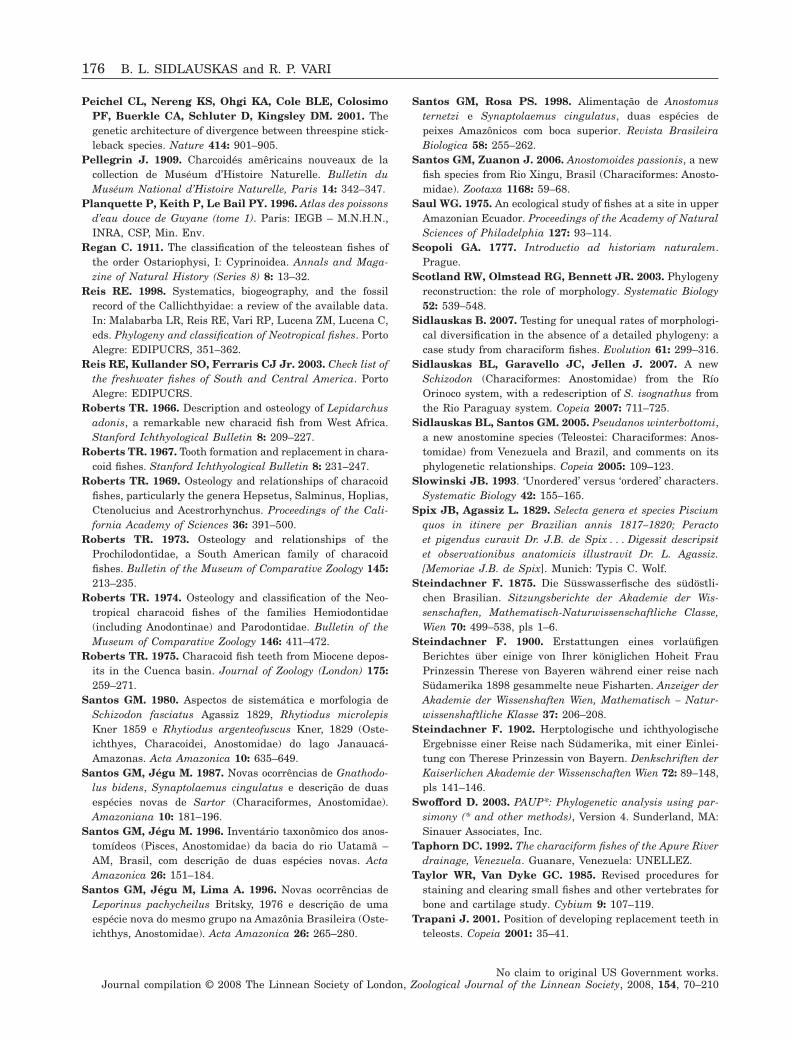
Peichel CL, Nereng KS, Ohgi KA, Cole BLE, ColosimoPF, Buerkle CA, Schluter D, Kingsley DM. 2001. Thegenetic architecture of divergence between threespine stick-leback species. Nature 414: 901–905.
Pellegrin J. 1909. Charcoidés amêricains nouveaux de lacollection de Muséum d’Histoire Naturelle. Bulletin duMuséum National d’Histoire Naturelle, Paris 14: 342–347.
Planquette P, Keith P, Le Bail PY. 1996. Atlas des poissonsd’eau douce de Guyane (tome 1). Paris: IEGB – M.N.H.N.,INRA, CSP, Min. Env.
Regan C. 1911. The classification of the teleostean fishes ofthe order Ostariophysi, I: Cyprinoidea. Annals and Maga-zine of Natural History (Series 8) 8: 13–32.
Reis RE. 1998. Systematics, biogeography, and the fossilrecord of the Callichthyidae: a review of the available data.In: Malabarba LR, Reis RE, Vari RP, Lucena ZM, Lucena C,eds. Phylogeny and classification of Neotropical fishes. PortoAlegre: EDIPUCRS, 351–362.
Reis RE, Kullander SO, Ferraris CJ Jr. 2003. Check list ofthe freshwater fishes of South and Central America. PortoAlegre: EDIPUCRS.
Roberts TR. 1966. Description and osteology of Lepidarchusadonis, a remarkable new characid fish from West Africa.Stanford Ichthyological Bulletin 8: 209–227.
Roberts TR. 1967. Tooth formation and replacement in chara-coid fishes. Stanford Ichthyological Bulletin 8: 231–247.
Roberts TR. 1969. Osteology and relationships of characoidfishes, particularly the genera Hepsetus, Salminus, Hoplias,Ctenolucius and Acestrorhynchus. Proceedings of the Cali-fornia Academy of Sciences 36: 391–500.
Roberts TR. 1973. Osteology and relationships of theProchilodontidae, a South American family of characoidfishes. Bulletin of the Museum of Comparative Zoology 145:213–235.
Roberts TR. 1974. Osteology and classification of the Neo-tropical characoid fishes of the families Hemiodontidae(including Anodontinae) and Parodontidae. Bulletin of theMuseum of Comparative Zoology 146: 411–472.
Roberts TR. 1975. Characoid fish teeth from Miocene depos-its in the Cuenca basin. Journal of Zoology (London) 175:259–271.
Santos GM. 1980. Aspectos de sistemática e morfologia deSchizodon fasciatus Agassiz 1829, Rhytiodus microlepisKner 1859 e Rhytiodus argenteofuscus Kner, 1829 (Oste-ichthyes, Characoidei, Anostomidae) do lago Janauacá-Amazonas. Acta Amazonica 10: 635–649.
Santos GM, Jégu M. 1987. Novas ocorrências de Gnathodo-lus bidens, Synaptolaemus cingulatus e descrição de duasespécies novas de Sartor (Characiformes, Anostomidae).Amazoniana 10: 181–196.
Santos GM, Jégu M. 1996. Inventário taxonômico dos anos-tomídeos (Pisces, Anostomidae) da bacia do rio Uatamã –AM, Brasil, com descrição de duas espécies novas. ActaAmazonica 26: 151–184.
Santos GM, Jégu M, Lima A. 1996. Novas ocorrências deLeporinus pachycheilus Britsky, 1976 e descrição de umaespécie nova do mesmo grupo na Amazônia Brasileira (Oste-ichthys, Anostomidae). Acta Amazonica 26: 265–280.
Santos GM, Rosa PS. 1998. Alimentação de Anostomusternetzi e Synaptolaemus cingulatus, duas espécies depeixes Amazônicos com boca superior. Revista BrasileiraBiologica 58: 255–262.
Santos GM, Zuanon J. 2006. Anostomoides passionis, a newfish species from Rio Xingu, Brasil (Characiformes: Anosto-midae). Zootaxa 1168: 59–68.
Saul WG. 1975. An ecological study of fishes at a site in upperAmazonian Ecuador. Proceedings of the Academy of NaturalSciences of Philadelphia 127: 93–114.
Scopoli GA. 1777. Introductio ad historiam naturalem.Prague.
Scotland RW, Olmstead RG, Bennett JR. 2003. Phylogenyreconstruction: the role of morphology. Systematic Biology52: 539–548.
Sidlauskas B. 2007. Testing for unequal rates of morphologi-cal diversification in the absence of a detailed phylogeny: acase study from characiform fishes. Evolution 61: 299–316.
Sidlauskas BL, Garavello JC, Jellen J. 2007. A newSchizodon (Characiformes: Anostomidae) from the RíoOrinoco system, with a redescription of S. isognathus fromthe Rio Paraguay system. Copeia 2007: 711–725.
Sidlauskas BL, Santos GM. 2005. Pseudanos winterbottomi,a new anostomine species (Teleostei: Characiformes: Anos-tomidae) from Venezuela and Brazil, and comments on itsphylogenetic relationships. Copeia 2005: 109–123.
Slowinski JB. 1993. ‘Unordered’ versus ‘ordered’ characters.Systematic Biology 42: 155–165.
Spix JB, Agassiz L. 1829. Selecta genera et species Pisciumquos in itinere per Brazilian annis 1817–1820; Peractoet pigendus curavit Dr. J.B. de Spix . . . Digessit descripsitet observationibus anatomicis illustravit Dr. L. Agassiz.[Memoriae J.B. de Spix]. Munich: Typis C. Wolf.
Steindachner F. 1875. Die Süsswasserfische des südöstli-chen Brasilian. Sitzungsberichte der Akademie der Wis-senschaften, Mathematisch-Naturwissenschaftliche Classe,Wien 70: 499–538, pls 1–6.
Steindachner F. 1900. Erstattungen eines vorlaüfigenBerichtes über einige von Ihrer königlichen Hoheit FrauPrinzessin Therese von Bayeren während einer reise nachSüdamerika 1898 gesammelte neue Fisharten. Anzeiger derAkademie der Wissenshaften Wien, Mathematisch – Natur-wissenshaftliche Klasse 37: 206–208.
Steindachner F. 1902. Herptologische und ichthyologischeErgebnisse einer Reise nach Südamerika, mit einer Einlei-tung con Therese Prinzessin von Bayern. Denkschriften derKaiserlichen Akademie der Wissenschaften Wien 72: 89–148,pls 141–146.
Swofford D. 2003. PAUP*: Phylogenetic analysis using par-simony (* and other methods), Version 4. Sunderland, MA:Sinauer Associates, Inc.
Taphorn DC. 1992. The characiform fishes of the Apure Riverdrainage, Venezuela. Guanare, Venezuela: UNELLEZ.
Taylor WR, Van Dyke GC. 1985. Revised procedures forstaining and clearing small fishes and other vertebrates forbone and cartilage study. Cybium 9: 107–119.
Trapani J. 2001. Position of developing replacement teeth inteleosts. Copeia 2001: 35–41.
176 B. L. SIDLAUSKAS and R. P. VARI
No claim to original US Government works.Journal compilation © 2008 The Linnean Society of London, Zoological Journal of the Linnean Society, 2008, 154, 70–210
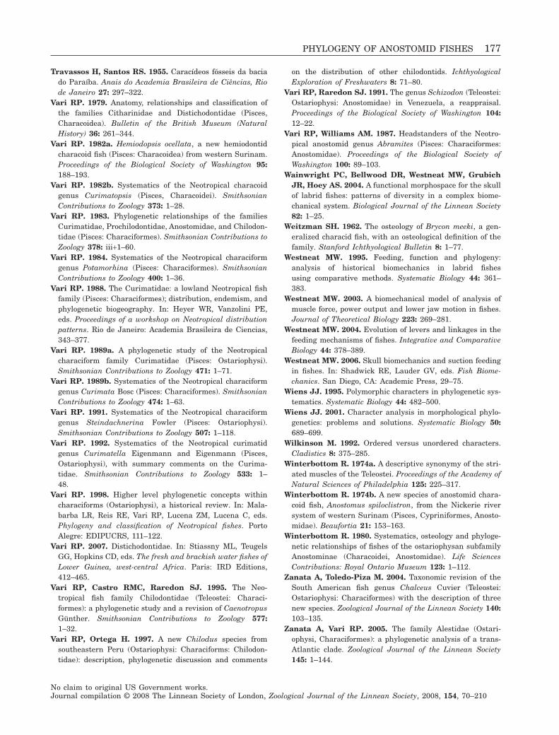
Travassos H, Santos RS. 1955. Caracídeos fósseis da baciado Paraíba. Anais do Academia Brasileira de Ciências, Riode Janeiro 27: 297–322.
Vari RP. 1979. Anatomy, relationships and classification ofthe families Citharinidae and Distichodontidae (Pisces,Characoidea). Bulletin of the British Museum (NaturalHistory) 36: 261–344.
Vari RP. 1982a. Hemiodopsis ocellata, a new hemiodontidcharacoid fish (Pisces: Characoidea) from western Surinam.Proceedings of the Biological Society of Washington 95:188–193.
Vari RP. 1982b. Systematics of the Neotropical characoidgenus Curimatopsis (Pisces, Characoidei). SmithsonianContributions to Zoology 373: 1–28.
Vari RP. 1983. Phylogenetic relationships of the familiesCurimatidae, Prochilodontidae, Anostomidae, and Chilodon-tidae (Pisces: Characiformes). Smithsonian Contributions toZoology 378: iii+1–60.
Vari RP. 1984. Systematics of the Neotropical characiformgenus Potamorhina (Pisces: Characiformes). SmithsonianContributions to Zoology 400: 1–36.
Vari RP. 1988. The Curimatidae: a lowland Neotropical fishfamily (Pisces: Characiformes); distribution, endemism, andphylogenetic biogeography. In: Heyer WR, Vanzolini PE,eds. Proceedings of a workshop on Neotropical distributionpatterns. Rio de Janeiro: Academia Brasileira de Ciencias,343–377.
Vari RP. 1989a. A phylogenetic study of the Neotropicalcharaciform family Curimatidae (Pisces: Ostariophysi).Smithsonian Contributions to Zoology 471: 1–71.
Vari RP. 1989b. Systematics of the Neotropical characiformgenus Curimata Bosc (Pisces: Characiformes). SmithsonianContributions to Zoology 474: 1–63.
Vari RP. 1991. Systematics of the Neotropical characiformgenus Steindachnerina Fowler (Pisces: Ostariophysi).Smithsonian Contributions to Zoology 507: 1–118.
Vari RP. 1992. Systematics of the Neotropical curimatidgenus Curimatella Eigenmann and Eigenmann (Pisces,Ostariophysi), with summary comments on the Curima-tidae. Smithsonian Contributions to Zoology 533: 1–48.
Vari RP. 1998. Higher level phylogenetic concepts withincharaciforms (Ostariophysi), a historical review. In: Mala-barba LR, Reis RE, Vari RP, Lucena ZM, Lucena C, eds.Phylogeny and classification of Neotropical fishes. PortoAlegre: EDIPUCRS, 111–122.
Vari RP. 2007. Distichodontidae. In: Stiassny ML, TeugelsGG, Hopkins CD, eds. The fresh and brackish water fishes ofLower Guinea, west-central Africa. Paris: IRD Editions,412–465.
Vari RP, Castro RMC, Raredon SJ. 1995. The Neo-tropical fish family Chilodontidae (Teleostei: Characi-formes): a phylogenetic study and a revision of CaenotropusGünther. Smithsonian Contributions to Zoology 577:1–32.
Vari RP, Ortega H. 1997. A new Chilodus species fromsoutheastern Peru (Ostariophysi: Characiforms: Chilodon-tidae): description, phylogenetic discussion and comments
on the distribution of other chilodontids. IchthyologicalExploration of Freshwaters 8: 71–80.
Vari RP, Raredon SJ. 1991. The genus Schizodon (Teleostei:Ostariophysi: Anostomidae) in Venezuela, a reappraisal.Proceedings of the Biological Society of Washington 104:12–22.
Vari RP, Williams AM. 1987. Headstanders of the Neotro-pical anostomid genus Abramites (Pisces: Characiformes:Anostomidae). Proceedings of the Biological Society ofWashington 100: 89–103.
Wainwright PC, Bellwood DR, Westneat MW, GrubichJR, Hoey AS. 2004. A functional morphospace for the skullof labrid fishes: patterns of diversity in a complex biome-chanical system. Biological Journal of the Linnean Society82: 1–25.
Weitzman SH. 1962. The osteology of Brycon meeki, a gen-eralized characid fish, with an osteological definition of thefamily. Stanford Ichthyological Bulletin 8: 1–77.
Westneat MW. 1995. Feeding, function and phylogeny:analysis of historical biomechanics in labrid fishesusing comparative methods. Systematic Biology 44: 361–383.
Westneat MW. 2003. A biomechanical model of analysis ofmuscle force, power output and lower jaw motion in fishes.Journal of Theoretical Biology 223: 269–281.
Westneat MW. 2004. Evolution of levers and linkages in thefeeding mechanisms of fishes. Integrative and ComparativeBiology 44: 378–389.
Westneat MW. 2006. Skull biomechanics and suction feedingin fishes. In: Shadwick RE, Lauder GV, eds. Fish Biome-chanics. San Diego, CA: Academic Press, 29–75.
Wiens JJ. 1995. Polymorphic characters in phylogenetic sys-tematics. Systematic Biology 44: 482–500.
Wiens JJ. 2001. Character analysis in morphological phylo-genetics: problems and solutions. Systematic Biology 50:689–699.
Wilkinson M. 1992. Ordered versus unordered characters.Cladistics 8: 375–285.
Winterbottom R. 1974a. A descriptive synonymy of the stri-ated muscles of the Teleostei. Proceedings of the Academy ofNatural Sciences of Philadelphia 125: 225–317.
Winterbottom R. 1974b. A new species of anostomid chara-coid fish, Anostomus spiloclistron, from the Nickerie riversystem of western Surinam (Pisces, Cypriniformes, Anosto-midae). Beaufortia 21: 153–163.
Winterbottom R. 1980. Systematics, osteology and phyloge-netic relationships of fishes of the ostariophysan subfamilyAnostominae (Characoidei, Anostomidae). Life SciencesContributions: Royal Ontario Museum 123: 1–112.
Zanata A, Toledo-Piza M. 2004. Taxonomic revision of theSouth American fish genus Chalceus Cuvier (Teleostei:Ostariophysi: Characiformes) with the description of threenew species. Zoological Journal of the Linnean Society 140:103–135.
Zanata A, Vari RP. 2005. The family Alestidae (Ostari-ophysi, Characiformes): a phylogenetic analysis of a trans-Atlantic clade. Zoological Journal of the Linnean Society145: 1–144.
PHYLOGENY OF ANOSTOMID FISHES 177
No claim to original US Government works.Journal compilation © 2008 The Linnean Society of London, Zoological Journal of the Linnean Society, 2008, 154, 70–210
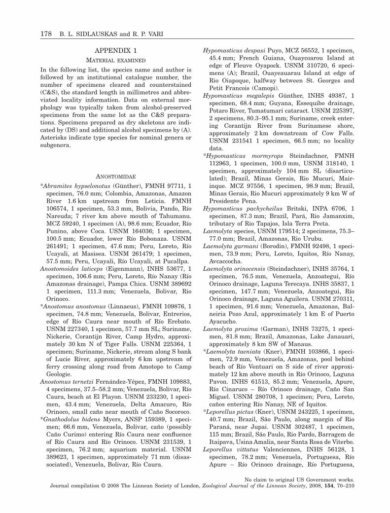
APPENDIX 1MATERIAL EXAMINED
In the following list, the species name and author isfollowed by an institutional catalogue number, thenumber of specimens cleared and counterstained(C&S), the standard length in millimetres and abbre-viated locality information. Data on external mor-phology was typically taken from alcohol-preservedspecimens from the same lot as the C&S prepara-tions. Specimens prepared as dry skeletons are indi-cated by (DS) and additional alcohol specimens by (A).Asterisks indicate type species for nominal genera orsubgenera.
ANOSTOMIDAE
*Abramites hypselonotus (Günther), FMNH 97711, 1specimen, 76.0 mm; Colombia, Amazonas, AmazonRiver 1.6 km upstream from Leticia. FMNH106574, 1 specimen, 53.3 mm, Bolivia, Pando, RíoNareuda; 7 river km above mouth of Tahumanu.MCZ 59240, 1 specimen (A), 98.6 mm; Ecuador, RíoPunino, above Coca. USNM 164036; 1 specimen,100.5 mm; Ecuador, lower Río Bobonaza. USNM261491; 1 specimen, 47.6 mm; Peru, Loreto, RíoUcayali, at Masisea. USNM 261479; 1 specimen,57.5 mm; Peru, Ucayali, Río Ucayali, at Pucallpa.
Anostomoides laticeps (Eigenmann), INHS 53677, 1specimen, 106.6 mm; Peru, Loreto, Río Nanay (RíoAmazonas drainage), Pampa Chica. USNM 3896921 specimen, 111.3 mm; Venezuela, Bolivar, RioOrinoco.
*Anostomus anostomus (Linnaeus), FMNH 109876, 1specimen, 74.8 mm; Venezuela, Bolivar, Entrerios,edge of Río Caura near mouth of Río Erebato.USNM 227340, 1 specimen, 57.7 mm SL; Suriname,Nickerie, Corantijn River, Camp Hydro, approxi-mately 30 km N of Tiger Falls. USNM 225364, 1specimen; Suriname, Nickerie, stream along S bankof Lucie River, approximately 6 km upstream offerry crossing along road from Amotopo to CampGeologie.
Anostomus ternetzi Fernández-Yépez, FMNH 109883,4 specimens, 37.5–58.2 mm; Venezuela, Bolivar, RíoCaura, beach at El Playon. USNM 233230, 1 speci-men, 43.4 mm; Venezuela, Delta Amacuro, RíoOrinoco, small caño near mouth of Caño Socoroco.
*Gnathodolus bidens Myers, ANSP 159389, 1 speci-men; 66.6 mm, Venezuela, Bolivar, caño (possiblyCaño Curimo) entering Río Caura near confluenceof Río Caura and Río Orinoco. USNM 231539, 1specimen, 76.2 mm; aquarium material. USNM389623, 1 specimen, approximately 71 mm (disas-sociated), Venezuela, Bolivar, Río Caura.
Hypomasticus despaxi Puyo, MCZ 56552, 1 specimen,45.4 mm; French Guiana, Ouaycoarou Island atedge of Fleuve Oyapock. USNM 310720, 6 speci-mens (A); Brazil, Ouayeauarau Island at edge ofRio Oiapoque, halfway between St. Georges andPetit Francois (Camopi).
Hypomasticus megalepis Günther, INHS 49387, 1specimen, 68.4 mm; Guyana, Essequibo drainage,Potaro River, Tumatumari cataract. USNM 225397,2 specimens, 80.3–95.1 mm; Suriname, creek enter-ing Corantijn River from Surinamese shore,approximately 2 km downstream of Cow Falls.USNM 231541 1 specimen, 66.5 mm; no localitydata.
*Hypomasticus mormyrops Steindachner, FMNH112963, 1 specimen, 100.0 mm, USNM 318140, 1specimen, approximately 104 mm SL (disarticu-lated); Brazil, Minas Gerais, Rio Mucuri, Mair-inque. MCZ 97556, 1 specimen, 98.9 mm; Brazil,Minas Gerais, Rio Mucuri approximately 9 km W ofPresidente Pena.
Hypomasticus pachycheilus Britski, INPA 6706, 1specimen, 87.3 mm; Brazil, Pará, Rio Jamanxim,tributary of Rio Tapajos, Isla Terra Preta.
Laemolyta species, USNM 179514; 2 specimens, 75.3–77.0 mm; Brazil, Amazonas, Rio Urubu.
Laemolyta garmani (Borodin), FMNH 92498, 1 speci-men, 73.9 mm; Peru, Loreto, Iquitos, Río Nanay,Avcaccocha.
Laemolyta orinocensis (Steindachner), INHS 35764, 1specimen, 76.5 mm, Venezuela, Anzoategui, RíoOrinoco drainage, Laguna Terecaya. INHS 35837, 1specimen, 147.7 mm; Venezuela, Anzoategui, RíoOrinoco drainage, Laguna Aguilera. USNM 270311,1 specimen, 91.6 mm; Venezuela, Amazonas, Bal-neiria Pozo Azul, approximately 1 km E of PuertoAyacucho.
Laemolyta proxima (Garman), INHS 73275, 1 speci-men, 81.8 mm; Brazil, Amazonas, Lake Janauari,approximately 8 km SW of Manaus.
*Laemolyta taeniata (Kner), FMNH 103866, 1 speci-men, 72.9 mm, Venezuela, Amazonas, pool behindbeach of Río Ventuari on S side of river approxi-mately 12 km above mouth in Río Orinoco, LagunaPavon. INHS 61513, 85.2 mm; Venezuela, Apure,Río Cinaruco – Río Orinoco drainage, Caño SanMiguel. USNM 280708, 1 specimen; Peru, Loreto,caños entering Río Nanay, NE of Iquitos.
*Leporellus pictus (Kner), USNM 243225, 1 specimen,40.7 mm; Brazil, São Paulo, along margin of RioParaná, near Jupai. USNM 302487, 1 specimen,115 mm; Brazil, São Paulo, Rio Pardo, Barragem deItaipava, Usina Amalia, near Santa Rosa de Viterbo.
Leporellus vittatus Valenciennes, INHS 56128, 1specimen, 78.2 mm; Venezuela, Portuguesa, RíoApure – Río Orinoco drainage, Río Portuguesa,
178 B. L. SIDLAUSKAS and R. P. VARI
No claim to original US Government works.Journal compilation © 2008 The Linnean Society of London, Zoological Journal of the Linnean Society, 2008, 154, 70–210
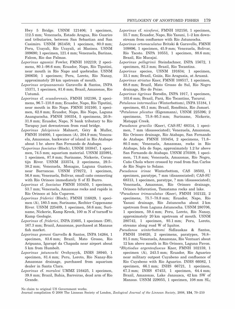
Hwy 5 Bridge. USNM 121406; 1 specimen,112.5 mm; Venezuela, Estado Aragua, Río Guaricoand tributaries, between San Sebastian and SanCasimiro. USNM 261458; 1 specimen, 80.0 mm;Peru, Ucayali, Río Ucayali, at Masisea. USNM389690; 1 specimen, 121.4 mm; Venezuela, Barinas,Falcon, Rio das Palmas.
Leporinus agassizi Fowler, FMNH 102219; 2 speci-mens, 80.1–98.6 mm; Ecuador, Napo, Río Tiputini,near mouth in Río Napo and quebradas. USNM280656; 1 specimen; Peru, Loreto, Río Nanay,approximately 20 km upstream of mouth.
Leporinus aripuanaensis Garavello & Santos, INPA15371, 1 specimen, 81.6 mm; Brazil, Amazonas, RioUatumâ.
Leporinus cf. ecuadorensis, FMNH 102198, 2 speci-mens, 96.7–110.8 mm; Ecuador, Napo, Río Tiputini,near mouth in Río Napo. FMNH 102160, 1 speci-men, 62.8 mm; Ecuador, Napo, Río Napo, LagunaAnangucocha. FMNH 100354, 5 specimens, 20.9–31.8 mm; Ecuador, Napo, N bank tributary to RíoTarapuy just downstream from road bridge.
Leporinus falcipinnis Mahnert, Géry & Muller,FMNH 104000, 1 specimen (A), 204.9 mm; Venezu-ela, Amazonas, backwater of island in Rio Atabapoabout 1 hr. above San Fernando de Atabapo.
*Leporinus fasciatus (Bloch), USNM 103847, 1 speci-men, 74.5 mm; aquarium material. USNM 225991,1 specimen, 87.8 mm; Suriname, Nickerie, Coran-tijn River. USNM 233574, 2 specimens, 29.5–38.2 mm; Venezuela, Monagas, Laguna Guatero,near Barrancas. USNM 279272, 1 specimen,56.8 mm; Venezuela, Bolivar, small caño connectingwith Río Orinoco immediately S of El Burro.
Leporinus cf. fasciatus FMNH 103450, 1 specimen,53.7 mm; Venezuela, Amazonas rocks and rapids inRío Orinoco at Isla Cupaven.
Leporinus friderici (Bloch), FMNH 116829, 1 speci-men (A), 180.5 mm; Suriname, Rechter CoppenameRiver. USNM 225409, 1 specimen, 58.6 mm; Suri-name, Nickerie, Kamp Kreek, 100 m N of turnoff toKamp Geologie.
Leporinus cf. friderici, INPA 21605, 1 specimen (DS),167.3 mm; Brazil, Amazonas, purchased at Manausfish market.
Leporinus gomesi Garvello & Santos, INPA 14264, 1specimen, 83.6 mm; Brazil, Mato Grosso, RioAripuana, Igarapé da Chapada near airport about5 km from Humbolt.
Leporinus jatuncochi Ovchynyyk, INHS 38940, 1specimen, 81.4 mm; Peru, Loreto, Río Nanay-RíoAmazonas drainage, purchased from aquariumdealer in Santa Clara.
Leporinus cf. moralesi UMMZ 216425, 1 specimen,39.8 mm; Brazil, Bahia, Barreiras, dead arm of RioGrande.
Leporinus cf. niceforoi, FMNH 102150, 1 specimen,55.7 mm; Ecuador, Napo, Río Yasuni, 1–2 km down-stream from confluence with Río Jatuncocha.
Leporinus ortomaculatus Britski & Garavello, FMNH109896, 1 specimen, 45.9 mm; Venezuela, Bolivar,Río Tacoto. INPA 10553, 1 specimen, 88.6 mm;Brazil, Rio Mucajaí.
Leporinus pellegrinii Steindachner, INPA 15672, 1specimen, 82.3 mm; Brazil, Rio Tocantins.
Leporinus species, USNM 191634; 1 specimen,33.1 mm; Brazil, Goiás, Rio Araguaia, at Aruanã.
Leporinus striatus Kner, FMNH 108517, 1 specimen,68.8 mm; Brazil, Mato Grosso do Sul, Rio Negrodrainage, Rio do Peixe.
Leporinus tigrinus Borodin, INPA 1917, 1 specimen,103.6 mm; Brazil, Pará, Rio Tocantins, Tucuruí.
Petulanos intermedius (Winterbottom), INPA 15184, 1specimen, 65.1 mm; Brazil, Rondônia, Rio Jamari.
*Petulanos plicatus (Eigenmann), USNM 225396; 2specimens, 75.8–95.3 mm; Suriname, Nickerie,Matappi Creek.
Pseudanos gracilis (Kner), CAS-SU 68314, 1 speci-men, ? mm (disassociated); Venezuela, Amazonas,Río Orinoco drainage, Río Atabapo, San Fernandode Atabapo. FMNH 103454, 2 specimens, 71.8–80.5 mm; Venezuela, Amazonas, rocks in RíoAtabapo, Isla de Sapo, approximately 1.2 hr aboveSan Fernando de Atabapo. USNM 270309, 1 speci-men, 71.8 mm; Venezuela, Amazonas, Río Negro,Caño Chola where crossed by road from San Carlosde Río Negro to Solano.
Pseudanos irinae Winterbottom, CAS 38502, 1specimen, paratype, ? mm (disassociated); CAS-SU68313, 1 specimen, paratype, ? mm (disassociated);Venezuela, Amazonas, Río Orinoco drainage,Orinoco bifurcation, Tamatama rocks and lake.
*Pseudanos trimaculatus (Kner), FMNH 102122, 2specimens, 75.7–78.8 mm; Ecuador, Napo, RíoYasuni drainage, Río Jatuncocha about 2 kmupstream from Laguna Jatuncocha. USNM 280706,1 specimen, 59.4 mm; Peru, Loreto, Río Nanay,approximately 20 km upstream of mouth. USNM280742, 1 specimen, 93.1 mm; Peru, Loreto,streams along road W of Iquitos.
Pseudanos winterbottomi Sidlauskas & Santos,FMNH 104020, 2 specimens, paratypes, 76.8–91.5 mm; Venezuela, Amazonas, Río Ventuari about12 km above mouth in Río Orinoco, Laguna Pavon.
*Rhytiodus argenteofuscus Kner, FMNH 102159, 1specimen (A), 243.5 mm; Ecuador, Rio Aguariconear military outpost Cuyabeno and confluence ofRio Cuyabeno with Rio Aguarico. INHS 66082, 1specimen, 66.1 mm; INHS 66721, 1 specimen,67.3 mm; INHS 67453, 1 specimen, 64.4 mm;Brazil, Amazonas, Lake Janauaca, 42 km SW ofManaus. USNM 229055, 1 specimen, 108 mm SL;
PHYLOGENY OF ANOSTOMID FISHES 179
No claim to original US Government works.Journal compilation © 2008 The Linnean Society of London, Zoological Journal of the Linnean Society, 2008, 154, 70–210
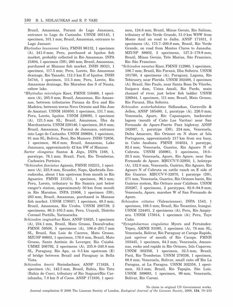
Brazil, Amazonas, Paraná do Lago Janauaca,entrance to Lago do Castanho. USNM 305145, 1specimen, 101.1 mm; Brazil, Amazonas, entrance toLago Januari.
Rhytiodus lauzannei Géry, FMNH 96132, 1 specimen(A), 241.0 mm; Peru, purchased at Iquitos fishmarket, probably collected in Río Amazonas. INPA21604, 1 specimen (DS), 260 mm; Brazil, Amazonas,purchased at Manaus fish market. INHS 39321, 1specimen, 117.5 mm; Peru, Loreto, Río Amazonasdrainage, Río Yanashi, 112.3 km E of Iquitos. INHS54744, 1 specimen, 111.5 mm; Peru, Loreto, RíoAmazonas drainage, Río Marañon due S of Nauta,oxbow lake.
*Rhytiodus microlepis Kner, FMNH 116468, 1 speci-men (A), 285.0 mm; Brazil, Amazonas, Rio Amazo-nas, between tributaries Parana da Eva and RioMadeira, between towns Novo Oriente and São Josedo Amatari. USNM 163850; 1 specimen, 131.7 mm;Peru, Loreto, Iquitos. USNM 229095, 1 specimen(A), 121.5 mm SL; Brazil, Amazonas, Ilha daMarchantaria. USNM 229146; 1 specimen, 28.0 mm;Brazil, Amazonas, Paraná de Janauaca, entranceinto Lago do Castanho. USNM 389694, 1 specimen,91 mm SL; Bolivia, Beni, Río Mamore. INHS 72856,1 specimen, 86.6 mm; Brazil, Amazonas, LakeJanauaca, approximately 42 km SW of Manaus.
Sartor elongatus Santos & Jégu, INPA 1168, 1,paratype, 76.1 mm; Brazil, Pará, Rio Trombetas,Cachoeira Porteira.
*Schizodon fasciatus Agassiz, FMNH 102211, 1 speci-men (A), 225.8 mm, Ecuador, Napo, Quebrada Zan-cudocoha, about 1 km upstream from mouth in RíoAguarico. FMNH 111351, 1 specimen, 96.1 mm;Peru, Loreto, tributary to Río Samira just belowranger’s station, approximately 50 km from mouthin Río Marañon. INPA 21606, 1 specimen (DS),283 mm; Brazil, Amazonas, purchased in Manausfish market. USNM 179507; 1 specimen, 49.3 mm;Brazil, Amazonas, Rio Urubu. USNM 280739; 2specimens, 86.2–103.5 mm; Peru, Ucayali, DistritoCoronel Portillo, Yarinacocha.
Schizodon isognathus Kner, ANSP 53825, 1 specimen(A), 234.1 mm, Brazil, Mato Grosso, Descalvados;FMNH 59508, 5 specimens (A), 106.4–201.7 mmSL, Brazil, San Luis de Caceres, Mato Grosso.MZUSP 88603, 1 specimen, 176.0 mm, Brazil, MatoGrosso, Santo Antônio do Leverger, Rio Cuiabá.UMMZ 206792, 2 specimens (A), 235.8–240.8 mmSL, Paraguay, Río Apa, ca 0.5 km upstream (E)of bridge between Brazil and Paraguay in BellaVista.
Schizodon knerii Steindachner, ANSP 171828, 1specimen (A), 142.5 mm, Brazil, Bahia, Rio Tatu(Bahia do Coco), tributary of Rio Itaguari/Rio Car-inhanha, 7.0 km S of Cocos. ANSP 171830, 1 speci-
men, 124.6 mm; Brazil, Minas Gerais, Rio Salinas,tributary of Rio Verde Grande, 51.0 km WSW fromMonte Azul on road to Jaíba. ANSP 171831, 3specimens (A), 175.7–200.9 mm, Brazil, Rio VerdeGrande, on road from Montes Claros to Janauba.MZUSP 88602, 2 specimens, 127.2–179.9 mm;Brazil, Minas Gerais, Três Marias, São Francisco,Rio São Francisco.
*Schizodon nasutus Kner, FMNH 112960, 1 specimen,106.7 mm; Brazil, Rio Paraná, Ilha Solteira. USNM181760, 4 specimens (A); Paraguay, Laguna, RíoTebicuary, near Florida. USNM 302488, 1 specimen(A); Brazil, São Paulo, near Santa Rosa De Viterbo,Itaipava dam, Usina Amali, Rio Pardo, mainchannel of river, just below fish ladder. USNM326944, 1 specimen, 111 mm; Brazil, Mato Grosso,Rio Paraná, Ilha Solteira.
Schizodon scotorhabdotus Sidlauskas, Garavello &Jellen, ANSP 165480, 1, paratype (A), 226.0 mm;Venezuela, Apure, Río Capanaparo, backwaterlagoon (mouth of Caño Las Varitas) near SanFernando de Apure-Puerto Paez highway. ANSP182897, 1, paratype (DS), 224 mm, Venezuela,Delta Amacuro, Río Orinoco on N shore at IslaPortuguesa, approximately 217 km from sea buoy,in Caño Anabata. FMNH 104024, 1 paratype,92.4 mm; Venezuela, Guarico, Río Aguaro N ofCabruta. USNM 256961, 5 specimens, 19.0–20.5 mm; Venezuela, Apure, Río Apure, near SanFernando de Apure. MBUCV-V-32950, 1, holotype(A), 132.0 mm, Venezuela, Guárico, Río Aguaro, RíoAguaro N of Cabruta on cattle ranch on E side ofRío Guarico. MBUCV-V-32970, 1 paratype (DS),271 mm, Venezuela, Guarico, Río Orinoco basin, RíoGuárico system, Río Orituco near Calabozo. USNM258267, 3 specimens, 2 paratypes, 82.8–94.9 mm;Venezuela, Apure, marshes W of San Fernando deApure.
Schizodon vittatus (Valenciennes), INPA 1543, 1specimen, 108.5 mm; Brazil, Rio Tocantins, Icangui.USNM 124401, 2 specimens (A); Peru, Río Ampiy-acu. USNM 175914, 1 specimen (A); Peru, TuyeCaño.
*Synaptolaemus cingulatus Myers and Fernández-Yépez, AMNH 91095, 1 specimen (A), 78 mm SL;Venezuela, Bolivar, Río Paraguay at Carapo Rapids,just upriver of mouth of Río Carapo. FMNH103445, 1 specimen, 64.3 mm; Venezuela, Amazo-nas, rocks and rapids in Río Orinoco, Isla Cupaven.USNM 302356, 1 specimen, 32.5 mm; Brazil,Pará, Rio Trombetas. USNM 279336, 1 specimen,88.8 mm; Venezuela, Bolivar, small caño off Río LaParagua, at La Paragua. USNM 302358, 1 speci-men, 32.3 mm; Brazil, Rio Tapajós, São Luis.USNM 389693, 1 specimen, 99 mm; Venezuela,Bolivar, Río Caroni.
180 B. L. SIDLAUSKAS and R. P. VARI
No claim to original US Government works.Journal compilation © 2008 The Linnean Society of London, Zoological Journal of the Linnean Society, 2008, 154, 70–210
CHARACIDAE
Brycon argenteus Meek and Hildebrand, USNM293136, 1 specimen, 100 mm; Panama, ComaracaKuna Yala, Quebrada Sigdi at Pingandi.
Brycon falcatus Müller and Troschel, USNM 226161,2 specimens, 71.3–78.3 mm; Suriname, Nickerie,Corantijn River at km 180, side channel of mainriver along Surinamese shore.
CHILODONTIDAE
Caenotropus maculosus (Eigenmann), USNM 231545,2 specimens, 46.5–48.2 mm; Guyana. USNM231546, 1 specimen, 108.0 mm; Suriname, Sara-maca River. USNM 389691, 1 specimen, 66.5 mm;Guyana, Tumatumari.
Caenotropus mestomorgmatos Vari, Castro &Raredon, INHS 43988, 2 specimens, 65.9–69.6 mm;Peru, Loreto, Río Amazonas drainage, Río Nanay,Pampa Chica, 4.54 km W of Iquitos.
Caenotropus labyrinthicus (Kner), FMNH 100377, 3specimens, 45.6–57.9 mm; Venezuela, Monagas,Cocos between Puerto Ordaz & Barrancas, 300 kmfrom sea buoy. USNM 231543, 1 specimen,64.8 mm; Brazil, Amazonas, Rio Negro, São Gabrieldo Cachoeira. USNM 231544, 1 specimen; Venezu-ela, Amazonas, upper Río Negro.
Chilodus gracilis Isbrücker and Nijssen, USNM232358; 5 specimens, 27.7–29.6 mm; Brazil, Pará,Rio Inhangapi, 40 km E of Belém.
Chilodus punctatus Müller and Troschel, FMNH100351, 7 specimens, 22.4–58.7 mm; Ecuador,Napo, Río Cuyabeno-Río Aguarico drainage,Laguna Avcacocha. FMNH 102061, 1 specimen,58.1 mm; Ecuador, Napo, Río Yasuni, Río Jatunco-cha, about 2 km upstream from Laguna Jatunco-cha. USNM 280444, 2 specimens, 55.0–63.5 mm;Peru, Loreto, Quebrada Corrientillo at road fromIquitos to Río Nanay. USNM 231542, 14 specimens,45.7–58.7 mm; Peru, Loreto, Río Nanay.
CITHARINIDAE
Citharinus citharus (St. Hilaire), USNM 52146, 1specimen, 218.7 mm (only gill arches C and S);Sudan, junction of Nile and Atbara Rivers.
Citharinus sp., USNM 231554, 2 specimens 56.3–64.5 mm; Burkina Faso, Upper Volta, Black VoltaRiver.
CURIMATIDAE
Curimata cyprinoides (Linneaus), USNM 225619, 1specimen, 79.2 mm; Suriname, Nickerie, Makilik-abore Kreek.
Curimata inornata Vari, USNM 268597, 1 paratype,115.7 mm; Brazil, Pará, Rio Tapajos, São Luis.
Curimata vittata (Kner), FMNH 101490, 3 specimens,40.0–49.4 mm; Ecuador, Napo, Río Aguarico drain-age, Laguna Grande de Cuyabeno and Río Cuya-beno. USNM 231434, 1 specimen, 53.7 mm; Brazil,Amazonas, Rio Negro.
Curimatella alburna (Müller & Troschel), FMNH101529, 1 specimen, 62.7 mm; Ecuador, Napo,laguna open to Río Lagartococha, about 25 kmupstream from mouth in Río Aguarico.
Curimatella dorsalis (Eigenmann & Eigenmann),USNM 243235, 2 specimens, 51.5–53.7 mm; Brazil,Amazonas, Rio Solimões, upriver of Coari.
Curimatopsis macrolepis (Steindachner), FMNH100352, 5 specimens, 25.4–41.5 mm; Ecuador,Napo, Río Yasuni, Quebrada to Río Jatuncocha,approximately 2 km upstream from Laguna Jatun-cocha. USNM 190285, 1 specimen, 34.7 mm; Peru,Loreto, Iquitos.
Curimatopsis microlepis Eigenmann & Eigenmann,USNM 268867, 1 specimen, 75.8 mm; Brazil, Ama-zonas, Rio Solimões, lago near Beruri.
Cyphocharax abramoides (Kner), USNM 267953, 1specimen, 100.3 mm; Brazil, Pará, Rio Xingu, BeloMonte.
Potamorhina laticeps (Valenciennes), USNM 121325,1 specimen, 129.3 mm; Venezuela, Zulia, LagoMaracaibo basin.
Potamorhina latior (Spix & Agassiz), FMNH 106591,1 specimen, 82.5 mm; Bolivia, Pando, Lago de laAnguilla, Tahumanu. USNM 228691, 1 specimen,97.6 mm; Brazil, Amazonas, near Manaus.
Psectrogaster curviventris Eigenmann & Kennedy,FMNH 108026, 2 specimens, 43.6–48.0 mm; Para-guay, Alto Paraguay, Riacho Coeyu, tributary of RíoParaguay.
Pseudocurimata patiae (Eigenmann), USNM 287597,1 specimen, 89.5 mm, Colombia, Narino, Río Patia.
Steindachnerina conspersa (Holmberg), USNM252224, 1 specimen, 83.2 mm; Paraguay, PresidenteHayes, off Trans-Chaco Highway at km 50.
DISTICHODONTIDAE
Distichodus sp., USNM 175436, 1 specimen, 62.5 mm;Congo, Stanley Pool.Paradistichodus dimidiatus Pellegrin, USNM
231556, 2 specimens, 45.6–46.3 mm; Ghana, DayiRiver at Gbefi.
Xenocharax spilurus Günther, USNM 227693, 1 speci-men, 89.3 mm; Gabon, Allonha II, Lac Ezanga.
HEMIODONTIDAE
Hemiodus ocellatus (Vari), USNM 225593, 1 paratype,99.6 mm; Suriname, Nickerie District, CorantijnRiver.
PHYLOGENY OF ANOSTOMID FISHES 181
No claim to original US Government works.Journal compilation © 2008 The Linnean Society of London, Zoological Journal of the Linnean Society, 2008, 154, 70–210
Hemiodus quadrimaculatus Pellegrin, FMNH116862, 2 specimens, 55.3–68.2 mm; Suriname,Morocco Sula.
PARODONTIDAE
Parodon suborbitalis Valenciennes, USNM 231552, 2specimens, 55.0–58.1 mm; Colombia, Río Salado.
Parodon guyanensis Géry, FMNH 116878, 2 speci-mens, 59.1–63.4 mm; Suriname, Sula Calliandra.
PROCHILODONTIDAE
Ichthyoelephas humeralis (Günther), FMNH 80715, 1specimen, 107.6 mm; Ecuador, Río Guayas basin,Río Palenque at Centro Cientifico Río Palenque.USNM 290374, 2 specimens, 130.4–133.6 mm;Ecuador, Quevedo fish market.
Prochilodus costatus Valenciennes, USNM 357391, 1specimen, 103.5 mm; Brazil, Rio Jetuitae basin.
Prochilodus magdalenae Steindachner, USNM 79231,2 specimens, 91.9–103.4 mm; Colombia, Calamar.
Prochilodus mariae Eigenmann, USNM 270243, 2specimens, 92.6–97.1 mm; Venezuela, Bolivar, RíoOrinoco. FMNH 103683, 1 specimen, 105.4 mm;Venezuela, Guarico, lagoon about 4 km from E sideof Río Aguaro, N of Cabruta.
Prochilodus nigricans Agassiz, USNM 280609, 1specimen, 78.5 mm; Peru, Ucayali, District CoronelPortillo, Yarinacocha.
Prochilodus reticulatus Valenciennes, USNM 121326,3 specimens, 110.7–113.1 mm; Venezuela, LagoMaracaibo basin.
Prochilodus rubrotaeniatus Jardine, USNM 225419, 2specimens, 89.0–100.4 mm; Suriname, Nickerie,Corantijn River.
Prochilodus vimboides Kner, MCZ 20169, 2 speci-mens, 93.4 and ? mm (disassociated); Brazil, Rio deJaneiro, Rio Paraiba. USNM 345752, 1 specimen,114.8 mm; Brazil, Minas Gerais, Rio Jequitai basin.
Semaprochilodus insignis (Jardine), USNM 290148, 1specimen, 102.5 mm; Brazil, Amazonas, vicinity ofManaus.
Semaprochilodus kneri (Pellegrin), USNM 279325, 1specimen, 99.3 mm; Venezuela, Amazonas, RíoOrinoco, near Puerto Ayachucho. FMNH 85634, 1specimen, 86.3 mm; Venezuela, Amazonas, ponds0.3 km S of Puerto Nuevo, towards Puerto Ayacucho.
Semaprochilodus taeniurus (Valenciennes), USNM289798, 1 specimen, 78.2 mm; Brazil, Amazonas,near Manaus.
182 B. L. SIDLAUSKAS and R. P. VARI
No claim to original US Government works.Journal compilation © 2008 The Linnean Society of London, Zoological Journal of the Linnean Society, 2008, 154, 70–210
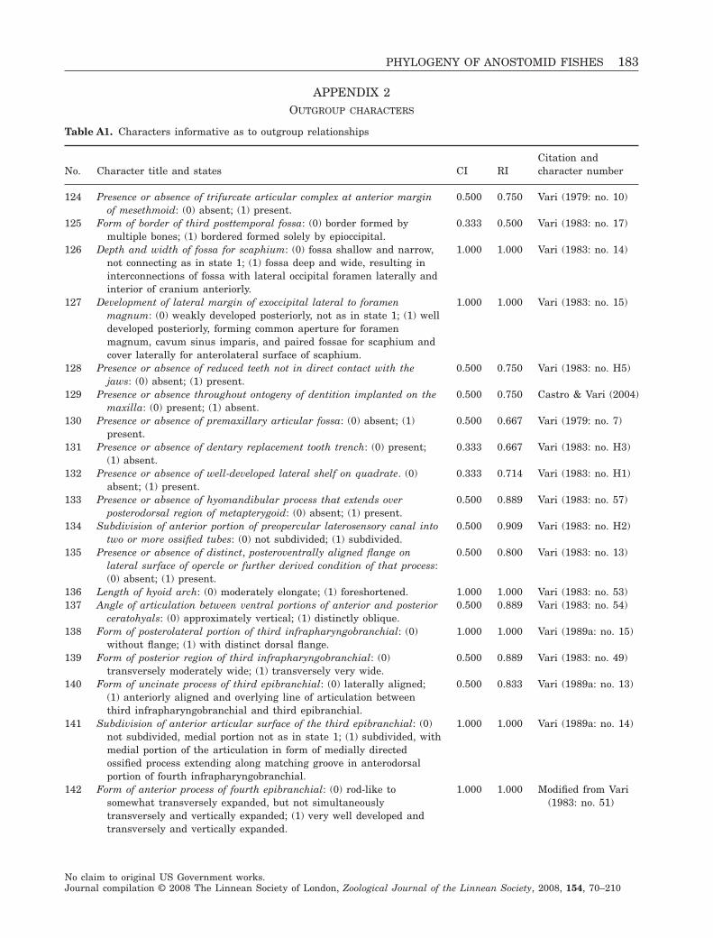
APPENDIX 2OUTGROUP CHARACTERS
Table A1. Characters informative as to outgroup relationships
No. Character title and states CI RICitation andcharacter number
124 Presence or absence of trifurcate articular complex at anterior marginof mesethmoid: (0) absent; (1) present.
0.500 0.750 Vari (1979: no. 10)
125 Form of border of third posttemporal fossa: (0) border formed bymultiple bones; (1) bordered formed solely by epioccipital.
0.333 0.500 Vari (1983: no. 17)
126 Depth and width of fossa for scaphium: (0) fossa shallow and narrow,not connecting as in state 1; (1) fossa deep and wide, resulting ininterconnections of fossa with lateral occipital foramen laterally andinterior of cranium anteriorly.
1.000 1.000 Vari (1983: no. 14)
127 Development of lateral margin of exoccipital lateral to foramenmagnum: (0) weakly developed posteriorly, not as in state 1; (1) welldeveloped posteriorly, forming common aperture for foramenmagnum, cavum sinus imparis, and paired fossae for scaphium andcover laterally for anterolateral surface of scaphium.
1.000 1.000 Vari (1983: no. 15)
128 Presence or absence of reduced teeth not in direct contact with thejaws: (0) absent; (1) present.
0.500 0.750 Vari (1983: no. H5)
129 Presence or absence throughout ontogeny of dentition implanted on themaxilla: (0) present; (1) absent.
0.500 0.750 Castro & Vari (2004)
130 Presence or absence of premaxillary articular fossa: (0) absent; (1)present.
0.500 0.667 Vari (1979: no. 7)
131 Presence or absence of dentary replacement tooth trench: (0) present;(1) absent.
0.333 0.667 Vari (1983: no. H3)
132 Presence or absence of well-developed lateral shelf on quadrate. (0)absent; (1) present.
0.333 0.714 Vari (1983: no. H1)
133 Presence or absence of hyomandibular process that extends overposterodorsal region of metapterygoid: (0) absent; (1) present.
0.500 0.889 Vari (1983: no. 57)
134 Subdivision of anterior portion of preopercular laterosensory canal intotwo or more ossified tubes: (0) not subdivided; (1) subdivided.
0.500 0.909 Vari (1983: no. H2)
135 Presence or absence of distinct, posteroventrally aligned flange onlateral surface of opercle or further derived condition of that process:(0) absent; (1) present.
0.500 0.800 Vari (1983: no. 13)
136 Length of hyoid arch: (0) moderately elongate; (1) foreshortened. 1.000 1.000 Vari (1983: no. 53)137 Angle of articulation between ventral portions of anterior and posterior
ceratohyals: (0) approximately vertical; (1) distinctly oblique.0.500 0.889 Vari (1983: no. 54)
138 Form of posterolateral portion of third infrapharyngobranchial: (0)without flange; (1) with distinct dorsal flange.
1.000 1.000 Vari (1989a: no. 15)
139 Form of posterior region of third infrapharyngobranchial: (0)transversely moderately wide; (1) transversely very wide.
0.500 0.889 Vari (1983: no. 49)
140 Form of uncinate process of third epibranchial: (0) laterally aligned;(1) anteriorly aligned and overlying line of articulation betweenthird infrapharyngobranchial and third epibranchial.
0.500 0.833 Vari (1989a: no. 13)
141 Subdivision of anterior articular surface of the third epibranchial: (0)not subdivided, medial portion not as in state 1; (1) subdivided, withmedial portion of the articulation in form of medially directedossified process extending along matching groove in anterodorsalportion of fourth infrapharyngobranchial.
1.000 1.000 Vari (1989a: no. 14)
142 Form of anterior process of fourth epibranchial: (0) rod-like tosomewhat transversely expanded, but not simultaneouslytransversely and vertically expanded; (1) very well developed andtransversely and vertically expanded.
1.000 1.000 Modified from Vari(1983: no. 51)
PHYLOGENY OF ANOSTOMID FISHES 183
No claim to original US Government works.Journal compilation © 2008 The Linnean Society of London, Zoological Journal of the Linnean Society, 2008, 154, 70–210
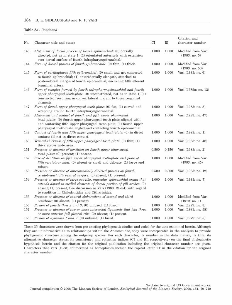
Table A1. Continued
No. Character title and states CI RICitation andcharacter number
143 Alignment of dorsal process of fourth epibranchial: (0) dorsallydirected, not as in state 1; (1) orientated anteriorly with extensionover dorsal surface of fourth infrapharyngobranchial.
1.000 1.000 Modified from Vari(1983: no. 5)
144 Form of dorsal process of fourth epibranchial: (0) thin; (1) thick. 1.000 1.000 Modified from Vari(1983: no. 50)
145 Form of cartilaginous fifth epibranchial: (0) small and not connectedto fourth epibranchial; (1) anterodorsally elongate, attached toposterodorsal margin of fourth epibranchial, encircling fifth efferentbranchial artery.
1.000 1.000 Vari (1983: no. 6)
146 Form of complex formed by fourth infrapharyngobranchial and fourthupper pharyngeal tooth-plate: (0) unconstricted, not as in state 1; (1)constricted, resulting in convex lateral margin to those conjoinedelements.
1.000 1.000 Vari (1989a: no. 12)
147 Form of fourth upper pharyngeal tooth-plate: (0) flat; (1) curved andwrapping around fourth infrapharyngobranchial.
1.000 1.000 Vari (1983: no. 8)
148 Alignment and contact of fourth and fifth upper pharyngealtooth-plates: (0) fourth upper pharyngeal tooth-plate aligned withand contacting fifth upper pharyngeal tooth-plate; (1) fourth upperpharyngeal tooth-plate angled and contacting fourth epibranchial.
1.000 1.000 Vari (1983: no. 47)
149 Contact of fourth and fifth upper pharyngeal tooth-plate: (0) in directcontact; (1) not in direct contact.
1.000 1.000 Vari (1983: no. 1)
150 Vertical thickness of fifth upper pharyngeal tooth-plate: (0) thin; (1)thick across wide area.
1.000 1.000 Vari (1983: no. 48)
151 Presence or absence of dentition on fourth upper pharyngealtooth-plate: (0) present; (1) absent.
0.500 0.750 Vari (1983: no. 2)
152 Size of dentition on fifth upper pharyngeal tooth-plate and plate offifth ceratobranchial. (0) absent or small and delicate; (1) large androbust.
1.000 1.000 Modified from Vari(1983: no. 45)
153 Presence or absence of anteromedially directed process on fourthceratobranchial’s ventral surface: (0) absent; (1) present.
0.500 0.800 Vari (1983: no. 12)
154 Presence or absence of large sac-like, muscular epibranchial organ thatextends dorsal to medial elements of dorsal portion of gill arches: (0)absent; (1) present, See discussion in Vari (1983: 21–24) with regardto condition in Chilodontidae and Citharinidae.
1.000 1.000 Vari (1983: no. 7)
155 Presence or absence of ventral elaborations of second and thirdvertebrae: (0) absent; (1) present.
1.000 1.000 Modified from Vari(1979: no. 1)
156 Fusion of postcleithra 2 and 3; (0) unfused; (1) fused. 1.000 1.000 Vari (1979: no. 3)157 Presence or absence of two or more intercostal ligaments that join three
or more anterior full pleural ribs: (0) absent; (1) present.1.000 1.000 Vari (1983: no. 58)
158 Fusion of hypurals 1 and 2: (0) unfused; (1) fused. 1.000 1.000 Vari (1979: no. 5)
These 35 characters were drawn from pre-existing phylogenetic studies and coded for the taxa examined herein. Althoughthey are uninformative as to relationships within the Anostomidae, they were incorporated in the analysis to providephylogenetic structure among the outgroup species. For each character, its number in the data matrix, its title, itsalternative character states, its consistency and retention indices (CI and RI, respectively) on the final phylogenetichypothesis herein and the citation for the original publication including the original character number are given.Characters that Vari (1983) enumerated as homoplasies include the capital letter ‘H’ in the citation for the originalcharacter number.
184 B. L. SIDLAUSKAS and R. P. VARI
No claim to original US Government works.Journal compilation © 2008 The Linnean Society of London, Zoological Journal of the Linnean Society, 2008, 154, 70–210
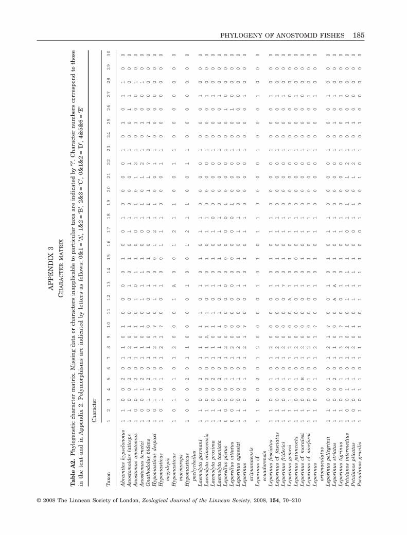
AP
PE
ND
IX3
CH
AR
AC
TE
RM
AT
RIX
Tab
leA
2.P
hyl
ogen
etic
char
acte
rm
atri
x.M
issi
ng
data
orch
arac
ters
inap
plic
able
topa
rtic
ula
rta
xaar
ein
dica
ted
by‘?
’.C
har
acte
rn
um
bers
corr
espo
nd
toth
ose
inth
ete
xtan
din
App
endi
x2.
Pol
ymor
phis
ms
are
indi
cate
dby
lett
ers
asfo
llow
s:0&
1=
‘A’,
1&2
=‘B
’,2&
3=
‘C’,
0&1&
2=
’D’,
4&5&
6=
‘E’
Taxo
n
Ch
arac
ter
12
34
56
78
910
11
12
13
14
15
16
17
18
19
20
21
22
23
24
25
26
27
28
29
30
Abr
amit
esh
ypse
lon
otu
s1
10
02
01
10
10
00
01
00
10
00
01
01
01
10
0A
nos
tom
oid
esla
tice
ps1
10
02
01
20
10
01
01
01
10
00
11
00
11
00
0A
nos
tom
us
anos
tom
us
10
01
20
11
01
01
01
10
01
10
12
10
10
01
00
An
osto
mu
ste
rnet
zi1
00
12
01
10
10
11
11
00
11
01
21
01
00
10
0G
nat
hod
olu
sbi
den
s1
01
12
01
10
00
01
01
00
11
11
?0
?1
00
00
0H
ypom
asti
cus
des
paxi
10
10
10
12
1?
01
10
01
21
10
01
10
00
00
00
Hyp
omas
ticu
sm
egal
epis
10
10
10
12
1?
00
10
01
11
00
01
10
00
00
00
Hyp
omas
ticu
sm
orm
yrop
s1
01
01
01
20
00
1A
00
12
11
00
11
00
00
00
0
Hyp
omas
ticu
spa
chyc
hei
lus
10
00
20
11
00
00
10
01
21
10
01
10
00
00
00
Lae
mol
yta
garm
ani
11
00
20
11
01
10
10
10
11
00
00
10
00
10
00
Lae
mol
yta
orin
ocen
sis
11
00
20
11
A1
10
10
10
11
00
00
10
00
10
00
Lae
mol
yta
prox
ima
11
00
20
11
01
10
10
10
10
00
00
10
00
10
00
Lae
mol
yta
taen
iata
11
00
20
11
01
10
10
10
11
00
00
10
00
10
00
Lep
orel
lus
pict
us
00
00
10
12
00
00
00
00
00
10
00
10
01
00
00
Lep
orel
lus
vitt
atu
s0
00
01
01
20
00
00
00
00
01
00
01
00
10
00
0L
epor
inu
sag
assi
zi1
10
01
01
20
00
01
01
01
10
00
01
00
00
00
0L
epor
inu
sar
ipu
anae
nsi
s1
10
01
01
21
?0
01
01
01
10
00
01
00
01
00
0
Lep
orin
us
cf.
ecu
ador
ensi
s1
10
02
01
20
00
01
01
01
10
00
01
00
01
00
0
Lep
orin
us
fasc
iatu
s1
10
01
01
20
00
01
01
01
10
00
01
00
01
00
0L
epor
inu
scf
.fa
scia
tus
11
00
10
12
00
00
10
10
11
00
00
10
00
10
00
Lep
orin
us
frid
eric
i1
10
01
01
20
00
0?
01
01
10
00
01
00
01
00
0L
epor
inu
sgo
mes
i1
11
01
01
20
00
A1
01
01
10
00
01
00
00
00
0L
epor
inu
sja
tun
coch
i1
10
01
01
20
00
01
00
01
10
00
01
00
01
00
0L
epor
inu
scf
.m
oral
esi
11
00
B0
12
00
00
10
10
11
00
00
10
00
00
00
Lep
orin
us
cf.
nic
efor
oi1
10
01
01
20
00
01
01
01
10
00
01
00
01
00
0L
epor
inu
sor
tom
acu
latu
s1
10
01
01
21
?0
01
01
01
10
00
01
00
01
00
0
Lep
orin
us
pell
egri
nii
11
00
10
12
1?
00
10
10
11
00
00
10
00
10
00
Lep
orin
us
stri
atu
s1
10
02
01
10
00
AA
01
01
10
00
01
01
01
00
0L
epor
inu
sti
grin
us
11
00
10
13
1?
00
10
10
11
00
00
10
00
10
00
Pet
ula
nos
inte
rmed
ius
10
01
10
12
01
01
11
10
01
10
12
10
10
00
00
Pet
ula
nos
plic
atu
s1
00
11
01
20
10
11
11
00
11
01
21
01
00
00
0P
seu
dan
osgr
acil
is1
10
11
01
10
10
11
11
00
10
01
01
01
00
00
0
PHYLOGENY OF ANOSTOMID FISHES 185
© 2008 The Linnean Society of London, Zoological Journal of the Linnean Society, 2008, 154, 70–210
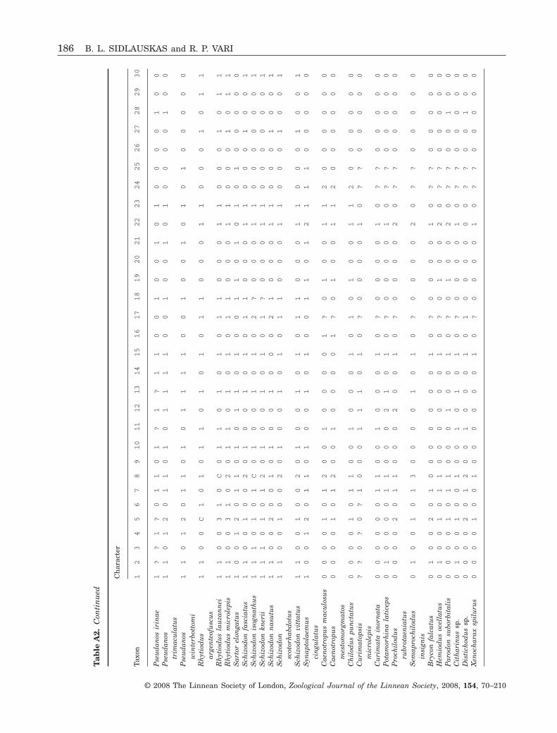
Tab
leA
2.C
onti
nu
ed
Taxo
n
Ch
arac
ter
12
34
56
78
910
11
12
13
14
15
16
17
18
19
20
21
22
23
24
25
26
27
28
29
30
Pse
ud
anos
irin
ae1
??
1?
01
10
1?
1?
11
00
10
01
01
00
00
10
0P
seu
dan
ostr
imac
ula
tus
11
01
20
11
01
01
11
10
01
00
10
10
00
01
00
Pse
ud
anos
win
terb
otto
mi
11
01
20
11
01
01
11
10
01
00
10
10
10
00
00
Rh
ytio
du
sar
gen
teof
usc
us
11
00
C1
01
01
10
10
10
11
00
01
10
00
10
11
Rh
ytio
du
sla
uza
nn
ei1
10
03
10
C0
11
01
01
01
10
00
11
00
01
01
1R
hyt
iod
us
mic
role
pis
11
00
31
02
01
10
10
10
11
00
01
10
00
10
11
Sar
tor
elon
gatu
s1
00
12
01
10
10
11
01
00
11
01
01
01
00
00
0S
chiz
odon
fasc
iatu
s1
10
01
00
20
10
01
01
01
10
00
11
00
01
00
1S
chiz
odon
isog
nat
hu
s1
11
01
01
C0
10
01
01
02
?0
00
11
00
00
00
1S
chiz
odon
kner
ii1
11
01
01
20
10
01
01
01
?0
00
11
00
00
00
1S
chiz
odon
nas
utu
s1
10
02
00
10
10
01
00
02
10
00
11
00
01
00
1S
chiz
odon
scot
orh
abd
otu
s1
10
01
00
20
10
01
01
01
10
00
11
00
01
00
1
Sch
izod
onvi
ttat
us
11
00
10
02
01
00
10
10
11
00
01
10
00
10
01
Syn
apto
laem
us
cin
gula
tus
10
01
20
11
01
00
10
10
01
10
12
11
10
00
00
Cae
not
ropu
sm
acu
losu
s0
00
01
01
20
01
00
00
1?
01
00
11
20
00
00
0C
aen
otro
pus
mes
tom
orgm
atos
00
00
10
12
00
10
00
01
?0
10
01
12
00
00
00
Ch
ilod
us
pun
ctat
us
00
00
10
11
00
10
00
10
10
10
01
12
00
00
00
Cu
rim
atop
sis
mic
role
pis
??
0?
0?
10
00
11
10
10
?0
00
01
0?
?0
00
00
Cu
rim
ata
inor
nat
a0
00
00
01
10
01
00
01
0?
00
00
10
??
00
00
0P
otam
orh
ina
lati
ceps
01
00
00
11
00
02
10
10
?0
00
01
0?
?0
00
00
Pro
chil
odu
sru
brot
aen
iatu
s0
00
02
01
10
00
20
01
0?
00
00
20
??
00
00
0
Sem
apro
chil
odu
sin
sign
is0
10
01
01
30
00
01
01
0?
00
00
20
??
00
00
0
Bry
con
falc
atu
s0
10
02
01
00
00
00
01
0?
00
00
10
??
00
00
0H
emio
du
soc
ella
tus
01
00
10
11
00
00
00
10
?0
10
02
0?
?0
00
00
Par
odon
subo
rbit
alis
00
00
10
11
00
01
00
10
?0
10
02
0?
?0
01
00
Cit
har
inu
ssp
.0
10
01
00
10
01
01
01
0?
00
00
10
??
00
00
0D
isti
chod
us
sp.
00
00
20
12
00
10
00
10
10
00
00
0?
?0
01
00
Xen
och
arax
spil
uru
s0
00
01
00
10
00
00
01
0?
00
00
10
??
00
00
0
186 B. L. SIDLAUSKAS and R. P. VARI
© 2008 The Linnean Society of London, Zoological Journal of the Linnean Society, 2008, 154, 70–210
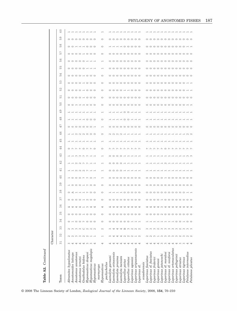
Tab
leA
2.C
onti
nu
ed
Taxo
n
Ch
arac
ter
31
32
33
34
35
36
37
38
39
40
41
42
43
44
45
46
47
48
49
50
51
52
53
54
55
56
57
58
59
60
Abr
amit
esh
ypse
lon
otu
s3
22
00
40
01
11
11
01
10
11
00
00
00
00
00
1A
nos
tom
oid
esla
tice
ps4
23
10
40
A0
01
11
?1
10
11
00
00
00
00
00
1A
nos
tom
us
anos
tom
us
43
30
04
01
00
22
3?
12
01
11
00
00
00
01
01
An
osto
mu
ste
rnet
zi4
33
00
40
10
02
23
?1
20
11
10
00
00
00
10
1G
nat
hod
olu
sbi
den
s4
2C
?1
11
11
?2
??
01
20
11
11
11
10
01
10
1H
ypom
asti
cus
des
paxi
32
20
03
2?
??
??
??
10
00
10
00
00
11
00
01
Hyp
omas
ticu
sm
egal
epis
31
20
04
00
00
11
1?
10
00
10
00
00
11
00
01
Hyp
omas
ticu
sm
orm
yrop
s4
12
00
40
00
11
11
01
01
01
00
00
01
10
00
1
Hyp
omas
ticu
spa
chyc
hei
lus
41
20
04
00
01
11
10
10
10
10
00
00
11
00
01
Lae
mol
yta
garm
ani
43
30
04
01
00
00
0?
12
01
10
00
00
00
01
01
Lae
mol
yta
orin
ocen
sis
44
40
04
01
00
00
0?
12
01
10
00
00
00
01
01
Lae
mol
yta
prox
ima
44
40
04
01
00
00
0?
12
01
10
00
00
00
01
01
Lae
mol
yta
taen
iata
44
40
04
01
00
00
0?
12
01
10
00
00
00
01
01
Lep
orel
lus
pict
us
42
B0
04
00
00
11
1?
11
00
00
01
00
00
00
01
Lep
orel
lus
vitt
atu
s4
23
00
40
00
01
11
?1
10
00
00
10
00
00
00
1L
epor
inu
sag
assi
zi4
22
10
40
00
01
11
?1
10
11
00
00
00
00
00
1L
epor
inu
sar
ipu
anae
nsi
s4
12
10
40
00
01
11
?1
10
11
00
00
00
00
00
1L
epor
inu
scf
.ec
uad
oren
sis
42
21
04
00
00
11
1?
11
01
10
00
00
00
00
01
Lep
orin
us
fasc
iatu
s4
23
10
40
00
01
11
?1
10
11
00
00
00
00
00
1L
epor
inu
scf
.fa
scia
tus
42
31
04
00
00
11
1?
11
01
10
00
00
00
00
01
Lep
orin
us
frid
eric
i4
33
10
40
00
01
11
?1
10
11
00
00
00
00
00
1L
epor
inu
sgo
mes
i4
12
10
40
00
01
11
?1
10
11
00
00
00
00
00
1L
epor
inu
sja
tun
coch
i3
33
12
31
00
01
11
?1
10
11
00
00
00
00
00
1L
epor
inu
scf
.m
oral
esi
33
31
04
00
01
12
10
11
01
10
00
00
00
00
01
Lep
orin
us
cf.
nic
efor
oi4
33
10
40
00
01
11
?1
10
11
00
00
00
00
00
1L
epor
inu
sor
tom
acu
latu
s4
33
10
40
00
01
11
?1
10
11
00
00
00
00
00
1L
epor
inu
spe
lleg
rin
ii4
33
00
40
00
01
11
?1
10
11
00
00
00
00
00
1L
epor
inu
sst
riat
us
32
20
04
00
00
11
1?
11
01
10
00
00
00
00
01
Lep
orin
us
tigr
inu
s3
33
10
40
00
01
11
?1
10
11
00
00
00
00
00
1P
etu
lan
osin
term
ediu
s4
23
00
40
10
01
13
?1
20
11
10
11
00
00
10
1P
etu
lan
ospl
icat
us
42
30
04
01
00
11
3?
12
01
11
01
10
00
01
01
PHYLOGENY OF ANOSTOMID FISHES 187
© 2008 The Linnean Society of London, Zoological Journal of the Linnean Society, 2008, 154, 70–210
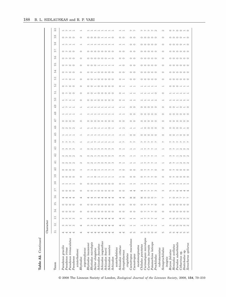
Tab
leA
2.C
onti
nu
ed
Taxo
n
Ch
arac
ter
31
32
33
34
35
36
37
38
39
40
41
42
43
44
45
46
47
48
49
50
51
52
53
54
55
56
57
58
59
60
Pse
ud
anos
grac
ilis
43
30
04
01
00
22
3?
12
01
11
00
10
00
01
01
Pse
ud
anos
irin
ae4
33
00
40
10
02
23
?1
20
1?
??
??
??
??
??
?P
seu
dan
ostr
imac
ula
tus
43
30
04
01
00
22
3?
12
01
11
00
00
00
01
01
Pse
ud
anos
win
terb
otto
mi
43
30
04
01
00
22
3?
12
01
11
00
10
00
01
01
Rh
ytio
du
sar
gen
teof
usc
us
44
30
04
00
01
22
11
11
01
10
00
00
00
01
01
Rh
ytio
du
sla
uza
nn
ei4
33
00
40
01
12
22
11
10
11
00
00
00
00
10
1R
hyt
iod
us
mic
role
pis
43
30
04
00
11
22
21
11
01
10
00
00
00
01
01
Sar
tor
elon
gatu
s4
33
01
31
10
01
11
?1
20
11
11
11
10
00
10
1S
chiz
odon
fasc
iatu
s4
44
00
40
01
13
33
11
10
11
00
00
01
00
10
1S
chiz
odon
isog
nat
hu
s4
44
00
40
01
12
23
11
10
11
00
00
01
00
11
1S
chiz
odon
kner
ii4
44
00
40
01
13
33
11
10
11
00
00
01
00
11
1S
chiz
odon
nas
utu
s4
44
00
40
01
12
23
11
00
11
00
00
01
10
11
1S
chiz
odon
scot
orh
abd
otu
s4
44
00
40
01
13
33
11
10
11
00
00
01
00
10
1
Sch
izod
onvi
ttat
us
44
40
04
00
11
33
31
11
01
10
00
00
10
01
01
Syn
apto
laem
us
cin
gula
tus
43
30
04
01
00
13
1?
12
01
11
11
10
00
01
01
Cae
not
ropu
sm
acu
losu
s5
11
00
B4
10
01
11
?0
10
00
11
10
00
00
00
?C
aen
otro
pus
mes
tom
orgm
atos
51
10
0E
41
00
11
1?
01
00
01
11
00
00
00
0?
Ch
ilod
us
pun
ctat
us
51
10
0E
41
00
11
1?
01
00
01
11
00
00
00
0?
Cu
rim
atop
sis
mic
role
pis
0?
??
?0
??
??
??
??
00
00
01
11
00
00
00
0?
Cu
rim
ata
inor
nat
a0
??
??
0?
??
??
??
?0
00
00
11
10
00
00
00
?P
otam
orh
ina
lati
ceps
0?
??
?0
??
??
??
??
00
00
01
11
00
00
00
0?
Pro
chil
odu
sru
brot
aen
iatu
s5
11
00
73
10
01
11
?0
00
00
10
10
00
00
00
2
Sem
apro
chil
odu
sin
sign
is5
11
00
73
10
01
11
?0
00
00
10
10
00
00
00
2
Bry
con
falc
atu
s5
33
00
75
10
05
33
?0
00
00
11
00
00
00
00
0H
emio
du
soc
ella
tus
55
50
00
??
??
??
??
00
00
01
10
00
00
00
0?
Par
odon
subo
rbit
alis
45
50
03
61
00
11
1?
10
00
11
10
00
00
00
00
Cit
har
inu
ssp
.5
22
00
74
10
02
22
?0
00
00
11
10
00
00
00
0D
isti
chod
us
sp.
52
20
07
41
00
22
2?
01
00
11
11
00
00
01
11
Xen
och
arax
spil
uru
s5
22
00
74
10
02
22
?0
00
00
11
10
00
00
00
0
188 B. L. SIDLAUSKAS and R. P. VARI
© 2008 The Linnean Society of London, Zoological Journal of the Linnean Society, 2008, 154, 70–210

Tab
leA
2.C
onti
nu
ed
Taxo
n
Ch
arac
ter
61
62
63
64
65
66
67
68
69
70
71
72
73
74
75
76
77
78
79
80
81
82
83
84
85
86
87
88
89
90
Abr
amit
esh
ypse
lon
otu
s1
10
00
01
10
00
11
00
10
00
00
01
10
01
01
0A
nos
tom
oid
esla
tice
ps1
10
01
01
10
00
11
01
10
00
10
01
10
01
01
1A
nos
tom
us
anos
tom
us
11
00
10
0?
?0
12
11
11
20
11
10
11
10
10
00
An
osto
mu
ste
rnet
zi1
10
01
00
??
01
21
11
12
01
11
01
11
01
00
0G
nat
hod
olu
sbi
den
s0
1?
01
10
??
11
20
01
10
11
?1
01
11
11
00
0H
ypom
asti
cus
des
paxi
11
00
00
11
00
00
10
01
00
00
00
11
00
10
10
Hyp
omas
ticu
sm
egal
epis
11
00
00
11
00
00
10
01
00
00
00
11
00
10
10
Hyp
omas
ticu
sm
orm
yrop
s1
10
00
01
10
00
01
00
10
00
00
01
10
01
01
0
Hyp
omas
ticu
spa
chyc
hei
lus
11
00
00
11
00
00
10
01
00
00
00
11
00
10
10
Lae
mol
yta
garm
ani
11
00
10
11
00
01
10
11
10
01
00
11
00
10
11
Lae
mol
yta
orin
ocen
sis
11
00
10
11
00
01
10
11
10
01
00
11
00
10
11
Lae
mol
yta
prox
ima
11
00
10
11
00
01
10
11
10
01
00
11
00
10
11
Lae
mol
yta
taen
iata
11
00
10
11
00
01
10
11
10
01
00
11
00
10
11
Lep
orel
lus
pict
us
11
01
00
10
00
00
10
00
00
00
00
11
00
00
10
Lep
orel
lus
vitt
atu
s1
10
10
01
00
00
01
00
00
00
00
01
10
00
01
0L
epor
inu
sag
assi
zi1
10
00
01
10
00
11
00
10
00
00
01
10
01
01
0L
epor
inu
sar
ipu
anae
nsi
s1
10
00
01
10
00
11
00
10
00
00
01
10
01
01
0L
epor
inu
scf
.ec
uad
oren
sis
11
10
00
11
00
01
10
01
00
00
00
11
00
10
10
Lep
orin
us
fasc
iatu
s1
10
00
01
10
00
11
00
10
00
00
01
10
01
01
0L
epor
inu
scf
.fa
scia
tus
11
00
00
11
00
01
10
01
00
01
00
11
00
10
10
Lep
orin
us
frid
eric
i1
10
00
01
10
00
11
00
10
00
00
01
10
01
01
0L
epor
inu
sgo
mes
i1
10
00
01
10
00
11
00
10
00
00
01
10
01
01
0L
epor
inu
sja
tun
coch
i1
10
00
01
10
00
11
00
10
00
10
01
10
01
01
0L
epor
inu
scf
.m
oral
esi
11
00
00
11
00
01
10
01
00
00
00
11
00
10
10
Lep
orin
us
cf.
nic
efor
oi1
10
00
01
10
00
01
00
10
00
00
01
10
01
01
0L
epor
inu
sor
tom
acu
latu
s1
10
00
01
10
00
11
00
10
00
00
01
10
01
01
0L
epor
inu
spe
lleg
rin
ii1
10
00
01
10
00
11
00
10
00
10
01
10
01
01
0L
epor
inu
sst
riat
us
11
00
00
11
00
01
10
01
00
00
00
11
00
10
10
Lep
orin
us
tigr
inu
s1
10
00
01
10
00
11
00
10
00
10
01
10
01
01
0P
etu
lan
osin
term
ediu
s1
10
01
00
??
01
21
11
12
11
11
01
11
01
10
0P
etu
lan
ospl
icat
us
11
00
10
0?
?0
12
11
11
21
11
10
11
10
11
00
PHYLOGENY OF ANOSTOMID FISHES 189
© 2008 The Linnean Society of London, Zoological Journal of the Linnean Society, 2008, 154, 70–210

Tab
leA
2.C
onti
nu
ed
Taxo
n
Ch
arac
ter
61
62
63
64
65
66
67
68
69
70
71
72
73
74
75
76
77
78
79
80
81
82
83
84
85
86
87
88
89
90
Pse
ud
anos
grac
ilis
11
00
10
0?
?0
02
10
11
20
01
10
11
10
10
1A
Pse
ud
anos
irin
ae?
??
??
00
??
00
21
01
12
00
11
01
11
0?
01
1P
seu
dan
ostr
imac
ula
tus
11
00
10
0?
?0
02
10
11
20
01
10
11
10
10
11
Pse
ud
anos
win
terb
otto
mi
11
00
10
0?
?0
02
10
11
20
11
10
11
10
10
11
Rh
ytio
du
sar
gen
teof
usc
us
11
10
10
11
00
01
10
01
10
01
00
11
00
10
11
Rh
ytio
du
sla
uza
nn
ei1
11
01
01
10
00
11
00
11
00
10
01
10
01
01
1R
hyt
iod
us
mic
role
pis
11
10
10
11
00
01
10
01
10
01
00
11
00
10
11
Sar
tor
elon
gatu
s0
1?
01
10
??
01
21
11
12
11
11
11
11
11
00
0S
chiz
odon
fasc
iatu
s1
11
01
01
11
00
11
00
10
00
10
01
10
01
01
1S
chiz
odon
isog
nat
hu
s1
11
01
01
10
00
01
00
10
00
10
01
10
01
01
1S
chiz
odon
kner
ii1
11
01
01
10
00
11
00
10
00
10
01
10
01
01
1S
chiz
odon
nas
utu
s1
11
01
01
10
00
01
00
10
00
10
01
10
01
01
1S
chiz
odon
scot
orh
abd
otu
s1
11
01
01
10
00
11
00
10
00
10
01
10
01
01
1
Sch
izod
onvi
ttat
us
11
10
10
11
10
01
10
01
00
00
00
11
00
10
11
Syn
apto
laem
us
cin
gula
tus
11
00
11
0?
?0
12
10
11
21
11
10
11
11
10
00
Cae
not
ropu
sm
acu
losu
s1
00
01
0?
??
00
00
00
01
0?
?0
00
02
00
01
1C
aen
otro
pus
mes
tom
orgm
atos
10
00
10
??
?0
00
00
00
10
??
00
00
20
00
11
Ch
ilod
us
pun
ctat
us
10
00
10
??
?0
00
00
00
10
??
00
00
21
00
11
Cu
rim
atop
sis
mic
role
pis
10
02
10
??
?0
20
00
11
?0
??
00
00
20
10
11
Cu
rim
ata
inor
nat
a1
00
21
0?
??
02
00
01
1?
0?
?0
00
02
00
01
1P
otam
orh
ina
lati
ceps
10
02
10
??
?0
20
00
11
?0
??
00
00
20
00
11
Pro
chil
odu
sru
brot
aen
iatu
s1
10
00
0?
??
02
00
01
00
0?
?0
01
10
00
01
1
Sem
apro
chil
odu
sin
sign
is1
10
00
0?
??
02
00
01
00
0?
?0
01
10
00
01
1
Bry
con
falc
atu
s1
00
21
0?
??
02
00
00
0?
??
?0
00
02
01
01
1H
emio
du
soc
ella
tus
10
02
10
??
?0
20
00
01
??
??
00
00
20
00
11
Par
odon
subo
rbit
alis
10
02
10
??
?0
20
00
10
??
??
?0
00
20
00
10
Cit
har
inu
ssp
.1
00
21
0?
??
02
00
0?
1?
??
?0
00
02
00
01
1D
isti
chod
us
sp.
0?
?2
10
??
?0
20
00
01
2?
??
00
00
20
10
11
Xen
och
arax
spil
uru
s1
00
21
0?
??
02
00
00
0?
??
?0
00
02
01
01
0
190 B. L. SIDLAUSKAS and R. P. VARI
© 2008 The Linnean Society of London, Zoological Journal of the Linnean Society, 2008, 154, 70–210
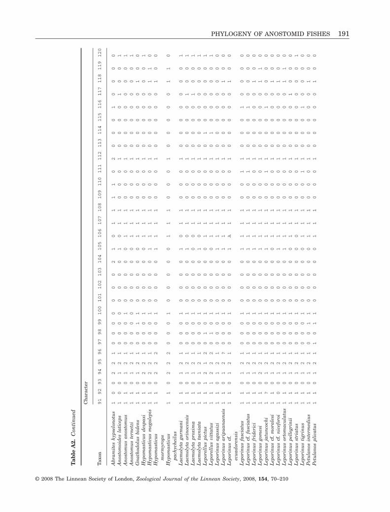
Tab
leA
2.C
onti
nu
ed
Taxo
n
Ch
arac
ter
91
92
93
94
95
96
97
98
99
100
101
102
103
104
105
106
107
108
109
110
111
112
113
114
115
116
117
118
119
120
Abr
amit
esh
ypse
lon
otu
s1
00
22
10
00
00
00
21
01
11
10
20
00
10
00
0A
nos
tom
oid
esla
tice
ps1
00
12
10
00
00
00
01
01
10
00
10
00
01
00
1A
nos
tom
us
anos
tom
us
11
01
21
00
01
00
00
01
11
00
01
00
00
00
01
An
osto
mu
ste
rnet
zi1
10
12
10
00
00
00
00
11
10
00
10
00
00
00
1G
nat
hod
olu
sbi
den
s1
10
12
10
01
10
00
00
11
10
00
10
00
00
10
0H
ypom
asti
cus
des
paxi
11
02
21
00
01
00
00
01
11
00
01
00
00
00
01
Hyp
omas
ticu
sm
egal
epis
11
02
21
00
01
00
00
11
11
00
01
00
00
01
10
Hyp
omas
ticu
sm
orm
yrop
s1
10
22
20
00
10
00
01
11
10
00
10
00
00
10
0
Hyp
omas
ticu
spa
chyc
hei
lus
11
02
22
00
01
00
00
01
11
00
01
00
00
01
10
Lae
mol
yta
garm
ani
11
01
21
00
01
00
00
10
11
00
01
00
00
00
01
Lae
mol
yta
orin
ocen
sis
11
01
21
00
01
00
00
10
11
00
01
00
00
10
01
Lae
mol
yta
prox
ima
11
01
21
00
01
00
00
00
11
00
01
00
00
10
01
Lae
mol
yta
taen
iata
11
01
21
00
01
00
00
10
11
00
01
00
00
00
01
Lep
orel
lus
pict
us
11
12
12
01
01
00
00
01
11
00
01
01
00
00
01
Lep
orel
lus
vitt
atu
s1
11
22
20
10
10
00
00
11
10
00
10
10
00
00
1L
epor
inu
sag
assi
zi1
10
12
10
00
10
00
01
11
10
00
10
00
00
00
0L
epor
inu
sar
ipu
anae
nsi
s1
10
12
?0
00
10
00
01
11
10
00
10
00
00
10
0L
epor
inu
scf
.ec
uad
oren
sis
11
01
21
00
01
00
00
1A
11
00
01
00
00
01
00
Lep
orin
us
fasc
iatu
s1
10
12
10
00
10
00
01
11
10
01
10
00
10
00
0L
epor
inu
scf
.fa
scia
tus
11
01
21
00
01
00
00
11
11
00
11
00
01
00
00
Lep
orin
us
frid
eric
i1
10
12
10
00
10
00
01
11
10
00
10
00
00
10
0L
epor
inu
sgo
mes
i1
10
22
10
00
10
00
01
11
10
00
10
00
00
11
0L
epor
inu
sja
tun
coch
i1
?0
12
10
00
10
00
01
11
10
01
10
00
10
00
0L
epor
inu
scf
.m
oral
esi
11
01
21
00
01
00
00
01
11
00
01
00
00
01
00
Lep
orin
us
cf.
nic
efor
oi1
00
12
10
00
10
00
01
11
10
00
10
00
00
10
0L
epor
inu
sor
tom
acu
latu
s1
10
12
10
00
10
00
01
11
10
00
10
00
00
11
0L
epor
inu
spe
lleg
rin
ii1
10
12
10
00
10
00
00
11
10
00
10
00
01
00
0L
epor
inu
sst
riat
us
11
01
21
00
01
00
00
00
11
00
01
00
00
00
01
Lep
orin
us
tigr
inu
s1
10
12
10
00
10
00
00
11
10
01
10
00
10
00
0P
etu
lan
osin
term
ediu
s1
10
12
10
01
10
00
00
11
10
00
10
00
00
10
0P
etu
lan
ospl
icat
us
11
01
21
10
11
00
00
01
11
00
01
00
00
01
00
PHYLOGENY OF ANOSTOMID FISHES 191
© 2008 The Linnean Society of London, Zoological Journal of the Linnean Society, 2008, 154, 70–210
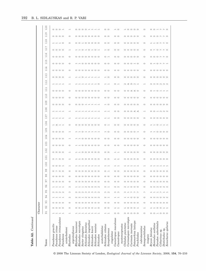
Tab
leA
2.C
onti
nu
ed
Taxo
n
Ch
arac
ter
91
92
93
94
95
96
97
98
99
100
101
102
103
104
105
106
107
108
109
110
111
112
113
114
115
116
117
118
119
120
Pse
ud
anos
grac
ilis
10
01
21
00
00
00
00
01
11
00
01
00
00
01
00
Pse
ud
anos
irin
ae1
00
12
10
0?
0?
00
00
A1
10
00
10
00
00
10
0P
seu
dan
ostr
imac
ula
tus
10
01
21
00
00
00
00
01
11
00
01
00
00
01
00
Pse
ud
anos
win
terb
otto
mi
10
01
21
00
01
00
00
01
11
00
01
00
00
00
01
Rh
ytio
du
sar
gen
teof
usc
us
10
02
32
00
01
11
01
10
11
00
01
10
00
10
01
Rh
ytio
du
sla
uza
nn
ei1
00
22
10
00
11
11
11
01
10
00
11
01
01
00
0R
hyt
iod
us
mic
role
pis
10
02
21
00
01
11
11
10
11
00
01
10
10
10
00
Sar
tor
elon
gatu
s1
10
12
10
00
10
00
00
01
10
00
10
00
00
10
0S
chiz
odon
fasc
iatu
s1
00
23
20
00
10
00
01
01
11
00
11
00
01
00
0S
chiz
odon
isog
nat
hu
s1
00
23
20
00
10
00
01
01
11
00
11
00
00
00
1S
chiz
odon
kner
ii1
00
23
20
00
10
00
01
01
11
00
11
00
01
00
1S
chiz
odon
nas
utu
s1
10
23
20
00
10
00
01
01
11
00
11
00
00
00
1S
chiz
odon
scot
orh
abd
otu
s1
00
22
20
00
10
00
01
01
11
00
01
00
01
00
1
Sch
izod
onvi
ttat
us
10
02
32
00
01
00
00
10
11
10
01
10
00
10
00
Syn
apto
laem
us
cin
gula
tus
10
01
21
00
11
00
00
01
11
00
01
00
01
00
00
Cae
not
ropu
sm
acu
losu
s0
10
13
20
00
11
10
00
00
00
00
00
00
00
00
1C
aen
otro
pus
mes
tom
orgm
atos
01
01
32
00
01
11
00
00
00
00
01
00
00
00
00
Ch
ilod
us
pun
ctat
us
01
01
32
00
01
00
00
00
00
00
02
00
00
00
01
Cu
rim
atop
sis
mic
role
pis
00
01
11
00
01
00
00
00
00
00
0A
00
00
00
00
Cu
rim
ata
inor
nat
a0
00
0?
00
00
10
00
00
00
00
0A
D0
00
00
00
0P
otam
orh
ina
lati
ceps
00
00
?0
00
01
00
00
00
0A
00
A2
00
10
00
00
Pro
chil
odu
sru
brot
aen
iatu
s0
10
0?
00
00
10
00
01
00
A0
00
10
00
00
00
0
Sem
apro
chil
odu
sin
sign
is0
10
0?
00
00
10
00
00
00
10
00
10
00
00
00
0
Bry
con
falc
atu
s0
10
21
20
00
10
00
01
10
00
00
20
00
00
10
0H
emio
du
soc
ella
tus
01
02
12
00
01
00
00
11
00
00
12
00
10
?1
00
Par
odon
subo
rbit
alis
10
02
12
00
01
00
00
00
01
00
00
00
00
00
01
Cit
har
inu
ssp
.0
00
0?
00
00
10
00
00
10
20
01
20
0?
??
??
?D
isti
chod
us
sp.
01
02
12
00
01
00
00
01
02
00
12
00
??
??
??
Xen
och
arax
spil
uru
s0
10
21
20
00
10
00
00
10
20
01
20
01
00
00
0
192 B. L. SIDLAUSKAS and R. P. VARI
© 2008 The Linnean Society of London, Zoological Journal of the Linnean Society, 2008, 154, 70–210

Tab
leA
2.C
onti
nu
ed
Taxo
n
Ch
arac
ter
121
122
123
124
125
126
127
128
129
130
131
132
133
134
135
136
137
138
139
140
141
142
143
144
145
146
147
148
149
150
Abr
amit
esh
ypse
lon
otu
s0
00
00
00
01
00
11
10
11
01
00
10
10
00
11
1A
nos
tom
oid
esla
tice
ps0
00
00
00
01
00
11
10
11
01
00
10
10
00
11
1A
nos
tom
us
anos
tom
us
11
00
00
00
10
01
11
01
10
10
01
01
00
01
11
An
osto
mu
ste
rnet
zi1
00
00
00
01
00
11
10
11
01
00
10
10
00
11
1G
nat
hod
olu
sbi
den
s0
00
00
00
01
00
11
10
11
01
00
10
10
00
11
1H
ypom
asti
cus
des
paxi
1?
00
00
00
10
01
11
01
10
10
01
01
00
01
11
Hyp
omas
ticu
sm
egal
epis
00
00
00
00
10
01
11
01
10
10
01
01
00
01
11
Hyp
omas
ticu
sm
orm
yrop
s0
00
00
00
01
00
11
10
11
01
00
10
10
00
11
1
Hyp
omas
ticu
spa
chyc
hei
lus
00
00
00
00
10
01
11
01
10
10
01
01
00
01
11
Lae
mol
yta
garm
ani
00
00
00
00
10
01
11
01
10
10
01
01
00
01
11
Lae
mol
yta
orin
ocen
sis
00
00
00
00
10
01
11
01
10
10
01
01
00
01
11
Lae
mol
yta
prox
ima
00
00
00
00
10
01
11
01
10
10
01
01
00
01
11
Lae
mol
yta
taen
iata
00
00
00
00
10
01
11
01
10
10
01
01
00
01
11
Lep
orel
lus
pict
us
01
10
00
00
10
01
11
01
10
10
01
01
00
01
11
Lep
orel
lus
vitt
atu
s0
11
00
00
01
00
11
10
11
01
00
10
10
00
11
1L
epor
inu
sag
assi
zi0
00
00
00
01
00
11
10
11
01
00
10
10
00
11
1L
epor
inu
sar
ipu
anae
nsi
s0
00
00
00
01
00
11
10
11
01
00
10
10
00
11
1L
epor
inu
scf
.ec
uad
oren
sis
00
00
00
00
10
01
11
01
10
10
01
01
00
01
11
Lep
orin
us
fasc
iatu
s0
00
00
00
01
00
11
10
11
01
00
10
10
00
11
1L
epor
inu
scf
.fa
scia
tus
00
00
00
00
10
01
11
01
10
10
01
01
00
01
11
Lep
orin
us
frid
eric
i0
00
00
00
01
00
11
10
11
01
00
10
10
00
11
1L
epor
inu
sgo
mes
i0
20
00
00
01
00
11
10
11
01
00
10
10
00
11
1L
epor
inu
sja
tun
coch
i0
00
00
00
01
00
11
10
11
01
00
10
10
00
11
1L
epor
inu
scf
.m
oral
esi
00
00
00
00
10
01
11
01
10
10
01
01
00
01
11
Lep
orin
us
cf.
nic
efor
oi0
00
00
00
01
00
11
10
11
01
00
10
10
00
11
1L
epor
inu
sor
tom
acu
latu
s0
00
00
00
01
00
11
10
11
01
00
10
10
00
11
1L
epor
inu
spe
lleg
rin
ii0
00
00
00
01
00
11
10
11
01
00
10
10
00
11
1L
epor
inu
sst
riat
us
10
00
00
00
10
01
11
01
10
10
01
01
00
01
11
Lep
orin
us
tigr
inu
s0
00
00
00
01
00
11
10
11
01
00
10
10
00
11
1P
etu
lan
osin
term
ediu
s0
00
00
00
01
00
11
10
11
01
00
10
10
00
11
1P
etu
lan
ospl
icat
us
00
00
00
00
10
01
11
01
10
10
01
01
00
01
11
PHYLOGENY OF ANOSTOMID FISHES 193
© 2008 The Linnean Society of London, Zoological Journal of the Linnean Society, 2008, 154, 70–210
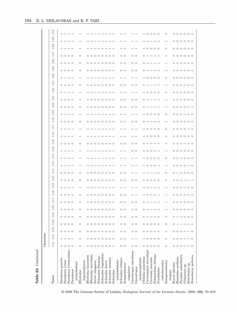
Tab
leA
2.C
onti
nu
ed
Taxo
n
Ch
arac
ter
121
122
123
124
125
126
127
128
129
130
131
132
133
134
135
136
137
138
139
140
141
142
143
144
145
146
147
148
149
150
Pse
ud
anos
grac
ilis
01
00
00
00
10
01
11
01
10
10
01
01
00
01
11
Pse
ud
anos
irin
ae0
10
00
00
01
00
11
10
11
01
00
10
10
00
11
1P
seu
dan
ostr
imac
ula
tus
00
00
00
00
10
01
11
01
10
10
01
01
00
01
11
Pse
ud
anos
win
terb
otto
mi
01
00
00
00
10
01
11
01
10
10
01
01
00
01
11
Rh
ytio
du
sar
gen
teof
usc
us
00
00
00
00
10
01
11
01
10
10
01
01
00
01
11
Rh
ytio
du
sla
uza
nn
ei0
00
00
00
01
00
11
10
11
01
00
10
10
00
11
1R
hyt
iod
us
mic
role
pis
00
00
00
00
10
01
11
01
10
10
01
01
00
01
11
Sar
tor
elon
gatu
s0
00
00
00
01
00
11
10
11
01
00
10
10
00
11
1S
chiz
odon
fasc
iatu
s0
00
00
00
01
00
11
10
11
01
00
10
10
00
11
1S
chiz
odon
isog
nat
hu
s0
00
00
00
01
00
11
10
11
01
00
10
10
00
11
1S
chiz
odon
kner
ii0
00
00
00
01
00
11
10
11
01
00
10
10
00
11
1S
chiz
odon
nas
utu
s0
00
00
00
01
00
11
10
11
01
00
10
10
00
11
1S
chiz
odon
scot
orh
abd
otu
s0
00
00
00
01
00
11
10
11
01
00
10
10
00
11
1
Sch
izod
onvi
ttat
us
00
00
00
00
10
01
11
01
10
10
01
01
00
01
11
Syn
apto
laem
us
cin
gula
tus
00
00
00
00
10
01
11
01
10
10
01
01
00
01
11
Cae
not
ropu
sm
acu
losu
s0
10
00
00
11
01
11
00
11
01
00
10
10
00
11
1C
aen
otro
pus
mes
tom
orgm
atos
01
00
00
01
10
11
10
01
10
10
01
01
00
01
11
Ch
ilod
us
pun
ctat
us
01
00
00
01
10
11
10
01
10
10
01
01
00
01
11
Cu
rim
atop
sis
mic
role
pis
00
00
11
10
10
10
00
10
01
01
10
10
11
10
10
Cu
rim
ata
inor
nat
a0
00
01
11
01
01
00
01
00
10
11
01
01
11
01
0P
otam
orh
ina
lati
ceps
00
00
11
10
10
10
00
10
01
01
10
10
11
10
10
Pro
chil
odu
sru
brot
aen
iatu
s0
00
00
11
11
00
10
11
00
10
11
01
01
11
01
0
Sem
apro
chil
odu
sin
sign
is0
01
00
11
11
00
10
11
00
10
11
01
01
11
01
0
Bry
con
falc
atu
s0
00
00
00
00
00
00
00
00
00
00
00
00
00
00
0H
emio
du
soc
ella
tus
00
01
10
00
00
10
00
00
00
00
00
00
00
00
00
Par
odon
subo
rbit
alis
00
01
10
00
01
00
10
00
00
00
00
00
00
00
00
Cit
har
inu
ssp
.?
??
10
00
01
10
00
01
00
01
00
00
00
00
00
0D
isti
chod
us
sp.
??
?1
00
00
01
01
00
00
10
01
00
00
00
00
00
Xen
och
arax
spil
uru
s0
00
10
00
00
10
00
00
00
00
10
00
00
00
00
0
194 B. L. SIDLAUSKAS and R. P. VARI
© 2008 The Linnean Society of London, Zoological Journal of the Linnean Society, 2008, 154, 70–210
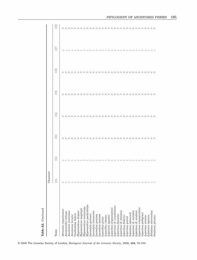
Tab
leA
2.C
onti
nu
ed
Taxo
n
Ch
arac
ter
151
152
153
154
155
156
157
158
Abr
amit
esh
ypse
lon
otu
s1
10
00
01
0A
nos
tom
oid
esla
tice
ps1
10
00
01
0A
nos
tom
us
anos
tom
us
11
00
00
10
An
osto
mu
ste
rnet
zi1
10
00
01
0G
nat
hod
olu
sbi
den
s1
10
00
01
0H
ypom
asti
cus
des
paxi
11
00
00
10
Hyp
omas
ticu
sm
egal
epis
11
00
00
10
Hyp
omas
ticu
sm
orm
yrop
s1
10
00
01
0H
ypom
asti
cus
pach
ych
eilu
s1
10
00
01
0L
aem
olyt
aga
rman
i1
10
00
01
0L
aem
olyt
aor
inoc
ensi
s1
10
00
01
0L
aem
olyt
apr
oxim
a1
10
00
01
0L
aem
olyt
ata
enia
ta1
10
00
01
0L
epor
ellu
spi
ctu
s1
10
00
01
0L
epor
ellu
svi
ttat
us
11
00
00
10
Lep
orin
us
agas
sizi
11
00
00
10
Lep
orin
us
arip
uan
aen
sis
11
00
00
10
Lep
orin
us
cf.
ecu
ador
ensi
s1
10
00
01
0L
epor
inu
sfa
scia
tus
11
00
00
10
Lep
orin
us
cf.
fasc
iatu
s1
10
00
01
0L
epor
inu
sfr
ider
ici
11
00
00
10
Lep
orin
us
gom
esi
11
00
00
10
Lep
orin
us
jatu
nco
chi
11
00
00
10
Lep
orin
us
cf.
mor
ales
i1
10
00
01
0L
epor
inu
scf
.n
icef
oroi
11
00
00
10
Lep
orin
us
orto
mac
ula
tus
11
00
00
10
Lep
orin
us
pell
egri
nii
11
00
00
10
Lep
orin
us
stri
atu
s1
10
00
01
0L
epor
inu
sti
grin
us
11
00
00
10
Pet
ula
nos
inte
rmed
ius
11
00
00
10
Pet
ula
nos
plic
atu
s1
10
00
01
0
PHYLOGENY OF ANOSTOMID FISHES 195
© 2008 The Linnean Society of London, Zoological Journal of the Linnean Society, 2008, 154, 70–210
Tab
leA
2.C
onti
nu
ed
Taxo
n
Ch
arac
ter
151
152
153
154
155
156
157
158
Pse
ud
anos
grac
ilis
11
00
00
10
Pse
ud
anos
irin
ae1
10
00
01
0P
seu
dan
ostr
imac
ula
tus
11
00
00
10
Pse
ud
anos
win
terb
otto
mi
11
00
00
10
Rh
ytio
du
sar
gen
teof
usc
us
11
00
00
10
Rh
ytio
du
sla
uza
nn
ei1
10
00
01
0R
hyt
iod
us
mic
role
pis
11
00
00
10
Sar
tor
elon
gatu
s1
10
00
01
0S
chiz
odon
fasc
iatu
s1
10
00
01
0S
chiz
odon
isog
nat
hu
s1
10
00
01
0S
chiz
odon
kner
ii1
10
00
01
0S
chiz
odon
nas
utu
s1
10
00
01
0S
chiz
odon
scot
orh
abd
otu
s1
10
00
01
0S
chiz
odon
vitt
atu
s1
10
00
01
0S
ynap
tola
emu
sci
ngu
latu
s1
10
00
01
0C
aen
otro
pus
mac
ulo
sus
11
00
00
10
Cae
not
ropu
sm
esto
mor
gmat
os1
10
00
01
0C
hil
odu
spu
nct
atu
s1
10
00
?1
0C
uri
mat
opsi
sm
icro
lepi
s1
01
10
00
0C
uri
mat
ain
orn
ata
10
11
00
00
Pot
amor
hin
ala
tice
ps1
01
10
00
0P
roch
ilod
us
rubr
otae
nia
tus
10
11
00
00
Sem
apro
chil
odu
sin
sign
is1
01
10
00
0B
ryco
nfa
lcat
us
00
00
00
00
Hem
iod
us
ocel
latu
s0
01
00
00
1P
arod
onsu
borb
ital
is0
00
00
00
0C
ith
arin
us
sp.
10
00
11
01
Dis
tich
odu
ssp
.0
00
01
10
1X
enoc
har
axsp
ilu
rus
00
00
11
01
196 B. L. SIDLAUSKAS and R. P. VARI
© 2008 The Linnean Society of London, Zoological Journal of the Linnean Society, 2008, 154, 70–210
APPENDIX 4SYNAPOMORPHY LIST
The 32 numbered clades in Figure 7 and in the listbelow appear in all of the most parsimonious treesrecovered during phylogenetic analysis. Lists of thecharacter state transitions that optimize at thebranch subtending each numbered clade in the strictconsensus phylogeny are given, with unambiguouslyoptimized synapomorphies appearing first (indicatedby bold arrows), followed by ambiguous optimizations(indicated by normal arrows). Transitions optimizedas autapomorphies of nominal genera or subgenerarepresented by only one species in the analysis alsoappear below. Each synapomorphic character statetransition is followed by a brief description of eachsynapomorphy. Finally, information appears onwhether that state transition is a reversal of a pre-vious character state change within the Anostomidae,whether other clades within the Anostomidae haveacquired that character state independently, andwhether additional state transitions in the characterin question occur within the clade defined by thesynapomorphy. Not all state changes in each charac-ter are necessarily listed below; such informationappears in the individual character descriptions.
Because character optimizations were performedon a strict consensus phylogeny, caution must beexercised in interpreting some synapomorphiesinvolving clades that contain basal polytomies (i.e.clades 8, 10, 18, 25 and 26). In some cases, alterna-tive resolutions of the polytomies would shift theinterpretation of the synapomorphies up or down onehierarchical level. For example, state 1 of character79 (aspect ratio of metapterygoid–quadrate fenestra)is interpreted on the strict consensus as an ambigu-ous synapomorphy for clade 25 (ACCTRAN optimi-zation) with a reversal to state 0 in clade 26. Werethe basal polytomy in clade 25 resolved as in themajority-rule consensus (Fig. 6), state 1 of character79 would optimize instead as an unambiguous syna-pomorphy for a clade containing Pseudanos winter-bottomi and clade 27.
Clade 1: Family Anostomidae• Character 31, state 5 → 4; four teeth on premaxilla;
further modified in clades 5, 9, 11 and Leporinus cf.moralesi
• Character 37, state 4 → 0; dentary teeth spade orchisel-shaped, tapering to pointed or blunt distalmargin with or without additional cusping; furthermodified in Hypomasticus despaxi, L. jatuncochiand clade 32
• Character 38, state 1 → 0; dentary teeth with large,well-developed posterior lamina; reversed in clade21
• Character 45, state 0 → 1; ascending process ofpremaxilla large, well-developed and distinctly tri-angular overall
• Character 50, state 1 → 0; anterolateral flange ofmaxilla merges with ventral portion of maxilla onlateral face of ventral portion; reversed in clade 25
• Character 51, state 1 → 0; anterolateral andventral flanges of maxilla rotated relative to eachother along main axis of bone with result thatventral portion of maxilla lies in parasagittal planeand anterolateral flange of bone lies in transverseplane; reversed in clade 31
• Character 62, state 0 → 1; dentary with shortlaterosensory canal segment, terminating at, orposterior to, approximate longitudinal midpoint ofbone
• Character 65, state 1 → 0; retroarticular laterallysituated in lower jaw and deeply recessed intopocket formed primarily by dentary and to lesserdegree by anguloarticular; line drawn along ventralmargin of retroarticular clearly bisects ventralportion of dentary; reversed in clade 13
• Character 73, state 0 →1; distinct process on lateralsurface of ectopterygoid present; reversed inGnathodolus
• Character 83, state 0 → 1; interopercular–mandibular ligament elongate, interopercle notablyseparated from retroarticular
• Character 84, state 0 → 1; presence of large shelf onlateral surface of preopercle that serves as origin forportion of adductor mandibulae muscle
• Character 85, state 2 → 0; anterodorsal process ofpreopercle that extends between symplectic andquadrate well-developed with broad triangular orrhomboidal form; further modified in clade 25
• Character 90, state 1 → 0; posterodorsal margin ofopercle with slight or pronounced concavity;reversed in clade 13 with subsequent transitionback to state 0 in clade 27, polymorphic in Pseu-danos gracilis
• Character 91, state 0 →1; curved anterior process ofthird epibranchial that extends medially over dorsalsurface of fourth infrapharyngobranchial present
• Character 106, state 0 → 1; supraneural dorsal toWeberian apparatus present; reversed in clades 9and 13, subsequently reversed in clade 25 and againin Sartor, polymorphic in Leporinus cf. ecuadorensisand Pseudanos irinae.
• Character 107, state 0 → 1; presence of two or moreintercostal ligaments uniting in total four or more ofanterior full ribs
• Character 134, state 0 → 1; anterior portion ofpreopercular laterosensory canal subdivided intotwo or more ossified tubes
• Character 22, state 1 → 0 (ACCTRAN); articularfacets of vomer with palatines angled toward each
PHYLOGENY OF ANOSTOMID FISHES 197
No claim to original US Government works.Journal compilation © 2008 The Linnean Society of London, Zoological Journal of the Linnean Society, 2008, 154, 70–210
other anteriorly; further modified in clades 5 and 13,reversed to state 0 in clade 21, further modified inclade 27, reversed to state 0 in clade 32
• Character 33, state 1 → 2 (ACCTRAN); second andthird teeth of premaxilla with two cusps; modified tostate 3 in Leporellus vittatus and clade 8, state2 occurring again in clade 9, Leporinus agassizi,L. aripuanaensis and L. cf. ecuadorensis, furthermodified in Schizodon and clade 23, polymorphic forstates 2 and 3 in Gnathodolus
• Character 8, state 1 → 2 (DELTRAN); two interme-diate pores along ossified portion of sensory canalof second infraorbital; reversed in Hypomasticuspachycheilus, Rhytiodus argenteofuscus, Schizodonnasutus and clade 21 with subsequent transitionback to state 2 in Petulanos, transition to state 3 inLeporinus tigrinus, polymorphic for states 2 and 3 inRhytiodus lauzannei and Schizodon isognathus
• Character 60, state 0 → 1 (DELTRAN, ambiguitydue to inapplicability of character to Chilodontidaeand Curimatidae); dentary replacement toothtrench large and with distinct ventral fenestra
• Character 108, state 0 → 1 (DELTRAN); tenbranched dorsal-fin rays present
• Character 24, state ? → 0 (ambiguous due to inap-plicability of character to outgroups other thanChilodontidae); lateral ethmoid–ectopterygoid liga-ment inserts on ventral surface of anterior wing oflateral ethmoid; further modified in Synaptolaemus
Vari (1983) also detailed five myological synapomor-phies for the Anostomidae which are listed below.These were not reinvestigated due to lack of appropri-ate material for dissection for many pertinent taxa.
• Presence of triangular, autogenous section of levatorarcus palatini that arises from posterodorsal portionof orbital cavity and inserts on anterodorsal marginof hyomandibular
• Subdivision of A1 portion of adductor mandibulae• Posterior part of A2 portion of adductor mandibulae
medial of levator arcus palatini large and withpartial origin on hyomandibular
• A3 and A2 portions of the adductor mandibulae withanterior dorsal expansion resulting in contact ofthese muscles with each other dorsal to tendon thatattaches to main body of A2
• Absence of Aw portion of adductor mandibulae
Clade 2: Leporellus• Character 17, state 1 →0; ventral process of meseth-
moid runs vertically or nearly so, anterior portionof vomer positioned directly ventral to ventralprocess of mesethmoid; acquired independently inAbramites and clade 25
• Character 26, state 0 → 1; presence of fenestra onportion of lateral wing of lateral ethmoid situated
dorsal to lateral ethmoid–ectopterygoid ligament;acquired independently in Anostomoides
• Character 32, state 1 → 2; symphyseal tooth ofpremaxilla with two cusps; acquired independentlyin, Anostomoides, Gnathodolus, Petulanos, Hypo-masticus despaxi, Leporinus agassizi, L. cf. ecuador-ensis, clade 9 and in either clade 10 with reversal inclade 11 or in L. fasciatus and L. cf. fasciatus(optimization ambiguous)
• Character 64, state 0 → 1; retroarticular elongateand cup-shaped, with its length in lateral view morethan three times its height, with interopercular–mandibular ligament attaching in cup-shapeddepression near anterior margin of bone
• Character 93, state 0 →1; second basibranchial withprominent lateral flanges resulting in overall shaperesembling violin
• Character 98, state 0 → 1; presence of inflectionpoint in joint between anterior and posterior cera-tohyals and corresponding process of anterior cera-tohyal running ventral to posterior ceratohyal
• Character 114, state 0 → 1; lobes of caudal fincovered with scales
• Character 123, state 0 → 1; caudal fin with widedark stripes extending across much of each lobe;stripe(s) of dorsal lobe run from anterodorsal toposteroventral; stripe(s) of ventral lobe run fromanteroventral to posterodorsal
• Character 15, state 1 → 0 (DELTRAN); anteriorportion of mesethmoid ventrally directed and withstrong hook; acquired independently in Hypomasti-cus, Leporinus jatuncochi and Schizodon nasutus
• Character 22, state 1 → 0 (DELTRAN); articularfacets of vomer with palatines angled toward eachother anteriorly; state 0 acquired independently inclade 7 and Sartor (reversal after transition to state2 in clade 27)
• Character 120, state 0 → 1 (DELTRAN); completestripe of approximately height of one scale or greaterrunning along lateral line from opercle to caudalpeduncle in majority of examined specimens;acquired independently in Leporinus striatus andclade 13
• Character 122, state 0 → 1 (DELTRAN); most scaleswith small, dark spot lying under scale creatingoverall appearance of row of such spots along centerof each scale row; acquired independently in Anos-tomus anostomus, Pseudanos gracilis, P. irinae, andP. winterbottomi.
Clade 3: Abramites, Anostomoides, Anostomus,Gnathodolus, Hypomasticus, Laemolyta, Leporinus,Petulanos, Pseudanos, Rhytiodus, Sartor, Schizodon,Synaptolaemus• Character 1, state 0 → 1; antorbital relatively large,
with distinct horizontally directed flange running
198 B. L. SIDLAUSKAS and R. P. VARI
No claim to original US Government works.Journal compilation © 2008 The Linnean Society of London, Zoological Journal of the Linnean Society, 2008, 154, 70–210
ventral to nasal cavity; flat bony plate may joinvertical and horizontal portions of antorbital tovarying degrees
• Character 13, state 0 → 1; anteroventral portion ofsixth infraorbital not greatly expanded and overlap-ping anteroventral process of sphenotic; reversed inAbramites hypselonotus and Anostomus anostomus,polymorphic in Hypomasticus mormyrops andLeporinus striatus
• Character 18, state 0 → 1; distinct ventromedialplate-like process of mesethmoid extends ventrallyand posteriorly towards vomer; reversed inLaemolyta proxima
• Character 49, state 0 → 1; area of attachment ofprimordial ligament on medial surface of maxillalocated within ventral one-half of longest axis ofbone
• Character 52, state 1 → 0; ventral portion of maxillanarrow, only slightly expanded in axis transverse tolong axis of bone; reversed in clade 29
• Character 76, state 0 → 1; posterodorsal and poste-rior processes of quadrate entirely or almost entirelyjoined by thin intervening plate of bone with lengthof unfused portion of posterodorsal process muchless than twice width of process
• Character 87, state 0 → 1; symplectic long withposterior margin of bone situated proximate toventral margin of hyomandibular
• Character 118, state 0 → 1; series of one or moredark spots with distinct margins present alonglateral-line scale row, each spot approximately oneto two scales in height; reversed in Hypomasticusdespaxi, Leporinus agassizi, L. pellegrinii andclades 9, 10 and 12, acquired independently in clade26 and in either clade 29 with reversal in Synapto-laemus or in Petulanos and clade 32 (optimizationambiguous)
• Character 3, state 0 → 1 (ACCTRAN); supraorbitalwith deeply concave, distinctly notched anterior oranterolateral margin; reversed in clade 8, acquiredindependently in Schizodon knerii, S. isognathusand Gnathodolus bidens
• Character 96, state 2 → 1 (ACCTRAN); one or twocomplete rows of teeth on tooth-plate of fifth cera-tobranchial; reversed in clades 6 and 15, reacquiredin clade 17
• Character 105, state 0 → 1 (ACCTRAN); presence ofdorsal process of third vertebra overlapping neuralcomplex laterally; reversed in Hypomasticusdespaxi, H. pachycheilus, Laemolyta proxima, Lep-orinus cf. moralesi, L. striatus, L. tigrinus, L. pelle-grinii, and clade 25
• Character 120, state 1 → 0 (ACCTRAN); completestripe along lateral-line absent; reversed in Hypo-masticus despaxi, Leporinus striatus and clade 13;lost independently in clades 17, 26 and 29
• Character 122, state 1 → 0 (ACCTRAN); centers ofscales same colour as, or lighter than, scale margins;no series of small dark spots along center of eachscale row; reversed in clade 25 with independentreacquisition in Pseudanos trimaculatus and clade27, followed by another reversal in Anostomusanostomus
• Character 33, state 1 → 2 (DELTRAN); two cusps oneach of second and third teeth of premaxilla; furthermodified to state 3 in Leporinus friderici, L. cf.moralesi, L. cf. niceforoi, L. ortomaculatus andclades 10 and 12, with subsequent modification tostate 4 in Schizodon and clade 23
• Character 68, state 0 → 1 (DELTRAN, ambiguitydue to inapplicability of character to outgroups);process of palatine with distinct lateral, anterolat-eral or anteroventral orientation
• Character 95, state 1 → 2 (DELTRAN); most teethon fifth upper pharyngeal tooth-plate bicuspid butwith occasional teeth unicuspid; further modified tostate 3 in Rhytiodus argenteofuscus and Schizodon
Clade 4: Hypomasticus• Character 16, state 0 → 1; mesethmoid with two
distinct lateral processes that fit into concavities onmedial surface of premaxilla
• Character 46, state 1→0; premaxilla with vertical oranterodorsal inclination
• Character 55, state 0 → 1; ascending process ofanguloarticular very wide, forming pronounced ver-tical ridge perpendicular to dentary; acquired inde-pendently in Schizodon
• Character 56, state 0 → 1; dorsal portion of ascend-ing process of anguloarticular in form of deeptrough; acquired independently in Schizodonnasutus
• Character 17, state 1 → 2 (ACCTRAN); ventralprocess of mesethmoid posteroventrally aligned;reversed in Hypomasticus megalepis, acquired inde-pendently in clade 20
• Character 22, state 0 → 1 (ACCTRAN); articularfacets of vomer with palatines parallel and widelyseparated; acquired independently in clade 13,reversed in clade 21 with further modification inclade 27 and subsequent reversal in clade 32.
• Character 15, state 1 → 0 (DELTRAN); anteriorportion of mesethmoid ventrally directed and withstrong hook; acquired independently in Leporellus,Leporinus jatuncochi and Schizodon nasutus
Clade 5: Hypomasticus despaxi, H. megalepis• Character 9, state 0 → 1; fourth and fifth infraorbit-
als fused; acquired independently in Leporinusaripuanaensis, L. ortomaculatus, L. tigrinus and L.pellegrinii, polymorphic in Laemolyta orinocensis.
PHYLOGENY OF ANOSTOMID FISHES 199
No claim to original US Government works.Journal compilation © 2008 The Linnean Society of London, Zoological Journal of the Linnean Society, 2008, 154, 70–210
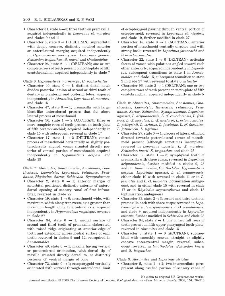
• Character 31, state 4 →3; three teeth on premaxilla;acquired independently in Leporinus cf. moralesiand clades 9 and 11
• Character 3, state 0 → 1 (DELTRAN); supraorbitalwith deeply concave, distinctly notched anterioror anterolateral margin; acquired independentlyin Hypomasticus mormyrops, Leporinus gomesi,Schizodon isognathus, S. knerii and Gnathodolus
• Character 96, state 2 → 1 (DELTRAN); one or twocomplete rows of teeth present on tooth-plate of fifthceratobranchial; acquired independently in clade 7
Clade 6: Hypomasticus mormyrops, H. pachycheilus• Character 40, state 0 → 1; distinct distal notch
divides posterior lamina of second or third tooth ofdentary into anterior and posterior lobes; acquiredindependently in Abramites, Leporinus cf. moralesi,and clade 15
• Character 47, state 0 → 1; premaxilla with large,block-like anterodorsal process that fits abovelateral process of mesethmoid
• Character 96, state 1 → 2 (ACCTRAN); three ormore complete rows of teeth present on tooth-plateof fifth ceratobranchial; acquired independently inclade 15 with subsequent reversal in clade 17
• Character 17, state 1 → 2 (DELTRAN); ventralprocess of mesethmoid horizontally or slightly pos-terodorsally aligned, vomer situated directly pos-terior of ventral portion of mesethmoid; acquiredindependently in Hypomasticus despaxi andclade 19
Clade 7: Abramites, Anostomoides, Anostomus, Gna-thodolus, Laemolyta, Leporinus, Petulanos, Pseu-danos, Rhytiodus, Sartor, Schizodon, Synaptolaemus• Character 2, state 0 → 1; anterior margin of
antorbital positioned distinctly anterior of antero-dorsal opening of sensory canal of first infraor-bital; reversed in clade 27
• Character 19, state 1 → 0; mesethmoid wide, withmaximum width along transverse axis greater thanmaximum length along longitudinal axis; acquiredindependently in Hypomasticus megalepis, reversedin clade 27
• Character 34, state 0 → 1; medial surface ofsecond and third teeth of premaxilla elaborated,with raised ridge originating at anterior edge oftooth and extending across medial surface of eachtooth; reversed in clades 9 and 12, reacquired inAnostomoides
• Character 48, state 0 → 1; maxilla having verticalor posterodorsal orientation, with dorsal tip ofmaxilla situated directly dorsal to, or distinctlyposterior of, ventral margin of bone
• Character 72, state 0 → 1; ectopterygoid verticallyorientated with vertical through anterodorsal limit
of ectopterygoid passing through ventral portion ofectopterygoid; reversed in Leporinus cf. niceforoiand clade 19, further modified in clade 27
• Character 15, state 0 → 1 (ACCTRAN); anteriorportion of mesethmoid ventrally directed and withstrong hook; reversed in Leporinus jatuncochi andSchizodon nasutus
• Character 22, state 1 → 0 (DELTRAN); articularfacets of vomer with palatines angled toward eachother anteriorly; acquired independently in Leporel-lus, subsequent transitions to state 1 in Anosto-moides and clade 15, subsequent transition to state2 in clade 27 with reversal to state 0 in Sartor
• Character 96, state 2 → 1 (DELTRAN); one or twocomplete rows of teeth present on tooth-plate of fifthceratobranchial; acquired independently in clade 5
Clade 8: Abramites, Anostomoides, Anostomus, Gna-thodolus, Laemolyta, Rhytiodus, Petulanos, Pseu-danos, Sartor, Schizodon, Synaptolaemus, Leporinusagassizi, L. aripuanaensis, L. cf. ecuadorensis, L. frid-erici, L. cf. moralesi, L. cf. niceforoi, L. ortomaculatus,L. pellegrinii, L. striatus, L. fasciatus, L. cf. fasciatus,L. jatuncochi, L. tigrinus• Character 27, state 0 → 1; process of lateral ethmoid
directed towards posterolateral corner of meseth-moid present (although sometimes incomplete);reversed in Leporinus agassizi, L. cf. moralesi,Schizodon knerii, S. isognathus and clade 25
• Character 32, state 1 → 3; symphyseal tooth ofpremaxilla with three cusps; reversed in Leporinusaripuanaensis, further modified in clades 9, 23and 30, Anostomoides, Gnathodolus, Hypomasticusdespaxi, Leporinus agassizi, L. cf. ecuadorensis,either clade 10 with reversal in clade 11 or in L.fasciatus and L. cf. fasciatus (optimization ambigu-ous), and in either clade 15 with reversal in clade17 or in Rhytiodus argenteofuscus and clade 18(optimization ambiguous)
• Character 33, state 2 → 3; second and third teeth onpremaxilla each with three cusps; reversed in Lepo-rinus agassizi, L. aripuanaensis, L. cf. ecuadorensis,and clade 9, acquired independently in Leporellusvittatus, further modified in Schizodon and clade 23
• Character 94, state 2 → 1; one or two full rows ofteeth present on fifth upper pharyngeal tooth-plate;reversed in Abramites and clade 15
• Character 3, state 1 → 0 (ACCTRAN); supraor-bital with smoothly convex, straight or slightlyconcave anteroventral margin; reversal, subse-quent reversal in Gnathodolus, Schizodon kneriiand S. isognathus.
Clade 9: Abramites and Leporinus striatus• Character 5, state 1 → 2; two intermediate pores
present along ossified portion of sensory canal of
200 B. L. SIDLAUSKAS and R. P. VARI
No claim to original US Government works.Journal compilation © 2008 The Linnean Society of London, Zoological Journal of the Linnean Society, 2008, 154, 70–210
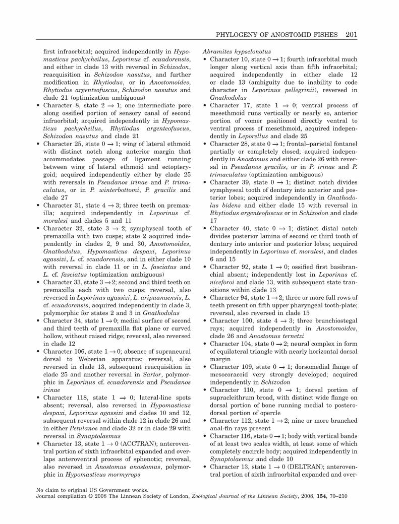
first infraorbital; acquired independently in Hypo-masticus pachycheilus, Leporinus cf. ecuadorensis,and either in clade 13 with reversal in Schizodon,reacquisition in Schizodon nasutus, and furthermodification in Rhytiodus, or in Anostomoides,Rhytiodus argenteofuscus, Schizodon nasutus andclade 21 (optimization ambiguous)
• Character 8, state 2 → 1; one intermediate porealong ossified portion of sensory canal of secondinfraorbital; acquired independently in Hypomas-ticus pachycheilus, Rhytiodus argenteofuscus,Schizodon nasutus and clade 21
• Character 25, state 0 → 1; wing of lateral ethmoidwith distinct notch along anterior margin thataccommodates passage of ligament runningbetween wing of lateral ethmoid and ectoptery-goid; acquired independently either by clade 25with reversals in Pseudanos irinae and P. trima-culatus, or in P. winterbottomi, P. gracilis andclade 27
• Character 31, state 4 → 3; three teeth on premax-illa; acquired independently in Leporinus cf.moralesi and clades 5 and 11
• Character 32, state 3 → 2; symphyseal tooth ofpremaxilla with two cusps; state 2 acquired inde-pendently in clades 2, 9 and 30, Anostomoides,Gnathodolus, Hypomasticus despaxi, Leporinusagassizi, L. cf. ecuadorensis, and in either clade 10with reversal in clade 11 or in L. fasciatus andL. cf. fasciatus (optimization ambiguous)
• Character 33, state 3 → 2; second and third teeth onpremaxilla each with two cusps; reversal, alsoreversed in Leporinus agassizi, L. aripuanaensis, L.cf. ecuadorensis, acquired independently in clade 3,polymorphic for states 2 and 3 in Gnathodolus
• Character 34, state 1 → 0; medial surface of secondand third teeth of premaxilla flat plane or curvedhollow, without raised ridge; reversal, also reversedin clade 12
• Character 106, state 1 → 0; absence of supraneuraldorsal to Weberian apparatus; reversal, alsoreversed in clade 13, subsequent reacquisition inclade 25 and another reversal in Sartor, polymor-phic in Leporinus cf. ecuadorensis and Pseudanosirinae
• Character 118, state 1 → 0; lateral-line spotsabsent; reversal, also reversed in Hypomasticusdespaxi, Leporinus agassizi and clades 10 and 12,subsequent reversal within clade 12 in clade 26 andin either Petulanos and clade 32 or in clade 29 withreversal in Synaptolaemus
• Character 13, state 1 → 0 (ACCTRAN); anteroven-tral portion of sixth infraorbital expanded and over-laps anteroventral process of sphenotic; reversal,also reversed in Anostomus anostomus, polymor-phic in Hypomasticus mormyrops
Abramites hypselonotus• Character 10, state 0 → 1; fourth infraorbital much
longer along vertical axis than fifth infraorbital;acquired independently in either clade 12or clade 13 (ambiguity due to inability to codecharacter in Leporinus pellegrinii), reversed inGnathodolus
• Character 17, state 1 → 0; ventral process ofmesethmoid runs vertically or nearly so, anteriorportion of vomer positioned directly ventral toventral process of mesethmoid, acquired indepen-dently in Leporellus and clade 25
• Character 28, state 0 → 1; frontal–parietal fontanelpartially or completely closed; acquired indepen-dently in Anostomus and either clade 26 with rever-sal in Pseudanos gracilis, or in P. irinae and P.trimaculatus (optimization ambiguous)
• Character 39, state 0 → 1; distinct notch dividessymphyseal tooth of dentary into anterior and pos-terior lobes; acquired independently in Gnathodo-lus bidens and either clade 15 with reversal inRhytiodus argenteofuscus or in Schizodon and clade17
• Character 40, state 0 → 1; distinct distal notchdivides posterior lamina of second or third tooth ofdentary into anterior and posterior lobes; acquiredindependently in Leporinus cf. moralesi, and clades6 and 15
• Character 92, state 1 → 0; ossified first basibran-chial absent; independently lost in Leporinus cf.niceforoi and clade 13, with subsequent state tran-sitions within clade 13
• Character 94, state 1 → 2; three or more full rows ofteeth present on fifth upper pharyngeal tooth-plate;reversal, also reversed in clade 15
• Character 100, state 4 → 3; three branchiostegalrays; acquired independently in Anostomoides,clade 26 and Anostomus ternetzi
• Character 104, state 0 → 2; neural complex in formof equilateral triangle with nearly horizontal dorsalmargin
• Character 109, state 0 → 1; dorsomedial flange ofmesocoracoid very strongly developed; acquiredindependently in Schizodon
• Character 110, state 0 → 1; dorsal portion ofsupracleithrum broad, with distinct wide flange ondorsal portion of bone running medial to postero-dorsal portion of opercle
• Character 112, state 1 → 2; nine or more branchedanal-fin rays present
• Character 116, state 0 →1; body with vertical bandsof at least two scales width, at least some of whichcompletely encircle body; acquired independently inSynaptolaemus and clade 10
• Character 13, state 1 → 0 (DELTRAN); anteroven-tral portion of sixth infraorbital expanded and over-
PHYLOGENY OF ANOSTOMID FISHES 201
No claim to original US Government works.Journal compilation © 2008 The Linnean Society of London, Zoological Journal of the Linnean Society, 2008, 154, 70–210
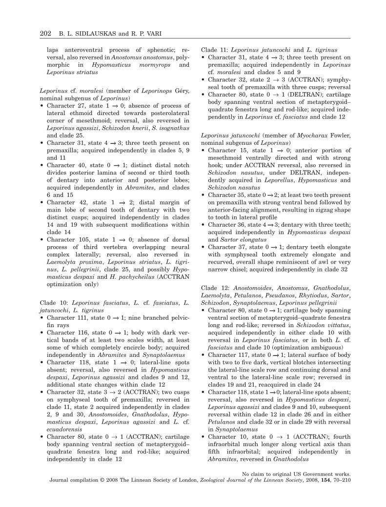
laps anteroventral process of sphenotic; re-versal, also reversed in Anostomus anostomus, poly-morphic in Hypomasticus mormyrops andLeporinus striatus
Leporinus cf. moralesi (member of Leporinops Géry,nominal subgenus of Leporinus)• Character 27, state 1 → 0; absence of process of
lateral ethmoid directed towards posterolateralcorner of mesethmoid; reversal, also reversed inLeporinus agassizi, Schizodon knerii, S. isognathusand clade 25.
• Character 31, state 4 → 3; three teeth present onpremaxilla; acquired independently in clades 5, 9and 11
• Character 40, state 0 → 1; distinct distal notchdivides posterior lamina of second or third toothof dentary into anterior and posterior lobes;acquired independently in Abramites, and clades6 and 15
• Character 42, state 1 → 2; distal margin ofmain lobe of second tooth of dentary with twodistinct cusps; acquired independently in clades14 and 19 with subsequent modifications withinclade 14
• Character 105, state 1 → 0; absence of dorsalprocess of third vertebra overlapping neuralcomplex laterally; reversal, also reversed inLaemolyta proxima, Leporinus striatus, L. tigri-nus, L. pellegrinii, clade 25, and possibly Hypo-masticus despaxi and H. pachycheilus (ACCTRANoptimization only)
Clade 10: Leporinus fasciatus, L. cf. fasciatus, L.jatuncochi, L. tigrinus• Character 111, state 0 → 1; nine branched pelvic-
fin rays• Character 116, state 0 → 1; body with dark ver-
tical bands of at least two scales width, at leastsome of which completely encircle body; acquiredindependently in Abramites and Synaptolaemus
• Character 118, state 1 → 0; lateral-line spotsabsent; reversal, also reversed in Hypomasticusdespaxi, Leporinus agassizi and clades 9 and 12,additional state changes within clade 12
• Character 32, state 3 → 2 (ACCTRAN); two cuspson symphyseal tooth of premaxilla; reversed inclade 11, state 2 acquired independently in clades2, 9 and 30, Anostomoides, Gnathodolus, Hypo-masticus despaxi, Leporinus agassizi and L. cf.ecuadorensis
• Character 80, state 0 → 1 (ACCTRAN); cartilagebody spanning ventral section of metapterygoid–quadrate fenestra long and rod-like; acquiredindependently in clade 12
Clade 11: Leporinus jatuncochi and L. tigrinus• Character 31, state 4 → 3; three teeth present on
premaxilla; acquired independently in Leporinuscf. moralesi and clades 5 and 9
• Character 32, state 2 → 3 (ACCTRAN); symphy-seal tooth of premaxilla with three cusps; reversal
• Character 80, state 0 → 1 (DELTRAN); cartilagebody spanning ventral section of metapterygoid–quadrate fenestra long and rod-like; acquired inde-pendently in Leporinus cf. fasciatus and clade 12
Leporinus jatuncochi (member of Myocharax Fowler,nominal subgenus of Leporinus)• Character 15, state 1 → 0; anterior portion of
mesethmoid ventrally directed and with stronghook; under ACCTRAN reversal, also reversed inSchizodon nasutus, under DELTRAN, indepen-dently acquired in Leporellus, Hypomasticus andSchizodon nasutus
• Character 35, state 0 → 2; at least two teeth presenton premaxilla with strong ventral bend followed byanterior-facing alignment, resulting in zigzag shapeto tooth in lateral profile
• Character 36, state 4 → 3; dentary with three teeth;acquired independently in Hypomasticus despaxiand Sartor elongatus
• Character 37, state 0 → 1; dentary teeth elongatewith symphyseal tooth extremely elongate andrecurved, overall shape reminiscent of awl or verynarrow chisel; acquired independently in clade 32
Clade 12: Anostomoides, Anostomus, Gnathodolus,Laemolyta, Petulanos, Pseudanos, Rhytiodus, Sartor,Schizodon, Synaptolaemus, Leporinus pellegrinii• Character 80, state 0 → 1; cartilage body spanning
ventral section of metapterygoid–quadrate fenestralong and rod-like; reversed in Schizodon vittatus,acquired independently in either clade 10 withreversal in Leporinus fasciatus, or in both L. cf.fasciatus and clade 10 (optimization ambiguous)
• Character 117, state 0 → 1; lateral surface of bodywith two to five dark, vertical blotches intersectingthe lateral-line scale row and continuing dorsal andventral to the lateral-line scale row; reversed inclades 19 and 21, reacquired in clade 24
• Character 118, state 1 →0; lateral-line spots absent;reversal, also reversed in Hypomasticus despaxi,Leporinus agassizi and clades 9 and 10, subsequentreversal within clade 12 in clade 26 and in eitherPetulanos and clade 32 or in clade 29 with reversalin Synaptolaemus
• Character 10, state 0 → 1 (ACCTRAN); fourthinfraorbital much longer along vertical axis thanfifth infraorbital; acquired independently inAbramites, reversed in Gnathodolus
202 B. L. SIDLAUSKAS and R. P. VARI
No claim to original US Government works.Journal compilation © 2008 The Linnean Society of London, Zoological Journal of the Linnean Society, 2008, 154, 70–210
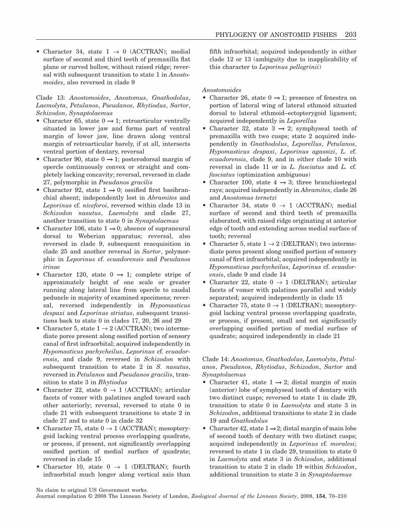
• Character 34, state 1 → 0 (ACCTRAN); medialsurface of second and third teeth of premaxilla flatplane or curved hollow, without raised ridge; rever-sal with subsequent transition to state 1 in Anosto-moides, also reversed in clade 9
Clade 13: Anostomoides, Anostomus, Gnathodolus,Laemolyta, Petulanos, Pseudanos, Rhytiodus, Sartor,Schizodon, Synaptolaemus• Character 65, state 0 → 1; retroarticular ventrally
situated in lower jaw and forms part of ventralmargin of lower jaw, line drawn along ventralmargin of retroarticular barely, if at all, intersectsventral portion of dentary, reversal
• Character 90, state 0 → 1; posterodorsal margin ofopercle continuously convex or straight and com-pletely lacking concavity; reversal, reversed in clade27, polymorphic in Pseudanos gracilis
• Character 92, state 1 → 0; ossified first basibran-chial absent; independently lost in Abramites andLeporinus cf. niceforoi, reversed within clade 13 inSchizodon nasutus, Laemolyta and clade 27,another transition to state 0 in Synaptolaemus
• Character 106, state 1 → 0; absence of supraneuraldorsal to Weberian apparatus; reversal, alsoreversed in clade 9, subsequent reacquisition inclade 25 and another reversal in Sartor, polymor-phic in Leporinus cf. ecuadorensis and Pseudanosirinae
• Character 120, state 0 → 1; complete stripe ofapproximately height of one scale or greaterrunning along lateral line from opercle to caudalpeduncle in majority of examined specimens; rever-sal, reversed independently in Hypomasticusdespaxi and Leporinus striatus, subsequent transi-tions back to state 0 in clades 17, 20, 26 and 29
• Character 5, state 1 → 2 (ACCTRAN); two interme-diate pores present along ossified portion of sensorycanal of first infraorbital; acquired independently inHypomasticus pachycheilus, Leporinus cf. ecuador-ensis, and clade 9, reversed in Schizodon withsubsequent transition to state 2 in S. nasutus,reversed in Petulanos and Pseudanos gracilis, tran-sition to state 3 in Rhytiodus
• Character 22, state 0 → 1 (ACCTRAN); articularfacets of vomer with palatines angled toward eachother anteriorly; reversal, reversed to state 0 inclade 21 with subsequent transitions to state 2 inclade 27 and to state 0 in clade 32
• Character 75, state 0 → 1 (ACCTRAN); mesoptery-goid lacking ventral process overlapping quadrate,or process, if present, not significantly overlappingossified portion of medial surface of quadrate;reversed in clade 15
• Character 10, state 0 → 1 (DELTRAN); fourthinfraorbital much longer along vertical axis than
fifth infraorbital; acquired independently in eitherclade 12 or 13 (ambiguity due to inapplicability ofthis character to Leporinus pellegrinii)
Anostomoides• Character 26, state 0 → 1; presence of fenestra on
portion of lateral wing of lateral ethmoid situateddorsal to lateral ethmoid–ectopterygoid ligament;acquired independently in Leporellus
• Character 32, state 3 → 2; symphyseal tooth ofpremaxilla with two cusps; state 2 acquired inde-pendently in Gnathodolus, Leporellus, Petulanos,Hypomasticus despaxi, Leporinus agassizi, L. cf.ecuadorensis, clade 9, and in either clade 10 withreversal in clade 11 or in L. fasciatus and L. cf.fasciatus (optimization ambiguous)
• Character 100, state 4 → 3; three branchiostegalrays; acquired independently in Abramites, clade 26and Anostomus ternetzi
• Character 34, state 0 → 1 (ACCTRAN); medialsurface of second and third teeth of premaxillaelaborated, with raised ridge originating at anterioredge of tooth and extending across medial surface oftooth; reversal
• Character 5, state 1 → 2 (DELTRAN); two interme-diate pores present along ossified portion of sensorycanal of first infraorbital; acquired independently inHypomasticus pachycheilus, Leporinus cf. ecuador-ensis, clade 9 and clade 14
• Character 22, state 0 → 1 (DELTRAN); articularfacets of vomer with palatines parallel and widelyseparated; acquired independently in clade 15
• Character 75, state 0 → 1 (DELTRAN); mesoptery-goid lacking ventral process overlapping quadrate,or process, if present, small and not significantlyoverlapping ossified portion of medial surface ofquadrate; acquired independently in clade 21
Clade 14: Anostomus, Gnathodolus, Laemolyta, Petul-anos, Pseudanos, Rhytiodus, Schizodon, Sartor andSynaptolaemus• Character 41, state 1 → 2; distal margin of main
(anterior) lobe of symphyseal tooth of dentary withtwo distinct cusps; reversed to state 1 in clade 29,transition to state 0 in Laemolyta and state 3 inSchizodon, additional transitions to state 2 in clade19 and Gnathodolus
• Character 42, state 1 →2; distal margin of main lobeof second tooth of dentary with two distinct cusps;acquired independently in Leporinus cf. moralesi;reversed to state 1 in clade 29, transition to state 0in Laemolyta and state 3 in Schizodon, additionaltransition to state 2 in clade 19 within Schizodon,additional transition to state 3 in Synaptolaemus
PHYLOGENY OF ANOSTOMID FISHES 203
No claim to original US Government works.Journal compilation © 2008 The Linnean Society of London, Zoological Journal of the Linnean Society, 2008, 154, 70–210
• Character 58, state 0 → 1; dentary squarish orrhomboidal with notable bend midway alonganteroventral margin; teeth and anterior marginof dentary inflected so that line drawn throughlong axis of symphyseal tooth of dentarypasses distinctly anterior of joint of quadrate andanguloarticular
• Character 43, state 1 → 3 (ACCTRAN); distalmargin of main lobe of third tooth of dentary withthree distinct cusps; subsequent transitions to state2 in clade 17 and state 0 in Laemolyta, reversed tostate 1 in Rhytiodus argenteofuscus and clade 31
• Character 77, state 0 → 1 (ACCTRAN); lateral shelfof quadrate moderate in length, extending posteri-orly past midpoint of symplectic but not beyondposterior limit of symplectic; reversed in Schizodon,modified to state 2 in clade 25 with subsequentmodification to state 0 in Gnathodolus
• Character 34, state 1 → 0 (DELTRAN); medialsurface of second and third teeth of premaxilla flatplane or curved hollow, without raised ridge origi-nating at anterior edge of tooth and extendingacross medial surface of tooth; reversal, reversedindependently in Leporinus pellegrinii and clade 9
Clade 15: Rhytiodus, Schizodon• Character 7, state 1 → 0; lamina located dorsal to
sensory canal of second infraorbital absent or poorlydeveloped with maximum height less than diameterof sensory canal; reversed in Schizodon isognathusand S. knerii
• Character 30, state 0 → 1; bony shelf above orbitformed by lateral ethmoid, frontal and sphenoticbones very wide, distance between lateral tips ofcontralateral sphenotics 80% or greater of length ofneurocranium from anterior tip of mesethmoid to tipof supraoccipital spine
• Character 40, state 0 → 1; distinct distal notchdivides posterior lamina of second or third tooth ofdentary into anterior and posterior lobes; acquiredindependently in Abramites, Leporinus cf. moralesiand clade 6
• Character 44, state 0 → 1; posterior lobes of dentaryteeth with one or more distinct cusps
• Character 63, state 0 → 1; sensory canal segment ofdentary with pore positioned along midlength ofcanal segment between posterior opening of canalproximate to anguloarticular and anterior limit ofcanal and occurring in posterior one-half of length ofdentary; acquired independently in Leporinus cf.ecuadorensis
• Character 94, state 1 → 2; three or more fullrows of teeth present on fifth upper pharyngealtooth-plate; reversal, reversed independently inAbramites
• Character 113, state 0 → 1; first four to six principalrays of upper lobe of caudal fin and last four to sixrays of lower lobe of caudal fin not distinctly sepa-rated but rather with margins in contact in adults;rays thickened and conjoined into rigid plate-likestructure in adults
• Character 5, state 2 → 1 (ACCTRAN); one interme-diate pore present along ossified portion of sensorycanal of first infraorbital; reversal with subsequenttransition to state 2 in Schizodon nasutus, alsoreversed in Petulanos and Pseudanos gracilis
• Character 32, state 3 → 4 (ACCTRAN); symphysealtooth of premaxilla with four cusps; reversed inclade 17; acquired independently in clade 23
• Character 39, state 0 → 1 (ACCTRAN); distinctnotch divides symphyseal tooth of dentary intoanterior and posterior lobes; acquired indepen-dently in Abramites and Gnathodolus bidens,reversed in Rhytiodus argenteofuscus
• Character 75, state 1 → 0 (ACCTRAN); mesoptery-goid with well-developed ventral triangular processthat significantly overlaps ossified portion of medialsurface of quadrate; reversal
• Character 95, state 2 → 3 (ACCTRAN); at least someteeth on fifth upper pharyngeal tooth-plate tricuspidand other teeth bicuspid; reversed in Schizodonscotorhabdotus and clade 17
• Character 96, state 1 → 2 (ACCTRAN); three ormore complete rows of teeth on tooth-plate of fifthceratobranchial; reversed in clade 17, acquired inde-pendently in clade 6
• Character 22, state 0 → 1 (DELTRAN); articularfacets of vomer with palatines parallel andwidely separated; state 1 acquired independently inAnostomoides
Clade 16: Rhytiodus• Character 6, state 0 → 1; walls of sensory canal of
first infraorbital thickened and laterosensory canallocated lateral to main lamina of bone; canal fusedonly weakly along its medial surface to main laminaof first infraorbital
• Character 11, state 0 → 1; flange of fifth infraorbitalpoorly developed or entirely absent, with at leastdorsal section of flange missing; acquired indepen-dently in Laemolyta
• Character 29, state 0 → 1; neurocranium distinctlydepressed; depth of neurocranium at angle in paras-phenoid about 30–32% of length of neurocraniumfrom anterior tip of mesethmoid to tip of supraoc-cipital spine; mesethmoid and horizontal portions ofvomer separated by narrow gap in lateral view
• Character 101, state 0 → 1; presence or absence ofanteriorly directed process of fourth pleural ribwithin Weberian apparatus
204 B. L. SIDLAUSKAS and R. P. VARI
No claim to original US Government works.Journal compilation © 2008 The Linnean Society of London, Zoological Journal of the Linnean Society, 2008, 154, 70–210
• Character 102, state 0 → 1; fourth pleural rib withanteriorly directed distal hook
• Character 104, state 0 → 1; neural complex in formof elongate triangle with nearly horizontal dorsalmargin
• Character 43, state 3 → 1 (ACCTRAN); distalmargin of main lobe of third tooth of dentary withonly single cusp; subsequent transition to state 2 inclade 17
• Character 77, state 0 → 1 (DELTRAN); lateral shelfof quadrate moderate in length, extending posteri-orly past midpoint of symplectic but not beyondposterior limit of symplectic; acquired indepen-dently in Laemolyta
• Character 5, state (1 or 2) → 3 (ancestral statereconstruction ambiguous under ACCTRAN andDELTRAN); three intermediate pores present alongossified portion of sensory canal of first infraorbital
Clade 17: Rhytiodus microlepis, R. lauzannei• Character 103, state 0 → 1; os suspensorium very
well-developed ventrally with distinct posterior cur-vature around anterior portion of swim bladder
• Character 115, state 0 → 1; 76 or more scalesincluding scales overlapping hypural plate presentin complete lateral-line series
• Character 120, state 1 → 0; complete stripe alonglateral-line absent; reversal, reversed indepen-dently in clades 20, 26 and 29
• Character 32, state 4 → 3 (ACCTRAN); symphysealtooth of premaxilla with three cusps; reversal
• Character 95, state 3 → 2 (ACCTRAN); most teethon fifth upper pharyngeal tooth-plate bicuspid butwith occasional teeth unicuspid; reversal, reversedindependently in Schizodon scotorhabdotus
• Character 96, state 2 → 1 (ACCTRAN); one or twocomplete rows of teeth present on tooth-plate of fifthceratobranchial; reversal
• Character 39, state 0 → 1 (DELTRAN); distinctnotch divides symphyseal tooth of dentary intoanterior and posterior lobes; acquired indepen-dently in Abramites, Gnathodolus and Schizodon
• Character 43, state (1, 3) → 2 (ancestral statereconstruction ambiguous under ACCTRAN andDELTRAN); distal margin of main lobe of thirdtooth of dentary with two distinct cusps
Clade 18: Schizodon• Character 33, state 3 → 4; second and third teeth of
premaxilla each with four cusps; acquired indepen-dently in clade 23
• Character 41, state 2 →3; distal margin of main lobeof symphyseal tooth of dentary with three distinctcusps; reversed in clade 19
• Character 42, state 2 →3; distal margin of main lobeof second tooth of dentary with three distinct cusps;
acquired independently in Synaptolaemus; reversedin clade 19
• Character 55, state 0 → 1; ascending process ofanguloarticular very wide, forming pronounced ver-tical ridge perpendicular to dentary; acquired inde-pendently in Hypomasticus
• Character 109, state 0 → 1; dorsomedial flange ofmesocoracoid very strongly developed; acquiredindependently in Abramites
• Character 77, state 1 → 0 (ACCTRAN); lateral shelfof quadrate elongate, extending posteriorly pastposterior margin of symplectic to terminate ventralto hyomandibular; reversal, reversed independentlyin Gnathodolus
• Character 32, state 3 → 4 (DELTRAN); symphysealtooth of premaxilla with four cusps; acquired inde-pendently in Rhytiodus argenteofuscus and clade 23
• Character 39, state 0 → 1 (DELTRAN); distinctnotch divides symphyseal tooth of dentary intoanterior and posterior lobes; acquired indepen-dently in Abramites, Gnathodolus and clade 17
• Character 43, state 1 → 3 (DELTRAN); distalmargin of main lobe of third tooth of dentary withthree distinct cusps; acquired independently inclade 25
• Character 95, state 2 → 3 (DELTRAN); at leastsome teeth on fifth upper pharyngeal tooth-platetricuspid and other teeth bicuspid; reversed inSchizodon scotorhabdotus, acquired independentlyin Rhytiodus argenteofuscus
• Character 96, state 1 → 2 (DELTRAN); one ortwo complete rows of teeth on tooth-plate of fifthceratobranchial; acquired independently in Rhy-tiodus argenteofuscus
Clade 19: Schizodon isognathus, S. nasutus• Character 17, state 1 → 2; ventral process of me-
sethmoid horizontally or slightly posterodorsallyaligned, vomer directly posterior of ventral portionof mesethmoid; acquired independently in eitherHypomasticus with reversal in Hypomasticus mega-lepis, or in both Hypomasticus despaxi and clade 6(optimization ambiguous)
• Character 41, state 3 → 2; distal margin of main(anterior) lobe of symphyseal tooth of dentary withtwo distinct cusps; reversal
• Character 42, state 3 →2; distal margin of main lobeof second tooth of dentary with two distinct cusps;reversal
• Character 59, state 0 → 1; long axes of dentaryand anguloarticular form distinctly obtuse angle;acquired independently in Schizodon knerii
• Character 72, state 1 → 0; ectopterygoid anterodor-sally inclined, with vertical through anterodorsallimit of ectopterygoid passing anterior to anterior
PHYLOGENY OF ANOSTOMID FISHES 205
No claim to original US Government works.Journal compilation © 2008 The Linnean Society of London, Zoological Journal of the Linnean Society, 2008, 154, 70–210
limit of ventral portion of ectopterygoid; reversal,reversed independently in Leporinus cf. niceforoi
• Character 117, state 1 → 0; lateral surface of bodywithout dark vertical blotches; reversal, alsoreversed in clade 21
Clade 20: Schizodon fasciatus, S. vittatus• Character 69, state 0 → 1; process of palatine very
long, length of process approximately two or moretimes width of process
• Character 120, state 1 → 0; complete stripe alonglateral-line absent; reversal, reversed indepen-dently in clades 17, 26 and 29
Clade 21: Anostomus, Gnathodolus, Laemolyta, Petul-anos, Pseudanos, Sartor, Synaptolaemus• Character 8, state 2 → 1; one intermediate pore
present along ossified portion of sensory canal ofsecond infraorbital; acquired independently inHypomasticus pachycheilus, Rhytiodus argenteofus-cus, Schizodon nasutus and clade 9
• Character 38, state 0 → 1; dentary teeth either withsmall, poorly developed posterior lamina or poste-rior lamina entirely absent
• Character46,state1→2;horizontallyorientatedpre-maxilla with premaxillary teeth directed anteriorly
• Character 117, state 1 → 0; lateral surface of bodywithout dark vertical blotches; reversal with subse-quent transition to state 1 in clade 24, reversedindependently in clade 19
• Character 22, state 1 → 0 (ACCTRAN); articularfacets of vomer with palatines angled toward eachother anteriorly; reversal, transition to state 2 inclade 27 with subsequent transition to state 0 ineither clade 32 or Sartor (ambiguity due to inabilityto code Gnathodolus for this character)
• Character 5, state 1 → 2 (DELTRAN); two interme-diate pores present along ossified portion of sensorycanal of first infraorbital; acquired independently inAnostomoides, Hypomasticus pachycheilus, Lepori-nus cf. ecuadorensis and clade 9, reversed to state 1in Pseudanos gracilis and Petulanos
• Character 75, state 0 → 1 (DELTRAN); mesoptery-goid lacking ventral process overlapping quadrate,or process, if present, small and not significantlyoverlapping ossified portion of medial surface ofquadrate; acquired independently in Anostomoides
Clade 22: Laemolyta• Character 11, state 0 → 1; flange of fifth infraorbital
posterior of sensory canal poorly developed orentirely absent, with at least dorsal section of flangemissing; acquired independently in Rhytiodus
• Character 41, state 2 → 0; distal margin of mainbody of symphyseal tooth of dentary with straightincisiform margin and no apparent cusps
• Character 42, state 2 → 0; distal margin of mainbody of second tooth of dentary with straight incisi-form margin and no apparent cusps
• Character 92, state 0 → 1; ossified first basibran-chial present; reversal, also reversed within clade 13in Schizodon nasutus and clade 27
• Character 43, state 3 → 0 (ACCTRAN); distalmargin of main body of third tooth of dentary withstraight incisiform margin and no apparent cusps
• Character 43, state 1 → 0 (DELTRAN); distalmargin of main body of third tooth of dentary withstraight incisiform margin and no apparent cusps
• Character 77, state 0 → 1 (DELTRAN); lateral shelfof quadrate moderate in length, extending posteri-orly past midpoint of symplectic but not beyondposterior limit of symplectic; acquired indepen-dently in Rhytiodus
Clade 23: Laemolyta orinocensis, L. proxima, L.taeniata• Character 32, state 3 → 4; symphyseal tooth of
premaxilla with four cusps; acquired independentlyin either clade 15 with reversal in clade 17, or inRhytiodus argenteofuscus and clade 18 (optimiza-tion ambiguous)
• Character 33, state 3 → 4; second and third teeth ofpremaxilla each with four cusps; acquired indepen-dently in Schizodon
Clade 24: Laemolyta orinocensis, L. proxima• Character 117, state 0 → 1; lateral surface of body
with two to five dark, vertical blotches intersectinglateral-line scale row and continuing dorsal andventral to lateral-line scale row; reversal
Clade 25: Anostomus, Gnathodolus, Petulanos, Pseu-danos, Sartor, Synaptolaemus• Character 4, state 0 → 1; origin of sensory canal of
first infraorbital situated directly dorsal to, or pos-terodorsal to, distal terminus of canal of firstinfraorbital where that canal contacts canal ofsecond infraorbital
• Character 12, state 0 → 1; sensory canal of sixthinfraorbital straight and lacking medial pore;acquired independently in Hypomasticus despaxiand H. mormyrops, reversed in Synaptolaemus andGnathodolus or in clade 31 with subsequent tran-sition to state 1 in Sartor (optimization ambigu-ous), polymorphic in Leporinus gomesi and L.striatus
• Character 14, state 0 → 1; sixth infraorbital pos-teriorly expanded and fused with sixth infraor-bital; reversed in clade 31
• Character 17, state 1 →0; ventral process of meseth-moid runs vertically or nearly so, anterior portion ofvomer positioned directly ventral to ventral process
206 B. L. SIDLAUSKAS and R. P. VARI
No claim to original US Government works.Journal compilation © 2008 The Linnean Society of London, Zoological Journal of the Linnean Society, 2008, 154, 70–210
of mesethmoid; acquired independently in Leporel-lus and Abramites
• Character 21, state 0 → 1; ventral surface of mainbody of vomer with pentagonal raised area inventral view medial to articulations with left andright palatines
• Character 27, state 1 → 0; absence of process oflateral ethmoid directed towards posterolateralcorner of mesethmoid; reversal, reversed indepen-dently in Leporinus agassizi, L. cf. moralesi, Schiz-odon isognathus and S. knerii.
• Character 50, state 0 → 1; anterolateral flange ofmaxilla merges with ventral portion of maxilla onanterior edge of ventral portion; reversal
• Character 67, state 1 → 0; palatine without distinctprocess extending away from main, rounded portionof bone cradled by dorsal portion of ectopterygoid
• Character 72, state 1 → 2; ectopterygoid posterodor-sally inclined with vertical through anterodorsallimit of ectopterygoid passing distinctly posterior ofventral portion of ectopterygoid
• Character 81, state 0 → 1; metapterygoid with threedistinct facets; vertical sheet dorsal to quadrate andsymplectic, horizontal or medially curved portionthat contacts mesopterygoid, and inclined planeproximate to hyomandibular that meets other facetsalong well defined margins
• Character 85, state 0 → 1; anterodorsal process ofpreopercle with thin, needle-like form
• Character 105, state 1 →0; absence of dorsal processof third vertebra overlapping neural complex later-ally; reversal, also reversed in Laemolyta proxima,Leporinus cf. moralesi, striatus, L. tigrinus, L. pel-legrinii, and possibly Hypomasticus despaxi andH. pachycheilus (ACCTRAN optimization only)
• Character 106, state 0 → 1; presence of supraneuraldorsal to Weberian apparatus; reversal, subsequenttransition to state 0 in Sartor, polymorphic in Pseu-danos irinae
• Character 25, state 0 → 1 (ACCTRAN); wing oflateral ethmoid with distinct notch along anteriormargin that accommodates passage of ligamentrunning between wing of lateral ethmoid andectopterygoid; acquired independently in clade 9,reversed in Pseudanos irinae and P. trimaculatus
• Character 53, state 0 → 1 (ACCTRAN); dorsalportion of maxilla in form of flat, medially-directedplate; reversed in Anostomus and Pseudanostrimaculatus
• Character 77, state 1 → 2 (ACCTRAN); lateral shelfof quadrate short, extending posteriorly to pointshort of midpoint of symplectic; subsequent transi-tion to state 0 in Gnathodolus
• Character 79, state 0 → 1 (ACCTRAN);metapterygoid–quadrate fenestra longer along axisrunning parallel to quadrate-metapterygoid joint
than along axis perpendicular to that joint; reversedin clade 26
• Character 122, state 0 → 1 (ACCTRAN); most scaleswith small, dark spot lying under scale creatingoverall appearance of row of such spots along centerof each scale row; reversed in Pseudanos trimacula-tus and clade 27 with subsequent transition to state1 in Anostomus anostomus
• Character 43, state 1 → 3 (DELTRAN); distalmargin of main lobe of third tooth of dentary withthree distinct cusps; reversed in clade 31, acquiredindependently in Schizodon
• Character 77, state 0 → 2 (DELTRAN); lateral shelfof quadrate short, extending posteriorly to pointshort of midpoint of symplectic; subsequent reversalto state 0 in Gnathodolus
Clade 26: Pseudanos gracilis, P. irinae, P. trimaculatus• Character 100, state 4 → 3; three branchiostegal
rays; acquired independently in Abramites, Anosto-moides, and Anostomus ternetzi
• Character 118, state 0 → 1; series of one or moredark spots with distinct margins present alonglateral-line scale row, each spot approximately oneto two scales in height, reversal, also reversed ineither clade 29 with subsequent reversal in Synap-tolaemus, or in Petulanos and clade 32 (optimizationambiguous)
• Character 120, state 1 → 0; complete stripe alonglateral-line absent; reversal, reversed indepen-dently in clades 17, 20 and 29
• Character 25, state 1 → 0 (ACCTRAN); wing oflateral ethmoid without distinct notch along ante-rior margin; reversal, reversed in Pseudanos gracilis
• Character 28, state 0 → 1 (ACCTRAN); frontal–parietal fontanel partially or completely closed;acquired independently in Abramites and Anosto-mus, reversed in Pseudanos gracilis
• Character 79, state 1 → 0 (ACCTRAN);metapterygoid–quadrate fenestra shorter along axisrunning parallel to quadrate-metapterygoid jointthan along axis perpendicular to that joint; reversal
Clade 27: Anostomus, Gnathodolus, Petulanos, Sartor,Synaptolaemus• Character 2, state 1 → 0; anterior margin of antor-
bital positioned just dorsal of, or dorsal and poste-rior of, anterodorsal opening of sensory canal of firstinfraorbital; reversal
• Character 19, state 0 → 1; mesethmoid narrow, withmaximum width along transverse axis much lessthan maximum length along longitudinal axis;reversal
• Character 22, state 0 → 2; articular facets of vomerwith palatines parallel and widely separated;
PHYLOGENY OF ANOSTOMID FISHES 207
No claim to original US Government works.Journal compilation © 2008 The Linnean Society of London, Zoological Journal of the Linnean Society, 2008, 154, 70–210
reversed in either clade 32 or Sartor (ambiguity dueto inability to code Gnathodolus for this character)
• Character 71, state 0 → 1 very small anterodorsalprocess of ectopterygoid running ventral to palatine,or process absent
• Character 89, state 1 → 0; joint of interhyal, metap-terygoid, symplectic and hyomandibular positionedat approximately same horizontal plane as anteriorportion of lateral shelf of metapterygoid
• Character 90, state 1 → 0; posterodorsal marginof opercle with slight or pronounced concavity;reversal
• Character 92, state 0 → 1; ossified first basibran-chial present; reversal with subsequent transition tostate 0 in Synaptolaemus, also reversed within clade13 in Schizodon nasutus and Laemolyta
• Character 74, state 0 → 1 (ACCTRAN); posteriorlydirected dorsolateral process of mesopterygoidpresent; reversed in clade 31 with subsequent tran-sition back to state 1 in Sartor
• Character 122, state 1 → 0 (ACCTRAN); centers ofscales same colour as, or lighter than, scale margins,no series of small dark spots along center of eachscale row; reversal with subsequent transition backto state 1 in Anostomus anostomus
• Character 25, state 0 → 1 (DELTRAN); wing oflateral ethmoid with distinct notch along anteriormargin that accommodates passage of ligamentrunning between wing of lateral ethmoid and ectop-terygoid; state 1 acquired independently in Pseu-danos gracilis and P. winterbottomi
• Character 79, state 0 → 1 (DELTRAN);metapterygoid–quadrate fenestra longer along axisrunning parallel to quadrate-metapterygoid jointthan along axis perpendicular to that joint; state 1acquired independently in Pseudanos winterbottomi
Clade 28: Anostomus• Character 28, state 0 → 1; frontal–parietal fontanel
partially or completely closed; acquired indepen-dently in Abramites and either clade 26 with re-versal in Pseudanos gracilis, or in P. irinae andP. trimaculatus (optimization ambiguous).
• Character 121, state 0 →1; one or more longitudinalstripes present dorsal and ventral to lateral-linescale row; acquired independently in Hypomasticusdespaxi and Leporinus striatus
• Character 53, state 1 → 0 (ACCTRAN); dorsalportion of maxilla rod-like, with tapered or pointedtip; reversal, reversed independently in Pseudanostrimaculatus
• Character 74, state 0 → 1 (DELTRAN); presence ofposteriorly directed dorsolateral process of mesop-terygoid; acquired independently in Petulanos andSartor
Clade 29: Gnathodolus, Petulanos, Sartor,Synaptolaemus• Character 41, state 2 → 1; distal margin of main
(anterior) lobe of symphyseal tooth of dentary withonly single cusp; reversal with subsequent transi-tion back to state 2 in Gnathodolus
• Character 42, state 2 → 1; distal margin of mainlobe of second tooth of dentary with only singlecusp; reversal, further modified to state 3 inSynaptolaemus
• Character 52, state 0 → 1; ventral portion of maxillavery wide, greatly expanded in direction transverseto long axis of bone; reversal
• Character 78, state 0 → 1; lateral shelf of quadrateterminates anteriorly prior to reaching articularcondyle
• Character 99, state 0 → 1; urohyal high, dorsal andventral margins meet at angle greater than 45°;reversed in Gnathodolus
• Character 120, state 1 → 0; complete stripe alonglateral-line absent; reversal, reversed indepen-dently in clades 17, 20 and 26
• Character 118, state 0 → 1 (ACCTRAN); series ofone or more dark spots with distinct marginspresent along lateral-line scale row, each spotapproximately one to two scales in height, reversalwith subsequent reversal in Synaptolaemus, alsoreversed in clade 26
• Character 53, state 0 → 1 (DELTRAN); dorsalportion of maxilla in form of flat, medially-directedplate; acquired independently in Pseudanos gracilisand P. winterbottomi
Clade 30: Petulanos• Character 5, state 2 → 1; one intermediate pore
present along ossified portion of sensory canal offirst infraorbital; reversal, also reversed in Pseu-danos gracilis, also reversed in Schizodon underACCTRAN optimization
• Character 8, state 1 → 2; two intermediate porespresent along ossified portion of sensory canal ofsecond infraorbital; reversal
• Character 32, state 3 → 2; symphyseal tooth ofpremaxilla with two cusps; state 2 acquired inde-pendently in clade 9, Anostomoides, Gnathodolus,Leporellus, Hypomasticus despaxi, Leporinus agas-sizi, L. cf. ecuadorensis, and in either clade 10 withreversal in clade 11 or in L. fasciatus and L. cf.fasciatus (optimization ambiguous).
• Character 88, state 0 → 1; ventral lamina of sym-plectic broad and triangular
• Character 74, state 0 → 1 (DELTRAN); presence ofposteriorly directed dorsolateral process of mesop-terygoid; acquired independently in Anostomus andSartor
208 B. L. SIDLAUSKAS and R. P. VARI
No claim to original US Government works.Journal compilation © 2008 The Linnean Society of London, Zoological Journal of the Linnean Society, 2008, 154, 70–210
• Character 118, state 0 → 1 (DELTRAN); series ofone or more dark spots with distinct marginspresent along lateral-line scale row; reversal;reversed independently in clades 26 and 32
Clade 31: Gnathodolus, Sartor, Synaptolaemus• Character 14, state 1 → 0; sixth infraorbital not
posteriorly expanded and entirely separate fromsuprapreopercle; reversal
• Character 43, state 3 →1; distal margin of main lobeof third tooth of dentary with only single cusp;reversal, also reversed in Rhytiodus or only in R.argenteofuscus (optimization ambiguous)
• Character 51, state 0 → 1; anterolateral flange ofmaxilla and ventral portion of bone without rotationrelative to each other, and both lying in parasagittalplane; reversal
• Character 66, state 0 → 1; dermal papillae presenton lower lip
• Character 86, state 0 → 1; anterior margin ofascending arm of preopercle with distinct triangularprocess overlapping joint between interhyal, hyo-mandibular, metapterygoid and symplectic inlateral view
• Character 12, state 1 → 0 (ACCTRAN); sensorycanal of sixth infraorbital tripartite or with form ofangled tube with medial pore situated at angle oftube; reversal with subsequent transition back tostate 1 in Sartor
• Character 74, state 1 → 0 (ACCTRAN); absence ofposteriorly directed dorsolateral process of mesop-terygoid; reversal with subsequent transition backto state 1 in Sartor
Synaptolaemus• Character 24, state 0 → 1; insertion of lateral
ethmoid–ectopterygoid ligament on dorsal surface ofanterior wing of lateral ethmoid
• Character 42, state 1 →3; distal margin of main lobeof second tooth of dentary with three distinct cusps;acquired independently in Schizodon
• Character 92, state 1 → 0; ossified first basibran-chial absent; reversal
• Character 116, state 0 →1; body with vertical bandsof at least two scales width, at least some of whichcompletely encircle body; acquired independently inAbramites and clade 10
• Character 118, state 1 → 0 (ACCTRAN); lateral-linespots absent; reversal
• Character 12, state 1 → 0 (DELTRAN); sensorycanal of sixth infraorbital tripartite or with form ofangled tube with medial pore situated at angle oftube; reversal, also reversed in Gnathodolus
Clade 32: Gnathodolus, Sartor• Character 35, state 0 → 1; at least two of teeth
present on premaxilla with distinct dorsal curvaturedistally; teeth appearing strongly ventrally bowedin lateral profile with distal tips of teeth positionedmore dorsally than base of teeth
• Character 37, state 0 → 1; dentary teeth elongatewith symphyseal tooth extremely elongate andrecurved, overall shape reminiscent of awl or verynarrow chisel; acquired independently in Leporinusjatuncochi
• Character 54, state 0 → 1; anterolateral and poste-rior portions of maxilla merge to form flat plateorientated perpendicular to process of maxilla thatcontacts cartilage of palatine
• Character 61, state 1 → 0; sensory canal of dentarygreatly reduced or absent
• Character 22, state 2 → 0 (ACCTRAN); articularfacets of vomer with palatines angled toward eachother anteriorly; reversal
• Character 36, state 4 → 1 (ACCTRAN); dentary withone tooth present
• Character 118, state 0 → 1 (DELTRAN); series ofone or more dark spots with distinct marginspresent along lateral-line scale row; reversal,reversed independently in Petulanos and clade 26
Gnathodolus• Character 3, state 0 → 1; supraorbital with deeply
concave, distinctly notched anterior or anterolateralmargin; reversal, also reversed in Schizodon isog-nathus and S. knerii
• Character 10, state 1 → 0; vertical extent of fourthand fifth infraorbitals approximately equal or fifthinfraorbital longer along vertical axis than fourthinfraorbital; reversal
• Character 20, state 0 → 1; vomer with two deepgrooves on ventral surface that receive dorsal sur-faces of mesopterygoid, metapterygoid and palatine
• Character 23, state 1 → 0; absence of lateralethmoid–ectopterygoid ligament
• Character 32, state 3 → 2; symphyseal toothof premaxilla with two cusps; state 2 acquiredindependently in Anostomoides, Gnathodolus,Leporellus, Petulanos, Hypomasticus despaxi,Leporinus agassizi, L. cf. ecuadorensis, clade 9,and in either clade 10 with reversal in clade 11 or inL. fasciatus and L. cf. fasciatus (optimizationambiguous)
• Character 39, state 0 → 1; distinct notch dividessymphyseal tooth of dentary into anterior and pos-terior lobes; acquired independently in Abramitesand either clade 15 with reversal in Rhytiodusargenteofuscus or in Schizodon and clade 17 (opti-mization ambiguous)
PHYLOGENY OF ANOSTOMID FISHES 209
No claim to original US Government works.Journal compilation © 2008 The Linnean Society of London, Zoological Journal of the Linnean Society, 2008, 154, 70–210
• Character 41, state 1 → 2; distal margin of main(anterior) lobe of symphyseal tooth of dentary withtwo distinct cusps; reversal
• Character 57, state 0 → 1; dentary extraordinarilyelongate, distance from joint with anguloarticular totip of dentary three times or more height of dentaryjust anterior to ascending process of anguloarticular
• Character 70, state 0 → 1; palatine in shape ofdorsoventrally elongate hourglass, with triangulardorsal and ventral portions connected by thin threadof bone
• Character 73, state 1 →0; absence of distinct processon posterolateral surface of the ectopterygoid;reversal
• Character 77, state 2 → 0; lateral shelf of quadrateelongate, extending posteriorly past posteriormargin of symplectic to terminate ventral to hyo-mandibular; reversal
• Character 12, state 1 → 0 (DELTRAN); sensorycanal of sixth infraorbital tripartite or with form ofangled tube with medial pore situated at angle oftube; reversal, also reversed in Synaptolaemus
• Character 36, state 4 → 1 (DELTRAN); one toothpresent on dentary
Sartor• Character 82, state 0 → 1; lateral surface of metap-
terygoid with well-developed lateral shelf
• Character 99, state 1 → 0; urohyal low, dorsal andventral margins meet at angle equal to or less than45°; reversal
• Character 106, state 1 → 0; absence of supraneuraldorsal to Weberian apparatus; reversal
• Character 12, state 0 → 1 (ACCTRAN); canal ofsixth infraorbital straight and lacking medial pore;reversal
• Character 36, state 1 → 3 (ACCTRAN); three teethpresent on dentary, acquired independently inHypomasticus despaxi and Leporinus jatuncochi
• Character 74, state 0 → 1 (ACCTRAN); presence ofposteriorly directed dorsolateral process of mesop-terygoid; reversal
• Character 22, state 2 → 0 (DELTRAN); articularfacets of vomer with palatines angled toward eachother anteriorly; reversal
• Character 36, state 4 → 3 (DELTRAN); three teethpresent on dentary; acquired independently inHypomasticus despaxi and Leporinus jatuncochi
• Character 74, state 0 → 1 (DELTRAN); presence ofposteriorly directed dorsolateral process of mesop-terygoid; acquired independently in Anostomus andPetulanos
210 B. L. SIDLAUSKAS and R. P. VARI
No claim to original US Government works.Journal compilation © 2008 The Linnean Society of London, Zoological Journal of the Linnean Society, 2008, 154, 70–210
Related Documents