PHT 226 Lab# 4 • Culture media Culture media • Streaking Streaking

PHT 226 Lab# 4 Culture mediaCulture media Streaking Streaking.
Dec 24, 2015
Welcome message from author
This document is posted to help you gain knowledge. Please leave a comment to let me know what you think about it! Share it to your friends and learn new things together.
Transcript

PHT 226 Lab# 4
•Culture mediaCulture media• StreakingStreaking

Culture Media
•The culture media is an artificial preparation contains the essential element and nutrient
in a proper concentration needed by the microorganism to grow.
•It may be-:•Liquid (broth)•Solid (containing agar)•Semisolid (containing low conc of agar)

Culture Media
•Most common ingredients-:
.1Essential elements and nutrients.
.2Solidifying agents.

Objectives of bacterial culture:
•To study bacterial morphology, physiology and pathogenic properties.
•At the clinical level:.1Isolation of bacteria in pure culture.2Identification.3Antimicrobial susceptibility
•Preparation of important bacterial products, e.g., toxins, antigens, vaccines … etc.

Essential Elements and Nutrients
.1A Source of Nitrogen: •(peptone )•meat extract,•yeast extract ,•amino acids & in organic compound.

Essential Elements and Nutrients
.2A Source of Carbon: •usually supplied in form of carbohydrates.
.3Inorganic salts: •normally present in peptone but added in
traces [e.g; Na, K, Ca, Mn, Fe, Co, Zn…….etc]
♙NaCl

Essential Elements and Nutrients
4. Buffers:
•added to resist any changed in pH of the medium .
Blood and milk are naturally occurring buffers.
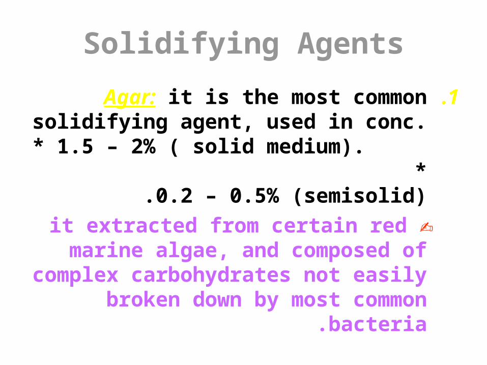
Solidifying Agents
.1Agar: it is the most common solidifying agent, used in conc. * 1.5 – 2% ( solid
medium). * 0.2 – 0.5% (semisolid) .
✍it extracted from certain red marine algae, and composed of complex carbohydrates not easily broken down by most common
bacteria.

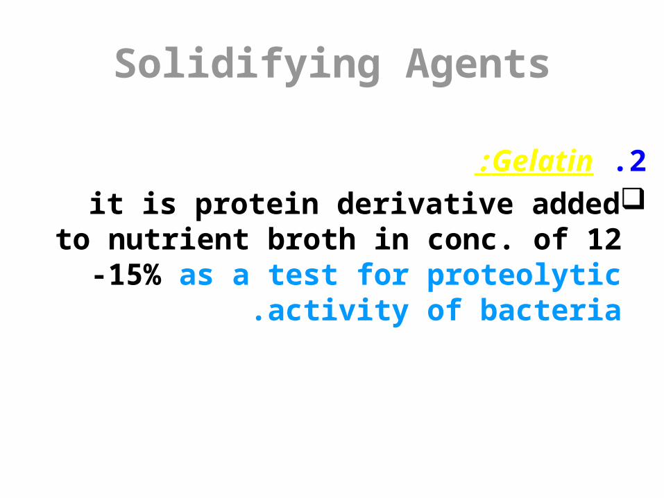
Solidifying Agents
2. Gelatin: it is protein derivative added to nutrient broth
in conc. of 12 -15% as a test for proteolytic activity of bacteria .

Classification of Media
•According to its content-:
.1Non synthetic complex medium; •contains extracts or digests of plant or
animal tissues such as beef, peptone & soybean digest.

Classification of Media
.2Synthetic Chemically defined medium;• contains known chemical compounds such
as: amino acids ,sugar,Vitamins & salts.
Classification of Media
.3Living medium; •it is a living cell of animal or plant in which
m.o could be cultivated such as viruses (intracellular tissue culture.)

Types of culture media:
•Simple media•Enriched media•Selective media•Indicator media•Selective and indicator media
Classification of Media•According to the purpose of use-:1 -Simple [Basic] media; •contain only the essential or basic nutrients
which support the growth of most common bacteria .
✎ e.g; • Nutrient agar (plate, slant)• Nutrient broth


Classification of Media
2 -Enriched media; in addition to basic nutrients, it contains
enriched substances such as blood, serum, vitamins to support the growth of most
pathogens can’t grow on ordinary simple media.
✎ e.g ;blood agar ,Chocolate agar ,Serum agar.

Blood agar

Chocolate agar(Neisseria spp.)
C.diphtheria

Classification of Media
3. Selective media; mostly used in case of mixture.Contains substances which selectively enhance the
growth of certain particular species and inhibit the growth of undesired bacterial species.
These inhibitory substances include: dyes, heavy metals, chemicals, antimicrobial agents…

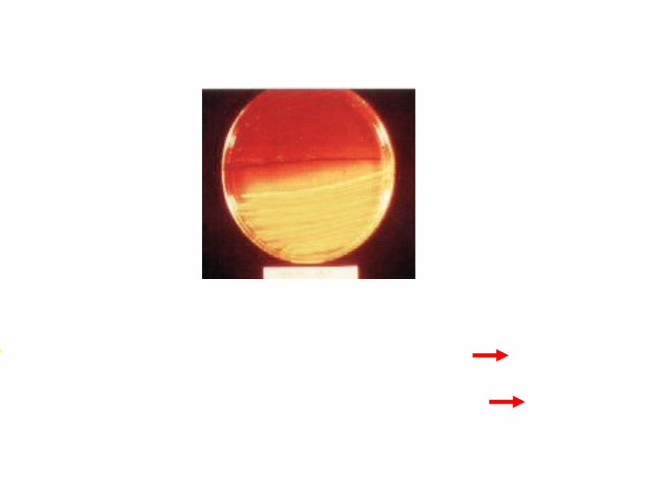
Examples of Selective Media
.1Mannitol salt agar; •it is selective medium for Staphylococcus
species because it contains high conc. of salt (about 7%).

2 -Lowenstein Jensen medium it is selective medium for M. tuberculosis;
•because it contains malachite green dye.•It contains also eggs which solidify the
medium without agar and make it enriched medium .


Examples of Selective Media
3 -MacConkey agar: •it is selective medium for gram –ve bacteria
because it contains bile salt which inhibit the growth of gram +ve bacteria.
Examples of Selective Media
4 -Cetrimide agar: it is highly selective medium for
pseudomonas species. It contains Cetrimide.Cetrimide is a surfactant that
inhibit the growth of all organism except pseudomonas.

Classification of Media
4 -Indicator media:• Contain substances which are affected by
the growth of organism to produce characteristic reaction such as indicators
which indicates acid production during sugar fermentation.

Examples of Diagnostic Media
.1Mannitol salt agar; It contains mannitol and phenol red indicator .S.aureus
is the only strain of staphylococcus species can ferment mannitol, leading to production of acid which can detected by yellow color around the
growth resulting from changing the color of the indicator .
Growth with yellow color around the colonies S.aureus
Growth without change in the color of the medium S.epidermidis

Examples of Diagnostic Media
2. MacConkey agar;
•Gram –ve bacteria classified to:
•It contains neutral red indicator which detect acid production due to fermentation of lactose which done by
lactose fermenter gram –ve bacteria such as E-coli and Klebsiella species to give rose pink colonies .
Lactose fermenter Lactose non-fermenter

Lactose fermenter Pink colonies
Lactose non-fermenter Pale colonies

Classification of Media
5. Selective and indicator media; •are both selective and diagnostic since they
permit the growth of a few desired species only & contain indicator which enable us to differentiate between the species which are
able to grow on these media.•e.g.; Mannitol salt agar ,
MacConkey agar.

Media for Fungi
❀ Sabouraud dextrose agar. similar in composition as those of
bacteria EXCEPT:.1They are acidic in nature (PH 3 -6)..2 contain readily fermentable
sugars e.g.; dextrose and maltose.
☞Culture of Fungi usually incubate at lower temperature than
bacteria (about 25°C) .

It is anaerobic medium due to presence of:It is anaerobic medium due to presence of: Sodium thioglycolate and cystaine which act Sodium thioglycolate and cystaine which act
as reducing agents.as reducing agents.
Fluid thioglycolate:Fluid thioglycolate:
Anaerobic mediaAnaerobic media
It is anaerobic medium due to presence of:It is anaerobic medium due to presence of: Meat particles (prepared from heart Meat particles (prepared from heart
muscles) muscles) Hematin& glutathione in meat particles Hematin& glutathione in meat particles
act as reducing agents.act as reducing agents.
Cooked meat medium:Cooked meat medium:

Anaerobic media

Anaerobic Jar - The nutrient agar plates employed for routine culturing do not contain reducing agents. - They can be used to culture anaerobes if they are placed inside an anaerobe jar ( a chamber from which the oxygen is removed). - Gas generator packets (e.g. GasPak) are placed in the jar along with plates or tubes containing conventional media destined to be incubated anaerobically. - In order to assure that oxygen actually was removed from the chamber, a strip of paper soaked in methylene blue dye is included in the jar. - It is blue when exposed to oxygen but will become colorless (white) when oxygen is absent.

GAS PAK SYSTEM FOR ANAEROBIC CULTIVATION
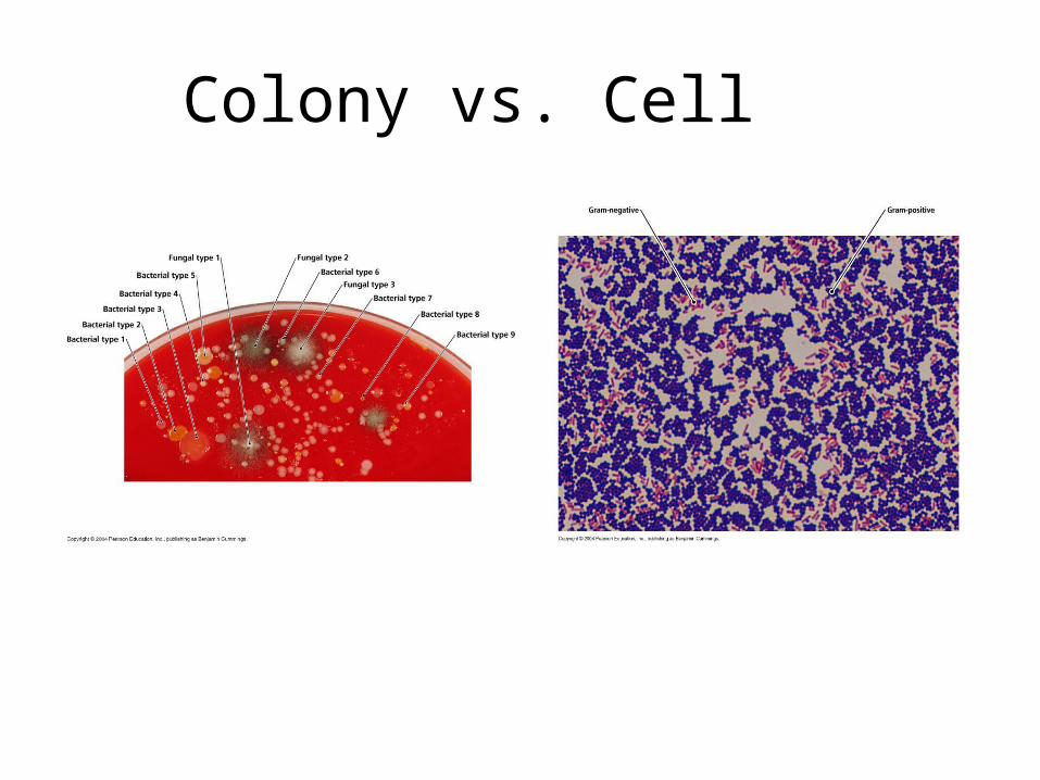
Colony vs. Cell
Colonies Cells

Introduction to Microbiological Equipments and Materials
•Inoculation: Culturing of sterile media with m.o [Inoculation loop].
•Incubation: Placing the culture into the incubator at optimum temperature for growth.
•Sterile: Free from any living microorganism
•Contamination: Introduction of undesirable m.o.

Description of ColoniesDescription of Colonies

Isolation of Pure Colonies Isolation of Pure Colonies of Microorganism of Microorganism
“Streak Plate Method”“Streak Plate Method”In natural environments, bacteria & In natural environments, bacteria &
other m.o exist in mixed other m.o exist in mixed population.population.
““Streak Plate Technique”Streak Plate Technique”The individual cells are separated from each other by certain distance on the surface of the agar.After incubation, each single deposited cell divide many times and finally form visible mass of growth “COLONY”.
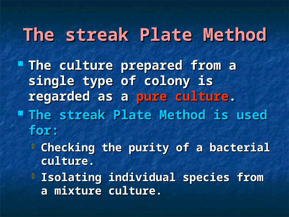
The streak Plate MethodThe streak Plate Method
The culture prepared from a The culture prepared from a single type of colony is regarded single type of colony is regarded as a as a pure culturepure culture..
The streak Plate Method is used The streak Plate Method is used for:for: Checking the purity of a bacterial Checking the purity of a bacterial
culture.culture. Isolating individual species from a Isolating individual species from a
mixture culture.mixture culture.


The streak Plate MethodThe streak Plate Method
Objective:-Objective:-
for isolation of individual for isolation of individual species of a mixed broth culture.species of a mixed broth culture.
Materials:-Materials:- Nutrient agar plate.Nutrient agar plate. Mixed broth culture of Mixed broth culture of Serratia Serratia
marcescens marcescens andand staph. aureus. staph. aureus.

The streak Plate MethodThe streak Plate Method
ProcedureProcedure::
S & SS & S
Aseptic technique Aseptic technique
Invert the plate and Invert the plate and Incubate for 24h at 37Incubate for 24h at 37℃℃
Drop of the cultureDrop of the cultureFlame & Cool
Flame & Cool
Flame & Cool


The streak Plate MethodThe streak Plate Method

The streak Plate MethodThe streak Plate Method


Sources of Sources of ContaminationContamination
Objective:Objective: To identify some of the To identify some of the sources of contamination present in the sources of contamination present in the lab. lab.
✔✔ in order to avoid them in order to avoid them Contamination from hands.Contamination from hands. Contamination from breath.Contamination from breath. Contamination from air.Contamination from air. Contamination from bench.Contamination from bench.

Sources of Sources of ContaminationContamination
Sterile nutrient agar plateSterile nutrient agar plate a-a- unwashed unwashed
b-b- washed with water washed with water
c-c- disinfected with alcohol disinfected with alcohol
d-d- control control incubate the plate Inverted at 37°c incubate the plate Inverted at 37°c
for 24 hr.for 24 hr. Record the appearance of the plateRecord the appearance of the plate
a b
c d
1. Contamination from hands:
Sources of Sources of ContaminationContamination
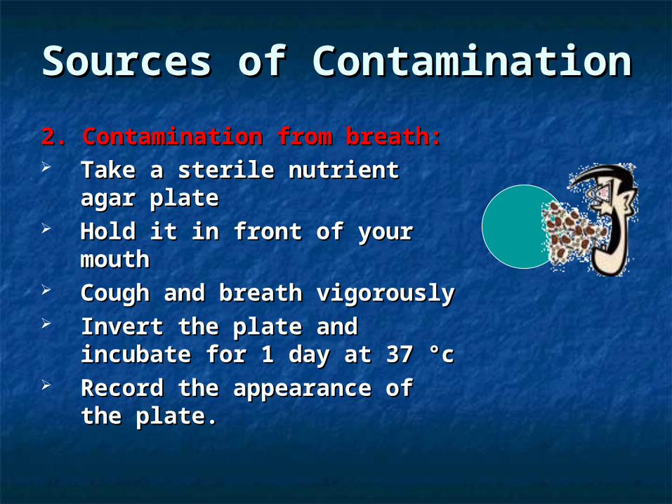
2. Contamination from breath:2. Contamination from breath: Take a sterile nutrient agar Take a sterile nutrient agar
plate plate Hold it in front of your mouthHold it in front of your mouth Cough and breath vigorouslyCough and breath vigorously Invert the plate and incubate Invert the plate and incubate
for 1 day at 37 °cfor 1 day at 37 °c Record the appearance of the Record the appearance of the
plate.plate.

Sources of Sources of ContaminationContamination
3.Contamination from air:3.Contamination from air: Expose one sterile Expose one sterile
nutrient agar plate on nutrient agar plate on the bench for 30 minthe bench for 30 min
Invert the plate and Invert the plate and incubate for 1 day at 37 incubate for 1 day at 37 °c°c
Record the colonial Record the colonial appearance appearance 30 min
Sources of Sources of ContaminationContamination
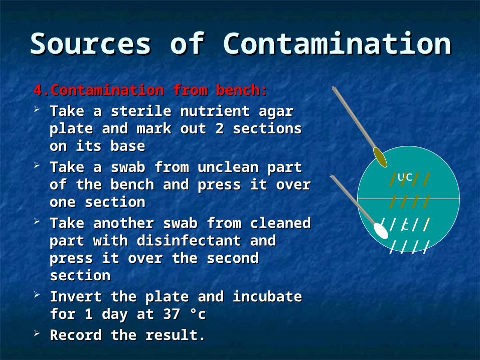
4.Contamination from bench:4.Contamination from bench: Take a sterile nutrient agar Take a sterile nutrient agar
plate and mark out 2 sections plate and mark out 2 sections on its baseon its base
Take a swab from unclean part Take a swab from unclean part of the bench and press it over of the bench and press it over one sectionone section
Take another swab from Take another swab from cleaned part with disinfectant cleaned part with disinfectant and press it over the second and press it over the second sectionsection
Invert the plate and incubate Invert the plate and incubate for 1 day at 37 °cfor 1 day at 37 °c
Record the result.Record the result.
uc
c
/////////
/////////

Thank you
Related Documents