MARINE ECOLOGY PROGRESS SERIES Mar Ecol Prog Ser Vol. 270: 83–102, 2004 Published April 14 INTRODUCTION Haptophyte microalgae are an important component of the world’s oceanic phytoplankton (Okada & McIn- tyre 1977), blooming seasonally at polar, equatorial and subtropical latitudes (Brown & Yoder 1994). The calcite- covered coccolithophorids such as Gephyrocapsa oceanica and Emiliania huxleyi dominate subtropical and sub-polar latitudes (Westbroek et al. 1994), and are significant globally in providing a long-term sink for inorganic carbon (Van der Wal et al. 1995, Paasche 2002). They also produce volatile dimethyl sulphide, which produces cloud-condensation nuclei, increasing cloud cover and affecting regional climates (Malin et al. 1994). In addition, some species (e.g. Chrysochromulina polylepis) are highly toxic to fin-fishes (Moestrup 1994). Monitoring of these and other phytoplankton groups is essential in order to follow seasonal successions, impacts of global warming on the marine environment, and harmful ecological events. While taxonomic monitoring of the 200 known hapto- phyte species by microscopy is possible (Jordan et al. © Inter-Research 2004 · www.int-res.com *Corresponding author: Email: [email protected] Photosynthetic pigments in 37 species (65 strains) of Haptophyta: implications for oceanography and chemotaxonomy Manuel Zapata 1 , S. W. Jeffrey 2, *, Simon W. Wright 3 , Francisco Rodríguez 1 , José L. Garrido 4 , Lesley Clementson 2 1 Centro de Investigacións Mariñas (CIMA), Consellería de Pesca, Xunta de Galicia, Apartado 13, 36620 Vilanova de Arousa, Spain 2 CSIRO Marine Research, GPO Box 1538, Hobart, Tasmania 7001, Australia 3 Australian Antarctic Division, Kingston, Tasmania 7050, Australia 4 Instituto de Investigacións Mariñas, Consejo Superior de Investigacións Cientificas (CSIC), Eduardo Cabello 6, 36208 Vigo, Spain ABSTRACT: The pigment compositions of 37 species (65 strains) of cultured haptophytes were analysed using improved HPLC methods. We distinguished 8 pigment types based on the distribution of 9 chlorophyll c (chl c) pigments and 5 fucoxanthin derivatives. All types contained chl c 2 and Mg- 2, 4-divinyl phaeoporphyrin a 5 monomethyl ester (MgDVP), fucoxanthin, diadinoxanthin and β,β- carotene. Pigment types were based on the following additional pigments: Type 1: chl c 1 ; Type 2: chl c 1 and chl c 2 -Pavlova gyrans-type; Type 3: chl c 1 and chl c 2 -monogalactosyl diacylglyceride ester (chl c 2 -MGDG [18:4/14:0]); Type 4: chl c 1 , chl c 3 and non-polar chl c 1 -like; Type 5: chl c 1 , chl c 3 , chl c 2 - MGDG [18:4/14:0] and 4-keto-fucoxanthin; Type 6: chl c 3 , monovinyl chl c 3 (MV-chl c 3 ), chl c 2 -MGDG [18:4/14:0], 19’-hexanoyloxyfucoxanthin and its 4-keto derivative, and traces of 19’-butanoyloxyfu- coxanthin; Type 7: similar to Type 6, minus MV-chl c 3 but with chl c 2 -MGDG [14:0/14:0] added; Type 8: similar to Type 6, minus MV-chl c 3 but with significant 19’-butanoyloxyfucoxanthin. Taxonomic associations ranged from single genera to multiple families – Type 1: Pavlovaceae, Isochrysidaceae and Pleurochrysidaceae; Type 2: Pavlovaceae; Type 3: Isochrysidaceae; Type 4: Prymnesium spp.; Type 5: Ochrosphaera spp.; Type 6: Nöelaerhabdaceae, notably Emiliania spp.; Type 7: Chrysochro- mulina spp.; Type 8: Phaeocystaceae, Prymnesiaceae and Isochrysidaceae. These pigment types showed a strong correlation with available phylogenetic trees, supporting a genetic basis for the pig- ment associations. The additional marker pigments offer oceanographers greater power for detecting haptophytes in mixed populations, while also distinguishing a greater proportion of them from diatoms. KEY WORDS: Haptophyta · HPLC · Chlorophylls c · Fucoxanthins · Pigment types · Phylogeny · Oceanography Resale or republication not permitted without written consent of the publisher

Welcome message from author
This document is posted to help you gain knowledge. Please leave a comment to let me know what you think about it! Share it to your friends and learn new things together.
Transcript
MARINE ECOLOGY PROGRESS SERIESMar Ecol Prog Ser
Vol. 270: 83–102, 2004 Published April 14
INTRODUCTION
Haptophyte microalgae are an important componentof the world’s oceanic phytoplankton (Okada & McIn-tyre 1977), blooming seasonally at polar, equatorial andsubtropical latitudes (Brown & Yoder 1994). The calcite-covered coccolithophorids such as Gephyrocapsaoceanica and Emiliania huxleyi dominate subtropicaland sub-polar latitudes (Westbroek et al. 1994), and aresignificant globally in providing a long-term sink forinorganic carbon (Van der Wal et al. 1995, Paasche
2002). They also produce volatile dimethyl sulphide,which produces cloud-condensation nuclei, increasingcloud cover and affecting regional climates (Malin et al.1994). In addition, some species (e.g. Chrysochromulinapolylepis) are highly toxic to fin-fishes (Moestrup 1994).Monitoring of these and other phytoplankton groups isessential in order to follow seasonal successions,impacts of global warming on the marine environment,and harmful ecological events.
While taxonomic monitoring of the 200 known hapto-phyte species by microscopy is possible (Jordan et al.
© Inter-Research 2004 · www.int-res.com*Corresponding author: Email: [email protected]
Photosynthetic pigments in 37 species (65 strains)of Haptophyta: implications for oceanography
and chemotaxonomy
Manuel Zapata1, S. W. Jeffrey2,*, Simon W. Wright3, Francisco Rodríguez1,José L. Garrido4, Lesley Clementson2
1Centro de Investigacións Mariñas (CIMA), Consellería de Pesca, Xunta de Galicia, Apartado 13, 36620 Vilanova de Arousa, Spain
2CSIRO Marine Research, GPO Box 1538, Hobart, Tasmania 7001, Australia3Australian Antarctic Division, Kingston, Tasmania 7050, Australia
4Instituto de Investigacións Mariñas, Consejo Superior de Investigacións Cientificas (CSIC), Eduardo Cabello 6, 36208 Vigo, Spain
ABSTRACT: The pigment compositions of 37 species (65 strains) of cultured haptophytes wereanalysed using improved HPLC methods. We distinguished 8 pigment types based on the distributionof 9 chlorophyll c (chl c) pigments and 5 fucoxanthin derivatives. All types contained chl c2 and Mg-2,4-divinyl phaeoporphyrin a5 monomethyl ester (MgDVP), fucoxanthin, diadinoxanthin and β,β-carotene. Pigment types were based on the following additional pigments: Type 1: chl c1; Type 2: chlc1 and chl c2-Pavlova gyrans-type; Type 3: chl c1 and chl c2-monogalactosyl diacylglyceride ester (chlc2-MGDG [18:4/14:0]); Type 4: chl c1, chl c3 and non-polar chl c1-like; Type 5: chl c1, chl c3, chl c2-MGDG [18:4/14:0] and 4-keto-fucoxanthin; Type 6: chl c3, monovinyl chl c3 (MV-chl c3), chl c2-MGDG[18:4/14:0], 19’-hexanoyloxyfucoxanthin and its 4-keto derivative, and traces of 19’-butanoyloxyfu-coxanthin; Type 7: similar to Type 6, minus MV-chl c3 but with chl c2-MGDG [14:0/14:0] added; Type8: similar to Type 6, minus MV-chl c3 but with significant 19’-butanoyloxyfucoxanthin. Taxonomicassociations ranged from single genera to multiple families – Type 1: Pavlovaceae, Isochrysidaceaeand Pleurochrysidaceae; Type 2: Pavlovaceae; Type 3: Isochrysidaceae; Type 4: Prymnesium spp.;Type 5: Ochrosphaera spp.; Type 6: Nöelaerhabdaceae, notably Emiliania spp.; Type 7: Chrysochro-mulina spp.; Type 8: Phaeocystaceae, Prymnesiaceae and Isochrysidaceae. These pigment typesshowed a strong correlation with available phylogenetic trees, supporting a genetic basis for the pig-ment associations. The additional marker pigments offer oceanographers greater power for detectinghaptophytes in mixed populations, while also distinguishing a greater proportion of them fromdiatoms.
KEY WORDS: Haptophyta · HPLC · Chlorophylls c · Fucoxanthins · Pigment types · Phylogeny ·Oceanography
Resale or republication not permitted without written consent of the publisher
Mar Ecol Prog Ser 270: 83–102, 2004
1995, Heimdal 1997), it is so time-consuming thatoceanographers routinely use photosynthetic pigmentprofiles as chemotaxonomic markers of phytoplanktongroups (Jeffrey et al. 1997b). In order to interpret pig-ment data from field samples, however, a thoroughknowledge of the pigment composition of each of thelikely species groups of the phytoplankton populationsis necessary. Unfortunately very few wide-ranging pig-ment surveys of algal classes have been published,exceptions being for diatoms (Stauber & Jeffrey 1988)and haptophytes (Jeffrey & Wright 1994). Dominantspecies in field samples should always be assessedmicroscopically in representative samples (Andersen etal. 1996, Wright & van den Enden 2000).
Knowledge of pigment characteristics of any group isalways limited by the resolution of current separationmethods. The haptophyte pigment study of Jeffrey &Wright (1994), which used the SCOR-UNESCO HPLCmethod of Wright et al. (1991), distinguished most of themarker carotenoids, but failed to resolve monovinyl anddivinyl analogues of chlorophyll c (e.g. chlorophylls c1
and c2) and additional fucoxanthin derivatives such as 4-keto-19’-hexanoyloxyfucoxanthin (Egeland et al. 2000).Nevertheless 4 useful pigment subgroups of the classwere determined. New advances in HPLC pigmenttechnology in the past decade (Jeffrey et al. 1999[review], Zapata et al. 2000) have allowed a new exam-ination of the pigment composition of this importantgroup of microalgae in the present work.
The recent methods of Garrido & Zapata (1997) andZapata et al. (2000), in which polymeric C18 or mono-meric C8 columns were used with pyridine as solventmodifier, have allowed separation of 11 chlorophyll cpigments (including chlorophylls c1 and c2) across algalclasses (Zapata et al. in press) and several new fucoxan-thin derivatives. Structural determinations of 2 ‘non-polar’ chlorophyll c pigments in Emiliania huxleyi andChrysochromulina polylepis showed them to be, notphytylated chlorophyll c derivatives (Nelson & Wake-ham 1989), but chlorophyll c2-monogalactosyl diacyl-glycerol esters (Garrido et al. 2000, Zapata et al. 2001).The finding of a chlorophyll attached to a massive lipidside-chain is unique in the photosynthetic literature,and this advance may provide new clues to the photo-synthetic mechanisms of these important marine species(Jeffrey & Anderson 2000).
Van Lenning et al. (2003) recently used the Zapata etal. (2000) technique to study the pigment content of 9species of Pavlovaceae, finding 3 pigment types thatcorresponded with phylogenetic relationships (based on18S rDNA) and morphological differences within thefamily.
In this paper, we re-examine the photosynthetic pig-ments of haptophyte cultures from 7 families (37 spe-cies; 65 strains) using the HPLC methods cited above.
Algal cultures were selected from 7 haptophyte fami-lies — Pavlovaceae, Phaeocystaceae, Prymnesiaceae,Isochrysidaceae, Noëlaerhabdaceae, Pleurochrysida-ceae and Hymenomonadaceae — and included manyglobally important species. Multiple isolates of singlespecies or genera from different geographic regions(e.g. Emiliania huxleyi, Phaeocystis antarctica andChrysochromulina spp.) were also analysed to deter-mine pigment variability. Of the 50 pigments separated,9 chlorophyll c pigments and 5 fucoxanthin derivativeswere useful indicators of 8 haptophyte pigment types.This new information shows the diversity of chlorophyllc and fucoxanthin pigments in the photosynthetic appa-ratus of haptophyte microalgae, and should provideuseful additional biomarkers for haptophytes in fieldstudies and new clues to photosynthetic mechanismsand phylogenetic relationships.
MATERIALS AND METHODS
Algal cultures. Haptophyte cultures (37 species, 65strains) were obtained from 3 sources: the CSIRO AlgalCulture Collection (Jeffrey & LeRoi 1997, CSIRO 1998),the Australian Antarctic Division, and the Provasoli-Guillard National Centre for Culture of Marine Phyto-plankton (CCMP). Strains, isolate information and cul-ture conditions (media and growth temperatures) arelisted in Table 1. Light irradiances were: 60 to 70 µmolquanta m–2 s–1 on 12 h:12 h light:dark cycles (CSIRO 42strains, CCMP 14 strains) and 40 µmol quanta m–2 s–1 on16:8 h light:dark cycles (Australian Antarctic Division,10 strains of Phaeocystis antarctica).
Sample preparation. Cultures were examined bylight microscopy before HPLC pigment analysis toensure the cells were in excellent health and morphol-ogy. Cells were harvested 4 to 6 h into the light cyclefrom cultures in exponential growth phase. We filtered10 ml of each culture onto 25 mm Whatman GF/F filtersusing less than 20 kPa vacuum. Filters were frozenimmediately at –25°C, and analysed within 12 h.
Pigment extraction. Frozen filters were extractedunder low light in Teflon-lined screw capped tubeswith 5 ml 95% methanol using a stainless steel spatulafor filter-grinding. The tubes were chilled in a beakerof ice and sonicated for 5 min in an UltrasonicsAustralia bath. Extracts were then filtered through25 mm diameter hydrophilic Teflon (PTFE) syringefilters (MFS HP020, 0.2 µm pore size) to remove celland filter debris. An aliquot (0.5 ml) of the methanolextract was mixed with 0.2 ml of water and 200 µl wasinjected immediately into the HPLC. This procedureavoids peak distortion of early eluting peaks (Zapata &Garrido 1991) and prevents the loss of non-polarpigments prior to injection.
84
Zapata et al.: Photosynthetic pigments in Haptophyta 85
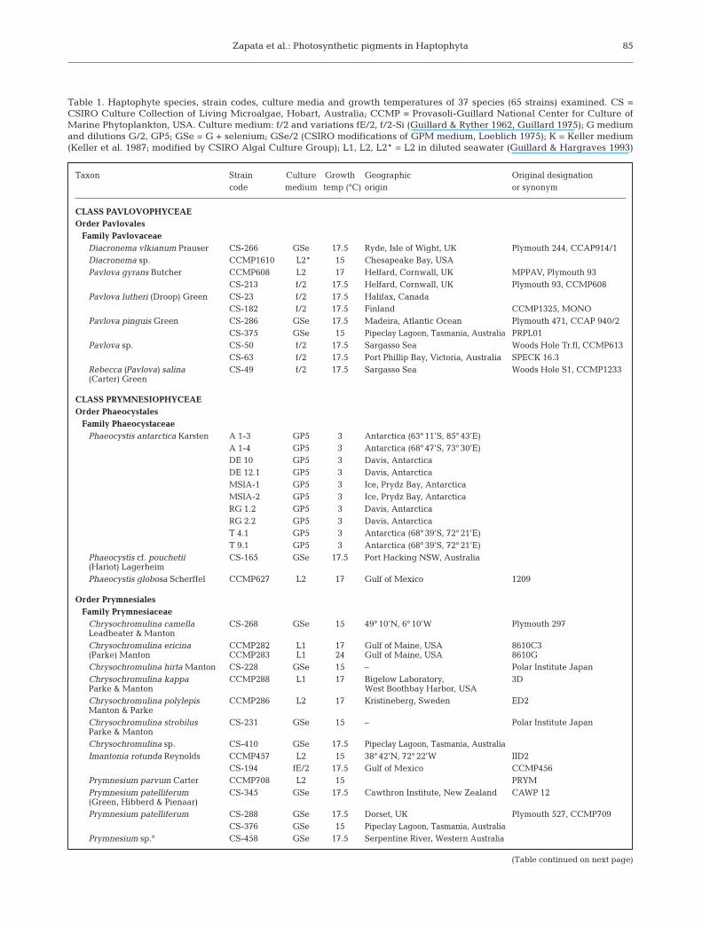
Table 1. Haptophyte species, strain codes, culture media and growth temperatures of 37 species (65 strains) examined. CS =CSIRO Culture Collection of Living Microalgae, Hobart, Australia; CCMP = Provasoli-Guillard National Center for Culture ofMarine Phytoplankton, USA. Culture medium: f/2 and variations fE/2, f/2-Si (Guillard & Ryther 1962, Guillard 1975); G mediumand dilutions G/2, GP5; GSe = G + selenium; GSe/2 (CSIRO modifications of GPM medium, Loeblich 1975); K = Keller medium(Keller et al. 1987; modified by CSIRO Algal Culture Group); L1, L2, L2* = L2 in diluted seawater (Guillard & Hargraves 1993)
Taxon Strain Culture Growth Geographic Original designationcode medium temp (°C) origin or synonym
CLASS PAVLOVOPHYCEAEOrder Pavlovales
Family PavlovaceaeDiacronema vlkianum Prauser CS-266 GSe 17.5 Ryde, Isle of Wight, UK Plymouth 244, CCAP914/1Diacronema sp. CCMP1610 L2* 15 Chesapeake Bay, USAPavlova gyrans Butcher CCMP608 L2 17 Helfard, Cornwall, UK MPPAV, Plymouth 93
CS-213 f/2 17.5 Helfard, Cornwall, UK Plymouth 93, CCMP608Pavlova lutheri (Droop) Green CS-23 f/2 17.5 Halifax, Canada
CS-182 f/2 17.5 Finland CCMP1325, MONOPavlova pinguis Green CS-286 GSe 17.5 Madeira, Atlantic Ocean Plymouth 471, CCAP 940/2
CS-375 GSe 15 Pipeclay Lagoon, Tasmania, Australia PRPL01Pavlova sp. CS-50 f/2 17.5 Sargasso Sea Woods Hole Tr.fl, CCMP613
CS-63 f/2 17.5 Port Phillip Bay, Victoria, Australia SPECK 16.3Rebecca (Pavlova) salina CS-49 f/2 17.5 Sargasso Sea Woods Hole S1, CCMP1233(Carter) Green
CLASS PRYMNESIOPHYCEAEOrder Phaeocystales
Family PhaeocystaceaePhaeocystis antarctica Karsten A 1-3 GP5 3 Antarctica (63° 11’S, 85° 43’E)
A 1-4 GP5 3 Antarctica (68° 47’S, 73° 30’E)DE 10 GP5 3 Davis, AntarcticaDE 12.1 GP5 3 Davis, AntarcticaMSIA-1 GP5 3 Ice, Prydz Bay, AntarcticaMSIA-2 GP5 3 Ice, Prydz Bay, AntarcticaRG 1.2 GP5 3 Davis, AntarcticaRG 2.2 GP5 3 Davis, AntarcticaT 4.1 GP5 3 Antarctica (68° 39’S, 72° 21’E)T 9.1 GP5 3 Antarctica (68° 39’S, 72° 21’E)
Phaeocystis cf. pouchetii CS-165 GSe 17.5 Port Hacking NSW, Australia(Hariot) LagerheimPhaeocystis globosa Scherffel CCMP627 L2 17 Gulf of Mexico 1209
Order PrymnesialesFamily Prymnesiaceae
Chrysochromulina camella CS-268 GSe 15 49° 10’N, 6° 10’W Plymouth 297Leadbeater & MantonChrysochromulina ericina CCMP282 L1 17 Gulf of Maine, USA 8610C3(Parke) Manton CCMP283 L1 24 Gulf of Maine, USA 8610GChrysochromulina hirta Manton CS-228 GSe 15 – Polar Institute JapanChrysochromulina kappa CCMP288 L1 17 Bigelow Laboratory, 3DParke & Manton West Boothbay Harbor, USAChrysochromulina polylepis CCMP286 L2 17 Kristineberg, Sweden ED2Manton & ParkeChrysochromulina strobilus CS-231 GSe 15 – Polar Institute JapanParke & MantonChrysochromulina sp. CS-410 GSe 17.5 Pipeclay Lagoon, Tasmania, AustraliaImantonia rotunda Reynolds CCMP457 L2 15 38° 42’N, 72° 22’W IID2
CS-194 fE/2 17.5 Gulf of Mexico CCMP456Prymnesium parvum Carter CCMP708 L2 15 PRYM Prymnesium patelliferum CS-345 GSe 17.5 Cawthron Institute, New Zealand CAWP 12(Green, Hibberd & Pienaar)Prymnesium patelliferum CS-288 GSe 17.5 Dorset, UK Plymouth 527, CCMP709
CS-376 GSe 15 Pipeclay Lagoon, Tasmania, AustraliaPrymnesium sp.a CS-458 GSe 17.5 Serpentine River, Western Australia
(Table continued on next page)
Mar Ecol Prog Ser 270: 83–102, 2004
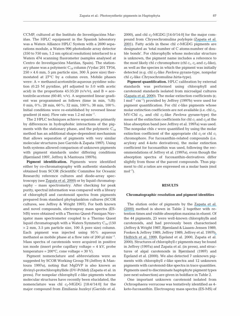
HPLC pigment analyses. We used 2 HPLC methods:the C8 method of Zapata et al. (2000), which was usedfor all haptophyte cultures, and the C18 method of Gar-rido & Zapata (1997), which was used for a subset of thecultures. The chromatographic equipment for the C8
method was a Waters 600 pump and a Waters 996diode-array detector (samples analysed at CSIROMarine Research, Hobart, Australia). The stationaryphase was a C8 column (Waters Symmetry, 150 ×4.6 mm, 3.5 µm particle size, 100 Å pore size) ther-mostated at 25°C either by means of a column oven, or a
25°C circulating water bath. Mobile phases were: A =methanol:acetonitrile: aqueous pyridine solution(0.25 M pyridine, pH adjusted to 5.0 with acetic acid) inthe proportions 50:25:25 (v/v/v), and B = acetonitrile:acetone (80:20 v/v). A segmented linear gradient was(time in min, % B): 0 min, 0%; 18 min, 40%; 22 min,100%; 38 min, 100%. Initial conditions were re-established by reversed linear gradient (4 min). Flowrate was 1 ml min–1.
The C18 HPLC method of Garrido & Zapata (1997) wasused to analyse 12 haptophyte species (14 strains) from
86
Table 1 (continued)
Taxon Strain Culture Growth Geographic Original designationcode medium temp (°C) origin or synonym
Order IsochrysidalesFamily Isochrysidaceae
Chrysotila lamellosa CS-272 GSe 17.5 UK Plymouth 408, CCAP 918/1Anand emend. Green & ParkeCricosphaera carterae CS-40 G 17.5 – F.T. Haxo Cr. cart.(Braarud & Fagerland) Braarudb
Dicrateria inornata Parke CCMP355 L2 15 – DICRATCS-254 f/2-Si 17.5 – CCMP355CS-267c GSe 17.5 Plymouth, UK Plymouth B, CCAP 915/1CCMP1323 L2 15 Isle of Man, UK Plymouth I, CCAP 927/1CS-22 f/2 17.5 Halifax, Canada
Isochrysis sp. CS-177 f/2 17.5 Tahiti, Society Islands CCMP1324, T-ISOPseudoisochrysis paradoxa Ott CS-186 f/2 17.5 York River, Virginia, USA CCMP715, VA12
Family NoëlaerhabdaceaeEmiliania huxleyi CCMP370 L2 15 Oslo fjord, Norway 451B(Lohmann) Hay & MohlerEmiliania huxleyi CCMP373 L2 15 Sargasso Sea BT-6
CS-57 f/2 17.5 Sargasso Sea BT-6CS-275-2 GSe 15 60°N 20°W, Iceland Basin Plymouth G1779GaCS-279 GSe 15 24° 27’N, 20° 24’W Plymouth DNN53/74/6CS-282 GSe 15 32°N 62°W, Sargasso Sea Plymouth M181CS-283 GSe 15 Durban, South Africa Plymouth M186CS-284 GSe 15 Sargasso Sea Plymouth MCH-1,CCMP375CS-363 G/2 15 Pipeclay Lagoon, TasmaniaCS-369 K 15 Pipeclay Lagoon, TasmaniaCS-370 K 15 Pipeclay Lagoon, Tasmania
Gephyrocapsa oceanica Kamptner CS-335 GSe/2 17.5 Jervis Bay, NSW, Australia
Order CoccolithalesFamily Pleurochrysidaceae
Pleurochrysis aff. carterae CS-287 GSe 17.5 Port Erin, Isle of Man, UK Plymouth 156, CCMP646(Braarud & Fagerland) Christiansen CCAP 961/5Pleurochrysis roscoffensis CCMP1588 L2 15 Narragansett Bay, USA CO791CChadfaud & Feldman
Family HymenomonadaceaeOchrosphaera neapolitana Schussnig CS-285 GSe 17.5 Salcombe, UK Plymouth 162, CCAP 923/1Ochrosphaera verrucosa Schussnig CCMP594 L2 15 Puerto Penasco, Mexico UW 390, Norris 20-6-11
UNKNOWNHaptophyte (unidentified) CS-124 G/2 25 Coral Sea
CS-260 fE/2 25 Dunk Island, Queensland, Australia
a Now identified as P. parvum (G. M. Hallegraeff pers. comm.)b C. carterae is also known as Pleurochrysis carterae (Braarud & Fagerland) Christensenc Dicrateria inornata Parke (CS-267) shows pigment pattern indistinguishable from that of Imantonia rotunda (CS-194)
Zapata et al.: Photosynthetic pigments in Haptophyta
CCMP, cultured at the Instituto de Investigacións Mar-iñas. The HPLC equipment in the Spanish laboratorywas a Waters Alliance HPLC System with a 2690 sepa-rations module, a Waters 996 photodiode array detector(350 to 750 nm; 1.2 nm optical resolution) interfaced to aWaters 474 scanning fluorometer (samples analysed atCentro de Investigacións Mariñas, Spain). The station-ary phase was a polymeric C18 column (Vydac 201 TP54,250 × 4.6 mm, 5 µm particle size, 300 Å pore size) ther-mostated at 27°C by a column oven. Mobile phaseswere: A = methanol:acetonitrile:aqueous pyridine solu-tion (0.25 M pyridine, pH adjusted to 5.0 with aceticacid) in the proportions 45:35:20 (v/v/v), and B = ace-tonitrile:acetone (60:40, v/v). A segmented linear gradi-ent was programmed as follows (time in min, %B):0 min, 0%; 28 min, 60%; 32 min, 100%; 38 min, 100%.Initial conditions were re-established by reversed lineargradient (4 min). Flow rate was 1.2 ml min–1.
The 2 HPLC techniques achieve separations primarilyby differences in hydrophobic interactions of the pig-ments with the stationary phase, and the polymeric C18
method has an additional shape-dependent mechanismthat allows separation of pigments with very similarmolecular structures (see Garrido & Zapata 1997). Usingboth systems allowed comparison of unknown pigmentswith pigment standards under differing conditions(Bjørnland 1997, Jeffrey & Mantoura 1997b).
Pigment identification. Pigments were identifiedeither by co-chromatography with authentic standardsobtained from SCOR (Scientific Commitee for OceanicResearch) reference cultures and diode-array spec-troscopy (see Zapata et al. 2000) or by liquid chromatog-raphy – mass spectrometry. After checking for peakpurity, spectral information was compared with a libraryof chlorophyll and carotenoid spectra from pigmentsprepared from standard phytoplankton cultures (SCORcultures, see Jeffrey & Wright 1997). For both knownand novel compounds, electrospray mass spectra (ES-MS) were obtained with a Thermo Quest-Finnigan Nav-igator mass spectrometer coupled to a Thermo Questliquid chromatograph with a Waters Symmetry C18 (150× 2 mm, 3.5 µm particle size, 100 Å pore size) column.Each pigment was injected using 95% aqueousmethanol as mobile phase at a flow rate of 200 µl min–1.Mass spectra of carotenoids were acquired in positiveion mode (insert probe capillary voltage = 4 kV, probetemperature = 200°C, cone voltage = 30 V).
Pigment nomenclature and abbreviations were assuggested by SCOR Working Group 78 (Jeffrey & Man-toura 1997a), noting that MgDVP is also known asdivinyl-protochlorophyllide (DV-Pchlid) (Zapata et al. inpress). For nonpolar chlorophyll c-like pigments whosemolecular structures have recently been elucidated, thenomenclature was chl c2-MGDG [18:4/14:0] for themajor compound from Emiliania huxleyi (Garrido et al.
2000), and chl c2-MGDG [14:0/14:0] for the major com-pound from Chrysochromulina polylepis (Zapata et al.2001). Fatty acids in these chl c-MGDG pigments aredesignated as ‘total number of C atoms:number of dou-ble bonds’. For chlorophylls whose molecular structureis unknown, the pigment name includes a reference tothe most likely chl c chromophore (chl c1, c2 and c3-like),as well as the species in which the pigment was initiallydetected (e.g. chl c2-like Pavlova gyrans-type, nonpolarchl c2-like Chrysochromulina hirta-type).
Pigment quantification. HPLC calibration by externalstandards was performed using chlorophyll andcarotenoid standards isolated from microalgal cultures(Zapata et al. 2000). The molar extinction coefficients (ε;l mol–1 cm–1) provided by Jeffrey (1997b) were used forpigment quantification. For chl c-like pigments whosemolar extinction coefficients are not available (i.e. chl c3,MV-Chl c3, and chl c2-like Pavlova gyrans-type) themean of the extinction coefficients for chl c1 and c2 at theblue absorption band (see Jeffrey et al. 1997a) was used.The nonpolar chls c were quantified by using the molarextinction coefficient of the appropriate chl c2 or chl c1
chromophore. For fucoxanthin-related compounds (i.e.acyloxy and 4-keto derivatives), the molar extinctioncoefficient for fucoxanthin was used, following the rec-ommendations of Jeffrey et al. (1997a), even though theabsorption spectra of fucoxanthin-derivatives differslightly from those of the parent compounds. Thus pig-ment to chl a ratios are expressed on a molar basis (molmol–1).
RESULTS
Chromatographic resolution and pigment identities
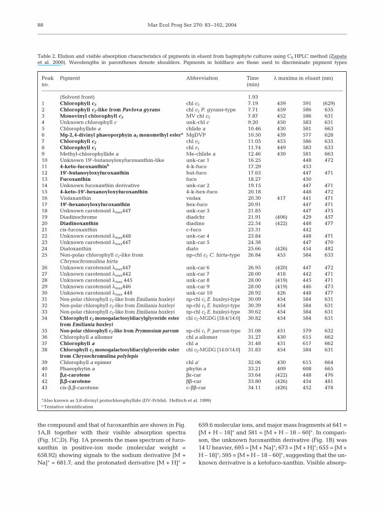
The elution order of pigments by the Zapata et al.(2000) method is shown in Table 2 together with re-tention times and visible absorption maxima in eluent. Ofthe 44 pigments, 25 were well-known chlorophylls andcarotenoids, and had previously been characterised(Jeffrey & Wright 1987, Bjørnland & Liaaen-Jensen 1989,Fookes & Jeffrey 1989, Jeffrey 1989, Jeffrey et al. 1997b,Helfrich et al. 1999, Egeland et al. 2000, Zapata et al.2000). Structures of chlorophyll c pigments may be foundin Jeffrey (1997a) and Zapata et al. (in press), and struc-tures of algal carotenoids in Bjørnland (1997) andEgeland et al. (2000). We also detected 7 unknown pig-ments with chlorophyll c-like spectra and 12 unknownpigments with carotenoid-like spectra in trace quantities.Pigments used to discriminate haptophyte pigment types(see next subsection) are given in boldface in Table 2.
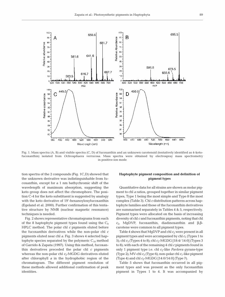
One important unknown carotenoid isolated fromOchrosphaera verrucosa was tentatively identified as 4-keto-fucoxanthin. Electrospray mass spectra (ES-MS) of
87
Mar Ecol Prog Ser 270: 83–102, 2004
the compound and that of fucoxanthin are shown in Fig.1A,B together with their visible absorption spectra(Fig. 1C,D). Fig. 1A presents the mass spectrum of fuco-xanthin in positive-ion mode (molecular weight =658.92) showing signals to the sodium derivative [M +Na]+ = 681.7, and the protonated derivative [M + H]+ =
659.6 molecular ions, and major mass fragments at 641 =[M + H – 18]+ and 581 = [M + H – 18 – 60]+. In compari-son, the unknown fucoxanthin derivative (Fig. 1B) was14 U heavier, 695 = [M + Na]+; 673 = [M + H]+; 655 = [M +H – 18]+; 595 = [M + H – 18 – 60]+, suggesting that the un-known derivative is a ketofuco-xanthin. Visible absorp-
88
Table 2. Elution and visible absorption characteristics of pigments in eluent from haptophyte cultures using C8 HPLC method (Zapata et al. 2000). Wavelengths in parentheses denote shoulders. Pigments in boldface are those used to discriminate pigment types
Peak Pigment Abbreviation Time λ maxima in eluant (nm)no. (min)
(Solvent front) 1.931 Chlorophyll c3 chl c3 7.19 459 591 (629)2 Chlorophyll c2-like from Pavlova gyrans chl c2 P. gyrans-type 7.71 459 586 6353 Monovinyl chlorophyll c3 MV chl c3 7.87 452 586 6314 Unknown chlorophyll c unk-chl c 9.20 450 583 6315 Chlorophyllide a chlide a 10.46 430 581 6636 Mg-2,4-divinyl phaeoporphyin a5 monomethyl estera MgDVP 10.50 439 577 6287 Chlorophyll c2 chl c2 11.05 453 586 6358 Chlorophyll c1 chl c1 11.74 449 583 6339 Methyl-chlorophyllide a Me-chlide a 12.46 430 581 66310 Unknown 19’-butanoyloxyfucoxanthin-like unk-car 1 16.25 448 47211 4-keto-fucoxanthinb 4-k-fuco 17.29 45312 19’-butanoyloxyfucoxanthin but-fuco 17.63 447 47113 Fucoxanthin fuco 18.27 45014 Unknown fucoxanthin derivative unk-car 2 19.15 447 47115 4-keto-19’-hexanoyloxyfucoxanthin 4-k-hex-fuco 20.18 448 47216 Violaxanthin violax 20.30 417 441 47117 19’-hexanoyloxyfucoxanthin hex-fuco 20.91 447 47118 Unknown carotenoid λmax447 unk-car 3 21.85 447 47519 Diadinochrome diadchr 21.91 (406) 429 45720 Diadinoxanthin diadino 22.54 (422) 448 47721 cis-fucoxanthin c-fuco 23.31 44222 Unknown carotenoid λmax448 unk-car 4 23.84 448 47123 Unknown carotenoid λmax447 unk-car 5 24.38 447 47024 Diatoxanthin diato 25.66 (426) 454 48225 Non-polar chlorophyll c2-like from np-chl c2 C. hirta-type 26.84 455 584 633
Chrysochromulina hirta26 Unknown carotenoid λmax447 unk-car 6 26.95 (420) 447 47227 Unknown carotenoid λmax442 unk-car 7 28.00 418 442 47128 Unknown carotenoid λmax 445 unk-car 8 28.00 (419) 445 47129 Unknown carotenoid λmax446 unk-car 9 28.00 (419) 446 47330 Unknown carotenoid λmax 448 unk-car 10 28.92 426 448 47731 Non-polar chlorophyll c2-like from Emiliania huxleyi np-chl c2 E. huxleyi-type 30.09 454 584 63132 Non-polar chlorophyll c2-like from Emiliania huxleyi np-chl c2 E. huxleyi-type 30.39 454 584 63133 Non-polar chlorophyll c2-like from Emiliania huxleyi np-chl c2 E. huxleyi-type 30.62 454 584 63134 Chlorophyll c2 monogalactosyldiacylglyceride ester chl c2-MGDG [18:4/14:0] 30.82 454 584 631
from Emiliania huxleyi35 Non-polar chlorophyll c1-like from Prymnesium parvum np-chl c1 P. parvum-type 31.08 451 579 63236 Chlorophyll a allomer chl a allomer 31.27 430 615 66237 Chlorophyll a chl a 31.48 431 617 66238 Chlorophyll c2 monogalactosyldiacylglyceride ester chl c2-MGDG [14:0/14:0] 31.83 454 584 631
from Chrysochromulina polylepis39 Chlorophyll a epimer chl a’ 32.06 430 615 66440 Phaeophytin a phytin a 33.21 409 608 66541 ββ,εε-carotene βε-car 33.64 (422) 448 47642 ββ,ββ-carotene ββ-car 33.80 (426) 454 48143 cis-β,β-carotene c-ββ-car 34.11 (426) 452 478
aAlso known as 3,8-divinyl protochlorophyllide (DV-Pchlid; Helfrich et al. 1999)b Tentative identification
Zapata et al.: Photosynthetic pigments in Haptophyta
tion spectra of the 2 compounds (Fig. 1C,D) showed thatthe unknown derivative was indistinguishable from fu-coxanthin, except for a 1 nm bathychromic shift of thewavelength of maximum absorption, suggesting theketo group does not affect the chromophore. The posi-tion C-4 for the keto substituent is suggested by analogywith the keto derivative of 19’-hexanoyloxyfucoxanthin(Egeland et al. 2000). Further confirmation of this tenta-tive structure by NMR (nuclear magnetic resonance)techniques is needed.
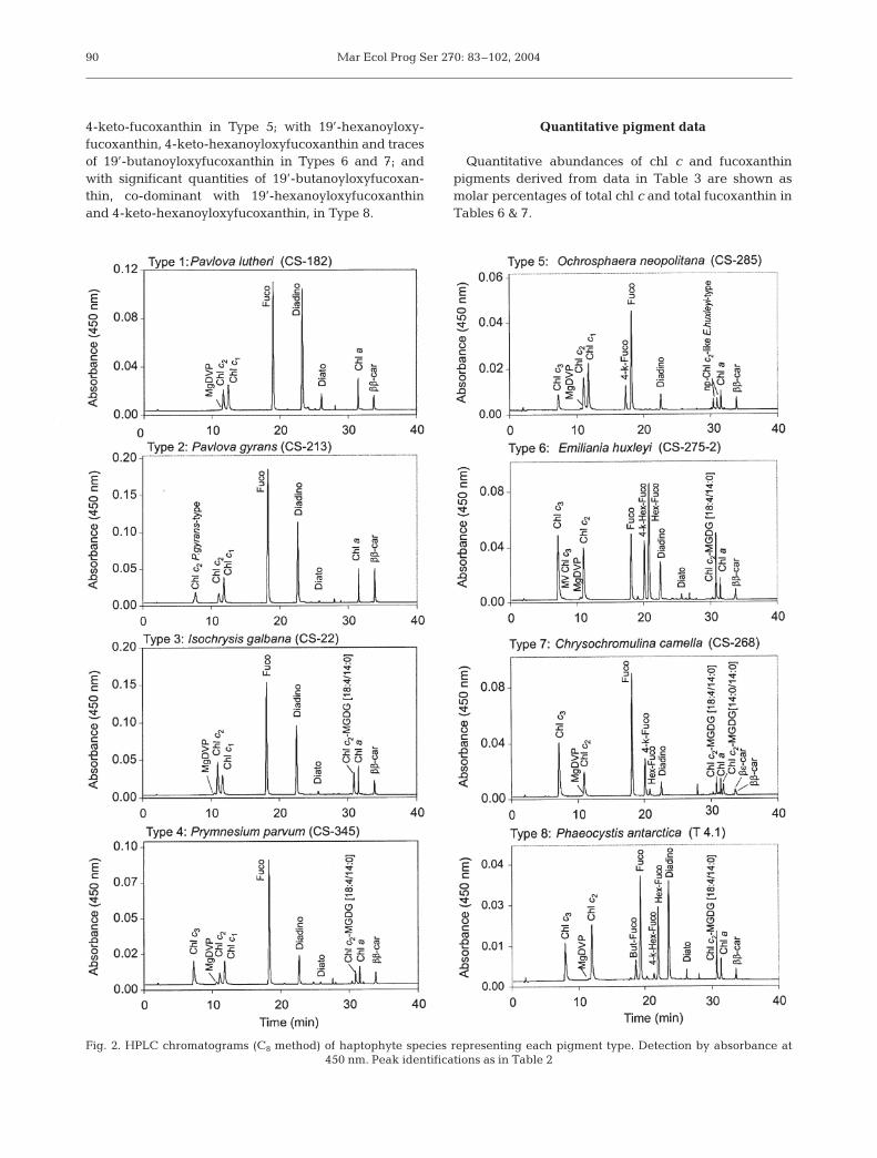
Fig. 2 shows representative chromatograms from eachof the 8 haptophyte pigment types found using the C8
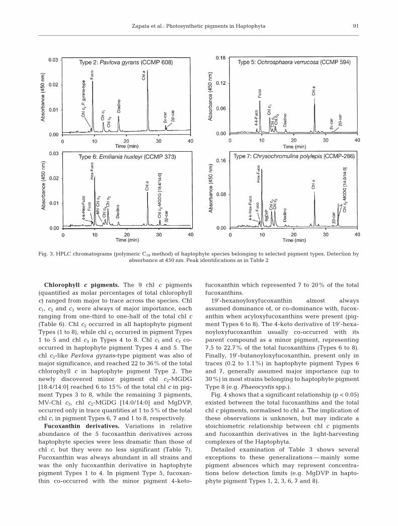
HPLC method. The polar chl c pigments eluted beforethe fucoxanthin derivatives while the non-polar chl cpigments eluted near chl a. Fig. 3 shows 4 selected hap-tophyte species separated by the polymeric C18 methodof Garrido & Zapata (1997). Using this method, fucoxan-thin derivatives preceded the polar chl c pigmentswhereas the non-polar chl c2-MGDG derivatives elutedafter chlorophyll a in the hydrophobic region of thechromatogram. The different pigment resolution ofthese methods allowed additional confirmation of peakidentities.
Haptophyte pigment composition and definition ofpigment types
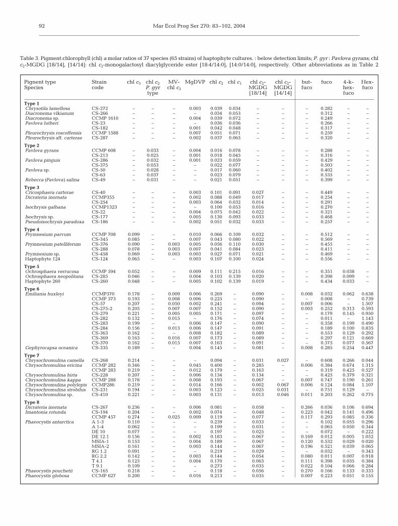
Quantitative data for all strains are shown as molar pig-ment to chl a ratios, grouped together in similar pigmenttypes, Type 1 being the most simple and Type 8 the mostcomplex (Table 3). Chl c distribution patterns across hap-tophyte families and those of the fucoxanthin derivativesare summarised separately in Tables 4 & 5, respectively.Pigment types were allocated on the basis of increasingdiversity of chl c and fucoxanthin pigments, noting that chlc2, MgDVP, fucoxanthin, diadinoxanthin and β,β-carotene were common to all pigment types.
Table 4 shows that MgDVP and chl c2 were present in allpigment types and were accompanied by chl c1 (Types 1 to5); chl c3 (Types 4 to 8); chl c2-MGDG [18:4/ 14:0] (Types 3to 8); with each of the remaining 4 chl c pigments found inonly 1 pigment type i.e. chl c2-like Pavlova gyrans-type(Type 2); MV-chl c3 (Type 6); non-polar chl c1-like pigment(Type 4) and chl c2-MGDG [14:0/14:0] (Type 7).
Table 5 shows that fucoxanthin occurred in all pig-ment types and was present as the only fucoxanthinpigment in Types 1 to 4. It was accompanied by
89
Fig. 1. Mass spectra (A, B) and visible spectra (C, D) of fucoxanthin and an unknown carotenoid (tentatively identified as 4-keto-fucoxanthin) isolated from Ochrosphaera verrucosa. Mass spectra were obtained by electrospray mass spectrometry
in positive-ion mode
Mar Ecol Prog Ser 270: 83–102, 2004
4-keto-fucoxanthin in Type 5; with 19’-hexanoyloxy-fucoxanthin, 4-keto-hexanoyloxyfucoxanthin and tracesof 19’-butanoyloxyfucoxanthin in Types 6 and 7; andwith significant quantities of 19’-butanoyloxyfucoxan-thin, co-dominant with 19’-hexanoyloxyfucoxanthinand 4-keto-hexanoyloxyfucoxanthin, in Type 8.
Quantitative pigment data
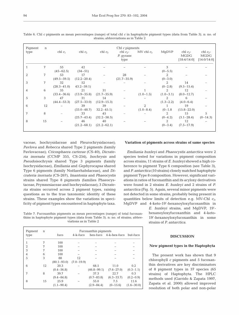
Quantitative abundances of chl c and fucoxanthinpigments derived from data in Table 3 are shown asmolar percentages of total chl c and total fucoxanthin inTables 6 & 7.
90
Fig. 2. HPLC chromatograms (C8 method) of haptophyte species representing each pigment type. Detection by absorbance at 450 nm. Peak identifications as in Table 2
Zapata et al.: Photosynthetic pigments in Haptophyta
Chlorophyll c pigments. The 9 chl c pigments(quantified as molar percentages of total chlorophyllc) ranged from major to trace across the species. Chlc1, c2 and c3 were always of major importance, eachranging from one-third to one-half of the total chl c(Table 6). Chl c2 occurred in all haptophyte pigmentTypes (1 to 8), while chl c1 occurred in pigment Types1 to 5 and chl c3 in Types 4 to 8. Chl c1 and c3 co-occurred in haptophyte pigment Types 4 and 5. Thechl c2-like Pavlova gyrans-type pigment was also ofmajor significance, and reached 22 to 36% of the totalchlorophyll c in haptophyte pigment Type 2. Thenewly discovered minor pigment chl c2-MGDG[18:4/14:0] reached 6 to 15% of the total chl c in pig-ment Types 3 to 8, while the remaining 3 pigments,MV-Chl c3, chl c2-MGDG [14:0/14:0] and MgDVP,occurred only in trace quantities at 1 to 5% of the totalchl c, in pigment Types 6, 7 and 1 to 8, respectively.
Fucoxanthin derivatives. Variations in relativeabundance of the 5 fucoxanthin derivatives acrosshaptophyte species were less dramatic than those ofchl c, but they were no less significant (Table 7).Fucoxanthin was always abundant in all strains andwas the only fucoxanthin derivative in haptophytepigment Types 1 to 4. In pigment Type 5, fucoxan-thin co-occurred with the minor pigment 4-keto-
fucoxanthin which represented 7 to 20% of the totalfucoxanthins.
19’-hexanoyloxyfucoxanthin almost alwaysassumed dominance of, or co-dominance with, fucox-anthin when acyloxyfucoxanthins were present (pig-ment Types 6 to 8). The 4-keto derivative of 19’-hexa-noyloxyfucoxanthin usually co-occurred with itsparent compound as a minor pigment, representing7.5 to 22.7% of the total fucoxanthins (Types 6 to 8).Finally, 19’-butanoyloxyfucoxanthin, present only intraces (0.2 to 1.1%) in haptophyte pigment Types 6and 7, generally assumed major importance (up to30%) in most strains belonging to haptophyte pigmentType 8 (e.g. Phaeocystis spp.).
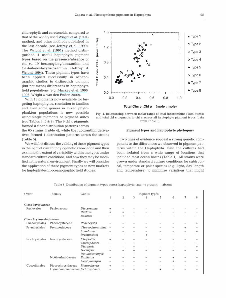
Fig. 4 shows that a significant relationship (p < 0.05)existed between the total fucoxanthins and the totalchl c pigments, normalised to chl a. The implication ofthese observations is unknown, but may indicate astoichiometric relationship between chl c pigmentsand fucoxanthin derivatives in the light-harvestingcomplexes of the Haptophyta.
Detailed examination of Table 3 shows severalexceptions to these generalizations — mainly somepigment absences which may represent concentra-tions below detection limits (e.g. MgDVP in hapto-phyte pigment Types 1, 2, 3, 6, 7 and 8).
91
Fig. 3. HPLC chromatograms (polymeric C18 method) of haptophyte species belonging to selected pigment types. Detection byabsorbance at 450 nm. Peak identifications as in Table 2
Mar Ecol Prog Ser 270: 83–102, 200492
Table 3. Pigment:chlorophyll (chl) a molar ratios of 37 species (65 strains) of haptophyte cultures. : below detection limits; P. gyr : Pavlova gyrans; chlc2-MGDG [18/14], [14/14]: chl c2-monogalactosyl diacylglyceride ester [18:4/14:0], [14:0/14:0], respectively. Other abbreviations as in Table 2
Pignent type Strain chl c3 chl c2 MV- MgDVP chl c2 chl c1 chl c2- chl c2- but- fuco 4-k- Hex-Species code P. gyr chl c3 MGDG MGDG fuco hex- fuco
type [18/14] [14/14] fuco
Type 1Chrysotila lamellosa CS-272 – – – 0.003 0.039 0.034 – – – 0.282 – –Diacronema vlkianum CS-266 – – – – 0.034 0.053 – – – 0.312 – –Diacronema sp. CCMP 1610 – – – 0.004 0.039 0.072 – – – 0.249 – –Pavlova lutheri CS-23 – – – – 0.036 0.036 – – – 0.266 – –
CS-182 – – – 0.001 0.042 0.048 – – – 0.317 – –Pleurochrysis roscoffensis CCMP 1588 – – – 0.007 0.051 0.071 – – – 0.259 – –Pleurochrysis aff. carterae CS-287 – – – 0.002 0.037 0.065 – – – 0.320 – –
Type 2Pavlova gyrans CCMP 608 – 0.033 – 0.004 0.016 0.078 – – – 0.288 – –
CS-213 – 0.025 – 0.001 0.018 0.045 – – – 0.316 – –Pavlova pinguis CS-286 – 0.032 – 0.001 0.023 0.059 – – – 0.429 – –
CS-375 – 0.053 – – 0.022 0.077 – – – 0.503 – –Pavlova sp. CS-50 – 0.028 – – 0.017 0.060 – – – 0.402 – –
CS-63 – 0.037 – – 0.023 0.079 – – – 0.535 – –Rebecca (Pavlova) salina CS-49 – 0.031 – – 0.021 0.051 – – – 0.399 – –
Type 3Cricosphaera carterae CS-40 – – – 0.003 0.101 0.091 0.027 – – 0.449 – –Dicrateria inornata CCMP355 – – – 0.002 0.088 0.049 0.017 – – 0.254 – –
CS-254 – – – 0.003 0.064 0.032 0.014 – – 0.291 – –Isochrysis galbana CCMP1323 – – – – 0.100 0.053 0.016 – – 0.270 – –
CS-22 – – – 0.004 0.075 0.042 0.022 – – 0.321 – –Isochrysis sp. CS-177 – – – 0.005 0.130 0.093 0.033 – – 0.468 – –Pseudoisochrysis paradoxa CS-186 – – – 0.002 0.051 0.032 0.033 – – 0.257 – –
Type 4Prymnesium parvum CCMP 708 0.099 – – 0.010 0.066 0.109 0.032 – – 0.512 – –
CS-345 0.085 – – 0.007 0.043 0.080 0.022 – – 0.569 – –Prymnesium patelliferum CS-376 0.090 – 0.003 0.005 0.056 0.110 0.030 – – 0.455 – –
CS-288 0.078 – 0.003 0.007 0.041 0.084 0.023 – – 0.411 – –Prymnesium sp. CS-458 0.069 – 0.003 0.003 0.027 0.071 0.021 – – 0.469 – –Haptophyte 124 CS-124 0.065 – – 0.003 0.107 0.100 0.024 – – 0.556 – –
Type 5Ochrosphaera verrucosa CCMP 594 0.052 – – 0.009 0.111 0.215 0.016 – – 0.351 0.038 –Ochrosphaera neopolitana CS-285 0.046 – – 0.004 0.103 0.139 0.020 – – 0.398 0.099 –Haptophyte 260 CS-260 0.048 – – 0.005 0.102 0.139 0.019 – – 0.434 0.033 –
Type 6Emiliania huxleyi CCMP370 0.178 – 0.009 0.006 0.269 – 0.090 – 0.008 0.032 0.062 0.638
CCMP 373 0.193 – 0.008 0.006 0.225 – 0.090 – – 0.008 – 0.739CS-57 0.207 – 0.050 0.002 0.241 – 0.094 – 0.007 0.006 – 1.507CS-275-2 0.205 – 0.007 0.007 0.152 – 0.090 – 0.003 0.252 0.313 0.593CS-279 0.221 – 0.005 0.005 0.171 – 0.097 – – 0.179 0.145 0.950CS-282 0.132 – 0.015 – 0.176 – 0.074 – – 0.011 – 1.143CS-283 0.199 – – 0.006 0.147 – 0.090 – – 0.358 0.199 0.490CS-284 0.156 – 0.013 0.006 0.147 – 0.091 – – 0.189 0.100 0.835CS-363 0.162 – – 0.008 0.182 – 0.089 – – 0.553 0.129 0.292CS-369 0.163 – 0.016 0.007 0.173 – 0.089 – – 0.297 0.121 0.669CS-370 0.162 – 0.015 0.007 0.163 – 0.091 – – 0.375 0.077 0.567
Gephyrocapsa oceanica CS-335 0.189 – – 0.004 0.145 – 0.081 – 0.008 0.285 0.254 0.447
Type 7Chrysochromulina camella CS-268 0.214 – – – 0.094 – 0.031 0.027 – 0.608 0.266 0.044Chrysochromulina ericina CCMP 282 0.346 – – 0.045 0.400 – 0.285 – 0.006 0.384 0.674 1.315
CCMP 283 0.219 – – 0.012 0.179 – 0.163 – – 0.319 0.425 0.527Chrysochromulina hirta CS-228 0.207 – – 0.006 0.134 – 0.134 – – 0.425 0.379 0.321Chrysochromulina kappa CCMP 288 0.178 – – 0.008 0.193 – 0.067 – 0.007 0.747 0.190 0.261Chrysochromulina polylepis CCMP286 0.219 – – 0.014 0.166 – 0.002 0.067 0.006 0.124 0.084 1.107Chrysochromulina strobilus CS-231 0.194 – – 0.003 0.123 – 0.025 0.031 – 0.751 0.129 –Chrysochromulina sp. CS-410 0.221 – – 0.003 0.131 – 0.013 0.046 0.011 0.203 0.262 0.775
Type 8Dicrateria inornata CS-267 0.236 – – 0.006 0.081 – 0.058 – 0.266 0.036 0.106 0.694Imantonia rotunda CS-194 0.204 – – 0.002 0.074 – 0.048 – 0.223 0.042 0.141 0.496
CCMP 457 0.274 – 0.025 0.009 0.119 – 0.077 – 0.117 0.293 0.085 0.336Phaeocystis antarctica A 1-3 0.110 – – – 0.239 – 0.033 – – 0.102 0.055 0.296
A 1-4 0.062 – – – 0.199 – 0.031 – – 0.065 0.050 0.344DE 10 0.077 – – – 0.197 – 0.025 – – 0.072 – 0.222DE 12.1 0.156 – – 0.002 0.183 – 0.067 – 0.169 0.012 0.005 1.052MSIA-1 0.153 – – 0.004 0.189 – 0.067 – 0.120 0.532 0.029 0.020MSIA-2 0.161 – – 0.003 0.144 – 0.067 – 0.196 0.521 0.039 0.065RG 1.2 0.091 – – – 0.219 – 0.029 – – 0.032 – 0.343RG 2.2 0.142 – – 0.003 0.144 – 0.054 – 0.080 0.011 0.007 0.918T 4.1 0.123 – – 0.004 0.170 – 0.063 – 0.111 0.398 0.035 0.384T 9.1 0.109 – – – 0.273 – 0.035 – 0.022 0.104 0.066 0.284
Phaeocystis pouchetii CS-165 0.218 – – – 0.118 – 0.056 – 0.270 0.166 0.133 0.333Phaeocystis globosa CCMP 627 0.200 – – 0.016 0.213 – 0.035 – 0.007 0.223 0.051 0.155
Zapata et al.: Photosynthetic pigments in Haptophyta
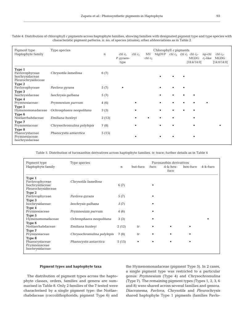
Pigment types and haptophyte taxa
The distribution of pigment types across the hapto-phyte classes, orders, families and genera are sum-marised in Table 8. Only 2 families of the 7 tested werecharacterised by a single pigment type: the Noëlae-rhabdaceae (coccolithophorids; pigment Type 6) and
the Hymenomonadaceae (pigment Type 5). In 2 cases,a single pigment type was restricted to a particulargenus: Prymnesium (Type 4) and Chrysochromulina(Type 7). The remaining pigment types (Types 1, 2, 3, 6and 8) were shared across several families and genera.Diacronema, Pavlova, Chrysotila and Pleurochrysisshared haptophyte Type 1 pigments (families Pavlo-
93
Table 4. Distribution of chlorophyll c pigments across haptophyte families, showing families with designated pigment type and type species withcharacteristic pigment patterns. n: no. of species (strains); other abbreviations as in Table 2
Pigment type Type species Chlorophyll c pigmentsHaptophyte family n chl c2 chl c3 MV MgDVP chl c2 chl c1 chl c2- np-chl chl c2-
P. gyrans- chl c3 MGDG c1-like MGDGtype [18:4/14:0] [14:0/14:0]
Type 1Pavlovophyceae Chrysotila lamellosa 6 (7)Isochrysidaceae • • •PleurochrysideceaeType 2Pavlovophyceae Pavlova gyrans 5 (7) • • • •Type 3Isochrysidaceae Isochrysis galbana 5 (7) • • • •Type 4Prymnesiaceae Prymnesium parvum 4 (6) • • • • • •Type 5Hymenomonadaceae Ochrosphaera neopolitana 3 (3) • • • • •Type 6Noëlaerhabdaceae Emiliana huxleyi 2 (12) • • • • •Type 7Prymnesiaceae Chrysochromulina polylepis 7 (8) • • • • •Type 8Phaeocystaceae Phaeocystis antarctica 5 (15)Prymnesiaceae • • • •Isochrysidaceae
Table 5. Distribution of fucoxanthin derivatives across haptophyte families. tr: trace; further details as in Table 4
Pigment type Type species Fucoxanthin derivativesHaptophyte family n but-fuco fuco 4-k-hex- hex-fuco 4-k-fuco
fuco
Type 1Pavlovophyceae Chrysotila lamellosaIsochrysidaceae 6 (7) •PleurochrysideceaeType 2Pavlovophyceae Pavlova gyrans 5 (7) •Type 3Isochrysidaceae Isochrysis galbana 5 (7) •Type 4Prymnesiaceae Prymnesium parvum 4 (6) •Type 5Hymenomonadaceae Ochrosphaera neopolitana 3 (3) • •Type 6Noëlaerhabdaceae Emiliana huxleyi 2 (12) tr • • •Type 7Prymnesiaceae Chrysochromulina polylepis 7 (8) tr • • •Type 8Phaeocystaceae Phaeocystis antarctica 5 (15) • • • •PrymnesiaceaeIsochrysidaceae
Mar Ecol Prog Ser 270: 83–102, 2004
vaceae, Isochrysidaceae and Pleurochrysidaceae);Pavlova and Rebecca shared Type 2 pigments (familyPavlovaceae); Cricosphaera carterae (CS-40), Dicrate-ria inornata (CCMP 355, CS-254), Isochrysis andPseudoisochrysis shared Type 3 pigments (familyIsochrysidaceae), Emiliania and Gephyrocapsa sharedType 6 pigments (family Noëlaerhabdaceae), and Di-crateria inornata (CS-267), Imantonia and Phaeocystisstrains shared Type 8 pigments (families Phaeocys-taceae, Prymnesiaceae and Isochrysidaceae); 3 Dicrate-ria strains occurred across 2 pigment types, raisingquestions as to the true taxonomic identity of thesestrains. These examples show the variations in speci-ficity of pigment types encountered in haptophyte taxa.
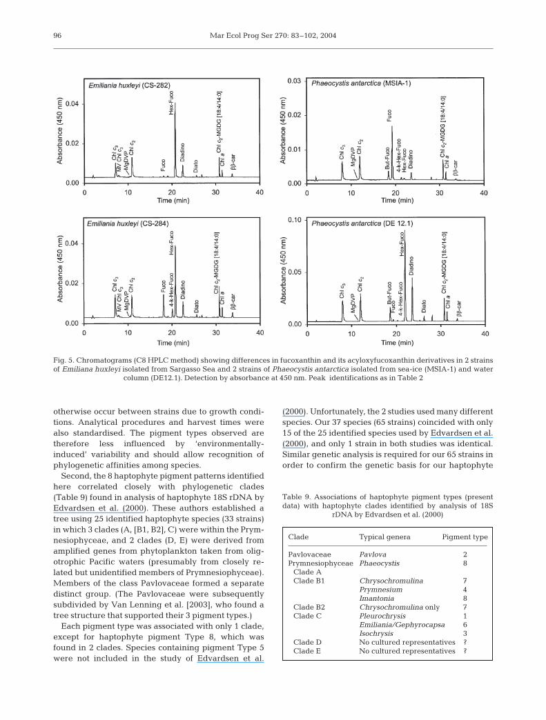
Variation of pigments across strains of same species
Emiliania huxleyi and Phaeocystis antarctica were 2species tested for variations in pigment compositionacross strains; 11 strains of E. huxleyi showed a high co-herence to pigment Type 6 composition (see Table 3),and P. antarctica (10 strains) closely matched haptophytepigment Type 8 composition. However, significant vari-ations in ratios of fucoxanthin and its acyloxy derivativeswere found in 2 strains E. huxleyi and 2 strains of P.antarctica (Fig. 5). Again, several minor pigments werenot detected in some strains, probably being present inquantities below limits of detection e.g. MV-Chl c3,MgDVP and 4-keto-19’-hexanoyloxyfucoxanthin in
E. huxleyi strains, and MgDVP, 19’-hexanoyloxyfucoxanthin and 4-keto-19’-hexanoyloxyfucoxanthin in somestrains of P. antarctica.
DISCUSSION
New pigment types in the Haptophyta
The present work has shown that 9chlorophyll c pigments and 5 fucoxan-thin derivatives are key discriminatorsof 8 pigment types in 37 species (65strains) of Haptophyta. The HPLCmethods used (Garrido & Zapata 1997,Zapata et al. 2000) allowed improvedresolution of both polar and non-polar
94
Table 7. Fucoxanthin pigments as mean percentages (range) of total fucoxan-thins in haptophyte pigment types (data from Table 3). n: no. of strains; abbre-
viations as in Table 2
Pigment n Fucoxanthin pigmentstype fuco 4-k-fuco hex-fuco 4-k-hex-fuco but-fuco
1 7 100 – – – –2 7 100 – – – –3 7 100 – – – –4 6 100 – – – –5 3 88 12 – – –
(80.1–93.0) (7.0–19.9)6 12 20.3 – 68.5 11.0 0.2
(0.4–56.8) (46.8–99.1) (7.6–27.0) (0.3–1.1)7 8 39.7 – 37.3 22.7 0.3
(9.4–84.8) (0.7–83.8) (6.3–33.7) (0.2–0.9)8 15 25.9 – 55.0 7.5 11.6
(1.1–90.4) (2.9–84.4) (0–15.6) (1.6–30.0)
Table 6. Chl c pigments as mean percentages (range) of total chl c in haptophyte pigment types (data from Table 3). n: no. ofstrains; abbreviations as in Table 2
Pigment n Chl c pigmentstype chl c1 chl c2 chl c3 chl c2- MV chl c3 MgDVP chl c2- chl c2-
P. gyrans MGDG MGDGtype [18:4/14:0] [14:0/14:0]
1 7 55 42 – – – 3 – –(45–62.5) (34–51) (0–5.5)
2 7 53 17 – 28 – 2 – –(49.5–59.5) (12.2–20.4) (21.7–35.9) (0–3.0)
3 7 32 52 – – – 2 14 –(28.3–41.0) 43.2–59.1) (0–2.8) (9.5–15.4)
4 6 35 21 31 – 1 2 12 –(33.4–36.6) (13.9–35.8) (21.7–35.9) (1.0–1.5) (1.0–3.1) (8.0–12.7)
5 3 47 31 14 – – 2 6 –(44.4–53.3) (27.5–33.0) (12.9–15.3) (1.3–2.2) (4.0–6.4)
6 12 – 39 39 – 2 1 19 –(33.0–48.7) 32.2–45.1) (1.0–8.4) (0–1.8 (15.8–22.0)
7 8 – 33 45 – – 2 15 5(25.7–43.4) (32.2–58.5) (0–4.2) (3.1–28.4) (0–14.3)
8 15 – 46 40 – – 2 12 –(21.2–68.1) (21.2–62.1) (0–3.4) (7.5–17.9)
Zapata et al.: Photosynthetic pigments in Haptophyta
chlorophylls and carotenoids, compared tothat of the widely used Wright et al. (1991)method, and other methods published inthe last decade (see Jeffrey et al. 1999).The Wright et al. (1991) method distin-guished 4 useful haptophyte pigmenttypes based on the presence/absence ofchl c3, 19’-hexanoyloxyfucoxanthin and19’-butanoyloxyfucoxanthin (Jeffrey &Wright 1994). These pigment types havebeen applied successfully in oceano-graphic studies to distinguish pigment(but not taxon) differences in haptophytefield populations (e.g. Mackey et al. 1996,1998, Wright & van den Enden 2000).
With 13 pigments now available for tar-geting haptophytes, resolution to familiesand even some genera in mixed phyto-plankton populations is now possibleusing single pigments or pigment suites(see Tables 4, 5 & 8). The 9 chl c pigmentsformed 8 clear distribution patterns acrossthe 65 strains (Table 4), while the fucoxanthin deriva-tives formed 4 distribution patterns across the strains(Table 5).
We will first discuss the validity of these pigment typesin the light of current phylogenetic knowledge and thenexamine the extent of variability within the types understandard culture conditions, and how they may be modi-fied in the natural environment. Finally we will considerthe application of these pigment types as new markersfor haptophytes in oceanographic field studies.
Pigment types and haptophyte phylogeny
Two lines of evidence suggest a strong genetic com-ponent to the differences we observed in pigment pat-terns within the Haptophyta. First, the cultures hadbeen isolated from a wide range of locations thatincluded most ocean basins (Table 1). All strains weregrown under standard culture conditions for subtropi-cal, temperate or polar species (e.g. light, day lengthand temperature) to minimise variations that might
95
Table 8. Distribution of pigment types across haptophyte taxa; +: present; –: absent
Order Family Genus Pigment types1 2 3 4 5 6 7 8
Class PavlovaceaePavlovales Pavlovaceae Diacronema + – – – – – – –
Pavlova + + – – – – – –Rebecca – + – – – – – –
Class PrymnesiophyceaePhaeocystales Phaeocystaceae Phaeocystis – – – – – – – +
Prymnesiales Prymnesiaceae Chrysochromulina – – – – – – + –Imantonia – – – – – – – +Prymnesium – – – + – – – –
Isochrysidales Isochrysidaceae Chrysotila + – – – – – – –Cricosphaera – – + – – – – –Dicrateria – – + – – – – +Isochrysis – – + – – – – –Pseudoisochrysis – – + – – – – –
Noëlaerhabdaceae Emiliania – – – – – + – –Gephyrocapsa – – – – – + – –
Coccolithales Pleurochrysidaceae Pleurochrysis + – – – – – – –Hymenomonadaceae Ochrosphaera – – – – + – – –
Fig. 4. Relationship between molar ratios of total fucoxanthins (Total fucos)and total chl c pigments to chl a across all haptophyte pigment types (data
from Table 3)
Type 1
Type 2
Type 3
Type 4
Type 5
Type 6
Type 7
Type 8
0.0 0.2 0.4 0.6 0.8 1.0
Total Chs c :Chl a (mole : mole)
Tota
l Fuc
os
: Chl
a(m
ole
: m
ole
)
1.6
1.2
0.8
0.4
0.0
Mar Ecol Prog Ser 270: 83–102, 2004
otherwise occur between strains due to growth condi-tions. Analytical procedures and harvest times werealso standardised. The pigment types observed aretherefore less influenced by ‘environmentally-induced’ variability and should allow recognition ofphylogenetic affinities among species.
Second, the 8 haptophyte pigment patterns identifiedhere correlated closely with phylogenetic clades(Table 9) found in analysis of haptophyte 18S rDNA byEdvardsen et al. (2000). These authors established atree using 25 identified haptophyte species (33 strains)in which 3 clades (A, [B1, B2], C) were within the Prym-nesiophyceae, and 2 clades (D, E) were derived fromamplified genes from phytoplankton taken from olig-otrophic Pacific waters (presumably from closely re-lated but unidentified members of Prymnesiophyceae).Members of the class Pavlovaceae formed a separatedistinct group. (The Pavlovaceae were subsequentlysubdivided by Van Lenning et al. [2003], who found atree structure that supported their 3 pigment types.)
Each pigment type was associated with only 1 clade,except for haptophyte pigment Type 8, which wasfound in 2 clades. Species containing pigment Type 5were not included in the study of Edvardsen et al.
(2000). Unfortunately, the 2 studies used many differentspecies. Our 37 species (65 strains) coincided with only15 of the 25 identified species used by Edvardsen et al.(2000), and only 1 strain in both studies was identical.Similar genetic analysis is required for our 65 strains inorder to confirm the genetic basis for our haptophyte
96
Fig. 5. Chromatograms (C8 HPLC method) showing differences in fucoxanthin and its acyloxyfucoxanthin derivatives in 2 strainsof Emiliana huxleyi isolated from Sargasso Sea and 2 strains of Phaeocystis antarctica isolated from sea-ice (MSIA-1) and water
column (DE12.1). Detection by absorbance at 450 nm. Peak identifications as in Table 2
Table 9. Associations of haptophyte pigment types (presentdata) with haptophyte clades identified by analysis of 18S
rDNA by Edvardsen et al. (2000)
Clade Typical genera Pigment type
Pavlovaceae Pavlova 2Prymnesiophyceae Phaeocystis 8
Clade AClade B1 Chrysochromulina 7
Prymnesium 4Imantonia 8
Clade B2 Chrysochromulina only 7Clade C Pleurochrysis 1
Emiliania/Gephyrocapsa 6Isochrysis 3
Clade D No cultured representatives ?Clade E No cultured representatives ?
Zapata et al.: Photosynthetic pigments in Haptophyta
pigment associations. In conclusion, we have some con-fidence in believing that our 8 haptophyte pigmenttypes match phylogenetic trends among the speciesstudied.
Possible pigment functions
Clear differences in relative quantities of the 9 chl cpigments and 5 fucoxanthin derivatives across the 65strains (Tables 6 & 7) probably result from functionaldifferences between them.
Chlorophyll c pigments. It is generally acceptedthat Chls c1, c2 and c3 have a light-harvesting role(Anderson & Barrett 1986, Wilhelm & Wiedemann1991, Green & Durnford 1996, Zapata et al. in press),and in our study these chl c pigments were alwayspresent as a major proportion of the total chl c (Table6). The chl c2-like Pavlova gyrans-type pigment,occurring in 35% of the total chl c pigments, may alsohave a light-harvesting role (Fawley 1989). The occur-rence of MgDVP in trace quantities in most hapto-phytes (Table 3) may signify its role as a biosyntheticintermediate in chlorophyll synthesis (Porra 1997,Porra et al. 1997). When present in larger quantities(e.g. in some prasinophytes), it occurs with chl a and bin the chlorophyll protein complexes and has a light-harvesting role (Brown 1985). The function of 2 othertrace pigments, MV-Chl c3 and chl c2-MGDG[14:0/14:0], is unknown.
The newly discovered minor pigment chl c2-MGDG[18:4/14:0] may also have a light-harvesting role (J. L.Garrido pers. comm.) or it may function in the assem-bly of light-harvesting pigment complexes (Hoober &Eggink 2001). It may act as a transporter of chl c2 fromthe MGDG-rich lipid bilayer of the inner chloroplastenvelope membrane to its final location in the light-harvesting pigment protein complexes of the thy-lakoids (Jeffrey & Anderson 2000). For a more com-plete discussion of chl c chemistry, distribution andfunction, see Zapata et al. (in press).
Fucoxanthin derivatives. The light-harvesting rolesof fucoxanthin and 19’-hexanoyloxyfucoxanthin wereestablished by Sieferman-Harms (1985) and Haxo(1985), respectively. When present in significant quan-tities, 19’-butanoyloxyfucoxanthin may have a similarrole. The function of the 4-keto derivatives is un-known. The universally distributed carotenoid pairdiadinoxanthin and diatoxanthin, present in all hapto-phytes examined, have a well-established photopro-tective function via the light-regulated epoxide cycle(Stransky & Hager 1970, Siefermann-Harms 1985,Demmig-Adams & Adams 1993, Moisan et al. 1998,Lohr & Wilhelm 1999). Further study is needed tounderstand the function and biosynthetic regulation of
all these important marine pigments, and their conse-quent reliability as chemotaxonomic indicators in fieldoceanography.
Quantitative variation of chlorophyll c andfucoxanthins across strains and pigment types
The data in Tables 6 & 7 show that the patterns ofrelative abundance for chl c and fucoxanthins, respec-tively, are clear-cut, but there is considerable variationaround the means for most pigments. While little isknown of chl c variability in haptophytes or other taxa,variation in fucoxanthins has previously been ob-served in haptophytes.
Wright & Jeffrey (1987) gave a first indication of thevariability of the relative proportions of fucoxanthin, 19’-hexanoyloxyfucoxanthin and 19’-butanoyloxyfu-coxanthin in 4 different isolates of Phaeocystis spp. – 3from the Southern Ocean (probably P. antarctica) and 1from the East Australian Current (probably P. globosa;Medlin et al. 1994). This trend was confirmed in the pre-sent work, in which 11 strains of Emiliana huxleyi and 10strains of P. antarctica were analysed (Tables 3 & 7).
While Emiliana huxleyi strains showed a strongcoherence with haptophyte pigment Type 6, and thoseof Phaeocystis antarctica with pigment Type 8, vari-ability in relative abundances of fucoxanthins andacyloxyfucoxanthins were indicated among the strainsof both species (Tables 3 & 7, Fig. 5). These results donot deny the validity of the haptophyte pigment types,but point to the need to understand those factors thatinfluence pigment variability within strains of the samespecies, isolated from different geographic areas, lightfields or populations.
Confirmation of, and explanations for, fucoxanthinvariability have been published in the past decade.Vaulot et al. (1994) observed 3 pigment clusters in 16strains of Phaeocystis isolated mainly from temperateoceanic areas, supporting some of the present observa-tions. In their Phaeocystis strains, both fucoxanthin and19’-hexanoyloxyfucoxanthin were dominant or co-dominant, but 19’-butanoyloxyfucoxanthin in theirstudy was never present except in minor or trace quan-tities. This pattern matches only 4 of our 11 Phaeocys-tis strains.
Jeffrey & Wright (1994) found 1 strain of Phaeocystissp. from the East Australian Current had fucoxanthinand lacked 19’-hexanoyloxyfucoxanthin, similar to thefinding of Breton et al. (1999) and Cottonec et al. (2001)with northern hemisphere Phaeocystis spp. None ofthe strains examined in the present work matched thispigment pattern.
Fucoxanthin/acyloxyfucoxanthin variability was pro-duced by iron limitation aided by light stress in
97
Mar Ecol Prog Ser 270: 83–102, 2004
1 Antarctic Phaeocystis strain (Van Leeuwe & Stefels1998). Iron limitation caused increased synthesis of19’-hexanoyloxyfucoxanthin and 19’-butanoyloxyfu-coxanthin at the expense of fucoxanthin. Buma et al.(1991) found differences in the 19’-hexanoyloxyfuco-xanthin to chl a ratios in Phaeocystis strains isolatedfrom both Antarctic and Atlantic ocean regions, andpigment ratios were also affected by experimentaldifferences in growth phase, temperature, morpho-logical cell type (flagellates or colonies) and varia-tions in day/night cycles. Stolte et al. (2000) alsofound that 19’-hexanoyloxyfucoxanthin was synthe-sised from fucoxanthin, with light acting as a modu-lating factor, in strains of Emiliania huxleyi grownunder conditions of light, phosphate and nitrate limi-tation.
The relative importance of nutritional, environmen-tal and genetic factors influencing fucoxanthin vari-ability needs to be fully evaluated in order to define thereliability of fucoxanthins as indicators of algal types inthe field.
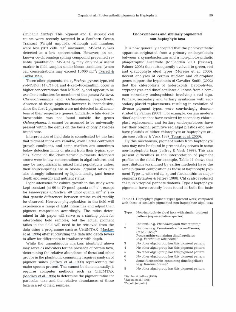
Comparison with previous surveys of Haptophyta
Table 10 highlights the advances in a comparisonof the 8 haptophyte pigment types identified in thepresent work with the 4 of Jeffrey & Wright (1994).The earlier study could not distinguish the pigmenta-tion of diatoms from that of 16 of 50 haptophytestrains studied (32%, their type 1). Most of these cannow be distinguished by the presence of chl c2
Pavlova gyrans-type and Chl c2-MGDG [18:4/14:0]and fall within the new haptophyte pigment Types 2and 3, respectively, with only 7 of the 65 haptophytestrains (11%) remaining in Type 1. Similarly, thosetaxa previously classified by Jeffrey & Wright (1994)as type 2 can now be further subdivided by the pres-
ence of chl c2-MGDG [18:4/14:0] and non-polar chl c1
(new Type 4) and 4-keto-fucoxanthin (new Type 5),respectively. The former type 3 of Jeffrey & Wright(1994) can now be subdivided into new Type 6 andnew Type 7 on the basis of MV-chl c3 and chl c2-MGDG [14:0/14:0], respectively. Type 4 of Jeffrey &Wright (1994) could not be further subdivided (newhaptophyte pigment Type 8), although chl c2-MGDG[18:4/14:0] was recognised as an additional charac-teristic.
Application of haptophyte pigment signatures inoceanography
The additional pigments and pigment patterns iden-tified in this study add power to biological oceano-graphic studies where one must detect algal pigmentsignatures in the presence of other taxa, some of whichhave potentially overlapping pigment compositions.
The recent analysis of 9 species from the Pavlo-vaceae by Van Lenning et al. (2003) found 3 pigmenttypes: A, B, C. While not all species tested were com-mon to our study, it is clear that their Pavlovo-phyceae pigment type A corresponds with our Type1, and their type B with our Type 2, with no irregu-larities. Their type C was based on the presence ofan additional pigment (thought to be the monovinylform of chl c2 Pavlova gyrans-type) that was found ina single species, Exanthemachrysis gayraliae, whichunfortunately was not included in our survey. How-ever this pigment appears to be a useful additionalmarker.
Several of the new marker pigments discussedabove are restricted to particular taxa and may beuseful for their detection in mixed populations. Ofparticular interest is MV-chl c3, a minor pigmentstrongly associated with the globally important species
98
Table 10. Comparison of Jeffrey & Wrights’ (1994) haptophyte pigment types (1 to 4), with Types 1 to 8 found in present work. tr: trace; further abbreviations as in Table 2
Jeffrey & Wright (1994) Present workType Distinguishing pigments Type Distinguishing pigments
1 [chl c1 + chl c2 ]a, fuco 1 Identical to Jeffrey & Wright (1994) type 12 Identical to Jeffrey & Wright (1994) type 1 + chl c2 P. gyrans-type3 Identical to Jeffrey & Wright (1994) type 1 + chl c2 MGDG [18:4/14:0]
2 [chl c1 + chl c2]a, chl c3, fuco 4 Identical to Jeffrey & Wright (1994) type 2 + chl c2 MGDG [18:4/14:0] + npchl c2
5 Identical to Jeffrey & Wright (1994) type 2 + chl c2 MGDG [18:4/14:0] + 4-k-fuco3 [chl c2]a, chl c3, fuco, hex-fucob, but-fuco (tr) 6 Identical to Jeffrey & Wright (1994) type 3 + MVchl c2 + chl c2 MGDG [18:4/14:0]
7 Identical to Jeffrey & Wright (1994) type 3 + chl c2 MGDG [18:4/14:0] + chl c2 MGDG [14:0/14:0]4 [chl c2]a, chl c3, fuco, hex-fucob, but-fuco 8 Identical to Jeffrey & Wright (1994) type 4 + chl c2 MGDG [18:4/14:0]
a[chl c1 + chl c2 ] were not resolved by Jeffrey & Wright (1994); table shows present understanding of previous resultsbPresent work shows that algae with hex-fuco also contain 4-k-hex-fuco
Zapata et al.: Photosynthetic pigments in Haptophyta
Emiliania huxleyi. This pigment and E. huxleyi cellcounts were recently targeted in a Southern OceanTransect (Wright unpubl.). Although cell numberswere low (263 cells ml–1 maximum), MV-chl c3 wasdetected at a low concentration. However, an un-known co-chromatographing compound prevented re-liable quantitation. MV-Chl c3 may only be a usefulmarker in field samples under bloom conditions (whencell concentrations may exceed 10000 ml–1; Tyrrell &Taylor 1995).
Three other pigments, chl c2 Pavlova gyrans-type, chlc2-MGDG [14:0/14:0], and 4-keto-fucoxanthin, occur inhigher concentrations than MV-chl c3 and appear to beexcellent indicators for members of the genera Pavlova,Chrysochromulina and Ochrosphaera, respectively.Absence of these pigments however is inconclusive,since the first 2 pigments were not detected in all mem-bers of their respective genera. Similarly, while 4-keto-fucoxanthin was not found outside the genusOchrosphaera, it cannot be assumed to be universallypresent within the genus on the basis of only 2 speciestested here.
Interpretation of field data is complicated by the factthat pigment ratios are variable, even under controlledgrowth conditions, and some markers are sometimesbelow detection limits or absent from their typical spe-cies. Some of the characteristic pigments describedabove were in low concentrations in algal cultures andmay be insignificant in mixed field populations unlesstheir source-species are in bloom. Pigment ratios arealso strongly influenced by light intensity (and hencedepth and season) and nutrient status.
Light intensities for culture growth in this study werekept constant (at 60 to 70 µmol quanta m–2 s–1, exceptfor Phaeocystis antarctica, 40 µmol quanta m–2 s–1) sothat genetic differences between strains could readilybe observed. However phytoplankton in the field willexperience a range of light intensities and adjust theirpigment composition accordingly. The ratios deter-mined in this paper will serve as a starting point forinterpreting field samples, but the actual pigmentratios in the field will need to be retrieved from thedata using a programme such as CHEMTAX (Mackeyet al. 1996) after subdividing the data into depth layersto allow for differences in irradiance with depth.
While the unambiguous markers identified abovemay serve as indicators for the presence of certain taxa,determining the relative abundance of these and othergroups in the planktonic community requires analysis ofpigment suites (Jeffrey et al. 1999) representing themajor species present. This cannot be done manually; itrequires computer methods such as CHEMTAX(Mackey et al. 1996) to determine the pigment ratios forparticular taxa and the relative abundances of thosetaxa in a set of field samples.
Endosymbioses and similarly pigmentednon-haptophyte taxa
It is now generally accepted that the photosyntheticapparatus originated from a primary endosymbiosisbetween a cyanobacterium and a non-photosyntheticphagotrophic eucaryote (McFadden 2001 [review],Palmer 2003) that subsequently evolved to green, redand glaucophyte algal types (Moreira et al. 2000).Recent analyses of certain nuclear and chloroplastgenes support the hypothesis of Cavalier-Smith (2002)that the chloroplasts of heterokonts, haptophytes,cryptophytes and dinoflagellates all arose from a com-mon secondary endosymbiosis involving a red alga.Primary, secondary and tertiary symbioses with sec-ondary plastid replacements, resulting in evolution ofdiverse pigment types, were convincingly demon-strated by Palmer (2003). For example, certain moderndinoflagellates that have evolved by secondary chloro-plast replacement and tertiary endosymbioses havelost their original primitive red algal plastids and nowhave plastids of either chlorophyte or haptophyte ori-gin (see Jeffrey & Vesk 1997, Tengs et al. 2000).
By this mechanism, pigment suites from haptophytetaxa may now be found in present-day oceans in somenon-haptophyte taxa (Jeffrey & Vesk 1997). This canpresent difficulties in the interpretation of pigmentprofiles in the field. For example, Table 11 shows thatmost diatoms (examined by earlier methods) have thesame pigment composition as those of haptophyte pig-ment Type 1, with chl c1, c2 and fucoxanthin as majorpigments (Stauber & Jeffrey 1988). Chl c3 also replacedchl c1 in 5 tropical pennate diatoms. Type 2 haptophytepigments have recently been found in both the toxic
99
Table 11. Haptophyte pigment types (present work) comparedwith those of similarly pigmented non-haptophyte algal taxa
Type Non-haptophyte algal taxa with similar pigmentpattern (representative species)
1 Diatoms (e.g. Phaeodactylum tricornutum)a
2 Diatoms (e.g. Pseudo-nitzschia multiseries,CCMP 1659)b
Fucoxanthin-containing dinoflagellates(e.g. Peridinium foliaceum)c
3 No other algal group has this pigment pattern4 No other algal group has this pigment pattern5 No other algal group has this pigment pattern6 No other algal group has this pigment pattern7 Some fucoxanthin-containing dinoflagellates
(e.g. Karenia brevis)b
8 No other algal group has this pigment pattern
aStauber & Jeffrey (1988)bZapata et al. (1998)cZapata (unpubl.)
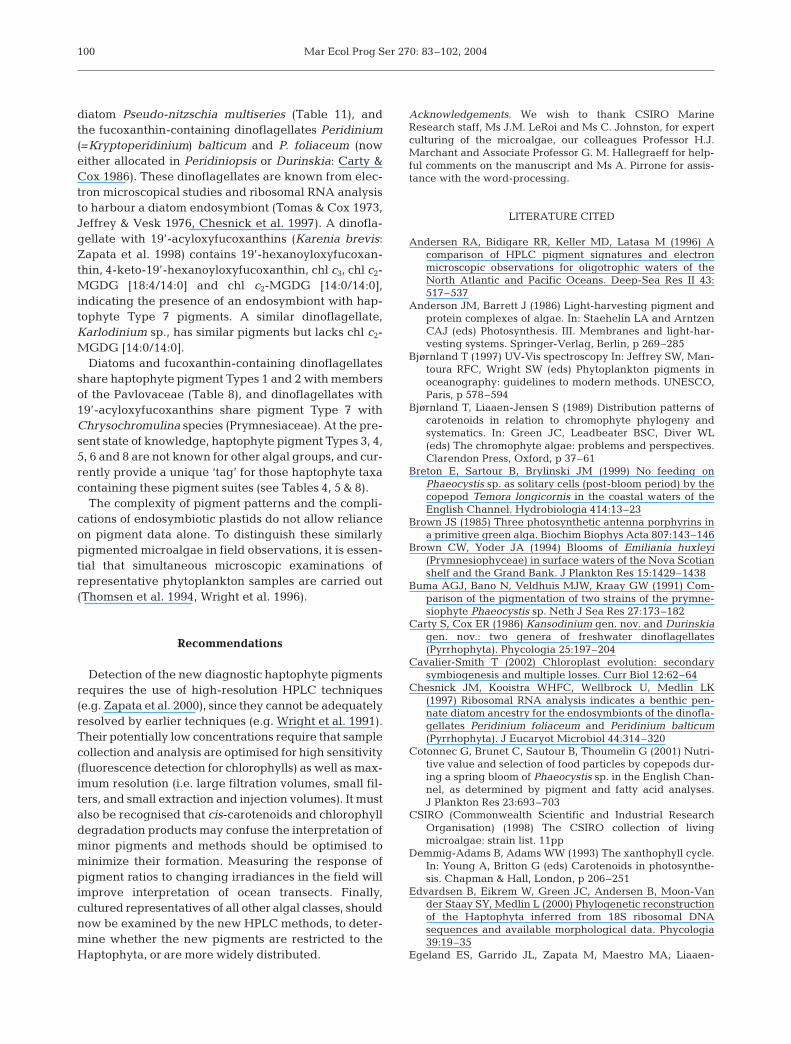
Mar Ecol Prog Ser 270: 83–102, 2004
diatom Pseudo-nitzschia multiseries (Table 11), andthe fucoxanthin-containing dinoflagellates Peridinium(=Kryptoperidinium) balticum and P. foliaceum (noweither allocated in Peridiniopsis or Durinskia: Carty &Cox 1986). These dinoflagellates are known from elec-tron microscopical studies and ribosomal RNA analysisto harbour a diatom endosymbiont (Tomas & Cox 1973,Jeffrey & Vesk 1976, Chesnick et al. 1997). A dinofla-gellate with 19’-acyloxyfucoxanthins (Karenia brevis:Zapata et al. 1998) contains 19’-hexanoyloxyfucoxan-thin, 4-keto-19’-hexanoyloxyfucoxanthin, chl c3, chl c2-MGDG [18:4/14:0] and chl c2-MGDG [14:0/14:0],indicating the presence of an endosymbiont with hap-tophyte Type 7 pigments. A similar dinoflagellate,Karlodinium sp., has similar pigments but lacks chl c2-MGDG [14:0/14:0].
Diatoms and fucoxanthin-containing dinoflagellatesshare haptophyte pigment Types 1 and 2 with membersof the Pavlovaceae (Table 8), and dinoflagellates with19’-acyloxyfucoxanthins share pigment Type 7 withChrysochromulina species (Prymnesiaceae). At the pre-sent state of knowledge, haptophyte pigment Types 3, 4,5, 6 and 8 are not known for other algal groups, and cur-rently provide a unique ‘tag’ for those haptophyte taxacontaining these pigment suites (see Tables 4, 5 & 8).
The complexity of pigment patterns and the compli-cations of endosymbiotic plastids do not allow relianceon pigment data alone. To distinguish these similarlypigmented microalgae in field observations, it is essen-tial that simultaneous microscopic examinations ofrepresentative phytoplankton samples are carried out(Thomsen et al. 1994, Wright et al. 1996).
Recommendations
Detection of the new diagnostic haptophyte pigmentsrequires the use of high-resolution HPLC techniques(e.g. Zapata et al. 2000), since they cannot be adequatelyresolved by earlier techniques (e.g. Wright et al. 1991).Their potentially low concentrations require that samplecollection and analysis are optimised for high sensitivity(fluorescence detection for chlorophylls) as well as max-imum resolution (i.e. large filtration volumes, small fil-ters, and small extraction and injection volumes). It mustalso be recognised that cis-carotenoids and chlorophylldegradation products may confuse the interpretation ofminor pigments and methods should be optimised tominimize their formation. Measuring the response ofpigment ratios to changing irradiances in the field willimprove interpretation of ocean transects. Finally,cultured representatives of all other algal classes, shouldnow be examined by the new HPLC methods, to deter-mine whether the new pigments are restricted to theHaptophyta, or are more widely distributed.
Acknowledgements. We wish to thank CSIRO MarineResearch staff, Ms J.M. LeRoi and Ms C. Johnston, for expertculturing of the microalgae, our colleagues Professor H.J.Marchant and Associate Professor G. M. Hallegraeff for help-ful comments on the manuscript and Ms A. Pirrone for assis-tance with the word-processing.
LITERATURE CITED
Andersen RA, Bidigare RR, Keller MD, Latasa M (1996) Acomparison of HPLC pigment signatures and electronmicroscopic observations for oligotrophic waters of theNorth Atlantic and Pacific Oceans. Deep-Sea Res II 43:517–537
Anderson JM, Barrett J (1986) Light-harvesting pigment andprotein complexes of algae. In: Staehelin LA and ArntzenCAJ (eds) Photosynthesis. III. Membranes and light-har-vesting systems. Springer-Verlag, Berlin, p 269–285
Bjørnland T (1997) UV-Vis spectroscopy In: Jeffrey SW, Man-toura RFC, Wright SW (eds) Phytoplankton pigments inoceanography: guidelines to modern methods. UNESCO,Paris, p 578–594
Bjørnland T, Liaaen-Jensen S (1989) Distribution patterns ofcarotenoids in relation to chromophyte phylogeny andsystematics. In: Green JC, Leadbeater BSC, Diver WL(eds) The chromophyte algae: problems and perspectives.Clarendon Press, Oxford, p 37–61
Breton E, Sartour B, Brylinski JM (1999) No feeding onPhaeocystis sp. as solitary cells (post-bloom period) by thecopepod Temora longicornis in the coastal waters of theEnglish Channel. Hydrobiologia 414:13–23
Brown JS (1985) Three photosynthetic antenna porphyrins ina primitive green alga. Biochim Biophys Acta 807:143–146
Brown CW, Yoder JA (1994) Blooms of Emiliania huxleyi(Prymnesiophyceae) in surface waters of the Nova Scotianshelf and the Grand Bank. J Plankton Res 15:1429–1438
Buma AGJ, Bano N, Veldhuis MJW, Kraay GW (1991) Com-parison of the pigmentation of two strains of the prymne-siophyte Phaeocystis sp. Neth J Sea Res 27:173–182
Carty S, Cox ER (1986) Kansodinium gen. nov. and Durinskiagen. nov.: two genera of freshwater dinoflagellates(Pyrrhophyta). Phycologia 25:197–204
Cavalier-Smith T (2002) Chloroplast evolution: secondarysymbiogenesis and multiple losses. Curr Biol 12:62–64
Chesnick JM, Kooistra WHFC, Wellbrock U, Medlin LK(1997) Ribosomal RNA analysis indicates a benthic pen-nate diatom ancestry for the endosymbionts of the dinofla-gellates Peridinium foliaceum and Peridinium balticum(Pyrrhophyta). J Eucaryot Microbiol 44:314–320
Cotonnec G, Brunet C, Sautour B, Thoumelin G (2001) Nutri-tive value and selection of food particles by copepods dur-ing a spring bloom of Phaeocystis sp. in the English Chan-nel, as determined by pigment and fatty acid analyses.J Plankton Res 23:693–703
CSIRO (Commonwealth Scientific and Industrial ResearchOrganisation) (1998) The CSIRO collection of livingmicroalgae: strain list. 11pp
Demmig-Adams B, Adams WW (1993) The xanthophyll cycle.In: Young A, Britton G (eds) Carotenoids in photosynthe-sis. Chapman & Hall, London, p 206–251
Edvardsen B, Eikrem W, Green JC, Andersen B, Moon-Vander Staay SY, Medlin L (2000) Phylogenetic reconstructionof the Haptophyta inferred from 18S ribosomal DNAsequences and available morphological data. Phycologia39:19–35
Egeland ES, Garrido JL, Zapata M, Maestro MA, Liaaen-
100
Zapata et al.: Photosynthetic pigments in Haptophyta
Jensen S (2000) Algal carotenoids. Part 64. Structure andchemistry of 4-keto-19’-hexanoyloxyfucoxanthin with anovel carotenoid end group. J Chem Soc Perkin Trans I, 8:1223–1230
Fawley MW (1989) A new form of chlorophyll c involved inlight harvesting. Plant Physiol (Rockville) 91:727–732
Fookes CJR, Jeffrey SW (1989) The structure of chlorophyllc3, a novel marine photosynthetic pigment. J Chem SocChem Commun 23:1827–1828
Garrido JL, Zapata M (1997) Reversed-phase high-perfor-mance liquid chromatography of mono- and divinyl-chlorophyll forms using pyridine-containing mobilephases and polymeric octadecylsilica. Chromatographia44:43–49
Garrido JL, Otero J, Maestro MA, Zapata M (2000) The mainnon-polar chlorophyll c from Emiliania huxleyi (Prymne-siophyceae) is a chlorophyll c2-monogalactosyldiacylglyc-eride ester: a mass spectrometry study. J Phycol 36:497–505
Green BR, Durnford DG (1996) The chlorophyll-carotenoidproteins of oxygenic photosynthesis. Annu Rev PlantPhysiol Plant Mol Biol 47:685–714
Guillard RRL (1975) Culture of phytoplankton for feedingmarine invertebrates. In: Smith WL, Chanley M (eds) Cul-ture of marine invertebrate animals. Plenum Press, NewYork, p 29–60
Guillard RRL, Hargraves PE (1993) Stichochrysis immobilis isa diatom not a chrysophyte. Phycologia 32:234–236
Guillard RRL, Ryther JH (1962) Studies of marine planktondiatoms. I. Cyclotella nana Hustedt and Detonula confer-vacea (Cleve) Gran. Can J Microbiol 8:229–239
Haxo FT (1985) Photosynthetic action spectrum of the coccol-ithophorid Emiliania huxleyi: (Haptophyceae): 19’-hexa-noyloxyfucoxanthin as antenna pigment. J Phycol 21:282–287
Heimdal BR (1997) Modern coccolithophorids. In: Tomas CR(ed) Marine phytoplankton: a guide to naked flagellatesand coccolithophorids, Academic Press, London, p147–247
Helfrich M, Ross A, King GC, Turner AG, Larkum AWD(1999) Identification of [8-vinyl]-protochlorophyllide a inphototrophic prokaryotes and algae: chemical and spec-troscopic properties. Biochim Biophys Acta 1410:262–272
Hoober JK, Eggink LL (2001) A potential role of chlorophyllsb and c in assembly of light-harvesting complexes. FEBSLett 489:1–3
Jeffrey SW (1989) Chlorophyll c pigments and their distribu-tion in the chromophyte algae. In: Green JC, LeadbeaterBSC, Diver WL (eds) The chromophyte algae: problemsand perspectives. Clarendon Press, Oxford, p 13–36
Jeffrey SW (1997a) Structural relationships between algalchlorophylls. In: Jeffrey SW, Mantoura RFC, Wright SW(eds) Phytoplankton pigments in oceanography: guide-lines to modern methods. UNESCO monographs onoceanographic methodology, Vol 10. UNESCO, Paris, p566–571
Jeffrey SW (1997b) Chlorophyll and carotenoid extinction co-efficients. In: Jeffrey SW, Mantoura RFC, Wright SW (eds)Phytoplankton pigments in oceanography: guidelines tomodern methods, UNESCO monographs on oceano-graphic methodology, Vol 10. UNESCO, Paris, p 595-596
Jeffrey SW, Anderson JM (2000) Emiliania huxleyi (Hapto-phyta) holds promising insights for photosynthesis. J Phy-col 35:449–452
Jeffrey SW, LeRoi JM (1997) Simple procedures for growingSCOR reference microalgal cultures. In: Jeffrey SW, Man-toura RFC, Wright SW (eds) Phytoplankton pigments in
oceanography: guidelines to modern methods, UNESCOmonographs on oceanographic methodology, Vol 10.UNESCO, Paris, p 181–205
Jeffrey SW, Mantoura RFC (1997a) Pigment abbreviationsused by SCOR WG 78. In: Jeffrey SW, Mantoura RFC,Wright SW (eds) Phytoplankton pigments in oceanogra-phy: guidelines to modern methods, UNESCO mono-graphs on oceanographic methodology, Vol 10. UNESCO,Paris, p 564–565
Jeffrey SW, Mantoura RFC (1997b) Minimum criteria foridentifying phytoplankton pigments. In: Jeffrey SW, Man-toura RFC, Wright SW (eds) Phytoplankton pigments inoceanography: guidelines to modern methods, UNESCOmonographs on oceanographic methodology, Vol 10.UNESCO, Paris, p 631–632
Jeffrey SW, Vesk M (1976) Further evidence for a membrane-bound endosymbiont within the dinoflagellate Peridiniumfoliaceum. J Phycol 12:450–455
Jeffrey SW, Vesk M (1997) Introduction to marine phyto-plankton and their pigment signatures. In: Jeffrey SW,Mantoura RFC, Wright SW (eds) Phytoplankton pigmentsin oceanography: guidelines to modern methods,UNESCO monographs on oceanographic methodology,Vol 10. UNESCO, Paris p 37–84
Jeffrey SW, Wright SW (1987) A novel spectrally distinct com-ponent in preparations of chlorophyll c from the microalgaEmiliania huxleyi (Prymnesiophyceae). Biochim BiophysActa 894:180–184
Jeffrey SW, Wright SW (1994) Photosynthetic pigments in theHaptophyta. In: Green JC, Leadbeater BSC (eds) The hap-tophyte algae. Clarendon Press, Oxford, p 111–132
Jeffrey SW, Wright SW (1997) Qualitative and quantitativeHPLC analysis of SCOR reference algal cultures. In: Jef-frey SW, Mantoura RFC, Wright SW (eds) Phytoplanktonpigments in oceanography: guidelines to modern meth-ods, UNESCO monographs on oceanographic methodol-ogy, Vol 10. UNESCO, Paris, p 343–360
Jeffrey SW, Mantoura RFC, Bjørnland T (1997a) Data for theidentification of 47 key phytoplankton pigments. In: Jef-frey SW, Mantoura RFC, Wright SW (eds) Phytoplanktonpigments in oceanography: guidelines to modern meth-ods, UNESCO monographs on oceanographic methodol-ogy, Vol 10. UNESCO, Paris, p 449–559
Jeffrey SW, Mantoura RFC, Wright SW (eds) (1997b) Phyto-plankton pigments in oceanography: guidelines to mod-ern methods, UNESCO monographs on oceanographicmethodology, Vol 10, UNESCO, Paris
Jeffrey SW, Wright SW, Zapata M (1999) Recent advances inHPLC pigment analysis of phytoplankton. Mar FreshwRes 50:879–896
Jordan RW, Kleijne A, Heimdal BR, Green JC (1995) A glos-sary of the extant Haptophyta of the world. J Mar BiolAssoc UK 74:769–814
Keller MD, Selvin RC, Claus W, Guillard RRL (1987) Mediafor the culture of oceanic ultraphytoplankton. J Phycol 23:633–638
Loeblich AR (1975) A seawater medium for dinoflagellatesand the nutrition of Cachonia niei. J Phycol 11:80–86
Lohr M, Wilhelm C (1999) Algae displaying the diadinoxan-thin cycle possess the violaxanthin cycle. Proc Natl AcadSci USA 96:8784–8798
Mackey MD, Mackey DJ, Higgins HW, Wright SW (1996)CHEMTAX – a program for estimating class abundancefrom chemical markers: application to HPLC measure-ments of phytoplankton. Mar Ecol Prog Ser 144:265–283
Mackey DJ, Higgins HW, Mackey MD, Holdsworth D (1998)Algal class abundances in the western Equatorial Pacific:
101
Mar Ecol Prog Ser 270: 83–102, 2004
estimation from HPLC measurements of chloroplast pig-ments using CHEMTAX. Deep-Sea Res II 45:1441–1468
Malin G, Liss PS, Turner SM (1994) Dimethyl sulfide: produc-tion and atmospheric consequences. The haptophytealgae. Clarendon Press, Oxford, p 303–320
McFadden GI (2001) Primary and secondary endosymbiosisand the origin of plastids. J Phycol 37:951–959
Medlin LK, Lange M, Baumann MEM (1994) Genetic differ-entiation among three colony-forming species of Phaeo-cystis: further evidence for the phylogeny of the Prymne-siophyta. Phycologia 33:199–212
Moestrup Ø (1994) Economic aspects: ‘blooms’, nuisance spe-cies, and toxins. In: Green JC, Leadbeater BSC (eds) Thehaptophyte algae. Clarendon Press, Oxford, p 265–285
Moisan TA, Olaizola M, Mitchell BG (1998) Xanthophyllcycling in Phaeocystis antarctica: changes in cellular fluo-rescence. Mar Ecol Progr Ser 169:113–121
Moreira D, Le Guyader H, Philippe H (2000) The origin of redalgae and the evolution of chloroplasts. Nature 405:69–72
Nelson JR, Wakeham SG (1989) A phytol-substituted chloro-phyll c from Emiliania huxleyi (Prymnesiophyceae). J Phy-col 25:761–766
Okada H, McIntyre A (1977) Seasonal distribution of the mod-ern coccolithophores in the Western North AtlanticOcean. Mar Biol 54:319–328
Paasche E (2002) A review of the coccolithophorid Emilianiahuxleyi (Prymnesiophyceae), with particular reference togrowth, coccolith formation, and calcification-photosyn-thesis interactions. Phycologia 40:503–529
Palmer JD (2003) The symbiotic birth and spread of plastids:how many times and whodunit? J Phycol 39:4–11
Porra RJ (1997) Recent progress in porphyrin and chlorophyllbiosynthesis. Photochem Photobiol 65:492–516
Porra RJ, Pfündel EE, Engel N (1997) Metabolism and func-tion of photosynthetic pigments. In: Jeffrey SW, MantouraRFC, Wright SW (eds) Phytoplankton pigments inoceanography: guidelines to modern methods, UNESCOmonographs on oceanographic methodology, Vol 10.UNESCO, Paris, p 85–126.
Siefermann-Harms D (1985) Carotenoids in photosynthesis. I.Location in photosynthetic membranes and light-harvest-ing function. Biochim Biophys Acta 811:325–355
Stauber JL, Jeffrey SW (1988) Photosynthetic pigments infifty-one species of marine diatoms. J Phycol 24:158–172
Stolte W, Kraay GW, Noodeloos AAM, Riegman R (2000)Genetic and physiological variation in pigment composi-tion of Emiliania huxleyi (Prymnesiophyceae) and thepotential use of its pigment ratios as a quantitative physio-logical marker. J Phycol 36:529–539
Stransky H, Hager A (1970) Das Carotinoidmuster und dieVerbreitung des lichtinduzierten Xanthophyllcyclus inverschiedenen Algenklassen. Arch Mikrobiol 73:315–323
Tengs T, Dahlberg OJ, Schalchian-Tabrizi K, Klaveness D,Rudi K, Delwiche CF, Jakobsen KS (2000) Phylogeneticanalyses indicate that the 19’-hexanoyloxyfucoxanthin-containing dinoflagellates have tertiary plastids of hapto-phyte origin. Mol Biol Evol 17:718–729
Thomsen HA, Buck KR, Chavez FP (1994) Haptophytes ascomponents of marine phytoplankton. In: Green J, Lead-beater B (eds) The haptophyte algae, Oxford UniversityPress, Oxford, p 187–208
Tomas RN, Cox ER (1973) Observations on the symbiosis ofPeridinium balticum and its intracellular alga. I. Ultra-structure. J Phycol 9:304–323
Tyrrell T, Taylor AH (1995) A modelling study of Emilianiahuxleyi in the NE Atlantic. J Mar Syst 9:83–112
Van der Wal P, Kempers RS, Veldhuis MJW (1995) Production
and downward flux of organic matter and calcite in aNorth Sea bloom of the coccolithophore Emiliania huxleyi.Mar Ecol Prog Ser 126:247–265
Van Leeuwe MA, Stefels J (1998) Effects of iron and lightstress on the biochemical composition of Antarctic Phaeo-cystis sp. (Prymnesiophyceae). II. Pigment composition.J Phycol 34:496–503
Van Lenning K, Latasa M, Estrada M, Sáez AG, Medlin L,Probert I, Véron B, Young J (2003) Pigment signatures andphylogenetic relationships of the Pavlovophyceae (Hapto-phyta). J Phycol 39:379–389
Vaulot D, Birrien JL, Marie D, Casotti R, Veldhuis MJW, KraayGW, Chrétiennot-Dinet MJ (1994) Morphology, ploidy, pig-ment composition, and genome size of cultured strains ofPhaeocystis (Prymnesiophyceae). J Phycol 30:1022–1035
Westbroek P, van Hinte JE, Brummer GJ, Veldhuis M, Brown-lee C, Green JC, Harris R, Heimdal BR (1994) Emilianiahuxleyi as a key to biosphere-geosphere interactions. In:Green J, Leadbeater B (eds) The haptophyte algae.Oxford University Press, Oxford, p 321–334
Wilhelm C, Wiedemann T (1991) Evidence of protein boundchlorophyll c3 in a light-harvesting protein isolated fromthe flagellate alga Prymnesium parvum (Prymnesio-phyceae). Photosynthetica 25:249–255
Wright SW, Jeffrey SW (1987) Fucoxanthin pigment markersof marine phytoplankton analysed by HPLC and HPLTC.Mar Ecol Prog Ser 38:259–266
Wright SW, van den Enden RL (2000) Phytoplankton commu-nity structure and stocks in the East Australian marginalice zone (BROKE survey, January-March 1996) deter-mined by CHEMTAX analysis of HPLC pigment signa-tures. Deep-Sea Res II 47:2363–2400
Wright SW, Jeffrey SW, Mantoura RFC, Llewellyn CA, Bjørn-land T, Repeta D, Welschmeyer N (1991) Improved HPLCmethod for the analysis of chlorophylls and carotenoidsfrom marine phytoplankton. Mar Ecol Prog Ser 77:183–196
Wright SW, Thomas DP, Marchant HJ, Higgins HW, MackeyMD, Mackey DJ (1996) Analysis of phytoplankton of theAustralian sector of the Southern Ocean: comparisons ofmicroscopy and size-frequency data with interpretationsof pigment HPLC data using the CHEMTAX matrix fac-torisation program. Mar Ecol Prog Ser 144:285–298
Zapata M, Garrido JL (1991) Influence of injection conditionsin reversed-phase high-performance liquid chromatogra-phy of chlorophylls and carotenoids. Chromatographia 31:589–594
Zapata M, Freire J, Garrido JL (1998) Pigment composition ofseveral harmful algae determined by HPLC using pyri-dine-containing mobile phase and a polymeric octadecyl-silica column. In: Reguera B, Blanco J, Fernández ML,Wyatt T (eds) Harmful algae. Xunta de Galicia and Inter-governmental Oceanographic Commission of UNESCO,Santiago de Compostele, p 304–307
Zapata M, Rodríguez F, Garrido JL (2000) Separation ofchlorophylls and carotenoids from marine phytoplankton:a new HPLC method using a reversed phase C8 columnand pyridine-containing mobile phases. Mar Ecol Prog Ser195:29–45
Zapata M, Edvardsen B, Rodríguez F, Maestro MA, Garrido JL(2001) Chlorophyll c2 monogalactosyldiacylglyceride ester(chl c2-MGDG). A novel marker pigment for Chrysochro-mulina species (Haptophyta). Mar Ecol Prog Ser 219:85–98
Zapata M, Garrido JL, Jeffrey SW (in press) Chlorophyll cpigments: current status. In: Grimm B, Porra RJ, RüdigerW, Scheer U (eds) Chlorophylls and bacteriochlorophylls:biochemistry, biophysics and biological functions, KluwerAcademic Publishers, Dordrecht
102
Editorial responsibility: Otto Kinne (Editor) Oldendorf/Luhe, Germany
Submitted: July 10, 2003; Accepted: December 18, 2003Proofs received from author(s): March 22, 2004
Related Documents