Journal of Experimental Botany, Vol. 63, No. 4, pp. 1619–1636, 2012 doi:10.1093/jxb/err402 Advance Access publication 25 January, 2012 REVIEW PAPER Photosynthesis, photorespiration, and light signalling in defence responses Saijaliisa Kangasja ¨ rvi 1, *, Jenny Neukermans 2 , Shengchun Li 2 , Eva-Mari Aro 1 and Graham Noctor 2 1 Department of Biochemistry and Food Chemistry, University of Turku, FI-20014 Turku, Finland 2 Institut de Biologie des Plantes, UMR CNRS 8618, Universite ´ de Paris sud 11, 91405 Orsay cedex, France * To whom correspondence should be addressed. E-mail: saijaliisa.kangasjarvi@utu.fi Received 19 September 2011; Revised 6 November 2011; Accepted 16 November 2011 Abstract Visible light is the basic energetic driver of plant biomass production through photosynthesis. The constantly fluctuating availability of light and other environmental factors means that the photosynthetic apparatus must be able to operate in a dynamic fashion appropriate to the prevailing conditions. Dynamic regulation is achieved through an array of homeostatic control mechanisms that both respond to and influence cellular energy and reductant status. In addition, light availability and quality are continuously monitored by plants through photo- receptors. Outside the laboratory growth room, it is within the context of complex changes in energy and signalling status that plants must regulate pathways to deal with biotic challenges, and this can be influenced by changes in the highly energetic photosynthetic pathways and in the turnover of the photosynthetic machinery. Because of this, defence responses are neither simple nor easily predictable, but rather conditioned by the nutritional and signalling status of the plant cell. This review discusses recent data and emerging concepts of how recognized defence pathways interact with and are influenced by light-dependent processes. Particular emphasis is placed on the potential roles of the chloroplast, photorespiration, and photoreceptor-associated pathways in regulating the outcome of interactions between plants and pathogenic organisms. Key words: Photoreceptor, photorespiration, photosynthesis, plant immunity, reactive oxygen species, signalling. Introduction Plants are constantly challenged by fungal, bacterial, and viral pathogens that may cause enormous economic losses in agriculture and also have an ecological impact in nature. On a molecular level, disease resistance necessitates tight cross-communication between different signalling pathways in plants. It has become well known that recognition of pathogen-derived molecules is enabled by immune recep- tors, which elicit signalling cascades in which organelles carry out vital functions in determining appropriate im- mune reactions against a variety of biotic stress agents. Chloroplasts have the potential to act as delicate environ- mental sensors, since they harbour numerous metabolic pathways that are readily unbalanced by environmental fluctuations. In photosynthetic light reactions, the thylakoid membrane protein complexes photosystem II (PSII), cytochrome b 6 /f complex, photosystem I (PSI), and ATP synthase harness solar energy into chemical form. The reducing equivalents and ATP produced are subsequently utilized in various metabolic and regulatory pathways in chloroplasts and other cellular compartments. Besides Abbreviations: APX, ascorbate peroxidase; CAT, catalase; CRY, cryptochrome; ET, ethylene; ETI, effector-triggered immunity; GPX, glutathione peroxidase; GR, glutathione reductase; H 2 O 2 , hydrogen peroxide; HR, hypersensitive response; JA, jasmonic acid; MAMP, microbe-associated molecular pattern; MAPK, mitogen- activated protein kinase; MTI, MAMP-triggered immunity; NDH, NADPH dehydrogenase; NDPK, nucleotide-diphophate kinase; NO, nitric oxide; 1 O 2 , singlet oxygen; ÁO 2 -, superoxide; PCD, programmed cell death; PAL, phenylalanine ammonia lyase; PHOT, phototropin; PHY, phytochrome; PGR5, PROTON GRADIENT REGULATION 5; PRX, peroxiredoxin; PSI, photosystem I; PSII, photosystem II; RuBP, ribulose 1, 5-bisphosphate; RCC, red chlorophyll catabolite; RCCR, RCC reductase, R:FR, red to far red light ratio; ROS, reactive oxygen species, SA, salicylic acid; TCV; Turnip crinkle virus, (TIR)-NBS-LRR, (toll/interleukin-1 receptor)- nucleotide-binding site leucine-rich repeat; TMV; Tobacco mosaic virus. ª The Author [2012]. Published by Oxford University Press [on behalf of the Society for Experimental Biology]. All rights reserved. For Permissions, please e-mail: [email protected] Downloaded from https://academic.oup.com/jxb/article/63/4/1619/445149 by guest on 06 August 2022

Welcome message from author
This document is posted to help you gain knowledge. Please leave a comment to let me know what you think about it! Share it to your friends and learn new things together.
Transcript

Journal of Experimental Botany, Vol. 63, No. 4, pp. 1619–1636, 2012doi:10.1093/jxb/err402 Advance Access publication 25 January, 2012
REVIEW PAPER
Photosynthesis, photorespiration, and light signalling indefence responses
Saijaliisa Kangasjarvi1,*, Jenny Neukermans2, Shengchun Li2, Eva-Mari Aro1 and Graham Noctor2
1 Department of Biochemistry and Food Chemistry, University of Turku, FI-20014 Turku, Finland2 Institut de Biologie des Plantes, UMR CNRS 8618, Universite de Paris sud 11, 91405 Orsay cedex, France
* To whom correspondence should be addressed. E-mail: [email protected]
Received 19 September 2011; Revised 6 November 2011; Accepted 16 November 2011
Abstract
Visible light is the basic energetic driver of plant biomass production through photosynthesis. The constantly
fluctuating availability of light and other environmental factors means that the photosynthetic apparatus must be
able to operate in a dynamic fashion appropriate to the prevailing conditions. Dynamic regulation is achieved
through an array of homeostatic control mechanisms that both respond to and influence cellular energy and
reductant status. In addition, light availability and quality are continuously monitored by plants through photo-
receptors. Outside the laboratory growth room, it is within the context of complex changes in energy and signalling
status that plants must regulate pathways to deal with biotic challenges, and this can be influenced by changes inthe highly energetic photosynthetic pathways and in the turnover of the photosynthetic machinery. Because of this,
defence responses are neither simple nor easily predictable, but rather conditioned by the nutritional and signalling
status of the plant cell. This review discusses recent data and emerging concepts of how recognized defence
pathways interact with and are influenced by light-dependent processes. Particular emphasis is placed on the
potential roles of the chloroplast, photorespiration, and photoreceptor-associated pathways in regulating the
outcome of interactions between plants and pathogenic organisms.
Key words: Photoreceptor, photorespiration, photosynthesis, plant immunity, reactive oxygen species, signalling.
Introduction
Plants are constantly challenged by fungal, bacterial, andviral pathogens that may cause enormous economic losses
in agriculture and also have an ecological impact in nature.
On a molecular level, disease resistance necessitates tight
cross-communication between different signalling pathways
in plants. It has become well known that recognition of
pathogen-derived molecules is enabled by immune recep-
tors, which elicit signalling cascades in which organelles
carry out vital functions in determining appropriate im-mune reactions against a variety of biotic stress agents.
Chloroplasts have the potential to act as delicate environ-mental sensors, since they harbour numerous metabolic
pathways that are readily unbalanced by environmental
fluctuations. In photosynthetic light reactions, the thylakoid
membrane protein complexes photosystem II (PSII),
cytochrome b6/f complex, photosystem I (PSI), and ATP
synthase harness solar energy into chemical form. The
reducing equivalents and ATP produced are subsequently
utilized in various metabolic and regulatory pathwaysin chloroplasts and other cellular compartments. Besides
Abbreviations: APX, ascorbate peroxidase; CAT, catalase; CRY, cryptochrome; ET, ethylene; ETI, effector-triggered immunity; GPX, glutathione peroxidase; GR,glutathione reductase; H2O2, hydrogen peroxide; HR, hypersensitive response; JA, jasmonic acid; MAMP, microbe-associated molecular pattern; MAPK, mitogen-activated protein kinase; MTI, MAMP-triggered immunity; NDH, NADPH dehydrogenase; NDPK, nucleotide-diphophate kinase; NO, nitric oxide; 1O2, singlet oxygen;�O2-, superoxide; PCD, programmed cell death; PAL, phenylalanine ammonia lyase; PHOT, phototropin; PHY, phytochrome; PGR5, PROTON GRADIENTREGULATION 5; PRX, peroxiredoxin; PSI, photosystem I; PSII, photosystem II; RuBP, ribulose 1, 5-bisphosphate; RCC, red chlorophyll catabolite; RCCR, RCCreductase, R:FR, red to far red light ratio; ROS, reactive oxygen species, SA, salicylic acid; TCV; Turnip crinkle virus, (TIR)-NBS-LRR, (toll/interleukin-1 receptor)-nucleotide-binding site leucine-rich repeat; TMV; Tobacco mosaic virus.ª The Author [2012]. Published by Oxford University Press [on behalf of the Society for Experimental Biology]. All rights reserved.For Permissions, please e-mail: [email protected]
Dow
nloaded from https://academ
ic.oup.com/jxb/article/63/4/1619/445149 by guest on 06 August 2022
photosynthesis, chloroplasts also host crucial steps in the
biosynthesis of amino acids, hormones, vitamins, secondary
metabolites, and lipids, which comprise basic components
of metabolic pathways but also carry out important
functions in stress resistance and signalling in plant cells.
The photosynthetic electron transfer chain may undergo
tremendous changes in redox potential due to metabolic
variations caused by environmental cues. Pathways of cyclicelectron flow, involving thylakoid NADPH dehydrogenase
(NDH) and the PROTON GRADIENT REGULATION 5
(PGR5)-containing protein complexes, have a crucial role in
balancing the function of linear electron transfer (Munekage
et al., 2002; DalCorso et al., 2008; Suorsa et al., 2009).
Additionally, the ferredoxin–thioredoxin system is a well
known mechanism that activates light-driven metabolic
reactions in chloroplasts. Thioredoxins become reduced viaPSI and ferredoxin–thioredoxin reductase in illuminated
leaves, and provide a link between the activity of photosyn-
thetic light reactions and activation of key enzymes of
photosynthetic carbon fixation (Buchanan and Balmer,
2005).
Besides reduction of disulphide bridges through thiore-
doxin activity, oxidation of protein thiols by reactive
oxygen species (ROS) may equally well induce alterationsin the activity of enzymes and regulatory proteins. The
chloroplast thiol redox state may thus also influence the
‘retrograde signals’ that originate from various processes in
chloroplasts and regulate gene expression in the nucleus
(Fernandez and Strand, 2008). Importantly, light-driven
redox chemistry also provides plants with a mechanism for
generation of ROS, which is a key player in the relay of
stress signals in photosynthetic tissues (Foyer and Noctor,2000; Gechev et al., 2006). Indeed, transient increases in the
levels of ROS in chloroplasts have vital signalling roles in
the onset of immune reactions upon attempted infection in
different cell types (Dat et al., 2000; Joo et al., 2005; Foyer
and Noctor, 2009; Kangasjarvi et al., 2009).
Since they are highly responsive to environmental cues,
chloroplasts may carry out versatile functions in defence
signalling, and are capable of inducing highly specificresponses upon infection by various types of plant pathogens
(Kachroo et al., 2003; Kariola et al., 2005; Muhlenbock
et al., 2008). The potential of chloroplasts to modulate
immune reactions in plants has been evidenced by identifi-
cation of a number of ‘lesion-mimic mutants’, which display
spontaneous activation of defence pathways due to a mal-
function in a chloroplastic process (Lorrain et al., 2003).
Besides generation of ROS, formation of nitric oxide(NO) as well as reactions for biosynthesis of the plant
hormones salicylic acid (SA) and jasmonic acid (JA) occur
in chloroplasts, and contribute to the specificity of immune
reactions (Kunkel and Brooks, 2002; Boller and He, 2009).
However, as this review emphasizes, the final defence
output results from extensive cross-communication between
organellar and cytosolic components, including photoperi-
odic and hormonal signals, which together regulate cellularfunctions and modulate gene expression in the nucleus
(Fedoroff, 2006; Roberts and Paul, 2006; Griebel and Zeier,
2008). Indeed, the specificity of defence reactions relies on
the concerted action of organellar signals and cytosolic
networks, which may have both synergistic and antagonistic
interactions with each other.
Light could impact on plant defence responses in
numerous ways. Based on current knowledge, three of the
most important routes are likely to be (i) by influencing the
general energy and reductant status of the cell (e.g.NADPH, ATP, and carbon skeletons) and thereby the ‘fuel’
available to launch and sustain the response against
invaders; (ii) by effects on ROS production in the chloro-
plast or peroxisome, but perhaps also through more indirect
effects on ROS generation by the mitochondrial electron
transport chain or components such as NADPH oxidases;
and (iii) signalling through photoreceptors, which are
increasingly recognized as influencing the outcome ofpathogenesis responses. The first two routes are obviously
linked because both ROS production and removal depend
at least partly on reductants, but emerging links between all
three mechanisms point to a highly integrated system of
regulation that acts to achieve outcomes appropriate to the
prevailing environmental conditions. This review describes
some of the recent advances in identifying components
involved in this network, and discusses the physiologyunderlying some of the interactions.
Overview of defence strategies against different types ofpathogens in plants
Plant pathogens are generally classified as biotrophs, hemi-
biotrophs, and necrotrophs depending on their lifestyle.
Biotrophic and hemibiotrophic pathogens utilize plant-
derived metabolites for growth, and therefore aim to maintain
the cellular integrity of the host plant, at least during the early
phases of infection. Necrotrophs, in contrast, often invade the
plant tissue through wounded sites, and induce necrosis to
utilize the cellular components of the collapsing host tissue.Therefore, plants deploy different strategies to combat
different types of pathogens.
In general terms, resistance against biotrophic pathogens
mainly involves SA-dependent signalling pathways, and
culminates in a localized cell death termed the hypersensi-
tive response (HR) in the vicinity of the pathogen entry site.
Pathways utilizing JA and ethylene (ET), on the other hand,
act against necrotrophic pathogens (Kunkel and Brooks,2002). SA and JA/ET signalling pathways have been
considered mutually antagonistic, but a growing number of
reports have also demonstrated the existence of synergistic
interactions between the two pathways in plants (Shah et al.,
1999; Devadas et al., 2002). Moreover, increasing evidence
points to a multifaceted role for the phytohormone abscisic
acid (ABA) in determining the extent of SA- and JA/ET-
dependent responses (for recent reviews, see Asselbergh et al.,2008; Cao et al., 2011; Robert-Seilaniatz et al., 2011).
Recognition of an invading pathogen promotes a massive
reprogramming of gene expression to elicit specific defence
reactions in the infected plant. Perception of conserved
microbial features, termed microbe-associated molecular
1620 | Kangasjarvi et al.D
ownloaded from
https://academic.oup.com
/jxb/article/63/4/1619/445149 by guest on 06 August 2022

patterns (MAMPs), by plasma membrane-spanning pattern
recognition receptors induces a repertoire of immune
responses including closure of stomata to limit pathogen
entry sites, changes in host gene expression, proteome, and
metabolome, and fortification of the cell wall. Such
mechanisms are known as MAMP-triggered immunity
(MTI; Boller and He, 2009), and are sufficient to prevent
a range of microbes from colonizing the host tissue.However, host-adapted pathogens have evolved effector
molecules that target the mechanisms of MTI in attempts
to enhance pathogen growth. Plants, in turn, have co-
evolved intracellular nucleotide-binding site leucine-rich
repeat (NBS-LRR) resistance proteins, which specifically
recognize the presence of effector molecules, and elicit
a second layer of immune reactions, which may be highly
specific and often accompany the HR at the attempted siteof infection (Jones and Dangl, 2006). This second layer of
induced resistance is termed effector-triggered immunity
(ETI; Boller and He, 2009).
Allocation of recourses for the onset of immune reactions
and biosynthesis of protective compounds causes a demand
for energy in the infected tissue. However, several studies
have reported that photosynthesis becomes down-regulated,
and that plants shift towards non-assimilatory metabolismin response to various types of pathogens or phytophagous
insects (reviewed by Roberts et al., 2006; Bolton, 2009;
Major et al., 2010; Kerchev et al., 2011). Such metabolic
shifts are plant driven and tightly coordinated, and, depend-
ing on the type of plant–pathogen interaction, the extent of
the response may vary from a single cell at the site of
infection (Scharte et al., 2005) to an entire leaf that contains
uninfected areas as well (Meyer et al., 2001). For example,when source leaves of tobacco were infected with Phytoph-
thora nicotianae, full activation of defence responses and the
HR was preceded by interruption of photosynthetic electron
transfer and down-regulation of photosynthetic activity
during the first hours after the inoculation (Scharte et al.,
2005). Infection of bean (Phaseolus vulgaris) leaves with
Colletotrichum lindemuthianum, in contrast, led to necrosis
and successive down-regulation of photosynthesis at laterstages of infection (Meyer et al., 2001). Collapse of
photosynthetic activity inevitably leads to a metabolic
transition from source to sink in infected tissues. The
resulting demand for carbohydrates and energy becomes
compensated through increased activities of cell wall
invertases, hexose transporters, the oxidative pentose phos-
phate pathway, and respiratory metabolism (Essmann et al.,
2008; Scharte et al., 2009). Such reprogramming of primarycarbon metabolism may further enhance the expression of
defence-related genes, and favour the production of second-
ary compounds with antimicrobial activity (Bolton, 2009).
Since chloroplasts host a number of defence-related
pathways, it is not surprising that host-targeted pathogen
effector proteins may target chloroplastic functions, pre-
sumably to inhibit chloroplast-derived defence signals. The
bacterial effector HopI1 of Pseudomonas syringae, a hemi-biotrophic bacterial pathogen, localizes to chloroplasts,
where it disrupts the structural organization of the thylakoid
membrane and suppresses SA production, presumably to aid
in successful host colonization by the pathogen (Jelenska
et al., 2007). HopU1, on the other hand, is a mono-ADP-
ribosyltransferase that targets chloroplast RNA-binding
proteins (Fu et al., 2007). Whether and how chloroplasts
recognize such bacterial effectors is a matter of increasing
interest in the research field.
The role of chloroplasts in triggering ROS/redox-dependent events in defence signalling
Perception of an invading pathogen commonly promotes
transient increases in the generation of ROS, which act assecondary messengers and may elicit an HR in infected
tissues. The specificity of ROS signals is determined by
multiple interacting factors, including the localization,
chemical identity, and abundance of ROS. Within the cell,
ROS abundance is determined by ROS lifetime, itself
determined by a multilayered antioxidant network. It has
also become clear that ROS do not act as damaging agents
that cause cell death merely through excessive oxidationof cellular components, but rather they elicit active cell
death programmes, whereby ROS are perceived by as yet
unidentified receptor molecules.
In early stages of infection, ROS accumulation is
triggered by plasma membrane-bound NADPH oxidases
and typically occurs in the apoplast (Torres and Dangl,
2005), but the contribution of chloroplastic and peroxi-
somal ROS production to plant immunity has also beendescribed (Karpinski et al., 1999; Vandenabeele et al.,
2004). Intracellular ROS produced as hydrogen peroxide
(H2O2) in the peroxisomes can interact with specific
NADPH oxidases to govern SA-dependent defence metab-
olism and resistance (Chaouch et al., 2011). The relation-
ship between organelle-derived ROS and induction of
defence gene expression or the HR, however, is not
straightforward and strongly depends on the interactingpathogen and host (Belhaj et al., 2009; Zurbriggen et al.,
2009). Moreover, it seems that besides initiating ROS
signals, chloroplasts also perceive, mediate, and even
amplify ROS signals that originate from the apoplast (Joo
et al., 2005). Indeed, the role of chloroplastic ROS pro-
duction in coordinating cell death or modulating defence
outputs appears to be highly specific in targeting various
types of invading pathogens. Presumably, the action ofchloroplast-derived ROS depends on the activation state of
other defence signalling components as well as antioxidant
agents inside and outside of the organelles.
Besides the fact that immune reactions employ ROS
generated in different cellular compartments, the emerging
picture on oxidative signalling is further complicated by
differential ROS-dependent functions in infected and neigh-
bouring cells. Whereas oxidative bursts in the apoplast andchloroplast often elicit an HR in infected cells, NADPH
oxidase activity and consequent accumulation of extracellu-
lar ROS appear to contain the HR in neighbouring
uninfected cells (Torres and Dangl, 2005). Moreover,
vascular tissues seem to respond specifically to light-induced
Light signalling in defence responses | 1621D
ownloaded from
https://academic.oup.com
/jxb/article/63/4/1619/445149 by guest on 06 August 2022

ROS signals in disease resistance (Lorrain et al., 2004). Such
cell-specific functions are accompanied by specific metabolic
properties and unique characteristics of ROS tolerance in
bundle sheath cells (Fryer et al., 2003; Kangasjarvi et al.,
2009).
High light, ROS, and defence signalling throughchloroplasts
Photosynthetic electron transfer reactions comprise a signif-
icant source of ROS due to formation of highly reactive
intermediates in light-exposed green tissues. In the reaction
centre of PSII, singlet oxygen (1O2) is produced viareactions between excited triplet reaction centre chlorophyll
P680 and molecular oxygen, especially when plants are
exposed to high irradiance levels inducing PSII photo-
inhibition (Hideg et al., 2002; Aro et al., 2005). Upon over-
reduction of the electron transfer chain, molecular oxygen
may also drain electrons from several sites, notably the
highly reducing components in and after PSI, which results
in formation of superoxide (�O2�) and H2O2 in the chloro-
plast stroma (Asada, 1999). Production of ROS and photo-
damage to PSII are linked to the high turnover rate of the
D1 reaction centre protein, which becomes degraded and
replaced by de novo protein synthesis in the so-called PSII
repair cycle (Mulo et al., 2008). By this means, plants avoid
more extensive oxidative damage to other components of
the photosynthetic electron transfer chain.
FtsH protease is one of the multiple components thatmediate the coordinated turnover of D1, and analysis of
Arabidopsis thaliana variegated mutants deficient in FtsH
has indisputably demonstrated its importance for mainte-
nance of chloroplast integrity (Miura et al., 2007; Yu et al.,
2008). It is therefore notable that infection of tobacco
(Nicotiana benthamiana) leaves with Tobacco mosaic virus
(TMV) diminished the level of the FtsH protease called
DS9, which presumably disturbed the PSII repair cycle andtherefore caused inhibition of photosynthetic electron trans-
port (Seo et al., 2000). This, in turn, was accompanied by
HR-like cell death in TMV-infected tobacco leaves (Seo
et al., 2000). Moreover, transgenic tobacco plants contain-
ing increased amounts of the DS9 protein showed delayed
onset of TMV-induced HR (Seo et al., 2000). These
observations suggest that programmed inhibition of the
PSII repair cycle through specific down-regulation of pro-tease activity may provide plants with a mechanism to elicit
ROS production and cell death upon infection.
Chloroplasts also comprise an early target for a specific
mitogen-activated protein kinase (MAPK) cascade, which
promotes ROS production and the HR upon viral infection
(Liu et al., 2007). This MAPK cascade was first identified in
tobacco leaves, and consists of a MAPK kinase, NtMEK2,
and its downstream MAPKs SIPK, Ntf4, and WIPK (Liuet al., 2007). In transgenic tobacco, inducible overexpression
of NtMEK2 resulted in a rapid and drastic decline in
photosynthetic carbon assimilation, which was proposed to
mimic excess light conditions and therefore lead to pro-
duction of �O2� and H2O2 in chloroplasts. Notably, these
metabolic disturbances preceded the light-dependent onset
of HR in tobacco leaves (Liu et al., 2007).
Studies of mutants have also revealed the contribution of
photosynthetic electron transport and light-induced ROS
production to the onset of cell death in response to bacterial
pathogens (Bechtold et al., 2008). Much of this understand-
ing has been obtained by utilizing lesion-mimic mutants
that show enhanced HR-like cell death under high irradi-ance levels (Lorrain et al., 2003). One of the best known
examples is the lesion simulating disease 1 (lsd1) mutant,
which fails to limit the spread of the HR, and undergoes
runaway cell death when infected by avirulent pathogens or
upon exposure to excess irradiance levels (Dietrich et al.,
1994; Mateo et al., 2004). This has been linked to failure of
lsd1 to up-regulate genes encoding Cu/Zn superoxide
dismutase and catalase 1 (CAT1), which act as antioxidantenzymes in chloroplasts and peroxisomes, respectively
(Mateo et al., 2004). The significance of changes in CAT1
transcripts remains unclear, as this catalase is not highly
expressed in leaf tissues, and knockout mutants show
virtually unchanged leaf catalase activity (Mhamdi et al.,
2010a). Lesion formation becomes diminished in a double
mutant combination of lsd1 and chlorophyll a/b binding
harvesting organelle specific (cao), which displays reducedPSII antenna size due to deficient folding of the light-
harvesting antenna proteins (Mateo et al., 2004; Klimyuk
et al., 1999). This indicates that the HR-eliciting redox
signal that evidently promotes cell death in lsd1 involves
PSII electron transport (Mateo et al., 2004). Further
analysis provided evidence that the light-dependent defence
and death signals most probably originate from reduction
of the plastoquinone pool, and are relayed to the nucleargenome through the cytosolic components LSD1, EN-
HANCED DISEASE RESISTANCE 1 (EDS1), and PHY-
TOALEXIN DEFICIENT4 (PAD4), which form central
regulatory nodes in plant immunity (Muhlenbock et al.,
2008).
Chloroplast defence signalling may also involve an
S-sulphocysteine synthase, CS26, which may contribute to
the maintenance of redox balance in chloroplasts (Bermudezet al., 2010). This protein catalyses the incorporation of
thiosulphate into O-acetyl serine to form S-sulphocysteine,
which can then be converted to cysteine (Bermudez et al.,
2010). A mutation in CS26 results in severely stunted
growth, accumulation of ROS, and transcriptional activation
of both SA- and JA-responsive defence genes (Bermudez
et al., 2010). Even though the molecular targets for
S-sulphocysteine remain to be demonstrated, it is clear thatthe function of CS26 is indispensable for light acclimation
and appropriate defence signalling in plants. CS26 is
predicted to localize to the chloroplast lumen, where its
reaction product S-sulphocysteine has been hypothesized to
mediate redox regulation of the thylakoid protein kinase,
STN7 (Bermudez et al., 2010). Intriguingly, STN7 is
a strictly redox-regulated protein kinase responsible for
phosphorylation of the PSII light-harvesting antenna pro-teins, although its precise physiological role in light
acclimation and chloroplast signalling has been a matter of
1622 | Kangasjarvi et al.D
ownloaded from
https://academic.oup.com
/jxb/article/63/4/1619/445149 by guest on 06 August 2022

intensive debate (Bellafiore et al., 2005; Rochaix, 2007;
Tikkanen et al., 2011).
The impact of chloroplast ROS on immune reactions was
also explored by a different approach, using transgenic
tobacco plants that overexpress a plastid-targeted cyano-
bacterial flavodoxin, and are therefore unable to generate
high levels of ROS in chloroplasts (Zurbriggen et al., 2009).
These mutant plants displayed attenuated cell death uponinfection by a non-host pathogen Xanthomonas campestris,
whereas neither the synthesis of SA or JA nor the
expression of defence-related genes was affected by the
presence of the flavodoxin in chloroplasts (Zurbriggen et al.,
2009). In this particular plant–pathogen interaction, ROS
formation in chloroplasts is evidently not required to induce
defence gene expression in the nucleus.
Mutant screens have also identified chloroplastic compo-nents that might represent targets for microbial effector
molecules. A highly conserved Arabidopsis chloroplast pro-
tein RESISTANCE TO PHYTOPHTORA (RPH1) is re-
quired for activation of specific immune reactions against
Phytophthora brassiceae, an oomycete postulated to excrete
a chloroplast-targeted effector molecule that may interact
with RPH1 (Belhaj et al., 2009). RPH1 is a putative
membrane protein with three predicted transmembranehelixes and an unknown molecular function (Belhaj et al.,
2009). Notably, rph1 mutant plants display runaway cell
death but reduced oxidative burst by plasma membrane
NADPH oxidase when exposed to Phytophthora (Belhaj
et al., 2009). These observations suggest that chloroplasts
communicate through RPH1 to elicit ROS production in
the apoplast, presumably to contain the spread of the
lesion. Intriguingly, rph1 plants show wild-type resistanceto another oomycete Hyaloperonospora arabidopsidis, as
well as to P. syringae and the necrotrophic fungal pathogen
Botrytis cinerea (Belhaj et al., 2009), thus elegantly demon-
strating the specificity of signalling effects mediated by
a chloroplastic component.
Chlorophyll metabolism as a source of chloroplast ROSsignals
Besides photosynthetic electron transfer reactions, the
photoactive nature of chlorophyll provides a mechanism
for ROS formation in chloroplasts. Uncontrolled accumu-
lation of phototoxic intermediates of chlorophyll biosynthe-sis or degradation may lead to generation of ROS in
chloroplasts, as evidenced by lesion-mimic phenotypes of
mutants deficient in distinct steps in the biosynthesis or
degradation of chlorophyll (Lorrain et al., 2003). Detailed
analysis of these mutants has revealed that even though
many of them share the common property of a light-
dependent HR, cell death is not simply a consequence of
ROS accumulation in chloroplasts. Rather, defects inindividual components of chlorophyll metabolism elicit
immune responses in a highly specific manner.
The chlorophyll biosynthesis mutant fluorescent (flu) has
provided an elegant model to study singlet oxygen signalling
in chloroplasts. Dark treatment of flu results in accumulation
of protochlorophyllide, which causes a massive release of1O2 upon re-illumination (op den Camp et al., 2003). The
accumulation of 1O2 leads to rapid and selective transcrip-
tional reprogramming, and finally induces programmed cell
death (PCD) in flu plants (op den Camp et al., 2003). The
induced genes include both SA and JA defence marker
genes (Danon et al., 2005), suggesting a role for chloroplast
singlet oxygen in synergistic interactions between thedifferent hormonal pathways.
It is notable that light-dependent release of 1O2 alone is
not sufficient to induce the PCD response in flu seedlings,
and suppressor screens have resulted in identification of
components that are required for induction of cell death in
re-illuminated flu plants. These include the chloroplastic
EXECUTER proteins (Wagner et al., 2004) as well as
EDS1 (Ochsenbein et al., 2006) and the blue light receptorcryptochrome (CRY1; Danon et al., 2006). Blue light was
also found to be required to trigger cell death in flu plants
(Danon et al., 2006). Transcript profiling of flu single and
flu cry1 double mutants indicated, however, that only
a subset of 1O2-induced genes require CRY1activity (Danon
et al., 2006). Thus, blue light has the ability to influence
chloroplast 1O2 signalling in a highly specific manner.
The vast majority of genes activated by 1O2 in flu weredifferent from those induced by treating plants with methyl
viologen, a herbicide that induces generation of �O2�
through PSI activity in chloroplasts (op den Camp et al.,
2003). Nevertheless, H2O2 signalling seems to interact with
signals that originate from 1O2 in chloroplasts. Overexpres-
sion of the H2O2-metabolizing enzyme, thylakoid ascorbate
peroxidase (tAPX), in the flu background led to enhanced1O2-dependent gene expression and cell death as comparedwith the parental flu plants (Laloi et al., 2007). These
findings lead to the conclusion that 1O2 signalling is fine-
tuned by antagonistic effects of H2O2 in chloroplasts.
Components that participate in the degradation of
chlorophyll may also promote highly specific signalling
effects in leaves. A specific Arabidopsis CHLOROPHYL-
LASE 1 (AtCHL1) operates at the initial step of the
chlorophyll degradation pathway and becomes transcrip-tionally induced upon treatment by necrotrophic pathogens,
JA, or wounding, presumably to control the release of ROS
by chlorophyll molecules that become released from the
thylakoid membrane upon tissue damage (Kariola et al.,
2005). Silencing of AtCHL1 rendered the mutant resistant
against the necrotrophic bacterial pathogen Erwinia caroto-
vora but susceptible to the necrotrophic fungal pathogen
Alternaria brassicicola (Kariola et al., 2005). Even thoughboth of these pathogens have a necrotrophic lifestyle,
resistance against Erwinia employs both JA/ET and SA
pathways (Kariola et al., 2005), whereas resistance against
Alternaria depends on JA signalling. AtCHL1 was sug-
gested to fine-tune the balance between SA- and JA-
dependent signalling pathways, and thus the tolerance of
plants to these different types of plant pathogens, by
modulating ROS levels (Kariola et al., 2005). Even thoughit remains unclear how the extent of ROS accumulation
determines the extent of SA/JA-dependent signals, it is clear
Light signalling in defence responses | 1623D
ownloaded from
https://academic.oup.com
/jxb/article/63/4/1619/445149 by guest on 06 August 2022

that signals elicited by ROS cross-communicate with other
signalling components to determine the final outcome of the
defence reaction. Recent data suggest that the glutathione
system may be an important modulator of both SA and JA
signalling in response to ROS production. In gr1 mutants
lacking expression of one of the two Arabidopsis genes
encoding glutathione reductase (GR), a suite of JA-
associated genes was repressed, including genes involved inJA synthesis and signalling, but also downstream genes such
as AtCHL1 (Mhamdi et al., 2010b).
Chloroplast degeneration, senescence, and cell deathregulation under low light
Lesion-mimic mutants that show premature yellowing
conditionally under moderate light intensity and become
rescued upon growth under high light have also been
identified in Arabidopsis mutant screens (Mateo et al., 2006;
Trotta et al., 2011). These include constitutive expression of
PR genes 5 (cpr5), defence no death 1 (dnd1), and the
recently identified pp2a-b’c, which displays reduced expres-sion of a regulatory subunit B’c of the heterotrimeric
protein phosphatase 2A (PP2A). In cpr5, the mutation lies
in a gene coding for a membrane protein of unknown
function, whereas dnd1 is deficient in cyclic nucleotide-gated
cation channel 2 (CNGC2) (Clough et al., 2000; Ali et al.,
2007). The low-light-enhanced phenotypes of cpr5 and dnd1
were discussed in terms of increased foliar SA levels, but no
functional connection between CPR5 and DND1 has beenreported (Mateo et al., 2006). PP2A-B’c and CPR5, instead,
appear to be functionally connected (Trotta et al., 2011).
Knock-down pp2a-b’c plants show senescence-like symp-
toms including premature yellowing and eventually cell
death in leaves, which is accompanied by accumulation of
H2O2 through a pathway that requires functional CPR5
(Trotta et al., 2011). Similarly to cpr5, the pp2a-b’c mutant
shows constitutive activation of both SA- and JA-depen-dent defence pathways. In contrast to cpr5, however, pp2a-
b’c leaves do not contain increased levels of SA or JA.
Rather, the constitutive defence response is associated with
hypomethylation of DNA and increased levels of methio-
nine salvage pathway components in pp2a-b’c leaves (Trottaet al., 2011).
The slow degeneration of cells in pp2a-b’c leaves is
accompanied by disintegration of chloroplasts, which con-tain peculiar thylakoid-deficient extrusions (Trotta et al.,
2011). Similar swollen chloroplasts with homogenous pro-
trusions were also observed in wounded leaves of the lethal
leaf spot 1 (lls1) mutant of maize (Zea mays) (Gray et al.,
2002). Subsequent work demonstrated that LLS1 in
an orthologue for Arabidopsis ACCELERATED CELL
DEATH 1 (ACD1), which encodes a pheophorbide a oxy-
genase (PaO) that functions in chlorophyll degradation andperforms a reaction that yields red chlorophyll catabolite
(RCC) (Yang et al., 2004). Also mutants lacking RCC
reductase (RCCR) develop a light-dependent lesion-mimic
phenotype and are designated acd2 (Mach et al., 2001).
Besides the proposed role as a photosensitizer, pheophorbide
a may also promote the onset of cell death through an as yet
unidentified light-independent mechanism. This became
evident upon induction of senescence during a prolonged
5 d dark treatment of Arabidopsis wild-type and antisense-
ACD1 plants (Hirashima et al., 2009). Under these experi-
mental conditions, antisense-ACD1 plants showed accumu-
lation of H2O2 and enhanced cell death (Hirashima et al.,
2009). Whether accumulation of pheophorbide a leads toperturbation of cellular homeostasis and thus induces
a general alarm signal or whether pheophorbide a itself acts
as a signalling molecule has not yet been resolved. Even so,
senescence-associated components that ensure controlled
degradation of chlorophyll in ageing leaves indisputably
also hold the potential to elicit defence reactions upon
infection. Moreover, enhanced senescence seems to repre-
sent a mechanism for induction of cell death under lowirradiance and in the dark.
Antioxidant systems in defence signalling
In parallel with the recognition of ROS as key signallingmolecules, the function of antioxidant enzymes and ROS
scavenging in the fine-tuning of defence reactions has also
become widely accepted. Plants possess versatile antioxidant
systems to ensure that H2O2 is maintained at low levels
during basic leaf metabolism (Pastori and Foyer, 2002;
Mittler et al., 2004). In chloroplasts, the low molecular
weight antioxidants ascorbate and glutathione contribute
chemically to the quenching of ROS, and comprise a keyredox buffer in plant cells (Mittler et al., 2004; Foyer and
Noctor, 2009). H2O2 can also be enzymatically detoxified by
APX, peroxiredoxin (PRXs), or glutathione peroxidase
(GPX) activities. It should be noted that the last class of
enzymes is misnamed and probably mainly uses thioredoxin
rather than glutathione as its in vivo reductant (Noctor
et al., 2011, and references cited therein). The extent of the
functional overlap between these systems still remainsunclear, though one difference is that APX is H2O2 specific
while the other peroxidases can also use small organic
peroxides. A peculiar characteristic of chloroplast APXs
and PRXs is that they are prone to inactivation when
H2O2 accumulates in excess (Asada, 1999; Konig et al.,
2002; Kitajima et al., 2006; Kitajima, 2008). While the
physiological significance of this phenomenon has not been
experimentally demonstrated, one can assume that suchROS-mediated inactivation of antioxidant enzymes could
provide plants with a mechanism to amplify further the
ROS burst in chloroplasts (Kitajima, 2008). In leaf perox-
isomes, a specific catalase, annotated CAT2 in some species
(e.g. Arabidopsis) but differently in other species (e.g. CAT1
in tobacco), is considered to act as the major H2O2
detoxifying enzyme (Mhamdi et al., 2010a).
The chloroplast Cu/Zn superoxide dismutase, CSD2,seems to have a particularly important role in controlling
ROS levels in infected tissues (Mateo et al., 2004). In pp2a-
b’c mutant leaves, the constitutive defence responses and
elevated ROS levels associate with an increased level of
CSD2 (Trotta et al., 2011). This is paralleled by an elevated
1624 | Kangasjarvi et al.D
ownloaded from
https://academic.oup.com
/jxb/article/63/4/1619/445149 by guest on 06 August 2022

level of aconitase, which is a classical enzyme in the
mitochondrial citric acid cycle, but has also been assigned
a function in the regulation of CSD2 by binding to the 5#-untranslated region of CSD2 mRNA in the cytosol (Moeder
et al., 2007). Tobacco plants with reduced aconitase levels
displayed increased tolerance against methyl viologen in-
duced photo-oxidative stress, which further implies that
aconitase operates in the antioxidant network in plants(Moeder et al., 2007). Moreover, aconitase was found to
promote cell death at early phases of infection, but to
restrict the spread of the lesions at later time points
(Moeder et al., 2007). Thus, CSD2 and aconitase seem to
mediate complex interactions in defence signalling in plants.
Of the chloroplast H2O2-scavenging enzymes, GPX7 and
PRXQ seem to be specifically involved in the fine-tuning of
defence reactions according to internal and external cues.Arabidopsis plants lacking GPX7 become vulnerable to
photo-oxidative stress, but at the same time acquire re-
sistance against infection by Pseudomonas strains (Chang
et al., 2009). Another study indicated a role for PRXQ in
mediating responses against Botrytis, a necrotrophic fungus
(Kiba et al., 2005). In knock-down pp2a-b’c mutant leaves,
both GPX7 and PRXQ accumulate less than in the wild
type, and these adjustments are associated with slightresistance against both Pseudomonas and Botrytis strains
(Trotta et al., 2011).
A key outstanding point concerns the extent to which
defence-related ROS signalling is mediated—as well as
controlled—by the antioxidative system. One long discussed
possibility is that ROS-induced perturbations of the gluta-
thione pool trigger changes in protein thiol status, thereby
transmitting ROS signals (Foyer and Noctor, 2009; Noctoret al., 2011, and references cited therein). There is a close
relationship between expected intracellular H2O2 availabil-
ity (which is not easy to quantify directly) and the redox
state of the glutathione pool (Mhamdi et al., 2010a).
Pathogen responses triggered by catalase deficiency in
Arabidopsis cat2 mutants are dependent on GR activity
(Mhamdi et al., 2010b), while knocking out a specific
NADPH oxidase activity in the cat2 background largelyannuls both cat2-triggered SA signalling and cat2-triggered
changes in glutathione (Chaouch et al., 2011). Together,
these observations point to a crucial role for glutathione as
a modulator of H2O2 signals during SA-dependent defence
responses, in addition to its role as an antioxidant. In-
terestingly, the chloroplast is one of the major sites in which
oxidized glutathione accumulates in response to increased
intracellular H2O2, even when this oxidant is produced inthe peroxisomes (Smith et al., 1985; Queval et al., 2011a).
Oxidant-induced accumulation of glutathione is associated
with induction and activation of enzymes involved in
sulphur assimilation (Bick et al., 2001; Queval et al., 2009),
and could contribute to links between sulphur nutrition and
pathogen resistance that have been described (Bloem et al.,
2007; Zechmann et al., 2007). Moreover, since the GR/
glutathione system can affect pathogen resistance, includinggenes involved in both JA and SA signalling (Ball et al.,
2004; Parisy et al., 2007; Mhamdi et al., 2010b), this
chloroplast response could be functionally significant in
determining how intracellular ROS activate the expression
of defence hormone signalling. Changes in ascorbate
content have also been well documented to modulate
pathogenesis responses, with ascorbate-deficient mutants
showing constitutive activation of PR genes and related
effects (Pastori et al., 2003; Conklin and Barth, 2004; Pavet
et al., 2005).
Photorespiratory metabolism and defence: thephysiology of photorespiration
Despite the focus on production of ROS at the plasma-lemma, it is clear that the plant cell contains numerous
intracellular sources of ROS, notably located in the
chloroplasts, but also in peroxisomes and mitochondria.
Redox states in all these compartments can be modified by
photosynthesis, with photorespiration in particular involv-
ing complex intercompartmental cycling through redox
shuttles (Hanning and Heldt, 1993; Igamberdiev and
Gardestrom, 2003). Thus, factors that alter the rate ofphotorespiration could impact on the probability of ROS
accumulation in several organelles. Photorespiration most
obviously affects ROS production in the peroxisome, where
glycollate oxidation can produce abundant amounts of
H2O2 as part of the photorespiratory carbon recycling
pathway (Noctor et al., 2002; Foyer and Noctor, 2003).
However, photorespiration-linked changes in redox cycling
could also alter NAD(P) redox states in the chloroplast andmitochondrion, and thus the rate of ROS production in
these compartments (Scheibe et al., 2005; Foyer et al.,
2009).
Peroxisomes are a rich source of oxidative and related
signals (Nyathi and Baker, 2006). An important role for
peroxisomal metabolism in some biotic interactions is
supported by the observation that these organelles congre-
gate at the site of invasion during exposure of cells to fungi(Lipka et al., 2005). Studies on catalase-deficient plants, in
which ROS signals are conditional on increased photorespi-
ration, have demonstrated the potential of this pathway to
trigger pathogenesis-linked reactions (Chamnongpol et al.,
1996, 1998; Du and Klessig, 1997; Takahashi et al., 1997;
Chaouch et al., 2010, 2011). Impaired stomatal function has
been shown to trigger cell death and pathogenesis responses
under high light conditions (Mateo et al., 2004), an effectthat is most probably linked to increased photorespiration.
Other evidence pointing to a role for photorespiratory
metabolism in defence comes from studies of plants with
altered peroxisomal serine:glyoxylate aminotransferase activity
(Taler et al., 2004).
Higher irradiance should favour increased photorespir-
atory flux. Because the rate of photorespiration is
inextricably linked to photosynthetic metabolism, bothphotosynthesis and photorespiration should show a similar
dependence on irradiance. Thus, supersaturating irradiances
should not drive photorespiratory metabolism at much
higher rates than those observed at saturating light (unless
associated with increased leaf temperature or decreased CO2
Light signalling in defence responses | 1625D
ownloaded from
https://academic.oup.com
/jxb/article/63/4/1619/445149 by guest on 06 August 2022

diffusion to the chloroplast from outside the leaf). The
irradiance required to saturate photosynthesis is both
species specific and influenced by the history of a given leaf
or plant, as well as other factors such as temperature. For
example, photosynthesis generally reaches its ceiling rate at
;500 lmol m�2 s�1 in Arabidopsis grown under typical
controlled conditions, but higher irradiances (1000–
1500 lmol m�2 s�1) may be required to saturate photosyn-thesis in wheat leaves (e.g. Veljovic-Jovanovic et al., 2001;
Novitskaya et al., 2002). For sun-exposed leaves of plants
growing in the field, saturation of photosynthesis (and,
therefore, photorespiration) can require even higher irradi-
ances. It is important to note that many of the advances in
dissecting pathogenesis responses (e.g. in Arabidopsis) have
been obtained on plants grown at lower (sometimes much
lower) irradiances than those required to saturate photo-synthesis, and it is possible that responses are somewhat
different when photosynthesis and photorespiration are
more rapid.
Higher temperatures will favour increased photorespir-
atory flux, even if the irradiance remains constant. This is
because the ribulose 1,5-bisphosphate (RuBP) carboxylatio-
n:oxygenation ratio (C:O) depends on (i) the intrinsic
preference of Rubisco for CO2 compared with O2 (specific-ity factor) and (ii) the stromal concentration of CO2 relative
to that of O2 (Keys, 1999; Von Caemmerer, 2000). Both of
these factors decrease with increasing temperature, thus
favouring photorespiration relative to overall photosyn-
thetic rates and providing a satisfying ecophysiological
explanation of the geographical distribution of C3 and C4
species. At low temperatures, photorespiration is likely to
be slow, both because photosynthetic metabolism is slowand because C:O is relatively high.
Decreased stomatal conductance will promote photores-
piration as the stromal CO2 concentration drops, thus
decreasing the C:O ratio. Among the factors triggering
stomatal closure in the light, drought and salt/osmotic stress
are prominent. However, many bacterial pathogens that
gain entry into the leaf through the stomata, such as the
well studied P. syringae, can also trigger this response(Melotto et al., 2008).
Because of the above factors, the rate of H2O2 production
through the peroxisomal glycollate oxidase reaction will, up
to a limit, increase with increased irradiance, temperature, or
stomatal closure. The last could also favour chloroplast ROS
production if RuBP oxygenation is not able to sustain
metabolism by completely replacing CO2 fixation. In this
case, the regeneration of NADP+, the main acceptor for theelectron transport chain, could be slowed, possibly favouring
ROS production in the chloroplast (Fig. 1).
Catalase down-regulation and defence responses toenhanced photorespiratory H2O2
Other than the rate of photorespiration itself, a key player
determining whether ROS associated with this pathway
contribute to defence responses is likely to be catalase
activity. ROS are distinguished by their high reactivity and
by their ongoing metabolism through an active antioxidant
system. While the first property makes them suitable as
signal molecules, the second means that cells can potentially
control the probability that ROS interact with signalling
components by regulating key antioxidative systems, in-
dependently of the rate of ROS generation. In catalase-
deficient tobacco lines, enhanced peroxisomal H2O2 avail-
ability can trigger SA-related pathogenesis responses(Chamnongpol et al., 1996, 1998; Du and Klessig, 1997;
Takahashi et al., 1997). This includes cell death, though this
does not occur through simple generalized oxidative dam-
age but rather through a PCD-like phenomenon (Dat et al.,
2003). Recent characterization of Arabidopsis gene-specific
cat2 knockouts has opened up the possibility of genetic
studies to analyse the relationship between enhanced
peroxisomal H2O2 and defence responses. These haveestablished that lesion formation in this line is daylength
dependent (Queval et al., 2007) and conditional on SA
synthesis through the isochorismate pathway that is acti-
vated during the response to biotrophic pathogens
(Chaouch and Noctor, 2010; Chaouch et al., 2010). Thus,
the sid2 mutation, which blocks isochorismate synthesis,
also blocks a range of pathogenesis responses that are
otherwise activated in cat2 (Chaouch et al., 2010). Usingtargeted and non-targeted metabolite analysis, it was shown
that metabolic signatures triggered by the cat2 mutation
were highly similar to those that follow challenge with
virulent and avirulent bacteria (Chaouch et al., 2011).
Further, the atrbohF mutation specifically affected meta-
bolic signatures triggered by the cat2 mutation and by
bacterial challenge in a similar manner (Chaouch et al.,
2011). Together, these findings show that H2O2 producedinside the cell can contribute strongly to the activation of
the isochorismate-dependent SA synthesis pathway and,
therefore, downstream reactions (Fig. 1).
While these studies have unequivocally demonstrated that
genetically engineered catalase deficiency can act similarly
to pathogen challenge to trigger defence pathways, it is not
yet established that catalase down-regulation is an impor-
tant part of pathogenesis responses. Nevertheless, literaturestudies have described several possible levels at which such
regulation could occur. These include down-regulation of
expression of the major leaf catalase in tobacco exposed to
pathogens or SA (Dorey et al., 1998) and more direct
inhibition of enzyme activity by SA, NO, or inhibitors that
are yet to be fully characterized (Beffagna and Lutzu, 2007;
Vlot et al., 2009). Other mechanisms regulating catalase
include a G-box binding factor (GBF1) that interacts withthe CAT2 promoter, and this mechanism has been impli-
cated in regulating leaf senescence (Smykowski et al., 2010).
In mammalian cells, programmed degradation of catalase
may trigger autophagic cell death (Yu et al., 2006). Studies
on catalase turnover in several plant species have identified
the protein as one of the most labile in leaf cells. The fast
turnover of catalase is light dependent, whereas resynthesis
to replenish the catalase pool may be negatively affected bystresses such as cold and salt (Volk and Feierabend, 1989;
Hertwig et al., 1992; Streb and Feierabend, 1996; Schmidt
1626 | Kangasjarvi et al.D
ownloaded from
https://academic.oup.com
/jxb/article/63/4/1619/445149 by guest on 06 August 2022

et al., 2006). Finally, although catalase is considered to be
mainly peroxisomal, the details of its import mechanisms
and their regulation remain to be definitively elucidated
(Mhamdi et al., 2010a). It is possible that mechanisms that
act to down-regulate leaf catalase may be part of eventscontributing to the general increase in intracellular ROS
that are necessary to activate pathways such as SA synthesis
(Fig. 1).
Light perception in pathogen defence
In addition to effects of light quantity on redox status, light
quality is important in pathogen defence. For example, in
light conditions such as shading, where the red:far red light
ratio (R:FR) is altered, the response to pathogens is
decreased. This has been observed in the sav3 (SHADEAVOIDANCE 3) mutant (Moreno et al., 2009). It has been
proposed that shading, characterized by a low R:FR,
reduces plant sensitivity to jasmonates (Moreno et al.,
2009). Thus, in addition to the effects of light on redox and
energetic processes, interactions with light quality and
photoreceptor signalling are influential in the plant defence
response.
It is now well established that in addition to its influenceon plant growth and development, light signalling is
required to establish an efficient response in several plant–
pathogen interactions (Genoud et al., 2002; Zeier et al.,
2004; Chandra-Shekara et al., 2006; Griebel and Zeier,
2008). When Arabidopsis plants are inoculated in the dark
with an avirulent strain of Pseudomonas syringae, they are
not able to accumulate SA and this is accompanied by the
failure to induce expression of the phenylpropanoid path-
way enzyme, phenylalanine ammonia lyase (PAL) (Zeier
et al., 2004). Not only SA biosynthesis, but also SAperception is controlled by light. When treatment of
Arabidopsis leaves with exogenous SA is performed in dim
light or in the dark, expression of the SA-induced defence
gene PR-1 is compromised (Genoud et al., 2002). Light
regulation of defence responses is relevant not only during
artificial darkening but also within light/dark cycles that
naturally occur. However, a daytime-dependent difference
in P. syringae-induced plant defences did not result from thecircadian rhythm (Griebel and Zeier, 2008). Light availabil-
ity is particularly important during the first hours after
inoculation, as the absence of light during the early plant–
pathogen interaction negatively affects development of an
HR at later stages of the interaction (Griebel and Zeier,
2008).
Plant photoreceptors and defence
At least four classes of photoreceptors have been identified in
Arabidopsis. The phytochromes are now known to be a familyof five genes in Arabidopsis (PHYA–PHYE) and are most
important in sensing red and far-red light (Rockwell et al.,
2006; Franklin and Quail, 2010). Three distinct classes of
specific UV-A/blue light sensors are known: cryptochromes
(CRY1 and CRY2), phototropins (PHOT1 and PHOT2),
Fig. 1. Photosynthetic and photorespiratory ROS production and some of the factors that may promote their contribution to salicylic
acid-related defence responses. C:O ratio, relative rates of carboxylation and oxygenation catalysed by Rubisco; glycolate 2-P, glycolate
2-phosphate; ROS, reactive oxygen species.
Light signalling in defence responses | 1627D
ownloaded from
https://academic.oup.com
/jxb/article/63/4/1619/445149 by guest on 06 August 2022

and Zeitlupes (ZTL, FKF1, and LKP2) (Imaizumi et al.,
2003; Lin and Shalitin, 2003; Christie, 2007; Demarsy and
Fankhauser, 2009; Kami et al., 2010). A third member of
the cryptochromes related to DNA photolyases, known as
CRY3 (or cry-DASH), has been observed in Arabidopsis.
The search for other photoreceptors is still ongoing in
higher plants. First, higher plants possess a UV-B receptor
with broad roles in photomorphogenesis, but its molecularnature is still elusive (Jenkins, 2009). Secondly, the putative
photoreceptor role of zeaxanthin in stomatal opening
remains to be resolved (Talbott et al., 2003). Finally, a novel
photoreceptor might be responsible for green light-mediated
rapid stem elongation (Folta and Maruhnich, 2007).
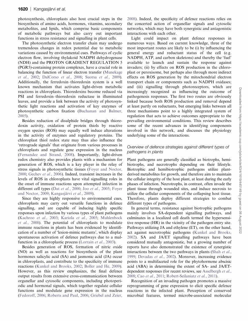
Several studies have shown that specific photoreceptors
can influence defence responses (Fig. 2). Systemic acquired
resistance (SAR) usually requires molecular recognitionevents such as gene-for-gene-based resistance, in which
disease resistance (R) genes notably include the large NBS-
LRR class. The constitutive shade-avoidance 1 mutant (csa1)
carries a mutation in a defence response-related protein
(TIR-NBS-LRR), resulting in a dominant negative effect on
phytochrome signalling. Moreover, this mutant shows de-
creased resistance against pathogenic Pseudomonas. Thus,
csa1 provides one of several pieces of evidence thatphytochrome and defence signalling interact (Faigon-Soverna
et al., 2006). It is also demonstrated that CRY1 positively
regulates R protein-mediated resistance to avirulent
P. syringae RPT2 in incompatible plant–pathogen interac-
tions (Wu and Yang, 2010).
Genoud et al. (2002) demonstrated that phytochrome
signalling pathways can activate both SA perception and
HR development triggered by avirulent P. syringae. In
particular, protein phosphatase 7 (AtPP7) has been identi-
fied as a modulator of phytochrome signals and has been
found to interact with nucleotide-diphosphate kinase 2
(NDPK2), an upstream element involved in the modulation
of the SA-dependent defence pathway by light (Genoud
et al., 2008). However, the use of Arabidopsis photoreceptor
double mutants has shown that the induction of defence
responses at inoculation sites is not or only slightlymodulated when cryptochrome, phototropin, or phyto-
chrome photoreception is diminished. This contrasts with
SAR, which depends on phytochrome photoreception, but
can be established without functional cryptochrome or
phototropin signalling pathways (Griebel and Zeier, 2008).
Chandra-Shekara et al. (2006) reported that the HR
triggered by Turnip crinkle virus (TCV) and resistance to
viral infection is influenced by light, but independent of thephotoreceptors phytochrome A and phytochrome B. When
Di-17, which is a TCV-resistant line when inoculated in the
light, was inoculated with TCV or TMV following extended
darkness before the regular day/night rhythm, development
of HR was absent and the virus spread systemically. HRT is
a putative resistance protein which confers the HR and
resistance to TCV. When this protein was overexpressed in
phyA or phyB mutant backgrounds, neither phytochromewas required for development of an HR resembling that
seen in Di-17 (Chandra-Shekara et al., 2006). The absence
of light does not affect the induction of SA by TCV,
although SA applied in the dark was unable to induce SA-
mediated signalling leading to resistance or PR-1 gene
expression. Thus, both light and SA are key players in
host–virus interactions (Chandra-Shekara et al., 2006).
Additionally, the blue-light photoreceptors CRY2 and
Biotrophs
PHY
JA
CRY
SA
Defense genesPR-1, PR-5, PDF1.2, others
COI1 / JAZ 1/MYC2 / PFT1
Necrotrophs,herbivores
PHYAPHYB
PHYAPHYBCRY1
PSI2 / NDPK2 / PP7 COP1
RED
BLUE
PHYBPHYCPHYDPHYE
CRY1CRY2PHOT1PHOT2
PHYA
CRY2PHOT2PHOT1
ABA
Fig. 2. Possible roles of photoreceptors in salicylic acid (SA) and jasmonic acid (JA)-related signalling pathways. CRY, cryptochrome;
PHOT, phototropin; PHY, phytochrome. For discussion, see text.
1628 | Kangasjarvi et al.D
ownloaded from
https://academic.oup.com
/jxb/article/63/4/1619/445149 by guest on 06 August 2022

PHOT2 are specifically required for maintaining the stabil-
ity of the HRT protein (and thereby resistance to TCV) by
interacting with and negatively regulating the activity of
COP1 (CONSTITUTIVELY PHOTOMORPHOGENIC 1).
COP1 is an E3 ubiquitin ligase, which is known to target
proteins for 26S proteasome-mediated degradation. CRY1
and PHOT1, in contrast, influence HRT-mediated resistance
without affecting the stability of the R protein (Jeong et al.,2010).
Despite these observations, not all inducible local plant
defences require the presence of light. In Arabidopsis
Col-0 leaves inoculated with P. syringae avrRPM1, contin-
uous darkness did not affect biosynthesis of the Arabidopsis
phytoalexin, camalexin, JA accumulation, or expression of
GST1, a ROS-induced glutathione S-transferase (Zeier et al.,
2004). JAs are plant hormones that regulate many physio-logical processes, including pathogen defence. The charac-
terization of several mutant lines deficient in JA biosynthesis
and signalling has provided evidence of links between JA and
phytochrome signalling. JASMONATE INSENSITIVE 1
(jar1) has the same locus as FAR-RED INSENSITIVE219
(fin219), which has been demonstrated to interact with
GSTU20 in response to light (Chen et al., 2007). The JA
receptor COI1 (CORONATINE INSENSITIVE1), a centralcomponent of JA signalling, is necessary for a number of
high irradiance responses in far-red light, and this requires
stability of another important JA signalling component,
JAZ1 (JASMONATE ZIM DOMAIN; Robson et al.,
2010). It has also been recently shown that the PHYTO-
CHROME AND FLOWERING TIME1 (PFT1) gene,
which encodes the mediator 25 subunit of the plant
Mediator complex, is a key regulator of JA-regulatedtranscription and is required for resistance to leaf-infecting
necrotrophic fungal pathogens (Kidd et al., 2009).
As well as pathogen responses, defence against the attack
of insect herbivores is influenced by light. There is evidence
that plant responses to herbivores and shading are in
competition with each other, which may become crucial
when, for example, plants under a canopy face such an
attack. This is known as the plant dilemma, in which theplant must prioritize expression of shade avoidance
responses or induction of chemical defences (Ballare, 2009).
It has been shown that shade can down-regulate plant
defences and so increase the leaf area eaten by herbivores
(Izaguirre et al., 2006; Moreno et al., 2009). Thus, in shade
conditions, priority will be given to reallocation of carbon
resources to minimize the risk of competition (Kami et al.,
2010). As discussed above, shading, which is characterizedby low R:FRs, decreases plant sensitivity to jasmonates
(Moreno et al., 2009). Thus, shade may weaken the defence
response by repressing JA synthesis and signalling.
Circadian rhythms, growth daylength, and ROS
The plant circadian clock controls several elements of plant
biochemistry and physiology and spans a period close to
24 h. An outcome of circadian control is gating, implying
that equal stimuli applied at different times of the day can
lead to different intensities of a specific plant response
(Hotta et al., 2007). A link between defence and circadian
signalling has been based on the fact that PCC1 (PATHO-
GEN AND CIRCADIAN CONTROLLED1) and PAL1
follow a circadian expression pattern, but the functional
significance of this is not yet clear. So far, the expression of
these rhythmically expressed pathogen/defence-related genes
has also been found to be inducible by pathogens, signallingmolecules, and abiotic stresses (Weyman et al., 2006).
However, the effect of infections at different times of the
day on the induction of gene expression or the pattern of
expression in circadian clock-defective mutants has not yet
been investigated (Roden and Ingle, 2009).
Although the role of the circadian clock remains unclear,
recent findings suggest that signalling pathways related to
daylength may be important in governing the outcome ofROS-triggered signalling. In the Arabidopsis cat2 mutant,
SA accumulation and associated responses do not occur in
short days (8 h light/16 h dark). The failure to up-regulate
these defences in these conditions does not seem to be
trivially linked to an insufficiently severe oxidative stress
(Queval et al., 2007). Furthermore, responses in other
Arabidopsis lesion-mimic mutants such as lsd1 and mips1
have also been shown to be influenced by the light regime(Dietrich et al., 1994; Meng et al., 2009).
The phenotypic differences in the response to H2O2 in
cat2 growing in short and long days are preceded and
accompanied by daylength-specific cat2-dependent changes
in gene expression. Daylength-specific patterns include
oxidative stress-associated genes, which are generally more
strongly induced in short days, and pathogenesis-related
gene expression, which is more evident in long days (Quevalet al., 2007, 2011b; Chaouch et al., 2010). Interestingly, the
effect of daylength is not confined to oxidative stress, but
also influences transcriptomic responses to the CO2 level
(Queval et al., 2011b). Neither is the oxidative stress–
daylength interaction confined to the cat2 background,
because the outcome of equal time exposure to ozone can
also be influenced by the growth photoperiod context
(Vollsnes et al., 2009). Further evidence that daylengthmodulates redox regulation of defence-linked gene expres-
sion is supported by analysis of gr1 mutants lacking
expression of the cytosolic/peroxisomal isoform of GR.
Although these mutants show neither phenotypic evidence
of oxidative stress nor increased ROS signals, their rela-
tively oxidized leaf glutathione status affects JA-associated
gene expression in a manner dependent on growth day-
length (Mhamdi et al., 2010b). As noted above, links havebeen described between ROS, SA, JA, photoreceptors,
flowering, and defence reactions (Genoud et al., 2002;
Martinez et al., 2004; Danon et al., 2005; Robson et al.,
2010).
Conclusions and perspectives
Although it is well established that plant defence is under
genetic control, the outcome of defence signalling is also
Light signalling in defence responses | 1629D
ownloaded from
https://academic.oup.com
/jxb/article/63/4/1619/445149 by guest on 06 August 2022

influenced by environmental conditions and nutritional
status. Understandably, much of the focus on defence
signalling has been on cytosol–nuclear interactions (e.g.
Mou et al., 2003; Kaminaka et al., 2006), but the
chloroplast, as the engine of plant growth, also plays
a crucial role. This organelle houses several important steps
in the synthesis of phytohormones involved in defence, such
as SA, JA, and ABA. As the ultimate source of photo-assimilate, the chloroplast also contributes to sugar status,
which can influence the SA pathway and interact with
signalling through other phytohormones such as ABA that
are involved in biotic challenge (Finkelstein and Gibson,
2002; Roitsch et al., 2003; Asselbergh et al., 2008).
Moreover, several recent studies have shown that chloro-
plast-located proteins are involved in cross-talk with the
cytosol and nucleus to govern the outcome of defencesignalling. Further important information in this area is
likely to be generated by the continued use of genetic
studies in amenable species such as Arabidopsis.
The chloroplast is potentially a major source of ROS. It
is the most important cellular player in production of 1O2,
and is also traditionally considered to be the major in-
tracellular producer of partially reduced oxygen species
such as �O2� and H2O2. However, these latter molecules can
also be produced in substantial amounts by other organ-
elles, notably peroxisomes and mitochondria (Foyer and
Noctor, 2003), and a key outstanding question concerns the
importance of different subcellular compartments in ROS
production during plant responses to pathogens. Full
resolution of this issue has been hampered by the absence
of techniques able to generate quantitative information with
sufficient resolution. Because of the reactivity of ROS andthe complex redox matrix of plant tissues, most techniques
used to detect intracellular ROS have hitherto been semi-
quantitative (Queval et al., 2008). As well as the question of
spatial differences, the role of different ROS and their
interactions (Gadjev et al., 2006) remain to be fully
elucidated. More insight into these questions is likely to be
provided following the emergence of in vivo sensors that are
able to report on specific ROS in a reliable, quantitative,and compartment-specific manner.
The most important redox parameter in defence responses
might not be ROS titre per se. While the plastoquinone and
TRX pools are key players in generating signals from the
photosynthetic electron transport chain, an increasing num-
ber of studies are also providing insight into the important
role of antioxidants, such as ascorbate and glutathione, in
redox regulation. It is a striking but often overlooked factthat plants with decreased amounts of major antioxidative
enzymes (APX and catalase) show clear evidence of
oxidative stress despite a failure to display sustained
increases in detectable ROS (e.g. Rizhsky et al., 2002;
Chaouch et al., 2010, 2011). This probably reflects the
potency of the intracellular antioxidative system in ROS
homeostasis, and several observations suggest that ROS-
triggered modulation of components such as glutathionemay be one route by which oxidative signals are perceived
by the plant cell (Mhamdi et al., 2010a, b; Noctor et al.,
2011). Comprehensive high-throughput proteomics technol-
ogies are likely to be particularly important in elucidating
the network of post-translational modifications involved in
redox regulation.
Intriguing information is accumulating on the role of
photoreceptor-mediated light signalling, circadian rhythms,
and daylength in determining or toning the outcome of
defence responses. Such effects are clearly of potentialrelevance to horticulture and agriculture, as they could
contribute to seasonal variations in plant susceptibility to
disease and other stresses. Photoreceptor pathways could be
important, for example, in determining the daylength
dependence of responses to intracellular H2O2. However,
light modulation of oxidative stress responses could be
dependent on chloroplast pathways such as those discussed
in the first part of this review. Future studies shouldcontinue to throw further light on the complexity of the
integrated circuitry that governs how plants cope with the
attempts of microorganisms and herbivores to gain access
to their resources.
Acknowledgements
This work was financially supported by the EU Marie Curie
ITN network COSI (project GA-215174) and the Academy
of Finland (CoE project 118637, 218157, and 130595). We
are grateful to Markus Teige, University of Vienna, Austria
for his excellent work as coordinator of the COSI ITN.
References
Ali R, Ma W, Lemtiri-Chlieh F, Tsaltas D, Leng Q, von
Bodman S, Berkowitz GA. 2007. Death don’t have no mercy and
neither does calcium: Arabidopsis CYCLIC NUCLEOTIDE GATED
CHANNEL2 and innate immunity. The Plant Cell 19, 1081–1095.
Aro EM, Suorsa M, Rokka A, Allahverdiyeva Y, Paakkarinen V,
Saleem A, Battchikova N, Rintamaki E. 2005. Dynamics of
photosystem II: a proteomic approach to thylakoid protein complexes.
Journal of Experimental Botany 56, 347–356.
Asada K. 1999. The water–water cycle in chloroplasts: scavenging of
active oxygens and dissipation of excess photons. Annual Review of
Plant Physiology and Plant Molecular Biology 50, 601–639.
Asselbergh D, De Vleesschauwer D, Hofte M. 2008. Global
switches and fine-tuning: ABA modulates plant–pathogen defense.
Molecular Plant-Microbe Interactions 21, 709–719.
Ball L, Accotto G, Bechtold U, et al. 2004. Evidence for a direct link
between glutathione biosynthesis and stress defense gene expression
in Arabidopsis. The Plant Cell 16, 2448–2462.
Ballare CL. 2009. Illuminated behaviour: phytochrome as a key
regulator of light foraging and plant anti-herbivore defence. Plant, Cell
and Environment 32, 713–725.
Bechtold U, Richard O, Zamboni A, Gapper C, Geisler M,
Pogson B, Karpinski S, Mullineaux PM. 2008. Impact of
chloroplastic- and extracellular-sourced ROS on high light-responsive
1630 | Kangasjarvi et al.D
ownloaded from
https://academic.oup.com
/jxb/article/63/4/1619/445149 by guest on 06 August 2022

gene expression in Arabidopsis. Journal of Experimental Botany 59,
121–133.
Beffagna N, Lutzu I. 2007. Inhibition of catalase activity is an early
response of Arabidopsis thaliana cultured cells to the phytotoxin
fusicoccin. Journal of Experimental Botany 58, 4183–4194.
Belhaj K, Lin B, Mauch F. 2009. The chloroplast protein RPH1 plays
a role in the immune response of Arabidopsis to. Phytophthora
brassicae. The Plant Journal 58, 287–298.
Bellafiore S, Bameche F, Peltier G, Rochaix JD. 2005. State
transitions and light adaptation require chloroplast thylakoid protein
kinase STN7. Nature 433, 892–895.
Bermudez MA, Paez-Ochoa MA, Gotor C, Romero LC. 2010.
Arabidopsis S-sulfocysteine synthase activity is essential for
chloroplast function and long-day light-dependent redox control. The
Plant Cell 2, 403–416.
Bick JA, Setterdahl AT, Knaff DB, Chen Y, Pitcher LH,
Zilinskas BA, Leustek T. 2001. Regulation of the plant-type 5’-
adenylyl sulfate reductase by oxidative stress. Biochemistry 40,
9040–9048.
Bloem E, Haneklaus S, Salac I, Wickenhauser P, Schnug E.
2007. Facts and fiction about sulfur metabolism in relation to plant–
pathogen interactions. Plant Biology 9, 596–607.
Boller T, He SY. 2009. Innate immunity in plants: an arms race
between pattern recognition receptors in plants and effectors in
microbial pathogens. Science 324, 742–744.
Bolton MD. 2009. Primary metabolism and plant defense—fuel for
the fire. Molecular Plant-Microbe Interactions 22, 487–947.
Buchanan BB, Balmer Y. 2005. Redox regulation: a broadening
horizon. Annual Review of Plant Biology 56, 187–220.
Cao FY, Yoshioka K, Desveaux D. 2011. The roles of ABA in plant–
pathogen interactions. Journal of Plant Research 124, 489–499.
Chamnongpol S, Willekens H, Langebartels C, Van Montagu M,
Inze D, Van Camp W. 1996. Transgenic tobacco with a reduced
catalase activity develops necrotic lesions and induces pathogenesis-
related expression under high light. The Plant Journal 10, 491–503.
Chamnongpol S, Willekens H, Moeder W, Langebartels C,
Sandermann H Jr, Van Montagu M, Inze D, Van Camp W. 1998.
Defense activation and enhanced pathogen tolerance induced by
H2O2 in transgenic plants. Proceedings of the National Academy of
Sciences, USA 95, 5818–5823.
Chandra-Shekara AC, Gupte M, Navarre D, Raina S, Raina R,
Klessig D, Kachroo P. 2006. Light-dependent hypersensitive
response and resistance signaling against Turnip Crinkle Virus in
Arabidopsis. The Plant Journal 45, 320–334.
Chang CC, Slesak I, Jorda L, Sotnikov A, Melzer M, Miszalski Z,
Mullineaux PM, Parker JE, Karpinska B, Karpinski S. 2009.
Arabidopsis chloroplastic glutathione peroxidases play a role in cross
talk between photooxidative stress and immune responses. Plant
Physiology 150, 670–683.
Chaouch S, Noctor G. 2010. myo-Inositol abolishes salicylic acid-
dependent cell death and pathogen defence responses triggered by
peroxisomal H2O2. New Phytologist 188, 711–718.
Chaouch S, Queval G, Noctor G. 2011. . AtrbohF is a crucial modulator
of defence-associated metabolism and a key actor in the interplay
between intracellular oxidative stress and pathogenesis responses in
Arabidopsis. The Plant Journal doi: 10.1111/j.1365313X.2011.04816.x.
Chaouch S, Queval G, Vanderauwera S, Mhamdi A,
Vandorpe M, Langlois-Meurinne M, Van Breusegem F,
Saindrenan P, Noctor G. 2010. Peroxisomal H2O2 is coupled to
biotic defense responses by ICS1 in a daylength-dependent manner.
Plant Physiology 153, 1692–1705.
Chen IC, Huang IC, Liu MJ, Wang ZG, Chung SS, Hsieh HL.
2007. Glutathione S-transferase interacting with far-red insensitive 219
is involved in phytochrome A-mediated signaling in Arabidopsis. Plant
Physiology 143, 1189–1202.
Christie JM. 2007. Phototropin blue-light receptors. Annual Review of
Plant Biology 58, 21–45.
Clough SJ, Fengler KA, Yu I, Lippok B, Smith RK, Bent AF. 2000.
The Arabidopsis dnd1 ‘defense, no death’ gene encodes a mutated
cyclic nucleotide-gated ion channel. Proceedings of the National
Academy of Sciences, USA 97, 9323–9328.
Conklin PL, Barth C. 2004. Ascorbic acid, a familiar small
molecule intertwined in the response of plants to ozone, pathogens,
and the onset of senescence. Plant, Cell and Environment 17,
959–970.
DalCorso G, Pesaresi P, Masiero S, Aseeva E, Schunemann D,
Finazzi G, Joliot P, Barbato R, Leister D. 2008. A complex
containing PGRL1 and PGR5 is involved in the switch between linear
and cyclic electron flow in Arabidopsis. Cell 132, 273–285.
Danon A, Miersch O, Felix G, Camp RG, Apel K. 2005. Concurrent
activation of cell death-regulating signalling pathways by singlet
oxygen in Arabidopsis thaliana. The Plant Journal 41, 68–80.
Danon A, Coll NS, Apel K. 2006. Cryptochrome-1-dependent
execution of programmed cell death induced by singlet oxygen in
Arabidopsis thaliana. Proceedings of the National Academy of
Sciences, USA 103, 17036–17041.
Dat JF, Pellinen R, Beeckman T, Van De Cotte B,
Langebartels C, Kangasjarvi J, Inze D, Van Breusegem F. 2003.
Changes in hydrogen peroxide homeostasis trigger an active cell
death process in tobacco. The Plant Journal 33, 1–12.
Dat J, Vandenabeele S, Vranova E, Van Montagu M, Inze D, Van
Breusegem F. 2000. Dual action of the active oxygen species during
plant stress responses. Cellular and Molecular Life Sciences 57,
779–795.
Demarsy E, Fankhauser C. 2009. Higher plants use LOV to perceive
blue light. Current Opinion in Plant Biology 12, 69–74.
Devadas SK, Enyedi A, Raina R. 2002. The Arabidopsis hrl1
mutation reveals novel overlapping roles for salicylic acid, jasmonic
acid and ethylene signalling in cell death and defence against
pathogens. The Plant Journal 30, 467–480.
Dietrich RA, Delaney TP, Uknes SJ, Ward ER, Ryals JA,
Dangl JL. 1994. Arabidopsis mutants simulating disease resistance
response. Cell 77, 565–577.
Dorey S, Baillieul F, Saindrenan P, Fritig B, Kauffmann S.
1998. Tobacco class I and II catalases are differentially expressed
during elicitor-induced hypersensitive cell death and localized
acquired resistance. Molecular Plant-Microbe Interactions 11,
1102–1109.
Light signalling in defence responses | 1631D
ownloaded from
https://academic.oup.com
/jxb/article/63/4/1619/445149 by guest on 06 August 2022

Du H, Klessig DF. 1997. Role for salicylic acid in the activation of
defense responses in catalase-deficient transgenic tobacco. Molecular
Plant-Microbe Interactions 10, 922–925.
Essmann J, Schmitz-Thom I, Schon H, Sonnewald S, Weis E,
Scharte J. 2008. RNA interference-mediated repression of cell wall
invertase impairs defense in source leaves of tobacco. Plant
Physiology 147, 1288–1299.
Faigon-Soverna A, Harmon FG, Storani L, Karayekov E,
Staneloni RJ, Gassmann W, Mas P, Casal JJ, Kay SA,
Yanovsky MJ. 2006. A constitutive shade-avoidance mutant
implicates TIR-NBS-LRR proteins in Arabidopsis photomorphogenic
development. The Plant Cell 18, 2919–2928.
Fedoroff N. 2006. Redox regulatory mechanisms in cellular stress
responses. Annals of Botany 98, 289–300.
Fernandez AP, Strand A. 2008. Retrograde signalling and plant
stress: plastid signals initiate cellular stress responses. Current Opinion
in Plant Biology 11, 509–513.
Finkelstein RR, Gibson SI. 2002. ABA and sugar interactions
regulating development: cross-talk or voices in a crowd? Current
Opinion in Plant Biology 5, 26–32.
Folta KM, Maruhnich SA. 2007. Green light: a signal to slow down
or stop. Journal of Experimental Botany 58, 3099–3111.
Foyer CH, Bloom AJ, Queval G, Noctor G. 2009. Photorespiratory
metabolism: genes, mutants, energetics, and redox signaling. Annual
Review of Plant Biology 60, 455–484.
Foyer CH, Noctor G. 2000. Oxygen processing in photosynthesis:
regulation and signalling. New Phytologist 146, 359–388.
Foyer CH, Noctor G. 2003. Redox sensing and signalling associated
with reactive oxygen produced in chloroplasts and mitochondria.
Physiologia Plantarum 119, 355–364.
Foyer CH, Noctor G. 2009. Redox regulation in photosynthetic
organisms: signaling, acclimation, and practical implications.
Antioxidants and Redox Signaling 11, 861–905.
Franklin KA, Quail PH. 2010. Phytochrome functions in Arabidopsis
development. Journal of Experimental Botany 61, 11–24.
Fryer MJ, Ball L, Oxborough K, Karpinski S, Mullineaux PM,
Baker NR. 2003. Control of Ascorbate Peroxidase 2 expression by
hydrogen peroxide and leaf water status during excess light stress
reveals a functional organisation of Arabidopsis leaves. The Plant
Journal 33, 691–705.
Fu ZQ, Guo M, Jeong BR, Tian F, Elthon TE, Cerny RL,
Staiger D, Alfano JR. 2007. A type III effector ADP-ribosylates
RNA-binding proteins and quells plant immunity. Nature 447,
284–288.
Gadjev I, Vanderauwera S, Gechev TS, Laloi C, Minkov IN,
Shulaev V, Apel K, Inze D, Mittler R, Van Breusegem F. 2006.
Transcriptomic footprints disclose specificity of reactive oxygen
species signaling in Arabidopsis. Plant Physiology 141, 436–445.
Gechev TS, Van Breusegem F, Stone JM, Denev I, Laloi C. 2006.
Reactive oxygen species as signals that modulate plant stress
responses and programmed cell death. Bioessays 28, 1091–1101.
Genoud T, Buchala AJ, Chua NH, Metraux JP. 2002.
Phytochrome signalling modulates the SA-perceptive pathway in
Arabidopsis. The Plant Journal 31, 87–95.
Genoud T, Cruz MTS, Kulisic T, Sparla F, Fankhauser C,
Metraux JP. 2008. The protein phosphatase 7 regulates
phytochrome signaling in Arabidopsis. PLoS One 3, e2699.
Gray J, Janick-Buckner D, Buckner B, Close PS, Johal GS.
2002. Light-dependent death of maize lls1 cells is mediated by mature
chloroplasts. Plant Physiology 130, 1894–1907.
Griebel T, Zeier J. 2008. Light regulation and daytime dependency of
inducible plant defenses in Arabidopsis: phytochrome signaling
controls systemic acquired resistance rather than local defense. Plant
Physiology 147, 790–801.
Hanning I, Heldt HW. 1993. On the function of mitochondrial
metabolism during photosynthesis in spinach (Spinacia oleracea L.)
leaves. Plant Physiology 103, 1147–1154.
Hertwig B, Streb P, Feierabend J. 1992. Light dependence of
catalase synthesis and degradation in leaves and the influence of
interfering stress conditions. Plant Physiology 100, 1547–1553.
Hideg E, Barta C, Kalai T, Vass I, Hideg K, Asada K. 2002.
Detection of singlet oxygen and superoxide with fluorescent sensors in
leaves under stress by photoinhibition or UV radiation. Plant and Cell
Physiology 43, 1154–1164.
Hirashima M, Tanaka R, Tanaka A. 2009. Light-independent cell
death induced by accumulation of pheophorbide a in Arabidopsis
thaliana. Plant and Cell Physiology 50, 719–729.
Hotta CT, Gardner MJ, Hubbard KE, Baek SJ, Dalchau N,
Suhita D, Dodd AN, Webb AAR. 2007. Modulation of environmental
responses of plants by circadian clocks. Plant, Cell and Environment
30, 333–349.
Igamberdiev AU, Gardestrom P. 2003. Regulation of NAD- and
NADP-dependent isocitrate dehydrogenases by reduction levels of
pyridine nucleotides in mitochondria and cytosol of pea leaves.
Biochichima et Biophysica Acta 1606, 117–125.
Imaizumi T, Tran HG, Swartz TE, Briggs WR, Kay SA. 2003. FKF1
is essential for photoperiodic-specific light signalling in Arabidopsis.
Nature 426, 302–306.
Izaguirre MM, Mazza CA, Biondini M, Baldwin IT, Ballare CL. 2006.
Remote sensing of future competitors: impacts on plant defenses.
Proceedings of the National Academy of Sciences, USA 103, 7170–7174.
Jelenska J, Yao N, Vinatzer BA, Wright CM, Brodsky JL,
Greenberg JT. 2007. A J domain virulence effector of Pseudomonas
syringae remodels host chloroplasts and suppresses defenses.
Current Biology 17, 499–508.
Jenkins GI. 2009. Signal transduction in responses to UV-B radiation.
Annual Review of Plant Biology 60, 407–431.
Jeong RD, Chandra-Shekara AC, Barman SR, Navarre D,
Klessig DF, Kachroo A, Kachroo P. 2010. Cryptochrome 2 and
phototropin 2 regulate resistance protein-mediated viral defense by
negatively regulating an E3 ubiquitin ligase. Proceedings of the
National Academy of Sciences, USA 107, 13538–13543.
Jones JD, Dangl JL. 2006. The plant immune system. Nature 444,
323–329.
Joo JH, Wang S, Chen JG, Jones AM, Fedoroff NV. 2005.
Different signaling and cell death roles of heterotrimeric G protein
alpha and beta subunits in the Arabidopsis oxidative stress response
to ozone. The Plant Cell 17, 957–970.
1632 | Kangasjarvi et al.D
ownloaded from
https://academic.oup.com
/jxb/article/63/4/1619/445149 by guest on 06 August 2022

Kachroo A, Lapchyk L, Fukushige H, Hildebrand D, Klessig D,
Kachroo P. 2003. Plastidial fatty acid signaling modulates salicylic
acid- and jasmonic acid-mediated defense pathways in the
Arabidopsis ssi2 mutant. The Plant Cell 15, 2952–2965.
Kami C, Lorrain S, Hornitschek P, Fankhauser C. 2010. Light-
regulated plant growth and development. Cuurent Topics in
Developmental Biology 91, 29–66.
Kaminaka H, Nake C, Epple E, et al. 2006. bZIP10-LSD1
antagonism modulates basal defense and cell death in Arabidopsis
following infection. EMBO Journal 25, 4400–4411.
Kangasjarvi S, Nurmi M, Tikkanen M, Aro EM. 2009. Cell-
specific mechanisms and systemic signalling as emerging themes in
light acclimation of C3 plants. Plant, Cell and Environment 32,
1230–1240.
Kariola T, Brader G, Li J, Palva ET. 2005. Chlorophyllase 1,
a damage control enzyme, affects the balance between defense
pathways in plants. The Plant Cell 17, 282–294.
Karpinski S, Reynolds H, Karpinska B, Wingsle G, Creissen G,
Mullineaux P. 1999. Systemic signalling and acclimation in
response to excess excitation energy in Arabidopsis. Science 284,
654–657.
Kerchev PI, Fenton B, Foyer CH, Hancock RD. . 2011. Plant
responses to insect herbivory: interactions between photosynthesis,
reactive oxygen species and hormonal signalling pathways. Plant, Cell
and Environment 35, 441–453.
Keys AJ. 1999. Biochemistry of photorespiration and the
consequences for plant performance. In: Bryant JA, Burrell MM,
Kruger NJ, eds. Plant carbohydrate biochemistry. Oxford: BIOS
Scientific, 147–161.
Kiba A, Nishihara M, Tsukatani N, Nakatsuka T, Kato Y,
Yamamura S. 2005. A peroxiredoxin Q homolog from gentians is
involved in both resistance against fungal disease and oxidative stress.
Plant and Cell Physiology 46, 1007–1015.
Kidd BN, Edgar CI, Kumar KK, Aitken EA, Schenk PM,
Manners JM, Kazan K. 2009. The mediator complex subunit PFT1 is
a key regulator of jasmonate-dependent defense in Arabidopsis. The
Plant Cell 21, 2237–2252.
Kitajima S, Tomizawa K, Shigeoka S, Yokota A. 2006. An inserted
loop region of stromal ascorbate peroxidase is involved in its hydrogen
peroxide-mediated inactivation. FEBS Journal 273, 2704–2710.
Kitajima S. 2008. Hydrogen peroxide-mediated inactivation of two
chloroplastic peroxidases, ascorbate peroxidase and 2-cys
peroxiredoxin. Photochemistry and Photobiology 84, 1404–1409.
Klimyuk VI, Persello-Cartieaux F, Havaux M, Contard-David P,
Schuenemann D, Meiherhoff K, Gouet P, Jones JD,
Hoffman NE, Nussaume L. 1999. A chromodomain protein encoded
by the arabidopsis CAO gene is a plant-specific component of the
chloroplast signal recognition particle pathway that is involved in LHCP
targeting. The Plant Cell 11, 87–99.
Konig J, Baier M, Horling F, Kahmann U, Harris G,
Schurmann P, Dietz KJ. 2002. The plant-specific function of 2-Cys
peroxiredoxin-mediated detoxification of peroxides in the redox-
hierarchy of photosynthetic electron flux. Proceedings of the National
Academy of Sciences, USA 16, 5738–4573.
Kunkel BN, Brooks DM. 2002. Cross talk between signaling pathways
in pathogen defense. Current Opinion in Plant Biology 5, 325–331.
Laloi C, Stachowiak M, Pers-Kamczyc E, Warzych E, Murgia I,
Apel K. 2007. Cross-talk between singlet oxygen- and hydrogen
peroxide-dependent signaling of stress responses in Arabidopsis
thaliana. Proceedings of the National Academy of Sciences, USA 104,
672–677.
Lin CT, Shalitin D. 2003. Cryptochrome structure and signal
transduction. Annual Review of Plant Biology 54, 469–496.
Lipka V, Dittgen J, Bednarek P, et al. 2005. Pre- and postinvasion
defenses both contribute to nonhost resistance in Arabidopsis.
Science 310, 1180–1183.
Liu Y, Ren D, Pike S, Pallardy S, Gassmann W, Zhang S. 2007.
Chloroplast-generated reactive oxygen species are involved in
hypersensitive response-like cell death mediated by a mitogen-
activated protein kinase cascade. The Plant Journal 51, 941–954.
Lorrain S, Lin B, Auriac MC, Kroj T, Saindrenan P, Nicole M,
Balague C, Roby D. 2004. VASCULAR ASSOCIATED DEATH1,
a novel GRAM domain-containing protein, is a regulator of cell death
and defense responses in vascular tissues. The Plant Cell 16,
2217–2232.
Lorrain S, Vailleau F, Balague C, Roby D. 2003. Lesion mimic
mutants: keys for deciphering cell death and defense pathways in
plants? Trends in Plant Science 8, 263–271.
Mach JM, Castillo AR, Hoogstraten R, Greenberg JT. 2001. The
Arabidopsis-accelerated cell death gene ACD2 encodes red
chlorophyll catabolite reductase and suppresses the spread of disease
symptoms. Proceedings of the National Academy of Sciences, USA
98, 771–776.
Major IT, Nicole MC, Duplessis S, Seguin A. 2010. Photosynthetic
and respiratory changes in leaves of poplar elicited by rust infection.
Photosynthesis Research 104, 41–48.
Martınez C, Pons E, Prats G, Leon J. 2004. Salicylic acid regulates
flowering time and links defence responses and reproductive
development. The Plant Journal 37, 209–217.
Mateo A, Funck D, Muhlenbock P, Kular B, Mullineaux PM,
Karpinski S. 2006. Controlled levels of salicylic acid are required for
optimal photosynthesis and redox homeostasis. Journal of
Experimental Botany 57, 1795–1807.
Mateo A, Muhlenbock P, Rusterucci C, et al. 2004. LESION
SIMULATING DISEASE 1 is required for acclimation to conditions that
promote excess excitation energy. Plant Physiology 136, 2818–2830.
Melotto M, Underwood W, He SY. 2008. Role of stomata in plant
innate immunity and foliar bacterial diseases. Annual Review of
Phytopathology 46, 101–122.
Meng PH, Raynaud C, Tcherkez G, et al. 2009. Crosstalks
between myo-inositol metabolism, programmed cell death and basal
immunity in Arabidopsis. PLoS One 4, e7364.
Meyer S, Saccardy-Adji K, Rizza F, Genty B. 2001. Inhibition of
photosynthesis by Colletotrichum lindemuthianum in bean leaves
determined by chlorophyll fluorescence imaging. Plant, Cell and
Environment 24, 927–955.
Mhamdi A, Hager J, Chaouch S, et al. 2010b. Arabidopsis
GLUTATHIONE REDUCTASE 1 is essential for the metabolism of
Light signalling in defence responses | 1633D
ownloaded from
https://academic.oup.com
/jxb/article/63/4/1619/445149 by guest on 06 August 2022

intracellular H2O2 and to enable appropriate gene expression through
both salicylic acid and jasmonic acid signaling pathways. Plant
Physiology 153, 1144–1160.
Mhamdi A, Queval G, Chaouch S, Vanderauwera S, Van
Breusegem F, Noctor G. 2010a. Catalases in plants: a focus on
Arabidopsis mutants as stress-mimic models. Journal of Experimental
Botany 61, 4197–4220.
Mittler R, Vanderauwera S, Gollery M, Van Breusegem F. 2004.
The reactive oxygen gene network in plants. Trends in Plant Science
9, 490–498.
Miura E, Kato Y, Matsushima R, Albrecht V, Laalami S,
Sakamoto W. 2007. The balance between protein synthesis and
degradation in chloroplasts determines leaf variegation in Arabidopsis
yellow variegated mutants. The Plant Cell 19, 1313–1328.
Moeder W, Del Pozo O, Navarre DA, Martin GB, Klessig DF.
2007. Aconitase plays a role in regulating resistance to oxidative stress
and cell death in Arabidopsis and Nicotiana benthamiana. Plant
Molecular Biology 63, 273–287.
Moreno JE, Tao Y, Chory J, Ballare CL. 2009. Ecological
modulation of plant defense via phytochrome control of jasmonate
sensitivity. Proceedings of the National Academy of Sciences, USA
106, 4935–4940.
Mou Z, Fan W, Dong X. 2003. Inducers of plant systemic acquired
resistance regulate NPR1 function through redox changes. Cell 113,
935–944.
Muhlenbock P, Szechynska-Hebda M, Plaszczyca M, Baudo M,
Mullineaux PM, Parker JE, Karpinska B, Karpinski S. 2008.
Chloroplast signaling and LESION SIMULATING DISEASE1 regulate
crosstalk between light acclimation and immunity in Arabidopsis. The
Plant Cell 20, 2339–2356.
Mulo P, Sirpio S, Suorsa M, Aro EM. 2008. Auxiliary proteins
involved in the assembly and sustenance of photosystem II..
Photosynthesis Research 98, 489–501.
Munekage Y, Hojo M, Meurer J, Endo T, Tasaka M, Shikanai T.
2002. PGR5 is involved in cyclic electron flow around photosystem I
and is essential for photoprotection in Arabidopsis. Cell 110,
361–371.
Noctor G, Veljovic-Jovanovic S, Driscoll S, Novitskaya L,
Foyer CH. 2002. Drought and oxidative load in the leaves of C3
plants: a predominant role for photorespiration? Annals of Botany 89,
841–850.
Noctor G, Mhamdi A, Queval G, Chaouch S, Han Y,
Neukermans J, Foyer CH. 2011. Glutathione in plants—an
integrated overview. Plant, Cell and Environment 35, 454–484.
Novitskaya L, Trevanion S, Driscoll SD, Foyer CH, Noctor G.
2002. How does photorespiration modulate leaf amino acid contents?
A dual approach through modelling and metabolite analysis. Plant, Cell
and Environment 25, 821–836.
Nyathi Y, Baker A. 2006. Plant peroxisomes as a source of signalling
molecules. Biochimica et Biophysica Acta 1763, 1478–1495.
Ochsenbein C, Przybyla D, Danon A, Landgraf F, Gobel C,
Imboden A, Feussner I, Apel K. 2006. The role of EDS1 (enhanced
disease susceptibility) during singlet oxygen-mediated stress
responses of Arabidopsis. The Plant Journal 47, 445–456.
op den Camp RG, Przybyla D, Ochsenbein C, et al. 2003. Rapid
induction of distinct stress responses after the release of singlet
oxygen in Arabidopsis. The Plant Cell 15, 2320–2332.
Parisy V, Poinssot B, Owsianowski L, Buchala A, Glazebrook J,
Mauch F. 2007. Identification of PAD2 as a c-glutamylcysteine
synthetase highlights the importance of glutathione in disease
resistance in Arabidopsis. The Plant Journal 49, 159–172.
Pastori GM, Foyer CH. 2002. Common components, networks, and
pathways of cross-tolerance to stress. The central role of ‘redox’ and
abscisic acid-mediated controls. Plant Physiology 129, 460–468.
Pastori GM, Kiddle G, Antoniw J, Bernard S, Veljovic-
Jovanovic S, Verrier PJ, Noctor G, Foyer CH. 2003. Leaf vitamin C
contents modulate plant defense transcripts and regulate genes that
control development through hormone signaling. The Plant Cell 15,
939–951.
Pavet V, Olmos E, Kiddle G, Mowla S, Kumar S, Antoniw J,
Alvarez ME, Foyer CH. 2005. Ascorbic acid deficiency activates cell
death and disease resistance responses in Arabidopsis. Plant
Physiology 139, 1291–1303.
Queval G, Hager J, Gakiere B, Noctor G. 2008. Why are literature
data for H2O2 contents so variable? A discussion of potential
difficulties in quantitative assays of leaf extracts. Journal of
Experimental Botany 59, 135–146.
Queval G, Issakidis-Bourguet E, Hoeberichts FA, Vandorpe M,
Gakiere B, Vanacker H, Miginiac-Maslow M, Van Breusegem F,
Noctor G. 2007. Conditional oxidative stress responses in the
Arabidopsis photorespiratory mutant cat2 demonstrate that redox
state is a key modulator of daylength-dependent gene expression and
define photoperiod as a crucial factor in the regulation of H2O2-
induced cell death. The Plant Journal 52, 640–657.
Queval G, Jaillard D, Zechmann B, Noctor G. 2011a. Increased
intracellular H2O2 availability preferentially drives glutathione
accumulation in vacuoles and chloroplasts. Plant, Cell and
Environment 34, 21–32.
Queval G, Neukermans J, Vanderauwera S, Van Breusegem F,
Noctor G. 2011b. Daylength is a key regulator of transcriptomic
responses to both CO2 and H2O2 in Arabidopsis. Plant, Cell and
Environment 35, 374–87.
Queval G, Thominet D, Vanacker H, Miginiac-Maslow M,
Gakiere B, Noctor G. 2009. H2O2-activated up-regulation of
glutathione in Arabidopsis involves induction of genes encoding
enzymes involved in cysteine synthesis in the chloroplast. Molecular
Plant 2, 344–356.
Rizhsky L, Hallak-Herr E, Van Breusegem F, Rachmilevitch S,
Barr J, Rodermel S, Inze D, Mittler R. 2002. Double antisense
plants lacking ascorbate peroxidase and catalase are less sensitive to
oxidative stress than single antisense plants lacking ascorbate
peroxidase or catalase. The Plant Journal 32, 329–342.
Robert-Seilaniantz A, Grant M, Jones JD. 2011. Hormone
crosstalk in plant disease and defense: more than just jasmonate–
salicylate antagonism. Annual Review of Phytopathology 49,
317–343.
Roberts MR, Paul ND. 2006. Seduced by the dark side: integrating
molecular and ecological perspectives on the influence of light on plant
defence against pests and pathogens. New Phytologist 170, 677–699.
1634 | Kangasjarvi et al.D
ownloaded from
https://academic.oup.com
/jxb/article/63/4/1619/445149 by guest on 06 August 2022

Robson F, Okamoto H, Patrick E, Harris SR, Wasternack C,
Brearley C, Turner JG. 2010. Jasmonate and phytochrome A
signaling in Arabidopsis wound and shade responses are integrated
through JAZ1 stability. The Plant Cell 22, 1143–1160.
Rochaix J. 2007. Role of thylakoid protein kinases in photosynthetic
acclimation. FEBS Letters 581, 2768–2775.
Rockwell NC, Su YS, Lagarias JC. 2006. Phytochrome structure
and signaling mechanisms. Annual Review of Plant Biology 57,
837–858.
Roden LC, Ingle RA. 2009. Lights, rhythms, infection: the role of light
and the circadian clock in determining the outcome of plant–pathogen
interactions. The Plant Cell 21, 2546–2552.
Roitsch T, Balibrea ME, Hofmann M, Proels R, Sinha AK. 2003.
Extracellular invertase: key metabolic enzyme and PR protein. Journal
of Experimental Botany 54, 513–524.
Scharte J, Schon H, Weis E. 2005. Photosynthesis and
carbohydrate metabolism in tobacco leaves during an incompatible
interaction with Phytophthora nicotianae. Plant, Cell and Environment
28, 1421–1435.
Scheibe R, Backhausen JE, Emmerlich V, Holtgrefe S. 2005.
Strategies to maintain redox homeostasis during photosynthesis
under changing conditions. Journal of Experimental Botany 56,
1481–1489.
Schmidt M, Grief J, Feierabend J. 2006. Mode of translational
activation of the catalase (cat1) mRNA of rye leaves (Secale cereale L.)
and its control through blue light and reactive oxygen. Planta 223,
835–846.
Seo S, Okamoto M, Iwai T, Iwano M, Fukui K, Isogai A,
Nakajima N, Ohashi Y. 2000. Reduced levels of chloroplast FtsH
protein in tobacco mosaic virus-infected tobacco leaves accelerate the
hypersensitive reaction. The Plant Cell 12, 917–932.
Shah J, Kachroo P, Klessig DF. 1999. The Arabidopsis ssi1
mutation restores pathogenesis-related gene expression in npr1 plants
and renders defensin gene expression salicylic acid dependent. The
Plant Cell 11, 191–206.
Smith IK, Kendall AC, Keys AJ, Turner JC, Lea PJ. 1985. The
regulation of the biosynthesis of glutathione in leaves of barley
(Hordeum vulgare L). Plant Science 41, 11–17.
Smykowski A, Zimmermann P, Zentgraf U. 2010. G-box binding
factor 1 reduces CATALASE2 expression and regulates the onset of
leaf senescence in Arabidopsis thaliana. Plant Physiology 153,
1321–1331.
Streb P, Feierabend J. 1996. Oxidative stress responses
accompanying photoinactivation of catalase in NaCl-treated rye
leaves. Botanica Acta 109, 125–132.
Suorsa M, Sirpio S, Aro EM. 2009. Towards characterization of the
chloroplast NAD(P)H dehydrogenase complex. Molecular Plant 6,
1127–1140.
Takahashi H, Chen Z, Du H, Liu Y, Klessig DF. 1997.
Development of necrosis and activation of disease resistance in
transgenic tobacco plants with severely reduced catalase levels. The
Plant Journal 11, 993–1005.
Talbott LD, Shmayevich IJ, Chung YS, Hammad JW, Zeiger E.
2003. Blue light and phytochrome-mediated stomatal opening in the
npq1 and phot1 phot2 mutants of Arabidopsis. Plant Physiology 133,
1522–1529.
Taler D, Galperin M, Benjamin I, Cohen Y, Kenigsbuch D. 2004.
Plant eR genes that encode photorespiratory enzymes confer
resistance against disease. The Plant Cell 16, 172–184.
Tikkanen M, Grieco M, Aro EM. 2011. Novel insights into plant
light-harvesting complex II phosphorylation and ‘state transitions’.
Trends in Plant Science 16, 126–131.
Torres MA, Dangl JL. 2005. Functions of the respiratory burst
oxidase in biotic interactions, abiotic stress and development. Current
Opinion in Plant Biology 8, 397–403.
Trotta A, Wrzaczek M, Scharte J, et al. 2011. Regulatory subunit
B#c of protein phosphatase 2A prevents unnecessary defense reactions
under low light in Arabidopsis. Plant Physiology 156, 1464–1480.
Vandenabeele S, Vanderauwera S, Vuylsteke M, Rombauts S,
Langebartels C, Seidlitz HK, Zabeau M, Van Montagu M, Inze D,
Van Breusegem F. 2004. Catalase deficiency drastically affects gene
expression induced by high light in Arabidopsis thaliana. The Plant
Journal 39, 45–58.
Veljovic-Jovanovic SD, Pignocchi C, Noctor G, Foyer CH. 2001.
Low ascorbic acid in the vtc-1 mutant of Arabidopsis is associated
with decreased growth and intracellular redistribution of the antioxidant
system. Plant Physiology 127, 426–435.
Vlot AC, Dempsey DMA, Klessig DF. 2009. Salicylic acid,
a multifaceted hormone to combat disease. Annual Review of
Phytopathology 47, 177–206.
Volk S, Feierabend J. 1989. Photoinactivation of catalase at low
temperature and its relevance to photosynthetic and peroxide
metabolism in leaves. Plant, Cell and Environment 12, 701–712.
Vollsnes AV, Erikson AB, Otterholt E, Kvaal K, Oxaal U,
Futsaether C. 2009. Visible foliar injury and infrared imaging show that
daylength affects short-term recovery after ozone stress in Trifolium
subterraneum. Journal of Experimental Botany 60, 3677–3686.
Von Caemmerer S. 2000. Biochemical models of leaf
photosynthesis. Collingwood, Australia: CSIRO Publishing.
Wagner D, Przybyla D, Camp ROD, et al. 2004. The genetic basis
of singlet oxygen-induced stress responses of Arabidopsis thaliana.
Science 306, 1183–1185.
Weyman PD, Pan ZQ, Feng Q, Gilchrist DG, Bostock RM. 2006. A
circadian rhythm-regulated tomato gene is induced by arachidonic acid
and Phythophthora infestans infection. Plant Physiology 140, 235–248.
Wu L, Yang HQ. 2010. CRYPTOCHROME 1 is implicated in
promoting R protein-mediated plant resistance to Pseudomonas
syringae in Arabidopsis. Molecular Plant 3, 539–548.
Yang M, Wardzala E, Johal GS, Gray J. 2004. The wound-inducible
Lls1 gene from maize is an orthologue of the Arabidopsis Acd1 gene,
and the LLS1 protein is present in non-photosynthetic tissues. Plant
Molecular Biology 54, 175–191.
Yu F, Liu X, Alsheikh M, Park S, Rodermel S. 2008. Mutations in
SUPPRESSOR OF VARIEGATION1, a factor required for normal
chloroplast translation, suppress var2-mediated leaf variegation in
Arabidopsis. The Plant Cell 20, 1786–1804.
Yu L, Wan F, Dutta S, Welsh S, Liu Z, Freundt E, Baehrecke EH,
Lenardo M. 2006. Autophagic programmed cell death by selective
Light signalling in defence responses | 1635D
ownloaded from
https://academic.oup.com
/jxb/article/63/4/1619/445149 by guest on 06 August 2022

catalase degradation. Proceedings of the National Academy of
Sciences, USA 103, 4952–4957.
Zechmann B, Zellnig G, Urbanek-Krajnc A, Muller M. 2007.
Artificial elevation of glutathione affects symptom development in ZYMV-
infected Cucurbita pepo L. plant. Archives of Virology 152, 747–762.
Zeier J, Pink B, Mueller MJ, Berger S. 2004. Light conditions
influence specific defence responses in incompatible plant–pathogen
interactions: uncoupling systemic resistance from salicylic acid and
PR-1 accumulation. Planta 219, 673–683.
Zurbriggen MD, Carrillo N, Tognetti VB, Melzer M, Peisker M,
Hause B, Hajirezaei MR. 2009. Chloroplast-generated reactive
oxygen species play a major role in localized cell death during the non-
host interaction between tobacco and Xanthomonas campestris pv.
vesicatoria. The Plant Journal 60, 962–973.
1636 | Kangasjarvi et al.D
ownloaded from
https://academic.oup.com
/jxb/article/63/4/1619/445149 by guest on 06 August 2022
Related Documents