Photonics meets excitonics: natural and artificial molecular aggregates The Harvard community has made this article openly available. Please share how this access benefits you. Your story matters Citation Saikin, Semion K., Alexander Eisfeld, Stéphanie Valleau, and Alán Aspuru-Guzik. 2013. "Photonics Meets Excitonics: Natural and Artificial Molecular Aggregates." Nanophotonics 2, no. 1: 21-38. Published Version doi:10.1515/nanoph-2012-0025 Citable link http://nrs.harvard.edu/urn-3:HUL.InstRepos:12362622 Terms of Use This article was downloaded from Harvard University’s DASH repository, and is made available under the terms and conditions applicable to Other Posted Material, as set forth at http:// nrs.harvard.edu/urn-3:HUL.InstRepos:dash.current.terms-of- use#LAA

Welcome message from author
This document is posted to help you gain knowledge. Please leave a comment to let me know what you think about it! Share it to your friends and learn new things together.
Transcript

Photonics meets excitonics: naturaland artificial molecular aggregates
The Harvard community has made thisarticle openly available. Please share howthis access benefits you. Your story matters
Citation Saikin, Semion K., Alexander Eisfeld, Stéphanie Valleau, and AlánAspuru-Guzik. 2013. "Photonics Meets Excitonics: Natural andArtificial Molecular Aggregates." Nanophotonics 2, no. 1: 21-38.
Published Version doi:10.1515/nanoph-2012-0025
Citable link http://nrs.harvard.edu/urn-3:HUL.InstRepos:12362622
Terms of Use This article was downloaded from Harvard University’s DASHrepository, and is made available under the terms and conditionsapplicable to Other Posted Material, as set forth at http://nrs.harvard.edu/urn-3:HUL.InstRepos:dash.current.terms-of-use#LAA
DOI 10.1515/nanoph-2012-0025 Nanophotonics 2013; 2(1): 21–38© 2013 Science Wise Publishing &
Review article
Semion K. Saikin* , Alexander Eisfeld *, St é phanie Valleau and Al á n Aspuru-Guzik*
Photonics meets excitonics: natural and artificial molecular aggregates Abstract: Organic molecules store the energy of absorbed
light in the form of charge-neutral molecular excitations –
Frenkel excitons. Usually, in amorphous organic materials,
excitons are viewed as quasiparticles, localized on single
molecules, which diffuse randomly through the structure.
However, the picture of incoherent hopping is not applica-
ble to some classes of molecular aggregates – assemblies of
molecules that have strong near-field interaction between
electronic excitations in the individual subunits. Molecular
aggregates can be found in nature, in photosynthetic com-
plexes of plants and bacteria, and they can also be produced
artificially in various forms including quasi-one dimen-
sional chains, two-dimensional films, tubes, etc. In these
structures light is absorbed collectively by many mole cules
and the following dynamics of molecular excitation pos-
sesses coherent properties. This energy transfer mechanism,
mediated by the coherent exciton dynamics, resembles the
propagation of electromagnetic waves through a structured
medium on the nanometer scale. The absorbed energy can
be transferred resonantly over distances of hundreds of
nanometers before exciton relaxation occurs. Furthermore,
the spatial and energetic landscape of molecular aggregates
can enable the funneling of the exciton energy to a small
number of molecules either within or outside the aggregate.
In this review we establish a bridge between the fields of
photo nics and excitonics by describing the present under-
standing of exciton dynamics in molecular aggregates.
Keywords: exciton; exciton dynamics; molecular aggregates;
J-aggregates; H-aggregates; light-harvesting complexes.
*Corresponding authors: Semion K. Saikin and Al á n Aspuru-Guzik, Department of Chemistry and Chemical Biology, Harvard University,
12 Oxford Street, Cambridge, MA 02138, USA,
e-mail: [email protected] ; [email protected];
and Alexander Eisfeld, Max-Planck-Institut f ü r Physik komplexer
Systeme, N ö thnitzer Str. 38, D-01187 Dresden, Germany ,
e-mail: [email protected]
Semion K. Saikin: Department of Physics, Kazan Federal University,
18 Kremlyovskaya Street, Kazan 420008, Russian Federation
St é phanie Valleau: Department of Chemistry and Chemical Biology,
Harvard University, 12 Oxford Street, Cambridge, MA 02138, USA
Edited by Volker Sorger
1 Introduction
Advances in nanotechnology supported by our under-
standing of material properties on the microscopic level
persistently drive the field of photonics to the nanometer
scale. With the development of photonic crystals [ 1 , 2 ] and
semiconductor optical cavities [ 3 , 4 ] the size of optical
devices, usually limited by light diffraction in free space,
can been scaled down to hundreds of nanometers just by
exploiting the material ’ s dielectric constant. Advances in
the design of plasmonic metamaterials [ 5 – 7 ] permitted
this limit to be pushed even further down to the order of
tens of nanometers. The natural question which arises is:
what will be the next limit and how can it be approached?
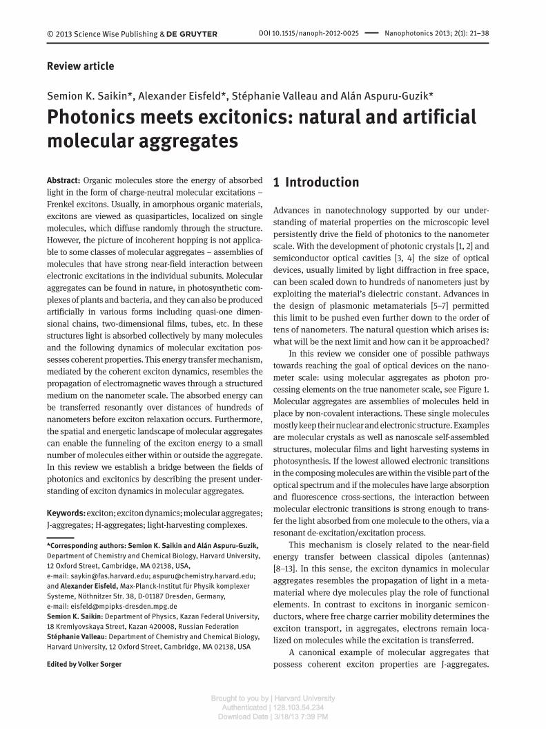
In this review we consider one of possible pathways
towards reaching the goal of optical devices on the nano-
meter scale: using molecular aggregates as photon pro-
cessing elements on the true nanometer scale, see Figure 1 .
Molecular aggregates are assemblies of molecules held in
place by non-covalent interactions. These single molecules
mostly keep their nuclear and electronic structure. Examples
are molecular crystals as well as nanoscale self-assembled
structures, molecular films and light harvesting systems in
photosynthesis. If the lowest allowed electronic transitions
in the composing molecules are within the visible part of the
optical spectrum and if the molecules have large absorption
and fluorescence cross-sections, the interaction between
molecular electronic transitions is strong enough to trans-
fer the light absorbed from one molecule to the others, via a
resonant de-excitation/excitation process.
This mechanism is closely related to the near-field
energy transfer between classical dipoles (antennas)
[ 8 – 13 ]. In this sense, the exciton dynamics in molecular
aggregates resembles the propagation of light in a meta-
material where dye molecules play the role of functional
elements. In contrast to excitons in inorganic semicon-
ductors, where free charge carrier mobility determines the
exciton transport, in aggregates, electrons remain loca-
lized on molecules while the excitation is transferred.
A canonical example of molecular aggregates that
possess coherent exciton properties are J-aggregates.
Brought to you by | Harvard UniversityAuthenticated | 128.103.54.234
Download Date | 3/18/13 7:39 PM

22 S.K. Saikin et al.: Photonics meets excitonics: natural and artificial © 2013 Science Wise Publishing &
These are aggregates of fluorescent molecules, discovered
about 80 years ago independently by Scheibe [ 14 , 15 ] and
Jelley [ 16 ]. They observed that the formation of aggre-
gates is accompanied by drastic changes in the optical
properties. The broad absorption line corresponding to
the molecular excitation is shifted to the red side of the
spectrum and becomes much narrower. Later, these types
of molecular aggregates were named J-aggregates after
Jelley. J-aggregates represent only one class of molecular
aggregates. For instance, there exist aggregates where the
absorption line is blue-shifted, so called H-aggregates.
Some aggregates exhibit several J-bands [ 17 ] and some
have both J-and H-bands [ 18 ]. In aggregates that consist
of non-equivalent monomers even more complicated
absorption structures can be found [ 19 ].
Molecular aggregates also appear as functional units
in nature. For instance, they form the absorbing and
energy transferring parts of light-harvesting complexes in
plants and some types of bacteria and algae [ 19 ]. In these
complexes the exciton absorbed by the antenna aggre-
gate has to be funneled to the reaction center – the part
where the exciton energy is used to create free charges to
be employed in a chemical reaction. The efficiency and
robustness of light absorption and exciton transfer in
light harvesting complexes may be crucial for the survival
of photosynthetic organisms under evolutionary pressure.
Shortly after the discovery of the self-assembled organic
dye aggregates the close connection of these artificial struc-
tures to the natural light harvesting systems was recognized
[ 20 , 21 ] and the exciton model of Frenkel [ 9 , 22 ] has been
used to explain the observed changes in optical properties
and the transfer of energy along the aggregate [ 21 ].
In Frenkel ’ s exciton theory, which is based on the
classical resonance interaction theory of Holtsmark [ 8 ],
the electronic excitation in the aggregate is not confined
to a single monomer, but it is coherently delocalized
over many monomers in the form of “ excitation waves ” .
Photonics Plasmonics
100 nm 10 nm 1 nm
Excitonics
Figure 1 Schematic illustration of the length scales character-
izing the different physical phenomena present the different fields
discussed in this review.
From superpositions of these excitation waves “ excita-
tion packets ” can be formed, which describe the coher-
ent motion of (localized) excitations [ 22 ]. Already in these
early works it was established that coupling to internal
and external vibrational modes, imperfections of the
aggregates and disorder induced by the environment
strongly influences the “ coherence size ” of the exciton
waves and modifies the optical and transport properties.
When the interaction with the environment (inter-
nal vibrations, solvent, etc.) is much stronger than the
resonant excitation transfer interaction, the excitation
becomes more or less localized on one monomer and the
transfer is no longer described by coherent exciton motion
but it becomes an incoherent hopping process. F ö rster
derived an elegant formula for the rate constants for trans-
port of excitation from one monomer to the other [ 23 , 24 ].
This rate is proportional to the overlap of the donor emis-
sion spectrum and the absorption spectrum of the accep-
tor molecule and depends on the inverse sixth power
of the distance between donor and acceptor. Typically,
in molecular aggregates, one is in a regime in-between
these two extreme cases, i.e., the transport is neither fully
coherent nor incoherent. This complicates the theoretical
modeling and the interpretation of experiments.
The remarkable optical and transport properties of
molecular aggregates have led to a variety of applications.
Right from the beginning, molecular aggregates were
employed as wavelength selective sensitizers in photo-
graphy [ 25 , 26 ]. Recent applications include the measure-
ment of membrane potentials [ 27 ] or the design of colorants
[ 28 ]. Some molecular aggregates form self-assembled supra-
molecular flexible fluorescent fibers [ 29 ] which may have
applications in thin-film optical and optoelectronic devices,
for example by employing optical bistability of J-aggregates
[ 30 ]. Molecular aggregates can be also utilized for sensing
applications. For instance, it has been demonstrated that a
large absorption cross section combined with fast exciton
diffusion may be used to enhance fluorescence from a small
number of dye molecules adsorbed or embedded in an
aggregate film [ 31 , 32 ]. Due to fast exciton diffusion within
the aggregate the excitons explore the aggregate. Once they
find a molecule with an electronic transition close to the
J-band the exciton can be transferred inelastically to the
adsorbed molecule. Moreover, dye aggregates might play
an important role in the development of efficient, low-cost
artificial light harvesting units (like organic solar cells) [ 33 ].
This review gives a brief, yet by no means complete,
overview of our experimental and theoretical under-
standing of excitons in molecular aggregates. Most of the
aspects in this work are covered at the advanced introduc-
tory level, and for a deeper understanding we refer to more
Brought to you by | Harvard UniversityAuthenticated | 128.103.54.234
Download Date | 3/18/13 7:39 PM

S.K. Saikin et al.: Photonics meets excitonics: natural and artificial 23© 2013 Science Wise Publishing &
detailed studies such as [ 19 , 34 – 38 ]. The review is struc-
tured as follows: In Section 2, we describe basic excitonic
properties of molecular aggregates starting with single
molecules and molecular dimers and then introducing
several examples of artificial and natural molecular aggre-
gates. In Section 3, we overview the main experimental
techniques that allow for probing the structural properties
of molecular aggregates and excitation dyna mics. Section
4 introduces the main theoretical approaches utilized for
modeling of excitons in aggregates. Section 5 shows how
molecular aggregates can be combined with photonic and
plasmonic structures. Finally, we conclude the review in
Section 6.
2 Basic properties of molecular aggregates
2.1 Properties of monomers
Molecules in the aggregates largely retain their electronic
and nuclear structure. Thus, it is natural to start our dis-
cussion with individual molecules interacting with light.
The interaction of a single molecule with light is schemati-
cally illustrated in Figure 2 . If the molecule has an opti-
cally allowed electronic transitions within the photon
spectrum it can be excited by absorbing light at the cor-
responding frequency. The transition time between the
ground and excited state of the molecule is of the order of
tens of attoseconds – the time that a single photon inter-
acts with a molecule. During this short time interval the
positions of the nuclei in the molecule are not changed.
Initially the molecule is in its ground state geometry, i.e.,
in a minimum of the electronic ground state potential | g ⟩
indicated by the blue wave-packet in Figure 2 . The equilib-
rium positions of atoms for the excited states, usually, are
different from those of the ground state. Therefore, after
the transition to the state | e ⟩ the molecule is in a transient
non-equilibrium state where molecular vibrational modes
are also excited (green wave-packet in Figure 2 ). Then, due
to the interaction of the molecule with its environment
the molecule relaxes towards the energetic minimum of
the excited state, i.e., the excited vibrational modes are
relaxed (wavy arrow and orange wavepacket in Figure 2 ).
This relaxation of the molecular geometry can also be
accompanied by a rearrangement of the environmental
molecules to a configuration with lower total energy. At
ambient conditions this process occurs over a timescale
of hundreds of femtoseconds to several picoseconds.
g
e
Vibrationalrelaxation
Non-radiative relaxation
FluroescenceAbsorptionE
nerg
y
Nuclear coordinate
Figure 2 Excitation/de-excitation processes in a single molecule
illustrated on a two-level molecular energy diagram. The parabolic
surfaces correspond to the ground state | g ⟩ and the first excited
| e ⟩ electronic states of the molecule. The horizontal axis indicates
the displacements of nuclei from their equilibrium positions. The
molecule, initially in the ground electronic and vibrational states,
is excited by absorbing light – blue arrow. During the absorption
process the positions of the nuclei are not changed. The light
absorption also induces molecular vibrations. Due to the
interaction with the environment the molecular vibrations are
equilibrated and the molecule relaxes to the bottom of the excited
electronic state – orange wavy arrow. Finally, the molecule relaxes
down to the ground electronic state by emitting a photon – red
arrow (fluorescence), or without photon emission – green dashed
arrow (non-radiative process). In the ground state the induced vibra-
tions are also equilibrated due to interaction with the environment.
Finally, on timescales of tens of picoseconds to nano-
seconds the molecule relaxes to its electronic ground state
either emitting a photon (fluorescence) or transferring
energy to other degrees of freedom, for instance vibrations
(non-radiative relaxation [ 39 ]). Usually, the absorption
and emission spectra exhibit a progression of peaks stem-
ming from the coupling to vibrational modes with high
energy (~150 meV). These peaks are broadened by the
same order of magnitude, due to coupling to a multitude
of low energy modes of the molecule and the surround-
ings. Emission takes place from the thermally relaxed
excited state, which is typically located in the low energy
wing of the absorption spectrum. The relaxation energy
is related to the energy difference between the maxima of
the absorption and emission spectra, the so-called Stokes
shift.
The absorption efficiency of a particular electronic
transition can often be characterized by the correspond-
ing transition dipole – a matrix element of a dipole opera-
tor between the ground and the excited molecular states.
Brought to you by | Harvard UniversityAuthenticated | 128.103.54.234
Download Date | 3/18/13 7:39 PM
24 S.K. Saikin et al.: Photonics meets excitonics: natural and artificial © 2013 Science Wise Publishing &
= | | .d e q r g⟨ ⋅ ⟩� �
(1)
Here and in the following we use an arrow symbol to indi-
cate three-dimensional vectors in real space. The dipole
operator is characterized by its charge q and the posi-
tion operator .r� The transition dipole is not a measur-
able value and is defined up to a complex phase factor
(if magnetic interactions can be neglected, which is often
the case, then the wavefunction can be chosen real and
d�
becomes a real vector). The absorption and emission
strengths of the respective transition are proportional to
2| | .d�
In the absorption spectra the transition frequency is
determined by the energy difference ε between the ground
and excited state.
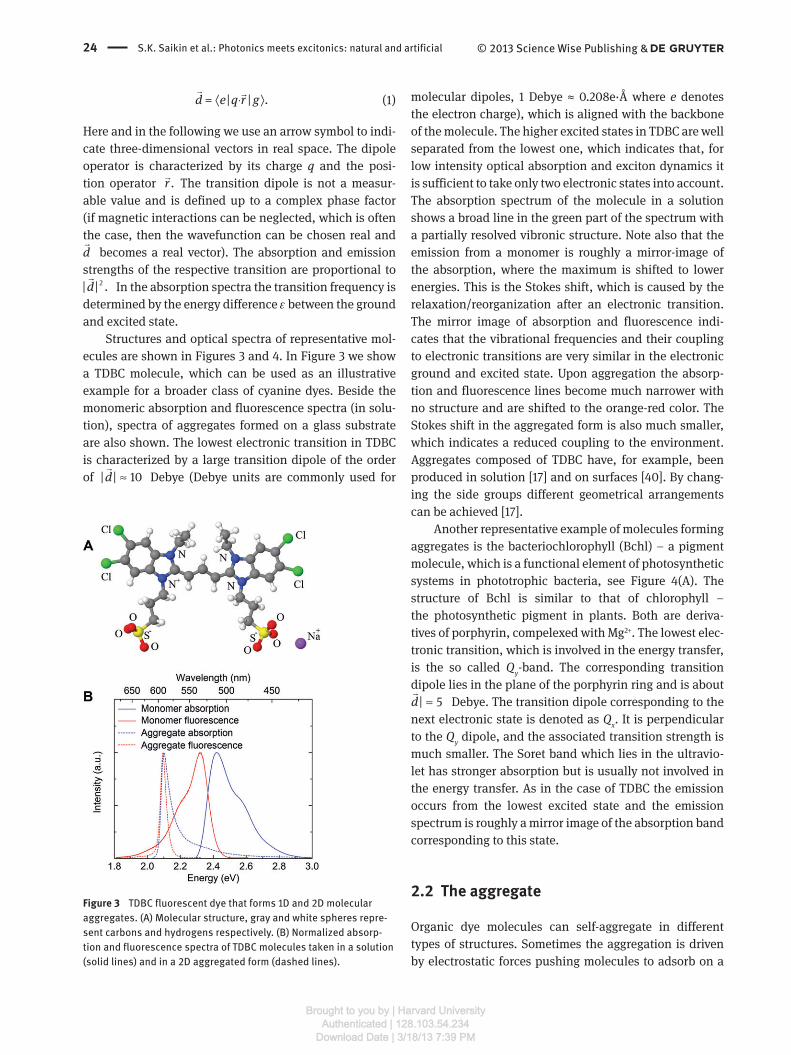
Structures and optical spectra of representative mol-
ecules are shown in Figures 3 and 4. In Figure 3 we show
a TDBC molecule, which can be used as an illustrative
example for a broader class of cyanine dyes. Beside the
monomeric absorption and fluorescence spectra (in solu-
tion), spectra of aggregates formed on a glass substrate
are also shown. The lowest electronic transition in TDBC
is characterized by a large transition dipole of the order
of | | 10d ≈�
Debye (Debye units are commonly used for
Figure 3 TDBC fluorescent dye that forms 1D and 2D molecular
aggregates. (A) Molecular structure, gray and white spheres repre-
sent carbons and hydrogens respectively. (B) Normalized absorp-
tion and fluorescence spectra of TDBC molecules taken in a solution
(solid lines) and in a 2D aggregated form (dashed lines).
molecular dipoles, 1 Debye ≈ 0.208e ‧ Å where e denotes
the electron charge), which is aligned with the backbone
of the molecule. The higher excited states in TDBC are well
separated from the lowest one, which indicates that, for
low intensity optical absorption and exciton dynamics it
is sufficient to take only two electronic states into account.
The absorption spectrum of the molecule in a solution
shows a broad line in the green part of the spectrum with
a partially resolved vibronic structure. Note also that the
emission from a monomer is roughly a mirror-image of
the absorption, where the maximum is shifted to lower
energies. This is the Stokes shift, which is caused by the
relaxation/reorganization after an electronic transition.
The mirror image of absorption and fluorescence indi-
cates that the vibrational frequencies and their coupling
to electronic transitions are very similar in the electronic
ground and excited state. Upon aggregation the absorp-
tion and fluorescence lines become much narrower with
no structure and are shifted to the orange-red color. The
Stokes shift in the aggregated form is also much smaller,
which indicates a reduced coupling to the environment.
Aggregates composed of TDBC have, for example, been
produced in solution [ 17 ] and on surfaces [ 40 ]. By chang-
ing the side groups different geometrical arrangements
can be achieved [ 17 ].
Another representative example of molecules forming
aggregates is the bacteriochlorophyll (Bchl) – a pigment
molecule, which is a functional element of photo synthetic
systems in phototrophic bacteria, see Figure 4 (A). The
structure of Bchl is similar to that of chlorophyll –
the photosynthetic pigment in plants. Both are deriva-
tives of porphyrin, compelexed with Mg 2 + . The lowest elec-
tronic transition, which is involved in the energy transfer,
is the so called Q y -band. The corresponding transition
dipole lies in the plane of the porphyrin ring and is about
| 5d ≈�
Debye. The transition dipole corresponding to the
next electronic state is denoted as Q x . It is perpendicular
to the Q y dipole, and the associated transition strength is
much smaller. The Soret band which lies in the ultravio-
let has stronger absorption but is usually not involved in
the energy transfer. As in the case of TDBC the emission
occurs from the lowest excited state and the emission
spectrum is roughly a mirror image of the absorption band
corresponding to this state.
2.2 The aggregate
Organic dye molecules can self-aggregate in different
types of structures. Sometimes the aggregation is driven
by electrostatic forces pushing molecules to adsorb on a
Brought to you by | Harvard UniversityAuthenticated | 128.103.54.234
Download Date | 3/18/13 7:39 PM
S.K. Saikin et al.: Photonics meets excitonics: natural and artificial 25© 2013 Science Wise Publishing &
surface; sometimes hydrophobic parts of the molecules
repel the water and tend to collect together such as in
synthetic tube aggregates [ 17 ].
A specific property of molecular aggregates with
strong inter-monomer couplings is that the absorption and
fluorescent spectra substantially differ from the spectra of
the molecules which form the aggregates while the elec-
tronic structure of the molecules is not modified. The
intermolecular distance in an aggregate is large enough
that the electron tunneling between different molecules
can be neglected. Therefore, only the Coulomb interac-
tion between the electronic transitions in monomers is
responsible for the spectral modifications and the inter-
molecular energy transfer. This interaction is similar to
the non-radiative near-field coupling between plasmonic
structures, which is mediated by virtual photons. It turns
out that the major contribution of the Coulomb interaction
in the spectra of aggregates is from the excitation transfer
via resonant transitions. We will refer to this main inter-
action term between the monomers as F ö rster coupling.
The F ö rster coupling, to a good approximation, can be
described by restricting to dipole transitions only (see
Figure 4 (A) Bacteriochlorophyll (Bchl) molecule – light absorbing
pigment that is a basic structural element of light-harvesting
complexes in phototrophic bacteria. Its structure is similar to
chlorophyll – the photosynthetic pigment in plants.; (B) Absorption
and fluorescent spectra of Bchl molecules.
Monomer 1 Monomer 2
Ene
rgy
e e
g g
Figure 5 Illustration of the excitation transfer due to resonant
near-field interaction between the state | e ⟩ | g ⟩ and | g ⟩ | e ⟩ .
below Eq. 2). The interaction is visualized in Figure 5 for
a situation in which initially monomer 1 is electronically
excited and monomer 2 is in the electronic ground state.
Then, in a resonant process molecule 1 is deexcited and
simultaneously the second monomer is excited. While this
time-ordered discrete picture is easy for visualization in
reality the exciton energy is transferred coherently. For
large distances between the monomers the interaction
between monomers 1 and 2 can be approximated by the
dipole-dipole term
12 1 2 1 12 2 123
12
1 ˆ ˆ= ( -3( )( )),V d d d R d RR
⋅ ⋅ ⋅� � � �
(2)
where R 12
denotes the distance between the monomers,
12R̂ is the corresponding direction vector, and the vectors
nd�
are the transition dipoles introduced in Eq. (1).
If the intermolecular distance is comparable with the
size of the molecules, then the interaction between the
two monomers can no longer be described adequately
using the point dipole-dipole interaction of Eq. (2).
Often, it is then sufficient to replace the point-dipoles in
Eq. (2) by extended dipoles [ 41 ]. For even higher accuracy
more elaborate schemes have been developed (see e.g.,
[ 42 ]).
Before discussing general molecular aggregates, let
us describe some basic results for the case of two coupled
identical monomers, ignoring nuclear degrees of freedom
for the moment. The eigenstates of the uncoupled mono-
mers can be taken as a basis. These states are | g ⟩ | g ⟩ , | e ⟩ | g ⟩ ,
| g ⟩ | e ⟩ , | e ⟩ | e ⟩ . While the states | g ⟩ | g ⟩ and | e ⟩ | e ⟩ are still suit-
able to describe the ground state and the doubly excited
state of the dimer, respectively, the two degenerate single
exciton states | e ⟩ | g ⟩ and | g ⟩ | e ⟩ are no longer eigenstates of
the coupled system, because of the resonant transfer inter-
action. The corresponding Hamiltonian is written as H = ε
1 | 1 ⟩ ⟨ 1 | + ε
2 | 2 ⟩ ⟨ 2 | + V
12 ( | 1 ⟩ ⟨ 2 | + | 2 ⟩ ⟨ 1 | ) (we choose the energy of the
monomer ground state as zero), where we have introduced
Brought to you by | Harvard UniversityAuthenticated | 128.103.54.234
Download Date | 3/18/13 7:39 PM

26 S.K. Saikin et al.: Photonics meets excitonics: natural and artificial © 2013 Science Wise Publishing &
the shorthand notation | 1 ⟩ ≡ | e ⟩ | g ⟩ and | 2 ⟩ ≡ | g ⟩ | e ⟩ or equiva-
lently in a matrix representation
1 12
12 2
= .V
H Vε
ε⎛ ⎞⎜ ⎟⎝ ⎠
(3)
For identical energies of excited states ε 1 = ε
2 ≡ ε
the eigenstates of the dimer are superpositions
1
| = (| | | | )2
e g e g±⟩ ⟩ ⟩± ⟩ ⟩ where the electronic excitation
is coherently delocalized over both monomers. The corre-
sponding eigenenergies are ε ± = ε ± V
12 . Note that the mag-
nitude and sign of the interaction depends sensitively on
the distance and orientation of the two monomers. Figure
6 illustrates how the relative orientations of molecular
transition dipoles change the frequency of electronic
excitations in a dimer. If the transition frequencies of the
involved monomers are equal and the transition dipoles
are parallel, only the transition between the ground and
the | + ⟩ state is optically allowed. It is detuned by the
energy V 12
from that of the non-interacting monomers.
From Eq. (2) one can see that for case (a), where the transi-
tion dipoles are orthogonal to the distance vector between
the monomers one has positive 2 3
12 12| | / .V d R∼ For the case
(b) the interaction is negative with 2 3
12 12-2 | | / .V d R∼ There-
fore, the alinement of the molecules shown in Figure 6 (A)
and (B) will be seen as a blue and red shift of the absorp-
tion line, respectively. As it has been noted before, the
states with only one excited monomer such as | g ⟩ | e ⟩ and
Figure 6 Interaction between electronic transitions dipoles (blue
arrows) of two molecules. The directions of the arrows reflect the
relative phase of the transition dipoles. For the considered
molecular alignments only one of the optical transitions is allowed
for the dimer (green arrows). (A) The optically allowed transition
of the dimer is shifted to the blue part of the spectrum, which is
similar to H-aggregation; (B) the optically allowed transition is
shifted to the red, which correspond to J-aggregation.
| e ⟩ | g ⟩ are not eigenstates of the system anymore. Thus, if
one of these state is populated initially, the energy will
oscillate between them back and forth coherently.
Let us briefly mention that the dimer often appears as
a first step of aggregation [ 43 ] and thus has been investi-
gated intensively (see e.g., [ 44 – 49 ]) . This interest in the
dimer is also due to the fact that many of the more realis-
tic models for aggregates where the environment and the
internal vibrations are included can only be solved effi-
ciently for the dimer.
The theoretical description of the dimer can easily be
extended to arbitrarily arranged monomers. As in the case
of the dimer the total ground state is taken as a product
of states where all monomers are in the ground state
a
e 1| = | ...| .ggl Ng g g⟩ ⟩ ⟩ We are interested in the properties of
aggregates in the linear absorption regime. Therefore, it is
sufficient to take into account only the states with at most
one electronic excitation on the aggregate, such as
| n ⟩ ≡ | g ⟩ 1 ... | e ⟩ n ... | g ⟩ N , (4)
in which monomer n is electronically excited and all other
monomers are in their electronic ground state. In this one-exciton manifold approximation the Hamiltonian can be
written as
=1
= | | | |N
en nm
n nmH n n V n mε ⟩⟨ + ⟩⟨∑ ∑
(5)
where V nm is the Coulomb dipole-dipole interaction, see
Eq. 2, which transfers excitation from monomer n to m .
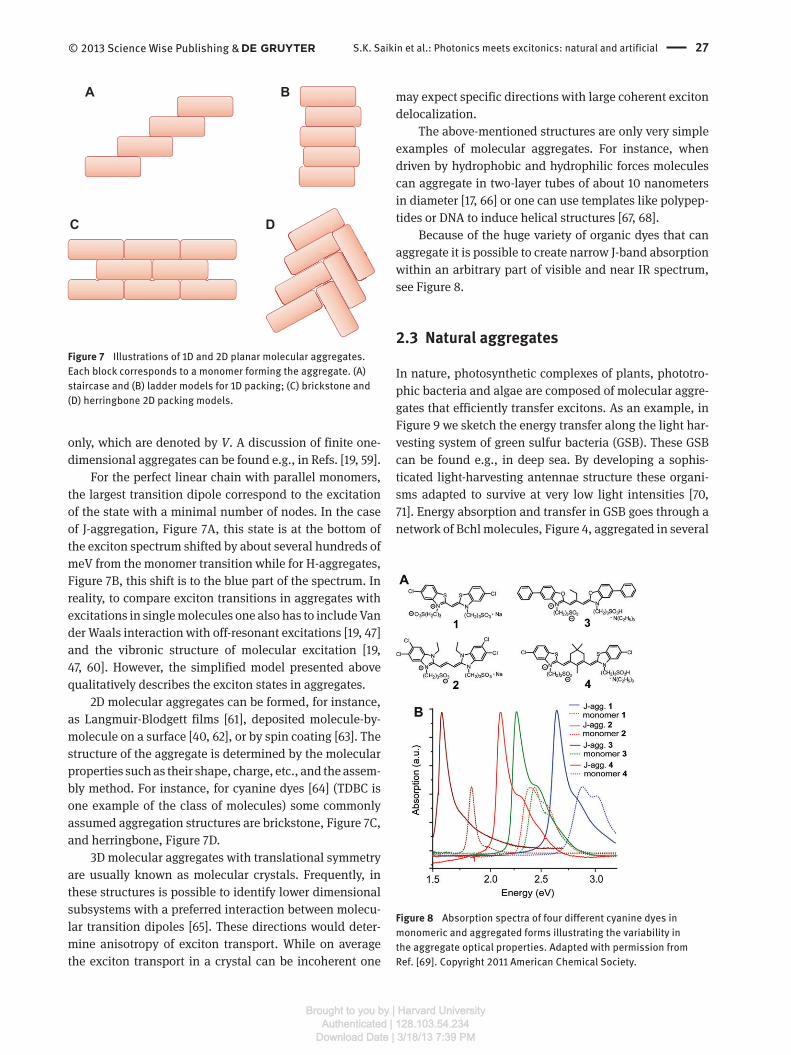
Examples of commonly discussed 1D and 2D planar
structures are shown in Figure 7 . While 1D models are well
studied theoretically (see e.g., Refs. [ 50 – 58 ]) and are fre-
quently used to characterize excitons in self-assembled
molecular aggregates, it is not clear whether ideal 1D
molecular chains are formed in experiments [ 34 ]. Usually,
they are subsystems of higher dimensional structures
such as films or crystals.
The 1D model of molecular aggregates is convenient
because many results can be obtained analytically [ 19 , 59 ].
For a perfect very long chain with N molecules the eigen-
states are well described by “ exciton waves ”
2
=1
1| = |
N i jnN
jn
e nN
π
φ ⟩ ⟩∑
(6)
with the corresponding eigenenergies
2= 2 cos .j
jE VNπ
ε ⎛ ⎞+ ⎝ ⎠
(7)
In the last equation, for the sake of simplicity, we have
considered the interactions between nearest neighbors
Brought to you by | Harvard UniversityAuthenticated | 128.103.54.234
Download Date | 3/18/13 7:39 PM
S.K. Saikin et al.: Photonics meets excitonics: natural and artificial 27© 2013 Science Wise Publishing &
only, which are denoted by V . A discussion of finite one-
dimensional aggregates can be found e.g., in Refs. [ 19 , 59 ].
For the perfect linear chain with parallel monomers,
the largest transition dipole correspond to the excitation
of the state with a minimal number of nodes. In the case
of J-aggregation, Figure 7 A, this state is at the bottom of
the exciton spectrum shifted by about several hundreds of
meV from the monomer transition while for H-aggregates,
Figure 7 B, this shift is to the blue part of the spectrum. In
reality, to compare exciton transitions in aggregates with
excitations in single molecules one also has to include Van
der Waals interaction with off-resonant excitations [ 19 , 47 ]
and the vibronic structure of molecular excitation [ 19 ,
47 , 60 ]. However, the simplified model presented above
qualitatively describes the exciton states in aggregates.
2D molecular aggregates can be formed, for instance,
as Langmuir-Blodgett films [ 61 ], deposited molecule-by-
molecule on a surface [ 40 , 62 ], or by spin coating [ 63 ]. The
structure of the aggregate is determined by the molecular
properties such as their shape, charge, etc., and the assem-
bly method. For instance, for cyanine dyes [ 64 ] (TDBC is
one example of the class of molecules) some commonly
assumed aggregation structures are brickstone, Figure 7 C,
and herringbone, Figure 7 D.
3D molecular aggregates with translational symmetry
are usually known as molecular crystals. Frequently, in
these structures is possible to identify lower dimensional
subsystems with a preferred interaction between molecu-
lar transition dipoles [ 65 ]. These directions would deter-
mine anisotropy of exciton transport. While on average
the exciton transport in a crystal can be incoherent one
A B
C D
Figure 7 Illustrations of 1D and 2D planar molecular aggregates.
Each block corresponds to a monomer forming the aggregate. (A)
staircase and (B) ladder models for 1D packing; (C) brickstone and
(D) herringbone 2D packing models.
Figure 8 Absorption spectra of four different cyanine dyes in
monomeric and aggregated forms illustrating the variability in
the aggregate optical properties. Adapted with permission from
Ref. [ 69 ]. Copyright 2011 American Chemical Society.
may expect specific directions with large coherent exciton
delocalization.
The above-mentioned structures are only very simple
examples of molecular aggregates. For instance, when
driven by hydrophobic and hydrophilic forces molecules
can aggregate in two-layer tubes of about 10 nanometers
in dia meter [ 17 , 66 ] or one can use templates like polypep-
tides or DNA to induce helical structures [ 67 , 68 ].
Because of the huge variety of organic dyes that can
aggregate it is possible to create narrow J-band absorption
within an arbitrary part of visible and near IR spectrum,
see Figure 8 .
2.3 Natural aggregates
In nature, photosynthetic complexes of plants, phototro-
phic bacteria and algae are composed of molecular aggre-
gates that efficiently transfer excitons. As an example, in
Figure 9 we sketch the energy transfer along the light har-
vesting system of green sulfur bacteria (GSB). These GSB
can be found e.g., in deep sea. By developing a sophis-
ticated light-harvesting antennae structure these organi-
sms adapted to survive at very low light intensities [ 70 ,
71 ]. Energy absorption and transfer in GSB goes through a
network of Bchl molecules, Figure 4 , aggregated in several
Brought to you by | Harvard UniversityAuthenticated | 128.103.54.234
Download Date | 3/18/13 7:39 PM
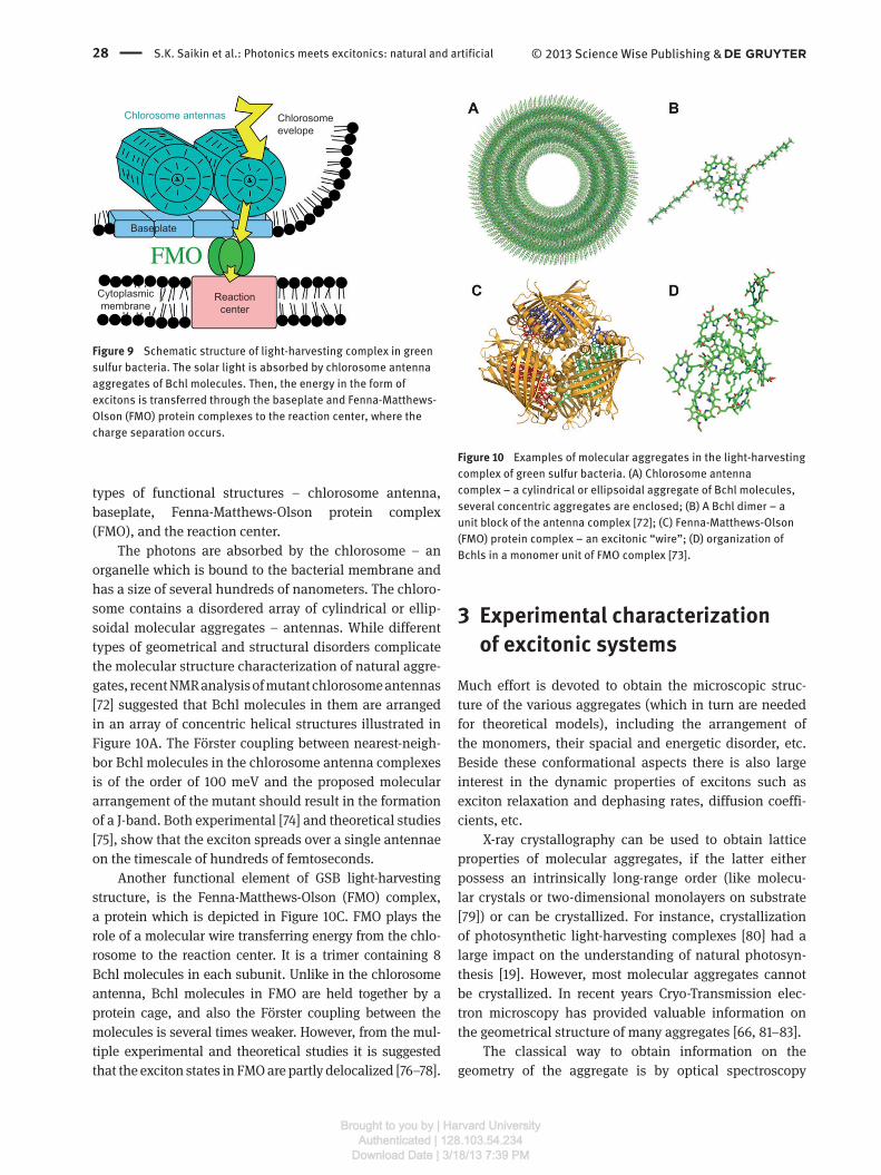
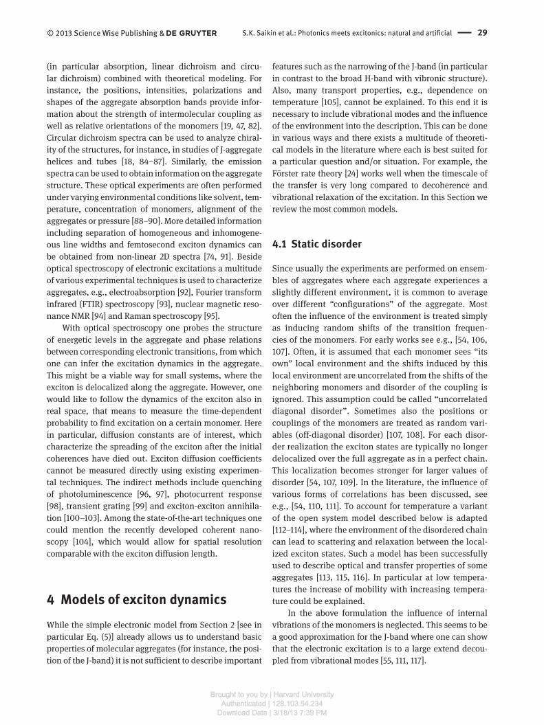
28 S.K. Saikin et al.: Photonics meets excitonics: natural and artificial © 2013 Science Wise Publishing &
types of functional structures – chlorosome antenna,
baseplate, Fenna-Matthews-Olson protein complex
(FMO), and the reaction center.
The photons are absorbed by the chlorosome – an
organelle which is bound to the bacterial membrane and
has a size of several hundreds of nanometers. The chloro-
some contains a disordered array of cylindrical or ellip-
soidal molecular aggregates – antennas. While different
types of geometrical and structural disorders complicate
the molecular structure characterization of natural aggre-
gates, recent NMR analysis of mutant chlorosome antennas
[ 72 ] suggested that Bchl molecules in them are arranged
in an array of concentric helical structures illustrated in
Figure 10 A. The F ö rster coupling between nearest-neigh-
bor Bchl molecules in the chlorosome antenna complexes
is of the order of 100 meV and the proposed molecular
arrangement of the mutant should result in the formation
of a J-band. Both experimental [ 74 ] and theoretical studies
[ 75 ], show that the exciton spreads over a single antennae
on the timescale of hundreds of femtoseconds.
Another functional element of GSB light-harvesting
structure, is the Fenna-Matthews-Olson (FMO) complex,
a protein which is depicted in Figure 10 C. FMO plays the
role of a molecular wire transferring energy from the chlo-
rosome to the reaction center. It is a trimer containing 8
Bchl molecules in each subunit. Unlike in the chlorosome
antenna, Bchl molecules in FMO are held together by a
protein cage, and also the F ö rster coupling between the
molecules is several times weaker. However, from the mul-
tiple experimental and theoretical studies it is suggested
that the exciton states in FMO are partly delocalized [ 76 – 78 ].
3 Experimental characterization of excitonic systems
Much effort is devoted to obtain the microscopic struc-
ture of the various aggregates (which in turn are needed
for theoretical models), including the arrangement of
the monomers, their spacial and energetic disorder, etc.
Beside these conformational aspects there is also large
interest in the dynamic properties of excitons such as
exciton relaxation and dephasing rates, diffusion coeffi-
cients, etc.
X-ray crystallography can be used to obtain lattice
properties of molecular aggregates, if the latter either
possess an intrinsically long-range order (like molecu-
lar crystals or two-dimensional monolayers on substrate
[ 79 ]) or can be crystallized. For instance, crystallization
of photosynthetic light-harvesting complexes [ 80 ] had a
large impact on the understanding of natural photosyn-
thesis [ 19 ]. However, most molecular aggregates cannot
be crystallized. In recent years Cryo-Transmission elec-
tron microscopy has provided valuable information on
the geometrical structure of many aggregates [ 66 , 81 – 83 ].
The classical way to obtain information on the
geometry of the aggregate is by optical spectroscopy
Figure 10 Examples of molecular aggregates in the light-harvesting
complex of green sulfur bacteria. (A) Chlorosome antenna
complex – a cylindrical or ellipsoidal aggregate of Bchl molecules,
several concentric aggregates are enclosed; (B) A Bchl dimer – a
unit block of the antenna complex [ 72 ]; (C) Fenna-Matthews-Olson
(FMO) protein complex – an excitonic “ wire ” ; (D) organization of
Bchls in a monomer unit of FMO complex [ 73 ].
Chlorosomeevelope
Baseplate
Cytoplasmicmembrane
Reactioncenter
Chlorosome antennas
Figure 9 Schematic structure of light-harvesting complex in green
sulfur bacteria. The solar light is absorbed by chlorosome antenna
aggregates of Bchl molecules. Then, the energy in the form of
excitons is transferred through the baseplate and Fenna-Matthews-
Olson (FMO) protein complexes to the reaction center, where the
charge separation occurs.
Brought to you by | Harvard UniversityAuthenticated | 128.103.54.234
Download Date | 3/18/13 7:39 PM
S.K. Saikin et al.: Photonics meets excitonics: natural and artificial 29© 2013 Science Wise Publishing &
(in particular absorption, linear dichroism and circu-
lar dichroism) combined with theoretical modeling. For
instance, the positions, intensities, polarizations and
shapes of the aggregate absorption bands provide infor-
mation about the strength of intermolecular coupling as
well as relative orientations of the monomers [ 19 , 47 , 82 ].
Circular dichroism spectra can be used to analyze chiral-
ity of the structures, for instance, in studies of J-aggregate
helices and tubes [ 18 , 84 – 87 ]. Similarly, the emission
spectra can be used to obtain information on the aggregate
structure. These optical experiments are often performed
under varying environmental conditions like solvent, tem-
perature, concentration of monomers, alignment of the
aggregates or pressure [ 88 – 90 ]. More detailed information
including separation of homogeneous and inhomogene-
ous line widths and femtosecond exciton dynamics can
be obtained from non-linear 2D spectra [ 74 , 91 ]. Beside
optical spectroscopy of electronic excitations a multitude
of various experimental techniques is used to characterize
aggregates, e.g., electroabsorption [ 92 ], Fourier transform
infrared (FTIR) spectroscopy [ 93 ], nuclear magnetic reso-
nance NMR [ 94 ] and Raman spectroscopy [ 95 ].
With optical spectroscopy one probes the structure
of energetic levels in the aggregate and phase relations
between corresponding electronic transitions, from which
one can infer the excitation dynamics in the aggregate.
This might be a viable way for small systems, where the
exciton is delocalized along the aggregate. However, one
would like to follow the dynamics of the exciton also in
real space, that means to measure the time-dependent
probability to find excitation on a certain monomer. Here
in particular, diffusion constants are of interest, which
characterize the spreading of the exciton after the initial
coherences have died out. Exciton diffusion coefficients
cannot be measured directly using existing experimen-
tal techniques. The indirect methods include quenching
of photoluminescence [ 96 , 97 ], photocurrent response
[ 98 ], transient grating [ 99 ] and exciton-exciton annihila-
tion [ 100 – 103 ]. Among the state-of-the-art techniques one
could mention the recently developed coherent nano-
scopy [ 104 ], which would allow for spatial resolution
comparable with the exciton diffusion length.
4 Models of exciton dynamics While the simple electronic model from Section 2 [see in
particular Eq. (5)] already allows us to understand basic
properties of molecular aggregates (for instance, the posi-
tion of the J-band) it is not sufficient to describe important
features such as the narrowing of the J-band (in particular
in contrast to the broad H-band with vibronic structure).
Also, many transport properties, e.g., dependence on
temperature [ 105 ], cannot be explained. To this end it is
necessary to include vibrational modes and the influence
of the environment into the description. This can be done
in various ways and there exists a multitude of theoreti-
cal models in the literature where each is best suited for
a particular question and/or situation. For example, the
F ö rster rate theory [ 24 ] works well when the timescale of
the transfer is very long compared to decoherence and
vibrational relaxation of the excitation. In this Section we
review the most common models.
4.1 Static disorder
Since usually the experiments are performed on ensem-
bles of aggregates where each aggregate experiences a
slightly different environment, it is common to average
over different “ configurations ” of the aggregate. Most
often the influence of the environment is treated simply
as inducing random shifts of the transition frequen-
cies of the monomers. For early works see e.g., [ 54 , 106 ,
107 ]. Often, it is assumed that each monomer sees “ its
own ” local environment and the shifts induced by this
local environment are uncorrelated from the shifts of the
neighboring monomers and disorder of the coupling is
ignored. This assumption could be called “ uncorrelated
diagonal disorder ” . Sometimes also the positions or
couplings of the monomers are treated as random vari-
ables (off-diagonal disorder) [ 107 , 108 ]. For each disor-
der realization the exciton states are typically no longer
delocalized over the full aggregate as in a perfect chain.
This localization becomes stronger for larger values of
disorder [ 54 , 107 , 109 ]. In the literature, the influence of
various forms of correlations has been discussed, see
e.g., [ 54 , 110 , 111 ]. To account for temperature a variant
of the open system model described below is adapted
[ 112 – 114 ], where the environment of the disordered chain
can lead to scattering and relaxation between the local-
ized exciton states. Such a model has been successfully
used to describe optical and transfer properties of some
aggregates [ 113 , 115 , 116 ]. In particular at low tempera-
tures the increase of mobility with increasing tempera-
ture could be explained.
In the above formulation the influence of internal
vibrations of the monomers is neglected. This seems to be
a good approximation for the J-band where one can show
that the electronic excitation is to a large extend decou-
pled from vibrational modes [ 55 , 111 , 117 ].
Brought to you by | Harvard UniversityAuthenticated | 128.103.54.234
Download Date | 3/18/13 7:39 PM
30 S.K. Saikin et al.: Photonics meets excitonics: natural and artificial © 2013 Science Wise Publishing &
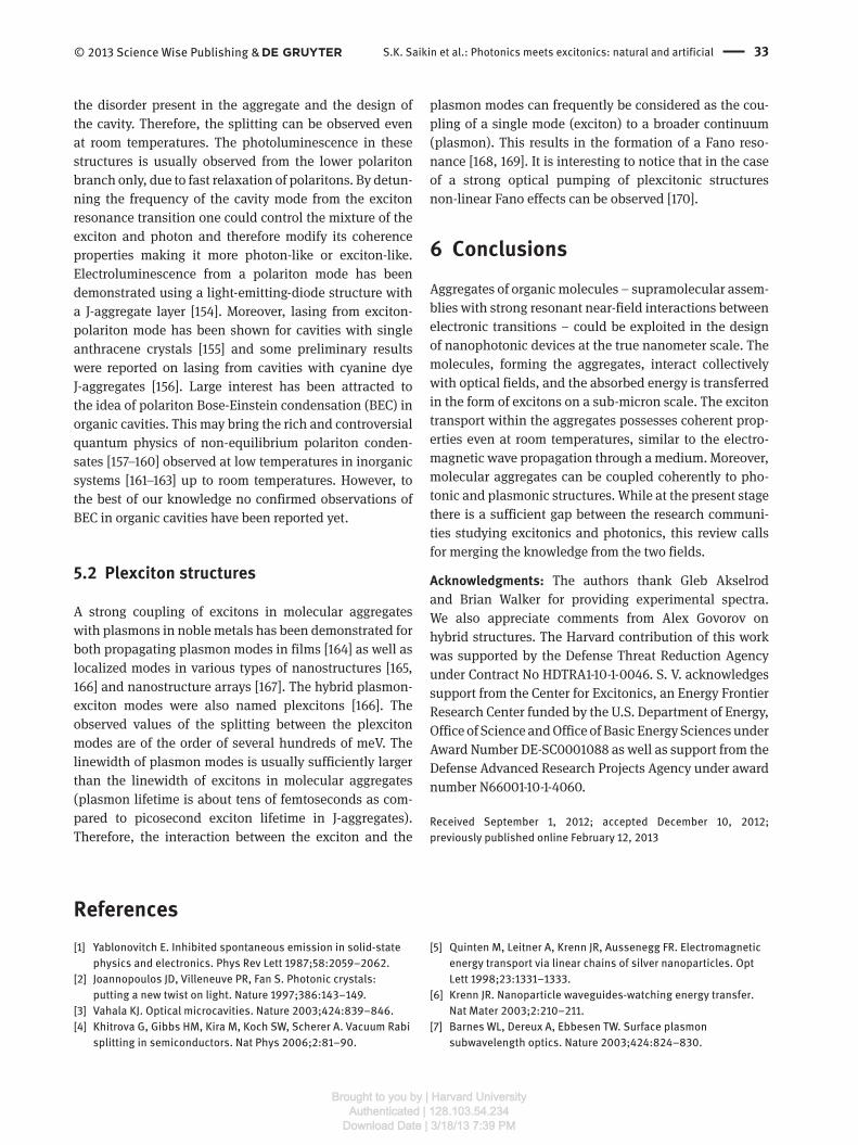
4.2 Dynamic disorder, Haken-Strobel-Reineker model
Fluctuations of the monomer transition frequencies or
the intermolecular couplings that are fast compared to
the exciton transfer time-scale are usually referred to as
dynamic disorder. Dynamical fluctuations play a dual
role in exciton transport [ 118 – 120 ]. In structures with
large static disorder excitons are localized. Dynamic fluc-
tuations in this case remove localization. However, if the
fluctuations are strong this results in the dynamical locali-
zation of the exciton. A seminal model to treat these fast
fluctuations is due to Haken, Strobl and Reineker [ 121 – 123 ],
where the dynamical fluctuations are described by real sto-
chastic processes, ε n ( t ) and V nm ( t ) and modeled using the
density matrix [ 121 ] or stochastic Schr ö dinger [ 124 ] equa-
tions. In the original work the stochastic processes have
been chosen as white noise, i.e., delta-correlated in time.
This model has been shown to capture coherent and inco-
herent excitation transfer on the same footing. Moreover,
one should notice that the equation describing Haken-
Strobl-Reineker (HRS) model for 1D systems is equivalent
to telegrapher ’ s equation, which further reflects the simi-
larity between the exciton propagation in molecular aggre-
gates and a wave propagation in a medium.
The Haken-Strobl-Reineker model, with several
extensions, has been utilized to study exciton dynamics in
natural and artificial molecular aggregates [ 120 , 123 – 127 ].
The drawback of this model is that the exciton population
does not relax to thermal equilibrium. There have been
many investigations and extensions which have included
colored noise and tried to cure the thermalization issue,
e.g., [ 128 , 129 ]. The original model also does not include
the effect of strongly coupled high frequency modes of
the monomers which is essential to describe the vibronic
structure present in many H-aggregates. The recent devel-
opment of so-called quantum state diffusion methods
[ 130 , 131 ] can be considered as an extension of the HSR
model which cures these deficiencies at the cost of colored
complex noise.
Figure 11 A illustrates how the HRS model can be
applied to exciton propagation on a brickstone lattice
of TDBC dyes at ambient conditions [ 124 ]. The second
moment of the exciton wavefunction, Figure 11 B shows
that the initial propagation on the time scale of 20 – 30 fs is
ballistic or wave-like, which is associated with the quad-
ratic dependence of the second moment on time. For the
longer timescale the time dependence is linear, which
characterizes a diffusive motion.
4.3 The vibronic model
Let us first consider a single monomer, where only the two
lowest electronic states are taken into account, see Figure
2 . In the Born-Oppenheimer approximation [ 132 ] we write
the nuclear Hamiltonian of the electronic ground and
excited states as
/ 2 /1= ( ( ))
2
g e g en nj n nj
jH P U Q+∑
(8)
where Q nj are the (mass-scaled) nuclear coordinates of
monomer n and P nj are the corresponding momenta.
Figure 11 (A) Time dynamics of exciton wave function on a 2D lattice of TDBC molecules subject to static disorder (linewidth σ = 70 meV)
and dynamic fluctuations (linewidth Γ = 30 meV) of molecular electronic transitions. At time zero the exciton is localized in the center of
the lattice. The contour plots show the population of lattice sites at four different times. (B) Second moments of the wave function cor-
responding to the exciton dynamics shown in (A). Two different transport regimes can be observed: the ballistic or a wave-like propagation
on timescales of 20 – 30 fs ( M (2) ~ t 2 ) and the diffusive motion ( M (2) ~ t ) at longer times. Reprinted with permission from Ref. [ 124 ]. Copyright
2012, American Institute of Physics.
Brought to you by | Harvard UniversityAuthenticated | 128.103.54.234
Download Date | 3/18/13 7:39 PM

S.K. Saikin et al.: Photonics meets excitonics: natural and artificial 31© 2013 Science Wise Publishing &
U g / e ( Q nj ) is the Born-Oppenheimer potential in the elec-
tronic ground/excited state (see Figure 2 ).
Then, for a molecular aggregate the Hamiltonian in the
electronic ground state is given by ( ) el el=1= | | .
Ng gnn
H H g g⟩⟨∑
We denote by V nm the transition dipole-dipole interac-
tion between monomer n and m , which, for simplicity,
is taken to be independent of nuclear coordinates. The
Hamiltonian in the one-exciton manifold, Eq. (4), is then
given by
=1 , =1
= | | | |,N N
en nm
n n mH W n n V n m⟩⟨ + ⟩⟨∑ ∑
(9)
with the “ collective ” Born-Oppenheimer surfaces
=Ne g
n n mm nW H H
≠+∑ . Note that the structure of Eq. (9) is
very similar to the purely electronic Hamiltonian (5). The
only difference is that now the energies ε n are replaced by
operators for the nuclear motion. Upon transfer of exci-
tation the nuclear wavepacket, according to Figure 2 , is
no longer in its equilibrium position. Thus the excitation
transfer is linked to excitation of nuclear motion. This
vibrational model is sometimes combined with the static
disorder approach [ 111 , 133 , 134 ]. The vibrational model
offers a clear idea regarding why the J-band does not
exhibit vibrational structure and the H-band does.
The coupling to vibrations usually slows down the
propagation of the exciton, however, it can help to over-
come energetic barriers caused by differences in the elec-
tronic transition energies. In this way efficient directed
transport along a biased chain can be achieved.
Often it is justified to approximate the Born-Oppen-
heimer potentials, Eq. (8), as harmonic potentials where
the surface of the electronically excited state is just
shifted relative to that of the ground electronic state,
i.e., we have 2 2 2
=1
1= ( )
2
Mgn nj nj njj
H P Qω+∑ and for the excited
electronic state 2 2 2
=1
1= ( ( - ) )
2
Men n nj nj nj njj
H P Q Qε ω+ + Δ∑
where ε n denotes the energy difference between the
minima of the upper and lower potential energy surface
and the vibrational frequency of mode j is denoted by
ω nj and the shift between the minima of the excited and
ground state harmonic potential is denoted by Δ Q nj . The
potential surfaces are sketched in Figure 2 for the case
of one mode. This harmonic approximation is widely
employed in the literature, e.g., [ 46 , 55 , 134 – 138 ].
4.4 Open system approaches
Open system approaches are closely related to the
vibronic model discussed in the previous subsection. In
these approaches one typically considers an infinite set
of harmonic oscillators forming the bath. It is easy to
see that the Hamiltonian (9) can be written as H e = H sys +
H sys – bath + H bath with H sys given by the purely electronic part
(5) with transition energies ε n that already include static
overall shifts induced by the environment. The bath is
taken as b = gath nn
H H∑ and the system-bath coupling is
described by a linear coupling of the electronic excita-
tion of a monomer to the set of vibrational modes, i.e.,
s = | | .ys bath n m nj njn jH Qπ π κ− ⟩⟨ ⊗∑ ∑ � The coupling constant
njκ� describes the coupling of the excitation on monomer
n to the harmonic oscillator with frequency ω nj . Typi-
cally, the frequency spectrum of bath oscillators is taken
to be continuous so that the coupling constant becomes
a continuous function of frequency. The open system
approaches have been used to describe optical and trans-
fer properties of light harvesting systems [ 139 , 140 ] and
can also be applied to explain properties of organic dye
aggregates [ 141 ]. We note that the HRS model can be con-
sidered as a special case of the open system models for
which the bath is Markovian (i.e., memoryless).
4.5 Mixed QM/MM
With the increase of computer capabilities, in recent years
it has become possible to simulate aggregates starting from
a microscopic description [ 142 – 145 ]. Since a full quantum
mechanical treatment is still out of reach, one uses mixed
quantum classical approaches. In these approaches usually
the nuclei are propagated classically in the electronic ground
state using molecular dynamics. Along these nuclear tra-
jectories one calculates the (time-dependent) transition
energies using quantum chemistry methods. These time-
dependent transition energies can then be used to obtain
input parameters for open system models or to use them
directly as the energy fluctuations in HSR model. However,
both approaches have their problems, since one is restricted
to a classical propagation in the electronic ground state.
4.6 Semi-empirical approach based on polarizabilities
While the approaches discussed above start from a micro-
scopic description of the exciton-phonon coupling and
the environment, in the semi-empirical approaches one
can take the standpoint that all the relevant information
of the coupling of an electronic transition to other degrees
of freedom is already contained in the shape of the absorp-
tion spectrum. The absorption spectrum in turn is linked
to the polarizabilities of the monomers.
Brought to you by | Harvard UniversityAuthenticated | 128.103.54.234
Download Date | 3/18/13 7:39 PM

32 S.K. Saikin et al.: Photonics meets excitonics: natural and artificial © 2013 Science Wise Publishing &
The induced electric moment of a monomer depends on
the local field at that monomer, which is produced by the
electric moments of all the other monomers and the external
field. This leads to a set of coupled equations from which one
can extract, for a given arrangement of the monomers, the
polarizability of the aggregate and thus the optical proper-
ties. Such equations have been derived using classical treat-
ments [ 10 ] or quantum approaches [ 117 , 146 ]. This method
implicitly assumes that the frequency dependent polariz-
ability does not change (beside an overall shift in energy)
when going from the monomer to the aggregate. Recently,
it has been demonstrated that using measured monomer
spectra as input one can accurately describe the band-shape
of J- and H-aggregates [ 60 , 147 ]. This approach also gives an
intuitive explanation of the vibrational structure and the
broadenings of the absorption bands of the aggregate.
4.7 Beyond the single-exciton approximation
Photonic device applications may require structures oper-
ating in a nonlinear response regime, where interactions
between excitons cannot be neglected. For example,
thin J-aggregate films switches based on optical bista-
bility have been suggested in [ 30 ]. At higher intensities,
however, an additional loss-channel – exciton-exciton
annihilation – appears. The underlying physical process
is similar to the Auger effect, followed by a fast non-radi-
ative energy conversion. Within an approximate picture
this can be viewed as a process by which two excitons
localized on neighboring molecules are combined into a
higher-energy electronic excitation of a single molecule.
Then, the resulting excitation quickly decays to the lowest
excited state or ground state dissipating the exceeding
energy through vibrations. A detailed kinematic model
for the exciton-exciton annihilation in molecular crystals
of different dimensions has been introduced by Suna in
[ 148 ], and a similar master equation approach for exciton
annihilation dynamics in natural photosynthetic com-
plexes has been suggested in [ 149 ]. While the microscopic
approaches discussed above are still valid, the molecular
Hamiltonian (5) should be extended to account for at least
two excited states per each monomer [ 38 , 150 ].
5 Hybrid excitonic structures In this section we specifically focus on J-aggregates.
Their large absorption and fluorescence cross-sections
combined with a narrow line width and a small Stokes shift
allow for coherent coupling between excitons and photons
in optical cavities or excitons and plasmon modes in metal
structures. This property can be utilized as an interface
between photonic, plasmonic and excitonic circuits.
5.1 Exciton-polariton structures
Most of the studies of strong coupling between excitons in
J-aggreagtes and photons have been done using organic
microcavities [ 151 – 154 ]. A schematic illustration of an
organic microcavity – a planar λ /2 cavity with a J-aggre-
gate layer embedded in it – is shown in Figure 12 A. Strong
coupling of cavity photons with excitons in a J-aggregate
result in formation of exciton-polariton modes. This is
associated with the splitting of a cavity mode – vacuum
Rabi splitting, see Figure 12 B.
Within a simple model assuming only one exciton
state and a single photon mode the value of the Rabi split-
ting, Ω R , is just
R X v= ,acd EΩ ⋅� �
(10)
where Xd�
is the transition dipole of the narrow exciton
transition, vacE�
is the vacuum field in the position
where the J-aggregate is located, and we assume that the
vacuum field does not vary substantially on the scale of
the aggregate. In J-aggregates the value of the exciton
transition dipole scales as with the number N * of coher-
ently coupled molecules involved *
X .d N∼�
The value of
N * is usually much smaller than N – the total number of
molecules in the aggregate, since the environment leads
to localization of the exciton states. The value of the Rabi
splitting observed for J-aggregates strongly coupled with
optical cavities ranges from several tens of meV [ 151 , 152 ]
to several hundreds of meV [ 102 , 153 , 154 ] depending on
Figure 12 (A) Structure of a planar λ /2 organic microcavity with
a thin layer of J-aggregates embedded. (B) An example of a cavity
photon and a J-aggregate exciton energy level anticrossing.
Reprinted with permission from Ref. [ 102 ]. Copyright 2010 by the
American Physical Society.
Brought to you by | Harvard UniversityAuthenticated | 128.103.54.234
Download Date | 3/18/13 7:39 PM
S.K. Saikin et al.: Photonics meets excitonics: natural and artificial 33© 2013 Science Wise Publishing &
the disorder present in the aggregate and the design of
the cavity. Therefore, the splitting can be observed even
at room temperatures. The photoluminescence in these
structures is usually observed from the lower polariton
branch only, due to fast relaxation of polaritons. By detun-
ning the frequency of the cavity mode from the exciton
resonance transition one could control the mixture of the
exciton and photon and therefore modify its coherence
properties making it more photon-like or exciton-like.
Electroluminescence from a polariton mode has been
demonstrated using a light-emitting-diode structure with
a J-aggregate layer [ 154 ]. Moreover, lasing from exciton-
polariton mode has been shown for cavities with single
anthracene crystals [ 155 ] and some preliminary results
were reported on lasing from cavities with cyanine dye
J-aggregates [ 156 ]. Large interest has been attracted to
the idea of polariton Bose-Einstein condensation (BEC) in
organic cavities. This may bring the rich and controversial
quantum physics of non-equilibrium polariton conden-
sates [ 157 – 160 ] observed at low temperatures in inorganic
systems [ 161 – 163 ] up to room temperatures. However, to
the best of our knowledge no confirmed observations of
BEC in organic cavities have been reported yet.
5.2 Plexciton structures
A strong coupling of excitons in molecular aggregates
with plasmons in noble metals has been demonstrated for
both propagating plasmon modes in films [ 164 ] as well as
localized modes in various types of nanostructures [ 165 ,
166 ] and nanostructure arrays [ 167 ]. The hybrid plasmon-
exciton modes were also named plexcitons [ 166 ]. The
observed values of the splitting between the plexciton
modes are of the order of several hundreds of meV. The
linewidth of plasmon modes is usually sufficiently larger
than the linewidth of excitons in molecular aggregates
(plasmon lifetime is about tens of femtoseconds as com-
pared to picosecond exciton lifetime in J-aggregates).
Therefore, the interaction between the exciton and the
plasmon modes can frequently be considered as the cou-
pling of a single mode (exciton) to a broader continuum
(plasmon). This results in the formation of a Fano reso-
nance [ 168 , 169 ]. It is interesting to notice that in the case
of a strong optical pumping of plexcitonic structures
non-linear Fano effects can be observed [ 170 ].
6 Conclusions Aggregates of organic molecules – supramolecular assem-
blies with strong resonant near-field interactions between
electronic transitions – could be exploited in the design
of nanophotonic devices at the true nanometer scale. The
molecules, forming the aggregates, interact collectively
with optical fields, and the absorbed energy is transferred
in the form of excitons on a sub-micron scale. The exciton
transport within the aggregates possesses coherent prop-
erties even at room temperatures, similar to the electro-
magnetic wave propagation through a medium. Moreover,
molecular aggregates can be coupled coherently to pho-
tonic and plasmonic structures. While at the present stage
there is a sufficient gap between the research communi-
ties studying excitonics and photonics, this review calls
for merging the knowledge from the two fields.
Acknowledgments: The authors thank Gleb Akselrod
and Brian Walker for providing experimental spectra.
We also appreciate comments from Alex Govorov on
hybrid structures. The Harvard contribution of this work
was supported by the Defense Threat Reduction Agency
under Contract No HDTRA1-10-1-0046. S. V. acknowledges
support from the Center for Excitonics, an Energy Frontier
Research Center funded by the U.S. Department of Energy,
Office of Science and Office of Basic Energy Sciences under
Award Number DE-SC0001088 as well as support from the
Defense Advanced Research Projects Agency under award
number N66001-10-1-4060.
Received September 1, 2012; accepted December 10, 2012;
previously published online February 12, 2013
References [1] Yablonovitch E. Inhibited spontaneous emission in solid-state
physics and electronics. Phys Rev Lett 1987;58:2059 – 2062.
[2] Joannopoulos JD, Villeneuve PR, Fan S. Photonic crystals:
putting a new twist on light. Nature 1997;386:143 – 149.
[3] Vahala KJ. Optical microcavities. Nature 2003;424:839 – 846.
[4] Khitrova G, Gibbs HM, Kira M, Koch SW, Scherer A. Vacuum Rabi
splitting in semiconductors. Nat Phys 2006;2:81 – 90.
[5] Quinten M, Leitner A, Krenn JR, Aussenegg FR. Electromagnetic
energy transport via linear chains of silver nanoparticles. Opt
Lett 1998;23:1331 – 1333.
[6] Krenn JR. Nanoparticle waveguides-watching energy transfer.
Nat Mater 2003;2:210 – 211.
[7] Barnes WL, Dereux A, Ebbesen TW. Surface plasmon
subwavelength optics. Nature 2003;424:824 – 830.
Brought to you by | Harvard UniversityAuthenticated | 128.103.54.234
Download Date | 3/18/13 7:39 PM

34 S.K. Saikin et al.: Photonics meets excitonics: natural and artificial © 2013 Science Wise Publishing &
[8] Holtsmark J. Ü ber die Absorption in Na-Dampf. Z Physik A
1925;34:722 – 729.
[9] Frenkel J. Zur Theorie der Resonanzverbreiterung von
Spektrallinien. Z Phys A 1930;59:198 – 207.
[10] DeVoe H. Optical properties of molecular aggregates. I.
Classical model of electronic absorption and refraction. J Chem
Phys 1964;41:393 – 400.
[11] Philpott MR. Some modern aspects of exciton theory. Adv
Chem Phys 1973;23:227 – 341.
[12] Zimanyi EN, Silbey RJ. Unified treatment of coherent and
incoherent electronic energy transfer dynamics using classical
electrodynamics. J Chem Phys 2010;133:144107, pages 1 – 10.
[13] Briggs JS, Eisfeld A. Equivalence of quantum and classical
coherence in electronic energy transfer. Phys Rev E
2011;83:051911, pages 1 – 4.
[14] Scheibe G. Ü ber die Ver ä nderlichkeit des Absorptions-
spektrums einiger Sensibilisierungsfarbstoffe und deren
Ursache. Angew Chem 1936;49:563.
[15] Scheibe G. Ü ber die Ver ä nderlichkeit der Absorptionsspektren
in L ö sungen und die Nebenvalenzen als ihre Ursache. Angew
Chem 1937;50:212 – 219.
[16] Jelley EE. Spectral absorption and fluorescence of dyes in the
molecular state. Nature 1936;138:1009 – 1010.
[17] Kirstein S, Daehne S. J-aggregates of amphiphilic cyanine
dyes: self-organization of artificial light harvesting complexes.
Int J Photoenergy 2006;20363, pages 1–21.
[18] Norden B. Linear and circular-dichroism of polymeric pseudo-
isocyanine. J Phys Chem 1977;81:151 – 159.
[19] van Amerongen H, Valkunas L, van Grondelle R. Photosynthetic
excitons. World Scientific, Singapore 2000.
[20] Scheibe G. Ü ber den Mechanismus der Sensibilisierung
photochemischer Reaktionen durch Farbstoffe, insbesondere
der Assimilation. Naturwissenschaften 1937;49:795.
[21] Franck J, Teller E. Migration and photochemical action of
excitation energy in crystals. J Chem Phys 1938;6:861 – 872.
[22] Frenkel J. On the transformation of light into heat in solids.
I. Phys Rev 1931;37:17 – 44.
[23] F ö rster T. Zwischenmolekulare Energiewanderung und
Fluoreszenz. Ann Phys (Leipzig) 1948;437:55 – 75.
[24] F ö rster T. Delocalized excitation and excitation transfer; in
Sinanoğlu, editor, Modern Quantum Chemistry; chapter III B 1,
pages 93 – 137; Academic Press 1965.
[25] Tani T. J-aggregates in spectral sensitization of photographic
materials; in T. Kobayashi, editor, J-Aggregates. World
Scientific 1996.
[26] Takechi K, Sudeep PK, Kamat PV. Harvesting infrared
photons with tricarbocyanine dye clusters. J Phys Chem B
2006;110:16169 – 16173.
[27] Reers M, Smith TW, Chen LB. J-aggregate formation of a
carbocyanine as a quantitative fluorescent indicator of
membrane-potential. Biochemistry 1991;30:4480 – 4486.
[28] Horn D, Rieger J. Organic nanoparticles in the aqueous
phase-theory, experiment, and use. Angew Chem Int Ed
2001;40:4331 – 4361.
[29] Higgins DA, Kerimo J, Vanden Bout DA, Barbara PF. A molecular
yarn: near-field optical studies of self-assembled, flexible,
fluorescent fibers. J Am Chem Soc 1996;118:4049 – 4058.
[30] Malyshev VA, Glaeske H, Feller KH. Intrinsic optical bistablility
of an ultrathin film consisting of oriented linear aggregates.
J Chem Phys 2000;113:1170 – 1176.
[31] M ö bius D, Kuhn H. Energy transfer in monolayers with cyanine
dye Scheibe aggregates, J Appl Phys 1988;64:5138 – 5141.
[32] Akselrod GM, Walker BJ, Tisdale WA, Bawedi MG, Bulovi ć V.
Twenty-fold enhancement of molecular fluoresence by coupling
to a J-aggredate critically coupled resonator. ACS Nano
2012;6:467 – 471.
[33] D ä hne S. Nanostrukturierte J-Aggregate als potenzielle
Lichtsammelsysteme f ü r Photosynthesen. Bunsen-Magazin
2002;4:81 – 92.
[34] W ü rthner F, Kaise TE, Saha-M ö ller CR. J-aggregates: from
serendipitous discovery to supramolecular engineering
of functional dye materials. Angew Chem Int Ed 2011;50:
3376 – 3410.
[35] Davydov A. Theory of molecular excitons. McGraw-Hill 1962.
[36] Kobayashi T, editor. J-aggregates. World Scientific 1996.
[37] Scholes GD, Rumbles G. Excitons in nanoscale systems. Nat
Mater 2006;5:683 – 696.
[38] K ü hn O, Lochbrunner S. Quantum efficiency in complex
systems, part II: from molecular aggregates to organic
solar cells; Semiconductors and Semimetals 85; chapter
Quantum Dynamics and Spectroscopy of Excitons in Molecular
Aggregates, pages 47 – 81; Academic Press, San Diego (2011).
[39] Medvedev ES, Osherov VI. Radiationless transitions in
polyatomic molecules. volume 57; Springer-Verlag 1995.
[40] Bradley M, Tischler J, Bulović V. Layer-by-layer J-aggregate thin
films with a peak absorption constant of 106 wavenumbers.
Adv Mater 2005;17:1881 – 1886.
[41] Czikklely V, Forsterling HD, Kuhn H. Extended dipole model for
aggregates of dye molecules. Chem Phys Lett 1970;6:207 – 210.
[42] Krueger BP, Scholes GD, Fleming GR. Calculation of couplings
and energy-transfer pathways between the pigments of LH2
by the ab-initio transition density cube method. J Phys Chem B
1998;102:5378 – 5386.
[43] Kopainsky B, Hallermeier JK, Kaiser W. The first step of
aggregation of PIC: the dimerization. Chem Phys Lett
1981;83:498 – 502.
[44] Fulton RL, Gouterman M. Vibronic coupling. II. Spectra of
dimers. J Chem Phys 1964;41:2280 – 2286.
[45] K ü hn O, Renger T, May V. Theory of exciton-vibrational
dynamics in molecular dimers. Chem Phys 1996;204:99 – 114.
[46] Seibt J, Lohr A, W ü rthner F, Engel V. Circular dichroism and
absorption spectroscopy of merocyanine dimer aggregates:
molecular properties and exciton transfer dynamics from
time-dependent quantum calculations. Phys Chem Phys
2007;9:6214 – 6218.
[47] Eisfeld A. A simple method to obtain information on the
conformation of dipole-dipole coupled dimers. Chem Phys Lett
2007;445:321 – 324.
[48] Womick JM, Moran AM. Exciton coherence and energy
transport in the light-harvesting dimers of allophycocyanin.
J Phys Chem B 2009;113:15747 – 15759.
[49] Guthmuller J, Zutterman F, Champagne B. Multimode simulation
of dimer absorption spectra from first principles calculations:
application to the 3,4,9,10-perylenetetracarboxylic diimide
dimer. J Chem Phys 2009;131:154302, pages 1 – 8.
[50] Moffitt W, Fitts DD, Kirkwood JG. Critique of the theory of
optical activity of helical polymers. Proc Natl Acad Sci USA
1957;43:723 – 730.
[51] Holstein T. Studies of polaron motion: part II. The “ small ”
polaron. Ann Phys (NY) 1959;8:343 – 389.
Brought to you by | Harvard UniversityAuthenticated | 128.103.54.234
Download Date | 3/18/13 7:39 PM
S.K. Saikin et al.: Photonics meets excitonics: natural and artificial 35© 2013 Science Wise Publishing &
[52] Hofelich F. Die Bewegung eines Exzitons entlang eines
Polymers unter dem Einfluβ der Gitterschwingungen. Z Phys B
1966;5:208.
[53] Bierman A. Exciton wave packet localization on an impurity. J
Chem Phys 1970;52:4987 – 4995.
[54] Knapp EW. Lineshapes of molecular aggregates, exchange
narrowing and intersite correlation. Chem Phys 1984;85:73 – 82.
[55] Scherer POJ, Fischer SF. On the theory of vibronic structure of
linear aggregates. Application to pseudoisocyanin (PIC). Chem
Phys 1984;86:269.
[56] Spano FC. Fermion excited states in one-dimensional
molecular aggregates with site disorder: nonlinear optical
response. Phys Rev Lett 1991;67:3424 – 3427.
[57] Knoester J. Nonlinear optical line shapes of disordered
molecular aggregates: motional narrowing and the effect of
intersite correlations. J Chem Phys 1993;99:8466 – 8479.
[58] Hoffmann M, Schmidt K, Fritz T, Hasche T, Agranovich VM,
Leo K. The lowest energy Frenkel and charge-transfer excitons
in quasi-one-dimensional structures: application to MePTCDI
and PTCDA crystals. Chem Phys 2000;258:73 – 96.
[59] May V, K ü hn O. Charge and energy transfer dynamics in
molecular systems. WILEY-VCH 2000.
[60] Eisfeld A, Briggs JS. The J- and H-bands of organic dye
aggregates. Chem Phys 2006;324:376 – 384.
[61] Pavinatto F, Gameiro A Jr., Hidalgo A, Dinelli L, Romualdo L,
Batista A, Neto NB, Ferreira M, Oliveira O Jr. Langmuir and
Langmuir-Blodgett (LB) films of tetrapyridyl metalloporphyrins.
Appl Surf Sci 2008;254:5946 – 5952.
[62] Proehl H, Nitsche R, Dienel T, Leo K, Fritz T. In situ differential
reflectance spectroscopy of thin crystalline films of PTCDA on
different substrates. Phys Rev B 2005;71:165207, pages 1 – 14.
[63] Misawa K, Ono H, Minoshima K, Kobayashi T. New fabrication
method for highly oriented J aggregates dispersed in polymer
films. Appl Phys Lett 1993;63:577 – 579.
[64] Mishra A, Behera RK, Behera PK, Mishra BK, Behera GB.
Cyanines during the 1990s: a review. Chem Rev
2000;100:1973 – 2011.
[65] Schwoerer M, Wolf H. Organic molecular solids. Wiley-VCH
2006.
[66] Eisele DM, Cone CW, Bloemsma EA, Vlaming SM, van der
Kwaak CGF, Silbey RJ, Bawendi MG, Knoester J, Rabe JP, Vanden
Bout DA. Utilizing redox-chemistry to elucidate the nature
of exciton transitions in supramolecular dye nanotubes. Nat
Chem 2012;4:655 – 662.
[67] Stryer L, Blout ER. Optical rotatory dispersion of dyes bound
to macromoleculescationic dyespolyglutamic acid complexes.
J Am Chem Soc 1961;83:1411 – 1418.
[68] Seifert JL, Connor RE, Kushon SA, Wang M, Armitage BA.
Spontaneous assembly of helical cyanine dye aggregates on
DNA nanotemplates. J Am Chem Soc 1999;121:2987 – 2995.
[69] Walker BJ, Dorn A, Bulov í V, Bawendi MG. Color-selective
photocurrent enhancement in coupled J-aggregate/nanowires
formed in solution. Nano Lett 2011;11:2655 – 2659.
[70] Overmann J, Cypionka H, Pfennig N. An extremely low-light-
adapted phototrophic sulfur bacterium from the Black Sea.
Limnol Oceanogr 1992;37:150 – 155.
[71] Beatty JT, Overmann J, Lince MT, Manske AK, Lang AS,
Blankenship RE, Dover CLV, Martinson TA, Plumley FG. An
obligately photosynthetic bacterial anaerobe from a deep-sea
hydrothermal vent. Proc Natl Acad Sci USA 2005;102:9306 – 9310.
[72] Ganapathy S, Oostergetel GT, Wawrzyniak PK, Reus M, Chew
AGM, Buda F, Boekema EJ, Bryant DA, Holzwarth AR, de Groot
HJM. Alternating syn-anti bacteriochlorophylls form concentric
helical nanotubes in chlorosomes. Proc Natl Acad Sci USA
2009;106:8525 – 8530.
[73] Tronrud DE, Wen J, Gay L, Blankenship RE. The structural basis
for the difference in absorbance spectra for the FMO antenna
protein from various green sulfur bacteria. Photosynth Res
2009;100:79 – 87.
[74] Dostal J, Mancal T, Augulis R, Vacha F, Psencik J, Zigmantas
D. Two-dimensional electronic spectroscopy reveals
ultrafast energy diffusion in chlorosomes. J Am Chem Soc
2012;134:11611 – 11617.
[75] Fujita T, Brookes JC, Saikin SK, Aspuru-Guzik A. Memory-
assisted exciton diffusion in the chlorosome light-harvesting
antenna of green sulfur bacteria. J Phys Chem Lett
2012;3:2357 – 2361.
[76] Milder M, Br ü ggemann B, van Grondelle R, Herek J. Revisiting
the optical properties of the FMO protein. Photosynth Res
2010;104:257 – 274.
[77] Renger T, May V. Ultrafast exciton motion in photosynthetic
antenna systems: the FMO-Complex. J Phys Chem A
1998;102:4381 – 4391.
[78] Adolphs J, Renger T. How proteins trigger excitation energy
transfer in the FMO complex of green sulfur bacteria. Biophys J
2006;91:2778 – 2797.
[79] M ü ller M, Ikonomov J, Sokolowski M. Structure of epitaxial
layers of KCl on Ag(100). Surf Sci 2011;605:1090 – 1094.
[80] Olson JM. The FMO protein. Photosynth Res 2004;80:181 – 187.
[81] von Berlepsch H, B ö ttcher C, Ouart A, Burger C, D ä hne S, Kirstein
S. Supramolecular structures of J-aggregates of carbocyanine
dyes in solution. J Phys Chem B 2000;104:5255 – 5262.
[82] Vlaming SM, Augulis R, Stuart MCA, Knoester J, van Loosdrecht
PHM. Exciton spectra and the microscopic structure of
self-assembled porphyrin nanotubes. J Phys Chem B
2009;113:2273 – 2283.
[83] Oostergetel GT, Reus M, Chew AGM, Bryant DA, Boekema EJ,
Holzwarth AR. FEBS Lett 2007;581:5435 – 5439.
[84] Spitz C, D ä hne S, Ouart A, Abraham H-W. Proof of chirality of
J-aggregates spontaneously and enantioselectively generated
from achiral dyes. J Phys Chem B 2000;104:8664 – 8669.
[85] Didraga C, Klugkist JA, Knoester J. Optical properties of helical
cylindrical molecular aggregates: the homogeneous limit. J
Phys Chem B 2002;106:11474 – 11486.
[86] Didraga C, Pug ž lys A, Hania PR, von Berlepsch H, Duppen K,
Knoester J. Structure, spectroscopy, and microscopic model
of tubular carbocyanine dye aggregates. J Phys Chem B
2004;108:14976 – 14985.
[87] Eisfeld A, Kniprath R, Briggs J. Theory of the absorption and
circular dichroism spectra of helical molecular aggregates. J
Chem Phys 2007;126:104904, pages 1 –13 .
[88] Renge I, Wild UP. Solvent, temperature, and excitonic effects
in the optical spectra of pseudoisocyanine monomer and
J-aggregates. J Phys Chem A 1997;101:7977 – 7988.
[89] Lindrum M, Chan IY. High pressure investigation of absorption
spectra of J-aggregates. J Chem Phys 1996;104:5359 – 5364.
[90] Kangur L, Leiger K, Freiberg A. Evidence for high-pressure-
induced rupture of hydrogen bonds in LH2 photosynthetic
antenna pigment-protein complexes. J Phys Conf Ser
2008;121:112004, pages 1 – 5.
Brought to you by | Harvard UniversityAuthenticated | 128.103.54.234
Download Date | 3/18/13 7:39 PM
36 S.K. Saikin et al.: Photonics meets excitonics: natural and artificial © 2013 Science Wise Publishing &
[91] Stiopkin I, Brixner T, Yang M, Fleming GR. Heterogeneous
exciton dynamics revealed by two-dimensional optical
spectroscopy. J Phys Chem B 2006;110:20032 – 20037.
[92] Luchowski R, Krawczyk S. Stark effect spectroscopy of
exciton states in the dimer of acridine orange. Chem Phys
2003;293:155 – 166.
[93] Ilharco LM, de Barros RB. Aggregation of pseudoisocyanine
iodide in cellulose acetate films: structural characterization
by FTIR. Langmuir 2000;16:9331 – 9337.
[94] van Rossum BJ, Steensgaard DB, Mulder FM,
Boender GJ, Schaffner K, Holzwarth AR, de Groot HJM. A
refined model of the chlorosomal antennae of the green
bacterium Chlorobium tepidum from proton chemical shift
constraints obtained with high-field 2-D and 3-D MAS NMR
dipolar correlation spectroscopy. Biochem 2001;40:
1587 – 1595.
[95] Leng WN, W ü rthner F, Kelley AM. Resonance Raman intensity
analysis of merocyanine dimers in solution. J Phys Chem B
2004;108:10284 – 10294.
[96] Lyons B, Monkman A. The role of exciton diffusion in energy
transfer between polyfluorene and tetraphenyl porphyrin.
Phys Rev B 2005;71:235201, pages 1 – 5.
[97] Lunt RR, Giebink NC, Belak AA, Benziger JB, Forrest SR.
Exciton diffusion lengths of organic semiconductor thin
films measured by spectrally resolved photoluminescence
quenching. J Appl Phys 2009;105:053711, pages 1 – 7.
[98] Ghosh AK, Feng T. Merocynanine organic solar cells. J Appl
Phys 1978;49:5982 – 5989.
[99] Salcedo J, Siegman A, Dlott D, Fayer M. Dynamics of energy
transport in molecular crystals: the picosecond transient-
grating method. Phys Rev Lett 1978;41:131 – 134.
[100] Moll J, Harrison WJ, Brumbaugh DV, Muenter AA. Exciton
annihilation in J-aggregates probed by femtosecond
fluorescence upconversion. J Phys Chem A 2000;104:
8847 – 8854.
[101] Shaw PE, Ruseckas A, Peet J, Bazan GC, Samuel IDW.
Exciton-exciton annihilation in mixed-phase polyfluorene
films. Adv Funct Mater 2010;20:155 – 161.
[102] Akselrod G, Tischler Y, Young E, Nocera D, Bulovic V.
Exciton-exciton annihilation in organic polariton
microcavities. Phys Rev B 2010;82:113106, pages 1 – 4.
[103] Marciniak H, Li X-Q, W ü rthner F, Lochbrunner S.
One-dimensional exciton diffusion in perylene bisimide
aggregates. J Phys Chem A 2011;115:648 – 654.
[104] Aeschlimann M, Brixner T, Fischer A, Kramer C, Melchior P,
Pfeiffer W, Schneider C, Str ü ber C, Tuchscherer P, Voronine
DV. Coherent two-dimensional nanoscopy. Science
2011;333:1723 – 1726.
[105] Scheblykin IG. Temperature dependence of exciton transport
in J-aggredates; in J-aggregates. World Scientific 2012.
[106] Schreiber M, Toyozawa Y. Numerical experiments on
the absorption lineshape of the exciton under lattice-
vibrations. I. The overall lineshape. J Phys Soc Jap
1982;51:1528 – 1536.
[107] Fidder H, Knoester J, Wiersma DA. Optical properties of
disordered molecular aggregates: a numerical study. J Chem
Phys 1991;95:7880 – 7890.
[108] Malyshev VA, Dominguez-Adame F. Motional narrowing effect
in one-dimensional Frenkel chains with configurational
disorder. Chem Phys Lett 1999;313:255 – 260.
[109] Malyshev VA. Localization length of a 1-D exciton and
temperature dependence of the radiative lifetime in frozen dye
solutions with J-aggregates. Opt Spectrosc 1991;71:505 – 506.
[110] Dom í nguez-Adame F, Malyshev VA. Frenkel excitons in
one-dimensional systems with correlated disorder. J Lumin
1999;83 – 4:61 – 67.
[111] Walczak P, Eisfeld A, Briggs JS. Exchange narrowing of
the J-band of molecular dye aggregates. J Chem Phys
2008;128:044505, pages 1 – 12.
[112] Heijs DJ, Malyshev VA, Knoester J. Decoherence of excitons
in multichromophore systems: thermal line broadening
and destruction of superradiant emission. Phys Rev Lett
2005;95:177402, pages 1 – 4.
[113] Bednarz M, Malyshev VA, Knoester J. Temperature dependent
fluorescence in disordered frenkel chains: interplay of equili-
bration and local band-edge level structure. Phys Rev Lett
2003;91:217401, pages 1 – 4.
[114] Vlaming SM, Malyshev VA, Knoester J. Nonmonotonic energy
harvesting efficiency in biased exciton chains. J Chem Phys
2007;127:154719, pages 1 – 8.
[115] Tomioka A, Miyano K. Numerical study of excitons in
a two-dimensional organic dye aggregate. Phys Rev B
1996;54:2963 – 2967.
[116] Gallos LK, Pimenov AV, Scheblykin IG, Van der Auweraer M,
Hungerford G, Varnavsky OP, Vitukhnovsky AG, Argyrakis
P. A kinetic model for J-aggregate dynamics. J Phys Chem B
2000;104:3918 – 3923.
[117] Briggs JS, Herzenberg A. Bandshapes in polymer spectra. Mol
Phys 1971;21:865 – 879.
[118] Logan DE, Wolynes PG. Localizability and dephasing of
dipolar excitons in topologically disordered systems. J Chem
Phys 1987;87:7199 – 7207.
[119] Evensky DA, Scalettar RT, Wolynes PG. Localization
and dephasing effects in a time-dependent Anderson
Hamiltonian. J Phys Chem 1990;94:1149 – 1154.
[120] Rebentrost P, Mohseni M, Kassal I, Lloyd S, Aspuru-Guzik A.
Environment-assisted quantum transport. New J Phys
2009;11:033003, pages 1 – 12.
[121] Haken H, Reineker P. The coupled coherent and incoherent
motion of excitons and its influence on the line shape of
optical absorption. Z Phys 1972;249:253 – 268.
[122] Haken H, Strobl G. An exactly solvable model for coherent and
incoherent exciton motion. Z Phys 1973;262:135 – 148.
[123] Reineker P, Krenke VM. Exciton dynamics in molecular
crystals and aggregates. Volume 94 of Springer tracts in
modern physics. Springer Verlag 1982.
[124] Valleau S, Saikin SK, Yung M-H, Aspuru-Guzik A. Exciton
transport in thin-film cyanine dye J-aggregates. J Chem Phys
2012;137:034109, pages 1 – 13.
[125] Madhukar A, Post W. Exact solution for the diffusion of a
particle in a medium with site diagonal and off-diagonal
dynamic disorder. Phys Rev Lett 1977;39:1424 – 1427.
[126] Plenio MB, Huelga SF. Dephasing-assisted transport:
quantum networks and biomolecules. New J Phys
2008;10:113019, pages 1 – 14.
[127] Eisfeld A, Briggs JS. Classical master equation for excitonic
transport under the influence of an environment. Phys Rev E
2012;85:046118.
[128] Č á pek V. Generalized Haken-Strobl-Reineker model of
excitation transfer. Z Phys B 1985;60:101 – 105.
Brought to you by | Harvard UniversityAuthenticated | 128.103.54.234
Download Date | 3/18/13 7:39 PM

S.K. Saikin et al.: Photonics meets excitonics: natural and artificial 37© 2013 Science Wise Publishing &
[129] Rahman TS, Knox RS, Kenkre VM. Theory of depolarization of
fluorescence in molecular pairs. Chem Phys 1979;44:197 – 211.
[130] Di ó si L, Strunz WT. The non-Markovian stochastic Schr ö dinger
equation for open systems. Phys Lett A 1997;235:569 – 573.
[131] Roden J, Eisfeld A, Wolff W, Strunz WT. Influence of
complex exciton-phonon coupling on optical absorption
and energy transfer of quantum aggregates. Phys Rev Lett
2009;103:058301, pages 1 – 4.
[132] Azumi T, Matsuzaki K. What does the term “ Vibronic
Coupling ” Mean? Photochem Photobiol 1977;25:315 – 326.
[133] Spano FC. Temperature dependent emission in disordered
herringbone aggregates of conjugated oligomers. Phys Rev B
2005;71:235208, pages 1 – 13.
[134] van Dijk L, Spano FC, Bobbert PA. Theory of exciton dynamics
in molecular aggregates in presence of polaronic effects.
Chem Phys Lett 2012;529:69 – 73.
[135] Merrifield RE. Theory of the vibrational structure of molecular
excition states. J Chem Phys 1964;40:445 – 450.
[136] Perrin MH, Gouterman M. Vibronic coupling. IV. Trimers and
trigonal molecules. J Chem Phys 1967;46:1019 – 1028.
[137] Eisfeld A, Braun L, Strunz WT, Briggs JS, Beck J, Engel V.
Vibronic energies and spectra of molecular dimers. J Chem
Phys 2005;122:134103, pages 1 – 10.
[138] Seibt J, Winkler T, Renziehausen K, Dehm V, W ü rthner F, Meyer
H-D, Engel V. Vibronic transitions and quantum dynamics
in molecular oligomers: a theoretical analysis with an
application to aggregates of perylene bisimides. J Phys Chem
A 2009;113:13475 – 13482.
[139] Ishizaki A, Fleming GR. Unified treatment of quantum
coherent and incoherent hopping dynamics in electronic
energy transfer: reduced hierarchy equation approach.
J Chem Phys 2009;130:234111, pages 1 – 10.
[140] Moix J, Wu J, Huo P, Coker D, Cao J. Efficient energy transfer
in light-harvesting systems, III: The influence of the eighth
bacteriochlorophyll on the dynamics and efficiency in FMO.
J Phys Chem Lett 2011;2:3045 – 3052.
[141] Roden J, Strunz WT, Eisfeld A. Spectral properties of molecular
oligomers. A non-Markovian quantum state diffusion
approach. Int J Mod Phys B 2010;24:5060 – 5067.
[142] Schulten K, Tesch M. Coupling of protein motion to electron
transfer: molecular dynamics and stochastic quantum
mechanics study of photosynthetic reaction centers. Chem
Phys 1991;158:421 – 446.
[143] Damjanovi ć A, Kosztin I, Kleinekath ö fer U, Schulten K.
Excitons in a photosynthetic light-harvesting system: a
combined molecular dynamics, quantum chemistry, and
polaron model study. Phys Rev E 2002;65:031919, pages
1 – 24.
[144] Olbrich C, Jansen TLC, Liebers J, Aghtar M, Str ü mpfer J,
Schulten K, Knoester J, Kleinekath ö fer U. From atomistic
modeling to excitation transfer and two-dimensional
spectra of the FMO light-harvesting complex. J Phys Chem B
2011;115:8609 – 8621.
[145] Shim S, Rebentrost P, Valleau S, Aspuru-Guzik A. Atomistic
study of the long-lived quantum coherences in the
Fenna-Matthews-Olson complex. Biophys J 2012;102:
649 – 660.
[146] McLachlan AD, Ball MA. Hypochromism and optical rotation in
helical polymers. Mol Phys 1964;8:581 – 595.
[147] Eisfeld A, Briggs JS. The shape of the J-band of pseudoiso-
cyanine. Chem Phys Lett 2007;446:354 – 358.
[148] Suna A. Kinematics of exciton-exciton annihilation in
molecular crystals. Phys Rev B 1970;1:1716 – 1739.
[149] Paillotin G, Swenberg CE, Breton J, Geacintov NE. Analysis
of picosecond laser-induced fluorescence phenomena in
photosynthetic membranes utilizing a master equation
approach. Biophys J 1979;25:513 – 533.
[150] Knoester J, Spano FC. Unusual behavior of two-photon
absorption from three-level molecules in a one-dimensional
lattice. Phys Rev Lett 1995;74:2780 – 2783.
[151] Lidzey DG, Bradley DDC, Virgili T, Armitage A, Skolnick MS,
Walker S. Room temperature polariton emission from strongly
coupled organic semiconductor microcavities. Phys Rev Lett
1999;82:3316 – 3319.
[152] Schouwink P, Berlepsch HV, D ä hne L, Mahrt RF. Observation
of strong exciton-photon coupling in an organic microcavity.
Chem Phys Lett 2001;344:352 – 356.
[153] Hobson PA, Barnes WL, Lidzey DG, Gehring GA, Whittaker
DM, Skolnick MS, Walker S. Strong exciton-photon coupling
in a low-Q all-metal mirror microcavity. Appl Phys Lett
2002;81:3519 – 3521.
[154] Tischler JR, Bradley MS, V. Bulovi ć , Song JH, Nurmikko A.
Strong coupling in a microcavity LED. Phys Rev Lett
2005;95:036401, pages 1 – 4.
[155] Kena-Cohen S, Forrest SR. Room-temperature polariton
lasing in an organic single-crystal microcavity. Nat Photon
2010;4:371 – 375.
[156] Akselrod GM, Young ER, Bradley MS, Bulovic V. Room
temperature organic polariton lasing by intra-cavity pumping;
in TSRC workshop on spontaneous coherence and collective
dynamics, July 4-8, 2011-Telluride, Colorado 2011.
[157] Moskalenko SA, Snoke DW. Bose-Einstein condensation of
excitons and biexcitons and coherent nonlinear optics with
excitons. Cambridge University Press 2000.
[158] Deng H, Haug H, Yamamoto Y. Exciton-polariton Bose-Einstein
condensation. Rev Mod Phys 2010;82:1489 – 1537.
[159] Butov LV, Kavokin AV. The behaviour of exciton-polaritons. Nat
Photon 2012;6:2.
[160] Deveaud-Pl é dran B. The behaviour of exciton-polaritons. Nat
Photon 2012;6:205.
[161] Kasprzak J, Richard M, Kundermann S, Baas A, Jeambrun JMJ,
Keeling P, Marchetti FM, Szyma ń ska MH, Andr é R, Staehli JL,
Savona V, Littlewood PB, Deveaud B, Dang LS. Bose-Einstein
condensation of exciton polaritons. Nature 2006;443:409 – 414.
[162] Utsunomiya S, Tian L, Roumpos G, Lai CW, Kumada N,
Fujisawa T, Kuwata-Gonokami M, Loffler A, Hofling S, Forchel
A, Yamamoto Y. Observation of Bogoliubov excitations in
exciton-polariton condensates. Nat Phys 2008;4:700 – 705.
[163] Amo A, Sanvitto D, Laussy FP, Ballarini D, Valle ED, Martin MD,
Lemaitre A, Bloch J, Krizhanovskii DN, Skolnick MS, Tejedor C,
Vina L. Collective fluid dynamics of a polariton condensate in
a semiconductor microcavity. Nature 2009;457:291 – 295.
[164] Bellessa J, Bonnand C, Plenet JC, Mugnier J. Strong coupling
between surface plasmons and excitons in an organic
semiconductor. Phys Rev Lett 2004;93:036404, pages 1 – 4.
[165] Sugawara Y, Kelf TA, Baumberg JJ, Abdelsalam ME, Bartlett
PN. Strong coupling between localized plasmons and organic
excitons in metal nanovoids. Phys Rev Lett 2006;97:266808,
pages 1 – 4.
[166] Fofang NT, Park T-H, Neumann O, Mirin NA, Nordlander P,
Halas NJ. Plexcitonic nanoparticles: plasmon-exciton coupling
in nanoshell-J-aggregate complexes. Nano Lett 2008;8:3481.
Brought to you by | Harvard UniversityAuthenticated | 128.103.54.234
Download Date | 3/18/13 7:39 PM

38 S.K. Saikin et al.: Photonics meets excitonics: natural and artificial © 2013 Science Wise Publishing &
[167] Bellessa J, Symonds C, Vynck K, Lemaitre A, Brioude A, Beaur
L, Plenet JC, Viste P, Felbacq D, Cambril E, Valvin P. Giant Rabi
splitting between localized mixed plasmon-exciton states in a
two-dimensional array of nanosize metallic disks in an organic
semiconductor. Phys Rev B 2009;80:033303, pages 1 – 4.
[168] Fano U. Effects of configuration interaction on intensities and
phase shifts. Phys Rev 1961;118:1866 – 1878.
[169] Zhang W, Govorov AO, Bryant GW. Semiconductor-metal
nanoparticle molecules: hybrid excitons and non-linear Fano
effect. Phys Rev Lett 1997;97:146804, pages 1 – 4.
[170] Fofang NT, Grady NK, Fan Z, Govorov AO, Halas NJ. Plexciton
dynamics: exciton-plasmon coupling in a J-aggregate-Au
nanoshell complex provides a mechanism for nonlinearity.
Nano Lett 2011;11:1556 – 1560.
Brought to you by | Harvard UniversityAuthenticated | 128.103.54.234
Download Date | 3/18/13 7:39 PM
Related Documents