Photochemical Preparation of Silver Nanoparticles Supported on Zeolite Crystals Moussa Zaarour, † Mohamad El Roz, † Biao Dong, † Richard Retoux, ‡ Roy Aad, § Julien Cardin, § Christian Dufour, § Fabrice Gourbilleau, § Jean-Pierre Gilson, † and Svetlana Mintova* ,† † LCS, ‡ CRISMAT, and § CIMAP, ENSICAEN, Universite ́ de Caen, CNRS, 6 bd du Mare ́ chal Juin, 14050 Caen, France * S Supporting Information ABSTRACT: A facile and rapid photochemical method for preparing supported silver nanoparticles (Ag-NPs) in a suspension of faujasite type (FAU) zeolite nanocrystals is described. Silver cations are introduced by ion exchange into the zeolite and subsequently irradiated with a Xe−Hg lamp (200 W) in the presence of a photoactive reducing agent (2-hydroxy-2- methylpropiophenone). UV−vis characterization indicates that irradiation time and intensity (I 0 ) influence significantly the amount of silver cations reduced. The full reduction of silver cations takes place after 60 s of a polychromatic irradiation, and a plasmon band of Ag-NPs appears at 380 nm. Transmission electron microscopy combined with theoretical calculation of the plasmon absorbance band using Mie theory shows that the Ag-NPs, stabilized in the micropores and on the external surface of the FAU zeolite nanocrystals, have an almost spheroidal shape with diameters of 0.75 and 1.12 nm, respectively. Ag-NPs, with a homogeneous distribution of size and morphology, embedded in a suspension of FAU zeolites are very stable (∼8 months), even without stabilizers or capping agents. ■ INTRODUCTION Silver nanoparticles are currently the focus of intensive research because of their catalytic, antibacterial, and optical properties. 1 Their interesting optical properties, more precisely, their plasmonic properties, make them highly desirable in various applications like sensors, 2 OLEDs, 3 and photovoltaic solar cells. 4−6 Their plasmonic properties depend strongly on their size, morphology, and density. 7 Therefore, a systematic modification of these parameters allows the tuning of their plasmonic response over the whole visible spectrum. In solution, silver nanoparticles agglomerate to form clusters or even large particles and hence lose their optical properties. To prevent this and maintain the plasmonic properties, Ag-NPs are usually charged or dispersed in media such as noble gases, organic scaffolds, or a porous matrix (zeolites). 7 Zeolites are crystalline aluminosilicates with a framework made of SiO 4 and AlO 4 tetrahedra, in which the negatively charged Al sites are neutralized by the charge-balancing extra- framework cations, such as K + , Li + , Na + , etc. 8 These charge compensating cations are noncovalently bonded to the zeolite and offer the possibility of replacing them with organic molecules or metals with interesting properties for optical applications. 9−14 In fact, the use of zeolites for optical applications has already attracted the attention of researchers because of their diverse structures, high porosity, high thermal stability, and availability in different sizes and morphologies. 10 The incorporation of zeolites into an optical device offers the possibility of stabilizing and organizing the photoactive guests as well as preventing intermolecular interactions that reduce or even quench their photophysical properties. 11 As a result, several studies reported the use of zeolites as suitable supports for Ag-NPs. 12−14 Ag-NPs are generally prepared by either thermal reduction, 15 microwave reduction, 16 sonochemical reduction, 17 chemical reduction, 18 or photoreduction of silver cations (Ag + ) in solution. 19−21 Thermal treatment is a classical way to prepare Ag-NPs: the solution containing silver cations is heated in the presence of a reducing agent such as H 2 or CO, introduced Received: February 19, 2014 Revised: May 7, 2014 Published: May 8, 2014 Article pubs.acs.org/Langmuir © 2014 American Chemical Society 6250 dx.doi.org/10.1021/la5006743 | Langmuir 2014, 30, 6250−6256

Welcome message from author
This document is posted to help you gain knowledge. Please leave a comment to let me know what you think about it! Share it to your friends and learn new things together.
Transcript

Photochemical Preparation of Silver Nanoparticles Supported onZeolite CrystalsMoussa Zaarour,† Mohamad El Roz,† Biao Dong,† Richard Retoux,‡ Roy Aad,§ Julien Cardin,§
Christian Dufour,§ Fabrice Gourbilleau,§ Jean-Pierre Gilson,† and Svetlana Mintova*,†
†LCS, ‡CRISMAT, and §CIMAP, ENSICAEN, Universite de Caen, CNRS, 6 bd du Marechal Juin, 14050 Caen, France
*S Supporting Information
ABSTRACT: A facile and rapid photochemical method for preparing supported silver nanoparticles (Ag-NPs) in a suspensionof faujasite type (FAU) zeolite nanocrystals is described. Silver cations are introduced by ion exchange into the zeolite andsubsequently irradiated with a Xe−Hg lamp (200 W) in the presence of a photoactive reducing agent (2-hydroxy-2-methylpropiophenone). UV−vis characterization indicates that irradiation time and intensity (I0) influence significantly theamount of silver cations reduced. The full reduction of silver cations takes place after 60 s of a polychromatic irradiation, and aplasmon band of Ag-NPs appears at 380 nm. Transmission electron microscopy combined with theoretical calculation of theplasmon absorbance band using Mie theory shows that the Ag-NPs, stabilized in the micropores and on the external surface ofthe FAU zeolite nanocrystals, have an almost spheroidal shape with diameters of 0.75 and 1.12 nm, respectively. Ag-NPs, with ahomogeneous distribution of size and morphology, embedded in a suspension of FAU zeolites are very stable (∼8 months), evenwithout stabilizers or capping agents.
■ INTRODUCTION
Silver nanoparticles are currently the focus of intensive researchbecause of their catalytic, antibacterial, and optical properties.1
Their interesting optical properties, more precisely, theirplasmonic properties, make them highly desirable in variousapplications like sensors,2 OLEDs,3 and photovoltaic solarcells.4−6 Their plasmonic properties depend strongly on theirsize, morphology, and density.7 Therefore, a systematicmodification of these parameters allows the tuning of theirplasmonic response over the whole visible spectrum.In solution, silver nanoparticles agglomerate to form clusters
or even large particles and hence lose their optical properties.To prevent this and maintain the plasmonic properties, Ag-NPsare usually charged or dispersed in media such as noble gases,organic scaffolds, or a porous matrix (zeolites).7
Zeolites are crystalline aluminosilicates with a frameworkmade of SiO4 and AlO4 tetrahedra, in which the negativelycharged Al sites are neutralized by the charge-balancing extra-framework cations, such as K+, Li+, Na+, etc.8 These chargecompensating cations are noncovalently bonded to the zeoliteand offer the possibility of replacing them with organic
molecules or metals with interesting properties for opticalapplications.9−14 In fact, the use of zeolites for opticalapplications has already attracted the attention of researchersbecause of their diverse structures, high porosity, high thermalstability, and availability in different sizes and morphologies.10
The incorporation of zeolites into an optical device offers thepossibility of stabilizing and organizing the photoactive guestsas well as preventing intermolecular interactions that reduce oreven quench their photophysical properties.11 As a result,several studies reported the use of zeolites as suitable supportsfor Ag-NPs.12−14
Ag-NPs are generally prepared by either thermal reduction,15
microwave reduction,16 sonochemical reduction,17 chemicalreduction,18 or photoreduction of silver cations (Ag+) insolution.19−21 Thermal treatment is a classical way to prepareAg-NPs: the solution containing silver cations is heated in thepresence of a reducing agent such as H2 or CO, introduced
Received: February 19, 2014Revised: May 7, 2014Published: May 8, 2014
Article
pubs.acs.org/Langmuir
© 2014 American Chemical Society 6250 dx.doi.org/10.1021/la5006743 | Langmuir 2014, 30, 6250−6256

directly into the reaction mixture, or generated in situ.15
Microwave-assisted reduction presents several advantages like amore homogeneous heating, a shorter reaction time, and aneasier nucleation of Ag-NPs.16 The sonochemical process isused for the preparation of Ag-NPs when the particle sizestrongly depends on the type of reducing agent applied.17
Using sodium borohydride, a strong reducing agent, results inspherical Ag-NPs 10 nm in size, while with a weak reducingagent, such as sodium citrate, the formation of Ag-NPs ∼3 nmin size is observed.17 Moreover, chemical treatment is anefficient process for preparing Ag-NPs in solution with goodcontrol of their size and shape. The key to a rational design is tofind the proper combination of silver precursors, a reducingagent, stabilizers, and reaction conditions (rate and pH).18 Thedrawback of these methods is the use of relatively largeamounts of reducing agents and their subsequent elimination inan additional step. Furthermore, Ag-NPs are generally sensitiveto heat and oxygen; the addition of capping and stabilizingagents is usually recommended to preserve their structure andprevent their aggregation in solution.The photoassisted synthesis (photoreduction) of silver is
another method used for the preparation of Ag-NPs, albeit lesspracticed. It allows the preparation of stable nanoparticles ofAg-NPs by irradiation of a reaction mixture with a light source(laser or lamp) in the presence of photoreducing agentswithout the need to introduce stabilizers or surfactants.19−21
Their size and the time needed for their preparation are directlyproportional to the irradiation power of the light source. Forexample, with a low-power lamp (4 W), irradiation for 9 h isneeded to produce 19 nm diameter Ag-NPs,21 while with astronger source (150 W), the reaction takes only 45 min.However, in the latter case, the Ag-NPs are polydisperse andsmaller than when they are prepared using a lower-energylamp.21
In this work, we report a facile and rapid preparation of Ag-NPs, supported on FAU type zeolite crystals, using aphotochemical reduction method. The silver cations areintroduced into the FAU zeolite nanocrystals (Si/Al = 1.2)by ion exchange and subsequently reduced (to Ag0) byirradiation with a Xe−Hg lamp (200 W) for 5−60 s. Theirradiation intensity and time have a direct influence on the Agnanoparticles and their plasmonic properties. The micro-structure and optical properties of the Ag-NPs are monitoredby high-resolution transmission electron microscopy(HRTEM) and UV−vis spectroscopy, respectively.
■ EXPERIMENTAL SECTIONRaw Materials. Silver nitrate (AgNO3) was purchased from Alfa
Aesar, and 2-hydroxy-2-methylpropiophenone [C6H5COC-(CH3)2OH], benzophenone (C13H10O), and benzaldehyde (C7H6O)were purchased from Sigma-Aldrich. Aluminum hydroxide [Al(OH)3,Sigma-Aldrich], sodium hydroxide (NaOH, Sigma-Aldrich, 97%), andcolloidal silica (SiO2, Ludox-HS 30, 30 wt % SiO2, pH 9.8, Aldrich)were used to prepare FAU type zeolite crystals (zeolite X) accordingto the procedure below; these reagents were used without furtherpurification.Preparation of a FAU Type Zeolite Suspension. The FAU
nanocrystals were synthesized from a clear precursor suspension with a9:0.9:10:200 Na2O:Al2O3:SiO2:H2O ratio. The suspension wasprepared by mixing Al(OH)3, NaOH, and SiO2 with doubly distilledwater and subjected to vigorous stirring at room temperature until aclear suspension was obtained. Then the resulting clear suspension wasaged for 24 h at room temperature prior to the hydrothermalsynthesis. The crystallization was performed at 150 °C for 45 min in a
conventional oven. The zeolite nanocrystals were purified bycentrifugation (25000 rpm for 4 h) and dispersed in doubly distilledwater (dd H2O) until the pH of the decanted suspension reached 7.Finally, the purified FAU zeolite sample was centrifuged and decanted;the resulting FAU slurry is further ion-exchanged as described below.
Ion Exchange of FAU Zeolites. The FAU slurry (100 mg) wassonicated in a 10 mL aqueous solution of AgNO3 (0.1 M) for 6 h, andthe product was purified by centrifugation and washed three timeswith doubly distilled water to remove any excess of silver. The ion-exchanged FAU zeolite in a water suspension will hereafter be termedFAU-Ag+. The as-prepared suspension was freeze-dried; the color ofthe FAU-Ag+ sample prior reduction is white. The FAU-Ag+ powdersample was redispersed in water (0.25 g/L in H2O). One milliliter ofthis FAU-Ag+ suspension containing 2.64 × 10⁻⁴ mg of Ag+ was thenadded to 0.1 mL of 2-hydroxy-2-methylpropiophenone (0.096 M inethanol, 99%). This mixture was stirred while being irradiated with aXe−Hg polychromatic lamp with an intensity varying from 20 to 100%for 5−60 s.
Preparation of Ag-NPs in FAU Zeolites. UV irradiation of theFAU-Ag+ suspension (1 mL) was conducted with a polychromaticXe−Hg lamp (LC8-01A spot light, Hamamatsu, L10852, 200 W)using a UV light guide (model A10014-50-0110). This device wasmounted at the entrance of the reactor containing the zeolitesuspensions to establish a “homogeneous” irradiation. The UVirradiation intensity (I0) at full capacity of the lamp measured with alight power meter (from Hamamatsu) was 55 mW/cm2 at 366 nm. Allsamples were stirred in the reactor during the reduction process. TheXe−Hg polychromatic lamp intensity varied from 20 to 100% for 5−60 s. The FAU suspensions containing Ag-NPs will hereafter betermed FAU-Ag.
Characterization of Ag-NPs in FAU Zeolite Suspensions. TheUV−vis absorption spectra of the zeolite suspensions containing Ag-NPs were recorded on a Thermo-electron evolution 500 UV−visspectrometer working in transmission and in quartz cuvettes with a 1cm path length. The zeolite suspension prior to ion exchange was usedas a reference for all UV−vis measurements.
Dynamic Light Scattering (DLS) Analysis. The hydrodynamicdiameters of the as-prepared suspensions of zeolite nanoparticles andthose containing Ag-NPs were determined with a Malvern ZetasizerNano instrument using a backscattering geometry (scattering angle of173°, He−Ne laser with a 3 mW output power at a wavelength of632.8 nm). The DLS analyses were performed on samples with a solidconcentration of 1 wt %.
High-Resolution Transmission Electron Microscopy (HRTEM). Thecrystal size, morphology, crystallinity, and chemical composition ofsuspensions of FAU-containing Ag-NPs were characterized usingHRTEM coupled with an energy dispersive analysis (EDS) on a 200kV JEOL 2010 FEG STEM electron microscope (tilt of ±42°)equipped with an EDS (energy dispersive spectrometer, Si/Lidetector; a double tilt sample holder was used). The suspensionswere sonicated for 15 min prior to their deposition on a holey carbonsupported on a nickel grid.
Theoretical Modeling of the Ag Plasmon Band. Thesimulation of the UV−vis absorbance spectra was performed usingthe Mie theory.22 Simulations were conducted using the refractiveindex of water taken from the IAPWS23 or an adjustable constantrefractive index for the surrounding medium. The refractive index ofsilver was taken from ref 24. The refractive index of silver was size-corrected to take into account the reduction of the electron mean freepath in nanoparticles. The size correction was conducted byintroducing an additional surface contribution into the electronicscattering to retain the Drude part of the dielectric function using theelectron scattering rate given by Kriebig:25
γ γν
= + gD
20F
(1)
where γ0 is the bulk scattering rate and νF is the Fermi velocity ofsilver. The nanoparticle diameter (D) and a phenomenologicaldimensionless parameter (g) are related to the shape of the particlesas well as the material properties and the surrounding dielectric host
Langmuir Article
dx.doi.org/10.1021/la5006743 | Langmuir 2014, 30, 6250−62566251
matrix. For spherical nanoparticles, g is estimated to be ∼0.7 ± 0.1.26
The values of γ0 and νF for silver were taken from ref 27.
■ RESULTS AND DISCUSSION
The photochemical treatment of materials is a fast and efficientmethod for producing electrons. It requires the use of a photo-reducing agent or a photoactive species that becomeselectronically excited and rapidly transfers electrons to theAg+ cations (reducing them into Ag0), upon irradiation at aspecific wavelength. The compatibility between the photo-reducing agent and the irradiation source is of primeimportance because the catalyst should absorb in the samerange as the light excitation to generate the free electrons.Besides the reducing activity, the catalyst is used as a stabilizingagent for the produced species.In this work, three photoreducing agents, benzophenone,
benzaldehyde, and 2-hydroxy-2-methylpropiophenone, are usedas photocatalysts. The first two show a very low reactivity in theFAU-Ag+ suspension, while the last one is the most compatiblewith the zeolite suspension and consequently used in allexperiments. To ensure the miscibility between the zeolitesuspension and the reducing agent, 2-hydroxy-2-methylpropio-phenone was first dissolved in ethanol and subsequently addedto the FAU-Ag+ suspension (in water); the mixture was stirredvigorously during the reaction to ensure homogeneoussuspensions free of sedimentation.The photo-reducing agent used is from the family of
hydroxyl-methylphenone, which is well-known to undergo a
homolytic C−C bond scission (α-cleavage) upon exposure toUV irradiation giving rise to a pair of radicals28 (Scheme 1).After the radical formation, the following processes arepossible: (1) recombination of radicals going back to theoriginal compound or (2) deactivation of radicals by dissolvedoxygen present in the reaction medium (Scheme 1). (3) In thepresence of silver cations, the radicals can undergo a redoxprocess giving rise to silver nanoparticles (Ag-NPs) in additionto the cationic form of the radicals that rearrange to form theircorresponding ketones. At the end of the reaction, the reducingagent is fully dissociated and will be no longer available tostabilize the produced Ag-NPs.
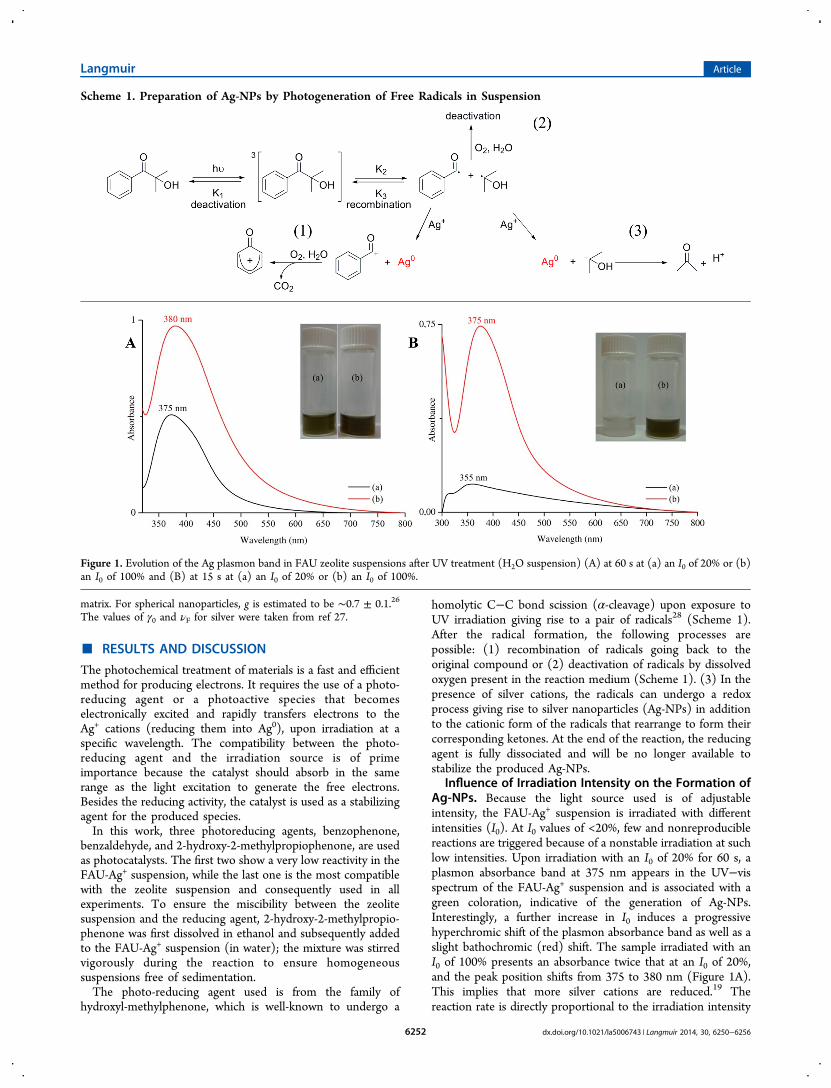
Influence of Irradiation Intensity on the Formation ofAg-NPs. Because the light source used is of adjustableintensity, the FAU-Ag+ suspension is irradiated with differentintensities (I0). At I0 values of <20%, few and nonreproduciblereactions are triggered because of a nonstable irradiation at suchlow intensities. Upon irradiation with an I0 of 20% for 60 s, aplasmon absorbance band at 375 nm appears in the UV−visspectrum of the FAU-Ag+ suspension and is associated with agreen coloration, indicative of the generation of Ag-NPs.Interestingly, a further increase in I0 induces a progressivehyperchromic shift of the plasmon absorbance band as well as aslight bathochromic (red) shift. The sample irradiated with anI0 of 100% presents an absorbance twice that at an I0 of 20%,and the peak position shifts from 375 to 380 nm (Figure 1A).This implies that more silver cations are reduced.19 Thereaction rate is directly proportional to the irradiation intensity
Scheme 1. Preparation of Ag-NPs by Photogeneration of Free Radicals in Suspension
Figure 1. Evolution of the Ag plasmon band in FAU zeolite suspensions after UV treatment (H2O suspension) (A) at 60 s at (a) an I0 of 20% or (b)an I0 of 100% and (B) at 15 s at (a) an I0 of 20% or (b) an I0 of 100%.
Langmuir Article
dx.doi.org/10.1021/la5006743 | Langmuir 2014, 30, 6250−62566252

used. At a low intensity, a small number of radicals aregenerated, because of the competing deactivation process(Scheme 1); only a few radicals are available for the reductionof silver cations, explaining the low intensity of the plasmonabsorbance band. At a high irradiation intensity (I0 = 100%),many free radicals are generated and are rapidly consumed.This favors the reduction of silver over the deactivation of freeradicals and leads to the full reduction of silver cations afteronly 60 s. This assumption is confirmed by running thereaction for a shorter time (15 s) (Figure 1B). At a lowirradiation intensity (I0 = 20%), a very low intensity bandappears at 355 nm with no change in the color of thesuspension. This suggests that the number of free radicalsgenerated is not enough to reduce the silver cations. With an I0of 100%, an intense absorption at 375 nm is associated with theappearance of a dark green color, indicating that the reaction isalmost complete after irradiation for 15 s. For comparison, theUV−visible spectra of the FAU-Ag+ suspension without andwith a photoreducing agent (2-hydroxy-2-methylpropiophe-none) at time zero are taken and presented in Figure S1 of theSupporting Information. It is clearly seen that after mixing, noabsorption band is observed in the spectrum, while the plasmonband appears after 15 s (Figure 1). These results demonstratethat the photoreducing agent upon exposure to UV irradiationis giving rise to a pair of radicals, and the sequential processesare followed as presented in Scheme 1.Influence of Irradiation Time on the Formation of Ag-
NPs. The irradiation of a silver-containing zeolite suspension isconducted for different times while fixing the intensity of thelight source at 100%. As observed in Figure 2, a plasmon band
appears at 365 nm, associated with a pale green coloration ofthe suspension after irradiation for 5 s. This highlights the highefficiency of this mode of reduction, i.e., formation of Ag-NPsafter irradiation for only 5 s instead of several hours for thechemical reduction method.21 Increasing the reaction timeleads to a continuous hyperchromic shift of the plasmon bandand a darkening of the zeolite suspension, indicative of theprogressive formation of Ag-NPs. On the basis of the bandintensity, more than 75% of the silver cations are reduced afteronly 15 s. Furthermore, all silver cations are reduced afterirradiation for 60 s with no increase in the plasmon bandintensity with longer irradiation times [2 or 3 min (Figure S2 of
the Supporting Information)]. Besides, an additional amount ofreducing agent is added to the samples and then subjected toirradiation. No notable change in the plasmon band is observedunder these conditions; thus, we conclude that the silvercations are fully reduced after treatment for only 1 min. Instead,a decrease in plasmon band intensity takes place at a longirradiation time (5 min), probably because of a partialdestruction of the zeolite nanoparticles caused by the harshconditions used. Interestingly, no change in the absorbancewavelength is noticed; this indicates that the reaction does notoccur via the formation of small Ag seeds growing into largerparticles during the reaction.21 Because of the fast reactionprocess, Ag-NPs of a similar size are progressively produced.The DLS study of the FAU-Ag suspensions obtained under
different irradiation conditions shows a monomodal particlesize distribution of particles with an average diameter of 70 nm(Figure 3), matching the DLS from a pure FAU zeolite
suspension (Figure S3 of the Supporting Information). Thisindicates that the silver nanoparticles are completely associatedwith the zeolite crystals and no evidence of separate Ag-NPs isobserved in the suspensions.The DLS results are confirmed by a HRTEM study (Figure 4
and Figure S4 of the Supporting Information). The zeolitecrystals contain silver nanoparticles predominantly located inthe channels of FAU crystals; however, several Ag-NPs are seenon the zeolite surface. At high magnifications, the zeolitenanoparticles show a high degree of crystallinity and well-aligned crystal fringes. As previously mentioned, because of theshort reaction period, the Ag nanoparticles are of similar sizeand shape and appear as spheres with a diameter in the range of0.7−1.1 nm. More than 80% of the Ag-NPs are located in thesuper cages (1.1 nm) and some in the sodalite cages (0.7 nm)of the FAU zeolite crystals. The entire FAU zeolite crystalcontaining Ag-NPs at high magnification is shown in Figure4C; the homogeneous distribution of Ag-NPs all over thezeolite crystal can be seen. The regular distribution of Ag-NPsin the zeolite crystals is observed at high magnifications, asshown as an inset of the enlarged selected area in Figure 4B.However, some Ag-NPs with a size of 5−6 nm can be seen onthe zeolite external surface (Figure 4A).The zeolite crystals containing Ag-NPs are further
characterized by EDS-TEM (Figure 5).
Figure 2. Evolution of the Ag plasmon band in FAU suspensions afterUV treatment at 100% for (a) 5, (b) 10, (c) 15, and (d) 60 s.
Figure 3. DLS curves of FAU-Ag suspensions after irradiation with anI0 of 100% for (a) 5, (b) 10, (c) 15, and (d) 60 s.
Langmuir Article
dx.doi.org/10.1021/la5006743 | Langmuir 2014, 30, 6250−62566253

The zeolite main components, i.e., Al, Si, and Na, aremeasured and presented in Figure 5. A Si:Al ratio of ∼1.2,which is characteristic of the FAU type zeolite (X), is measured.Additionally, the partial ion exchange of sodium for silver isevidenced. Besides, the chemical composition of the ion-exchanged (FAU-Ag+) and reduced (FAU-Ag) zeolite samplesis determined and presented in Table S1 of the SupportingInformation. The results confirm that the silver content doesnot change; thus, only reduction of the silver cations occurred,leading to the formation of Ag-NPs, which is proven by UV−vismeasurements.Stability of Ag-NPs. The stability of Ag-NPs in the FAU
zeolite is of prime importance. It is expected that thephotoreductant has a dual action during the photoreduction
process: (1) reducing the silver cations and simultaneously (2)stabilizing the Ag-NPs from further oxidation. However, wehave already mentioned that, in our case, the reducing agent isfragmented during the reaction and so produces free radicals.Hence, it is not capable of ensuring the stability of the reactionproducts. The zeolite is therefore used as an inorganic matrix tostabilize the Ag-NPs.10 In Figure 6A, the plasmon band of Ag-NPs stabilized in the FAU zeolite, and a control test with thesame amount of nonsupported and reduced silver in a watersolution is presented. It is clearly seen that the Ag-NPs in FAUzeolite exhibit an intense and nearly symmetrical bandreflecting their high degree of monodispersity, already shownby HREM. Moreover, the Ag-NPs in a water solution have avery broad and weak band extending from 365 to 800 nm
Figure 4. HRTEM images of FAU crystals containing Ag-NPs. (A) Ag-NPs within the cages and on the external surface of the zeolite crystals(arrows). (B) Ag-NPs in the zeolite cages only. The inset shows an enlarged selected area showing the silver nanoparticle in the zeolites. (C) Ag-NPshomogeneously distributed in the entire crystalline FAU zeolite particle. This sample was obtained after UV treatment at 100% for 60 s.
Figure 5. Elemental composition of the FAU-Ag sample measured by EDX-HRTEM (C corresponds to the holey carbon film, and Ni is comingfrom the grid used in the TEM experiment). The inset shows the elemental analysis of the FAU-Ag sample expressed in weight and atomic percent.
Figure 6. (A) Plasmon band of Ag-NPs in (a) a FAU-Ag suspension and (b) pure Ag-NPs in water (I0 = 100% for 10 s). (B) Evolution of theplasmon band for (a) FAU-Ag and (b) pure Ag in water after 60 min.
Langmuir Article
dx.doi.org/10.1021/la5006743 | Langmuir 2014, 30, 6250−62566254
(Figure 6A, black line). This band indicates the presence of Ag-NPs of different sizes. Hence, the first role of the zeolite crystalsis to induce a homogeneous formation of Ag-NPs with adefined location within the zeolite pores, and on the zeolitesurface. One hour after irradiation, the plasmon band of Ag-NPs supported on FAU zeolite crystals is unchanged (Figure6B), while the corresponding band for free Ag-NPs in a watersolution totally disappears. This evolution is accompanied bythe discoloration of the suspension, from light brown tocolorless. For the FAU-Ag suspension, no change in suspensioncolor, plasmon band position, or intensity is observed afterseveral months, while in the absence of zeolite, the Ag-NPsstart to be oxidized after reduction for only a few minutes(Figure S5 of the Supporting Information).Theoretical Calculations of Ag-NPs Based on the
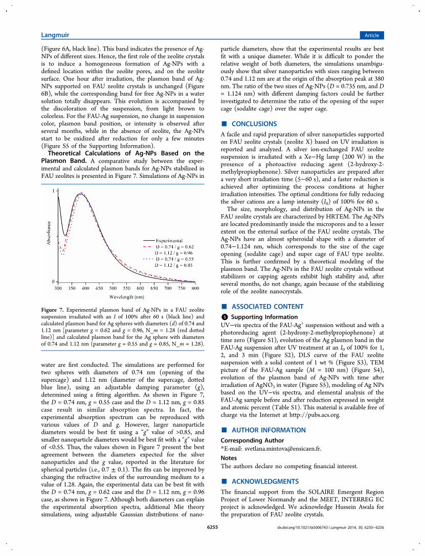
Plasmon Band. A comparative study between the exper-imental and calculated plasmon bands for Ag-NPs stabilized inFAU zeolites is presented in Figure 7. Simulations of Ag-NPs in
water are first conducted. The simulations are performed fortwo spheres with diameters of 0.74 nm (opening of thesupercage) and 1.12 nm (diameter of the supercage, dottedblue line), using an adjustable damping parameter (g),determined using a fitting algorithm. As shown in Figure 7,the D = 0.74 nm, g = 0.55 case and the D = 1.12 nm, g = 0.85case result in similar absorption spectra. In fact, theexperimental absorption spectrum can be reproduced withvarious values of D and g. However, larger nanoparticlediameters would be best fit using a “g” value of >0.85, andsmaller nanoparticle diameters would be best fit with a “g” valueof <0.55. Thus, the values shown in Figure 7 present the bestagreement between the diameters expected for the silvernanoparticles and the g value, reported in the literature forspherical particles (i.e., 0.7 ± 0.1). The fits can be improved bychanging the refractive index of the surrounding medium to avalue of 1.28. Again, the experimental data can be best fit withthe D = 0.74 nm, g = 0.62 case and the D = 1.12 nm, g = 0.96case, as shown in Figure 7. Although both diameters can explainthe experimental absorption spectra, additional Mie theorysimulations, using adjustable Gaussian distributions of nano-
particle diameters, show that the experimental results are bestfit with a unique diameter. While it is difficult to ponder therelative weight of both diameters, the simulations unambigu-ously show that silver nanoparticles with sizes ranging between0.74 and 1.12 nm are at the origin of the absorption peak at 380nm. The ratio of the two sizes of Ag-NPs (D = 0.735 nm, and D= 1.124 nm) with different damping factors could be furtherinvestigated to determine the ratio of the opening of the supercage (sodalite cage) over the super cage.
■ CONCLUSIONSA facile and rapid preparation of silver nanoparticles supportedon FAU zeolite crystals (zeolite X) based on UV irradiation isreported and analyzed. A silver ion-exchanged FAU zeolitesuspension is irradiated with a Xe−Hg lamp (200 W) in thepresence of a photoactive reducing agent (2-hydroxy-2-methylpropiophenone). Silver nanoparticles are prepared aftera very short irradiation time (5−60 s), and a faster reduction isachieved after optimizing the process conditions at higherirradiation intensities. The optimal conditions for fully reducingthe silver cations are a lamp intensity (I0) of 100% for 60 s.The size, morphology, and distribution of Ag-NPs in the
FAU zeolite crystals are characterized by HRTEM. The Ag-NPsare located predominantly inside the micropores and to a lesserextent on the external surface of the FAU zeolite crystals. TheAg-NPs have an almost spheroidal shape with a diameter of0.74−1.124 nm, which corresponds to the size of the cageopening (sodalite cage) and super cage of FAU type zeolite.This is further confirmed by a theoretical modeling of theplasmon band. The Ag-NPs in the FAU zeolite crystals withoutstabilizers or capping agents exhibit high stability and, afterseveral months, do not change, again because of the stabilizingrole of the zeolite nanocrystals.
■ ASSOCIATED CONTENT
*S Supporting InformationUV−vis spectra of the FAU-Ag+ suspension without and with aphotoreducing agent (2-hydroxy-2-methylpropiophenone) attime zero (Figure S1), evolution of the Ag plasmon band in theFAU-Ag suspension after UV treatment at an I0 of 100% for 1,2, and 3 min (Figure S2), DLS curve of the FAU zeolitesuspension with a solid content of 1 wt % (Figure S3), TEMpicture of the FAU-Ag sample (M = 100 nm) (Figure S4),evolution of the plasmon band of Ag-NPs with time afterirradiation of AgNO3 in water (Figure S5), modeling of Ag NPsbased on the UV−vis spectra, and elemental analysis of theFAU-Ag sample before and after reduction expressed in weightand atomic percent (Table S1). This material is available free ofcharge via the Internet at http://pubs.acs.org.
■ AUTHOR INFORMATIONCorresponding Author*E-mail: [email protected].
NotesThe authors declare no competing financial interest.
■ ACKNOWLEDGMENTSThe financial support from the SOLAIRE Emergent RegionProject of Lower Normandy and the MEET, INTERREG ECproject is acknowledged. We acknowledge Hussein Awala forthe preparation of FAU zeolite crystals.
Figure 7. Experimental plasmon band of Ag-NPs in a FAU zeolitesuspension irradiated with an I of 100% after 60 s (black line) andcalculated plasmon band for Ag spheres with diameters (d) of 0.74 and1.12 nm [parameter g = 0.62 and g = 0.96, N_m = 1.28 (red dottedline)] and calculated plasmon band for the Ag sphere with diametersof 0.74 and 1.12 nm (parameter g = 0.55 and g = 0.85, N_m = 1.28).
Langmuir Article
dx.doi.org/10.1021/la5006743 | Langmuir 2014, 30, 6250−62566255

■ REFERENCES(1) Flores-Lopez, N. S.; Castro-Rosas, J.; Ramirez-Bon, R.; Mendoza-Cordova, A.; Larios-Rodriguez, E.; Flores-Acosta, M. Synthesis andproperties of crystalline silver nanoparticles supported in naturalzeolite chabazite. J. Mol. Struct. 2012, 1028, 110−115.(2) Tung, N. H.; Chikae, M.; Ukita, Y.; Viet, P. H.; Takamura, Y.Sensing technique of silver nanoparticles as labels for immunoassayusing liquid electrode plasma atomic emission spectrometry. Anal.Chem. 2012, 84, 1210−1213.(3) Liu, F.; Rao, B. S.; Nunzi, J.-M. A dye functionalized silver−silicacore−shell nanoparticle organic light emitting diode. Org. Electron.2011, 12, 1279−1284.(4) Noh, H. S.; Cho, E. H.; Kim, H. M.; Han, Y. D.; Joo, J. Organicsolar cells using plasmonics of Ag nanoprisms. Org. Electron. 2013, 14,278−285.(5) Kulkarni, A. P.; Noone, K. M.; Munechika, K.; Guyer, S. R.;Ginger, D. S. Plasmon-enhanced charge carrier generation in organicphotovoltaic films using silver nanoprisms. Nano Lett. 2010, 10, 1501−1505.(6) Atwater, H. A.; Polman, A. Plasmonics for improved photovoltaicdevices. Nat. Mater. 2010, 3, 205−213.(7) Cuong, N. H.; Nguyen, H. M. T.; Nguyen, M. T. Theoreticalmodeling of optical properties of Ag8 and Ag14 silver clustersembedded in an LTA sodalite zeolite cavity. Phys. Chem. Chem.Phys. 2013, 15, 15404−15415.(8) Miyanaga, T.; Suzuki, Y.; Matsumoto, N.; Narita, S.; Ainai, T.;Hoshino, H. Formation of Ag clusters in zeolite X studied by in situEXAFS and infrared spectroscopy. Microporous Mesoporous Mater.2013, 168, 213−220.(9) Lee, J.-H.; Hwang, J.-H.; Nam, J.-M. DNA-tailored plasmonicnanoparticles for biosensing applications. WIREs Nanomedicine andNanobiotechnology 2013, 5, 96−109.(10) Zaarour, M.; Dong, B.; Naydenova, I.; Retoux, R.; Mintova, S.Progress in zeolite synthesis promotes advanced applications.Microporous Mesoporous Mater. 2014, 189, 11−21.(11) Devaux, A.; Calzaferri, G.; Miletto, I.; Cao, P.; Belser, P.;Bruhwiler, D.; Khorev, O.; Robert Haner, R.; Kunzmann, A. Self-absorption and luminescence quantum yields of dye-zeolite Lcomposites. J. Phys. Chem. C 2013, 117, 23034−23047.(12) Yang, M.; Fujino, T. Silver nanoparticles on zeolite surface forlaser desorption/ionization mass spectrometry of low molecularweight compounds. Chem. Phys. Lett. 2013, 576, 61−64.(13) Guerra, R.; Lima, E.; Viniegra, M.; Guzman, A.; Lara, V. Growthof Escherichia coli and Salmonella typhi inhibited by fractal silvernanoparticles supported on zeolites. Microporous Mesoporous Mater.2012, 147, 267−273.(14) Liu, Y.; Zhu, Z.; Liu, G.; Xu, Z.; Kuznicki, S. M.; Zhang, H. Anovel method to improve crystallinity of supported nanoparticles usinglow melting point metals. J. Phys. Chem. C 2011, 115, 14591−14597.(15) Cavicchioli, M.; Varanda, L. C.; Massabni, A. C.; Melnikov, P.Silver nanoparticles synthesized by thermal reduction of a silver(I)−aspartame complex in inert atmosphere. Mater. Lett. 2005, 28, 3585−3589.(16) Aswathy, B.; Avadhani, G. S.; Sumithra, I. S.; Suji, S.; Sony, G.Microwave assisted synthesis and UV-Vis spectroscopic studies ofsilver nanoparticles synthesized using vanillin as a reducing agent. J.Mol. Liq. 2011, 159, 165−169.(17) Wani, I. A.; Ganguly, A.; Ahmed, J.; Ahmad, T. Silvernanoparticles: Ultrasonic wave assisted synthesis, optical character-ization and surface area studies. Mater. Lett. 2011, 65, 520−522.(18) Rycenga, M.; Cobley, C. M.; Zeng, J.; Li, W.; Moran, C. H.;Zhang, Q.; Qin, D.; Xia, Y. Controlling the synthesis and assembly ofsilver nanostructures for plasmonic applications. Chem. Rev. 2011, 111,3669−3712.(19) Jia, H.; Zeng, J.; Song, W.; An, J.; Zhao, B. Preparation of silvernanoparticles by photo-reduction for surface-enhanced Ramanscattering. Thin Solid Films 2006, 2, 281−287.(20) Tsuji, T.; Okazaki, Y.; Tsuji, M. Photo-induced morphologicalconversions of silver nanoparticles prepared using laser ablation in
water-enhanced morphological conversions using halogen etching. J.Photochem. Photobiol., A 2008, 194, 247−253.(21) Kshirsagar, P.; Sangaru, S. S.; Malvindi, M. A.; Martiradonna, L.;Cingolani, R.; Pomp, P. P. Synthesis of highly stable silvernanoparticles by photoreduction and their size fractionation byphase transfer method. Colloids Surf., A 2011, 392, 264−270.(22) Bohren, C. F.; Huffman, D. R. Absorption and Scattering of Lightby Small Particles; John Wiley & Sons: New York, 1998.(23) International Association for the Properties of Water and Steam.Release on Refractive Index of Ordinary Water Substance as aFunction of Wavelength, Temperature and Pressure, 1997.(24) Johnson, P. B.; Christy, R. W. Optical Constants of the NobleMetals. Phys. Rev. 1972, B6, 4370−4379.(25) Kreibig, U. Electronic properties of small silver particles: Theoptical constants and their temperature dependence. J. Phys. F: Met.Phys. 1974, 4, 999−1014.(26) Lerme, J.; Baida, H.; Bonnet, C.; Broyer, M.; Cottancin, E.;Crut, A.; Maioli, P.; Del Fatti, N.; Valle, F.; Pellarin, M. SizeDependence of the Surface Plasmon Resonance Damping in MetalNanospheres. J. Phys. Chem. Lett. 2010, 1, 2922−2928.(27) Zeman, E. J.; Schatz, G. C. An accurate electromagnetic theorystudy of surface enhancement factors for silver, gold, copper, lithium,sodium, aluminum, gallium, indium, zinc, and cadmium. J. Phys. Chem.1987, 91, 634−643.(28) Andrzejewska, E. Photopolymerization kinetics of multifunc-tional monomers. Prog. Polym. Sci. 2001, 26, 605−665.
Langmuir Article
dx.doi.org/10.1021/la5006743 | Langmuir 2014, 30, 6250−62566256
Related Documents