Photoacoustic imaging in cancer detection, diagnosis, and treatment guidance Srivalleesha Mallidi, Geoffrey P. Luke, and Stanislav Emelianov Department of Biomedical Engineering, University of Texas at Austin, Austin, TX 78712, USA Abstract Imaging modalities play an important role in the clinical management of cancer, including screening, diagnosis, treatment planning, and therapy monitoring. Owing to increased research efforts in the past two decades, photoacoustic imaging – a non-ionizing, non-invasive technique capable of visualizing optical absorption properties of tissue at reasonable depth, with spatial resolution of ultrasound – has emerged. Ultrasound-guided photoacoustics is regarded for its ability to provide in vivo morphological and functional information about the tumor within the surrounding tissue. With the recent advent of targeted contrast agents, photoacoustics is capable of in vivo molecular imaging, thus facilitating further molecular and cellular characterization of cancer. This review examines the role of photoacoustics and photoacoustic-augmented imaging techniques in comprehensive cancer detection, diagnosis and treatment guidance. Introduction Cancer is a vicious disease that killed approximately 570,000 people in 2010 in the United States alone [1]. To develop successful therapeutic strategies and prevent recurrence of the disease, structural, functional and metabolic properties of this disease need to be well- characterized. The research efforts are not only limited to developing new treatments and discovering the root cause for the disease, but also in developing imaging technologies that can aid in early detection of cancer and can provide comprehensive real-time information on the tumor properties. Currently, ultrasound imaging (USI), magnetic resonance imaging (MRI), X-ray computed tomography (CT), and nuclear imaging techniques like positron emission tomography (PET) and single photon emission computed tomography (SPECT) are being used to detect tumors in patients [2]. With the development of various targeted contrast agents, these imaging techniques are also able to provide molecular information of the malignant tumor tissue. However, microscopic optical imaging techniques have higher resolution (~0.1–100 μm) than USI (50–500 μm), MRI (10–100μm), CT (50–200 μm), PET (1–2 mm) and SPECT (1–2 mm), and can detect a lower number of cancer cells per imaging voxel [3]. Traditional diffusive regime optical imaging techniques, such as diffuse optical tomography (DOT), have high detection sensitivity; however, the resolution is limited to ~5 mm. The need for an imaging technique that can provide high optical contrast images at microscale resolution and at a reasonable penetration depth has now been filled with photoacoustic imaging (PAI). © 2011 Elsevier Ltd. All rights reserved. Corresponding author: Emelianov, S. ([email protected]). Publisher's Disclaimer: This is a PDF file of an unedited manuscript that has been accepted for publication. As a service to our customers we are providing this early version of the manuscript. The manuscript will undergo copyediting, typesetting, and review of the resulting proof before it is published in its final citable form. Please note that during the production process errors may be discovered which could affect the content, and all legal disclaimers that apply to the journal pertain. NIH Public Access Author Manuscript Trends Biotechnol. Author manuscript; available in PMC 2012 May 1. Published in final edited form as: Trends Biotechnol. 2011 May ; 29(5): 213–221. doi:10.1016/j.tibtech.2011.01.006. NIH-PA Author Manuscript NIH-PA Author Manuscript NIH-PA Author Manuscript

Welcome message from author
This document is posted to help you gain knowledge. Please leave a comment to let me know what you think about it! Share it to your friends and learn new things together.
Transcript

Photoacoustic imaging in cancer detection, diagnosis, andtreatment guidance
Srivalleesha Mallidi, Geoffrey P. Luke, and Stanislav EmelianovDepartment of Biomedical Engineering, University of Texas at Austin, Austin, TX 78712, USA
AbstractImaging modalities play an important role in the clinical management of cancer, includingscreening, diagnosis, treatment planning, and therapy monitoring. Owing to increased researchefforts in the past two decades, photoacoustic imaging – a non-ionizing, non-invasive techniquecapable of visualizing optical absorption properties of tissue at reasonable depth, with spatialresolution of ultrasound – has emerged. Ultrasound-guided photoacoustics is regarded for itsability to provide in vivo morphological and functional information about the tumor within thesurrounding tissue. With the recent advent of targeted contrast agents, photoacoustics is capable ofin vivo molecular imaging, thus facilitating further molecular and cellular characterization ofcancer. This review examines the role of photoacoustics and photoacoustic-augmented imagingtechniques in comprehensive cancer detection, diagnosis and treatment guidance.
IntroductionCancer is a vicious disease that killed approximately 570,000 people in 2010 in the UnitedStates alone [1]. To develop successful therapeutic strategies and prevent recurrence of thedisease, structural, functional and metabolic properties of this disease need to be well-characterized. The research efforts are not only limited to developing new treatments anddiscovering the root cause for the disease, but also in developing imaging technologies thatcan aid in early detection of cancer and can provide comprehensive real-time information onthe tumor properties. Currently, ultrasound imaging (USI), magnetic resonance imaging(MRI), X-ray computed tomography (CT), and nuclear imaging techniques like positronemission tomography (PET) and single photon emission computed tomography (SPECT) arebeing used to detect tumors in patients [2]. With the development of various targetedcontrast agents, these imaging techniques are also able to provide molecular information ofthe malignant tumor tissue. However, microscopic optical imaging techniques have higherresolution (~0.1–100 µm) than USI (50–500 µm), MRI (10–100µm), CT (50–200 µm), PET(1–2 mm) and SPECT (1–2 mm), and can detect a lower number of cancer cells per imagingvoxel [3]. Traditional diffusive regime optical imaging techniques, such as diffuse opticaltomography (DOT), have high detection sensitivity; however, the resolution is limited to ~5mm. The need for an imaging technique that can provide high optical contrast images atmicroscale resolution and at a reasonable penetration depth has now been filled withphotoacoustic imaging (PAI).
© 2011 Elsevier Ltd. All rights reserved.Corresponding author: Emelianov, S. ([email protected]).Publisher's Disclaimer: This is a PDF file of an unedited manuscript that has been accepted for publication. As a service to ourcustomers we are providing this early version of the manuscript. The manuscript will undergo copyediting, typesetting, and review ofthe resulting proof before it is published in its final citable form. Please note that during the production process errors may bediscovered which could affect the content, and all legal disclaimers that apply to the journal pertain.
NIH Public AccessAuthor ManuscriptTrends Biotechnol. Author manuscript; available in PMC 2012 May 1.
Published in final edited form as:Trends Biotechnol. 2011 May ; 29(5): 213–221. doi:10.1016/j.tibtech.2011.01.006.
NIH
-PA Author Manuscript
NIH
-PA Author Manuscript
NIH
-PA Author Manuscript

PAI has shown tremendous potential in simultaneously providing structural, functional andmolecular information in pre-clinical studies. PAI can visualize tumor location deep within atissue, and is also able to provide information on tumor vasculature [4] or to monitorangiogenesis [5]. PAI can also obtain information on hemoglobin oxygen saturation at highresolution and contrast, without the use of exogenous contrast agents [4], which is asignificant advantage when compared with other tumor hypoxia imaging techniques (e.g.blood oxygen level dependent-MRI and PET). Another advantage of PAI is compatibilitywith widely available USI techniques [6]; when combined, PAI and USI can simultaneouslyprovide anatomical and functional information in tumors. For example, an in vivo study onhuman breast tissue has shown that an ultrasound image can depict the structure of ductalcarcinoma, while photoacoustic (PA) images show the associated scattered distribution ofvascularization [7].
With the availability of various targeted contrast agents, such as gold nanoparticles(AuNPs), several new avenues have opened up for in vivo molecular PAI. This hasfacilitated highly sensitive and specific detection of tumors. In addition, PAI, combined withother complementary imaging techniques, has shown promise in cancer treatment guidance.Although several reviews on the basics and applications of PAI are available, noexamination of the recent developments in molecular PAI of cancer and the ability of PAI tomonitor treatment is currently available; hence, we review these topics.
Basic principles of photoacoustic imagingPAI (also known as optoacoustic imaging) capitalizes on the photoacoustic effect firstdescribed by Alexander Graham Bell in 1880 [8]. In this review, we provide only basicprinciples (Box 1) towards understanding PAI applicability to cancer detection andtreatment guidance. Briefly, absorbed laser energy causes a rapid thermoelastic expansion oftissue, resulting in the generation of a wide-band ultrasound wave. The ultrasound wave isdetected with a transducer that converts the mechanical acoustic waves to electric signals.The captured signals are then processed to form an image [6,9,10]. A typical PAI setup(Figure 1a) consists of two main components: a laser (generally a tunable, nanosecondpulsed laser) and a USI system. Most ultrasound image processing techniques translatedirectly to PAI. Delay and sum beamforming or more advanced computed tomographyalgorithms used in USI are also used in PAI to obtain images [10,11]. Other reviews haveprovided detailed information on equations governing the PA effect, various PA systemconfigurations, and the image processing algorithms [6,9–12].
The contrast in PA images is largely determined by the wavelength-dependent µa (EquationI). The changes in optical absorption as a function of wavelength for a variety ofendogenous chromophores, including melanin, oxy- (HbO2) and deoxy-hemoglobin (Hb) arepresented in Figure 1b. Because of the large variations in µa, spectroscopic or multi-wavelength imaging techniques are used for characterization of tumor tissue opticalproperties. For example, HbO2 and Hb have different wavelength-dependent opticalabsorption properties, which allows spectroscopic PAI to differentiate between arteries andveins. Furthermore, HbO2 and Hb have the same optical absorption at approximately 805nm, PAI at this and other wavelengths can be used to assess total hemoglobin and bloodoxygenation. PA contrast can also be enhanced by exogenous contrast agents (e.g. dyes ornanoparticles) (Figure 1c) that have distinct and tunable absorption spectra in the near-infrared (NIR) optical window (600–1100 nm) where endogenous chromophores, such ashemoglobin and water, have low absorption (Figure 1b).
Mallidi et al. Page 2
Trends Biotechnol. Author manuscript; available in PMC 2012 May 1.
NIH
-PA Author Manuscript
NIH
-PA Author Manuscript
NIH
-PA Author Manuscript

Cancer detection with endogenous photoacoustic contrastCancer detection using PAI with endogenous chromophores (e.g. hemoglobin and melanin)is an area of active research. For example, PAI has been used to monitor melanoma tumorgrowth over the course of two weeks [23]. Optical contrast was provided by a higherconcentration of melanin in the tumor relative to the surrounding tissue. PAI has also beenused to detect skin melanoma [4,24]. Figure 2a depicts a melanoma and surroundingvasculature obtained by spectroscopic PAI. The pseudo-colored image was reconstructedfrom PA images obtained at 584 nm and 764 nm laser illumination. These studies indicatethat PAI has the potential to identify, visualize, and track tumors and their associatedvasculature with high resolution.
Malignant tumors have dense and unorganized vasculature compared to normal tissue. Thehigh density of blood vessels in tumors enhances PA image contrast, thereby enabling tumordetection. For example, the Twente Photoacoustic Mammoscope was developed to detectbreast carcinoma was based on this principle [21,25]. An X-ray mammogram (Figure 2b–i)and a sonogram (Figure 2b–ii) are compared with a PA image (Figure 2b–iii) obtained witha 1064 nm optical source. Figure 2 shows that the mammoscope is more sensitive tomalignant indicators, such as vascularization, than conventional radiological techniques.PAI can also provide information on angiogenesis or changes in vasculature [5,26–28]. Asshown in Figure 2c, sequential PA images can be obtained safely and noninvasively atdifferent stages of tumor progression to monitor angiogenesis and to determine if a tumorhas progressed to malignancy [5]. Compared to other vascular imaging techniques, includingdynamic-enhanced MRI, CT perfusion, and functional PET [29], PAI detects tumorvasculature at a better or comparable resolution, without the use of exogenous contrastagents.
Besides imaging melanin and blood vessels, PAI systems have been employed formeasuring the oxygen content of blood to study hypoxia in tumors [30,31]. Hypoxia is oftenlinked to malignancy and resistance to therapy [32]. The amount of oxygen saturation inblood (SO2) can be estimated by comparing the PA signal strength of HbO2 and Hbobtained from spectroscopic PA images. Figure 2d shows in vivo functional imaging of amouse brain with a glioblastoma. The blue hypoxic region (circled) indicates the location ofthe tumor in the brain [31]. The results clearly depict that the tumor has a lower percentageSO2 than the surrounding normal tissue.
Metastatic spread of the primary tumor often leads to death in cancer patients. Highlysensitive detection of circulating tumor cells (CTCs) would greatly enhance overall patientsurvival, if treated. PAI has been used for detecting CTCs in the blood stream, with the goalof detecting metastasis. The label-free detection of CTCs in vivo in a blood vessel using PAIcould provide higher detection sensitivity (~100-fold) compared to existing ex vivo CTCdetection assays that use a small amount of blood. With the use of contrast agents targeted toCTCs, the PAI detection sensitivity can be enhanced further [33–35].
Cancer detection with exogenous photoacoustic contrast agentsThe sensitivity of the PAI technique to image deeply situated tumors can be increaseddrastically by utilizing exogenous contrast agents. The NIR-absorbing dyes, such asIRDye800CW [31,36], AlexaFluor 750 [37] and indocyanine green (ICG) [38], have beenused to enhance PA contrast. However, among the exogenous contrast agents, AuNPs haveattracted attention in nanoparticle-based PAI owing to their unique optical properties fromthe surface plasmon resonance (SPR) effect. Because of the SPR effect, AuNPs have anabsorbance that is orders of magnitude higher than NIR dyes. For example, goldnanospheres, nanorods, nanoshells, nanocages and nanobeacons have been used in PAI
Mallidi et al. Page 3
Trends Biotechnol. Author manuscript; available in PMC 2012 May 1.
NIH
-PA Author Manuscript
NIH
-PA Author Manuscript
NIH
-PA Author Manuscript
because of their tunable and strong longitudinal plasmon resonance in the NIR [39–46]. Thecytotoxicity of these nanoparticles is debatable and often emerges in a dose- and time-dependent manner for different types of nanoparticles; hence, further investigation intoAuNPs toxicity is necessary [47]. The details on toxicity and pharmacodynamics ofnanoparticles are beyond the scope of the review and are therefore not discussed here.
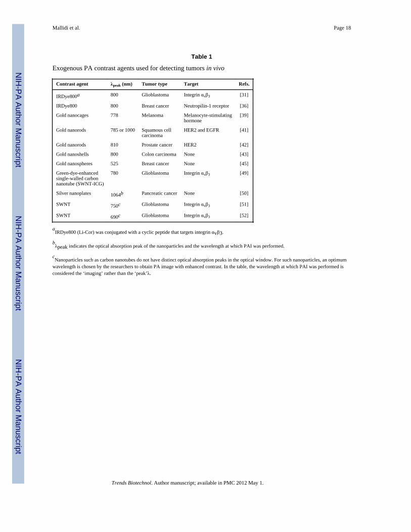
By attaching targeting moieties to exogenous agents, specific molecular informationregarding the tumors can be obtained. AuNPs with different optical absorption propertiescan be conjugated to cancer-specific biomarkers, such as growth factor receptors andintegrins (Table 1). By utilizing multiple targeted AuNPs, multiplex molecular labeling of atumor can be achieved and multi-wavelength PAI can image the heterogenous accumulationand interaction of AuNPs with cancer cells in vivo [41]. Fluorescent optical probes, such asquantum dots, also provide PA contrast and can be used for multiplex labeling of tumors[48].
There have been several advances in molecular PAI that help describe crucial functional andmolecular interactions between tumor cells and the surrounding micro-environment. Forexample, the feasibility of utilizing a multi-wavelength PAI technique to monitor molecularinteractions of epithelial growth factor receptor (EGFR)-targeted AuNPs in 3D tissuecultures and ex vivo tissue has been evaluated [44,53]. Briefly, AuNPs (spheres of 50 nmdiameter) functionalized with antibodies bind to EGFR. This specific targeting of AuNPs toEGFR causes plasmon resonance coupling between adjacent nanoparticles and changes theirabsorbance spectra so that it can be detected as a change in the PA signal amplitude.Overall, the results indicate that PAI together with bioconjugated AuNPs have the potentialto image nano-molecular interactions. Because of the concerns that nano-scale agents mightcause long-term toxicity in vivo, biodegradable gold nanoclusters have been developed as acontrast agent [54,55]. The size of these biodegradable nanoclusters, consisting of sub-5-nmAuNPs and a biodegradable polymer binder, is less than 100 nm. The nanoclusters are alsopH-sensitive and will biodegrade in the acidic environment of the endosome. Afterdegradation, the 5-nm AuNPs are excreted out of the body, thereby preventing toxicaccumulations.
Another recent advancement in PAI is the use of photo-activable probes to provide a target-dependent photoacoustic signal and they show superior specificity and sensitivity ascompared to the probes that donot interact with the target. For example, photo-activableprobes have been designed to specifically target matrix metalloprotease 2 (MMP-2), aprotease found to be overexpressed in many aggressive cancers[56]. The probe’s peptideplatform consists of an activatable cell-penetrating peptide (ACPP) that is recognizable byMMP-2, in both in vitro and mouse models. Before cleavage by MMP-2, the intact probeshows PA signals of similar intensity at the two wavelengths corresponding to theabsorption maxima of the chromophores BHQ3 (675 nm) and AlexaFluor 750 (750 nm).When the probe is cleaved by the appropriate enzyme, the BHQ3 dye associated with theCPP portion of the probe accumulates in the nearby cells, while the Alexa dye diffusesaway. This results in a PA signal visible only at 675 nm [56].
Recently, emphasis has been placed on multi-modal nanosystems that can enhance contrastin two or more imaging modalities, including microbubbles [57], perfluorocarbon-basednanobubbles [58], and nanowontons [59]. For example, nanowontons consist offerromagnetic (cobalt core) core coated with gold for biocompatibility and a unique shapethat enables optical absorption over a broad range of frequencies; the magnetic core acts asan MRI contrast agent and the gold coating provides the optical absorption contrast for PAI[59]. With the evolution of combined imaging strategies, these multi-modal nanostructureswill play a prominent role in cancer detection and treatment.
Mallidi et al. Page 4
Trends Biotechnol. Author manuscript; available in PMC 2012 May 1.
NIH
-PA Author Manuscript
NIH
-PA Author Manuscript
NIH
-PA Author Manuscript
Combination of PA with other imaging modalitiesAssessing complementary structural, functional, metabolic and molecular information on atumor with high accuracy is essential for cancer treatment. PAI primarily provides high-resolution images based on the optical contrast of tissue components, such as changes owingto abnormal vasculature or high melanin content; however, the overall anatomical structureof the tumor cannot be perceived by PAI alone. USI, a noninvasive technique whencombined with PAI, can be used to obtain anatomical details about the tumor and itssurrounding environment (Figure 3a). Both USI and PAI are highly synergistic: they utilizethe same transducer and receiver electronics and are non-ionizing imaging modalities(unlike CT and PET) [6]. Indeed, commercial ultrasound systems have been modified to PAIsystems for both small animal imaging and breast cancer imaging [12, 17, 60–63].
An imaging modality that can provide structural information at resolutions greater thanultrasound (~50–500 µm) is optical coherence tomography (OCT)(~10 µm), which relies onthe optical backscattering properties of tissue. A 3D combined OCT and PA system has beenconstructed that can image microvasculature circulation (Figure 3b) [64]. This combinedsystem could be used to image microvasculature of malignant skin lesions and providecomprehensive structural and functional information [65]. Other optical imaging modalities,such as fluorescence imaging (Figure 3c), have also been used with PAI to image braintumors in vivo [31]. PAI provides high-resolution structural images of tumor angiogenesis,whereas fluorescence imaging has high sensitivity to molecular probes for detecting tumorlocation.
Ultrasound-based elasticity imaging non-invasively assesses the biomechanical properties(i.e. the Young’s modulus) of tumors, which otherwise cannot be depicted from ultrasoundor PA images (Figure 3d). A combination of ultrasound and elasticity imaging has shownthe best results in detecting breast tumors in vivo with high sensitivity and specificity andcould potentially reduce unnecessary biopsies [66]. A combination of complementaryimaging techniques (e.g. elasticity coupled with USI and PAI) could further improve cancerdetection sensitivity and specificity by providing simultaneous information on anatomy(USI), angiogenesis (PAI), and changes in mechanical properties (elasticity imaging) of thetumor [6]. Another ultrasound-based imaging technique that can be combined with PAI ismagneto-motive USI (MMUS). MMUS has the potential to measure the biomechanicalproperties of the tissue with the aid of magnetic nanoparticles (Figure 3e) [67]. In MMUS,magnetic excitation is applied to induce motion of the magnetic nanoparticles within tissuesor organs. Mechanical properties of tissue can be evaluated based on tissue motion detectedin ultrasound images [68]. Based on the initial studies performed on tissue-mimickingphantoms, it can be predicted that the pathological changes in tissue, which are often relatedto changes in tissue mechanical properties, could be detected and differentiated usingMMUS. However, further investigation in murine tumor models is required to evaluate thetechnique in vivo. With the recent introduction of in vivo imaging systems (e.g. Vevo 2100by Visual Sonics Inc.) capable of both USI and PAI at microscopic or macroscopicresolution, the functional intricacies and the structural complexities of the tumor can besimultaneously visualized.
PAI for guiding, monitoring and evaluating therapyImaging techniques play a significant role in cancer therapy, from precise planning andguiding to evaluation of efficacy. In particular, PAI has shown potential in aiding therapiesby providing sequential monitoring of tumor functional properties such as changes in tumorvasculature before, during, and after therapeutic procedures. The therapeutic agents used forphotodynamic therapy (PDT)or photothermal therapy (PTT) can also act as PA contrast
Mallidi et al. Page 5
Trends Biotechnol. Author manuscript; available in PMC 2012 May 1.
NIH
-PA Author Manuscript
NIH
-PA Author Manuscript
NIH
-PA Author Manuscript

agents owing to their high optical absorption properties in the NIR region (e.g.,protoporphyrin IX for PDT and gold nanorods for PTT)[41,72].
PAI can assist in determining the location of the tumor, gauging the heterogeneities invasculature within the tumor, and observing the heterogeneities in the therapeutic agentaccumulation. For example, golden carbon nanotubes (GNTs) conjugated with an antibodyspecific to the lymphatic endothelial hyaluronan receptor-1 have been used to visualizeheterogeneities of endogenous low-absorbing mesenteric structures in nude mouse (Figure4a) [73]. The PA image provided information on heterogeneous accumulation of targetedGNTs (red regions in Figure 4a) to guide PTT to cause spatially specific thermal damage tothe lymphatic walls [73]. In another study, PAI was performed to identify the location of acolon adenocarcinoma tumor. The tumor was then specifically ablated by high-intensityfocused ultrasound (HIFU) with the guidance of PA images [74].
PAI has tremendous potential in guiding therapeutic procedures and could potentiallyprovide oncologists structural and functional information regarding tumors to facilitatepersonalized therapy by customizing the therapy dose. In addition, PAI can also aid incustomized delivery of drugs with use of multi-functional nanoparticles. An example of sucha nanoagent is a silver nanosystem consisting of a poly(lactic-co-glycolic acid) (PLGA)polymer core and outer silver cage network. The outer silver cage enhances contrast in PAIand the inner core of the nanosystem contains the drug doxorubicin (Figure 4b). Initialstudies indicated that the PLGA-based nanosystems have the potential to significantlyincrease contrast in PAI while delivering customized payloads of drug simultaneously to thetumor cells [75]. Another example for controlled and customized drug delivery is usinglight-triggered nano-constructs, such as microspheres containing the drug paclitaxcelencapsulated in hollow gold nanospheres (HAuNS). Depending on the concentration ofHAuNS in the tumor, the light dose can be adjusted to allow sufficient release of drugpaclitaxel [76]. In addition HAuNS can also act as a PTT agent. Very few in vitro studies arepublished illustrating these concepts and further investigations on in vivo tumor models arerequired for validation.
Monitoring functional or structural changes, such as changes in tumor tissue properties andvariations in vascular destruction during therapeutic procedures is critical for prognosis andfurther treatment. Several ex vivo studies show the potential of PAI in monitoringtherapeutic procedures. For example, it has been demonstrated using ex vivo liver tissue andchicken breast tissue that PAI is capable of sensitive detection of thermally-induced changesin tissue optical properties at depths of up to 30–50 mm with sub-millimeter resolutionduring thermal therapy [77,78]. In another study, PAI provided both high-resolution andhigh-tissue-contrast images to quantify changes in vessel morphology during PDT usingprotoporphyrin IX photosensitizer on a chicken chorioallantoic membrane tumor model[72].
In PTT, efficient optical absorbers, such as gold nanorods, are used to heat the tissue tohigher temperatures causing thermal damage. For example, a tumor loaded with goldnanorods shows significant temperature elevations in response to laser irradiation (Figure4c). Both PAI and USI can be used to obtain temperature maps of the tumor during PTT.Indeed, PA-based thermal imaging in the presence of strong optical absorbers has a highersignal-to-noise ratio when compared to USI-based thermal imaging [79]. The examplesprovided in this section clearly indicate the capability of PAI in guiding, customizing andmonitoring therapeutic procedures.
Mallidi et al. Page 6
Trends Biotechnol. Author manuscript; available in PMC 2012 May 1.
NIH
-PA Author Manuscript
NIH
-PA Author Manuscript
NIH
-PA Author Manuscript

Future outlookOverall, PAI could become a valuable tool for cancer detection and diagnosis, tumorcharacterization, and treatment guidance. By differentiating the optical properties of tissues,PAI is well-suited to measure functional properties of tumors in vivo. For example, multi-wavelength PAI can visualize vasculature and identify hypoxic conditions of the tumors.Current functional imaging techniques suffer from poor spatial resolution or inadequatepenetration depth. Conversely, PAI is non-ionizing and can image deep tissue structureswith acceptable spatial resolution.
For functional cellular and molecular imaging in vivo at sufficient depths, contrast agent-mediated photoacoustics holds great promise. Various PA contrast agents ranging fromplasmonic metal nanoparticles to FDA-approved dyes can be targeted to specific receptorsin intact living tissue. Compared with endogenous contrast of tissue, PA contrast agents canbe tuned to a specific wavelength to provide images with higher contrast and signal-to-noiseratio. As with any exogenous contrast agent, safety and biocompatibility of the agents mustbe further addressed.
PAI might also play an important role in several existing and emerging treatment modalities.PAI can image metal objects (e.g. needles, brachytherapy seeds, stents) and therefore guidetherapeutic interventions [80]. The temperature-dependence of the PA signal can provide thethermal imaging of tissue needed for thermal therapeutic approaches. Exogenous contrastagents used for molecular PAI can also be multiplexed to function as drug delivery carriersactivated internally or triggered remotely. Therefore, drug delivery and release is an activeand expanding area of PAI.
Fundamentally, PAI can be performed in real-time. Real-time imaging is critical in manydiagnostic and therapeutic applications of PAI. In deep-penetrating PAI, array-basedultrasound imaging probes are adopted, thus allowing real-time imaging of tissue whereframe rate is primarily limited by the pulse repetition frequency of the laser. Therefore, fromthe instrumentation perspective, high repetition rate, high pulse energy, single-wavelength,or tunable pulsed laser sources designed for PAI are needed. From a clinical perspective,PAI combined with ultrasound could be easily implemented and adapted by cliniciansfamiliar with ultrasonography. Biomedical photoacoustics have seen tremendous growth inthe past two decades yet photoacoustic imaging is still in its infancy. Therefore, wide andrapid advancement of photoacoustics, ranging from instrumentation development toregulatory approvals to pre-clinical and clinical utility, is anticipated in the next decade.
Box 1: Principles of PAI
To describe the photoacoustic effect, a simplified Equation I, assuming 1D plane-wavepropagation in a homogeneous medium, can be used:
[I]
Where p0 is the pressure rise immediately after thermo acoustic excitation in thephotoabsorber; β is the thermal coefficient of volume expansion; vs is the speed of soundin tissue; Cp is the specific heat capacity of the tissue at constant pressure; µa representsthe optical absorption coefficient of the photoabsorber; F0 is laser fluence at depth z = 0;Γ is the Gruneisen parameter or coefficient; and µeff is the effective extinction coefficientof the tissue, defined in Equation II as:
Mallidi et al. Page 7
Trends Biotechnol. Author manuscript; available in PMC 2012 May 1.
NIH
-PA Author Manuscript
NIH
-PA Author Manuscript
NIH
-PA Author Manuscript
[II]
where µs is the scattering coefficient of tissue [6,9,10,13]. In practice, opticalheterogeneities of the tissue and other effects can significantly complicte Equation I.
PAI spatial resolution is determined by overlap of optical and ultrasound beams[6,11,14]. For deep-penetrating PAI, the parameters of the ultrasound transducer are mostcritical: the axial resolution is inversely proportional to the transducer bandwidth (BW),and the lateral resolution is inversely proportional to the numerical aperture (NA) andcenter frequency of the transducer. Therefore, a transducer with high center frequency,high bandwidth, and large NA yields the best resolution images [6,11].
Penetration depth in PAI is limited by wavelength of laser illumination. Greaterpenetration depth can be achieved in the optical window (600–1100 nm) where tissueendogenous chromophores have less optical absorption. Imaging depths of up to 5–6 cmhave been achieved [15–17] using radiant exposures below the maximum permissibleexposure (MPE) imposed by the American National Standards Institute for human skin.The MPE limit at different wavelengths is 20 mJ/cm2 at 400<λ<700 nm;20×100.002(λ−700) mJ/cm2 at 700<λ<1050 nm; and 100 mJ/cm2 at 1050<λ<1500 nm [18].
There is a trade-off between imaging depth and resolution of PAI. As the imaging depthincreases, frequency-dependent attenuation decreases the BW and center frequency of theultrasound wave, resulting in poorer spatial resolution (Table I) [6]. Thus a number ofPAI systems have been developed to image both microscopic or macroscopic features oftumors [9]. Photoacoustic microscopy systems sacrifice imaging depth to improveresolution to as low as 50 µm[4,6,19,20]. With a highly focused laser beam, a lateralresolution of 5 µm can be reached to image superficial capillaries [19]. Conversely,macroscopic PAI systems, with resolution on the order of hundreds of microns, arepromising for tumor detection in deeper regions [15–17,21].
AcknowledgmentsPartial support for this work provided by NIH under grants EB008101 and CA149740 is greatly acknowledged. Theauthors would also like to thank all researchers and scientists who contributed to the field of photoacoustics and,therefore, made this review paper possible.
References1. Jemal A, et al. Cancer Statistics, 2010. CA Cancer J Clin. 2010; 60:277–300. [PubMed: 20610543]2. Fass L. Imaging and cancer: a review. Mol Oncol. 2008; 2:115–152. [PubMed: 19383333]3. Frangioni JV. New technologies for human cancer imaging. Journal of clinical oncology. 2008;
26:4012–4021. [PubMed: 18711192]4. Zhang HF, et al. Functional photoacoustic microscopy for high-resolution and noninvasive in vivo
imaging. Nat Biotech. 2006; 24:848–851.5. Siphanto RI, et al. Serial noninvasive photoacoustic imaging of neovascularization in tumor
angiogenesis. Opt. Express. 2005; 13:89–95. [PubMed: 19488331]6. Emelianov SY, et al. Synergy and applications of combined ultrasound, elasticity, and photoacoustic
imaging. IEEE International Ultrasonics Symposium. 2006:405–415.7. Jose J, et al. Imaging of tumor vasculature using Twente photoacoustic systems. Journal of
Biophotonics. 2009; 2:701–717. [PubMed: 19718681]8. Bell AG. Upon the production of sound by radiant energy. American Journal of Science. 1880;
20:305–324.
Mallidi et al. Page 8
Trends Biotechnol. Author manuscript; available in PMC 2012 May 1.
NIH
-PA Author Manuscript
NIH
-PA Author Manuscript
NIH
-PA Author Manuscript

9. Wang L. Multiscale photoacoustic microscopy and computed tomography. Nature Photonics. 2009;3:503–509. [PubMed: 20161535]
10. Oraevsky, A.; Karabutov, A. Optoacoustic Tomography. In: Vo-Dinh, T., editor. BiomedicalPhotonics Handbook. CRC Press; 2003. p. 34/31-34/34.
11. Xu M, Lihong VW. Photoacoustic imaging in biomedicine. Review of Scientific Instruments.2006; 77:041101.
12. Emelianov SY, et al. Photoacoustics for molecular imaging and therapy. Physics today. 2009;62:34–39. [PubMed: 20523758]
13. Oraevsky AA, et al. Measurement of tissue optical properties by time-resolved detection of laser-induced transient stress. Appl. Opt. 1997; 36:402–415. [PubMed: 18250688]
14. Oraevsky AA, et al. Lateral and z-axial resolution in laser optoacoustic imaging with ultrasonictransducers. Proc. of SPIE. 1995; 2389:198–208.
15. Esenaliev RO, et al. Sensitivity of laser opto-acoustic imaging in detection of small deeplyembedded tumors. IEEE Journal of Selected Topics in Quantum Electronics. 1999; 5:981–988.
16. Ku G, Wang LV. Deeply penetrating photoacoustic tomography in biological tissues enhancedwithan optical contrast agent. Opt. Lett. 2005; 30:507–509. [PubMed: 15789718]
17. Kim C, et al. Deeply penetrating in vivo photoacoustic imaging using a clinical ultrasound arraysystem. Biomed. Opt. Express. 2009; 1:278–284. [PubMed: 21258465]
18. American National Standards Institute. "American National Standard for the Safe Use of Lasers",ANSI Z136.1-2000. New York: American National Standards Institute; 2000.
19. Maslov K, et al. Optical-resolution photoacoustic microscopy for in vivo imaging of singlecapillaries. Opt. Lett. 2008; 33:929–931. [PubMed: 18451942]
20. Mallidi S, et al. Functional and morphological ultrasonic biomicroscopy for tissue engineers.Proceedings of the SPIE Medical Imaging: Ultrasonic Imaging and Signal Processing. 2006; 614761470Y61471-61477.
21. Manohar S, et al. Initial results of in vivo non-invasive cancer imaging in the human breast usingnear-infrared photoacoustics. Opt. Express. 2007; 15:12277–12285. [PubMed: 19547596]
22. Prahl, SA. Optical property spectra compiled by Scott Prahl. 2001. http://omlc.ogi.edu/spectra/.)23. Staley J, et al. Growth of melanoma brain tumors monitored by photoacoustic microscopy. Journal
of Biomedical Optics. 2010; 15:040510–040513. [PubMed: 20799777]24. Oh J-T, et al. Three-dimensional imaging of skin melanoma in vivo by dual-wavelength
photoacoustic microscopy. Journal of Biomedical Optics. 2006; 11:034032–034034.25. Manohar S, et al. The Twente Photoacoustic Mammoscope: system overview and performance.
Physics in Medicine and Biology. 2005; 50:2543. [PubMed: 15901953]26. Lao Y, et al. Noninvasive photoacoustic imaging of the developing vasculature during early tumor
growth. Physics in Medicine and Biology. 2008; 53:4203. [PubMed: 18635896]27. Kolkman, RGM., et al. Photoacoustic imaging of tumor angiogenesis; Photons Plus Ultrasound:
Imaging and Sensing 2008: The Ninth Conference on Biomedical Thermoacoustics,Optoacoustics, and Acousto-optics; 2008. p. 685602-685606.
28. Ku G, et al. Imaging of tumor angiogenesis in rat brains in vivo by photoacoustic tomography.Appl. Opt. 2005; 44:770–775. [PubMed: 15751858]
29. Turkbey B, et al. Imaging of tumor angiogenesis: functional or targeted? American journal ofroentgenology. 2009; 193:304–313. [PubMed: 19620425]
30. Lungu, et al. In vivo imaging and characterization of hypoxia-induced neovascularization andtumor invasion. 2006 Editorial Academy of the International Journal of Oncology.
31. Li M, et al. Simultaneous molecular and hypoxia imaging of brain tumors in vivo usingspectroscopic photoacoustic tomography. IEEE International Ultrasonics Symposium. 2008;96:481–489.
32. Hockel M, Vaupel P. Tumor hypoxia: definitions and current clinical, biologic, and molecularaspects. JNCI Journal of the National Cancer Institute. 2001
33. Zharov VP, et al. In vivo photoacoustic flow cytometry for monitoring of circulating single cancercells and contrast agents. Opt. Lett. 2006; 31:3623–3625. [PubMed: 17130924]
Mallidi et al. Page 9
Trends Biotechnol. Author manuscript; available in PMC 2012 May 1.
NIH
-PA Author Manuscript
NIH
-PA Author Manuscript
NIH
-PA Author Manuscript

34. Galanzha E, et al. In vivo, Noninvasive, Label-Free Detection and Eradication of CirculatingMetastatic Melanoma Cells Using Two-Color Photoacoustic Flow Cytometry with a Diode Laser.Cancer Research. 2009; 69:7926–7934. [PubMed: 19826056]
35. Weight RM, et al. Photoacoustic detection of metastatic melanoma cells in the human circulatorysystem. Opt. Lett. 2006; 31:2998–3000. [PubMed: 17001379]
36. Stantz KM, et al. Molecular imaging of neutropilin-1 receptor using photoacoustic spectroscopy inbreast tumors. Photons Plus Ultrasound: Imaging and Sensing. 2010:O1–O7.
37. Razansky D, et al. Multispectral photoacoustic imaging of fluorochromes in small animals. Opticsletters. 2007; 32:2891–2893. [PubMed: 17909608]
38. Kim G, et al. Indocyanine-green-embedded PEBBLEs as a contrast agent for photoacousticimaging. Journal of Biomedical Optics. 2007; 12:044020. [PubMed: 17867824]
39. Kim C, et al. In vivo molecular photoacoustic tomography of melanomas targeted bybioconjugated gold nanocages. ACS Nano. 2010; 4:4559–4564. [PubMed: 20731439]
40. Kim C, et al. In vivo photoacoustic tomography of chemicals: high-resolution functional andmolecular optical imaging at new depths. Chemical reviews. 110:2756–2782. [PubMed:20210338]
41. Li P, et al. In vivo photoacoustic molecular imaging with simultaneous multiple selective targetingusing antibody-conjugated gold nanorods. Optics Express. 2008; 16:18605–18615. [PubMed:19581946]
42. Agarwal A, et al. Targeted gold nanorod contrast agent for prostate cancer detection byphotoacoustic imaging. Journal of Applied Physics. 2007; 102:064701–064704.
43. Li M, et al. In-vivo photoacoustic microscopy of nanoshell extravasation from solid tumorvasculature. Journal of Biomedical Optics. 2009; 14:0105071–0105073.
44. Mallidi S, et al. Molecular specific optoacoustic imaging with plasmonic nanoparticles. OpticsExpress. 2007; 15:6583–6588. [PubMed: 19546967]
45. Zhang Q, et al. Gold nanoparticles as a contrast agent for in-vivo tumor imaging withphotoacoustic tomography. Nanotechnology. 2009; 20:395102. [PubMed: 19726840]
46. Pan D, et al. Molecular photoacoustic tomography with colloidal nanobeacons. AngewandteChemie. 2009; 121:4234–4237.
47. Lewinski N, et al. Cytotoxicity of nanoparticles. Small (Weinheim an der Bergstrasse, Germany).2008; 4:26–49.
48. Shashkov E, et al. Quantum dots as multimodal photoacoustic and photothermal contrast agents.Nano Letters. 2008; 8:3953–3958. [PubMed: 18834183]
49. De la Zerda A, et al. Ultrahigh Sensitivity Carbon Nanotube Agents for Photoacoustic MolecularImaging in Living Mice. Nano Letters. 2010; 10:2168–2172. [PubMed: 20499887]
50. Homan K, et al. Prospects of molecular photoacoustic imaging at 1064 nm wavelength. Opt. Lett.2010; 35:2663–2665. [PubMed: 20680092]
51. Xiang L, et al. Photoacoustic molecular imaging with antibody-functionalized single-walled carbonnanotubes for early diagnosis of tumor. Journal of Biomedical Optics. 2009; 14:021008. [PubMed:19405721]
52. De La Zerda A, et al. Carbon nanotubes as photoacoustic molecular imaging agents in living mice.Nature Nanotechnology. 2008; 3:557–562.
53. Mallidi S, et al. Multiwavelength Photoacoustic Imaging and Plasmon Resonance Coupling ofGold Nanoparticles for Selective Detection of Cancer. Nano Letters. 2009; 9:2825–2831.[PubMed: 19572747]
54. Yoon SJ, et al. Utility of biodegradable plasmonic nanoclusters in photoacoustic imaging. Opticsletters. 2010; 35:3751–3753. [PubMed: 21081985]
55. Tam JM, et al. Controlled assembly of biodegradable plasmonic nanoclusters for near-infraredimaging and therapeutic applications. ACS Nano. 2010; 4:2178–2184. [PubMed: 20373747]
56. Levi J, et al. Design, synthesis, and imaging of an activatable photoacoustic probe. Journal of theAmerican Chemical Society. 2010; 132:11264–11269. [PubMed: 20698693]
57. Kim C, et al. Multifunctional microbubbles and nanobubbles for photoacoustic and ultrasoundimaging. Journal of Biomedical Optics. 2010; 15:010510. [PubMed: 20210423]
Mallidi et al. Page 10
Trends Biotechnol. Author manuscript; available in PMC 2012 May 1.
NIH
-PA Author Manuscript
NIH
-PA Author Manuscript
NIH
-PA Author Manuscript
58. Wilson K, et al. Synthesis of a dual contrast agent for ultrasound and photoacoustic imaging.Photons Plus Ultrasound: Imaging and Sensing. 2010:M1–M5.
59. Bouchard LS, et al. Picomolar sensitivity MRI and photoacoustic imaging of cobalt nanoparticles.Proceedings of the National Academy of Sciences of the United States of America. 2009;106:4085–4089. [PubMed: 19251659]
60. Kolkman R, et al. Real-time in vivo photoacoustic and ultrasound imaging. Journal of BiomedicalOptics. 2008; 13:050510. [PubMed: 19021380]
61. Niederhauser JJ, et al. Combined ultrasound and optoacoustic system for real-time high-contrastvascular imaging in vivo. IEEE Transactions on Medical Imaging. 2005; 24:436–440. [PubMed:15822801]
62. Zemp RJ, et al. Photoacoustic imaging of the microvasculature with a high-frequency ultrasoundarray transducer. Journal of Biomedical Optics. 2007; 12:010501–010503. [PubMed: 17343475]
63. Jose J, et al. Imaging of tumor vasculature using Twente photoacoustic systems. Journal ofBiophotonics. 2009; 2:701–717. [PubMed: 19718681]
64. Zhang EZ, et al. Multimodal simultaneous photoacoustic tomography, optical resolutionmicroscopy, and OCT system. Photons Plus Ultrasound: Imaging and Sensing. 201075640U-75647.
65. Li L, et al. Three-dimensional combined photoacoustic and optical coherence microscopy for invivo microcirculation studies. Optics Express. 2009; 17:16450–16455. [PubMed: 19770860]
66. Zhi H, et al. Comparison of ultrasound elastography, mammography, and sonography in thediagnosis of solid breast lesions. Journal of ultrasound in medicine : official journal of theAmerican Institute of Ultrasound in Medicine. 2007; 26:807–815. [PubMed: 17526612]
67. Min Q, et al. Combined photoacoustic and magneto-acoustic imaging. IEEE Engineering inMedicine and Biology Society. 2009:4763–4766.
68. Mehrmohammadi M, et al. Pulsed Magnetomotive Ultrasound Imaging Using Ultrasmall MagneticNanoprobes. Molecular Imaging. 2010:1–11. [PubMed: 20128994]
69. Kim S, et al. Ultrasound and photoacoustic image-guided photothermal therapy using silica-coatedgold nanorods: in-vivo study. Proceedings of the 2010 IEEE Ultrasonics Symposium. 2010 inpress.
70. Wang L, et al. Combined photoacoustic and molecular fluorescence imaging in vivo. IEEEEngineering in Medicine and Biology Society. 2006:190–192.
71. Mallidi S, et al. Ultrasound-based imaging of nanoparticles: From molecular and cellular imagingto therapy guidance. IEEE International Ultrasonics Symposium. 2009:27–36.
72. Xiang L, et al. Real-time optoacoustic monitoring of vascular damage during photodynamictherapy treatment of tumor. Journal of Biomedical Optics. 2007; 12:014001. [PubMed: 17343476]
73. Kim J, et al. Golden carbon nanotubes as multimodal photoacoustic and photothermal high-contrast molecular agents. Nature Nanotechnology. 2009; 4:688–694.
74. Cui H, Yang X. In vivo imaging and treatment of solid tumor using integrated photoacousticimaging and high intensity focused ultrasound system. Medical Physics. 2010; 37:4777–4781.[PubMed: 20964197]
75. Homan K, et al. Silver nanosystems for photoacoustic imaging and image-guided therapy. Journalof Biomedical Optics. 2010; 15:021316. [PubMed: 20459238]
76. You J, et al. Near-infrared light triggers release of Paclitaxel from biodegradable microspheres:photothermal effect and enhanced antitumor activity. Small. 2010; 6:1022–1031. [PubMed:20394071]
77. Spirou GM, et al. Development and testing of an optoacoustic imaging system for monitoring andguiding prostate cancer therapies. Photons Plus Ultrasound: Imaging and Sensing. 2004
78. Larin K, et al. Monitoring of tissue coagulation during thermotherapy using optoacoustictechnique. J. Phys. D: Appl. Phys. 2005; 38:2645.
79. Shah J, et al. Photoacoustic imaging and temperature measurement for photothermal cancertherapy. Journal of Biomedical Optics. 2008; 13:034024. [PubMed: 18601569]
80. Su J, et al. Photoacoustic imaging of clinical metal needles in tissue. Journal of Biomedical Optics.2010; 15 021309-021306.
Mallidi et al. Page 11
Trends Biotechnol. Author manuscript; available in PMC 2012 May 1.
NIH
-PA Author Manuscript
NIH
-PA Author Manuscript
NIH
-PA Author Manuscript
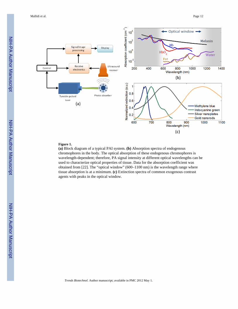
Figure 1.(a) Block diagram of a typical PAI system. (b) Absorption spectra of endogenouschromophores in the body. The optical absorption of these endogenous chromophores iswavelength-dependent; therefore, PA signal intensity at different optical wavelengths can beused to characterize optical properties of tissue. Data for the absorption coefficient wasobtained from [22]. The “optical window” (600–1100 nm) is the wavelength range wheretissue absorption is at a minimum. (c) Extinction spectra of common exogenous contrastagents with peaks in the optical window.
Mallidi et al. Page 12
Trends Biotechnol. Author manuscript; available in PMC 2012 May 1.
NIH
-PA Author Manuscript
NIH
-PA Author Manuscript
NIH
-PA Author Manuscript

Figure 2.(a) Overlaid maximum amplitude projections of PA images at 764 nm and 584 nm showinga tumor and its surrounding vasculature, respectively. The image clearly shows the vesselbranching and structure around the tumor. Adapted with permission from [4]. (b) Images ofthe breast of 57 year old woman with invasive ductal carcinoma: (i) X-ray mammogram; (ii)sonogram; (iii) PA image at 1064 nm. The X-ray mammogram and the sonogram depict thegross anatomical features of the tumor, but donot provide functional information. The highPA amplitude corresponds to abundant vasculature associated with malignant tumors. ThePA image clearly depicts that higher vascular densities are present in the tumor peripheryand the core of the tumor has minimum vasculature. Adapted with permission from [21]. (c)Pancreatic tumor cells were inoculated on a rat hind leg on day 1. PAI was used to monitorangiogenesis associated with the tumor growth. PA images obtained from the tumor regionon days 3, 7, 8 and 10 are maximum intensity projections of the photoacoustic sourcestrength in the xy-plane (i.e. top view on the tumor tissue). Adapted with permission from[5]. (d) In vivo functional imaging of a mouse brain with a glioblastoma xenograft obtainedusing PAI. Spectroscopic PAI (wavelengths from 764 nm to 824 nm) was used to detecthypoxia in a braintumor. The heat map represents the percentage oxygen saturation (SO2) inthe blood vessels (blue = hypoxic; red = hyperoxic). The area indicated by the red arrow isthe tumor. Adapted with permission from [31]. (e) A comparison of normal and brain tumorvasculature SO2in three mice. Three normal vessels and three tumor vessels were chosenfrom each SO2image that had been processed from spectroscopic PA images, such as theone shown in (d). The results clearly indicate that the percentage SO2 in tumors is lowerthan the surrounding normal tissue, thus indicating hypoxia. Adapted with permission from[31].
Mallidi et al. Page 13
Trends Biotechnol. Author manuscript; available in PMC 2012 May 1.
NIH
-PA Author Manuscript
NIH
-PA Author Manuscript
NIH
-PA Author Manuscript

Figure 3.Various optical- and ultrasound-based imaging techniques can be combined with PAI toprovide structural, functional and biomechanical properties of the tissue. (a)In vivo 3D USIand PAI of a subcutaneous tumor in a mouse injected with gold nanorods. The subcutaneoustumor appears as a bump in the 3D ultrasound image. The PA image shows theheterogenous localization of nanorods in tumor, which preferentially accumulate thereowing to the enhanced permeation and retention effect [69]. (b)In vivo PAI and OCT of theskin on the back of a nude mouse. The image represents a data fusion of OCT (structure ofthe skin) and PA (microvasculature) images. Adapted with permission from [64]. (c)Noninvasive in vivo fluorescence (FL) image acquired 24 hours after ICG injection in amouse with melanoma cells implanted in brain. Noninvasive in vivo PA images wereacquired with skin and skull intact, showing the vasculature in the brain. Adapted withpermission from [70]. (d) Gray-scale ultrasound image (left), PA image (center), andelasticity (right) images of a tissue-mimicking phantom with a single inclusion. Theinclusion had higher optical contrast and was harder compared to the background. (e)MMUS (top) and PA (bottom) images of a tissue-mimicking phantom with samplescontaining (left to right):a mixture of Fe3O4 nanoparticles and Au nanospheres; Aunanospheres only; PVA only (no nanoparticles); and Fe3O4 nanoparticles only. The MMUScolormap represents the displacement of the inclusions. The pure PVA sample (nonanoparticles) did not displace under magnetic excitation and showed no PA contrast. TheAu nanospheres also do not displace, but have high optical absorption and hence producegreater PA signals compared with Fe3O4 nanoparticles. The two samples containing Fe3O4nanoparticles had a displacement of about 100 µm [71].
Mallidi et al. Page 14
Trends Biotechnol. Author manuscript; available in PMC 2012 May 1.
NIH
-PA Author Manuscript
NIH
-PA Author Manuscript
NIH
-PA Author Manuscript
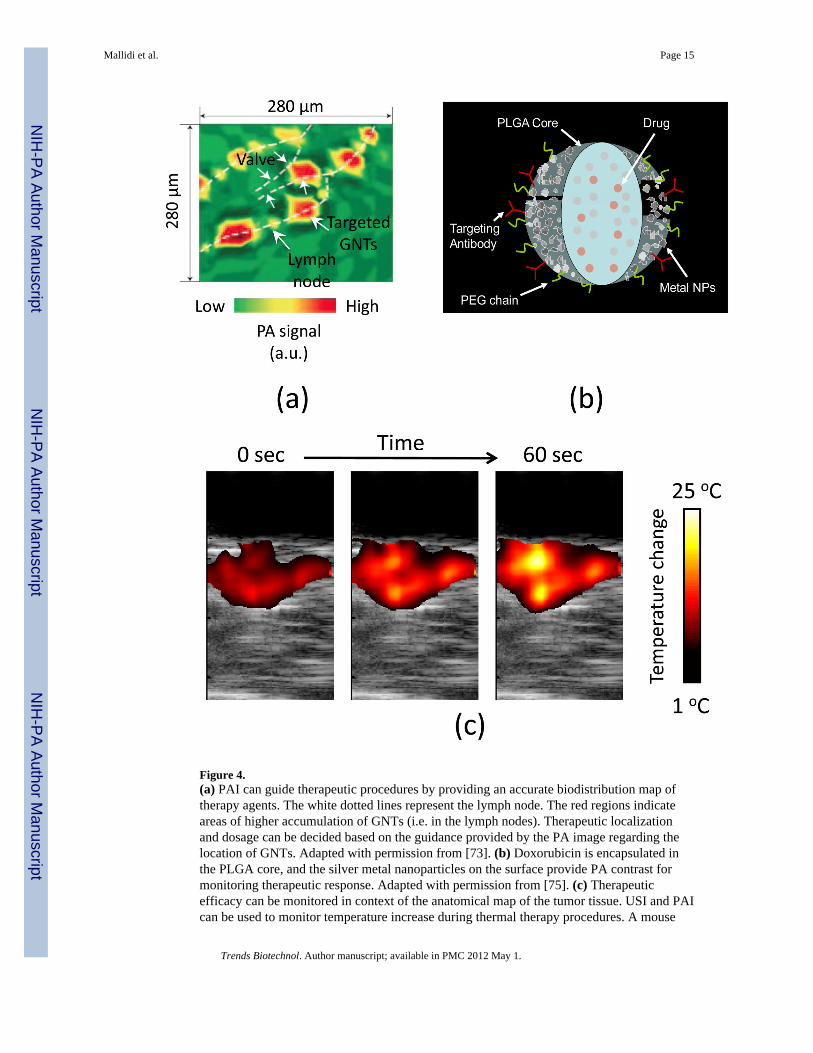
Figure 4.(a) PAI can guide therapeutic procedures by providing an accurate biodistribution map oftherapy agents. The white dotted lines represent the lymph node. The red regions indicateareas of higher accumulation of GNTs (i.e. in the lymph nodes). Therapeutic localizationand dosage can be decided based on the guidance provided by the PA image regarding thelocation of GNTs. Adapted with permission from [73]. (b) Doxorubicin is encapsulated inthe PLGA core, and the silver metal nanoparticles on the surface provide PA contrast formonitoring therapeutic response. Adapted with permission from [75]. (c) Therapeuticefficacy can be monitored in context of the anatomical map of the tumor tissue. USI and PAIcan be used to monitor temperature increase during thermal therapy procedures. A mouse
Mallidi et al. Page 15
Trends Biotechnol. Author manuscript; available in PMC 2012 May 1.
NIH
-PA Author Manuscript
NIH
-PA Author Manuscript
NIH
-PA Author Manuscript

was injected with optically absorbing gold nanorods that acted as both PA and photothermalagents. The tumor region was then irradiated with continuous-wave laser light at the peakabsorption wavelength. Images taken after 0, 30 and 60 seconds of treatment indicate thatthe relative temperature rise in the tumor is 25 °C. Each image represents a 10.5 mm × 20mm field of view.
Mallidi et al. Page 16
Trends Biotechnol. Author manuscript; available in PMC 2012 May 1.
NIH
-PA Author Manuscript
NIH
-PA Author Manuscript
NIH
-PA Author Manuscript

NIH
-PA Author Manuscript
NIH
-PA Author Manuscript
NIH
-PA Author Manuscript
Mallidi et al. Page 17
Table I
Common USI and PAI imaging parameters
Parameter Transducerfrequency (MHz)
Axial resolution(µm)
Lateralresolution (µm)
Imaging depth(mm)
Ultrasound 5 150 300 70
Ultrasound 20 75 165 30
Macroscopic PAI 5 150 300 40
Microscopic PAI 50 15 50 3
Optical resolutionPAI
75 15 5 0.7
Trends Biotechnol. Author manuscript; available in PMC 2012 May 1.
NIH
-PA Author Manuscript
NIH
-PA Author Manuscript
NIH
-PA Author Manuscript
Mallidi et al. Page 18
Table 1
Exogenous PA contrast agents used for detecting tumors in vivo
Contrast agent λpeak (nm) Tumor type Target Refs.
IRDye800a 800 Glioblastoma Integrin αvβ3 [31]
IRDye800 800 Breast cancer Neutropilin-1 receptor [36]
Gold nanocages 778 Melanoma Melanocyte-stimulatinghormone
[39]
Gold nanorods 785 or 1000 Squamous cellcarcinoma
HER2 and EGFR [41]
Gold nanorods 810 Prostate cancer HER2 [42]
Gold nanoshells 800 Colon carcinoma None [43]
Gold nanospheres 525 Breast cancer None [45]
Green-dye-enhancedsingle-walled carbonnanotube (SWNT-ICG)
780 Glioblastoma Integrin αvβ3 [49]
Silver nanoplates 1064b Pancreatic cancer None [50]
SWNT 750c Glioblastoma Integrin αvβ3 [51]
SWNT 690c Glioblastoma Integrin αvβ3 [52]
aIRDye800 (Li-Cor) was conjugated with a cyclic peptide that targets integrin αvβ3.
bλpeak indicates the optical absorption peak of the nanoparticles and the wavelength at which PAI was performed.
cNanoparticles such as carbon nanotubes do not have distinct optical absorption peaks in the optical window. For such nanoparticles, an optimum
wavelength is chosen by the researchers to obtain PA image with enhanced contrast. In the table, the wavelength at which PAI was performed isconsidered the ‘imaging’ rather than the ‘peak’λ.
Trends Biotechnol. Author manuscript; available in PMC 2012 May 1.
Related Documents