Photic history modifies susceptibility to retinal damage in albino trout W. TED ALLISON, 1,2 TED E. HALLOWS, 3 TRUDI JOHNSON, 2 CRAIG W. HAWRYSHYN, 2,5 and DONALD M. ALLEN 4 1 The University of Michigan, Department of Molecular, Cellular, and Developmental Biology, Ann Arbor, Michigan 2 Department of Biology, University of Victoria, Victoria, British Columbia, Canada 3 Utah Division of Wildlife Resources, Kamas State Hatchery, Kamas, Utah 4 Department of Science & Mathematics, University of Texas of the Permian Basin, Odessa, Texas (Received November 30, 2004; Accepted September 29, 2005! Abstract Albino vertebrates exposed to intense light typically lose photoreceptors via apoptosis, and thus serve as useful models of retinal degeneration. In contrast, albino rainbow trout exposed to intense light maintain populations of rod and cone nuclei despite substantial damage to rod outer segments ~ ROS!. The aim of this study was to differentiate between two hypotheses that could account for this divergent result: ~1! trout rod nuclei remain intact during light damage, or ~2! rod nuclei die but are replaced by cell proliferation. A further aim was to examine whether photic history modulates retinal damage, as in rodents. Albino and normally pigmented trout were moved from defined photic regimes into full daylight, while some were not moved to serve as protected controls. ROS were always maintained in pigmented fish and in albinos protected from full daylight. In albinos exposed to full daylight, ROS were removed over most of the central retina, whereas rod nuclei were maintained in the outer nuclear layer over 10 days. Pyknotic and TUNEL-labeled rod nuclei were abundant in affected albinos at all time-points tested. Rod death occurred without a decrease in the number of rod nuclei, confirming that proliferation must be replacing cells. Indeed a transient increase in proliferation was observed in retinal progenitors of albinos receiving 5 days of damaging light. This proliferative response was decreased with further damage. Cones remained intact even in areas where rod nuclei had degenerated. Pretreatment with light of moderate versus low intensity light affected the cell death and proliferative responses, and the ectopic localization of rod opsin. We conclude that apoptotic demise of rods, but not cones, occurred during light damage in retinas of albino trout and proliferative responses have a limited a capacity to replace lost rods. Keywords: Apoptosis, Salmonid, Retinal light damage, Proliferating cell nuclear antigen, Oncorhynchus mykiss Introduction Various animal models, including rodents and teleost fish, have been valuable in understanding the toxicology of light damage in the retina. Light damage leads to photoreceptor programmed cell death in albino rodents ~Shahinfar et al., 1991; Li et al., 1996!. Damage can be caused by constant illumination of moderate intensity, or by cyclical illumination of higher intensity ~Organis- ciak & Winkler, 1994!. Factors affecting the susceptibility of photoreceptors to light damage include diet, circadian factors, ocular pigmentation, and prior exposure to light ~ Li et al., 2003!. Recent progress has been made in identifying molecular pathways leading to photoreceptor apoptosis ~ Hao et al., 2002; Reme et al., 2003; Wenzel et al., 2005!. Thus, despite their nocturnal habit and rod-dominated retina, the data from light-damaged rodent retinas have contributed greatly to our understanding of the mechanisms of photoreceptor cell death and retinal degeneration in humans. Surface-dwelling diurnal fishes can be an attractive comple- ment to the study of nocturnal rodents for several reasons. Their higher cone:rod ratio has evolved to function in a bright-light environment, and may be considered more representative of the human fovea. Furthermore, the retina continues to grow through- out the life of the fish, and this feature provides a regenerative potential ~ Easter & Hitchcock, 2000; Stenkamp & Cameron, 2002; Raymond & Hitchcock, 2004!. The growth occurs through two primary mechanisms ~ Lyall, 1957; Ali, 1964; Johns, 1977!: ~1! addition of new retina by the circumferential germinal zone ~CGZ! at the retinal periphery; and ~2! stretching of the more central retina, with progenitor cells producing new rods to main- tain their density ~Johns & Fernald, 1981; Fernald, 1990!. Importantly, continuous ocular growth also gives the teleost retina a regenerative capacity that could facilitate understanding Address correspondence and reprint requests to: Craig W. Hawryshyn, Department of Biology, University of Victoria, Victoria, British Columbia, Canada, V8W 3N5. E-mail: [email protected]; Donald M. Allen, Depart- ment of Science & Mathematics, University of Texas of the Permian Basin, Odessa, TX 79762, USA. E-mail: [email protected] 5 Current address, Professor and Canada Research Chair in Visual Neuroscience and Behavior, Department of Biology, Queens University, Kingston, ON K7L 3N6 Canada. Visual Neuroscience ~2006!, 23, 25–34. Printed in the USA. Copyright © 2006 Cambridge University Press 0952-5238006 $16.00 DOI: 10.10170S0952523806231031 25

Welcome message from author
This document is posted to help you gain knowledge. Please leave a comment to let me know what you think about it! Share it to your friends and learn new things together.
Transcript
Photic history modifies susceptibility to retinal damagein albino trout
W. TED ALLISON,1,2 TED E. HALLOWS,3 TRUDI JOHNSON,2 CRAIG W. HAWRYSHYN,2,5
and DONALD M. ALLEN4
1The University of Michigan, Department of Molecular, Cellular, and Developmental Biology, Ann Arbor, Michigan2Department of Biology, University of Victoria, Victoria, British Columbia, Canada3Utah Division of Wildlife Resources, Kamas State Hatchery, Kamas, Utah4Department of Science & Mathematics, University of Texas of the Permian Basin, Odessa, Texas
(Received November 30, 2004; Accepted September 29, 2005!
Abstract
Albino vertebrates exposed to intense light typically lose photoreceptors via apoptosis, and thus serve as usefulmodels of retinal degeneration. In contrast, albino rainbow trout exposed to intense light maintain populations of rodand cone nuclei despite substantial damage to rod outer segments ~ROS!. The aim of this study was to differentiatebetween two hypotheses that could account for this divergent result: ~1! trout rod nuclei remain intact during lightdamage, or ~2! rod nuclei die but are replaced by cell proliferation. A further aim was to examine whether photichistory modulates retinal damage, as in rodents. Albino and normally pigmented trout were moved from definedphotic regimes into full daylight, while some were not moved to serve as protected controls. ROS were alwaysmaintained in pigmented fish and in albinos protected from full daylight. In albinos exposed to full daylight, ROSwere removed over most of the central retina, whereas rod nuclei were maintained in the outer nuclear layer over10 days. Pyknotic and TUNEL-labeled rod nuclei were abundant in affected albinos at all time-points tested. Roddeath occurred without a decrease in the number of rod nuclei, confirming that proliferation must be replacing cells.Indeed a transient increase in proliferation was observed in retinal progenitors of albinos receiving 5 days ofdamaging light. This proliferative response was decreased with further damage. Cones remained intact even in areaswhere rod nuclei had degenerated. Pretreatment with light of moderate versus low intensity light affected the celldeath and proliferative responses, and the ectopic localization of rod opsin. We conclude that apoptotic demise ofrods, but not cones, occurred during light damage in retinas of albino trout and proliferative responses have alimited a capacity to replace lost rods.
Keywords: Apoptosis, Salmonid, Retinal light damage, Proliferating cell nuclear antigen, Oncorhynchus mykiss
Introduction
Various animal models, including rodents and teleost fish, havebeen valuable in understanding the toxicology of light damage inthe retina. Light damage leads to photoreceptor programmed celldeath in albino rodents ~Shahinfar et al., 1991; Li et al., 1996!.Damage can be caused by constant illumination of moderateintensity, or by cyclical illumination of higher intensity ~Organis-ciak & Winkler, 1994!. Factors affecting the susceptibility ofphotoreceptors to light damage include diet, circadian factors,ocular pigmentation, and prior exposure to light ~Li et al., 2003!.Recent progress has been made in identifying molecular pathways
leading to photoreceptor apoptosis ~Hao et al., 2002; Reme et al.,2003; Wenzel et al., 2005!. Thus, despite their nocturnal habit androd-dominated retina, the data from light-damaged rodent retinashave contributed greatly to our understanding of the mechanismsof photoreceptor cell death and retinal degeneration in humans.
Surface-dwelling diurnal fishes can be an attractive comple-ment to the study of nocturnal rodents for several reasons. Theirhigher cone:rod ratio has evolved to function in a bright-lightenvironment, and may be considered more representative of thehuman fovea. Furthermore, the retina continues to grow through-out the life of the fish, and this feature provides a regenerativepotential ~Easter & Hitchcock, 2000; Stenkamp & Cameron,2002; Raymond & Hitchcock, 2004!. The growth occurs throughtwo primary mechanisms ~Lyall, 1957; Ali, 1964; Johns, 1977!:~1! addition of new retina by the circumferential germinal zone~CGZ! at the retinal periphery; and ~2! stretching of the morecentral retina, with progenitor cells producing new rods to main-tain their density ~Johns & Fernald, 1981; Fernald, 1990!.
Importantly, continuous ocular growth also gives the teleostretina a regenerative capacity that could facilitate understanding
Address correspondence and reprint requests to: Craig W. Hawryshyn,Department of Biology, University of Victoria, Victoria, British Columbia,Canada, V8W 3N5. E-mail: [email protected]; Donald M. Allen, Depart-ment of Science & Mathematics, University of Texas of the Permian Basin,Odessa, TX 79762, USA. E-mail: [email protected]
5Current address, Professor and Canada Research Chair in VisualNeuroscience and Behavior, Department of Biology, Queens University,Kingston, ON K7L 3N6 Canada.
Visual Neuroscience ~2006!, 23, 25–34. Printed in the USA.Copyright © 2006 Cambridge University Press 0952-5238006 $16.00DOI: 10.10170S0952523806231031
25
of repair mechanisms ~reviewed in Raymond & Hitchcock, 1997,2000; Stenkamp et al., 2001; Otteson & Hitchcock, 2003!. Popu-lations of progenitor cells are present in the central retina of fish,including rod progenitors in the outer nuclear layer ~ONL! and theproliferating inner nuclear layer cells ~PINCs!. The latter replenishthe former during natural development ~Julian et al., 1998!. ThePINCs may be replenished by glia dedifferentiating to proliferatingcells ~Fischer & Reh, 2003!. Thus, in addition to being effectivemodels of photoreceptor degeneration, the light-challenged teleostretina might also contribute to understanding how stem cells canbe signaled to replace lost photoreceptor cells.
Rainbow trout are an effective model for the study of light dam-age and regeneration in the retina. The cone photoreceptors, be-longing to four spectral classes, are patterned in a mosaic ~Browman& Hawryshyn, 1992; Beaudet et al., 1993; Beaudet et al., 1997;Allison et al., 2003! that can facilitate an understanding of mech-anisms of cone differentiation ~reviewed in Stenkamp & Cameron,2002!. The potential to regenerate photoreceptors in rainbow troutappears to be substantial, as indicated by the reappearance ofultraviolet-sensitive cones late in the fish’s natural life history ~Beau-det et al., 1997; Hawryshyn et al., 2003; Allison et al., 2005!. ThePINCs in rainbow trout increase their rate of proliferation in re-sponse to surgical injury ~Faillace et al., 2002!. Most importantly,raising albino rainbow trout in full sunlight or exposing them to3000 lux constant incandescent light leads to the loss of rod outersegments ~ROS! ~Allen & Hallows, 1997; Allen et al., 1999!.
Remarkably, the number of nuclei in the ONL, dominated byrod nuclei, did not decrease ~Allen & Hallows, 1997!. This resultdiffered from observations on light damage in rodents and otherfish, where the number of photoreceptor cells is reduced ~Allenet al., 1999; Vihtelic & Hyde, 2000!. It has been demonstrated thatthe loss of ROS in rainbow trout is localized to the central, matureretina ~Allen et al., 2001!. When albino trout were transferred todim light conditions following damage, the ROS and scotopicvisual sensitivity reappeared ~Allen & Hallows, 1997!. The dataled to the formation of two hypotheses ~Allen & Hallows, 1997!,which are not mutually exclusive of each other: ~1! that rods weresurviving the light damage, that is, ROS were pruned from the rodnuclei, which elaborated new ROS when fish were transferred todim-light conditions; and ~2! that rods were dying but beingreplaced immediately by generation of new rods from progenitorcells. The primary objective of this work was to discriminatebetween these two hypotheses.
Our examinations of cell death and proliferation in the currentwork support hypothesis ~2! above, that is, rods are dying andbeing replaced through the proliferation of retinal progenitors.During this experiment we found an unexpected decrease in thenumber of ONL nuclei in localized areas of the central retina ofaffected albinos, which had not been observed previously in trout.We hypothesized that this increased susceptibility resulted frompreexposure to moderately intense cyclic light rather than to beingreared in full daylight as in earlier studies. This hypothesis wasverified under a repeated protocol using two different pretreatments.
Materials and methods
Fish handling and dissections
Fish were maintained at Kamas State Fish Hatchery in SummitCounty, UT. The albino trout ~Oncorhynchus mykiss! utilized inthis study have an autosomal recessive mutation in the tyrosinasegene ~Bridges & von Limbach, 1972!.
Series 1On July 22, 2001, we transferred albino and normally pig-
mented rainbow trout from a covered raceway ~where they hadbeen raised completely protected from direct sunlight! into an openraceway with little opportunity for behavioral avoidance of sun-light. This methodology differed from previous experiments wheretrout were raised in outdoor raceways and sampled at various timesof the year ~Allen & Hallows, 1997; Allen et al., 1999, 2001!. Fishwere transferred serially such that eyes could be sampled afterperiods of 0, 2, 5, 10, and 20 days of exposure. Normally pig-mented fish ranged from 6.5 cm to 9.0 cm in standard length,whereas albinos ranged from 9 cm to 13 cm and were 2 monthsolder than the normally pigmented fish. This minor difference inage is unlikely to affect the qualitative outcomes of the experiment~as confirmed by our results from fish used in Series 2!. Regard-less, we do not statistically compare results between pigmentedand albino fish.
Series 2On July 26, 2002, fish which had been kept under indoor cyclic
fluorescent ~relatively dim! light for 1 month remained in place~not-pretreated! or were placed outdoors in shaded ~moderatelyintense! daylight for 10 days ~pretreated!. Fish from both groupswere identified by fin-clip and then placed in full sunlight such thatpretreated and not-pretreated fish were sampled after 0, 2, 5, and10 days. The 10-day duration of damage was selected because thisrepresented the time-point with the most dramatic change inproliferating cell nuclear antigen ~PCNA! and TUNEL labelingduring the previous experiment. Several of the albino fish assignedto the pretreated 0-day treatment group were lost to garter snakepredation, leaving fewer individuals per time-point for examina-tion. In these experiments ~Series 2!, albino and normally pig-mented trout were age-matched and ranged from 9 cm to 12 cm attime of sampling.
Light levels were measured at the water surface with a GammaScientific spectroradiometer. Daily fluctuation in daylight intensitywas measured with a recording pyroheliometer on site. Indoor fishwere on a 12-h light012-h dark cycle under fluorescent lights,whereas fish outdoors were exposed to 13.7-h light010.3-h darkduring treatment. The light levels for outdoor exposed fish, mea-sured at about noon on a clear day, was approximately 4 � 1016
Photons{cm�2{nm�1{s�1 integrated over 400–700 nm. The pre-treated trout ~in covered raceways! received light levels approxi-mately two orders of magnitude less than the exposed fish. Thenot-pretreated trout ~maintained indoors! were exposed to lightlevels approximately half the intensity of pretreated trout.
Enucleated eyes were immediately placed into ice-chilled fix-ative as appropriate to each method ~below!. The care and use ofanimals conformed to the principles adopted by the AmericanPhysiological Society and the Society for Neuroscience and theUniversity of Victoria Animal Care Committee under the auspicesof the Canadian Council on Animal Care.
Histology and labeling
Semithin sectioning was performed on eyes fixed in 2% glutar-aldehyde–2% paraformaldehyde in phosphate buffer ~pH 7.2! andembedded in epoxy resin as described previously ~Allen & Hal-lows, 1997; Allen et al., 1999, 2001!. The sclera was sliced opento admit fixative and in a manner to define the orientation ofthe eye.
26 W.T. Allison et al.
Other eyes from each treatment were processed for TUNELlabeling. They were fixed in 4% paraformaldehyde in phosphatebuffered saline ~PBS! and held on ice. These eyes were shippedovernight to Victoria, BC. They were rinsed in PBS and dissectedinto four quadrants. The embryonic fissure, which reaches theperipheral retina at the ventral-nasal aspect of the eye, was used asa landmark. Eyes were cryopreserved, frozen, and sectioned ~10-µmthickness! as described previously ~Allison et al., 2003!. Each ofthe four quadrants was frozen such that radial sections wereexamined from a known location in the retina. After rehydration,the sections were treated with 10 mg0ml proteinase K at 378C andpostfixed in 4% paraformaldehyde, using nuclease-free solutions.TUNEL labeling and negative controls were performed as permanufacture’s protocols ~Roche Biochemicals, Nutley, NJ!. Thesignal produced by TUNEL labeling was a green fluorescentproduct, which was visualized and documented as described pre-viously ~Allison et al., 2003!.
Other eyes from each treatment were processed for PCNAimmunohistochemistry to mark proliferative cells. Eyes wereplaced in cold fixative composed of 38% formaldehyde dilutedin 9 parts ethanol. These eyes were also shipped to Victoria,BC. The eyes were hemisected along the nasal-temporal axisand cryopreserved as above. Sectioning produced radial sections~10 mm! along the nasal-temporal axis. Sections were processedfor PCNA immunohistochemistry using standard protocols asreported previously ~Allison et al., 2003!. Primary antibody wasmouse-anti-PCNA ~PC-10; Santa Cruz Biotechnology, SantaCruz, CA! and secondary antibody was goat-anti-mouse conju-gated to alkaline phosphatase ~Vector Labs, Burlingame, CA!.Both antibodies were diluted 1:1000 in PBS with 1:50 heat-inactivated horse serum ~Sigma, St. Louis, MO!. Signal wasvisualized using BCIP0NBT as per manufacturer’s protocol~Gibco0BRL, Gaithersburg, MD!, which produces a dark purpleprecipitate.
Other immunohistochemistry was performed on sections adja-cent to those used for TUNEL labeling. Primary antibodies weremouse-anti-bovine rhodopsin ~K16-155C, provided by Dr. Har-grave, University of Florida, Gainesville ~Adamus et al., 1991!!,previously shown to label rods and one member of the doublecones in rainbow trout ~Veldhoen, 1996!. Presumably, the doublecone labeling represents the medium-wavelength sensitive conethat expresses a subclass of the rhodopsin gene, Rh2 ~Allison et al.,2003; Dann et al., 2004!. We also employed mouse monoclonalantibodies zpr-1 and zpr-3 ~Zebrafish International Resource Cen-ter, Eugene, OR! raised against zebrafish retinal antigens ~Larison& Bremiller, 1990!, that label double cones and rods of zebrafish,respectively. These antibodies were diluted 1:250 and visualizedwith 1:1000 goat-anti-mouse conjugated to Alexa-Fluor-594 ~redfluorescent signal; Molecular Probes, Eugene, OR!. Labeled cellswere counted in each section and the length of the section mea-sured using image analysis software ~Northern Eclipse, EmpixImaging, Inc., Cheektowaga, NY!. For PCNA quantifications thelabeled cells of the CGZ were not included in the counts. To assessproliferation in the central retina, we also counted the number ofPCNA labels in a single field of view ~approximately 300 mm! ofnasal retina that was one field of view removed from the opticnerve. We examined eyes from three individuals per treatment forthe Series 1 TUNEL measurements, and four individuals for allother analyses. In each instance we counted labeled cells from fourretinal sections. We compared values amongst treatments in SPSSsoftware using analysis of variance with a post-hoc pair-wiseTukey test ~a � 0.05!.
Results
Histology of light-damaged retinae
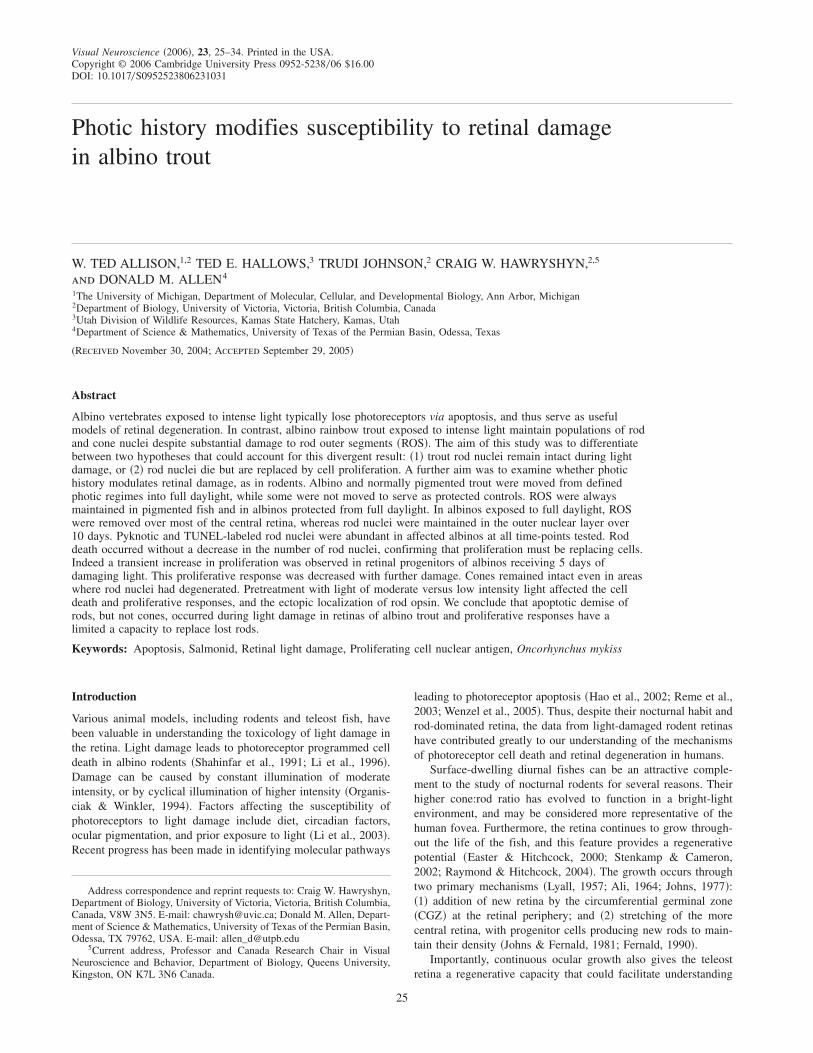
In pigmented fish, regardless of light treatment, sections in radialplane demonstrated that rod outer segments ~ROS! and normalretinal structure were maintained ~Fig. 1A!, although ROS wereshortened ~Table 1!. ROS were also maintained in albinos pro-tected from sunlight ~Fig. 1B! in both years. This was consistentwith previous light-damage paradigms on these fish ~Allen &Hallows, 1997; Allen et al., 1999, 2001!.
ROS were lost from central retinas of albino trout in fulldaylight ~Fig. 1C!, as expected. Rod nuclei were maintainedexcept in limited portions of the central retina of some individualsfrom day 2 to day 10 ~Table 1, Fig. 1D!. This limited ONL loss hadnot been observed in albino trout during previous experiments~Allen & Hallows, 1997; Allen et al., 1999, 2001! where the fishhad been raised in outdoor raceways. However, in much of thedamaged albino retina, rod nuclei were maintained despite ROSelimination. Also, rods close to the peripheral margins ~typicallywithin the most peripheral 0.5 mm of a 5–7 mm retinal circumfer-ence! maintained ROS, as observed previously ~Allen et al., 2001!.Cones remained intact in all retinal samples, including those inwhich rod nuclei had degenerated ~Fig. 1D!. Pyknotic nuclei wereobserved in all light-damaged albinos. Pyknotic nuclei were local-ized to the vitread half of the ONL in the central retina ~Fig. 1!.
TUNEL labeling increased during light damage
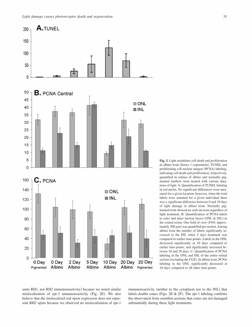
TUNEL labeling provided a clear demonstration that many rodphotoreceptors were dying during light treatment of albino fish inSeries 1 experiments ~Figs. 2A & 2B!. The TUNEL-positive nucleiwere localized to the vitread half of the ONL, and thus were rodnuclei. This was the same location where pyknotic nuclei wereobserved in semithin sections ~Fig. 1!. Furthermore, the increasesin TUNEL-positive rod nuclei were limited to the central retina,and were almost absent in the peripheral retina where ROS weremaintained ~Figs. 2A & 2B!. Some TUNEL-positive nuclei wereobserved in the peripheral retina, as expected in developing retinalareas ~Biehlmaier et al., 2001; Candal et al., 2005!. TUNEL-positive nuclei did not change in abundance in pigmented fishdespite light treatment ~Fig. 3A!. Low levels of TUNEL-positivenuclei were observed in untreated albinos, similar to pigmentedfish. However, each of the 12 albino fish treated with damaginglight had elevated TUNEL-positive nuclei in some location of theretina. TUNEL-positive nuclei in albino trout treated for 10 dayswere significantly higher than 0 days ~P � 0.030!. There was anobvious qualitative increase in cell death at the earliest duration oftreatment we examined ~2 days!. It is noteworthy that the increasesin TUNEL-positive label were observable coincident with thedisappearance of rod nuclei in central locations of histologicalsamples ~Table 1!. The significant increase in TUNEL labelingwas confirmed in Series 2 experiments, described below.
PCNA labeling transiently increased with light damage
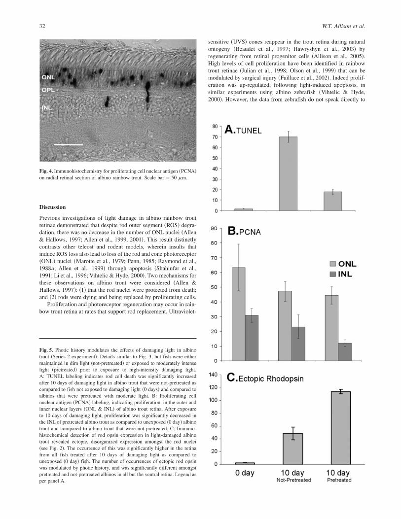
PCNA labeling ~Fig. 4! revealed the expected populations ofdividing cells based on previous examinations of rainbow trout~Julian et al., 1998! and other salmonids ~Candal et al., 2005!. Theperipheral retina ~CGZ! was strongly labeled and clusters ofdividing cells were apparent in both the outer and inner nuclearlayers ~ONL and INL! of the central retina. Because histological
Light damage causes photoreceptor death and regeneration 27
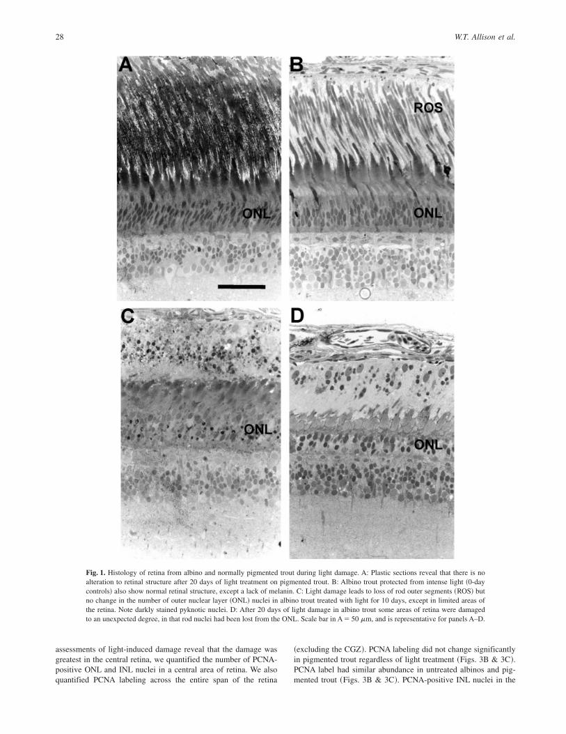
assessments of light-induced damage reveal that the damage wasgreatest in the central retina, we quantified the number of PCNA-positive ONL and INL nuclei in a central area of retina. We alsoquantified PCNA labeling across the entire span of the retina
~excluding the CGZ!. PCNA labeling did not change significantlyin pigmented trout regardless of light treatment ~Figs. 3B & 3C!.PCNA label had similar abundance in untreated albinos and pig-mented trout ~Figs. 3B & 3C!. PCNA-positive INL nuclei in the
Fig. 1. Histology of retina from albino and normally pigmented trout during light damage. A: Plastic sections reveal that there is noalteration to retinal structure after 20 days of light treatment on pigmented trout. B: Albino trout protected from intense light ~0-daycontrols! also show normal retinal structure, except a lack of melanin. C: Light damage leads to loss of rod outer segments ~ROS! butno change in the number of outer nuclear layer ~ONL! nuclei in albino trout treated with light for 10 days, except in limited areas ofthe retina. Note darkly stained pyknotic nuclei. D: After 20 days of light damage in albino trout some areas of retina were damagedto an unexpected degree, in that rod nuclei had been lost from the ONL. Scale bar in A � 50 mm, and is representative for panels A–D.
28 W.T. Allison et al.
central retina ~Fig. 3B! of albinos receiving 5 days of lighttreatment increased significantly as compared to untreated albinos~P � 0.005! or albinos exposed to 2 days of light ~P � 0.001!.
PCNA labeling of the ONL decreased at 10 days of lighttreatment, whether the quantification focused on the central retina~Fig. 3B! or the entire retina ~Fig. 3C!. The number of labelednuclei in the entire ONL was significantly lower at 10 dayscompared to 5 days of light treatment ~P � 0.010!. The number oflabeled nuclei at this location significantly increased between 10and 20 days of light treatment ~P � 0.010!.
Pretreatment light regimes affect cellular responses
We repeated the above experiments using fish that were main-tained in two different photic regimes to assay if this affected thecell death and proliferative responses. Our TUNEL-labeling ex-periment on retinae in Series 2 confirmed the Series 1 results.TUNEL-positive cells were qualitatively increased in each fish andwere localized to the vitread portion of the ONL ~i.e. they were rodnuclei!. TUNEL-positive nuclei were significantly higher duringlight damage ~Fig. 5A! in the retina for not-pretreated ~P � 0.002!and pretreated albino trout ~P � 0.002!. TUNEL-positive nucleiwere significantly lower in pretreated compared to the not-pretreated fish ~P � 0.005!. PCNA-positive ONL nuclei ~Fig. 5B!were significantly less abundant after ten days of treatment inSeries 2 ~P � 0.007!, very similar to Series 1; however, this waslimited to the pretreated retina. Given that reductions in rod nucleireached about the same final level at day 10 ~Table 1! in bothpretreated and not-pretreated groups, a lower death rate must havebeen coincident with a lower replacement rate. Indeed, PCNA-positive nuclei were significantly less abundant in the pretreated as
compared to the not-pretreated fish ~P � 0.010!. Thus, pretreat-ment with moderate intensity daylight led to decreases in bothTUNEL-positive and PCNA-positive labeling of nuclei in the ONLduring subsequent exposure to full daylight, such that the declinein number of rod nuclei were about the same as in the not-pretreated group.
Rod opsin localization as an index of pretreatment effect
We examined the immunohistochemical localization of rod opsinduring light damage as an additional index to measure the effect ofpretreatment photic conditions. Previous work has demonstratedthat opsin mislocalization increases during light damage in teleosts~Vihtelic & Hyde, 2000!. This was not completed on fish fromSeries 1 due to lack of materials. In normally pigmented fish, ourimmunohistochemistry revealed the expected localization of thelabel to the inner and outer segments of rods and one member ofthe double cones ~Fig. 2C!. We observed rod opsin expression tobe mislocalized in ONL of light-damaged albinos ~Fig 2D!. Themislocalization of rod opsin ~Fig. 4C! was significantly higher inthe light-damaged central retinae of pretreated ~P � 0.002! andnot-pretreated fish ~P � 0.015! when compared to controls. Therewas negligible mislocalization in the peripheral retina. The mis-localization of rod opsin was also significantly higher in thepretreated albinos than in the not-pretreated albinos ~P � 0.007!.A parallel to this was the observation of several rod opsin immu-noreactive cells in the INL: mislocalization of rod opsin in theINL was observed in the pretreated albinos ~Fig. 2D!, whereasno such INL mislocalization was observed in the not-pretreatedgroup. The ONL and INL rod opsin labeling appears to representmislocalized expression in rods, not double cones, ~i.e. it repre-
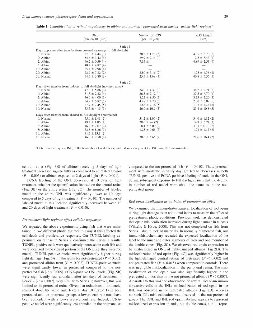
Table 1. Quantification of retinal morphology in albino and normally pigmented trout during various light regimesa
ONL~nuclei0100 mm!
Number of ROS~per 100 mm!
ROS Length~µm!
Series 1Days exposure after transfer from covered raceways to full daylight
0: Normal 57.0 6 4.44 ~3! 30.2 6 1.28 ~3! 47.5 6 6.70 ~3!0: Albino 54.6 6 3.42 ~4! 29.9 6 2.14 ~4! 2.5 6 8.43 ~4!2: Albino 46.2 6 0.59 ~4! 7.35 6— 4.89 6 2.53 ~4!5: Albino 40.2 6 4.87 ~4! — —
10: Albino 25.4 6 2.98 ~4! — —20: Albino 22.9 6 7.82 ~2! 2.80 6 3.16 ~2! 1.25 6 1.76 ~2!20: Normal 54.7 6 3.80 ~3! 25.5 6 1.68 ~3! 40.8 6 5.36 ~3!
Series 2Days after transfer from indoors to full daylight ~not-pretreated!
0: Normal 47.6 6 5.86 ~3! 34.0 6 4.37 ~3! 38.2 6 2.71 ~3!0: Albino 51.5 6 2.32 ~4! 36.5 6 2.12 ~4! 37.5 6 6.70 ~4!2: Albino 36.0 6 4.00 ~3! 8.22 6 8.50 ~3! 3.33 6 2.20 ~3!5: Albino 34.9 6 5.82 ~5! 4.48 6 4.70 ~5! 2.30 6 3.07 ~5!
10: Albino 27.7 6 7.45 ~5! 1.88 6 2.16 ~5! 1.05 6 1.22 ~5!10: Normal 53.5 6 4.13 ~5! 26.9 6 10.9 ~5! 25.4 6 10.8 ~5!
Days after transfer from shaded to full daylight ~pretreated!0: Normal 55.0 6 1.41 ~2! 31.2 6 1.06 ~2! 34.0 6 1.32 ~2!0: Albino 49.7 6 1.06 ~2! 28.0 6— ~2! 14.7 6 5.74 ~2!2: Albino 46.2 6 7.07 ~2! 8.4 6 5.09 ~2! 3.016 0.70 ~2!5: Albino 32.5 6 8.26 ~3! 1.25 6 0.65 ~3! 1.22 6 1.12 ~3!
10: Albino 31.7 6 13.1 ~2! — —10: Normal 46.8 6 2.58 ~2! 30.6 6 5.65 ~2! 21.6 6 16.4 ~2!
aOuter nuclear layer ~ONL! reflects number of rod nuclei, and rod outer segment ~ROS!. “—” Not measurable.
Light damage causes photoreceptor death and regeneration 29
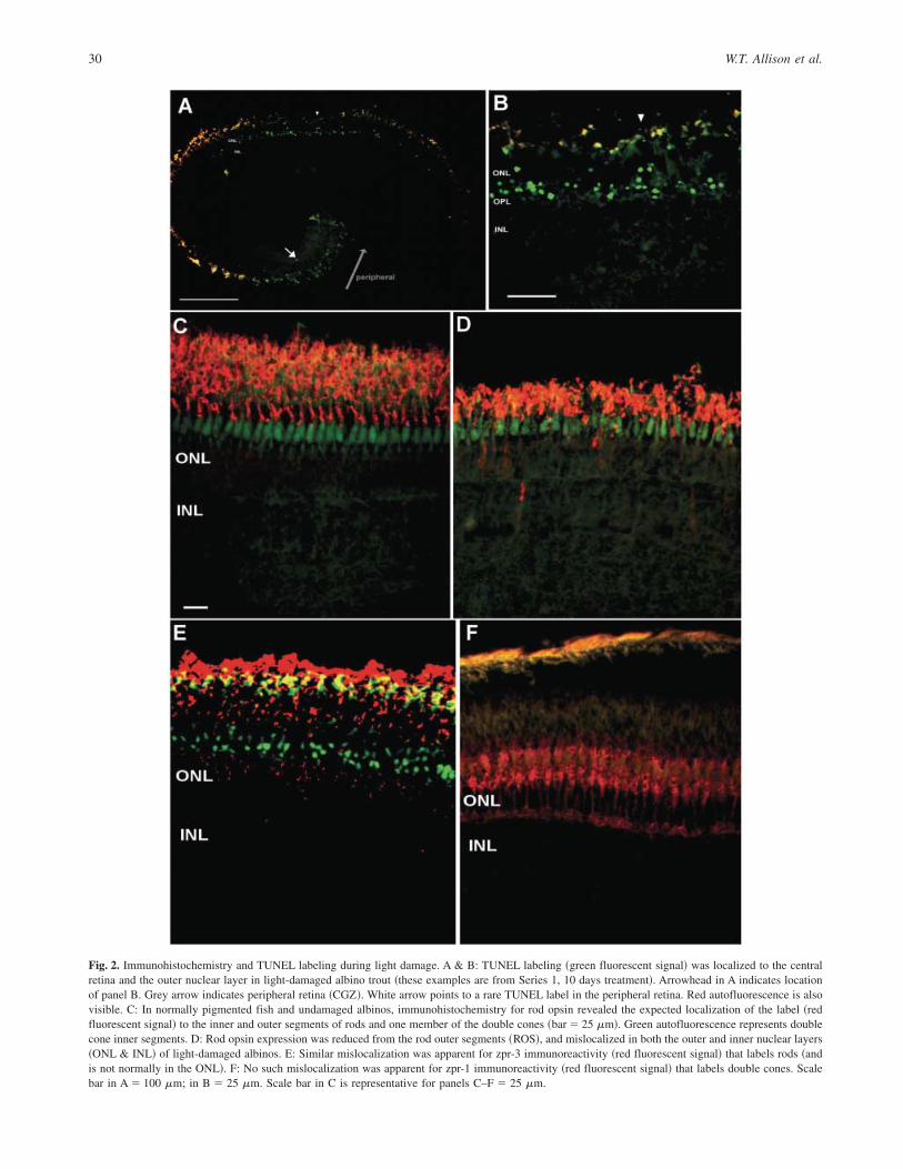
Fig. 2. Immunohistochemistry and TUNEL labeling during light damage. A & B: TUNEL labeling ~green fluorescent signal! was localized to the centralretina and the outer nuclear layer in light-damaged albino trout ~these examples are from Series 1, 10 days treatment!. Arrowhead in A indicates locationof panel B. Grey arrow indicates peripheral retina ~CGZ!. White arrow points to a rare TUNEL label in the peripheral retina. Red autofluorescence is alsovisible. C: In normally pigmented fish and undamaged albinos, immunohistochemistry for rod opsin revealed the expected localization of the label ~redfluorescent signal! to the inner and outer segments of rods and one member of the double cones ~bar � 25 mm!. Green autofluorescence represents doublecone inner segments. D: Rod opsin expression was reduced from the rod outer segments ~ROS!, and mislocalized in both the outer and inner nuclear layers~ONL & INL! of light-damaged albinos. E: Similar mislocalization was apparent for zpr-3 immunoreactivity ~red fluorescent signal! that labels rods ~andis not normally in the ONL!. F: No such mislocalization was apparent for zpr-1 immunoreactivity ~red fluorescent signal! that labels double cones. Scalebar in A � 100 mm; in B � 25 mm. Scale bar in C is representative for panels C–F � 25 mm.
30 W.T. Allison et al.
sents RH1, not RH2 immunoreactivity! because we noted similarmislocalization of zpr-3 immunoreactivity ~Fig. 2E!. We alsobelieve that the mislocalized rod opsin expression does not repre-sent RH2 opsin because we observed no mislocalization of zpr-1
immunoreactivity ~neither to the cytoplasm nor to the INL! thatlabels double cones ~Figs. 2E & 2F!. The zpr-1 labeling confirmsthe observation from semithin sections that cones are not damagedsubstantially during these light treatments.
Fig. 3. Light modulates cell death and proliferationin albino trout ~Series 1 experiment!. TUNEL andproliferating cell nuclear antigen ~PCNA! labeling,indicating cell death and proliferation, respectively,quantified in retinae of albino and normally pig-mented rainbow trout treated with various dura-tions of light. A: Quantification of TUNEL labelingin rod nuclei. No significant differences were mea-sured for a given location; however, when the totallabels were summed for a given individual therewas a significant difference between 0 and 10 daysof light damage in albino trout. Normally pig-mented trout showed no such increase regardless oflight treatment. B: Quantification of PCNA labelsin outer and inner nuclear layers ~ONL & INL! inthe central retina. One field of view ~FOV, approx-imately 300 mm!was quantified per section. Amongalbino trout the number of labels significantly in-creased in the INL when 5 days treatment wascompared to earlier time-points. Labels in the ONLdecreased significantly at 10 days compared toearlier time-points, and significantly increased be-tween 10 and 20 days. C: Quantification of PCNAlabeling in the ONL and INL of the entire retinalsection ~excluding the CGZ!. In albino trout, PCNAlabeling in the ONL significantly decreased at10 days compared to all other time-points.
Light damage causes photoreceptor death and regeneration 31
Discussion
Previous investigations of light damage in albino rainbow troutretinae demonstrated that despite rod outer segment ~ROS! degra-dation, there was no decrease in the number of ONL nuclei ~Allen& Hallows, 1997; Allen et al., 1999, 2001!. This result distinctlycontrasts other teleost and rodent models, wherein insults thatinduce ROS loss also lead to loss of the rod and cone photoreceptor~ONL! nuclei ~Marotte et al., 1979; Penn, 1985; Raymond et al.,1988a; Allen et al., 1999! through apoptosis ~Shahinfar et al.,1991; Li et al., 1996; Vihtelic & Hyde, 2000!. Two mechanisms forthese observations on albino trout were considered ~Allen &Hallows, 1997!: ~1! that the rod nuclei were protected from death;and ~2! rods were dying and being replaced by proliferating cells.
Proliferation and photoreceptor regeneration may occur in rain-bow trout retina at rates that support rod replacement. Ultraviolet-
sensitive ~UVS! cones reappear in the trout retina during naturalontogeny ~Beaudet et al., 1997; Hawryshyn et al., 2003! byregenerating from retinal progenitor cells ~Allison et al., 2005!.High levels of cell proliferation have been identified in rainbowtrout retinae ~Julian et al., 1998; Olson et al., 1999! that can bemodulated by surgical injury ~Faillace et al., 2002!. Indeed prolif-eration was up-regulated, following light-induced apoptosis, insimilar experiments using albino zebrafish ~Vihtelic & Hyde,2000!. However, the data from zebrafish do not speak directly to
Fig. 4. Immunohistochemistry for proliferating cell nuclear antigen ~PCNA!on radial retinal section of albino rainbow trout. Scale bar � 50 mm.
Fig. 5. Photic history modulates the effects of damaging light in albinotrout ~Series 2 experiment!. Details similar to Fig. 3, but fish were eithermaintained in dim light ~not-pretreated! or exposed to moderately intenselight ~pretreated! prior to exposure to high-intensity damaging light.A: TUNEL labeling indicates rod cell death was significantly increasedafter 10 days of damaging light in albino trout that were not-pretreated ascompared to fish not exposed to damaging light ~0 days! and compared toalbinos that were pretreated with moderate light. B: Proliferating cellnuclear antigen ~PCNA! labeling, indicating proliferation, in the outer andinner nuclear layers ~ONL & INL! of albino trout retina. After exposureto 10 days of damaging light, proliferation was significantly decreased inthe INL of pretreated albino trout as compared to unexposed ~0 day! albinotrout and compared to albino trout that were not-pretreated. C: Immuno-histochemical detection of rod opsin expression in light-damaged albinotrout revealed ectopic, disorganized expression amongst the rod nuclei~see Fig. 2!. The occurrence of this was significantly higher in the retinafrom all fish treated after 10 days of damaging light as compared tounexposed ~0 day! fish. The number of occurrences of ectopic rod opsinwas modulated by photic history, and was significantly different amongstpretreated and not-pretreated albinos in all but the ventral retina. Legend asper panel A.
32 W.T. Allison et al.

the above hypotheses regarding rainbow trout. In zebrafish, lightinduced a substantial loss of ONL nuclei and led to cone cell death~Vihtelic & Hyde, 2000!; therefore the mechanisms that maintainthe number of ONL nuclei in trout remain unknown.
Our results demonstrate that cell death occurs in rods of albinotrout exposed to damaging levels of light. Observations duringboth experiments ~Series 1 and 2! demonstrated a qualitativeincrease in TUNEL-positive rod nuclei in each of the 20 fishreceiving light damage. Both TUNEL-positive and pyknotic nucleiwere located in the vitread portion of the ONL, and were observedrarely in the peripheral regions of the retina, where light damage isless pronounced ~current study, Allen et al., 2001!. In Series 1,TUNEL label was increased at all time-points, including a signif-icant increase at 10 days of light damage. In Series 2, TUNEL labelwas significantly increased in fish that were either pretreated ornot-pretreated with light. Thus, three separate trials over 2 yearsdemonstrated a significant increase in TUNEL labeling in rodnuclei during 10 days of damaging light treatment.
The number of ONL nuclei did not decrease within the first10 days of light damage. This constant number of ONL nuclei wasobserved, except in limited portions of the central retina, despiteincreases in pyknotic and TUNEL-positive rod nuclei, and asubstantial loss of ROS. We observed no increase in TUNELlabeling nor ROS loss in normally pigmented trout. Taken together,these data confirm the hypothesis that rods are dying and beingreplaced by the progeny of proliferating cells present in the retina.
Consistent with the above conclusion, PCNA labeling demon-strated that light can modulate proliferation in albino fish. Lighthad no effect on PCNA in pigmented fish at the time-points tested.PCNA labeling in the INL doubled ~a significantly increase! at5 days of damaging light treatment in the central retina. Thisincrease in proliferation occurred at a time when rods were dying~at 2 & 5 days! but when the number of rods was not decreasing.The result is consistent with data ~Julian, 1997, also described inFaillace et al., 2002! that showed increases in PCNA label in thesame brood stock of albino trout as in the current work, and usingpreviously described light treatment regimes ~Allen & Hallows,1997; Allen et al., 1999, 2001!. A similar increase in PINCs hasbeen observed previously in trout after surgical damage ~Faillaceet al., 2002! and zebrafish following light damage ~Vihtelic &Hyde, 2000!.
When the entire span of the retina was considered ~excludingthe CGZ! significant decreases in PCNA labeling were measuredafter 10 days of light damage ~Fig. 3B!. This was coincident withthe peak of TUNEL labeling, and could represent a level ofdamage that includes progenitors. Such speculation would requirethe effect to be transient and include repair mechanisms, becausethe PCNA labeling subsequently increased in a significant manner.This increase in proliferation at 20 days could also be viewed as aresponse to the large loss of rod nuclei that occurred by day 10.
Although significant increases in PCNA labeling were ob-served, the effect of light on proliferation in albino trout did notalways mirror the dramatic increase in cell death. The methods weused allow only a “snap-shot” of the proliferative events thatoccurred. For example, proliferation in the trout retina varies in acircadian rhythm ~Julian et al., 1998!, and an increase in prolifer-ative rate may be more or less apparent during another portion ofthe day. Further experiments should incorporate considerations ofcircadian rhythms in proliferation ~Julian et al., 1998! and utilizecell fate mapping to demonstrate that the rate of rod addition ismodulated by light dosage. Similarly, our hypothesis predicted anincrease in ONL proliferation that was not apparent following the
increase in INL proliferation. Similar observations of increasedINL, but not ONL, proliferation were made by Julian ~1997! in asimilar experiment. It may be that our infrequent sampling regimemissed events in the ONL response. Alternatively, it cannot beexcluded that the INL progenitors are directly replacing lost rodswithout a substantial increase in ONL proliferation. Althoughcurrent models ~Raymond et al., 1988b; Raymond & Hitchcock,2000; Otteson et al., 2001! emphasize PINCs as replacing ONLprogenitors, no data exist to eliminate INL progenitors directlyreplacing lost rods in the absence of an amplifying proliferativeevent in the ONL.
Some rod nuclei were lost in albino trout by 20 days of lightexposure ~Series 1!, although cones remained intact. This resultdiffered from previous examinations of albino trout raised in fullsunlight which had shown no decrease in the number of ONLnuclei at the time of sampling ~Allen & Hallows, 1997; Allen et al.,1999, 2001!. The result has several interesting implications: ~1! itis a further demonstration that some rods in the central retina ofalbino trout can be eliminated by the current light regime, that is,the treatments used led to rod cell death; ~2! increases in prolifer-ative events were insufficient to replace all of the dying rods insome central locations; ~3! maintaining fish in moderately highintensity light has a protective effect and0or enhances the regen-erative capacity of the retina.
The latter implication was supported by experiments in Se-ries 2. Both PCNA and TUNEL labeling were significantly differ-ent between pretreated and not-pretreated groups. We also observeddisorganized and ectopic rod opsin expression in the ONL andINL, reminiscent of ectopic opsin observed in light-damagedzebrafish ~Vihtelic & Hyde, 2000!. The ectopic rod opsin expres-sion may represent degenerating rods and0or new rods that arebeing generated to replace rods and maintain the number of ONLnuclei. Regardless, the TUNEL, PCNA, and rod opsin data allsupport the hypothesis that photic history of the fish can modulatethe retina’s susceptibility to damage and0or its regenerative capac-ity. The TUNEL labeling supports a protective effect of pretreat-ment, as there was significantly less rod apoptosis in the pretreatedgroup. Furthermore, the PCNA data are not consistent with thepretreated fish having increased regenerative capacity. This repre-sents the first report we are aware of where pretreatment with lightmodulated the effects of light damage in a nonmammalian vertebrate.
Acknowledgments
We thank Dr. Paul Hargrave, University of Florida, and the ZebrafishInternational Resource Center, Eugene, OR ~supported by grant #RR12546from the NIH-NCRR! for providing us with antibodies. Support wasprovided by an Ashbel Smith Fellowship ~D.M.A.!, Alzheimer Society ofCanada and Canadian Institutes of Health Research Institute of AgingDoctoral Fellowship ~W.T.A.!, and an Operating Grant from the NaturalScience and Engineering Research Council of Canada ~C.W.H.!.
References
Adamus, G., Zam, Z.S., Arendt, A., Palczewski, K., McDowell, J.H.& Hargrave, P.A. ~1991!. Anti-rhodopsin monoclonal antibodies ofdefined specificity: Characterization and application. Vision Research31, 17–31.
Ali, M.A. ~1964!. Stretching of the retina during growth of salmon ~Salmosalar!. Growth 28, 83–89.
Allen, D.M. & Hallows, T.E. ~1997!. Solar pruning of retinal rods inalbino rainbow trout. Visual Neuroscience 14, 589–600.
Allen, D.M., Hendricks, G.C. & Hallows, T.E. ~2001!. Peripheral rodsevade light damage in albino trout. In New Insights into RetinalDegenerative Diseases, ed. Anderson, R.E., LaVail, M.M. & Hol-
Light damage causes photoreceptor death and regeneration 33
lyfield, J.G., pp. 287–300. New York: Kluwer Academic0PlenumPublishers.
Allen, D.M., Pipes, C., Deramus, K. & Hallows, T.E. ~1999!. Acomparison of light-induced rod degeneration in two teleost models. InRetinal Degenerative Diseases and Experimental Therapy. ed. Holly-field, J.G., Anderson, R.E. & LaVail, M.M., pp. 337–350. NewYork: Kluwer Academic0Plenum Publishers.
Allison, W.T., Dann, S.G., Helvik, J.V., Bradley, C., Moyer, H.D. &Hawryshyn, C.W. ~2003!. Ontogeny of ultraviolet-sensitive cones inthe retina of rainbow trout ~Oncorhynchus mykiss!. Journal of Com-parative Neurology 461, 294–306.
Allison, W.T., Dann, S.G., Veldhoen, K.M. & Hawryshyn, C.W.~2005!. Regeneration of cone photoreceptors during natural develop-ment. Journal of Comparative Neurology ~accepted pending revision,June 28th, 2005!.
Beaudet, L., Browman, H.I. & Hawryshyn, C.W. ~1993!. Optic nerveresponse and retinal structure in rainbow trout of different sizes. VisionResearch 33, 1739–1746.
Beaudet, L., Novales Flamarique, I. & Hawryshyn, C.W. ~1997!.Cone photoreceptor topography in the retina of sexually mature Pacificsalmonid fishes. Journal of Comparative Neurology 383, 49–59.
Biehlmaier, O., Neuhauss, S.C. & Kohler, K. ~2001!. Onset and timecourse of apoptosis in the developing zebrafish retina. Cell and TissueResearch 306, 199–207.
Bridges, W.R. & von Limbach, B. ~1972!. Inheritance of albanism inrainbow trout. Journal of Heredity 63, 152–153.
Browman, H.I. & Hawryshyn, C.W. ~1992!. Thyroxine induces a pre-cocial loss of ultraviolet photosensitivity in rainbow trout ~Oncorhyn-chus mykiss, Teleostei!. Vision Research 32, 2303–2312.
Candal, E., Anadon, R., DeGrip, W.J. & Rodriguez-Moldes, I. ~2005!.Patterns of cell proliferation and cell death in the developing retina andoptic tectum of the brown trout. Brain Research. Developmental BrainResearch 154, 101–119.
Dann, S.G., Allison, W.T., Levin, D.B., Taylor, J.S. & Hawryshyn,C.W. ~2004!. Salmonid opsin sequences undergo positive selection andindicate an alternate evolutionary relationship in Oncorhynchus. Jour-nal of Molecular Evolution 58, 400–412.
Easter, S.S. & Hitchcock, P.F. ~2000!. Stem cells and regeneration in theretina: What fish have taught us about neurogenesis. Neuroscientist 6,454–464.
Faillace, M.P., Julian, D. & Korenbrot, J.I. ~2002!. Mitotic activationof proliferative cells in the inner nuclear layer of the mature fish retina:Regulatory signals and molecular markers. Journal of ComparativeNeurology 451, 127–141.
Fernald, R.D. ~1990!. Teleost vision: Seeing while growing. Journal ofExperimental Zoology ~Suppl.! 5, 167–180.
Fischer, A.J. & Reh, T.A. ~2003!. Potential of Muller glia to becomeneurogenic retinal progenitor cells. Glia 43, 70–76.
Hao, W., Wenzel, A., Obin, M.S., Chen, C.K., Brill, E., Krasnop-erova, N.V., Eversole-Cire, P., Kleyner, Y., Taylor, A., Simon,M.I., Grimm, C., Reme, C.E. & Lem, J. ~2002!. Evidence for twoapoptotic pathways in light-induced retinal degeneration. Nature Ge-netics 32, 254–260.
Hawryshyn, C.W., Martens, G., Allison, W.T. & Anholt, B.R. ~2003!.Regeneration of ultraviolet-sensitive cones in the retinal cone mosaic ofthyroxin-challenged post-juvenile rainbow trout ~Oncorhynchus mykiss!.Journal of Experimental Biology 206, 2665–2673.
Johns, P.R. ~1977!. Growth of the adult goldfish eye. III. Source of the newretinal cells. Journal of Comparative Neurology 176, 343–357.
Johns, P.R. & Fernald, R.D. ~1981!. Genesis of rods in teleost fish retina.Nature 293, 141–142.
Julian, D. ~1997!. Cell birth and fate in the teleost retina. Ph.D. Thesis,pp. 146. University of California, San Francisco, San Francisco,California.
Julian, D., Ennis, K. & Korenbrot, J.I. ~1998!. Birth and fate ofproliferative cells in the inner nuclear layer of the mature fish retina.Journal of Comparative Neurology 394, 271–282.
Larison, K.D. & Bremiller, R. ~1990!. Early onset of phenotype and cellpatterning in the embryonic zebrafish retina. Development 109, 567–576.
Li, F., Cao, W. & Anderson, R.E. ~2003!. Alleviation of constant-light-induced photoreceptor degeneration by adaptation of adult albino rat tobright cyclic light. Investigative Ophthalmology and Visual Science 44,4968–4975.
Li, S., Chang, C.J., Abler, A.S., Fu, J., Tso, M.O. & Lam, T.T. ~1996!. Acomparison of continuous versus intermittent light exposure on apop-tosis. Current Eye Research 15, 914–922.
Lyall, A.H. ~1957!. The growth of the trout retina. Quarterly Journal ofMicroscopy Science 98, 101–110.
Marotte, L.R., Wye-Dvorak, J. & Mark, R.F. ~1979!. Retinotectalreorganization in goldfish–II. Effects of partial tectal ablation andconstant light on the retina. Neuroscience 4, 803–810.
Olson, A.J., Picones, A., Julian, D. & Korenbrot, J.I. ~1999!. Adevelopmental time line in a retinal slice from rainbow trout. Journalof Neuroscience Methods 93, 91–100.
Organisciak, D.T. & Winkler, B.S. ~1994!. Retinal light damage—practical and theoretical considerations. Progress in Retinal and EyeResearch 13, 1–29.
Otteson, D.C., D’Costa, A.R. & Hitchcock, P.F. ~2001!. Putative stemcells and the lineage of rod photoreceptors in the mature retina of thegoldfish. Developmental Biology 232, 62–76.
Otteson, D.C. & Hitchcock, P.F. ~2003!. Stem cells in the teleost retina:Persistent neurogenesis and injury-induced regeneration. Vision Re-search 43, 927–936.
Penn, J.S. ~1985!. Effects of continuous light on the retina of a fish,Notemigonus crysoleucas. Journal of Comparative Neurology 238,121–127.
Raymond, P.A., Bassi, C.J. & Powers, M.K. ~1988a!. Lighting conditionsand retinal development in goldfish: Photoreceptor number and struc-ture. Investigative Ophthalmology and Visual Science 29, 27–36.
Raymond, P.A. & Hitchcock, P.F. ~1997!. Retinal regeneration: Commonprinciples but a diversity of mechanisms. Advances in Neurology 72,171–184.
Raymond, P.A. & Hitchcock, P.F. ~2000!. How the neural retina regen-erates. Results and Problems in Cell Differentiation 31, 197–218.
Raymond, P.A. & Hitchcock, P.F. ~2004!. The teleost retina as a modelfor developmental and regeneration biology. Zebrafish 1, 257–271.
Raymond, P.A., Reifler, M.J. & Rivlin, P.K. ~1988b!. Regeneration ofgoldfish retina: rod precursors are a likely source of regenerated cells.Journal of Neurobiology 19, 431–463.
Reme, C.E., Grimm, C., Hafezi, F., Iseli, H.P. & Wenzel, A. ~2003!. Whystudy rod cell death in retinal degenerations and how? DocumentaOphthalmologica 106, 25–29.
Shahinfar, S., Edward, D.P. & Tso, M.O. ~1991!. A pathologic study ofphotoreceptor cell death in retinal photic injury. Current Eye Research10, 47–59.
Stenkamp, D.L. & Cameron, D.A. ~2002!. Cellular pattern formation inthe retina: Retinal regeneration as a model system. Molecular Vision 8,280–293.
Stenkamp, D.L., Powers, M.K., Carney, L.H. & Cameron, D.A. ~2001!.Evidence for two distinct mechanisms of neurogenesis and cellularpattern formation in regenerated goldfish retinas. Journal of Compar-ative Neurology 431, 363–381.
Veldhoen, K.M. ~1996!. Production of antiserum to a goldfish visualpigment opsin. Biology. M.Sc. Thesis, pp. 80. University of Victoria,Victoria, BC, Canada.
Vihtelic, T.S. & Hyde, D.R. ~2000!. Light-induced rod and cone celldeath and regeneration in the adult albino zebrafish ~Danio rerio!retina. Journal of Neurobiology 44, 289–307.
Wenzel, A., Grimm, C., Samardzija, M. & Reme, C.E. ~2005!. Molec-ular mechanisms of light-induced photoreceptor apoptosis and neuro-protection for retinal degeneration. Progress in Retinal Eye Research24, 275–306.
34 W.T. Allison et al.
Related Documents


