224 BIOCHIMICA ET BIOPHYSICA ACTA BBA 66414 PHOSPHOENOLPYRUVATE CARBOXYLASE DECARBOXYLATION CATALYZED REACTION IN CYTOSOL OF RAT ADIPOSE TISSUE O. MEYUHAS, CH. BOSHWITZ AND L. RESHEF Department of Biochemistry, The Hebrew University, Hadassah Medical School, Jerusalem (Israel) (Received April 8th, 1971) SUMMARY The activity of phosphoenolpyruvate carboxylase (ITP:oxaloacetate carboxy- lyase (transphosphorylating)) in crude extracts of adipose tissue and liver was meas- ured in the direction of phosphoenolpyruvate formation. Knowledge of the maximal rate of this process under these conditions is of importance in the evaluation of the physiological significance of changes in enzyme level induced by various treatments. The substrate (oxaloacetate) may be added either directly or generated (a) from malate catalyzed by malate dehydrogenase (E-malate :NAD + oxidoreductase, EC 1.1.1.37) in the presence of NAD+ and pyruvate, (b) from aspartate and a-ketoglutarate, cata- lyzed by aspartate aminotransferase (L-aspartate:2-oxoglutarate aminotransferase, EC 2.6.1.1, formerly known as glutamate:oxaloacetate transaminase). All these yielded a similar maximal activity. With the adipose tissue enzyme, the Km value for oxaloacetate, whether supplied directly or generated was 2.2-1o -5 M. With liver enzyme a similar Km value was obtained only if oxaloacetate was generated, while a higher value (3" IO-~ M) was found by directly adding oxaloacetate. Both oxaloacetate and malate, at high concentrations inhibit phosphoenol- pyruvate carboxylase activity. The inhibition of the adipose tissue enzyme, but not the liver enzyme, may be overcome by the addition of pyruvate. In the presence of pyruvate, Vmax for phosphoenolpyruvate formation was attained with 3-IO -3 M malate, as a source of oxaloacetate. Half maximal activity was found with 2.3" lO _4 M malate. This concentration of malate is physiological and therefore implies that the activity of the enzyme in vivo rarely exceeds its half maximal activity. INTRODUCTION The activity of the phosphoenolpyruvate carboxylase (ITP:oxaloacetate car- boxy-lyase (transphosphorylating)) in crude tissue extracts is determined by following either the rate of oxaloacetate decarboxylation 1 or the carboxylation of phosphoenol- pyruvate 2 as outlined by the following reaction: Biochim. t~iophys. Acta, 25 ° (1971) 224-237

Welcome message from author
This document is posted to help you gain knowledge. Please leave a comment to let me know what you think about it! Share it to your friends and learn new things together.
Transcript
224 BIOCHIMICA ET BIOPHYSICA ACTA
BBA 66414
PHOSPHOENOLPYRUVATE CARBOXYLASE DECARBOXYLATION CATALYZED REACTION IN CYTOSOL OF RAT ADIPOSE TISSUE
O. MEYUHAS, CH. B O S H W I T Z AND L. R E S H E F
Department of Biochemistry, The Hebrew University, Hadassah Medical School, Jerusalem (Israel)
(Received April 8th, 1971)
SUMMARY
The activity of phosphoenolpyruvate carboxylase (ITP:oxaloacetate carboxy- lyase (transphosphorylating)) in crude extracts of adipose tissue and liver was meas- ured in the direction of phosphoenolpyruvate formation. Knowledge of the maximal rate of this process under these conditions is of importance in the evaluation of the physiological significance of changes in enzyme level induced by various treatments. The substrate (oxaloacetate) may be added either directly or generated (a) from malate catalyzed by malate dehydrogenase (E-malate :NAD + oxidoreductase, EC 1.1.1.37) in the presence of NAD+ and pyruvate, (b) from aspartate and a-ketoglutarate, cata- lyzed by aspartate aminotransferase (L-aspartate:2-oxoglutarate aminotransferase, EC 2.6.1.1, formerly known as glutamate:oxaloacetate transaminase). All these yielded a similar maximal activity. With the adipose tissue enzyme, the Km value for oxaloacetate, whether supplied directly or generated was 2.2-1o -5 M. With liver enzyme a similar Km value was obtained only if oxaloacetate was generated, while a higher value (3" IO-~ M) was found by directly adding oxaloacetate.
Both oxaloacetate and malate, at high concentrations inhibit phosphoenol- pyruvate carboxylase activity. The inhibition of the adipose tissue enzyme, but not the liver enzyme, may be overcome by the addition of pyruvate. In the presence of pyruvate, Vmax for phosphoenolpyruvate formation was attained with 3-IO -3 M malate, as a source of oxaloacetate. Half maximal activity was found with 2.3" lO _4 M malate. This concentration of malate is physiological and therefore implies that the activity of the enzyme in vivo rarely exceeds its half maximal activity.
INTRODUCTION
The activity of the phosphoenolpyruvate carboxylase (ITP:oxaloacetate car- boxy-lyase (transphosphorylating)) in crude tissue extracts is determined by following either the rate of oxaloacetate decarboxylation 1 or the carboxylation of phosphoenol- pyruvate 2 as outlined by the following reaction:
Biochim. t~iophys. Acta, 25 ° (1971) 224-237

PHOSPHOENOLPYRUVATE CARBOXYLASE IN ADIPOSE TISSUE 225
Oxa loace t a t e + I T P .~ p h o s p h o e n o l p y r u v a t e + I D P + CO 2
Both of these reactions were described by UTTER AND KURAHASHI 3,4. The oxalo- acetate decarboxylation reaction was employed by NORDLIE AND LARDY 1 and in- volved a chemical determination of phosphoenolpyruvate formed by the ITP depen- dent decarboxylation of oxaloacetate. This method proved not to be sensitive enough as it failed to detect low enzyme activities like those found in adipose tissue 5. On the other hand, these low enzyme activities could be measured by the carboxylation reac- tion which determined the rate of NaH14C03 fixation in the presence of ITP, Mn 2÷ and phosphoenolpyruvate ~.
The use of this method revealed that the activity of adipose tissue phosphoenol- pyruvate carboxylase is subject to remarkable changes induced by a variety of treat- ments2, 6-s. These changes were closely co-related with those found in the rate of the glyceride glycerol synthesis from pyruvate s. Thus, a role for this enzyme in the regula- tion of the glyceroneogenic activity was indicated. To substantiate this indication and to show that this enzyme may be rate limiting in the glycerogenesis from pyruvate, the activity of the enzyme in the direction of glycerogenesis, i.e. decarboxylation, has to be established. However, the enzyme activity in adipose tissue has so far been assayed only by the carboxylation method and the rate of the decarboxylation cata- lyzed reaction is not known. Recently, CHANG et al. 9 who studied the kinetics of a purified mitochondrial enzyme preparation from pig liver, have shown that the rate of the oxaloacetate decarboxylation was I0 times faster than the rate of phospho- enolpyruvate carboxylation. I f this ratio also holds for the adipose tissue enzyme, the rate of the decarboxylation reaction would exceed the rate of the glyceride glycerol synthesis by more than an order of magnitude, and it would be impossible to maintain the supposition that phosphoenolpyruvate carboxylase indeed is a key enzyme in the synthesis of the glyceride glycerol.
Consequently, it became important to obtain direct measurements of the de- carboxylation activity in crude extracts of adipose tissue, and to quantitate the amount of the enzyme in this way.
EXPERIMENTAL PROCEDURE
A n i m a l s
Male albino rats from the Jerusalem Hebrew University breeding center (Wistar Origin) were fed on a local diet with a composition similar to the purina chow. Adrenalectomized rats were maintained on I~o NaC1 in the drinking water and used on the 5- Io th post-operative days. The rats used throughout the experiments were adrenalectomized and fasted for 24 h unless indicated otherwise in the text.
Mater ia l s
Malate, phosphoenolpyruvate, ITP, IDP, GTP, CTP, UTP, NAD +, NADH, L-aspartate and a-ketoglutarate were all Sigma preparations and used without further purification. NAD+-malate dehydrogenase (L-malate:NAD + oxidoreductase, EC i .I . 1.37) (specific activity 820 units/mg protein) was from Sigma. Lactate dehydro- genase (L-lactate :NAD + oxidoreductase, EC I.I.I.27) free of pyruvate kinase activity (specific activity of 360 units/mg protein) and pyruvate kinase (ATP :pyruvate phos-
Biochim. Biophys. Acta, 250 (1971) 224-237
226 O. MEYUHAS et al.
photransferase, EC 2.7.1.4o) (specific activity of I5o units/rag protein) were purchased from Boehringer and Sohne. Aspartate aminotransferase (L-aspartate:2-oxoglutarate aminotransferase, EC 2.6.1.1 formerly known as glutamate:oxaloacetate trans- aminase) was prepared from rat hearts. Supernatants obtained by centrifugation at I00 000 × g for 30 min were used without further purification. NaH'4CO3 was from Radiochemical Centre, Amersham, England.
Enzyme assay Adipose tissue was homogenized in 4 vol. of buffered sucrose 1° and liver in 9 vol.
of the same solution. Supernatants obtained by centrifugation at IOO ooo × g were used for enzyme assays.
Phosphoenolpyruvate carboxylase assay method Carboxylation assay. This was performed essentially according to CHANG AND
LANE 11 as adapted for crude adipose tissue extracts by BALLARD et al. 2. The fixation of "C labeled NaHlaCO3 was measured in the presence of phosphoenolpyruvate, IDP, MnC12, dithiothreitol, NADH, malate dehydrogenase and tissue extract. The following concentrations were included in the reaction mixture: Imidazole buffer, pH 6.6, ioo raM; phosphoenolpyruvate, 15 raM; MnC12, 20 mM; IDP, 1.25 raM; KHC03, 50 mM (containing 2 #C of NaH14COa); dithiothreitol, I raM; NADH, 2.5 raM; 2 units of malate dehydrogenase; and tissue extract in a total volume of i.o ml. The assay was carried out for IO and 20 rain, and the reaction was stopped by addition of trichloro- acetic acid to a final concentration of 3.30/0 . After each tube was gassed with CO 2 for 5 rain, the radioactivity was determined with Diotol as solvenO 2 in a Packard tricarb liquid scintillation spectrometer. A unit of enzyme activity is defined as i #mole of bicarbonate fixed per rain, at 37 °.
Decarboxylation assay. (a) Oxaloacetate added directly. The ITP and Mn 2+ depen- dent decarboxylation of oxaloacetate was measured by the determination of phospho- enolpyruvate formed at the end of the reaction. The assay reaction mixture contained the following components at their final concentrations. Tris-HC1 buffer, pH 8, 50 mM; MnCI 2, 0.75 raM; ITP, 0.75 mM; oxaloacetate, o.15 raM; and tissue extract in a total volume of I.O ml. The reaction mixture was initiated by the addition of freshly dis- solved oxaloacetate. At the end of 4 min and 2 rain at 37 ° for adipose tissue and liver supernatants respectively, the content of each tube was completely transferred to a tube containing 0. 9 ml of boiling water in a boiling water bath. Following 5 min boiling, the tubes were cooled in an ice bath, centrifuged and phosphoenolpyruvate determined by the method of CzoK AND ECKERT 13. Prior to the addition of pyruvate kinase, lactate dehydrogenase NADH and ADP were added to each tube for the removal of pyruvate. Tben, the amount of phosphoenolpyruvate was determined by the amount of NADH oxidized upon the addition of pyruvate kinase. I unit of enzyme activity is the amount of tissue supernatant which will catalyze the ITP dependent formation of I / ,mole of phosphoenolpyruvate per min at 37 °.
(b) Oxaloacetate generated from malate. In assay systems where oxaloacetate was generated from malate, the generating system contained the following components in their final concentrations. Tris-HC1 buffer, pH 8, 50 raM; malate dehydrogenase 1-2 units (not necessary to add) ; NAD +, I mM; MnCI 2, 0.75 mM; and malate, 20 mM;
Biochim. Biophys. Acta, 250 (1971) 224-237

PHOSPHOENOLPYRUVATE CARBOXYLASE IN ADIPOSE TISSUE 227
ITP, o.75 mM. The reaction mixture was brought to an equilibrium by incubation for 3 min at 37 ° followed by the addition of tissue supernatant which was equilibrated for another min. Then ITP was added to initiate phosphoenolpyruvate carboxylase activity. Henceforth the procedure was as outlined for oxaloacetate.
(c) Oxaloacetate generated from aspartate. In this case, similar to the previous generation system, an equilibrium of the system was first at tained by the addition of aspartate, a-ketoglutarate and aspartate aminotransferase =prerequisi te for maxi- mal activity) and incubation for 3 rain at 37 ° followed by the addition of tissue super- natant for another rain. Then phosphoenolpyruvate carboxylase activity was initiated by ITP. The complete assay system in total volume of i ml contained the following components in their final concentration: Tris-HC1 buffer, 50 raM; I-aspar- tate, i mM; a-ketoglutarate, I raM; MnC12, 0.75 raM; aspartate aminotransferase, 0.5 unit ; ITP, 0.75 mM; and tissue supernatant. At the end of the reaction, phospho- enolpyruvate was determined as previously described.
Aspartate aminotransferase activity This was determined spectrophotometrically according to BERGMEYER AND
BERNT 14.
Equilibrium constants The equilibrium constant used for the malate dehydrogenase system was taken
from STERN e[ al. 15.
T A B L E I
REQUIREMENTS FOR PHOSPHOENOLPYRUVATE CAROBOXYLASE DECARBOXYLATION CATALYZED REACTION
Adipose tissue and liver extracts were used as enzyme preparat ions. The reaction mixture con- tained the following components in their final concentrat ion :5omM Tris-HC1 buffer ,pH 8.o ; o. 75 mM ITP ; 0.75 mM MnCl~; o.i 5 mM. Oxaloacetic acid (freshly dissolved) and enzyme (o.o 5 ml of adipose tissue and 0.02 ml of liver supernatants) in total volume of I ml. Similar concentrat ions of other nucleotides (o.75 raM) or MgC12 concentrat ion as indicated in the table were added when indicated. The reaction was initiated by the addition of oxaloacetate, carried out at 37 ° for 2 min with liver enzyme and 4 rain with adipose tissue enzyme, and terminated by boiling. Phospho- enolpyruvate was determined as described under EXPERIMETNAL PROCEDURE.
Addition to or deletion from assay reaction mixture
Phosphoenolpyruvate formation (nmoles/min)
Adipose tissue Liver
a b
None (complete) 9 Tissue ext rac t deleted I I T P deleted 2 MnC12 deleted i Oxaloacetate deleted z I T P deleted, GTP added 5 I T P deleted, U T P added 3 I T P deleted, ATP added 2 I T P deleted, CTP added 2 MnC1, deleted, 0.75/~mole MgCI, added MnC12 deleted, i .o umole MgC1, added MnC12 deleted, 1. 5 ~moles MgCI, added
48 I
3 I
4 28 18 6 8
44
3
22 19 19
Biochim. Biophys. Acta, 250 (i97 I) 224-237

228 o. MEYUHAS et al.
160
w
~- 120
~Y
>--- 80
w a 40
o_
15
w < E g£,c
J~ o
& L , i ,
2 4 6 8 0 T INE (rain)
, b , I i I ll0 2i0 310 40 S0
TISSUE SUPERNATANT(pLiters)
Fig. I. Time course of the ITP-dependent phosphoenolpyruvate formation catalyzed by liver (O, Q) and adipose tissue supernatants (/k, A). Oxaloacetate was either added directly o.15 mM ((2), /k) or generated from malate (20 raM) in the presence of mAD+ (i mM) and pyruvate 0. 4 mM and catalyzed by malate dehydrogenase (0, A). In addition the reaction mixture contained in I ml final volume the following components: 5 ° mM Tris HCI buffer, pH 8; o.75 mM MnC1 v o.75 mM ITP and tissue supernatants. Reaction was stopped by boiling and phosphoenolpyru- vate determined as described under EXPERIMENTAL PROCEDURE. Each point is the average of 2 measurements.
Fig. 2. Linearity of phosphoenolpyruvate formation with respect to the amount of liver (O) and adipose tissue (©) supernatants added. Other conditions as outlined in Table I.
The equil ibr ium constant used for the aspartate aminotransferase system was t aken from HENSON AND CLELAND 16.
RESULTS
The decarboxylation catalyzed reaction by phosphoenolpyruvate carboxylase in crude extracts
As shown in Table I, phosphoenolpyruvate format ion was dependent on the presence of tissue extracts from adipose tissue or liver, on oxaloacetate, ITP, and Mn 2+. Replacing I T P by GTP or U T P gave lower activities and with CTP or A TP only residual ones (Table I). The concentra t ions of I T P and Mn ~+ used were found to be optimal. Mn 2+ was preferable to Mg ~+ ions added at opt imal concentrat ion. This is in accordance with results of HOLTEN AND NORDLIE lv, for liver cytosol phosphoenol- pyruva te carboxylase.
Enzymat i c activities proceeded l inearly for 3 and 4 rain for liver and adipose tissue respectively (Fig. I), and proport ional ly to the amounts of tissue extracts used for bo th the adipose tissue and liver enzymes (Fig. 2).
Recovery at the end of the assay of phosphoenolpyruvate added to the reaction mix ture was 81°/o . The loss was not dependent on the presence of I T P bu t was a cons tant por t ion of the added phosphoenolpyruvate (Fig. 3). Since ini t ia l velocities of phosphoenolpyruvate carboxylase were measured and therefore concentrat ions of phosphoenolpyruvate s tar ted from zero, the loss of phosphoenolpyruvate formed during the enzymat ic reaction was about 90/0 .
Dependence of enyzme activity upon the concentration of oxaloacetate In i t ia l velocity of phosphoenolpyruvate formation as a funct ion of oxaloacetate
Biochim. Biophys. Acta, 250 (1971) 224-237

PHOSPHOENOLPYRUVATE CARBOXYLASE IN ADIPOSE TISSUE 229
~180
,~c 160
o 120
~> 80
N ~o z
& 0'"
6 m 5 ~g
Z c W ~
, L , I , , , 0 40 80 120 160 180
P-ENOLPYRUVATE ADDED (nmotes)
S i
2O
I •
0 /.0 80 10-1xl/[makgt e]
'~'o 6~ 8~ ' IOO
[M A L ATE]x10 3
Fig. 3. Recovery of phosphoenolpyruva te added to the assay reaction mixture concentrat ion varied from 2o to 18o nmoles, containing liver superna tant . Conditions as in Table I except tha t I T P was deleted.
Fig. 4. Activi ty of adipose tissue phosphoenolpyruva te carboxylase as a function of the concen- t ra t ion of oxaloacetate. All other components were kept constant . Each point is the average of 2 measurements .
concentration are shown in Fig. 4. The concentrations of oxaloacetate ranged from 2.5" lO -5 to 4" lO-4 M. Above 2. lO _4 M, liver phosphoenolpyruvate carboxylase activ- i ty was markedly inhibited (Table II). On the other hand, with adipose tissue enzyme, the inhibition was smaller and inconsistent (Fig. 4, Table II), and the addition of pyruvate (0. 4 mM) overcame the inhibition (Tale II). No effect of pyruvate on the inhibition of liver enzyme by oxaloacetate was evident (Table II). The Km values obtained from the Lineweaver-Burk plots for oxaloacetate were 3.0" lO -4 M for liver enzyme and 2.6. lO -5 M for adipose tissue enzyme. The Kra value of the adipose tissue enzyme was not changed by the presence of pyruvate (data not presented).
Precursors for phosphoenolpyruvate formation I t is well known that oxaloacetate may be generated by several substrates in the
T A B L E I1
INHIBITION OF PHOSPHOENOLPYRUVATE FORMATION BY HIGH CONCENTRATIONS OF MALATE AND OXALOACETATE AND THE ]~FFECT OF PYRUVATE
Conditions and complete reaction mixture as described in Table I unless otherwise specified in the table.
Addition to reaction mixture Phosphoenolpyruvate formation ( nmoles / min)
Adipose tissue Liver
a b
None (complete) 20 Pyruvate , 0. 4/~mole 19 Malate, ioo/*moles 12 Malate, ioo/~moles + pyruvate , 0. 4/~mole 16 Oxaloacetate, 0. 4/~mole 13 Oxaloacetate, o. 4/zmole + pyruvate , 0. 4 #mole 18
24 24
9 I i
38
26 27
Bioehim. Biophys. Acta, 250 (i97x) 224-237

230 o. MEYOHAS et al .
TABLE I I I
PRECURSORS FOR PHOSPItOENOLPYRUVATE FORMATION IN ADIPOSE TISSUE AND LIVER CYTOSOL
Adipose tissue and liver cytosol were incubated at 37 ° wi th the subs t ra tes as outlined in the table. In addition the reaction mixture contained the following components in their final concentrat ions: 5 ° mM Tris-HC1 buffer, p H 8.o; o.75 mM MnCI~; o.75 mM ITP. At the end of 7 min the reaction was s topped by boiling and phosphoenolpyruva te determined as specified under EXPERIMENTAL PROCEDURE.
Expt. Addition or deletion Phosphoe**olpyruvate formation (nmo!es)
Adipose tissue Liver
A
B
Malate, io/*moles 71 Malate, IO/,moles + pyruvate , o. 4/zmole lO 3 Malate, I o #moles ; I T P deleted 14 Aspartate , IO/~nloles + a-ketoglutarate, io / ,moles 56 Aspartate , io /mlo les + a-ketoglutarate, io / tmoles ;
I T P deleted 15
Malate, i o / ,mo le s 56 Malate, IO/*moles 4- pyruvate , o.4/zmole 119 Malate, io/zmoles; I T P deleted 8 Citrate, IO/*moles 14 Citrate, IO #moles + ATP, o. 5 #mole
+ CoA, 0. 5 ltmole + MgCI~, 5/~moles 25 Citrate, io #moles + ATP, 0. 5//mole, + CoA, 0. 5 / , mo le
+ MgC12, 5/ tmoles; I T P deleted 14
74
8 134
8
cytosol. The efficacy of various sources of oxaloacetate was tested by following the formation of phosphoenolpyruvate. As shown in Table I I I , three systems were tested: malate, aspartate + a-ketoglutarate and citrate. In agreement with similar observa- tions made by SHRAGO AND LARDY is it was found that in liver cytosol, aspartate + a-ketoglutarate supported the formation of phosphoenolpyruvate to a greater extent than malate. With adipose tissue addition of an extra amount of transaminase equal- ized the ra te of phosphoenolpyruvate formation from a-ketoglutarate + aspartate to that obtained from malate. The results indicate that aspartate aminotransferase activity in adipose tissue cytosol is relatively low and malate may be considered the main precursor for phosphoenolpyruvate formation inside the cell.
Generation of oxaloacetate By generating oxaloacetate from either malate or aspartate, phosphoenol-
pyruvate carboxykinase activity could be assayed. As shown in Table V, the ITP dependent phosphoenolpyruvate carboxylase activities measured by generating oxaloacetate were comparable to those obtained by direct addition of oxaloacetate. With malate dehydrogenase system, at optimal malate concentration the addition of pyruvate was required for maximal enzyme activity is. The optimal concentration of pyruvate was found to be 0. 4 mM (Table IV). In tile presence of pyruvate, the rate of phosphoenolpyruvate formation was linear with time for about 5 rain and 8 rain for liver and adipose tissue respectively (Fig. I) and with the amount of tissue extract added.
Biochim. Biophys. Acta, 25 ° (1971) 224-237

PHOSPHOENOLPYRUVATE CARBOXYLASE IN ADIPOSE TISSUE 23I
T A B L E I V
E F F E C T OF P Y R U V A T E ON M A L A T E D E H Y D R O G E N A S E S Y S T E M
Condi t ions as specified in Table I, excep t t h a t oxa loace t a t e was replaced by m a l a t e (concent ra t ion as ou t l ined in the table), I /*mole N A D + and 2 un i t s of ma la t e dehydrogenase . The reac t ion mix- tu re con ta in ing all the componen t s excep t t i ssue e x t r a c t and I T P was i ncuba t ed a t 37 ° for 3 min followed b y the add i t ion of the t i ssue ex t rac t , i min la te r p h o s p h o e n o l p y r u v a t e fo rma t ion was i n i t i a t ed by the add i t ion of ITP. Resu l t s are expressed as percen t over control, lOO% ind ica tes be tween 20-40 nmoles p h o s p h o e n o l p y r u v a t e formed wi th adipose t i ssue e x t r a c t and 60 to 90 nmoles of p h o s p h o e n o l p y r u v a t e formed wi th l iver ex t rac t . N u m b e r in pa ren theses ind ica tes the n u m b e r of observat ions .
Addition or deletion o/ of control /o
Adipose tissue Liver
Malate , i o mM Malate , IO mM + p y r u v a t e , o.2 mM Malate , IO mM ± p y r u v a t e , o. 4 mM Malate, io mM + p y r u v a t e , o.8 mM Malate, 20 mM Malate , 20 mM + p y r u v a t e , 0. 4 mM P y r u v a t e , o. 4 raM; m a l a t e dele ted Malate, 5 ° mM Malate , 5 ° mM + p y r u v a t e , 0. 4 mM Malate , 5 ° mM + p y r u v a t e , o,8 mM
IOO IOO
148 195 ~: 44.5 (5) 198 -- 3 ° (3) 170 ± 7 .0 (3) 142 ioo (3) 177 ± 31 (4) 168 ~= 38 (4) 2 0
13 ° ± 46 (3) 14° 228 ~ 46 (3) 227 197 ± 7 ° (3) 2o6
The dependence of phosphoenolpyruvate formation on malate concentration in the case of malate dehydrogenase system and of aspartate and a-ketoglutarate in the case of aspartate aminotransferase system, are shown in Figs. 5-7- With the aspartate aminotransferase system (Fig. 5) increasing aspartate and a-ketogluta- rate concentration did not inhibit phosphoenolpyruvate carboxylase activity. Even at a concentration of IO -a M of each of these substrates, which upon equilibrium yielded an oxaloacetate concentration of 4" IO 4 M no inhibition was apparent. This is despite the fact that a direct addition of oxaloacetate at this concentration was inhibitory for phosphoenolpyruvate carboxylase activity (Table II).
T A B L E V
COMPARISON BETWEEN OXALOACETATE AND SYSTEMS WHICH GENERATE OXALOACETATE
Oxa loace t a t e was e i ther added d i rec t ly or replaced by (a) 20 mM malate , I / t m o l e NAD+, 2 un i t s m a l a t e dehydrogenase wi th or w i t h o u t 0. 4 mM p y r u v a t e ; (b) I mM aspa r t a t e , I mM a-ke tog lu- t a r a t e and 0. 5 un i t a s p a r t a t e aminot ransfe rase . W i t h e i ther of the two gene ra t i ng sys t ems the comple te reac t ion m i x t u r e (wi thou t I T P and t i ssue ext rac t ) was i n c u b a t e d for 3 min a t 37 °, t hen t i ssue e x t r a c t was added and i min l a t e r p h o s p h o e n o l p y r u v a t e fo rma t ion was i n i t i a t ed w i t h ITP. W h e n oxa loace t a t e was added, the reac t ion was in i t i a t ed by the oxa loace ta te .
Oxa!oacetate added or generated Phosphoenolpyruvate formation (nmoles/min)
Adipose tissue Liver
With I T P Without I T P With I T P Without I T P
Oxa loace t a t e io Genera ted f rom m a l a t e 8.5 Genera ted f rom ma la t e + p y r u v a t e 11.2 Genera t ed f rom a s p a r t a t e
1.2 41.5 2. 5 1-7 25.5 2.5 2.2 43 5
41 2
Biochim. Biophys. Acta, 25o (1971) 224-237

232 o. MEYUHAS 6t al.
w lO <c DE
_J-~ S o E gz
0
1Iv • ~ 6,25 510C
m ~- 3.75
J O 215C . . . . o ~ 0 40 80 LLI ~ 1.25
103"1/[ asp°r tote ] o_ 1 l
OI~ 0l ~ 013 Ol' 0
[ASPARTATE]*10 3
(:: L I
o 20 40 IO3.I/[0AA]
i I , I , I ~ r 10 20 30 40
[OXALOAC E TATE] xlO 5
Fig . 5. A c t i v i t y o f l i v e r p h o s p h o e n o l p y r u v a t e c a r b o x y l a s e as a f u n c t i o n fo t h e c o n c e n t r a t i o n s of a - k e t o g l u t a r a t e a n d a s p a r t a t e . The c o n c e n t r a t i o n s of t h e s e 2 s u b s t r a t e s w e r e k e p t e q u i m o l a r . O t h e r c o n d i t i o n s p e r t a i n i n g to t h i s s y s t e n l a r e o u t l i n e d in T a b l e V.
Fig . 6. A c t i v i t y of a d i p o s e t i s s u e p h o s p h o e n o l p y r u v a t e c a r b o x y l a s e as a f u n c t i o n of m a l a t e con- c e n t r a t i o n u s e d to g e n e r a t e o x a l o a c e t a t e . O t h e r w i s e c o n d i t i o n s a re as o u t l i n e d in T a b l e V, e x c e p t t h a t no p y r u v a t e w a s a d d e d . E a c h p o i n t is t h e a v e r a g e of 2 m e a s u r e m e n t s .
The Km values for oxaloacetate, calculated from the equilibrium constant of aspartate aminotransferase were 1.2.1o .5 M and 2. 7. IO -5 M (Table VI) for liver and adipose tissue respectively; these values were close to those found for adipose tissue enzyme with oxaloacetate added directly, but differed markedly from those found for liver with the direct addition of oxaloacetate. This low Km value also agrees with the findings recently reported for a purified enzyme preparation from rat liver cytoso125, and differs by an order of magnitude from the value reported by CHANG et al. 9, The present Km value for oxaloacetate approaches the physiological concentra- tion of this substrate 2~-24.
T A B L E V I
~ ' m V A L U E S O F P H O S P H O E N O L P Y R U V A T E C A R B O X Y L A S E F O R O X A L O A C E T A T t ~ A N D M A L A T E
C o m p l e t e r e a c t i o n m i x t u r e s as o u t l i n e d in T a b l e V. O x a l o a c e t a t e c o n c e n t r a t i o n w a s r a n g e d f r o m 1.25" lO -5 t o 4" l ° - 4 M. W i t h m a l a t e d e h y d r o g e n a s e s y s t e m t h e c o n c e n t r a t i o n of m a l a t e w a s v a r i e d f r o m 5 ' lO-4 to lO -1 M. W i t h m a l a t e d e h y d r o g e n a s e s y s t e m a n d p y r u v a t e (0. 4 raM) t h e c o n c e n t r a t i o n of m a l a t e w a s v a r i e d f r o m lO -4 t o 4 ' lO-1 M. W i t h a s p a r t a t e a m i n o t r a n s f e r a s e s y s t e m a s p a r t a t e a n d a - k e t o g l u t a r a t e c o n c e n t r a t i o n s r a n g e d f r o m 5 " I ° - e t o IO -s M, a n d b o t h s u b s t r a t e s w e r e k e p t a t e q u i m o l a r c o n c e n t r a t i o n s . W i t h o x a l o a c e t a t e g e n e r a t i n g s y s t e m s t h e /x" m v a l u e s for o x a l o a c e t a t e w e r e d e r i v e d f r o m t h e k n o w n e q u i l i b r i u m c o n s t a n t s o f t h e s e s y s t e m s 15,16.
Oxaloacetate added or generated Km value (raM)
For oxaloaeetate For malate
Adipose tissue Liver Adipose tissue Liver
O x a l o a c e t a t e G e n e r a t e d f r o m m a l a t e G e n e r a t e d f r o m m a l a t e + p y r u v a t e G e n e r a t e d f r o m a s p a r t a t e
2.6 • lO -5 3 " IO-a 1.25 • lO -5 1. 5 . i o 5
2. 7 • IO 5 i . 4. io -5
i .4" IO-S i .9" lO-3 2.3" lO -4 4-5 ' lO-4
Biochim. Biophys. Acta, 25 ° (1971) 224 -237
PHOSPHOENOLPYRUVATE CARBOXYLASE IN ADIPOSE TISSUE 233
~= s0 o E =
40
,D 30
z w
& 1 (
J I " t e
!,s 3,0 Lu 5 &
> - ' 0
' 110 ' 2 1 0 t 310 ~0
IN AL AT E] .103
w
J o_ t u ~ -
~z
, , . . . . <
10 20 30 /,O 50 TISSUE SUPERNATA NTS (IJ l i t e r ~ )
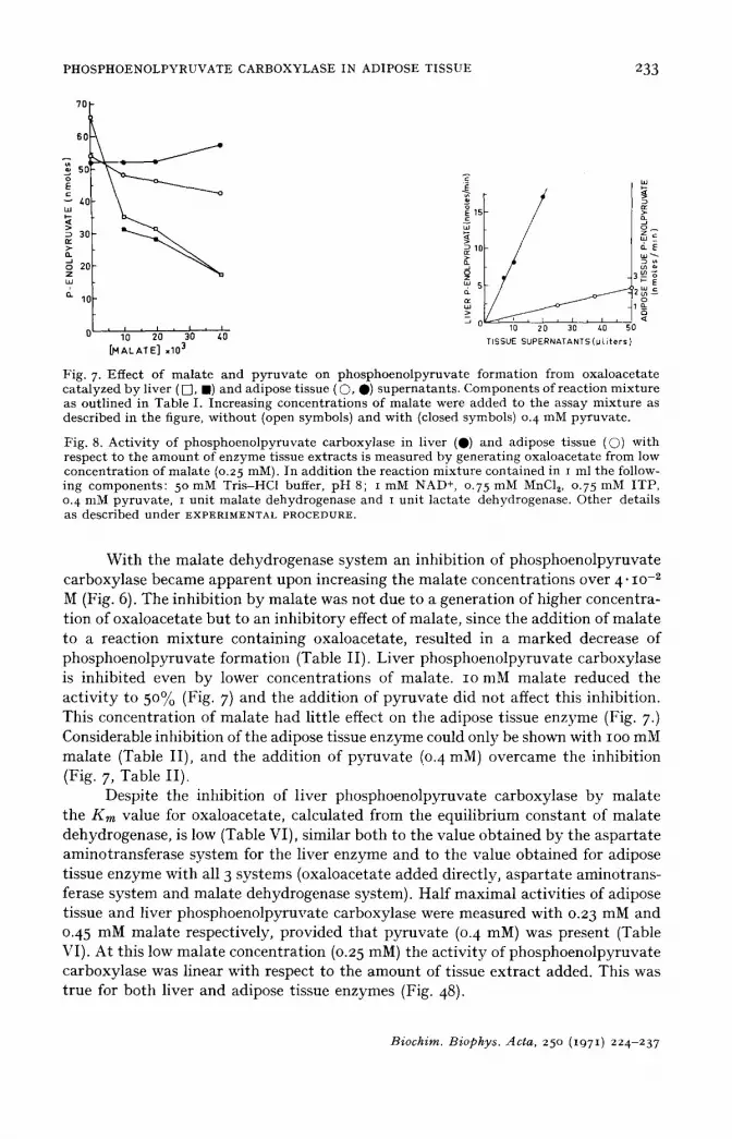
Fig. 7. Effect of malate and pyruvate on phosphoenolpyruvate formation from oxaloacetate catalyzed by liver ( [], l ) and adipose tissue ( ©, 0) supernatants. Components of reaction mixture as outlined in Table I. Increasing concentrations of malate were added to the assay mixture as described in the figure, without (open symbols) and with (closed symbols) 0. 4 nlM pyruvate.
Fig. 8. Activity of phosphoenolpyruvate carboxylase in liver (Q) and adipose tissue (Q) with respect to the amount of enzyme tissue extracts is measured by generating oxaloacetate from low concentration of malate (o.25 mM). In addition the reaction mixture contained in i ml the follow- ing components: 5 ° mM Tris-HC1 buffer, pH 8; i mM NAD +, 0.75 mM MnC12, 0.75 mM ITP, 0. 4 nlM pyruvate, i unit malate dehydrogenase and I unit lactate dehydrogenase. Other details as described under EXPERIMENTAL PROCEDURE.
W i t h the ma la t e dehydrogenase sys tem an inhib i t ion of phosphoeno lpy ruva te ca rboxylase became a p p a r e n t upon increasing the ma la t e concent ra t ions over 4" lO-2 M (Fig. 6). The inh ib i t ion b y ma la t e was not due to a genera t ion of higher concent ra- t ion of oxa loace ta t e b u t to an inh ib i to ry effect of mala te , since the add i t i on of ma la t e to a reac t ion mix tu re con ta in ing oxa loace ta te , resu l ted in a m a r k e d decrease of phosphoeno lpy ruva t e fo rmat ion (Table I I ) . L iver phosphoeno lpy ruva t e carboxylase is i nh ib i t ed even b y lower concen t ra t ions of mala te . IO mM ma la t e reduced the a c t i v i t y to 50% (Fig. 7) and the add i t i on of p y r u v a t e d id not affect this inhibi t ion . This concen t ra t ion of ma la t e had l i t t le effect on the adipose t issue enzyme (Fig. 7.) Considerable inhibi t ion of the adipose tissue enzyme could only be shown with IOO mM ma la t e (Table I I ) , and the add i t ion of p y r u v a t e (0. 4 raM) overcame the inhib i t ion (Fig. 7, Table I I ) .
Despi te the inhibi t ion of l iver phosphoeno lpyruva te carboxylase b y ma la t e the Km value for oxa loace ta te , ca lcu la ted from the equi l ib r ium cons t an t of ma la t e dehydrogenase , is low (Table VI), s imilar bo th to the value ob t a ined b y the a spa r t a t e amino t rans fe rase sys tem for the l iver enzyme and to the value ob ta ined for adipose t issue enzyme wi th all 3 sys tems (oxa loace ta te a d d e d di rec t ly , a s p a r t a t e amino t rans - ferase sys tem and mala te dehydrogenase system). Ha l f m a x i m a l act iv i t ies of adipose t issue and l iver phosphoeno lpyruva te carboxylase were measured wi th 0.23 mM and 0.45 mM ma la t e respect ively, p rov ided t ha t p y r u v a t e (0. 4 mM) was present (Table VI). A t th is low ma la t e concent ra t ion (0.25 raM) the ac t i v i t y of phosphoeno lpy ruva te ca rboxy lase was l inear wi th respect to the amoun t of tissue ex t r ac t added. This was t rue for bo th l iver and adipose t issue enzymes (Fig. 48).
Biochim. Biophys. Acta, 25 ° (1971) 224-237

234 o. MEYUHAS et al.
TABLE V I I
C O M P A R I S O N B E T W E E N C A R B O X Y L A T I O N A N D D E C A R B O X Y L A T I O N C A T A L Y Z E D R E A C T I O N S O F P H O S -
P H O E N O L P Y R U V A T E C A R B O X Y L A S E
Adipose tissue extracts prepared from animals t reated as outlined in the table were assayed for phosphoenolpyruva te carboxylase activity. A s imultaneous assay of the carboxylat ion reaction and decarboxylat ion reaction were carried out with each extract separately. For the measurement of the decarboxylat ion catalyzed reaction oxaloacetate was generated from malate (20 mM) in the presence of py ruva te o. 4 mM. The carboxylat ion reaction was performed as described under EXPERIMENTAL PROCEDURE. One unit of enzyme measured by the carboxylat ion reaction is the amoun t which catalyzed the fixation of i /*mole H14CO3- in the presence of phosphoenolpyruvate , Mn 2+ and I D P at 37 °. One unit of enzyme measured by the decarboxylat ion reaction is the a m o u n t of tissue ext rac t which catalyzed the I TP dependent formation of I /*mole of phosphoenolpyruva te per nlin at 37 °. Results are the means ± S.E.of the number of animals indicated in parentheses.
Treatment of animals Phosphoenolpyruvate carboxylase activity
Adipose tissue (munits/g tissue) Liver (units/g tissue)
Carboxylation Decarboxylation Carboxylation Decarboxylation
In t ac t fed (3) 39 :~ 2.04 84 ~: 16.6 2.33 ± 0.067 3.84 z~: 0.26 In tac t 24 h fasted (5) 288 -E 33 464 ~ 58 7.76 ± o.71 9.18 ~ 0.9 Adrenalectomized fed (5) 277 ± 26 468 • 95 Adrenalectomized 24 h
fasted (5) 486 ± 38 812 ~_ 47 7.98 ± 0.49 i i . i ~2 0.57
Ratio of decarboxylation to carboxylation reactions catalyzed by phosphoenolpyruvate carboxylasc
As pointed out earlier, the main purpose of the present s tudy was to compare tile rate of the reactions catalyzed by phosphoenolpyruvate carboxylase in the two directions with crude liver and adipose tissue extracts. With liver phosphoenolpyru- ra te carboxylase this comparison was not feasible as measurements of maximal activities of the decarboxylation catalyzed reaction could not be ensured satisfactorily.
1000 el e• 80(]
>- ,. 600 ~ , ~ / - x o e n ¢m n~ "~ 400
Lu 200 • o E
nV I I J I i I u 200 400 600
CARBOXYLATION (munits/g t issue )
Fig. 9. Relationship of carboxylat ion to decarboxylat ion catalyzed reactions. Adipose tissue superna tan t s were obtained from animals t reated variously as described in Table Vl i . Phospho- enolpyruvate carboxylase activity was assayed s imultaneously in both directions. The slope of the regression line and the intercept were calculated according to the following equations 3°. 27y -- aN + b27x and 27xy a x + b~x 2.
.Biochim. Biophys. Acta, 25 ° (1971) 224-237

PHOSPHOENOLPYRUVATE CARBOXYLASE IN ADIPOSE TISSUE 235
Nevertheless it is worth noting that the activities obtained (Table VII) were very close or slightly higher than the activities reported by FOSTER et al. 19. With adipose tissue on the other hand maximal activities measured simultaneously in both directions were made, and presented in Table VII . For comparison these measurements were made in adipose tissue extracts obtained from groups of animals that underwent various treatments. As shown in Table VII changes in enzyme activity induced by various t reatments in vivo were similar, whether measured in the backward or forward directions.
The individual figures of activities measured in the direction of decarboxylation were plotted against the simultaneous measured activities by the carboxylation method (Fig. 9). The regression line obtained indicates clearly a linear relationship between the two assays over a wide range of activities assayed. Each of the reactions was measured at near its own optimal pH. In the case of the decarboxylation cata- lyzed reaction the pH opt imum was found to be around 8 for all three systems used (oxaloacetate added directly, malate dehydrogenase system and aspartate amino- transferase system) while the carboxylation reaction was measured at pH 7.0. Under these conditions the slope of the regression line indicates a ratio of 1.66 between the decarboxylation to carboxylation catalyzed reactions. This ratio is far below the ratio reported for the purified phosphoenolpyruvate carboxylase from pig liver mito- chondria 9.
DISCUSSION
In their recent review on gluconeogenesis SCRUTTON AND UTTER 20 pointed out that the maximal capacity of a tissue to carry out a given sequence of reactions is dependent on the amounts of the key enzymes participating in it. In that respect, alteration in tile amounts of the key enzymes would contribute to alterations in the overall capacity and thus to the regulation of the pathway. Phosphoenolpyruvate carboxylase is considered as a key enzyme in gluconeogenesis in the liver and in glyceroneogenesis in adipose tissue",2°m. Changes in enzyme activities, brought about by various in vivo t reatments correlate closely with changes in the overall activity of the pathwayS, s° . However, its amount, as measured by the maximal activity, exceeds the overall capacity of the liver to produce glucose by a factor of 2 (ref. 20), and the rate of glyceroneogenesis in adipose tissue by a factor of 7 (Table viii , ref. 8). This implies either that phosphoenolpyruvate carboxylase is a key enzyme but that under all circumstances in vivo the enzyme functions at a fraction of its maximal activity. Alternatively, that phosphoenolpyruvate carboxylase catalyzed the formation of phosphoenolpyruvate at its maximal activity in vivo, but that it is not the key site of regulation of the gluconeogenic pathway. Analysis of the present experimental data support the view that the activity of phosphoenolpyruvate carboxylase inside the cell never exceeds one half of its measured maximal activity.
In adipose tissue, phosphoenolpyruvate carboxylase is located in the cytosol e while pyruvate, a glyceroneogenic substratee, 6-8 is carboxylated in the mitochon- dria26, 27. Recently it was shown 2Sthat if adipose tissue mitochondria were incubated with t4COo and pyruvate, the label appeared in citrate and malate which subsequently diffused out to the suspending medium. Consequently, by diffusing out of the mito- chondria these two substrates could generate oxaloacetate in vivo. Since, as shown in
Biochim. Biophys. Acta, 250 (1971) 224-237

236 o. MEYUHAS et al
Table I I I only malate but not citrate supported phosphoenolpyruvate formation and it was also preferable to aspartate, malate may be considered as the main precursor for phosphoenolpyruvate formation in adipose tissue.
The existence of an excess amount of dehydrogenases in animal cells enables to maintain these systems at a near equilibrium state in vivo 31. In that respect, our malate dehydrogenase system supplemented with pyruvate and brought to an equilib- rium resembles the in vivo system, phosphoenolpyruvate carboxylase activity becomes therefore dependent on the concentration of malate. Thus, 1/2-1/3 of the enzyme maximal activity was measured at a physiological concentration of malate22-24, 29.
In adipose tissue malate concentration does not change by various in vivo treatments 29, while slight changes were reported in liver 22-24. Therefore the activity of phosphoenolpyruvate carboxylase in vivo must depend principally on the ratio of NAD + to NADH in the cytosol. Yet as shown in Table VI, even under optimal conditions of this ratio, as exemplified by using an optimal concentration of reducible substrate (pyruvate) the activity of phosphoenolpyruvate carboxylase was only 1/2-1/3 of its maximal activity. Consequently it is clearly evident that under in vivo conditions the enzyme never functions at a rate which exceeds one half of its maximal activity.
Despite the fact that only a fraction of enzyme activity is measured at physio- logical concentration of malate the linear relationship between enzyme amounts and activities (Fig. 8) indicates the significance that changes in enzyme amount may bear on the regulation of phosphoenolpyruvate formation under conditions which resemble the in vivo situation.
ACKNOWLEDGEMENTS
The authors wish to thank Prof. B. Shapiro for his helpful and stimulating discussions in the course of this work. This work was supported in part by the Research Committee of the Hebrew University, Hadassah Medical School, Jerusalem, Israel, and by a grant from the National Institutes of Health, U.S.A., under PL.48o, Agreement No. o6-o16-1.
O.M. is a fellow of the California Foundation of Biochemical Research.
REFERENCES
i R. C. NORDLIE AND H. A. LARDY, J. Biol. Chem., 238 (1963) 2259. 2 F. J. BALLARD, R. W. HANSON AND G. A. LEVEILLE, J. Biol. Chem., 242 (1967) 2746. 3 M. F. UTTER AND K. KURAHASHI, J. Biol. Chem., 207 (1954) 787 • 4 M. F. IJTTER AND K. KURAHASHI, J. Biol. Chem., 207 (1954) 821. 5 J. W. YOUNG, E. SHRAGO AND H. A. LARDY, Biochemistry, 3 (1964) 1687. 6 L. RESHEF, R. V~ T. HANSON AND F. J. BALLARD, J. Biol. Chem., 244 (1969) 1994. 7 L. F-ESHEF, F. J. BALLARD AND R. W. HANSON, J. Biol. Chem., 244 (1969) 5577. 8 L. RESHEF, R. W. HANSON AND F. J. BALLARD, J. Biol. Chem., 245 (197 o) 5979. 9 H. C. CHANG, H. MARUYAMA, R. S. MILLER AND M. D. LANE, J. Biol. Chem., 241 (1966) 2421.
io H. V. HENNI~G, ]3. STUMPF, B. OHLY AND J. SUEBERT, Biochem. Z., 344 (1966) 274. I I tt . C. CHANG AND M. D. LANE, J. Biol. Chem., 241 (1966) 2413. 12 R. j . HERISURG, Anal. Chem., 32 (1953) 42. 13 1~. CzoK AND L. ECKERT, in H. U. BERGMEYER, Methods Enzymatic Anal., Academic Press,
New York, 1963, p. 224.
Biochim. Biophys. Acta, 25 ° (1971) 224-237
PHOSPHOENOLPYRUVATE CARBOXYLASE IN ADIPOSE TISSUE 237
14 H. U. BERGMEYER AND E. BERNT, in H. U. BERGMEYER, Methods Enzymatic Anal., Academic Press, New York, 1963, p. 837.
15 J. R. STERN, S. OCHOA AND F. LYNEN, J. Biol. Chem., 198 (1952) 313 . 16 C. P. HENSON AND W. W. CLELAND, Biochemistry, 3 (1964) 338. 17 D. D. HOLTEN AND R. C. NORDLIE, Biochemistry, 4 (1965) 723. 18 E. SHRAGO AND H. A. LARDY, J. Biol. Chem., 241 (1966) 663. 19 D. O. FOSTER, P. D. RAY AND H. A. LARDY, Biochemistry, 5 (1966) 563 • 20 M. C. SCRUTTON AND M. F. UTTER, Ann. Rev. Biochem., 34 (1968) 249. 21 L. RESHEF AND B. SHAPIRO, in B. JEANRENAUD AND D. HEPP, Adipose Tissue, George Th ieme
Verlag, S tu t t ga r t , Academic Press, New York, 197 o, p. 136. 22 H. J. HOROST, F. H. KREUTZ, M. REIM AND H. J. HUBENER, Biochem. Biophys. Res. Commun.,
4 (1961) 163 . 23 O, WIELAND AND G. LOFFLER, Biochem. Z., 339 (1963) 204. 24 J. R. WILLIAMSON, P. H. WRIGHT, W. J. MALAISSE AND J. ASHMORE, Biochem. Biophys. Res.
Commun., 24 (1066) 765 . 25 F. J. BALLARD, Biochem. J., 12o (197 o) 809. 26 I. B6TTGER, O. WIELAND, D. BREDICZKA AND D. PETTE, Eur. J. Biochem., 8 (1969) 113. 27 F. J. BALLARD, R. W. HANSON AND L. RESHEF, Biochem, J., 119 (197 o) 735. 28 M. S. PATEL AND N. W. HANSON, J. Biol. Chem., 245 (197 o) 13o2. 29 F. J. BALLARD AND R. W. HANSON, Biochem. J., 112 (1969) 195. 3 ° H, ARKIN AND R. R. COTTON, in Statistical Methods, Barnes and Nobel New York, 1967, p.
76 • 31 H. A. K]~EBS AND H. A. VEECH, Adv. Enzyme Regul., 7 (1968) 397.
Biochim. Biophys. Acta, 250 (1971) 224-237
Related Documents


![For Research Use Only PCK2 Polyclonal antibody · Background Information PCK2(phosphoenolpyruvate carboxykinase [GTP], mitochondrial) is also named as PEPCK2, PEPCK-M and belongs](https://static.cupdf.com/doc/110x72/60b24c18c6049f6cff2e0b4c/for-research-use-only-pck2-polyclonal-antibody-background-information-pck2phosphoenolpyruvate.jpg)