Phosphocaveolin-1 Enforces Tumor Growth and Chemoresistance in Rhabdomyosarcoma Fiorella Faggi 1,6 , Stefania Mitola 1 , Guglielmo Sorci 2,6 , Francesca Riuzzi 2,6 , Rosario Donato 2,6 , Silvia Codenotti 1,6 , Pietro Luigi Poliani 1 , Manuela Cominelli 1 , Raffaella Vescovi 1 , Stefania Rossi 1 , Stefano Calza 1 , Marina Colombi 1 , Fabio Penna 3,6 , Paola Costelli 3,6 , Ilaria Perini 4 , Maurilio Sampaolesi 4,5,6 , Eugenio Monti 1 , Alessandro Fanzani 1,6 * 1 Department of Molecular and Translational Medicine, University of Brescia, Brescia, Italy, 2 Department of Experimental Medicine and Biochemical Sciences, University of Perugia, Perugia, Italy, 3 Department of Experimental Medicine and Oncology, University of Torino, Torino, Italy, 4 Stem Cell Research Institute, University Hospital Gasthuisberg, Leuven, Belgium, 5 Human Anatomy Section, University of Pavia, Pavia, Italy, 6 Interuniversity Institute of Myology (IIM), Italy Abstract Caveolin-1 (Cav-1) can ambiguously behave as either tumor suppressor or oncogene depending on its phosphorylation state and the type of cancer. In this study we show that Cav-1 was phosphorylated on tyrosine 14 (pCav-1) by Src-kinase family members in various human cell lines and primary mouse cultures of rhabdomyosarcoma (RMS), the most frequent soft-tissue sarcoma affecting childhood. Cav-1 overexpression in the human embryonal RD or alveolar RH30 cells yielded increased pCav-1 levels and reinforced the phosphorylation state of either ERK or AKT kinase, respectively, in turn enhancing in vitro cell proliferation, migration, invasiveness and chemoresistance. In contrast, reducing the pCav-1 levels by administration of a Src-kinase inhibitor or through targeted Cav-1 silencing counteracted the malignant in vitro phenotype of RMS cells. Consistent with these results, xenotransplantation of Cav-1 overexpressing RD cells into nude mice resulted in substantial tumor growth in comparison to control cells. Taken together, these data point to pCav-1 as an important and therapeutically valuable target for overcoming the progression and multidrug resistance of RMS. Citation: Faggi F, Mitola S, Sorci G, Riuzzi F, Donato R, et al. (2014) Phosphocaveolin-1 Enforces Tumor Growth and Chemoresistance in Rhabdomyosarcoma. PLoS ONE 9(1): e84618. doi:10.1371/journal.pone.0084618 Editor: Natasha Kyprianou, University of Kentucky College of Medicine, United States of America Received May 28, 2013; Accepted November 15, 2013; Published January 10, 2014 Copyright: ß 2014 Faggi et al. This is an open-access article distributed under the terms of the Creative Commons Attribution License, which permits unrestricted use, distribution, and reproduction in any medium, provided the original author and source are credited. Funding: This work was supported by the Fondazione Cariplo grant to EM, Grant NEDD - Network Enabled Drug Design, Regione Lombardia to EM, ‘‘Associazione Italiana per la Ricerca sul Cancro’’ (MFAG Project n. 9161 to SM and Project 6021 to RD) and University of Brescia research fund (ex 60%) to AF and EM. The funders had no role in study design, data collection and analysis, decision to publish, or preparation of the manuscript. Competing Interests: The authors have declared that no competing interests exist. * E-mail: [email protected] Introduction Caveolins (i.e. Cav-1, Cav-2 and Cav-3) are 21–24 kDa membrane-associated proteins that mainly localize in the 50– 100 nm cholesterol-enriched invaginations of the plasma mem- brane, known as caveolae [1–3]. Caveolins are pivotally involved in multiple processes, including endocytosis, cholesterol homeostasis, regulation of signal transduction and caveolae biogenesis that requires the complementary action of Cavin family members [4– 6]. Cav-1, in particular, has been shown to mostly inhibit a large number of signaling pathways because of the presence of a caveolin scaffolding domain that allows the binding of a plethora of proteins, such as epidermal growth factor receptor, protein kinases C, endothelial nitric oxide synthase [7]. In response to a variety of stimuli such as growth factors, UV irradiation, mechanical and oxidative stress, Cav-1 can also be phosphorylated on tyrosine 14 (hereafter referred as to pCav-1) by members of the sarcoma kinases (Src-kinases) [8–10], in turn leading to activation of pathways linked to cell death or survival [11]. In cancer, there is mounting evidence that pCav-1 occurrence often predicts unfavorable outcome by supporting anchorage-independent cell growth, migration, invasiveness and multidrug resistance [11–15]. Rhabdomyosarcoma (RMS) is a pediatric soft-tissue cancer [16– 20] that arises from various muscle and non-muscle progenitors characterized by interrupted myogenesis [21–28]. The current classification includes two major histological variants, known as embryonal (ERMS) and alveolar (ARMS), with the former characterized by a complex genomic aetiogenesis [16,17] and the latter by the prevalent expression of chimeric transcription factors generated by the fusion of the paired box 3 or 7 with forkhead box O1 (Pax3-Foxo1 or Pax7-Foxo1) as a result of specific chromosomal translocations [29,30]. Previously we have shown that Cav-1 is consistently expressed in both the histological RMS variants [31,32]. Here, we provide further evidence that Cav-1 is consistently phosphorylated through a Src-dependent mechanism in various ERMS and ARMS cell lines, playing a pivotal role in tumor growth and chemoresistance. Results Cav-1 is phosphorylated through a Src-dependent mechanism in RMS cells The expression levels of Caveolin family members were analysed by western blot using four human RMS cell lines (embryonal RD, RD12, RD18 and alveolar RH30) and two mouse primary tumor cultures established from transgenic Myf6Cre/p53 2/2 and Myf6Cre/Pax3-Foxo1/p53 2/2 mice (em- bryonal U57810 and alveolar U23674, respectively) [21,24]. In PLOS ONE | www.plosone.org 1 January 2014 | Volume 9 | Issue 1 | e84618

Welcome message from author
This document is posted to help you gain knowledge. Please leave a comment to let me know what you think about it! Share it to your friends and learn new things together.
Transcript

Phosphocaveolin-1 Enforces Tumor Growth andChemoresistance in RhabdomyosarcomaFiorella Faggi1,6, Stefania Mitola1, Guglielmo Sorci2,6, Francesca Riuzzi2,6, Rosario Donato2,6,
Silvia Codenotti1,6, Pietro Luigi Poliani1, Manuela Cominelli1, Raffaella Vescovi1, Stefania Rossi1,
Stefano Calza1, Marina Colombi1, Fabio Penna3,6, Paola Costelli3,6, Ilaria Perini4, Maurilio Sampaolesi4,5,6,
Eugenio Monti1, Alessandro Fanzani1,6*
1 Department of Molecular and Translational Medicine, University of Brescia, Brescia, Italy, 2 Department of Experimental Medicine and Biochemical Sciences, University of
Perugia, Perugia, Italy, 3 Department of Experimental Medicine and Oncology, University of Torino, Torino, Italy, 4 Stem Cell Research Institute, University Hospital
Gasthuisberg, Leuven, Belgium, 5 Human Anatomy Section, University of Pavia, Pavia, Italy, 6 Interuniversity Institute of Myology (IIM), Italy
Abstract
Caveolin-1 (Cav-1) can ambiguously behave as either tumor suppressor or oncogene depending on its phosphorylationstate and the type of cancer. In this study we show that Cav-1 was phosphorylated on tyrosine 14 (pCav-1) by Src-kinasefamily members in various human cell lines and primary mouse cultures of rhabdomyosarcoma (RMS), the most frequentsoft-tissue sarcoma affecting childhood. Cav-1 overexpression in the human embryonal RD or alveolar RH30 cells yieldedincreased pCav-1 levels and reinforced the phosphorylation state of either ERK or AKT kinase, respectively, in turn enhancingin vitro cell proliferation, migration, invasiveness and chemoresistance. In contrast, reducing the pCav-1 levels byadministration of a Src-kinase inhibitor or through targeted Cav-1 silencing counteracted the malignant in vitro phenotypeof RMS cells. Consistent with these results, xenotransplantation of Cav-1 overexpressing RD cells into nude mice resulted insubstantial tumor growth in comparison to control cells. Taken together, these data point to pCav-1 as an important andtherapeutically valuable target for overcoming the progression and multidrug resistance of RMS.
Citation: Faggi F, Mitola S, Sorci G, Riuzzi F, Donato R, et al. (2014) Phosphocaveolin-1 Enforces Tumor Growth and Chemoresistance in Rhabdomyosarcoma. PLoSONE 9(1): e84618. doi:10.1371/journal.pone.0084618
Editor: Natasha Kyprianou, University of Kentucky College of Medicine, United States of America
Received May 28, 2013; Accepted November 15, 2013; Published January 10, 2014
Copyright: � 2014 Faggi et al. This is an open-access article distributed under the terms of the Creative Commons Attribution License, which permitsunrestricted use, distribution, and reproduction in any medium, provided the original author and source are credited.
Funding: This work was supported by the Fondazione Cariplo grant to EM, Grant NEDD - Network Enabled Drug Design, Regione Lombardia to EM,‘‘Associazione Italiana per la Ricerca sul Cancro’’ (MFAG Project n. 9161 to SM and Project 6021 to RD) and University of Brescia research fund (ex 60%) to AF andEM. The funders had no role in study design, data collection and analysis, decision to publish, or preparation of the manuscript.
Competing Interests: The authors have declared that no competing interests exist.
* E-mail: [email protected]
Introduction
Caveolins (i.e. Cav-1, Cav-2 and Cav-3) are 21–24 kDa
membrane-associated proteins that mainly localize in the 50–
100 nm cholesterol-enriched invaginations of the plasma mem-
brane, known as caveolae [1–3]. Caveolins are pivotally involved in
multiple processes, including endocytosis, cholesterol homeostasis,
regulation of signal transduction and caveolae biogenesis that
requires the complementary action of Cavin family members [4–
6]. Cav-1, in particular, has been shown to mostly inhibit a large
number of signaling pathways because of the presence of a
caveolin scaffolding domain that allows the binding of a plethora
of proteins, such as epidermal growth factor receptor, protein
kinases C, endothelial nitric oxide synthase [7]. In response to a
variety of stimuli such as growth factors, UV irradiation,
mechanical and oxidative stress, Cav-1 can also be phosphorylated
on tyrosine 14 (hereafter referred as to pCav-1) by members of the
sarcoma kinases (Src-kinases) [8–10], in turn leading to activation
of pathways linked to cell death or survival [11]. In cancer, there is
mounting evidence that pCav-1 occurrence often predicts
unfavorable outcome by supporting anchorage-independent cell
growth, migration, invasiveness and multidrug resistance [11–15].
Rhabdomyosarcoma (RMS) is a pediatric soft-tissue cancer [16–
20] that arises from various muscle and non-muscle progenitors
characterized by interrupted myogenesis [21–28]. The current
classification includes two major histological variants, known as
embryonal (ERMS) and alveolar (ARMS), with the former
characterized by a complex genomic aetiogenesis [16,17] and
the latter by the prevalent expression of chimeric transcription
factors generated by the fusion of the paired box 3 or 7 with
forkhead box O1 (Pax3-Foxo1 or Pax7-Foxo1) as a result of
specific chromosomal translocations [29,30].
Previously we have shown that Cav-1 is consistently expressed
in both the histological RMS variants [31,32]. Here, we provide
further evidence that Cav-1 is consistently phosphorylated through
a Src-dependent mechanism in various ERMS and ARMS cell
lines, playing a pivotal role in tumor growth and chemoresistance.
Results
Cav-1 is phosphorylated through a Src-dependentmechanism in RMS cells
The expression levels of Caveolin family members were
analysed by western blot using four human RMS cell lines
(embryonal RD, RD12, RD18 and alveolar RH30) and two
mouse primary tumor cultures established from transgenic
Myf6Cre/p532/2 and Myf6Cre/Pax3-Foxo1/p532/2 mice (em-
bryonal U57810 and alveolar U23674, respectively) [21,24]. In
PLOS ONE | www.plosone.org 1 January 2014 | Volume 9 | Issue 1 | e84618

PhosphoCav-1 Accelerates Rhabdomyosarcoma Growth
PLOS ONE | www.plosone.org 2 January 2014 | Volume 9 | Issue 1 | e84618
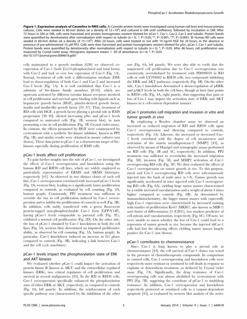
cells maintained in a growth medium (GM) we observed co-
expression of Cav-1 (both Tyr14-phosphorylated and total forms)
with Cav-2 and lack or very low expression of Cav-3 (Fig. 1A).
Instead, treatment of cells with a differentiation medium (DM)
lead to down-regulation of both Cav-1 and Cav-2 and increased
Cav-3 levels (Fig. 1A). It is well established that Cav-1 is a
substrate of Src-kinase family members [8-10], which are
upstream activated by different tyrosine kinase receptors involved
in cell proliferation and survival upon binding with ligands such as
hepatocyte growth factor (HGF), platelet-derived growth factor,
insulin and insulin-like growth factor [33–37]. Thus, treatment of
RD cells with HGF, a growth factor playing a pivotal role in RMS
progression [38–40], elicited increasing pSrc and pCav-1 levels
compared to untreated cells (Fig. 1B, western blot), in turn
promoting a rise in cell proliferation (Fig. 1B, Crystal violet assay).
In contrast, the effects promoted by HGF were counteracted by
co-treatment with a synthetic Src-kinase inhibitor, known as PP2
(Fig. 1B), and similar results were obtained in mouse cultures (not
shown). These data point to pCav-1 as a downstream target of Src-
kinases especially during proliferation of RMS cells.
pCav-1 levels affect cell proliferationTo gain further insights into the role of pCav-1, we investigated
the effects of Cav-1 overexpression and knockdown using the
human RD and RH30 cell lines, which are widely employed as
particularly representative of ERMS and ARMS histotypes,
respectively [41]. As observed in two distinct clones of each cell
line, Cav-1 overexpression correlated with increased pCav-1 levels
(Fig. 2A, western blot), leading to a significantly faster proliferation
compared to controls, as evaluated by cell counting (Fig. 2A,
bottom graph). Consistently, PP2 treatment was sufficient to
override the rise in cell proliferation induced by Cav-1 overex-
pression and to inhibit the proliferation of controls as well (Fig. 2B).
In addition, cells stably transfected with a green fluorescent
protein-tagged non-phosphorylatable Cav-1 form (GFP-Y14F),
having pCav-1 levels comparable to parental cells (Fig. 2C),
exhibited a normal cell proliferation (Fig. 2D). On the other side,
the loss of pCav-1 obtained by Cav-1 knockdown in both the cell
lines (Fig. 3A, western blot) determined an impaired proliferative
ability, as observed by cell counting (Fig. 3A, bottom graph). In
particular, Cav-1 knockdown induced an increase in G1 phase
compared to controls (Fig. 3B), indicating a link between Cav-1
and the cell cycle machinery.
pCav-1 levels impact the phosphorylation state of ERKand AKT kinases
We evaluated whether pCav-1 could impact the activation of
protein kinase B (known as AKT) and the extracellular regulated
kinases (ERK), two critical regulators of cell proliferation and
survival in several malignancies [42]. In the RD or RH30 cells,
Cav-1 overexpression specifically enhanced the phosphorylation
state of either ERK or AKT, respectively, as compared to controls
(Fig. 4A, left panels). In addition, the reinforcement of each
specific pathway was characterized by the inhibition of the other
one (Fig. 4A, left panels). We were also able to verify that the
augmented cell proliferation due to Cav-1 overexpression was
consistently overwhelmed by treatment with PD098059 in RD
cells or with LY294002 in RH30 cells, two compounds inhibiting
the ERK and AKT pathways, respectively (Fig. 4B). On the other
side, Cav-1 knockdown determined a down-regulation of pERK
and pAKT levels in both the cell lines, though at later time points
in RH30 cells (Fig. 4A, right panels), thus suggesting that gain or
loss of Cav-1 may impact the activation state of ERK and AKT
kinases in a cell-context dependent manner.
pCav-1 promotes cell migration and invasion in vitro andtumor growth in vivo
By employing a Boyden chamber assay we observed an
increased or reduced migration of RD and RH30 cells upon
Cav-1 overexpression and silencing compared to controls,
respectively (Fig. 5A). Likewise, the increased or decreased Cav-
1 levels correlated with the degree of cell invasiveness and
activation of the matrix metalloprotease-2 (MMP2) [43], as
observed by means of Matrigel and zymography assays performed
on RD cells (Fig. 5B and 5C, respectively). In addition, PP2
treatment was sufficient to overwhelm the increased migration
(Fig. 5D), invasion (Fig. 5E) and MMP2 activation of Cav-1
overexpressing RD cells (Fig. 5F). We then evaluated the effects of
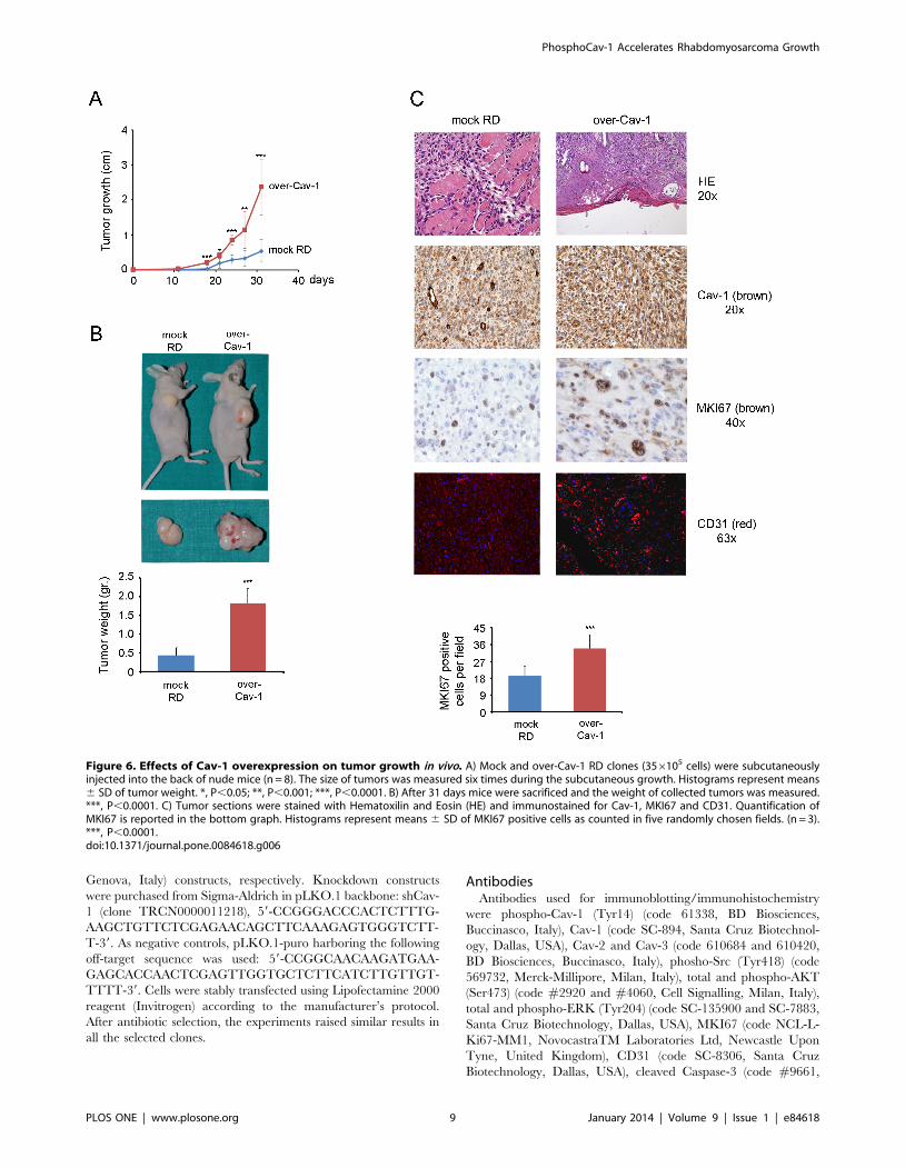
Cav-1 overexpression on tumor growth in vivo. To this purpose,
mock and Cav-1 overexpressing RD cells were subcutaneously
injected into the back of nude mice (n = 8). Tumor growth was
significantly accelerated in mice injected with Cav-1 overexpress-
ing RD cells (Fig. 6A), yielding huge tumor masses characterized
by a visible increased vascularization and a weight of about 4 times
higher compared to controls (Fig. 6B). As determined by
immunohistochemistry, the bigger tumor masses with expectedly
high Cav-1 expression were characterized by increased staining
with marker of proliferation Ki-67 (MKI67, also known as MIB-1)
and cluster of differentiation 31 (CD31), two markers predictive of
cell mitosis and vascularization, respectively (Fig. 6C). Of note, we
were unable to assess whether the loss of Cav-1 could lead to a
regression of tumor growth in vivo, because the injected shCav-1
cells had lost the silencing effects yielding tumor masses largely
positive for Cav-1 (not shown).
pCav-1 contributes to chemoresistanceSince Cav-1 is long known to play a pivotal role in
chemoresistance [44], the cell survival of Cav-1 clones was tested
in the presence of chemotherapeutic compounds. In comparison
to control cells, Cav-1 overexpressing and knockdown cells were
respectively more resistant or sensitized to cell death in response to
cisplatin or doxorubicin treatment, as deduced by Crystal violet
assay (Fig. 7A). Significantly, the drug resistance of Cav-1
overexpressing cells was almost abolished by co-treatment with
PP2 (Fig. 7B), suggesting the contribute of pCav-1 to multidrug
resistance. In addition, Cav-1 overexpression and knockdown
respectively protected or sensitized cells to a caspase-dependent
apoptosis [45], as evaluated by western blot analysis of the active
Figure 1. Expression analysis of Caveolins in RMS cells. A) Caveolin protein levels were investigated using human cell lines and mouse primarycultures. Cells were seeded in 60-mm dishes (at a density of 126104) and cultured in GM until confluence, followed by incubation in DM. After72 hours in GM or DM, cells were harvested and protein homogenates western blotted for pCav-1, Cav-1, Cav-2, Cav-3 and tubulin. Protein bandswere quantified by densitometry after normalization with respect to tubulin (n = 3). *, P,0.05; **, P,0.001; ***, P,0.0001. B) Human RD cells wereseeded in 60-mm dishes (at a density of 156104). After 24 hours, cells were treated or not with 10 ng/ml HGF for 24 hours, in the absence orpresence of pre-administered 10 mM PP2. Cells were then harvested and protein homogenates western blotted for pSrc, pCav-1, Cav-1 and tubulin.Protein bands were quantified by densitometry after normalization with respect to tubulin (n = 3). *, P,0.05. After 48 hours, cell proliferation wasmeasured by Crystal violet assay. Histograms represent means 6 SD of absorbance. (n = 4) *, P,0.05.doi:10.1371/journal.pone.0084618.g001
PhosphoCav-1 Accelerates Rhabdomyosarcoma Growth
PLOS ONE | www.plosone.org 3 January 2014 | Volume 9 | Issue 1 | e84618

Figure 2. Effects of Cav-1 overexpression on cell proliferation. RD and RH30 cells were stably transfected with either Cav-1 construct (over-Cav-1) or empty vector (mock). A) Parental cells and selected clones were seeded in 60-mm dishes (at a density of 156104). After 48 hours, cells wereharvested and protein homogenates western blotted for pCav-1, Cav-1 and tubulin. Protein bands were quantified by densitometry afternormalization with respect to tubulin (n = 3). **, P,0.001; ***, P,0.0001. At the same time points, cell proliferation was evaluated with Burker
PhosphoCav-1 Accelerates Rhabdomyosarcoma Growth
PLOS ONE | www.plosone.org 4 January 2014 | Volume 9 | Issue 1 | e84618

caspase-3 fragments (19 and 17 kDa) (Fig. 7C). This set of
experiments shows that overexpression or lack of Cav-1 cooperates
to protect from or enhance a caspase-dependent apoptosis in RMS
cells, respectively.
Discussion
Cav-1 and Cav-2 genes are located in a fragile site (known as
D7S522 locus) that is frequently deleted in human cancers [46],
indicating a common role as tumor suppressors. Consistent with
these observations, an impaired Cav-1 expression was found in
human lung [47], mammary [48], colon [49] and ovarian
carcinomas [50] or sarcomas as well [51]. Cav-1 and Cav-2
conventionally form plasma membrane hetero-oligomers that
negatively regulate the activity of several receptors involved in cell
proliferation and survival [52–54], and therefore their loss is
thought to facilitate tumor progression by deliberate activation of
different signaling pathways, as observed in the tumor prone Cav-
1 knock-out mouse model [55–59]. Yet, additional mechanisms
complicating the scenario have been described, since the
inhibitory interaction of Cav-1 with the epidermal growth factor
receptor can be hindered by the formation of a galectin-
glycoprotein lattice, thereby resulting in sustained receptorial
activity, as observed in breast tumor cells [60,61]. Beyond
functioning as a tumor suppressor, accumulating evidence have
more recently indicated that Cav-1, especially when phosphory-
lated in Tyr14, behaves as an ambiguous partner in cancer [11–
15], because of its ability to activate pathways involved in cell
migration and invasion, such as the Rho/ROCK and Focal
adhesion kinases signaling systems [62]. The data presented here
point to pCav-1 as a positive modulator of proliferation,
migration, invasiveness, chemoresistance in vitro and tumor growth
in vivo in RMS, the most frequent childhood soft tissue sarcoma
characterized by expression of myogenic markers and impaired
differentiation [21–28]. The malignant cell phenotype we
observed by Cav-1 overexpression was ascribable to the robust
increase of Cav-1 phosphorylation in Tyr14, likely due to an
increased accessibility of the intracellular Cav-1 pool to Src-family
members, as observed in other cellular models [63,64]. In this
regard, further studies are required to examine whether and how
Cav-1 overexpression may perturb the caveolae organization and
Cav-1 trafficking in RMS cells. Among its many features, Cav-1
has widely been reported to exert both inhibitory and activatory
functions on the RAS/ERK and AKT pathways depending on the
cellular context (see for a review [11–15]) and, more recently,
pCav1 was found to be specifically interconnected with ERK and
AKT signaling in mouse embryonic stem cells [65]. In this
context, it is quite of interest that we observed pCav-1 reinforcing
the proliferation of the embryonal RD or alveolar RH30 cells via
specific increase of either ERK or AKT signaling, respectively,
since activating RAS mutations leading to sustained ERK signaling
are solely detected in ERMS [66–70] and the Pax3-Foxo1 factor
strongly cooperates with activated AKT signaling in ARMS
malignancy [71,72]. An additional cue of interest was that we
observed pCav-1 cooperating to the overactivation of one specific
pathway and causally inhibition of the other one (or viceversa). It is
largely documented that AKT and ERK signaling influence with
each other by positive or negative crosstalks in a cell-context
dependent manner [73], and a recent report has shown that if one
of these two pathways is chemically inhibited in various RMS cell
lines, including RD and RH30 cells, they opportunistically
potentiate the other one to survive [74]. Intriguingly, the same
authors showed that the block of either pathway did not result in
the potentiation of the other one in the RMS TE671 cell line [74],
in which we have previously shown the lack of Cav-1 expression
[32], therefore cautiously suggesting that Cav-1 may participate to
the negative ERK/AKT crosstalk observed in some RMS cells.
Finally, we observed such a clear connection between Cav-1
and the cell cycle machinery of RMS cells, as analogously reported
for human endothelial cells [75]. Cav-1 knockdown induced an
accumulation in G1 phase and subsequent block of cell
proliferation characterized by down-regulation of both ERK and
AKT. Thus, the loss of Cav-1 could mimic to some extent the pro-
apoptotic effects produced by the concomitant chemical ERK/
AKT inhibition [74,76], thereby sensitizing RMS cells to cell
death in response to chemotherapeutic agents, definitely pointing
to pCav-1 as a therapeutically valuable target to overcome RMS
malignancy. In this perspective, since the recent growing body of
literature indicating a cooperation between Cav-1 and the
polymerase transcription released factor PTRF/Cavin-1 in cancer
progression [4,77–85], it will be certainly important to assess their
relative contribution in RMS malignancy.
Materials and Methods
All reagents were from Sigma-Aldrich (Milan, Italy), unless
otherwise stated.
Ethic statementThis study was carried out in strict accordance with the
recommendations in the Guide for the Care and Use of
Laboratory Animals of the University of Perugia (Italy). The
protocol was approved by the Committee on the Ethics of Animal
Experiments of the University of Perugia. All surgery was
performed under sodium pentobarbital anesthesia, and all efforts
were made to minimize suffering.
Cell cultureHuman RD and RH30 cells were purchased from the European
Collection of Cell Cultures (ECACC, Salisbury, UK). RD cells
harbor RAS [86,87] and P53 [88] mutations, while RH30 cells
express Pax3-Foxo1 [89] and harbor P53 mutations [88]. RD12
and RD18 cell lines were derived by random cloning of the RD
line [90,91]. The mouse primary cultures, namely U57810
(ERMS) and U23674 (ARMS), were established from the
transgenic Myf6Cre/p532/2 and Myf6Cre/Pax3-Foxo1/p532/2
mice, respectively [21,24]. Cells were routinely maintained under
standard conditions (37uC and 5% CO2 in humidified incubator)
chamber, as shown in the bottom graphs. Bar graphs represent means 6 SD of total cell numbers. (n = 3) ***, P,0.0001. B) Mock and over-Cav-1 cellswere seeded in 24-well plates (at a density of 156103). After 24 hours, cells were either treated with dimethylsulfoxide (DMSO, vehicle) or 10 mM PP2(replenished every 24 or 16 hours for RD and RH30 cells, respectively) for the indicated time points. Cell proliferation was then evaluated by Crystalviolet assay. Histograms represent means 6 SD of absorbance (n = 4). ***, P,0.0001; ****, P,2e-16. C) RD and RH30 cells were stably transfected withconstructs for either Cav-1 (22 kDa) or non-phosphorylatable GFP-Y14F (55 kDa). Parental cells and selected clones were seeded in 60-mm dishes (ata density of 126104). After 24 hours, cells were harvested and protein homogenates western blotted for pCav-1, Cav-1 and tubulin. Protein bandswere quantified by densitometry after normalization with respect to tubulin (n = 3). **, P,0.001; ***, P,0.0001. D) Under the same conditions seenabove, cell proliferation was evaluated by Crystal violet assay at the indicated time points. Histograms represent means 6 SD of absorbance (n = 4).*, P,0.05.doi:10.1371/journal.pone.0084618.g002
PhosphoCav-1 Accelerates Rhabdomyosarcoma Growth
PLOS ONE | www.plosone.org 5 January 2014 | Volume 9 | Issue 1 | e84618

in GM, composed by high-glucose Dulbecco’s modified eagle’s
medium (DMEM) supplemented with 10% foetal bovine serum
(FBS) in the presence of 100 mg/ml penicillin/streptomycin
antibiotics and 1% L-Glutamine (only for RH30 cells). To induce
myodifferentiation, 80% confluent cells were switched to DM,
composed by DMEM supplemented with 2% horse serum. Cells
Figure 3. Effects of Cav-1 knockdown on cell proliferation. RD and RH30 cells were stably transfected with either Cav-1 knockdown (shCav-1)or off-target (shOT) construct. A) Parental cells and selected clones were seeded in 60-mm dishes (at a density of 126104). After 48 hours, cells wereharvested and protein homogenates western blotted for pCav-1, Cav-1 and tubulin. Protein bands were quantified by densitometry afternormalization with respect to tubulin (n = 3). *, P,0.05; **, P,0.001; ***, P,0.0001. At the same time points, cell proliferation was evaluated withBurker chamber, as shown in the bottom graphs. Bar graphs represent means 6 SD of total cell numbers (n = 3). ***, P,0.0001. B) FACS analysis wasperformed on shCav-1 and shOT cells. Numbers in pie charts denote percent cells in the G1/S/G2-phases of the cell cycle (n = 3). *, P,0.05; ***, P,0.0001.doi:10.1371/journal.pone.0084618.g003
PhosphoCav-1 Accelerates Rhabdomyosarcoma Growth
PLOS ONE | www.plosone.org 6 January 2014 | Volume 9 | Issue 1 | e84618

PhosphoCav-1 Accelerates Rhabdomyosarcoma Growth
PLOS ONE | www.plosone.org 7 January 2014 | Volume 9 | Issue 1 | e84618

were treated with: HGF (10 ng/ml, ImmunoTools, Friesoythe,
Germany), a Src-kinase inhibitor known as PP2 (10 mM), the
chemotherapeutic drugs doxorubicin (0.15 ng/ml) and cisplatin
(2 mg/ml), the synthetic inhibitors of the RAS/ERK and AKT
cascades, respectively known as PD098059 (15 mM) and
LY294002 (10 mM).
Plasmids and transfectionThe overexpression of either the wild-type or non-phosphor-
ylatable Cav-1 was carried out by transfection of pCAGGS/Cav-1
(provided by F. Galbiati, University of Pittsburgh, USA) or
pEGFPN1/Cav-1(Y14F) (provided by D. Maggi, University of
Figure 4. Effects of Cav-1 levels on the phosphorylation state of ERK and AKT. Control, over-Cav-1 cells and shCav-1 cells were seeded in60-mm dishes (at a density of 126104). A) At the indicated time points, cells were harvested and protein homogenates western blotted for pAKT, AKT,pERK, ERK, pCav-1, Cav-1 and tubulin at the indicated time points. Protein bands corresponding to pAKT and pERK levels (as detected at 48 hours)were quantified by densitometry after normalization with respect to tubulin (n = 3). **, P,0.001; ***, P,0.0001. B) 24 hours after seeding, mock andover-Cav-1 cells were treated with 15 mM PD09859 (in the case of RD cell line) or 10 mM LY294002 (in the case of RH30 cell line) for up to 24 hours.Cell proliferation was then evaluated by Crystal violet assay. Histograms represent means 6 SD of absorbance (n = 4). *, P,0.05; **, P,0.001; ***, P,0.0001.doi:10.1371/journal.pone.0084618.g004
Figure 5. Effects of Cav-1 levels on cell migration and invasiveness. A) Cell migration was evaluated using a modified Boyden chamberassay. The migration index was calculated through the ratio between the mean number of migrated clones (over-Cav-1 and shCav-1) with respect totheir migrated controls, as counted in five randomly chosen fields (n = 3). ***, P,0.0001. B) Cell invasiveness of RD clones (over-Cav-1 and shCav-1) incomparison to controls was evaluated by Matrigel assay. Histograms represent mean 6 SD (n = 3). **, P,0.001. C) Gelatin-zymography assay wasperformed to evaluate the MMP2 activation in RD clones (over-Cav-1 and shCav-1) with respect to controls. D-E-F) Cell migration (D), invasiveness (E)and the MMP2 activation (F) were evaluated in RD clones (mock and over-Cav-1) treated or not with 10 mM PP2 for 24 hours. Histograms representmeans 6 SD (n = 3). *, P,0.05; **, P,0.001.doi:10.1371/journal.pone.0084618.g005
PhosphoCav-1 Accelerates Rhabdomyosarcoma Growth
PLOS ONE | www.plosone.org 8 January 2014 | Volume 9 | Issue 1 | e84618
Genova, Italy) constructs, respectively. Knockdown constructs
were purchased from Sigma-Aldrich in pLKO.1 backbone: shCav-
1 (clone TRCN0000011218), 59-CCGGGACCCACTCTTTG-
AAGCTGTTCTCGAGAACAGCTTCAAAGAGTGGGTCTT-
T-39. As negative controls, pLKO.1-puro harboring the following
off-target sequence was used: 59-CCGGCAACAAGATGAA-
GAGCACCAACTCGAGTTGGTGCTCTTCATCTTGTTGT-
TTTT-39. Cells were stably transfected using Lipofectamine 2000
reagent (Invitrogen) according to the manufacturer’s protocol.
After antibiotic selection, the experiments raised similar results in
all the selected clones.
AntibodiesAntibodies used for immunoblotting/immunohistochemistry
were phospho-Cav-1 (Tyr14) (code 61338, BD Biosciences,
Buccinasco, Italy), Cav-1 (code SC-894, Santa Cruz Biotechnol-
ogy, Dallas, USA), Cav-2 and Cav-3 (code 610684 and 610420,
BD Biosciences, Buccinasco, Italy), phosho-Src (Tyr418) (code
569732, Merck-Millipore, Milan, Italy), total and phospho-AKT
(Ser473) (code #2920 and #4060, Cell Signalling, Milan, Italy),
total and phospho-ERK (Tyr204) (code SC-135900 and SC-7883,
Santa Cruz Biotechnology, Dallas, USA), MKI67 (code NCL-L-
Ki67-MM1, NovocastraTM Laboratories Ltd, Newcastle Upon
Tyne, United Kingdom), CD31 (code SC-8306, Santa Cruz
Biotechnology, Dallas, USA), cleaved Caspase-3 (code #9661,
Figure 6. Effects of Cav-1 overexpression on tumor growth in vivo. A) Mock and over-Cav-1 RD clones (356105 cells) were subcutaneouslyinjected into the back of nude mice (n = 8). The size of tumors was measured six times during the subcutaneous growth. Histograms represent means6 SD of tumor weight. *, P,0.05; **, P,0.001; ***, P,0.0001. B) After 31 days mice were sacrificed and the weight of collected tumors was measured.***, P,0.0001. C) Tumor sections were stained with Hematoxilin and Eosin (HE) and immunostained for Cav-1, MKI67 and CD31. Quantification ofMKI67 is reported in the bottom graph. Histograms represent means 6 SD of MKI67 positive cells as counted in five randomly chosen fields. (n = 3).***, P,0.0001.doi:10.1371/journal.pone.0084618.g006
PhosphoCav-1 Accelerates Rhabdomyosarcoma Growth
PLOS ONE | www.plosone.org 9 January 2014 | Volume 9 | Issue 1 | e84618

Figure 7. Effects of Cav-1 levels on chemoresistance in vitro. A) Cells were seeded into a 24 well plate (at a density of 156103). After 24 hours,cells were treated with cisplatin (0.2 ng/ml) or doxorubicin (2 mg/ml) and DMSO as control for up to 24 hours. Crystal violet assay was then performedand the viability index was calculated by dividing the mean number of viable cells in Cav-1 overexpressing or silenced clones through their respectivecontrols Histograms represent means 6 SD of absorbance (n = 4). B) Cristal violet assay was carried out to evaluate the percentage of viable Cav-1
PhosphoCav-1 Accelerates Rhabdomyosarcoma Growth
PLOS ONE | www.plosone.org 10 January 2014 | Volume 9 | Issue 1 | e84618
Cell Signalling, Milan, Italy) and tubulin (code T5168, Sigma-
Aldrich, Milan, Italy).
ImmunoblottingProtein concentration was calculated by Bradford reagent assay.
Equal amounts of protein samples were separated by SDS-PAGE
under reducing conditions and transferred to polyvinylidine
fluoride membranes. Incubation with specific primary antibodies
was followed by horseradish peroxidase-conjugated secondary
antibodies (anti-mouse IgG from Santa Cruz Biotechnology,
Dallas, USA; anti-rabbit IgG from Thermo Scientific, Erembo-
degem, Belgium) and the resulting immunocomplexes were
visualized using enhanced chemiluminescence reagent (GeneSpin,
Milan, Italy). Immunoreactive bands were quantified using
densitometric analyses (Software Gel Pro Analyzer, version 4).
Total protein homogenates were obtained by harvesting cells in a
cold RIPA lysis buffer, composed by 20 mM Tris-HCl (pH 7.6),
1% Nonidet P40, 0.5% sodium deoxycholate, 0.1% SDS, 50 mM
NaCl and a cocktail of protease inhibitors (Roche, Milan, Italy)
plus phosphatase inhibitors (1 mM Na3VO4 and 4 mM NaF). For
Caveolin analysis, the Triton-insoluble membranous fractions
were obtained by centrifugation of cells harvested in a cold
Triton buffer, composed by 10 mM Tris-HCl (pH 8.0), 1% Triton
X-100, 5 mM EDTA, 150 mM NaCl, and a cocktail of protease
and phosphatase inhibitors (15,0006g for 15 minutes at 4uC).
Cell proliferation assayCells were seeded in 60-mm dishes (at a density of 156104).
After 24, 48 and 72 hours, cells were detached and counted with a
Burker chamber (in triplicate) by a phase contrast microscope.
Alternatively, cells were seeded into a 24 well plate (at a density of
156103). After 24, 48 or 72 hours, cells were harvested,
paraformaldehyde fixed and stained with Crystal violet (0.5% in
PBS with 20% methanol). Absorbance was then measured by
reading the plate at 570 nm emission wavelength. Images of cell
proliferation assays reflect representative results of at least four
independent experiments.
Cell cycle analysisCells were seeded in 100-mm dishes (at a density of 706104).
After 24 hours, cells were harvested, fixed in 1 ml of ice-cold
methanol and stained with 0.5 ml of propidium iodide (100 mg/
ml) containing 5 mg/ml pancreatic RNase (Agilent Technologies,
Wilmington, USA) overnight at 4uC. After gating out cellular
aggregates and debris, propidium iodide fluorescence was
measured using a CyFlowPartec flow cytometer (Partec Italia,
Milan, Italy). Data were analysed with FlowJo software (Tree Star,
Ashland, USA).
Chemotaxis assayCells (256103 cells in 50 ml of DMEM with 5% FBS) were
seeded in the upper compartment of a Boyden chamber,
containing gelatin-coated PVP-free polycarbonate filters (8 mm
pore size, Costar, Cambridge, USA) and DMEM with 10% FBS
in the lower compartment. As a negative control, 1% FBS medium
was used. After 5 hours of incubation at 37uC, cells migrated to the
lower side of the filter were stained with Diff-Quik (Dade-Behring,
Milan, Italy). Five random fields were counted for each triplicate
sample.
Matrigel assayMatrigel assay (BD Biosciences, Buccinasco, Italy) was used to
assess the cell invasive potential, according to the manufacturer’s
protocol. Briefly, cells were plated into upper inserts of 24-well
plates (at a density of 606103) and incubated at 37uC. Bottom
chambers were filled with 500 ml of DMEM supplemented with
10% FBS as a chemoattractant or DMEM only as a negative
control. After 24 hours, non-invading cells were gently removed
from the upper surface of inserts with a cotton swab and invaded
cells were methanol fixed and stained with Crystal violet (0.1% in
PBS with 20% methanol). The number of cells that invaded the
filter was counted using a bright-field microscope. Five randomly
selected fields were counted for each filter and experiments were
performed in triplicate.
Zymography assayThe activity of MMP2 was assayed by loading 40 ml of low
serum (0.1% FBS) cell conditioned medium into 10% SDS-PAGE
containing 1 mg/ml incorporated gelatin substrate. Following
electrophoresis, the gels were soaked in 2.5% Triton X-100 to
remove SDS and incubated for 24 hours at 37uC in the
Collagenase Buffer (50 mM Tris-HCl, 5 mM CaCl2, 0.02% Na
Azide, 0.005% Brij 35, 1 mM ZnCl2) with or without 5 mM
EDTA. Clear bands were visualized on the blue background after
staining with 0.1% Coomassie blue in 40% ethanol and 10%
acetic acid and destained in 30% methanol and 10% acetic acid.
Xenograft experimentsAthymic nude female mice (nu/nu, Harlan Laboratories,
Bresso, Italy) weighing ,20 gr were subcutaneously injected with
356105 RD cells in the back. Mice were inspected twice a week
and sacrificed by cervical dislocation after 5 weeks. Tumor volume
was calculated by the equation: tumor volume = x2y/2, where x
and y correspond to the width and thickness of the tissue,
respectively. Tumor masses were then excised, weighed and fixed
with 10% formalin in PBS (2 days at 4uC), extensively washed in
PBS, and paraffin-embedded.
ImmunohistochemistryImmunohistochemistry was performed on paraffin sections
according to the manufacturer’s protocol. Briefly, sections were
de-waxed, re-hydrated and endogenous peroxidase activity
blocked by 0.3% H2O2/methanol for 20 minutes. Heat-induced
antigen retrieval was performed using a thermostatic bath in
1 mmol/l EDTA (pH 8.0) or 1 mM Citrate buffer (pH 6.0).
Sections were then washed in TBS (pH 7.4) and incubated over-
night in TBS/1% bovine serum albumin with the specific
primary antibody. Single immunostain has been revealed by
ChemMATE EnVision HRP Labelled Polymer system (DAKO,
Glostrup, Denmark) or NovoLinkTM Polymer Detection System
(NovocastraTM Laboratories Ltd, Newcastle Upon Tyne, United
Kingdom) followed by diaminobenzydine as chromogen and
Hematoxylin and Eosin as counterstain.
overexpressing and control cells after treatment with either cisplatin or doxorubicin in the absence or presence of 10 mM PP2 for 24 hours.Histograms represent means 6 SD of absorbance (n = 4). *, P,0.05; **, P,0.001; **, P,0.0001 versus untreated cells. #, P,0.05; ##, P,0.001. C)Cleavage of Caspase-3 was evaluated by western blot analysis in over-Cav-1, shCav-1 RD clones and parental cells treated or not with doxorubicin forthe indicated time points.doi:10.1371/journal.pone.0084618.g007
PhosphoCav-1 Accelerates Rhabdomyosarcoma Growth
PLOS ONE | www.plosone.org 11 January 2014 | Volume 9 | Issue 1 | e84618

Statistical analysisLinear models fitted by ordinary least squares or generalised
least squares in case modelling the heteroscedasticity were used.
The differences between the groups were analyzed by unpaired
Student’s t test and One-Way ANOVA test (with Dunnet’s post-
test), using Prism 4 software for Windows (GraphPad Software,
San Diego, USA). Statements of significance were based on a p
value of less than 0.05.
Acknowledgments
We are grateful to Charles Keller (Oregon and Health University, USA)
for providing the primary mouse tumor cultures.
Author Contributions
Conceived and designed the experiments: AF FF SM GS RD EM.
Performed the experiments: FF SM GS FR S. Codenotti PLP M.
Cominelli RV SR IP. Analyzed the data: AF FF SM GS RD EM.
Contributed reagents/materials/analysis tools: M. Colombi FP PC MS.
Wrote the paper: AF. Performed the statistical analysis: S. Calza.
References
1. Williams TM, Lisanti MP (2004) The caveolin proteins. Genome Biol 5: 214.
2. Lajoie P, Nabi IR (2010) Lipid rafts, caveolae, and their endocytosis. Int RevCell Mol Biol 282: 135–163.
3. Parton RG, del Pozo MA (2013) Caveolae as plasma membrane sensors,
protectors and organizers. Nat Rev Mol Cell Biol 14: 98–112.
4. Hill MM, Bastiani M, Luetterforst R, Kirkham M, Kirkham A, et al. (2008)
PTRF-Cavin, a conserved cytoplasmic protein required for caveola formation
and function. Cell 132: 113–124.
5. Hansen CG, Nichols BJ (2010) Exploring the caves: cavins, caveolins and
caveolae. Trends Cell Biol 20: 177–186.
6. Briand N, Dugail I, Le Lay S (2011) Cavin proteins: New players in the caveolaefield. Biochimie 93: 71–77.
7. Boscher C, Nabi IR (2012) Caveolin-1: role in cell signaling. Adv Exp Med Biol729: 29–50.
8. Li S, Seitz R, Lisanti MP (1996) Phosphorylation of caveolin by src tyrosine
kinases. The alpha-isoform of caveolin is selectively phosphorylated by v-Src invivo. J Biol Chem 271: 3863–3868.
9. Volonte D, Galbiati F, Pestell RG, Lisanti MP (2001) Cellular stress induces the
tyrosine phosphorylation of caveolin-1 (Tyr(14)) via activation of p38 mitogen-activated protein kinase and c-Src kinase. Evidence for caveolae, the actin
cytoskeleton, and focal adhesions as mechanical sensors of osmotic stress. J Biol
Chem 276: 8094–8103.
10. Sanguinetti AR, Cao H, Corley Mastick C (2003) Fyn is required for oxidative-
and hyperosmotic-stress-induced tyrosine phosphorylation of caveolin-1.
Biochem J 376: 159–168.
11. Quest AF, Lobos-Gonzalez L, Nunez S, Sanhueza C, Fernandez JG, et al.
(2013) The caveolin-1 connection to cell death and survival. Curr Mol Med 13:266–281.
12. Burgermeister E, Liscovitch M, Rocken C, Schmid RM, Ebert MP (2008)
Caveats of caveolin-1 in cancer progression. Cancer Lett 268: 187–201.
13. Goetz JG, Lajoie P, Wiseman SM, Nabi IR (2008) Caveolin-1 in tumor
progression: the good, the bad and the ugly. Cancer Metastasis Rev 27: 715–
735.
14. Quest AF, Gutierrez-Pajares JL, Torres VA (2008) Caveolin-1: an ambiguous
partner in cell signalling and cancer. J Cell Mol Med 12: 1130–1150.
15. Sainz-Jaspeado M, Martin-Liberal J, Lagares-Tena L, Mateo-Lozano S, Garcia
del Muro X, et al. (2011) Caveolin-1 in sarcomas: friend or foe? Oncotarget 2:
305–312.
16. Xia SJ, Pressey JG, Barr FG (2002) Molecular pathogenesis of rhabdomyosar-
coma. Cancer Biol Ther 1: 97–104.
17. De Giovanni C, Landuzzi L, Nicoletti G, Lollini PL, Nanni P (2009) Molecularand cellular biology of rhabdomyosarcoma. Future Oncol 5: 1449–1475.
18. Perez EA, Kassira N, Cheung MC, Koniaris LG, Neville HL, et al. (2011)
Rhabdomyosarcoma in children: a SEER population based study. J Surg Res170: e243–251.
19. Parham DM, Alaggio R, Coffin CM (2012) Myogenic tumors in children andadolescents. Pediatr Dev Pathol 15: 211–238.
20. Parham DM, Barr FG (2013) Classification of rhabdomyosarcoma and its
molecular basis. Adv Anat Pathol 20: 387–397.
21. Keller C, Arenkiel BR, Coffin CM, El-Bardeesy N, DePinho RA, et al. (2004)
Alveolar rhabdomyosarcomas in conditional Pax3:Fkhr mice: cooperativity of
Ink4a/ARF and Trp53 loss of function. Genes Dev 18: 2614–2626.
22. Linardic CM, Downie DL, Qualman S, Bentley RC, Counter CM (2005)
Genetic modeling of human rhabdomyosarcoma. Cancer Res 65: 4490–4495.
23. Charytonowicz E, Cordon-Cardo C, Matushansky I, Ziman M (2009) Alveolarrhabdomyosarcoma: is the cell of origin a mesenchymal stem cell? Cancer Lett
279: 126–136.
24. Rubin BP, Nishijo K, Chen HI, Yi X, Schuetze DP, et al. (2011) Evidence for an
unanticipated relationship between undifferentiated pleomorphic sarcoma and
embryonal rhabdomyosarcoma. Cancer Cell 19: 177–191.
25. Rodriguez R, Rubio R, Menendez P (2012) Modeling sarcomagenesis using
multipotent mesenchymal stem cells. Cell Res 22: 62–77.
26. Zanola A, Rossi S, Faggi F, Monti E, Fanzani A (2012) Rhabdomyosarcomas: anoverview on the experimental animal models. J Cell Mol Med.
27. Fanzani A, Monti E, Donato R, Sorci G (2013) Muscular dystrophies share
pathogenetic mechanisms with muscle sarcomas. Trends Mol Med 19: 546–554.
28. Keller C, Guttridge DC (2013) Mechanisms of impaired differentiation in
rhabdomyosarcoma. FEBS J 280: 4323–4334.
29. Marshall AD, Grosveld GC (2012) Alveolar rhabdomyosarcoma - The
molecular drivers of PAX3/7-FOXO1-induced tumorigenesis. Skelet Muscle
2: 25.
30. Olanich ME, Barr FG (2013) A call to ARMS: targeting the PAX3-FOXO1
gene in alveolar rhabdomyosarcoma. Expert Opin Ther Targets 17: 607–623.
31. Rossi S, Poliani PL, Cominelli M, Bozzato A, Vescovi R, et al. (2011) Caveolin 1
is a marker of poor differentiation in Rhabdomyosarcoma. Eur J Cancer 47:
761–772.
32. Rossi S, Poliani PL, Missale C, Monti E, Fanzani A (2011) Caveolins in
rhabdomyosarcoma. J Cell Mol Med 15: 2553–2568.
33. Shor AC, Keschman EA, Lee FY, Muro-Cacho C, Letson GD, et al. (2007)
Dasatinib inhibits migration and invasion in diverse human sarcoma cell lines
and induces apoptosis in bone sarcoma cells dependent on SRC kinase for
survival. Cancer Res 67: 2800–2808.
34. Taniguchi E, Nishijo K, McCleish AT, Michalek JE, Grayson MH, et al. (2008)
PDGFR-A is a therapeutic target in alveolar rhabdomyosarcoma. Oncogene 27:
6550–6560.
35. Crose LE, Linardic CM (2011) Receptor tyrosine kinases as therapeutic targets
in rhabdomyosarcoma. Sarcoma 2011: 756982.
36. Bai Y, Li J, Fang B, Edwards A, Zhang G, et al. (2012) Phosphoproteomics
identifies driver tyrosine kinases in sarcoma cell lines and tumors. Cancer Res
72: 2501–2511.
37. Abraham J, Chua YX, Glover JM, Tyner JW, Loriaux MM, et al. (2012) An
adaptive Src-PDGFRA-Raf axis in rhabdomyosarcoma. Biochem Biophys Res
Commun 426: 363–368.
38. Sharp R, Recio JA, Jhappan C, Otsuka T, Liu S, et al. (2002) Synergism
between INK4a/ARF inactivation and aberrant HGF/SF signaling in
rhabdomyosarcomagenesis. Nat Med 8: 1276–1280.
39. Jankowski K, Kucia M, Wysoczynski M, Reca R, Zhao D, et al. (2003) Both
hepatocyte growth factor (HGF) and stromal-derived factor-1 regulate the
metastatic behavior of human rhabdomyosarcoma cells, but only HGF enhances
their resistance to radiochemotherapy. Cancer Res 63: 7926–7935.
40. Taulli R, Scuoppo C, Bersani F, Accornero P, Forni PE, et al. (2006) Validation
of met as a therapeutic target in alveolar and embryonal rhabdomyosarcoma.
Cancer Res 66: 4742–4749.
41. Hinson AR, Jones R, Crose LE, Belyea BC, Barr FG, et al. (2013) Human
rhabdomyosarcoma cell lines for rhabdomyosarcoma research: utility and
pitfalls. Front Oncol 3: 183.
42. De Luca A, Maiello MR, D’Alessio A, Pergameno M, Normanno N (2012) The
RAS/RAF/MEK/ERK and the PI3K/AKT signalling pathways: role in cancer
pathogenesis and implications for therapeutic approaches. Expert Opin Ther
Targets 16 Suppl 2: S17–27.
43. Onisto M, Slongo ML, Gregnanin L, Gastaldi T, Carli M, et al. (2005)
Expression and activity of vascular endothelial growth factor and metallopro-
teinases in alveolar and embryonal rhabdomyosarcoma cell lines. Int J Oncol 27:
791–798.
44. Hehlgans S, Cordes N (2011) Caveolin-1: an essential modulator of cancer cell
radio-and chemoresistance. Am J Cancer Res 1: 521–530.
45. Galluzzi L, Vitale I, Abrams JM, Alnemri ES, Baehrecke EH, et al. (2012)
Molecular definitions of cell death subroutines: recommendations of the
Nomenclature Committee on Cell Death 2012. Cell Death Differ 19: 107–120.
46. Engelman JA, Zhang XL, Lisanti MP (1998) Genes encoding human caveolin-1
and -2 are co-localized to the D7S522 locus (7q31.1), a known fragile site
(FRA7G) that is frequently deleted in human cancers. FEBS Lett 436: 403–410.
47. Racine C, Belanger M, Hirabayashi H, Boucher M, Chakir J, et al. (1999)
Reduction of caveolin 1 gene expression in lung carcinoma cell lines. Biochem
Biophys Res Commun 255: 580–586.
48. Lee SW, Reimer CL, Oh P, Campbell DB, Schnitzer JE (1998) Tumor cell
growth inhibition by caveolin re-expression in human breast cancer cells.
Oncogene 16: 1391–1397.
49. Bender FC, Reymond MA, Bron C, Quest AF (2000) Caveolin-1 levels are
down-regulated in human colon tumors, and ectopic expression of caveolin-1 in
colon carcinoma cell lines reduces cell tumorigenicity. Cancer Res 60: 5870–
5878.
PhosphoCav-1 Accelerates Rhabdomyosarcoma Growth
PLOS ONE | www.plosone.org 12 January 2014 | Volume 9 | Issue 1 | e84618

50. Wiechen K, Diatchenko L, Agoulnik A, Scharff KM, Schober H, et al. (2001)
Caveolin-1 is down-regulated in human ovarian carcinoma and acts as acandidate tumor suppressor gene. Am J Pathol 159: 1635–1643.
51. Wiechen K, Sers C, Agoulnik A, Arlt K, Dietel M, et al. (2001) Down-regulation
of caveolin-1, a candidate tumor suppressor gene, in sarcomas. Am J Pathol 158:833–839.
52. Razani B, Zhang XL, Bitzer M, von Gersdorff G, Bottinger EP, et al. (2001)Caveolin-1 regulates transforming growth factor (TGF)-beta/SMAD signaling
through an interaction with the TGF-beta type I receptor. J Biol Chem 276:
6727–6738.53. Matveev SV, Smart EJ (2002) Heterologous desensitization of EGF receptors
and PDGF receptors by sequestration in caveolae. Am J Physiol Cell Physiol282: C935–946.
54. Di Guglielmo GM, Le Roy C, Goodfellow AF, Wrana JL (2003) Distinctendocytic pathways regulate TGF-beta receptor signalling and turnover. Nat
Cell Biol 5: 410–421.
55. Drab M, Verkade P, Elger M, Kasper M, Lohn M, et al. (2001) Loss of caveolae,vascular dysfunction, and pulmonary defects in caveolin-1 gene-disrupted mice.
Science 293: 2449–2452.56. Razani B, Engelman JA, Wang XB, Schubert W, Zhang XL, et al. (2001)
Caveolin-1 null mice are viable but show evidence of hyperproliferative and
vascular abnormalities. J Biol Chem 276: 38121–38138.57. Capozza F, Williams TM, Schubert W, McClain S, Bouzahzah B, et al. (2003)
Absence of caveolin-1 sensitizes mouse skin to carcinogen-induced epidermalhyperplasia and tumor formation. Am J Pathol 162: 2029–2039.
58. Williams TM, Cheung MW, Park DS, Razani B, Cohen AW, et al. (2003) Lossof caveolin-1 gene expression accelerates the development of dysplastic
mammary lesions in tumor-prone transgenic mice. Mol Biol Cell 14: 1027–1042.
59. Lin MI, Yu J, Murata T, Sessa WC (2007) Caveolin-1-deficient mice haveincreased tumor microvascular permeability, angiogenesis, and growth. Cancer
Res 67: 2849–2856.60. Lajoie P, Partridge EA, Guay G, Goetz JG, Pawling J, et al. (2007) Plasma
membrane domain organization regulates EGFR signaling in tumor cells. J Cell
Biol 179: 341–356.61. Boscher C, Dennis JW, Nabi IR (2011) Glycosylation, galectins and cellular
signaling. Curr Opin Cell Biol 23: 383–392.62. Joshi B, Strugnell SS, Goetz JG, Kojic LD, Cox ME, et al. (2008)
Phosphorylated caveolin-1 regulates Rho/ROCK-dependent focal adhesiondynamics and tumor cell migration and invasion. Cancer Res 68: 8210–8220.
63. Hayer A, Stoeber M, Ritz D, Engel S, Meyer HH, et al. (2010) Caveolin-1 is
ubiquitinated and targeted to intralumenal vesicles in endolysosomes fordegradation. J Cell Biol 191: 615–629.
64. Hanson CA, Drake KR, Baird MA, Han B, Kraft LJ, et al. (2013)Overexpression of caveolin-1 is sufficient to phenocopy the behavior of a
disease-associated mutant. Traffic 14: 663–677.
65. Park JH, Han HJ (2009) Caveolin-1 plays important role in EGF-inducedmigration and proliferation of mouse embryonic stem cells: involvement of
PI3K/Akt and ERK. Am J Physiol Cell Physiol 297: C935–944.66. Marampon F, Ciccarelli C, Zani BM (2006) Down-regulation of c-Myc following
MEK/ERK inhibition halts the expression of malignant phenotype inrhabdomyosarcoma and in non muscle-derived human tumors. Mol Cancer 5:
31.
67. Langenau DM, Keefe MD, Storer NY, Guyon JR, Kutok JL, et al. (2007) Effectsof RAS on the genesis of embryonal rhabdomyosarcoma. Genes Dev 21: 1382–
1395.68. Marampon F, Bossi G, Ciccarelli C, Di Rocco A, Sacchi A, et al. (2009) MEK/
ERK inhibitor U0126 affects in vitro and in vivo growth of embryonal
rhabdomyosarcoma. Mol Cancer Ther 8: 543–551.69. Schaaf G, Hamdi M, Zwijnenburg D, Lakeman A, Geerts D, et al. (2010)
Silencing of SPRY1 triggers complete regression of rhabdomyosarcoma tumorscarrying a mutated RAS gene. Cancer Res 70: 762–771.
70. Storer NY, White RM, Uong A, Price E, Nielsen GP, et al. (2013) Zebrafish
rhabdomyosarcoma reflects the developmental stage of oncogene expressionduring myogenesis. Development 140: 3040–3050.
71. Petricoin EF, Espina V, Araujo RP, Midura B, Yeung C, et al. (2007)
Phosphoprotein pathway mapping: Akt/mammalian target of rapamycin
activation is negatively associated with childhood rhabdomyosarcoma survival.
Cancer Res 67: 3431–3440.
72. Jothi M, Nishijo K, Keller C, Mal AK (2012) AKT and PAX3-FKHR
cooperation enforces myogenic differentiation blockade in alveolar rhabdomyo-
sarcoma cell. Cell Cycle 11: 895–908.
73. Aksamitiene E, Kiyatkin A, Kholodenko BN (2012) Cross-talk between
mitogenic Ras/MAPK and survival PI3K/Akt pathways: a fine balance.
Biochem Soc Trans 40: 139–146.
74. Guenther MK, Graab U, Fulda S (2013) Synthetic lethal interaction between
PI3K/Akt/mTOR and Ras/MEK/ERK pathway inhibition in rhabdomyo-
sarcoma. Cancer Lett 337: 200–209.
75. Madaro L, Antonangeli F, Favia A, Esposito B, Biamonte F, et al. (2013) Knock
down of caveolin-1 affects morphological and functional hallmarks of human
endothelial cells. J Cell Biochem 114: 1843–1851.
76. Renshaw J, Taylor KR, Bishop R, Valenti M, De Haven Brandon A, et al.
(2013) Dual Blockade of the PI3K/AKT/mTOR (AZD8055) and RAS/MEK/
ERK (AZD6244) Pathways Synergistically Inhibits Rhabdomyosarcoma Cell
Growth In Vitro and In Vivo. Clin Cancer Res 19: 5940–5951.
77. Liu L, Pilch PF (2008) A critical role of cavin (polymerase I and transcript release
factor) in caveolae formation and organization. J Biol Chem 283: 4314–4322.
78. Aung CS, Hill MM, Bastiani M, Parton RG, Parat MO (2011) PTRF-cavin-1
expression decreases the migration of PC3 prostate cancer cells: role of matrix
metalloprotease 9. Eur J Cell Biol 90: 136–142.
79. Joshi B, Bastiani M, Strugnell SS, Boscher C, Parton RG, et al. (2012)
Phosphocaveolin-1 is a mechanotransducer that induces caveola biogenesis via
Egr1 transcriptional regulation. J Cell Biol 199: 425–435.
80. Bai L, Deng X, Li Q, Wang M, An W, et al. (2012) Down-regulation of the cavin
family proteins in breast cancer. J Cell Biochem 113: 322–328.
81. Gamez-Pozo A, Sanchez-Navarro I, Calvo E, Agullo-Ortuno MT, Lopez-Vacas
R, et al. (2012) PTRF/cavin-1 and MIF proteins are identified as non-small cell
lung cancer biomarkers by label-free proteomics. PLoS One 7: e33752.
82. Hill MM, Daud NH, Aung CS, Loo D, Martin S, et al. (2012) Co-regulation of
cell polarization and migration by caveolar proteins PTRF/Cavin-1 and
caveolin-1. PLoS One 7: e43041.
83. Inder KL, Zheng YZ, Davis MJ, Moon H, Loo D, et al. (2012) Expression of
PTRF in PC-3 Cells modulates cholesterol dynamics and the actin cytoskeleton
impacting secretion pathways. Mol Cell Proteomics 11: M111.012245.
84. Yi JS, Mun DG, Lee H, Park JS, Lee JW, et al. (2013) PTRF/Cavin-1 is
Essential for Multidrug Resistance in Cancer Cells. J Proteome Res.
85. Liu L, Xu HX, Wang WQ, Wu CT, Chen T, et al. (2013) Cavin-1 is essential for
the tumor-promoting effect of caveolin-1 and enhances its prognostic potency in
pancreatic cancer. Oncogene.
86. Chardin P, Yeramian P, Madaule P, Tavitian A (1985) N-ras gene activation in
the RD human rhabdomyosarcoma cell line. Int J Cancer 35: 647–652.
87. Stratton MR, Fisher C, Gusterson BA, Cooper CS (1989) Detection of point
mutations in N-ras and K-ras genes of human embryonal rhabdomyosarcomas
using oligonucleotide probes and the polymerase chain reaction. Cancer Res 49:
6324–6327.
88. Felix CA, Kappel CC, Mitsudomi T, Nau MM, Tsokos M, et al. (1992)
Frequency and diversity of p53 mutations in childhood rhabdomyosarcoma.
Cancer Res 52: 2243–2247.
89. Douglass EC, Valentine M, Etcubanas E, Parham D, Webber BL, et al. (1987) A
specific chromosomal abnormality in rhabdomyosarcoma. Cytogenet Cell Genet
45: 148–155.
90. Lollini PL, De Giovanni C, Landuzzi L, Nicoletti G, Scotlandi K, et al. (1991)
Reduced metastatic ability of in vitro differentiated human rhabdomyosarcoma
cells. Invasion Metastasis 11: 116–124.
91. Astolfi A, De Giovanni C, Landuzzi L, Nicoletti G, Ricci C, et al. (2001)
Identification of new genes related to the myogenic differentiation arrest of
human rhabdomyosarcoma cells. Gene 274: 139–149.
PhosphoCav-1 Accelerates Rhabdomyosarcoma Growth
PLOS ONE | www.plosone.org 13 January 2014 | Volume 9 | Issue 1 | e84618
Related Documents