Phosphatidylserine Targets Single-Walled Carbon Nanotubes to Professional Phagocytes In Vitro and In Vivo Nagarjun V. Konduru 1 , Yulia Y. Tyurina 1 , Weihong Feng 1 , Liana V. Basova 1 , Natalia A. Belikova 1 , Hu ¨ lya Bayir 1 , Katherine Clark 2 , Marc Rubin 2 , Donna Stolz 2 , Helen Vallhov 5 , Annika Scheynius 5 , Erika Witasp 6 , Bengt Fadeel 6 , Padmakar D. Kichambare 3 , Alexander Star 3 , Elena R. Kisin 4 , Ashley R. Murray 4 , Anna A. Shvedova 4 , Valerian E. Kagan 1,6 * 1 Center for Free Radical and Antioxidant Health, Graduate School of Public Health, Department of Environmental and Occupational Health, University of Pittsburgh, Pittsburgh, Pennsylvania, United States of America, 2 Department of Cell Biology & Physiology, University of Pittsburgh, Pittsburgh, Pennsylvania, United States of America, 3 Department of Chemistry, University of Pittsburgh, Pittsburgh, Pennsylvania, United States of America, 4 Pathology/Physiology Research Branch, Health Effects Laboratory Division (HELD), National Institute for Occupational Safety and Health (NIOSH), Morgantown, West Virginia, United States of America, 5 Clinical Allergy Research Unit, Department of Medicine Solna, Karolinska Institutet, Stockholm, Sweden, 6 Division of Biochemical Toxicology, Institute of Environmental Medicine, Karolinska Institutet, Stockholm, Sweden Abstract Broad applications of single-walled carbon nanotubes (SWCNT) dictate the necessity to better understand their health effects. Poor recognition of non-functionalized SWCNT by phagocytes is prohibitive towards controlling their biological action. We report that SWCNT coating with a phospholipid ‘‘eat-me’’ signal, phosphatidylserine (PS), makes them recognizable in vitro by different phagocytic cells - murine RAW264.7 macrophages, primary monocyte-derived human macrophages, dendritic cells, and rat brain microglia. Macrophage uptake of PS-coated nanotubes was suppressed by the PS-binding protein, Annexin V, and endocytosis inhibitors, and changed the pattern of pro- and anti-inflammatory cytokine secretion. Loading of PS-coated SWCNT with pro-apoptotic cargo (cytochrome c) allowed for the targeted killing of RAW264.7 macrophages. In vivo aspiration of PS-coated SWCNT stimulated their uptake by lung alveolar macrophages in mice. Thus, PS-coating can be utilized for targeted delivery of SWCNT with specified cargoes into professional phagocytes, hence for therapeutic regulation of specific populations of immune-competent cells. Citation: Konduru NV, Tyurina YY, Feng W, Basova LV, Belikova NA, et al. (2009) Phosphatidylserine Targets Single-Walled Carbon Nanotubes to Professional Phagocytes In Vitro and In Vivo. PLoS ONE 4(2): e4398. doi:10.1371/journal.pone.0004398 Editor: Arto Urtti, University of Helsinki, Finland Received August 6, 2008; Accepted December 16, 2008; Published February 9, 2009 This is an open-access article distributed under the terms of the Creative Commons Public Domain declaration which stipulates that, once placed in the public domain, this work may be freely reproduced, distributed, transmitted, modified, built upon, or otherwise used by anyone for any lawful purpose. Funding: This work was supported by NIOSH OH008282, NORA 92700Y, the Swedish Research Council, the Swedish Council for Working Life and Social Research, and the Human Frontier Science Program (HFSP). The funders had no role in study design, data collection and analysis, decision to publish, or preparation of the manuscript. Competing Interests: The authors have declared that no competing interests exist. * E-mail: [email protected] Introduction One of the major biomedical applications of carbon nanotubes (CNT) is their use as nanovectors in drug delivery paradigms. Professional phagocytes, particularly macrophages, are very attractive targets for selective drug delivery because these cells: i) host a variety of pathogens with significant public health impact, ii) play a critical role as orchestrators of inflammation as they regulate the production and release of pro- and anti-inflammatory mediators, reactive oxygen (ROS) and nitrogen species (RNS), particularly after exposure to particles [1], and iii) are significant contributors to the distribution of CNT in the body thus determining their potential toxic effects [2,3]. Importantly, non- functionalized nanotubes are poorly recognized by macrophages in vitro and in vivo resulting in the avoidance of CNTs from macrophages-mediated ‘‘surveillance’’ [4,5]. In contrast, functio- nalization of nanotubes induces their recognition by professional and non-professional macrophages and other cells [6–9]. Howev- er, the universal nature of the engulfment of covalently functionalized CNT by different types of cells precludes the possibility of their targeted delivery to specific cells [9]. This stimulated new lines of research on targeted interfacing of single walled carbon nanotubes (SWCNT) with living cells through specific coatings mimicking the cell surface [10]. In particular, glycopolymers - that mimic cell surface mucin glycoproteins and facilitate carbohydrate receptor interactions - have been developed to stimulate targeted engulfment of SWCNT by specific types of cells [10,11]. Macrophage recognition and uptake of apoptotic cells (also termed ‘‘efferocytosis’’) is an important type of cell/cell commu- nications regulating inflammation [12,13]. This interaction triggers not only effective clearance of apoptotic cells but also suppression of the inflammatory response [14,15] or of adaptive immunity [16], thus limiting local tissue responses and normally leading to a quiet cell removal [13]. In contrast, inefficient apoptotic cell clearance is pro-inflammatory and pro-immuno- genic. The recognition of apoptotic cells by macrophages is largely dependent on the appearance on the cell surface of an anionic PLoS ONE | www.plosone.org 1 February 2009 | Volume 4 | Issue 2 | e4398

Welcome message from author
This document is posted to help you gain knowledge. Please leave a comment to let me know what you think about it! Share it to your friends and learn new things together.
Transcript
Phosphatidylserine Targets Single-Walled CarbonNanotubes to Professional Phagocytes In Vitro and InVivoNagarjun V. Konduru1, Yulia Y. Tyurina1, Weihong Feng1, Liana V. Basova1, Natalia A. Belikova1, Hulya
Bayir1, Katherine Clark2, Marc Rubin2, Donna Stolz2, Helen Vallhov5, Annika Scheynius5, Erika Witasp6,
Bengt Fadeel6, Padmakar D. Kichambare3, Alexander Star3, Elena R. Kisin4, Ashley R. Murray4, Anna A.
Shvedova4, Valerian E. Kagan1,6*
1 Center for Free Radical and Antioxidant Health, Graduate School of Public Health, Department of Environmental and Occupational Health, University of Pittsburgh,
Pittsburgh, Pennsylvania, United States of America, 2 Department of Cell Biology & Physiology, University of Pittsburgh, Pittsburgh, Pennsylvania, United States of
America, 3 Department of Chemistry, University of Pittsburgh, Pittsburgh, Pennsylvania, United States of America, 4 Pathology/Physiology Research Branch, Health Effects
Laboratory Division (HELD), National Institute for Occupational Safety and Health (NIOSH), Morgantown, West Virginia, United States of America, 5 Clinical Allergy
Research Unit, Department of Medicine Solna, Karolinska Institutet, Stockholm, Sweden, 6 Division of Biochemical Toxicology, Institute of Environmental Medicine,
Karolinska Institutet, Stockholm, Sweden
Abstract
Broad applications of single-walled carbon nanotubes (SWCNT) dictate the necessity to better understand their healtheffects. Poor recognition of non-functionalized SWCNT by phagocytes is prohibitive towards controlling their biologicalaction. We report that SWCNT coating with a phospholipid ‘‘eat-me’’ signal, phosphatidylserine (PS), makes themrecognizable in vitro by different phagocytic cells - murine RAW264.7 macrophages, primary monocyte-derived humanmacrophages, dendritic cells, and rat brain microglia. Macrophage uptake of PS-coated nanotubes was suppressed by thePS-binding protein, Annexin V, and endocytosis inhibitors, and changed the pattern of pro- and anti-inflammatory cytokinesecretion. Loading of PS-coated SWCNT with pro-apoptotic cargo (cytochrome c) allowed for the targeted killing ofRAW264.7 macrophages. In vivo aspiration of PS-coated SWCNT stimulated their uptake by lung alveolar macrophages inmice. Thus, PS-coating can be utilized for targeted delivery of SWCNT with specified cargoes into professional phagocytes,hence for therapeutic regulation of specific populations of immune-competent cells.
Citation: Konduru NV, Tyurina YY, Feng W, Basova LV, Belikova NA, et al. (2009) Phosphatidylserine Targets Single-Walled Carbon Nanotubes to ProfessionalPhagocytes In Vitro and In Vivo. PLoS ONE 4(2): e4398. doi:10.1371/journal.pone.0004398
Editor: Arto Urtti, University of Helsinki, Finland
Received August 6, 2008; Accepted December 16, 2008; Published February 9, 2009
This is an open-access article distributed under the terms of the Creative Commons Public Domain declaration which stipulates that, once placed in the publicdomain, this work may be freely reproduced, distributed, transmitted, modified, built upon, or otherwise used by anyone for any lawful purpose.
Funding: This work was supported by NIOSH OH008282, NORA 92700Y, the Swedish Research Council, the Swedish Council for Working Life and Social Research,and the Human Frontier Science Program (HFSP). The funders had no role in study design, data collection and analysis, decision to publish, or preparation of themanuscript.
Competing Interests: The authors have declared that no competing interests exist.
* E-mail: [email protected]
Introduction
One of the major biomedical applications of carbon nanotubes
(CNT) is their use as nanovectors in drug delivery paradigms.
Professional phagocytes, particularly macrophages, are very
attractive targets for selective drug delivery because these cells: i)
host a variety of pathogens with significant public health impact, ii)
play a critical role as orchestrators of inflammation as they
regulate the production and release of pro- and anti-inflammatory
mediators, reactive oxygen (ROS) and nitrogen species (RNS),
particularly after exposure to particles [1], and iii) are significant
contributors to the distribution of CNT in the body thus
determining their potential toxic effects [2,3]. Importantly, non-
functionalized nanotubes are poorly recognized by macrophages in
vitro and in vivo resulting in the avoidance of CNTs from
macrophages-mediated ‘‘surveillance’’ [4,5]. In contrast, functio-
nalization of nanotubes induces their recognition by professional
and non-professional macrophages and other cells [6–9]. Howev-
er, the universal nature of the engulfment of covalently
functionalized CNT by different types of cells precludes the
possibility of their targeted delivery to specific cells [9]. This
stimulated new lines of research on targeted interfacing of single
walled carbon nanotubes (SWCNT) with living cells through
specific coatings mimicking the cell surface [10]. In particular,
glycopolymers - that mimic cell surface mucin glycoproteins and
facilitate carbohydrate receptor interactions - have been developed
to stimulate targeted engulfment of SWCNT by specific types of
cells [10,11].
Macrophage recognition and uptake of apoptotic cells (also
termed ‘‘efferocytosis’’) is an important type of cell/cell commu-
nications regulating inflammation [12,13]. This interaction
triggers not only effective clearance of apoptotic cells but also
suppression of the inflammatory response [14,15] or of adaptive
immunity [16], thus limiting local tissue responses and normally
leading to a quiet cell removal [13]. In contrast, inefficient
apoptotic cell clearance is pro-inflammatory and pro-immuno-
genic. The recognition of apoptotic cells by macrophages is largely
dependent on the appearance on the cell surface of an anionic
PLoS ONE | www.plosone.org 1 February 2009 | Volume 4 | Issue 2 | e4398
phospholipid, phosphatidylserine (PS), which is normally confined
to the cytosolic leaflet of plasma membrane [17,18]. Thus,
externalization of PS during apoptosis generates an ‘‘eat-me’’
signal for macrophages. Notably, non-apoptotic cells with
externalized PS can also be taken up by macrophages and
suppress ROS and RNS production [19]. PS externalization is one
of several features contributing to recognition of apoptotic cells.
Chemotactic factors such as lyso-PC [20], and bridging molecules
such as MFG-E8 [21] are some examples of other important
participants in the process of apoptotic cell clearance. Moreover,
additional recognition signals on the surface of apoptotic cells have
also been shown to be involved in this process, including proteins
such as annexin I and calreticulin [22,23]. However, PS remains a
universal component of the recognition pattern on the surface of
apoptotic cells [24], and recent studies have implicated several
different macrophage receptors in the process of PS-dependent
clearance of cell corpses [25,26]. In addition, knockout-mouse
studies have shown that PS-dependent clearance of apoptotic cells
is crucial for the maintenance of tissue homeostasis [27].
Therefore, we hypothesized that coating of SWCNT with PS will
interface them with macrophages and stimulate the recognition,
tethering and engulfment of nanotubes. Thus, PS-coated SWCNT
can be utilized for targeted delivery of specialized cargos -
regulators, inhibitors - into macrophages to control their functions
including inflammatory responses to SWCNT themselves. Here
we report that PS-coated SWCNTs are indeed readily taken up by
various classes of phagocytic cells (macrophages, microglia, and
dendritic cells). Further, using PS-coated SWCNTs we were able
to successfully deliver cytochrome c (cyt c), a pro-apoptotic death
signal – and cause apoptosis in macrophages. These studies thus
demonstrate that non-covalent modification of SWCNTs with
specific phospholipid molecules can be employed for targeted
delivery and regulation of professional phagocytes.
Materials and Methods
Reagents1,2-Dioleoyl-sn-Glycero-3-Phosphocholine (DOPC), 1,2-Dio-
leoyl-sn-Glycero-3-[Phospho-L-Serine] (Sodium Salt) (DOPS),
16:0-6:0 NBD PS, 1-Palmitoyl-2-[6-[(7-nitro-2-1,3-benzoxadia-
zol-4-yl)amino]dodecanoyl]-sn-Glycero-3-Phospho-L-Serine (Am-
monium Salt) and 16:0-6:0 NBD PC, 1-Palmitoyl-2-[6-[(7-nitro-2-
1,3-benzoxadiazol-4-yl)amino]dodecanoyl]-sn-Glycero-3-Phospho-
choline (Ammonium Salt) were from Avanti Polar Lipids Inc.
(Alabaster, AL). HEPES, MgCl2, KCl, NaCl, phenylmethylsulfonyl
fluoride, glutaraldehyde, osmium tetroxide, potassium ferricyanide,
diethylenetriaminepentaacetic acid (DTPA), zymosan and Hoechst
33342 were from Sigma-Aldrich (St. Louis, MO). RPMI, DMEM
medium, Ca2++Mg2+-free PBS were purchased from Invitrogen
Corporation (Grand Island, NY).
ParticlesSWCNT (CNI Inc., Houston, TX) produced by the high
pressure CO disproportionation process (HiPco) technique [28],
employing CO in a continuous-flow gas phase as the carbon
feedstock and Fe(CO)5 as the iron-containing catalyst precursor,
and purified by acid treatment to remove metal contaminates [29]
were used in the study. Chemical analysis trace metal (iron) in
SWCNT was performed at the Chemical Exposure and
Monitoring Branch (DART/NIOSH, Cincinnati, OH) using
nitric acid dissolution and inductively coupled plasma-atomic
emission spectrometry (ICP-AES). Analysis revealed that SWCNT
comprised of 0.23 weight % iron. For purity assessment of Hipco
SWCNT, we used several standard analytical techniques including
thermo gravimetric analysis with differential scanning calorimetry
(TGA-DSC), thermo-programming oxidation (TPO), and Raman
and Near-Infrared (NIR) spectroscopy [30]. Comparative analyt-
ical data obtained by TGA-DSC, TPO, NIR and Raman
spectroscopy revealed that more than 99% of carbon content in
the SWCNT HiPco product was accountable in CNT morphol-
ogy. Purified suspended HiPco SWCNT [29] were used in the
study. SWCNT were routinely tested for bacterial endotoxin (LPS)
contamination using the endpoint chromogenic LAL method, as
previously described [31]. The mean diameter and surface area of
SWCNT was 1–4 nm and 1040 m2/g. Surface area was
determined by Brunauer, Emmett, and Teller (BET) analysis,
and diameter and length was measured by TEM.
The chemical cutting of SWCNT was performed as reported
previously [32]. Purified SWCNT were dispersed in 4:1 mixture of
concentrated H2SO4 and 35% aqueous H2O2 and sonicated in
ultrasonic bath (Branson 1510 SonifierH, output power of 70 W at
40 KHz) for 24 hrs at 0uC. The dispersion was then heated to
70uC for 10 min for ‘‘polishing’’ the nanotubes. This solution was
then diluted 10-fold by deionized water and filtered through PTFE
membrane (100 mm pore size). The collected sample was
thoroughly washed with deionized water and vacuum dried at
110uC for 30 min. Thus obtained short SWCNT were dispersed
in 25 mM HEPES buffer (pH 7.4; containing 150 mM NaCl) by
sonication to final concentration 0.5 mg SWCNT/ml.
Transmission electron microscopy was conducted in a FEI-
Morgani TEM operated at 80 KV equipped with a soft imaging
system charge-coupled device (CCD) camera. TEM samples were
prepared by drop casting the solution on copper grid and the
excess drawn off with filter paper. The grid was negatively stained
with 2% uranyl acetate solution for a minute.
Zeta potential and particle size were determined on the Malvern
Zetasizer Nano (Malvern Instruments, Westborough, MA). The
analysis was conducted according to standard operating procedure
(Particle Technology Labs, Ltd, Downers Grove, IL) for this
instrumentation type.
Elemental carbon was analyzed using modified method NIOSH
5040 (NMAM 5040) [33]. After sonication and vortexing, samples
(50 ml) were spiked onto 1.45 cm2, clean quartz filter punch and
placed in Petri dish. The samples were allowed to dry overnight in
a dessicator located in the balance room. The quartz punch was
then analyzed with the Sunset thermal-optical carbon analyzer
(Sunset Laboratory Inc., Tigard, OR).
Atomic force microscopy (AFM)After sonication, 20 mL of SWCNT in 25 mM HEPES buffer
(pH 7.4) was placed on a freshly cleaved mica which was
subsequently rinsed with DI water while being spun at 950 rpm
to remove the excess of the buffer solution. The samples were air-
dried prior to imaging. AFM images were collected in a tapping
mode with a Multimode Nanoscope IIIa microscope (Digital
Instruments, Santa Barbara, CA) in air.
Coating of SWCNT with phospholipids and other cargoesIn all presented experiments, the SWCNT subjected to
chemical cutting by H2SO4 plus H2O2 have been utilized. The
morphology of thus obtained SWCNT preparations was assessed
by TEM, SEM as well as AFM. Following the chemical cutting of
SWCNT, the nanotubes were used for coating with cargoes
(phospholipids, cyt c). Therefore, neither phospholipids nor cyt c
were exposed to aggressive environments employed for the
SWCNT cutting protocol. SWCNT were sonicated with either
2.5 mM DOPC or 5 mM DOPC: DOPS at the ratio of 1:1 (3
cycles 30 s), then washed 4 times with 25 mM HEPES, pH 7.4.
Targeting Phagocytes by PS-CNT
PLoS ONE | www.plosone.org 2 February 2009 | Volume 4 | Issue 2 | e4398
After each washing, samples were centrifuged at 50,000 g for
30 min at 4uC. To prepare fluorescently labeled nanotubes,
SWCNT were sonicated with either PC or PC/PS liposomes
containing NBD-PC or NBD-PS (10% of total phospholipids),
respectively. For the PS-coated Annexin V treated SWCNT, PS-
coated SWCNT were incubated with Annexin V (25 mg/mg
SWCNT) in Annexin V binding buffer for 5 min at room
temperature and then washed twice with 25 mM HEPES, pH 7.4
to remove non-bound Annexin V. After each washing, PS-coated/
Annexin V treated SWCNT were centrifuged at 50,000 g for
15 min. To prepare PS/cyt c/SWCNT, nanoparticles (0.3 mg/
ml) were incubated in 25 mM HEPES buffer, pH 7.4 with 50 mM
cyt c for 30 min at room temperature. To remove non-bound cyt
c, SWCNT were washed twice with 25 mM HEPES buffer pH 7.4
and centrifuged at 50,000 g for 30 min at 4uC. After that, cyt c/
SWCNT were sonicated in the presence of 3 mM PC:PS
liposomes (at the ratio of 1:1) 3 cycles for 30 s and then washed
4 times with 25 mM HEPES. After each washing, samples were
centrifuged at 50,000 g for 30 min at 4uC. Coated SWCNT were
finally suspended in 25 mM HEPES pH 7.4 (containing a
transition metal chelator, DTPA (100 mM) to prevent oxidative
damage to lipids and protein) using the same volume as the
original suspension. Endotoxin content in SWCNT suspensions
and its amounts present in the medium during incubations were
approximately 300–500 times lower than those causing stimulation
of macrophages.
Determination of phospholipid contentPhospholipids from coated SWCNT were extracted using Folch
procedure [34] and separated by one dimensional HPTLC. Spots
of PS or PC were visualized by exposure to iodine vapors and
compared with authentic standards. Phospholipid phosphorus was
determined using sub-micro method [35].
AnimalsSpecific-pathogen-free adult female C57BL/6 mice (7–8 wk)
were supplied by Jackson Lab (Bar Harbor, ME) and weighed
20.360.2 g at time of use. Animals were individually housed in
AAALAC-approved NIOSH animal facilities in microisolator
cages for one week prior to use. Autoclaved Beta Chip bedding
(Northeastern Products Corp., Warrensburg, NY) was changed
weekly. Animals were supplied with water and Harlan Teklad,
7913, NIH-31 Modified Mouse/Rat Diet, Irradiated (Harlan
Teklad, Madison, WI) and housed under controlled light,
temperature and humidity conditions. Experiments were conduct-
ed under a protocol approved by the Animal Care and Use
Committee of the NIOSH. Mice were randomized into three
experimental groups treated either with non-coated SWCNT, PC-
coated SWCNT or PS-coated SWCNT on day 0. Animals were
sacrificed on day 1 following exposures.
Particulate InstillationPharyngeal aspiration was used for particulate administration to
C57BL/6 mice. Briefly, after anesthization with ketamine and
xylazine anesthesia (62.5 and 2.5 mg/kg, respectively), the mouse
was placed on a board in a near vertical position. The animal’s
tongue was extended with lined forceps and a suspension of
particulates (50 ml, non-coated SWCNT, PC-coated SWCNT or
PS-coated SWCNT at a dose of 40 mg/mouse) was placed in the
posterior of pharynx. The tongue was held until the suspension
was aspirated into the lungs. All mice in particle and PBS groups
survived this exposure procedure. This technique provides good
distribution of particles widely disseminated in a peri-bronchiolar
pattern within the alveolar region [36]. Animals treated with the
particulates recovered easily after anesthesia with no behavioral or
negative health outcomes.
Bronchoalveolar lavageMice were weighed and euthanized with intraperitoneal
injection of sodium pentobarbital (SPB, Fort Dodge Animal
Health, Fort Dodge, Iowa) (.100 mg/kg). The trachea was
cannulated with a blunted 22 gauge needle, and BAL was
performed using cold sterile Ca2++Mg 2+-free PBS at a volume of
0.9 ml for first lavage (kept separate) and 1.0 ml for subsequent
lavages. Approximately 5 ml of BAL fluid per mouse was collected
and pooled in sterile centrifuge tubes. Pooled BAL cells were
washed in Ca+2+Mg+2-free PBS by alternate centrifugation (8006g
for 10 min at 4uC) and resuspension.
CellsPrimary microglia was isolated from brains of postnatal day 5
rats as described [37]. More than 80% of the cell population was
represented by microglia as evidenced by immunostaining with the
microglia CD11 (mouse monoclonal anti-CD11 antibody were
from Novus Biologicals, Inc. (Littleton, Colorado, USA); Alexa
Fluor 488 goat anti-mouse secondary antibody were from
Molecular Probes (Eugene, Oregon USA).
Mononuclear cells were prepared from buffy coats (Karolinska
University Hospital Blood Bank, Stockholm, Sweden) obtained from
healthy adult blood donors by density gradient centrifugation using
Lymphoprep (Axis-Shield, Oslo, Norway) or Ficoll Paque (Amer-
sham Pharmacia Biotech AB, Uppsala, Sweden), as described
previously [38]. These studies were approved by the local ethical
committee at Karolinska Institutet, Stockholm. Briefly, cells were
washed and resuspended at 5.06106 cells/ml in RPMI-1640
medium. Monocytes were separated by adhesion to tissue culture
plastic for 1 h at 37uC with a 5% CO2 atmosphere and non-adherent
cells were removed by several washes with PBS. Human monocyte-
derived macrophages (HMDMs) were cultured for 3–4 days in
RPMI-1640 medium supplemented with 10% heat-inactivated FBS,
2 mM glutamine, 100 U/ml penicillin and 100 mg/ml streptomycin
(Gibco Invitrogen Corporation, Paisley, UK), and 50 ng/ml human
recombinant M-CSF (50 ng/ml = 7500 IU/ml) (R&D Systems,
Abingdon, UK). For isolation of dendritic cells, peripheral blood
mononuclear cells were harvested as described above, and washed
three times with PBS, followed by resuspension in MACS-buffer
(80 ml/107 cells) containing 0.5% BSA, 2 mM EDTA in PBS. Anti-
CD14 microbeads (Miltenyi Biotech, Bergisch Gladbach, Germany)
were added according to the manufacturer’s instructions. After
30 min at 4uC, the CD14-positive cells were separated from the
solution by autoMACS (Miltenyi Biotec), and analyzed by flow
cytometry (FACSCalibur, Becton Dickinson, Franklin Lakes, NJ) to
check for CD14+ cell purity. Monocyte-derived dendritic cells
(MDDC) were generated essentially as described before [39] by
culturing monocytes in RPMI 1640 medium, supplemented with
25 mg/mL gentamicin, 2 mM L-glutamine, 100 IU/ml penicillin,
100 mg/ml streptomycin, 50 mM b-mercaptoethanol, 10% heat-
inactivated FCS, and the recombinant cytokines GM-CSF (550 IU/
mL), and IL-4 (800 IU/mL), at a density of 4.06105 cells/ml, at
37uC in a 5% CO2 atmosphere. Both cytokines were from Biosource
International (Camarillo, CA). After 6–7 days, the cell surface
molecules CD1a, CD11c, CD14 and CD83 were analyzed by flow
cytometry as described below, to confirm an immature, phagocytosis-
competent phenotype with low CD83 expression.
HeLa cells and RAW264.7 macrophages (American Tissue
Culture Collections; ATCC) were grown in DMEM supplemented
with 10% heat inactivated fetal bovine serum (FBS), 100 units/ml
penicillin and 100 mg/ml streptomycin.
Targeting Phagocytes by PS-CNT
PLoS ONE | www.plosone.org 3 February 2009 | Volume 4 | Issue 2 | e4398
SH-SY5Y neuroblastoma cells (ATCC) were kept in
DMEM:F12 (1:1) supplemented with 2 mM glutamine, 1% non
essential amino acids and 15% fetal bovine serum (FBS).
Cell exposure to particlesCells (at density of 0.36106/ml for RAW 264.7 macrophages
and HeLa cells, 0.56106/ml for HMDM and MDDC, 36105/ml
for microglia and SH-SY5Y neuroblastoma cells) were exposed to
non-coated SWCNT or PC-coated SWCNT, PS-coated SWCNT
(0.02 mg/ml, calculated by concentration of SWCNT, or 0.1 mg/
ml for HMDM and MDDC) for 2 h (and 24 h for some
experiments using MDDC) in serum-free medium, except for
primary human phagocytes which were maintained in cell culture
medium supplemented with 10% heat-inactivated serum so that
cell viability was not compromised. For the purpose of uniformity
we present the condition of exposure to SWCNT using the ratios
of SWCNT (mg)/106 cells. The ratios SWCNT to cells were 125–
150 mg/106 cells. In the experiments with cyt c/PS-coated
SWCNT chloroquine (100 mM) was applied as endosome
disruptor and cells were incubated for 15 min at 37uC. Cells
were washed once with DMEM medium and then incubated in
DMEM medium containing 10% FBS for additional 2 h. At the
end pointed incubation, cells were washed with PBS, collected and
used for assessment of caspase 3/7 activity and externalized of PS.
Cytotoxicity was confirmed using Trypan blue exclusion as well as
LDH release (with CytoTox-ONETM Homogeneous Membrane
Integrity Assay (Promega, WI) kit).
Assessment of cytokinesRAW macrophages were seeded at 2.56105 cells/well 12 h
before treatment. Cells were incubated with 0.25 mg/ml zymosan
in the presence or absence of SWCNT, PS-coated SWCNT or
PC-coated SWCNT (all at the level of 150 mg/106 cells) in normal
culture medium for different time periods. At the end of
incubation, the medium was collected and subjected to 50,000 g
centrifugation at 4uC for 30 min to remove nanoparticles from the
solution. The supernatant was then used to measure cytokines.
R&D Quantikine H immunoassay kit (R&D system Inc. Minnea-
polis, MN) was used for measurements according to the
manufacturer’s manual.
Caspase 3/7 activity was measured using Caspase-Glo 3/7
Assay kit (Promega, Madison WI, USA).
PS exposure was determined by fluorescently microscopic
detection of annexin V as outlined in the annexin V-FITC
apoptosis detection kit (BioVision Research Products, Mountain
View, CA). Cells were analyzed under a Nikon ECLIPSE TE 200
fluorescence microscope (Tokyo, Japan) equipped with a digital
Hamamatsu CCD camera (C4742-95-12NBR) using the MetaI-
maging SeriesTM software version 4.6 (Universal Imaging Corp.,
Downingtown, PA). A minimum of 300 cells were analyzed per
experimental condition.
Flow cytometryFluorescence intensity of HMDM or MDDC incubated in the
presence or absence of NBD-labeled PC/PS-coated SWCNT was
measured with a FACScan flow cytometer (Becton Dickinson, San
Jose, CA) equipped with a 488 nm argon laser. Ten thousand
events were gated for live cells based on forward and side scatter
characteristics were collected for each sample and data were
analyzed using CellQuestPro software (Becton Dickinson). For
monitoring of DC phenotype, cells were labeled with fluorescent
phycoerythrin (PE)-conjugated monoclonal antibodies (mAbs)
specific for CD1a (Coulter Corporation, Hialeah, FL) and
CD11c (Becton Dickinson); and with fluorescein isothiocyanate
(FITC)-conjugated mouse mAbs specific for CD83 and CD14
(Becton Dickinson) according to the manufacturer’s instructions.
Control samples were labeled with isotype-matched antibodies
conjugated with the same fluorochrome. Fluorescence was
measured with a FACSCalibur flow cytometer (Becton Dickinson)
and data were analyzed using CellQuestPro.
Fluorescent microscopyCells were seeded on cover slides 18 h before treatment, 20 mg/
ml of various functionalized SWCNT were added and incubated
in DMEM with no phenol red at 37uC for 2 h. At the end of
incubation cells were gently washed 3 times with PBS, fixed by
2.5% paraformaldehyde at RT for 5 min and examined under a
Nikon ECLIPSE TE 200 fluorescence microscope (Tokyo, Japan)
equipped with a digital Hamamatsu charge-coupled device
camera (C4742–95-12NBR) and analyzed using the MetaImaging
SeriesTM software version 4.6 (Universal Imaging Corp., Down-
ingtown, PA).
Confocal microscopyRAW 264.7 macrophages were seeded on LabTek chamber
slides at a density of 56105 cells per well the previous day. The
normal DMEM medium was replaced with phenol red free RPMI
1640 medium the next day and incubated with LysoTracker Red-
DND99 (50 nM) for 1 h to label lysosomes. Prior to incubation
with LysoTracker Red, RAW 264.7 macrophages (56105 cells per
well) were incubated with cocktail of endocytosis inhibitors
containing a mixture of nystatin (25 mg/ml), genistein (200 mM),
chlorpromazine (6 mg/ml) and brefeldin A (10 mg/ml) for 30 min
as described [40]. Cells were then washed twice with PBS followed
by addition of various functionalized SWCNT (150 mg/106 cells).
The cells were incubated with particles for 5 minutes for the
purpose of confocal microscopy. Cells were then washed three
times with PBS and fixed using 2% paraformaldehyde. Nuclei
were stained with Hoechst 33342. Cells were imaged using
Olympus Fluoview 1000 confocal microscope.
Following incubation for 2 or 24 h with NBD-labeled SWCNT,
MDDCs were fixed in 4% formaldehyde for 15 min. Staining of
cell membranes was carried out using anti-HLA-DR mAb (Becton
Dickinson). A secondary goat anti-mouse mAb labeled with Alexa
Fluor 546 (Molecular Probes, Eugene, OR) was used for detection.
Slides were mounted with anti-fading Vectashield mounting
medium (Vector laboratories, Burlingame, CA). Fluorescent
images were acquired on a confocal laser-scanning microscope
(TCS SP2; Leica Microsystems, Mannheim, Germany) equipped
with one argon and two HeNe lasers. NBD was excited with a
488-nm laser line detecting light in the wavelength region of 560–
700 nm. Alexa 546 was excited by a 543-nm laser line with
detection of light in the region of 560–700 nm.
Scanning Electron MicroscopyMacrophages fixed in cold 2.5% glutaraldehyde were rinsed in
PBS, post-fixed in 1% Osmium Tetroxide (Electron Microscopy
Sciences, Hatfield, PA) with 0.1% potassium ferricyanide (Fisher
Scientific, Pittsburgh, PA) dehydrated through a graded series of
ethanol, from 30 to 100%, and then critical point dried in a critical
point dryer, Emscope CPD, (EMScope Lab, Ashford, Great
Britain). Following critical point drying, the samples were attached
to aluminum SEM specimen mounting stubs (Electron Microscopy
Sciences) and then sputter coated with a gold palladium alloy
(Sputter Coater 108 Auto, Cressington Scientific Instruments,
Cranberry Township, PA). Following processing, samples were
analyzed using a JEM 6330F microscope (JEOL, Peabody, MA).
Targeting Phagocytes by PS-CNT
PLoS ONE | www.plosone.org 4 February 2009 | Volume 4 | Issue 2 | e4398
Transmission Electron MicroscopyMacrophages were fixed in 2.5% glutaraldehyde for 1 h,
pelleted, and re-suspended in 3% gelatin in PBS, olidified at
4uC, and then re-fixed for 15 min. Pellets were washed 3 times in
PBS and then postfixed in 1% OsO4 and 1% K3Fe(CN)6 for
1 hour. After 3 PBS washes, the pellet was dehydrated through a
graded series of 30% to 100% ethanol then incubated in Polybed
812 epoxy resin (Polysciences, Warrington, PA) for 1 h. After
several changes of 100% resin over 24 h, pellet was cured at 37uCovernight with additional hardening at 65uC for 48 h. Ultrathin
(60 nm) sections were collected on 200 mesh grids and stained
with 2% uranyl acetate in 50% methanol for 10 min followed by
1% lead citrate for 7 min. Sections were observed on a JEM 1210
electron microscope (JEOL, Peabody, MA) at 80 kV.
StatisticsThe results are presented as mean6s.d. values from at least
three experiments, and statistical analyses were performed by
either paired/unpaired Student’s t-test or one-way ANOVA. The
statistical significance of differences was set at p, 0.05.
Results
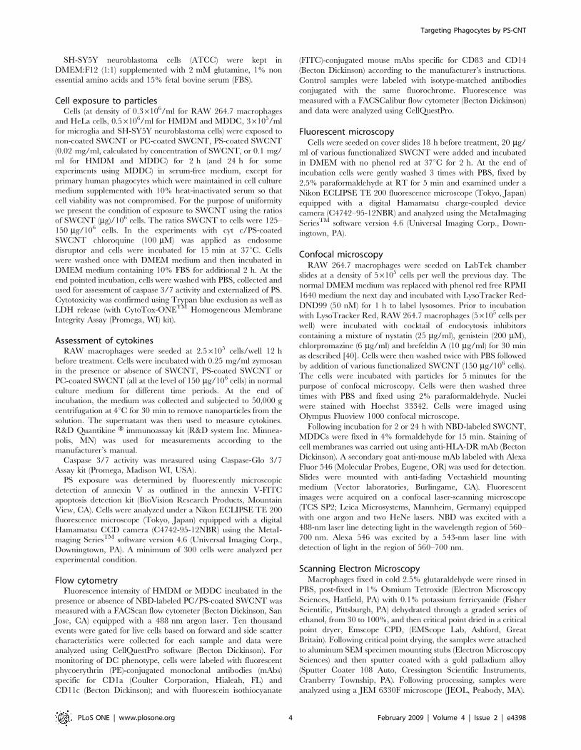
Physico-chemical characterization of functionalizedSWCNT
We prepared SWCNT coated with DOPC, as a control (PC-
coated SWCNT), or a mixture of DOPS, plus PC (PS-coated
SWCNT) by incubating nanotubes in the presence of liposomes
containing these lipids (PC or a mixture of PS plus PC at a molar
ratio of 1:1). Successful integration and the content of PS and PC
associated with SWCNT were confirmed by direct assessment of
the phosphorus content after HPTLC separation of phospholipids
(Fig. 1Aa). We found that PC-coated SWCNT contained
462633 nmol PC/mg SWCNT. Analysis of PS-coated SWCNT
revealed that amounts of integrated PS and PC on nanotubes were
approximately similar: 198614 and 223613 nmol of phospho-
lipids/mg SWCNT, respectively (Fig. 1Ab). Additionally, the
presence of PS on the surface of SWCNT was verified using
FITC-conjugated PS-specific protein, Annexin V, by measuring its
characteristic fluorescence (Fig 1B). After treatment of PS-coated
SWCNT with FITC-conjugated Annexin V, the appearance of a
robust fluorescence response was observed.
Analysis performed by NMAN 5040 revealed that purified
SWCNT were comprised of 99.7 wt% elemental carbon.
Determinations of carbon content for non-coated, PS-coated,
PC-coated and cyt c/PS-coated SWCNT showed that non-
covalent functionalization of SWCNT with phospholipids and
protein (cyt c) expectedly resulted in increased carbon content in
SWCNT suspensions. Based on our HPTLC-based determina-
tions of phospholipid content of PC- and PS-coated SWCNT, the
expected content of elemental carbon in the suspensions of these
coated SWCNT should be 81 and 61 ng/ml, respectively. Direct
estimates of elemental carbon content were 75 and 59 ng/ml for
SWCNT functionalized with PS and PC (Table S1). These
numbers correspond to 87% and 97% of theoretically calculated
content of the two phospholipids, respectively.
There was essentially no difference in the organization and
structure of SWCNT between non-coated samples and PS- or PC-
coated samples. Assessments of size distribution by dynamic light
scattering (DLS) showed that SWCNT (Fig. 1Ca), PS-coated
SWCNT and PC-coated SWCNT samples in suspensions had
very similar particle sizes in the range between 130–180 nm
(Table S1). Thus, SWCNTs were similarly dispersed in all
formulations used. Statistical analysis of isolated SWCNTs
measured by TEM revealed length distribution with a mean
length of 90635 nm (Fig. 1Cb). DLS determined the average
hydrodynamic diameter of the same sample as <135 nm. Average
particle sizes from DLS measurements for all samples are
summarized in Table S1. Small increases in particle size indicate
that no significant aggregation of SWCNTs takes place in solution.
Moreover, it is known that in microscopy, assessments may be
length-biased because they do not include nanotubes that cross the
edge of the micrograph and those that aggregate [41]. In fact,
longer nanotubes are ignored, because they are more likely to
reach out of view and aggregate with each other.
Evaluations of zeta potentials for SWCNT, PC-coated
SWCNT, PS-coated SWCNT, PS-NBD-coated SWCNT, PC-
NBD-coated SWCNT, and SWCNT containing cyt c showed that
they ranged from 240 to 250 mV (Supplemental Table 1).
Coating of SWCNT with phospholipids caused a slight increase in
the negative values of zeta potentials which, however, was not
significant between PC-coated SWCNT and PS-coated SWCNT.
Overall, the estimated negative values of zeta potentials corre-
spond with significant stability of dispersed non-coated and
phospholipids-coated SWCNTs in aqueous suspensions [42,43].
Transmission electron microscopy (TEM) of negatively stained
non-coated, PS-coated, and PC-coated SWCNT showed that they
had typical fibrous morphology and were represented mostly by
single nanotubes, as well as by ropes of nanotubes (Fig. 1Cb) and
entangled ‘‘bird’s nest’’-like aggregates. (Figure S1A). Calculated
percentage contributions of each of these ‘‘morphologies’’ in the
formulations used were 70, 25 and 5% for single SWCNT,
bundles and ‘‘bird’s-nest’’-like aggregates, respectively (Fig 1Cb
insert). PS-coated and PC-coated SWCNT had similar morphol-
ogy as non-coated SWCNT. Moreover, a thin layer of
phospholipids (PS/PC) (stained) is clearly visible on the sidewalls
of SWCNT by TEM (Figure S1B).
Cytotoxicity of SWCNTBoth Trypan Blue exclusion test and LDH release assay
demonstrated that neither non-coated SWCNT nor phospholip-
ids-coated SWCNT induced any cytotoxic effects in RAW 264.7
macrophages after 4 hrs of co-incubation (Figure S2). Moreover,
no impairment of viability was seen at 4 and 24 h of incubation of
SWCNT with primary human macrophages, using the Trypan
blue assay and the Hoechst 33342 staining method for
visualization of cell nuclei (data not shown).
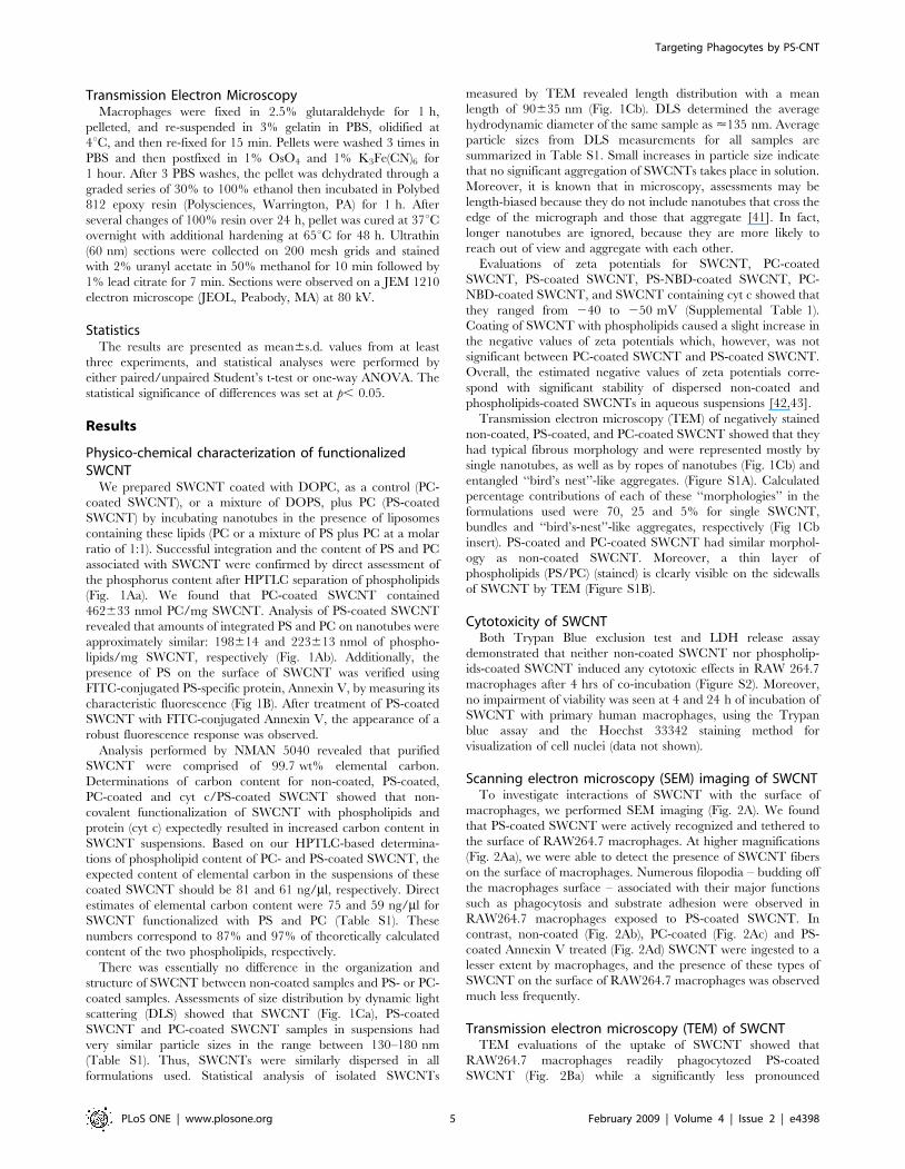
Scanning electron microscopy (SEM) imaging of SWCNTTo investigate interactions of SWCNT with the surface of
macrophages, we performed SEM imaging (Fig. 2A). We found
that PS-coated SWCNT were actively recognized and tethered to
the surface of RAW264.7 macrophages. At higher magnifications
(Fig. 2Aa), we were able to detect the presence of SWCNT fibers
on the surface of macrophages. Numerous filopodia – budding off
the macrophages surface – associated with their major functions
such as phagocytosis and substrate adhesion were observed in
RAW264.7 macrophages exposed to PS-coated SWCNT. In
contrast, non-coated (Fig. 2Ab), PC-coated (Fig. 2Ac) and PS-
coated Annexin V treated (Fig. 2Ad) SWCNT were ingested to a
lesser extent by macrophages, and the presence of these types of
SWCNT on the surface of RAW264.7 macrophages was observed
much less frequently.
Transmission electron microscopy (TEM) of SWCNTTEM evaluations of the uptake of SWCNT showed that
RAW264.7 macrophages readily phagocytozed PS-coated
SWCNT (Fig. 2Ba) while a significantly less pronounced
Targeting Phagocytes by PS-CNT
PLoS ONE | www.plosone.org 5 February 2009 | Volume 4 | Issue 2 | e4398
engulfment of PC-coated and non-coated SWCNT was docu-
mented (Fig. 2Bc). In addition, macrophages co-incubated with
PS-coated SWCNT had more endocytotic vesicles with entrapped
nanoparticles compared to macrophages incubated in the presence
of PC-coated or non-coated SWCNT (data not shown). At higher
magnifications (Fig. 2Ba), PS-coated SWCNT fibers within
phagosomes could be seen. To distinguish SWCNT from
autophagic bodies inside of phago-lysosomes we used a high
magnification TEM imaging (100,0006). Further, in order to
better identify nanoparticles in cells we performed TEM imaging
of nanotubes alone which were processed in a fashion similar to
that of cells for electron microscopy. SWCNT alone appear as
‘‘bamboo shoots’’ and resemble SWCNT that have been engulfed
by RAW264.7 macrophages (Fig. 2Bab). Markedly less of
‘‘bamboo shoot’’-like material was detected in RAW264.7
macrophages exposed to PC-coated or non–coated SWCNT
(Fig. 2Bc). Quantitative assessments of the number of nanotubes
present in an endosome within a 60 nm ultrafine section of cells
could be complicated by the fact that the same SWCNT could be
cut 1, 2 or more times, especially SWCNT bundles or ‘‘bird’s-
nest’’-like aggregates. Therefore, we presented the data as a plot of
percentage of active phagocytes in analyzed samples. We
considered as active macrophages those that engulfed at least
one nanoparticle. We found that amongst the various treatment
groups, 70% macrophages treated with PS-coated SWCNT
showed active phagocytosis with nanoparticles inside, which was
approximately twice the phagocytosis-positive population of cells
co-incubated with PC-coated SWCNT. Annexin V that masks PS
[44] and affects its recognition [45], effectively blocked the
recognition by 50% as indicated by a lower cell population with
active phagocytes (Fig. 2Bc). Notably, these TEM-based evalua-
tions of phagocytotic activity towards SWCNT were in good
Figure 1. Physico-chemical characterization of SWCNT. A. Evaluation of phospholipid content of PC-coated or PS-coated SWCNT. a) Typicalone-dimensional HPTLC of phospholipids extracted from PC-coated or PS-coated SWCNT. b) Phospholipid content of phospholipid-coated SWCNT. B.Typical fluorescence spectrum of Annexin V bound to PS-coated SWCNT. Note that a robust fluorescence response (Annexin V-FITC binding) wasrecorded from PS-coated but not from non-coated or PC-coated SWCNT. C. Physico-chemical characterization of bare SWCNT. a) Typical histogramsof size distribution assessed by dynamic light scattering; b) AFM images of bare SWCNT deposited on mica substrates; c) The height cross sectionanalysis of bare SWCNT.doi:10.1371/journal.pone.0004398.g001
Targeting Phagocytes by PS-CNT
PLoS ONE | www.plosone.org 6 February 2009 | Volume 4 | Issue 2 | e4398
agreement with independent assessments performed by confocal
microscopy of NBD-labeled phospholipid-coated SWCNT (see
below).
Uptake of SWCNT functionalized with fluorescentlylabeled phospholipids
To more quantitatively characterize uptake of phospholipid-
coated SWCNT by macrophages we utilized fluorescently labeled
PS, NBD-PS, for coating SWCNT; NBD-PC was utilized in
control experiments. After extensive washings, SWCNT incubated
with NBD-PS or NBD-PC displayed characteristic fluorescence
spectra confirming that the coating with phospholipids was
successful (Fig. 3A). We studied the time-course of uptake of
NBD-PC-coated SWCNT, NBD-PS-coated-SWCNT and NBD-
PS-coated and Annexin V treated SWCNT by RAW264.7
macrophages. To this end, we co-incubated RAW 264.7
macrophages with either NBD-PC- or NBD-PS coated nanopar-
ticles for up to 4 h (Fig. 3Ba). At this time point, viability test using
Trypan Blue exclusion revealed only 5.560.9% (n = 3) dead cells.
Uptake of NBD-PS-coated nanoparticles was time-dependent.
NBD-PS fluorescence was already detectable from cells after
Figure 2. SWCNT functionalized with PS but not with PC engulfed by murine RAW264.7 macrophages. A. Scanning electronmicrographs of RAW264.7 macrophages treated with SWCNT in vitro. RAW264.7 macrophages (0.36106 cells/ml) were incubated for 2 h with non-coated, PC- or PS-coated SWCNT. At the end of incubation macrophages were washed and fixed for SEM. a) Recognition of PS-coated SWCNT byRAW264.7 macrophages; (Note: The red arrows in all the electron micrographs in figure 2 point SWCNT. Sub-panel A-a representsscanning electron micrograph of SWCNT alone.) b) PC-coated SWCNT, c) non-coated, d) PS-coated Annexin V treated SWCNT are poorlyrecognized by macrophages. A total of 150 cells from each sample type were analyzed by SEM. B. Transmission electron micrographs of RAW264.7macrophages treated with SWCNT in vitro. RAW264.7 macrophages (0.36106 cells/ml) were incubated for 2 h with non-coated, PC- or PS-coatedSWCNT. At the end of incubation macrophages were washed and fixed for TEM. a) A high-power image of the macrophage with engulfed PS-coatedSWCNT. b) A typical TEM image of PS-coated SWCNT. Samples of SWCNT for TEM imaging were processed in a fashion similar to that of cells forelectron microscopy. Arrows indicate SWCNT. Representative TEM images are presented. c) Quantitative assessments of SWCNT phagocytosis by RAW264.7 macrophages. A total of 150 cells from each sample type were analyzed by TEM. Data are mean6S.D., n = 3, *p,0.05, PS-coated SWCNT vsSWCNT, PC-coated SWCNT and PS-coated Annexin V-treated. C. Effect of SWCNT on cytokine production by zymosan-stimulated RAW 264.7macrophages. Macrophages were seeded at 2.56105 cells/well in 48 well plates and co-incubated with zymosan (0.25 mg/ml) in the presence of non-coated, PC-coated and PS coated SWCNT (150 mg/106 cells). At the end of 2 hr incubation, TNF-a was measured in the medium. IL-10 and TGF-b weremeasured after 4 hr incubation. The cytokines were measured using R&D Quantikine H immunoassay kit. Data are mean6s.d., n = 3. ##p,0.05,SWCNT vs control. *p,0.05, PS-coated plus zymosan vs SWCNT plus zymozan and PC-coated plus zymosan.doi:10.1371/journal.pone.0004398.g002
Targeting Phagocytes by PS-CNT
PLoS ONE | www.plosone.org 7 February 2009 | Volume 4 | Issue 2 | e4398
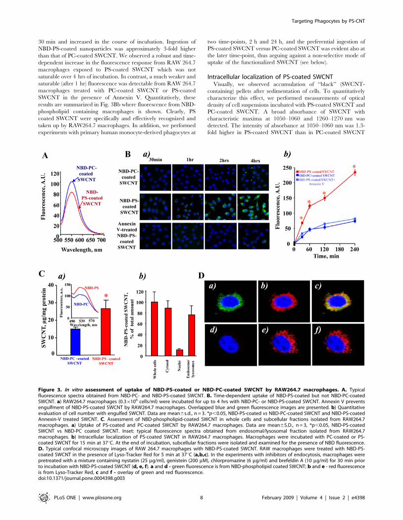
30 min and increased in the course of incubation. Ingestion of
NBD-PS-coated nanoparticles was approximately 3-fold higher
than that of PC-coated SWCNT. We observed a robust and time-
dependent increase in the fluorescence response from RAW 264.7
macrophages exposed to PS-coated SWCNT which was not
saturable over 4 hrs of incubation. In contrast, a much weaker and
saturable (after 1 hr) fluorescence was detectable from RAW 264.7
macrophages treated with PC-coated SWCNT or PS-coated
SWCNT in the presence of Annexin V. Quantitatively, these
results are summarized in Fig. 3Bb where fluorescence from NBD-
phospholipid containing macrophages is shown. Clearly, PS
coated SWCNT were specifically and effectively recognized and
taken up by RAW264.7 macrophages. In addition, we performed
experiments with primary human monocyte-derived phagocytes at
two time-points, 2 h and 24 h, and the preferential ingestion of
PS-coated SWCNT versus PC-coated SWCNT was evident also at
the later time-point, thus arguing against a non-selective mode of
uptake of the functionalized SWCNT (see below).
Intracellular localization of PS-coated SWCNTVisually, we observed accumulation of ‘‘black’’ (SWCNT-
containing) pellets after sedimentation of cells. To quantitatively
characterize this effect, we performed measurements of optical
density of cell suspensions incubated with PS-coated SWCNT and
PC-coated SWCNT. A broad absorbance of SWCNT with
characteristic maxima at 1050–1060 and 1260–1270 nm was
detected. The intensity of absorbance at 1050–1060 nm was 1.3-
fold higher in PS-coated SWCNT than in PC-coated SWCNT
Figure 3. In vitro assessment of uptake of NBD-PS-coated or NBD-PC-coated SWCNT by RAW264.7 macrophages. A. Typicalfluorescence spectra obtained from NBD-PC- and NBD-PS-coated SWCNT. B. Time-dependent uptake of NBD-PS-coated but not NBD-PC-coatedSWCNT. a) RAW264.7 macrophages (0.36106 cells/ml) were incubated for up to 4 hrs with NBD-PC- or NBD-PS-coated SWCNT. Annexin V preventsengulfment of NBD-PS-coated SWCNT by RAW264.7 macrophages. Overlapped blue and green fluorescence images are presented. b) Quantitativeevaluation of cell number with engulfed SWCNT. Data are mean6s.d., n = 3. *p,0.05, NBD-PS-coated vs NBD-PC-coated SWCNT and NBD-PS-coatedAnnexin-V treated SWCNT. C. Assessment of NBD-phospholipid-coated SWCNT in whole cells and subcellular fractions isolated from RAW264.7macrophages. a) Uptake of PS-coated and PC-coated SWCNT by RAW264.7 macrophages. Data are mean6S.D., n = 3, *p,0.05, NBD-PS-coatedSWCNT vs NBD-PC coated SWCNT. Inset: typical fluorescence spectra obtained from endosomal/lysosomal fraction isolated from RAW264.7macrophages. b) Intracellular localization of PS-coated SWCNT in RAW264.7 macrophages. Macrophages were incubated with PC-coated or PS-coated SWCNT for 15 min at 37uC. At the end of incubation, subcellular fractions were isolated and examined for the presence of NBD fluorescence.D. Typical confocal microscopy images of RAW 264.7 macrophages with NBD-PS-coated SWCNT. RAW macrophages were treated with NBD-PS-coated SWCNT in the presence of Lyso-Tracker Red for 5 min at 37uC (a,b,c). In the experiments with inhibitors of endocytosis, macrophages werepretreated with a mixture containing nystatin (25 mg/ml), genistein (200 mM), chlorpromazine (6 mg/ml) and brefeldin A (10 mg/ml) for 30 min priorto incubation with NBD-PS-coated SWCNT (d, e, f). a and d - green fluorescence is from NBD-phospholipid coated SWCNT; b and e - red fluorescenceis from Lyso-Tracker Red, c and f - overlay of green and red fluorescence.doi:10.1371/journal.pone.0004398.g003
Targeting Phagocytes by PS-CNT
PLoS ONE | www.plosone.org 8 February 2009 | Volume 4 | Issue 2 | e4398
samples. We further used fluorescently-labeled phospholipids to
characterize the presence of phospholipid-coated SWCNT in cells.
We found that NBD-PS coated SWCNT co-incubated with RAW
264.7 macrophages yielded a higher fluorescence response from
cell suspensions than NBD-PC coated SWCNT (Fig. 3Ca).
Further, the majority of fluorescence was associated with the
cytosolic fraction (containing endo-lysosomal vesicles) and was
minimal in the fraction of nuclei and cell debris (Fig. 3Cb). We
isolated the endo-lysosomal fraction from RAW 264.7 macro-
phages loaded with NBD-PS-coated SWCNT and found that
77618% (n = 3) of the fluorescence response was associated with
this fraction. This is in line with the recent demonstration of
deposition of SWCNT in endo-lysosomal compartments of
macrophages upon prolonged incubation [46].
Additionally, to prove co-localization of SWCNT with phago-
lysosomes we used confocal microscopy. To this end, we treated
RAW 264.7 macrophages (56105 cells/well) with either NBD-PS-
coated SWCNT or NBD-PC-coated SWCNT (150 mg/106 cells).
We found that NBD-PS-coated SWCNT were readily taken up by
macrophages. As shown in Fig. 3Da,b,c, a robust intracellular
fluorescence of NBD-PS (but not of NBD-PC) was co-localized, at
least in part, with a lysosomal marker, Lyso-Tracker Red.
Moreover, after pre-treatment of macrophages with the cocktail
of inhibitors of endocytosis (nystatin (25 mg/ml), genistein
(200 mM), chlorpromazine (6 mg/ml) and brefeldin A (10 mg/
ml)), the intensity of intracellular NBD-PS fluorescence was
drastically reduced (Fig. 3Dd,e,f). No fluorescence was detected
after treatment of macrophages with NBD-PC-coated SWCNT
(data not shown). This suggests that NBD-PS-coated SWCNT
were effectively taken up by macrophages through an endocytosis-
mediated process resulting in their significant accumulation in
endo-lysosomes.
Effect of SWCNT on cytokine release by RAW264.7macrophages
Apoptotic cells with externalized PS quench the production and
release of pro-inflammatory cytokines by macrophages [15,19]. To
assess whether PS-coated SWCNT display a similar effect, we
employed a standard stimulation of TNF-a formation in
RAW264.7 macrophages by zymosan [47,48] and evaluated the
effects of SWCNT. PS-coated SWCNT were more potent in
inhibiting TNF-a production by stimulated macrophages than
PC-coated or non-coated SWCNT (Fig. 2C). Thus, uptake of PS-
SWCNT is accompanied by typical PS-dependent suppression of
the pro-inflammatory macrophage response. It should be noted
that macrophages stimulated with SWCNT alone - in the absence
of zymosan - showed a significant increase in the TNF-aproduction up to 497624 pg/ml vs. 40610 (## P,0.05) in the
control. Notably, in PS-coated SWCNT, TNF-a levels dropped to
368633 pg/ml. No significant changes in the content of TNF-awas found after SWCNT coating with PC (445621 pg/ml). In
contrast, the production of anti-inflammatory cytokines is known
to be stimulated by PS-dependent pathways [16]. Stimulation of
RAW264.7 macrophages by zymosan resulted in the accumula-
tion of TGF-b and IL-10 at 4 hrs after the challenge. The
production of anti-inflammatory IL-10 and TGF-b by zymosan-
stimulated macrophages subsequently treated with PS-coated-
SWCNT increased 1.5- and 2-fold respectively, compared with
the effect of zymosan alone (Fig. 2C). This enhancement of IL-10
and TGF-b production was not observed after exposure of
zymosan-stimulated RAW264.7 macrophages to either non-
coated SWCNT or PC-coated SWCNT.
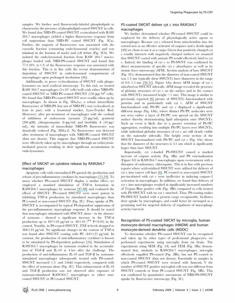
PS-coated SWCNT deliver cyt c into RAW264.7macrophages
We further determined whether PS-coated SWCNT could be
employed for the delivery of physiologically active agents to
macrophages. Because cyt c released from mitochondria into the
cytosol acts as an effective activator of caspases and a death-signal
[49] we chose to use it as a cargo. Given that positively charged cyt
c readily interacts with negatively charged surfaces we assumed
that SWCNT coated with anionic PS would effectively bind to cyt
c. Indeed, the binding of cyt c to PS-SWNT was confirmed by
direct measurements of specific cyt c absorbance as well as by
atomic force microscopy (AFM). Section analysis of bare SWCNT
(Fig. 1Cc) demonstrated that the diameter of non-coated SWCNT
was 1.3 nm (typically these SWCNTs have diameters in the range
of 0.6–1.5 nm [50,51]. Figure 4Aa shows AFM image of cyt c
adsorbed on SWCNT sidewalls. AFM image revealed the presence
of globular structures of cyt c on the surface and in the contact
with SWCNTs (measured height ,1 nm). This image is similar to
previously reported [6] picture of SWCNTs functionalized with
proteins and in particularly with cyt c. AFM of SWCNT
functionalized with PS/PC and cyt c displayed a significantly
different image (Fig. 4Ab), where distinct PS/PC molecules were
not seen; rather a layer of PS/PC was spread on the SWCNT
surface thereby demonstrating lipid adsorption onto SWCNTs.
Such an event is likely due to a greater retention of PS/PC
aggregation, resulting into multiple PS/PC layers over SWCNTs,
while individual globular structures of cyt c are still clearly visible
on the nanotube sidewalls. The height cross section of the
SWCNT functionalized with PS/PC and cyt c (Fig. 4Ac) shows
that the diameter of the structures is 3.5 nm which is significantly
larger than bare SWCNT.
Importantly, cyt c-loaded PS-SWCNT caused a marked
increase of caspase activity (Fig. 4Ba) and PS externalization
(Figure S3) in RAW264.7 macrophages upon co-treatment with a
disruptor of endosomes, chloroquine. This is in line with previous
reports where acid-oxidized SWCNT were utilized for delivery of
cyt c into cancer cell lines [6]. PC-coated or non-coated SWCNT
pre-incubated with cyt c were ineffective in inducing caspase-3
activation in macrophages. In addition, we found that delivery of
cyt c into macrophages resulted in significantly increased numbers
of Trypan Blue positive cells (Fig. 4Bb) compared to cells treated
with PS-SWCNT with no cyt c attached. Thus, PS-coating makes
PS-SWCNT loaded with a protein cargo recognizable, facilitates
their uptake by macrophages, and could hence be envisaged as a
promising tool for targeted delivery of regulators of macrophage
activity/survival.
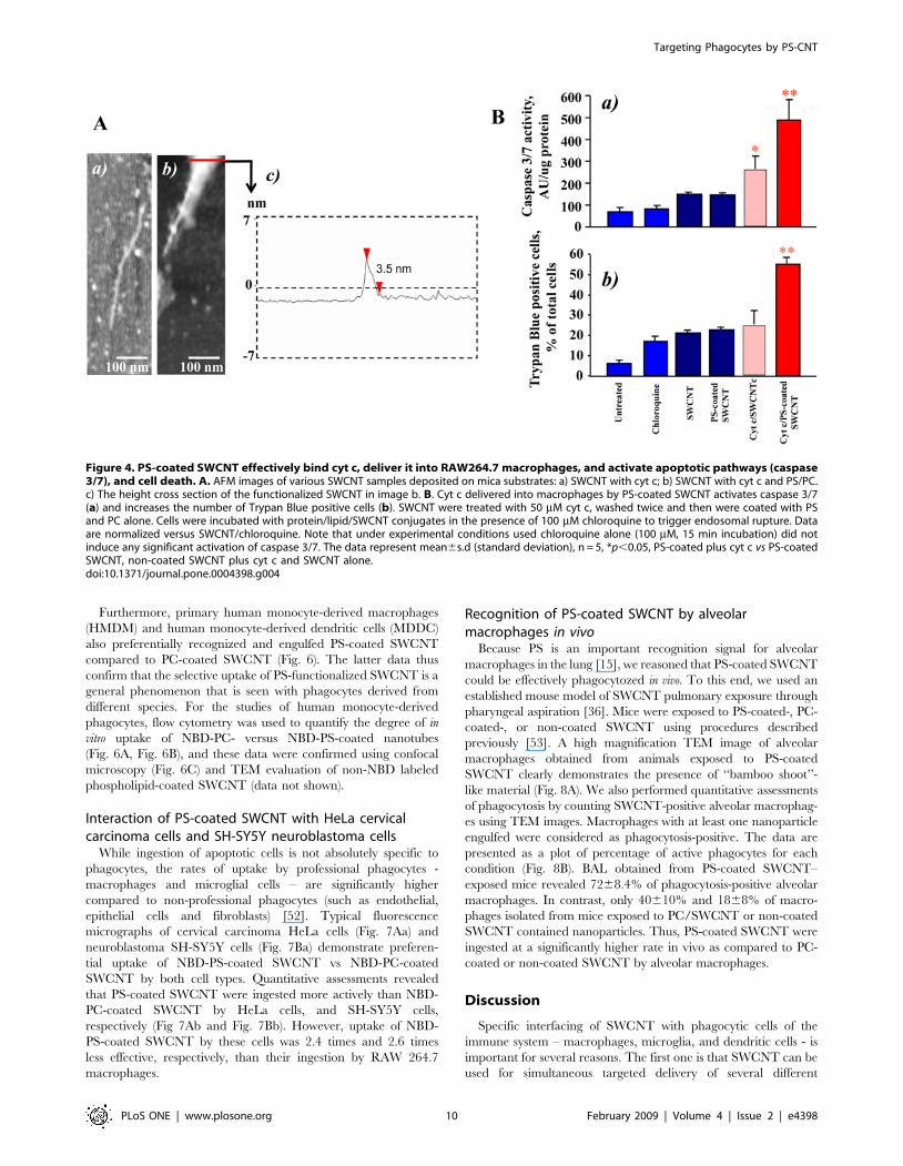
Recognition of PS-coated SWCNT by microglia, humanmonocyte-derived macrophages (HMDM) and humanmonocyte-derived dendritic cells (MDDC)
To determine whether PS-coated SWCNT can be recognized
and taken up by other types of professional phagocytes, we
performed experiments using microglia from rat brain. The
experiments using SEM (Fig. 5A) and TEM (Fig. 5Ba) demon-
strated that, similarly to RAW264.7 macrophages, microglia
effectively engulfed PS-coated (Fig. 5Bb), but not PC-coated or
non-coated SWCNT (data not shown). Essentially in samples in
which PS-coated SWCNT were treated with Annexin V the
number of SWCNT positive microglial cells was not different from
SWCNT controls or from PC-coated SWCNT (Fig. 5Bb). This
was confirmed by quantitative assessments of NBD-PS-SWCNT
uptake by fluorescence microscopy (Fig. 5C).
Targeting Phagocytes by PS-CNT
PLoS ONE | www.plosone.org 9 February 2009 | Volume 4 | Issue 2 | e4398
Furthermore, primary human monocyte-derived macrophages
(HMDM) and human monocyte-derived dendritic cells (MDDC)
also preferentially recognized and engulfed PS-coated SWCNT
compared to PC-coated SWCNT (Fig. 6). The latter data thus
confirm that the selective uptake of PS-functionalized SWCNT is a
general phenomenon that is seen with phagocytes derived from
different species. For the studies of human monocyte-derived
phagocytes, flow cytometry was used to quantify the degree of in
vitro uptake of NBD-PC- versus NBD-PS-coated nanotubes
(Fig. 6A, Fig. 6B), and these data were confirmed using confocal
microscopy (Fig. 6C) and TEM evaluation of non-NBD labeled
phospholipid-coated SWCNT (data not shown).
Interaction of PS-coated SWCNT with HeLa cervicalcarcinoma cells and SH-SY5Y neuroblastoma cells
While ingestion of apoptotic cells is not absolutely specific to
phagocytes, the rates of uptake by professional phagocytes -
macrophages and microglial cells – are significantly higher
compared to non-professional phagocytes (such as endothelial,
epithelial cells and fibroblasts) [52]. Typical fluorescence
micrographs of cervical carcinoma HeLa cells (Fig. 7Aa) and
neuroblastoma SH-SY5Y cells (Fig. 7Ba) demonstrate preferen-
tial uptake of NBD-PS-coated SWCNT vs NBD-PC-coated
SWCNT by both cell types. Quantitative assessments revealed
that PS-coated SWCNT were ingested more actively than NBD-
PC-coated SWCNT by HeLa cells, and SH-SY5Y cells,
respectively (Fig 7Ab and Fig. 7Bb). However, uptake of NBD-
PS-coated SWCNT by these cells was 2.4 times and 2.6 times
less effective, respectively, than their ingestion by RAW 264.7
macrophages.
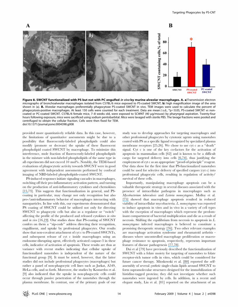
Recognition of PS-coated SWCNT by alveolarmacrophages in vivo
Because PS is an important recognition signal for alveolar
macrophages in the lung [15], we reasoned that PS-coated SWCNT
could be effectively phagocytozed in vivo. To this end, we used an
established mouse model of SWCNT pulmonary exposure through
pharyngeal aspiration [36]. Mice were exposed to PS-coated-, PC-
coated-, or non-coated SWCNT using procedures described
previously [53]. A high magnification TEM image of alveolar
macrophages obtained from animals exposed to PS-coated
SWCNT clearly demonstrates the presence of ‘‘bamboo shoot’’-
like material (Fig. 8A). We also performed quantitative assessments
of phagocytosis by counting SWCNT-positive alveolar macrophag-
es using TEM images. Macrophages with at least one nanoparticle
engulfed were considered as phagocytosis-positive. The data are
presented as a plot of percentage of active phagocytes for each
condition (Fig. 8B). BAL obtained from PS-coated SWCNT–
exposed mice revealed 7268.4% of phagocytosis-positive alveolar
macrophages. In contrast, only 40610% and 1868% of macro-
phages isolated from mice exposed to PC/SWCNT or non-coated
SWCNT contained nanoparticles. Thus, PS-coated SWCNT were
ingested at a significantly higher rate in vivo as compared to PC-
coated or non-coated SWCNT by alveolar macrophages.
Discussion
Specific interfacing of SWCNT with phagocytic cells of the
immune system – macrophages, microglia, and dendritic cells - is
important for several reasons. The first one is that SWCNT can be
used for simultaneous targeted delivery of several different
Figure 4. PS-coated SWCNT effectively bind cyt c, deliver it into RAW264.7 macrophages, and activate apoptotic pathways (caspase3/7), and cell death. A. AFM images of various SWCNT samples deposited on mica substrates: a) SWCNT with cyt c; b) SWCNT with cyt c and PS/PC.c) The height cross section of the functionalized SWCNT in image b. B. Cyt c delivered into macrophages by PS-coated SWCNT activates caspase 3/7(a) and increases the number of Trypan Blue positive cells (b). SWCNT were treated with 50 mM cyt c, washed twice and then were coated with PSand PC alone. Cells were incubated with protein/lipid/SWCNT conjugates in the presence of 100 mM chloroquine to trigger endosomal rupture. Dataare normalized versus SWCNT/chloroquine. Note that under experimental conditions used chloroquine alone (100 mM, 15 min incubation) did notinduce any significant activation of caspase 3/7. The data represent mean6s.d (standard deviation), n = 5, *p,0.05, PS-coated plus cyt c vs PS-coatedSWCNT, non-coated SWCNT plus cyt c and SWCNT alone.doi:10.1371/journal.pone.0004398.g004
Targeting Phagocytes by PS-CNT
PLoS ONE | www.plosone.org 10 February 2009 | Volume 4 | Issue 2 | e4398
regulators/inhibitors with a potential to release them in temporally
and spatially predetermined ways to control the bioactivity of a
specific cell population during physiologically critical events. As
macrophages can host a number of pathogens [54,55], nanocar-
riers can also be used for specific delivery of pro-apoptotic agents
to aid in the defense against intracellular pathogens [56].
Furthermore, macrophages and microglia are the major executors
of pro-/anti-inflammatory responses, and - along with antigen-
presenting dendritic cells - are important components of immune
reactions [57–60]. Specific targeting of cargoes/regulators to these
cells could be exploited for therapeutic regulation of numerous
immune functions, including the enhancement of immune
responses to prophylactic or therapeutic allergen-specific vaccines
through the coupling of allergens to nanocarriers. Finally,
SWCNT are among the most commonly used nanomaterials with
explosively expanding research and commercial applications [61].
Because the production and employment of industrial quantities of
SWCNT are becoming a reality, health risk concerns, particularly
due to occupational and environmental exposures, are emerging
[62]. Not only an unusually large surface area, but also unique
physical and chemical characteristics, redox features as well as
significant decoration with transition metals alert to a possibility of
unanticipated bioresponses resulting from interactions of SWCNT
with cells, tissues, and biofluids. In fact, recent studies have
demonstrated significant pulmonary and cardiovascular toxicity of
SWCNT associated with a robust inflammatory response and
early onset of fibrotic transition in mice [53,63,64]. In this context,
the enhancement of phagocytic recognition and uptake of
SWCNT through PS-functionalization may be important in order
to reduce the potential cytotoxicity of SWCNT.
Figure 5. Primary rat microglia recognized SWCNT functionalized with PS but not with PC. A. Scanning electron micrographs of microgliatreated with PS-coated SWCNT in vitro. Microglia (1.56105 cells/ml) were incubated for 2 h with PC- or PS-coated SWCNT. At the end of incubationmacrophages were washed and fixed for SEM. (Note: The red arrows in all the electron micrographs in figure 5 point SWCNT). B.Transmission electron micrographs of primary microglia exposed to SWCNT in vitro. Microglia (1.56105 cells/ml) were incubated with PC- or PS-coated SWCNT. At the end of incubation microglia was washed and fixed for TEM. a) Microglia exposed to PS-coated SWCNT; b) Quantitativeassessments of SWCNT phagocytosis by RAW 264.7 macrophages. A total of 60 cells from each sample type were analyzed by TEM. Data aremean6S.D., n = 3, *p,0.05, PS-coated SWCNT vs SWCNT, PC coated SWCNT and PS-coated Annexin V–treated SWCNT. C. In vitro assessment ofuptake of SWCNT coated with NBD-PS or NBD-PC by microglia. Microglia (1.56105 cells/ml) were incubated with NBD-PC- (a), NBD-PS-coated SWCNT(b) or NBD-PS-coated/Annexin V treated SWCNT (c). 1 – bright field image; 2 – blue fluorescence image, Hoechst 33342; 3 – green fluorescence image,NBD-labeled phospholipids; 4 – overlap of blue and green fluorescence images with image under bright field. d) Annexin V treatment of NBD-PS-coated SWCNT prevents their engulfment by microglia. Quantitative evaluation of cell number with engulfed SWCNT. Data are mean6s.d., n = 4.*p,0.05, NBD-PS-coated SWCNT vs NBD-PC-coated SWCNT and NBD-PS-coated Annexin V treated SWCNT.doi:10.1371/journal.pone.0004398.g005
Targeting Phagocytes by PS-CNT
PLoS ONE | www.plosone.org 11 February 2009 | Volume 4 | Issue 2 | e4398
Understanding of major principles of particle recognition by
macrophages has long been a controversial issue. Because non-
functionalized nanoparticles are prone to aggregation, sonication
of non-coated SWCNT was performed before adding them to
cells. Under these conditions, no significant uptake of non-coated
SWCNT by RAW 264.7 macrophages occurred during the 2 h
incubation period. In contrast, Dumortier et al. [65] have recently
reported that SWCNTs tend to re-aggregate and form large
clusters that are eventually (after 24 h of co-incubation) phagocy-
tozed by professional macrophages. It is likely that macrophage
uptake of big clusters of SWCNT formed during prolonged
incubation times is related to the reduced solubility of these non-
coated nanotubes. It has been also reported that geometry and
shape act as determinants of particle recognition [66]. Several
studies have demonstrated that functionalized SWCNT are
recognizable by cells and taken up through endocytosis-dependent
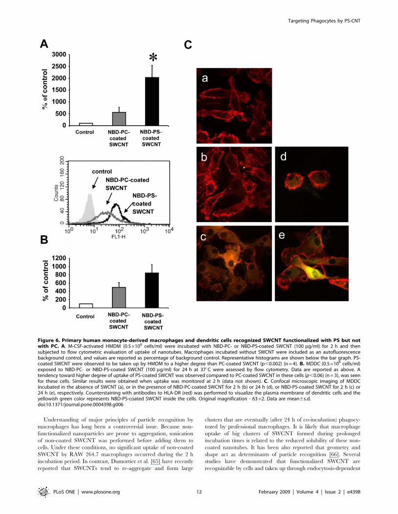
Figure 6. Primary human monocyte-derived macrophages and dendritic cells recognized SWCNT functionalized with PS but notwith PC. A. M-CSF-activated HMDM (0.56106 cells/ml) were incubated with NBD-PC- or NBD-PS-coated SWCNT (100 mg/ml) for 2 h and thensubjected to flow cytometric evaluation of uptake of nanotubes. Macrophages incubated without SWCNT were included as an autofluorescencebackground control, and values are reported as percentage of background control. Representative histograms are shown below the bar graph. PS-coated SWCNT were observed to be taken up by HMDM to a higher degree than PC-coated SWCNT (p,0.002) (n = 4). B. MDDC (0.56106 cells/ml)exposed to NBD-PC- or NBD-PS-coated SWCNT (100 mg/ml) for 24 h at 37uC were assessed by flow cytometry. Data are reported as above. Atendency toward higher degree of uptake of PS-coated SWCNT was observed compared to PC-coated SWCNT in these cells (p,0.06) (n = 3), was seenfor these cells. Similar results were obtained when uptake was monitored at 2 h (data not shown). C. Confocal microscopic imaging of MDDCincubated in the absence of SWCNT (a), or in the presence of NBD-PC-coated SWCNT for 2 h (b) or 24 h (d), or NBD-PS-coated SWCNT for 2 h (c) or24 h (e), respectively. Counterstaining with antibodies to HLA-DR (red) was performed to visualize the plasma membrane of dendritic cells and theyellowish green color represents NBD-PS-coated SWCNT inside the cells. Original magnification - 6362. Data are mean6s.d.doi:10.1371/journal.pone.0004398.g006
Targeting Phagocytes by PS-CNT
PLoS ONE | www.plosone.org 12 February 2009 | Volume 4 | Issue 2 | e4398
pathways [67,68]. By contrast, we and others reported that non-
functionalized SWCNT are neither effectively recognized nor
phagocytozed by macrophages [5,53,69]. The fact that PS-coating
leads to recognition and uptake of SWCNT suggests that it is the
lack of a recognition signal that is responsible for poor uptake of
non-functionalized carbon nanotubes by phagocytes. A variety of
specialized receptors on the macrophage surface have been
implicated in recognition and tethering of different particles,
including ultra-fine particles [64,67,70–72]. Recent studies
identified several novel macrophage receptors for which PS is a
specific high-affinity ligand [25,26], facilitating uptake of target
cells with externalized PS. PC is not specifically recognized by
these receptors and its presence on the cell surface does not
enhance recognition and uptake of cells by macrophages.
Therefore, we chose to use NBD-PC-coated SWCNT as controls
in our comparative experiments on assessments of SWCNT
uptake by macrophages. Further, we utilized Annexin V – a
protein known to selectively bind to PS (but not to PC) and mask
PS recognition and uptake by macrophages. The Annexin V
mediated suppression of uptake of NBD-PS-coated SWCNT (and
lack of fluorescence response from macrophages incubated in the
presence of NBD-PS coated SWCNT pre-treated with Annexin V)
was employed as an additional specific control of PS-dependent
recognition and uptake. Our data indicate that eclipsing of PS with
its specific ligand, Annexin V, completely blocks SWCNT
recognition by macrophage cell lines and primary phagocytes.
Most notably, these PS-dependent recognition patterns are
realized in vivo whereby alveolar macrophages display enhanced
uptake of PS-coated SWCNT during an inflammatory pulmonary
response induced by aspiration exposure. Together, our in vitro and
in vivo studies demonstrate that PS-functionalization renders
nanotubes appetizing to phagocytes. We believe that analysis of
SWCNT uptake by phagocytes based on the employment of
fluorescently-labeled phospholipids (NBD-PS and NBD-PC)
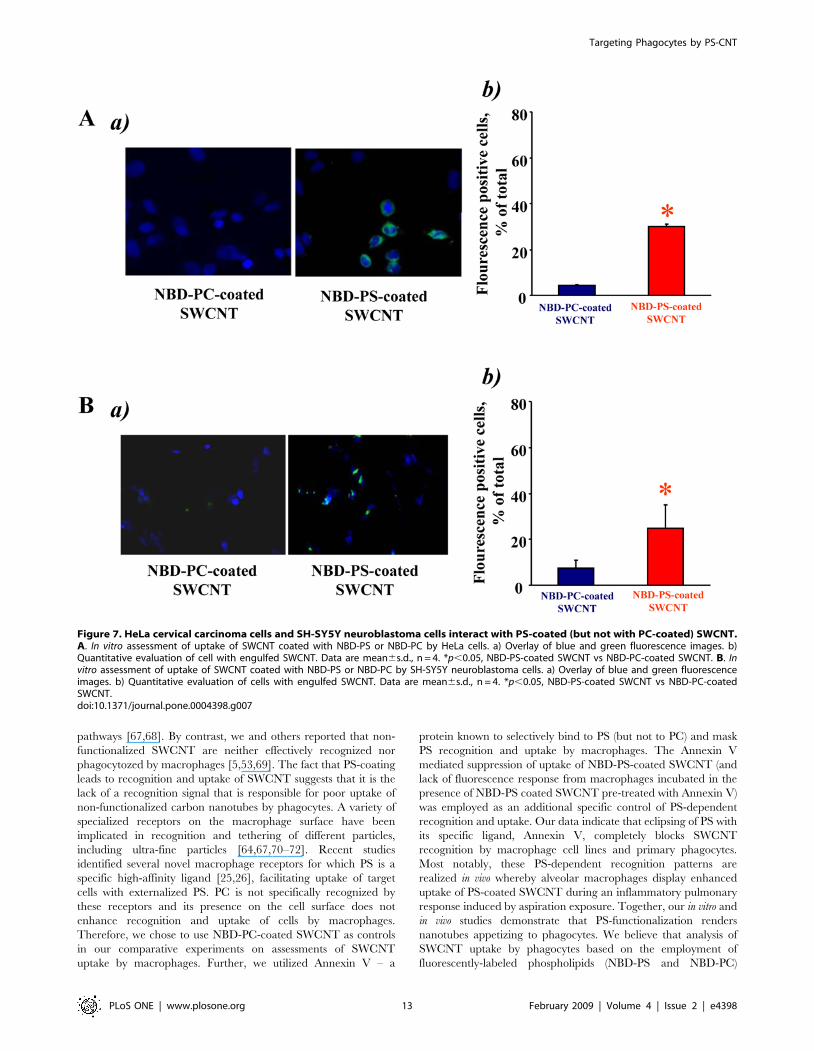
Figure 7. HeLa cervical carcinoma cells and SH-SY5Y neuroblastoma cells interact with PS-coated (but not with PC-coated) SWCNT.A. In vitro assessment of uptake of SWCNT coated with NBD-PS or NBD-PC by HeLa cells. a) Overlay of blue and green fluorescence images. b)Quantitative evaluation of cell with engulfed SWCNT. Data are mean6s.d., n = 4. *p,0.05, NBD-PS-coated SWCNT vs NBD-PC-coated SWCNT. B. Invitro assessment of uptake of SWCNT coated with NBD-PS or NBD-PC by SH-SY5Y neuroblastoma cells. a) Overlay of blue and green fluorescenceimages. b) Quantitative evaluation of cells with engulfed SWCNT. Data are mean6s.d., n = 4. *p,0.05, NBD-PS-coated SWCNT vs NBD-PC-coatedSWCNT.doi:10.1371/journal.pone.0004398.g007
Targeting Phagocytes by PS-CNT
PLoS ONE | www.plosone.org 13 February 2009 | Volume 4 | Issue 2 | e4398
provided more quantitatively reliable data. In this case, however,
the limitations of quantitative assessments might be due to a
possibility that fluorescently-labeled phospholipids could also
modify (promote or decrease) the uptake of these fluorescent
phospholipid coated SWCNT by macrophage. To minimize this
interference, mole fraction of fluorescently-labeled phospholipids
in the mixture with non-labeled phospholipids of the same type in
all experiments did not exceed 10 mol%. Notably, the TEM-based
evaluations of phagocytotic activity towards SWCNT were in good
agreement with independent assessments performed by confocal
imaging of NBD-labeled phospholipids-coated SWCNT.
PS-induced responses initiate signaling cascades in macrophages,
switching off their pro-inflammatory activation pattern, and turning
on the production of anti-inflammatory cytokines and chemokines
[15,73]. This suggests that functionalization in general, and PS-
coating in particular, may change not only recognition but also
pro-/anti-inflammatory behavior of macrophages interacting with
nanoparticles. In line with this, our experiments demonstrated that
PS coating of SWCNT could be utilized not only for directing
SWCNT to phagocytic cells but also as a regulator or ‘‘switch’’
affecting the profile of the produced and released cytokines in vitro
and in vivo [16,19]. Our studies show that PS-coating of SWCNT
confers on them a ‘‘zip-code’’ address directing their recognition,
engulfment, and uptake by professional phagocytes. Our results
show that non-covalent attachment of cyt c to PS-coated SWCNTs,
and subsequent release of cyt c inside macrophages using an
endosome-disrupting agent, effectively activated caspase-3 in these
cells, indicative of activation of apoptosis. These results are thus at
variance with recent studies indicating that cellular uptake of
functionalized nanotubes is independent of the nature of the
functional group [9]. It must be noted, however, that the latter
studies did not include professional phagocytes (macrophages) but
rather a panel of non-phagocytic cell lines such as Jurkat, A549,
HeLa cells, and so forth. Moreover, the studies by Kostarelos et al.
[9] also indicated that the uptake in non-phagocytic cells could
occur through passive penetration of the nanotubes through the
plasma membrane. In contrast, one of the primary goals of our
study was to develop approaches for targeting macrophages and
other professional phagocytes by cytotoxic agents using nanotubes
coated with PS as a specific ligand recognized by specialized plasma
membrane receptors [25,26]. We chose to use cyt c as a ‘‘death’’
signal. Cyt c is one of the key co-factors for the activation of
apoptosis in mammalian cells [62] and is known to be a difficult
cargo for targeted delivery into cells [6,74], thus justifying the
employment of cyt c as an appropriate ‘‘proof-of-principle’’ reagent.
Our data show for the first time that PS-functionalized nanotubes
could be used for selective delivery of specified cargoes (cyt c) into
professional phagocytic cells, resulting in regulation of activity/
survival of these cells.
Importantly, manipulating macrophage apoptosis can be a
valuable therapeutic strategy in several diseases associated with the
presence of intracellular pathogens in macrophages such as
Mycobacterium tuberculosis and Listeria monocytogenes. Molloy et al.
[75] showed that macrophage apoptosis resulted in reduced
viability of intracellular mycobacteria. L. monocytogenes was reported
to induce apoptosis in vitro and in vivo in a variety of cell types
with the exception of macrophages which represent the predom-
inant compartment of bacterial multiplication and die as a result of
necrosis. Shifting the equilibrium from necrosis to apoptosis in L.
monocytogenes infected macrophages is believed to constitute a
promising therapeutic strategy [76]. Two other relevant examples
are macrophage activation syndrome and rheumatoid arthritis -
diseases where uncontrolled macrophage proliferation or macro-
phage resistance to apoptosis, respectively, represents important
features of disease pathogenesis [77,78].
Dai et al [79] have previously described the functionalization of
SWCNT with a folate moiety for targeting of nanotubes to folate
receptor-rich tumor cells in vitro, which could be considered for
future cancer therapy. Mioskowski et al. [80] reported the self-
assembly of several synthetic single-chain lipids around SWCNT to
form supramolecular structures designed for the immobilization of
histidine-tagged proteins; they did not investigate whether such
nanotubes were ingested by cells. In addition, in a recent and
elegant study, Liu et al. [81] reported on the attachment of an
Figure 8. SWCNT functionalized with PS but not with PC engulfed in vivo by murine alveolar macrophages. A. a)Transmission electronmicrographs of bronchoalveolar macrophages isolated from C57BL/6 mice exposed to PS-coated SWCNT; b) high magnification image of the areashown in (a). B. Alveolar macrophages preferentially phagocytoze PS-coated SWCNT in vivo. TEM images were used to calculate the percent ofphagocytosis-positive macrophages. At least 150 cells were counted for each treatment. Data are mean6s.d., *p,0.05, PS-coated SWCNT vs non-coated or PC-coated SWCNT. C57BL/6 female mice, 7–8 weeks old, were exposed to SCWNT (40 mg/mouse) by pharyngeal aspiration. Twenty-fourhours following exposure, mice were sacrificed using sodium pentobarbital. Mice were lavaged with sterile PBS. The lavage fractions were pooled andcentrifuged to obtain the cellular fraction. Cells were then fixed for TEM.doi:10.1371/journal.pone.0004398.g008
Targeting Phagocytes by PS-CNT
PLoS ONE | www.plosone.org 14 February 2009 | Volume 4 | Issue 2 | e4398
RGD peptide to nanotubes coated with polyethylene glycol (PEG)
and subsequent in vivo administration to mice bearing integrin-
positive tumors. Efficient targeting of the nanotubes to the tumors
was achieved through this approach. The goal of our work was to
employ the naturally occurring ‘‘eat-me’’ signal, PS as a specific
ligand that is recognized by professional phagocytes. We provide
evidence that the uptake is specific, since nanotubes functionalized
with PC are not recognized as readily as PS-coated nanotubes and
because the PS-binding protein, Annexin V, can prevent uptake of
PS-coated nanotubes. Because the chemical cutting is associated
with the production of oxidative negatively-charged defects in
SWCNT (carboxy-groups [32]) it is possible that coating with
phospholipids could be slightly different in precut and non-precut
SWCNT. It should be noted, however, that binding of negatively
charged PS (at pH 7.4) is unlikely to occur at the sites of negatively
charged defects due to electrostatic repulsions. Previous studies
from our and other laboratories have established high-affinity
binding of cyt c with PS [82]. After the coating, binding of
positively-charged cyt c was likely associated with abundant
negatively-charged PS. We also provide evidence that the
mechanism of uptake is specific and occurs through endocytosis.
Finally, we show that a relevant cargo (pro-apoptotic cyt c) can be
delivered into macrophages, and upon disruption of endosomes,
the cargo is released and can perform its biological function
(activation of the caspase cascade).
Taken together, our findings highlight a novel strategy for the
controlled delivery of relevant cargoes into specific cell popula-
tions. Further developments of the novel principle established in
the current study, i.e. PS-coating of SWCNT for recognition and
ingestion, may exploit cargoes covalently-conjugated with specific
linkers that are hydrolysable extracellularly by activated macro-
phages (eg. superoxide-sensitive linkers) or intracellularly (eg.
esterase-sensitive conjugates) for targeted delivery and release to
regulate life-span and activity of phagocytes.
Supporting Information
Figure S1 TEM images of bare SWCNT (A) and PS-coated
SWCNT (B) deposited on mica substrates.
Found at: doi:10.1371/journal.pone.0004398.s001 (5.93 MB TIF)
Figure S2 Viability of RAW264.7 macrophages exposed to
SWCNT. RAW 264.7 macrophages were seeded at 16104 cells/
well on 96 well-plate 12 hrs before treatment. Cells were
incubated with non-coated SWCNT or SWCNT coated with
either PS or PC in serum and phenol red free DMEM containing
100 mM DTPA for up to 4 hrs. At the end of incubation, the
culture medium was collected, centrifuged to remove SWCNT
and used for LDH assay with CytotoxOne kit. Data are
mean6s.d., n = 3.
Found at: doi:10.1371/journal.pone.0004398.s002 (0.50 MB TIF)
Figure S3 PS-coated SWCNT effectively deliver cyt c into
RAW264.7 macrophages, and activate apoptotic pathways
(caspase 3/7). RAW264.7 macrophages were seeded at 0.56105
cells/well 12 hrs prior to treatment. Cells were incubated with
differently functionalized SWCNT in serum and phenol red free
DMEM containing 100 mM DTPA for 1 hr at 37uC. At the end of
incubation, cell were washed to remove SWCNT and treated with
chloroquine (100 mM) in complete culture medium at 37 uC for
15 min. After that, cells were washed with complete DMEM and
additionally incubated for 8 hrs. Cells were stained in binding
buffer containing annexin V (0.5 mg/ml) for 5 min and counter-
stained with Hoechst 33342 for nuclei at room temperature. The
cells were examined under a Nikon ECLIPSE TE 200
fluorescence microscope (Tokyo, Japan) equipped with a digital
Hamamatsu CCD camera (C4742-95-12NBR) and analyzed using
the MetaImaging SeriesTM software version 4.6 (Universal
Imaging Corp., Downingtown, PA). A minimum of 300
macrophages were counted per experimental condition. Data
are mean6s.d. (* p,0.01 vs untreated). Insert: Typical fluores-
cence image of RAW264.7 macrophages treated with cyt c/PS-
coated SWCNT (staining with annexin V).
Found at: doi:10.1371/journal.pone.0004398.s003 (1.39 MB TIF)
Table S1
Found at: doi:10.1371/journal.pone.0004398.s004 (0.03 MB
DOC)
Acknowledgments
DISCLAIMERS: The findings and conclusions in this report are those of
the authors and do not necessarily represent the view of the National
Institute for Occupational Safety and Health.
Author Contributions
Conceived and designed the experiments: NVK YYT WF LVB NAB HB
KC DS HV AS EW BF PDK AS ERK ARM AAS VEK. Performed the
experiments: NVK YYT WF LVB NAB HB KC MR DS HV AS EW BF
PDK ERK ARM AAS. Analyzed the data: NVK YYT WF LVB NAB HB
KC MR HV AS EW BF PDK AS ERK ARM AAS VEK. Contributed
reagents/materials/analysis tools: DS BF AS AAS. Wrote the paper: NVK
VEK.
References
1. Gonzalez-Flecha B (2004) Oxidant mechanisms in response to ambient air
particles. Mol Aspects Med 25: 169–182.
2. Oberdorster G, Sharp Z, Atudorei V, Elder A, Gelein R, et al. (2004)
Translocation of inhaled ultrafine particles to the brain. Inhal Toxicol 16:
437–445.
3. Elder A, Oberdorster G (2006) Translocation and effects of ultrafine particles
outside of the lung. Clin Occup Environ Med 5: 785–796.
4. Shvedova AA, Kisin ER, Mercer R, Murray AR, Johnson VJ, et al. (2005)
Unusual inflammatory and fibrogenic pulmonary responses to single-walled
carbon nanotubes in mice. Am J Physiol Lung Cell Mol Physiol 289: L698–708.
5. Kagan VE, Tyurina YY, Tyurin VA, Konduru NV, Potapovich AI, et al. (2006)
Direct and indirect effects of single walled carbon nanotubes on RAW 264.7
macrophages: role of iron. Toxicol Lett 165: 88–100.
6. Kam NW, Dai H (2005) Carbon nanotubes as intracellular protein transporters:
generality and biological functionality. J Am Chem Soc 127: 6021–6026.
7. Nimmagadda A, Thurston K, Nollert MU, McFetridge PS (2006) Chemical
modification of SWNT alters in vitro cell-SWNT interactions. J Biomed Mater
Res A 76: 614–625.
8. Klumpp C, Kostarelos K, Prato M, Bianco A (2006) Functionalized carbon
nanotubes as emerging nanovectors for the delivery of therapeutics. Biochim
Biophys Acta 1758: 404–412.
9. Kostarelos K, Lacerda L, Pastorin G, Wu W, Wieckowski S, et al. (2007)
Cellular uptake of functionalized carbon nanotubes is independent of functional
group and cell type. Nature Nanotechnology 2: 108–113.
10. Chen X, Tam UC, Czlapinski JL, Lee GS, Rabuka D, et al. (2006)
Interfacing carbon nanotubes with living cells. J Am Chem Soc 128: 6292–
6293.
11. Chen X, Lee GS, Zettl A, Bertozzi CR (2004) Biomimetic engineering of carbon
nanotubes by using cell surface mucin mimics. Angew Chem Int Ed Engl 43:
6111–6116.
12. Vandivier RW, Henson PM, Douglas IS (2006) Burying the dead: the impact of
failed apoptotic cell removal (efferocytosis) on chronic inflammatory lung
disease. Chest 129: 1673–1682.
13. Gardai SJ, Bratton DL, Ogden CA, Henson PM (2006) Recognition ligands on
apoptotic cells: a perspective. J Leukoc Biol 79: 896–903.
14. Fadok VA, Bratton DL, Konowal A, Freed PW, Westcott JY, et al. (1998)
Macrophages that have ingested apoptotic cells in vitro inhibit proinflammatory
cytokine production through autocrine/paracrine mechanisms involving TGF-
beta, PGE2, and PAF. J Clin Invest 101: 890–898.
15. Huynh ML, Fadok VA, Henson PM (2002) Phosphatidylserine-dependent
ingestion of apoptotic cells promotes TGF-beta1 secretion and the resolution of
inflammation. J Clin Invest 109: 41–50.
Targeting Phagocytes by PS-CNT
PLoS ONE | www.plosone.org 15 February 2009 | Volume 4 | Issue 2 | e4398
16. Hoffmann PR, Kench JA, Vondracek A, Kruk E, Daleke DL, et al. (2005)
Interaction between phosphatidylserine and the phosphatidylserine receptor
inhibits immune responses in vivo. J Immunol 174: 1393–1404.
17. Daleke DL (2003) Regulation of transbilayer plasma membrane phospholipid
asymmetry. J Lipid Res 44: 233–242.
18. Devaux PF, Lopez-Montero I, Bryde S (2006) Proteins involved in lipid
translocation in eukaryotic cells. Chem Phys Lipids 141: 119–132.
19. Serinkan BF, Gambelli F, Potapovich AI, Babu H, Di Giuseppe M, et al. (2005)
Apoptotic cells quench reactive oxygen and nitrogen species and modulate TNF-alpha/TGF-beta1 balance in activated macrophages: involvement of phospha-
tidylserine-dependent and -independent pathways. Cell Death Differ 12:
1141–1144.
20. Lauber K, Bohn E, Krober SM, Xiao YJ, Blumenthal SG, et al. (2003)
Apoptotic cells induce migration of phagocytes via caspase-3-mediated release of
a lipid attraction signal. Cell 113: 717–730.
21. Hanayama R, Tanaka M, Miwa K, Shinohara A, Iwamatsu A, et al. (2002)
Identification of a factor that links apoptotic cells to phagocytes. Nature 417:
182–187.
22. Arur S, Uche UE, Rezaul K, Fong M, Scranton V, et al. (2003) Annexin I is an
endogenous ligand that mediates apoptotic cell engulfment. Dev Cell 4:
587–598.
23. Obeid M, Tesniere A, Ghiringhelli F, Fimia GM, Apetoh L, et al. (2007)
Calreticulin exposure dictates the immunogenicity of cancer cell death. Nat Med
13: 54–61.
24. Martin SJ, Reutelingsperger CP, McGahon AJ, Rader JA, van Schie RC, et al.
(1995) Early redistribution of plasma membrane phosphatidylserine is a general
feature of apoptosis regardless of the initiating stimulus: inhibition by
overexpression of Bcl-2 and Abl. J Exp Med 182: 1545–1556.
25. Park D, Tosello-Trampont AC, Elliott MR, Lu M, Haney LB, et al. (2007) BAI1
is an engulfment receptor for apoptotic cells upstream of the ELMO/Dock180/
Rac module. Nature 450: 430–434.
26. Miyanishi M, Tada K, Koike M, Uchiyama Y, Kitamura T, et al. (2007)
Identification of Tim4 as a phosphatidylserine receptor. Nature 450: 435–439.
27. Hanayama R, Tanaka M, Miyasaka K, Aozasa K, Koike M, et al. (2004)
Autoimmune disease and impaired uptake of apoptotic cells in MFG-E8-
deficient mice. Science 304: 1147–1150.
28. Scott CD, Povitsky A, Dateo C, Gokcen T, Willis PA, et al. (2003) Iron catalyst
chemistry in modeling a high-pressure carbon monoxide nanotube reactor.
J Nanosci Nanotechnol 3: 63–73.
29. Gorelik O, Nikolaev P, Arepalli S (2000) Purification procedures for single-wall
carbon nanotubes.
30. Arepalli S, Nikolaev P, Gorelika O, Hadjievb VG, Holmesa W, et al. (2004)
Protocol for the characterization of single-wall carbon nanotube material
quality. Carbon 42(8–9): 1783–1791.
31. Vallhov H, Qin J, Johansson SM, Ahlborg N, Muhammed MA, et al. (2006) The
importance of an endotoxin-free environment during the production of
nanoparticles used in medical applications. Nano Lett 6: 1682–1686.
32. Liu J, Rinzler AG, Dai H, Hafner JH, Bradley RK, et al. (1998) Fullerene pipes.
Science 280: 1253–1256.
33. Birch ME (2003) MONITORING OF DIESEL PARTICULATE EXHAUST
IN THE WORKPLACE. NIOSH. pp 229–254.
34. Folch J, Lees M, Sloane Stanley GH (1957) A simple method for the isolation
and purification of total lipides from animal tissues. J Biol Chem 226: 497–509.
35. Bottcher CSF, Van Gent CM, Fries C (1961) A rapid and sensitive sub-micro
phosphorus determination. Anal Chim Acta 24: 203–204.
36. Rao GV, Tinkle S, Weissman DN, Antonini JM, Kashon ML, et al. (2003)
Efficacy of a technique for exposing the mouse lung to particles aspirated from
the pharynx. J Toxicol Environ Health A 66: 1441–1452.
37. Bettinger I, Thanos S, Paulus W (2002) Microglia promote glioma migration.
Acta Neuropathol 103: 351–355.
38. Buentke E, Heffler LC, Wallin RP, Lofman C, Ljunggren HG, et al. (2001) The
allergenic yeast Malassezia furfur induces maturation of human dendritic cells.Clin Exp Allergy 31: 1583–1593.
39. Romani N, Gruner S, Brang D, Kampgen E, Lenz A, et al. (1994) Proliferating
dendritic cell progenitors in human blood. J Exp Med 180: 83–93.
40. Ogretmen B, Pettus BJ, Rossi MJ, Wood R, Usta J, et al. (2002) Biochemical
mechanisms of the generation of endogenous long chain ceramide in response to
exogenous short chain ceramide in the A549 human lung adenocarcinoma cell
line. Role for endogenous ceramide in mediating the action of exogenous
ceramide. J Biol Chem 277: 12960–12969.
41. Wang S, Liang Z, Wang B, Zhang C (2006) Statistical characterization of single-
wall carbon nanotube length distribution. Nanotechnology 17: 634–639.
42. White B, Banerjee S, O’Brien S, Turro JN, Herman PI (2007) Zeta-Potential
Measurements of Surfactant-Wrapped Individual Single-Walled Carbon
Nanotubes. J Phys Chem 37: 13684–13690.
43. Mohanty B, Verma AK, Claesson P, Bohidar HB (2007) Physical and anti-
microbial characteristics of carbon nanoparticles prepared from lamp soot.
Nanotechnology 18: 9pp.
44. van Engeland M, Nieland LJ, Ramaekers FC, Schutte B, Reutelingsperger CP
(1998) Annexin V-affinity assay: a review on an apoptosis detection system based
on phosphatidylserine exposure. Cytometry 31: 1–9.
45. Vermes I, Haanen C, Steffens-Nakken H, Reutelingsperger C (1995) A novel
assay for apoptosis. Flow cytometric detection of phosphatidylserine expression
on early apoptotic cells using fluorescein labelled Annexin V. J ImmunolMethods 184: 39–51.
46. Porter AE, Gass M, Muller K, Skepper JN, Midgley PA, et al. (2007) Direct
imaging of single-walled carbon nanotubes in cells. Nature Nanotechnology 2:
713–717.
47. Vadiveloo PK (1999) Macrophages–proliferation, activation, and cell cycleproteins. J Leukoc Biol 66: 579–582.
48. Underhill DM (2003) Macrophage recognition of zymosan particles. J EndotoxinRes 9: 176–180.
49. Kagan VE, Tyurin VA, Jiang J, Tyurina YY, Ritov VB, et al. (2005)
Cytochrome c acts as a cardiolipin oxygenase required for release ofproapoptotic factors. Nature Chemical Biology 1: 223–232.
50. Yang H, Wang SC, Mercier P, Akins DL (2006) Diameter-selective dispersion ofsingle-walled carbon nanotubes using a water-soluble, biocompatible polymer.
Chem Commun (Camb). pp 1425–1427.
51. Gotovac S, Yang CM, Hattori Y, Takahashi K, Kanoh H, et al. (2007)
Adsorption of polyaromatic hydrocarbons on single wall carbon nanotubes ofdifferent functionalities and diameters. J Colloid Interface Sci 314: 18–24.
52. Parnaik R, Raff MC, Scholes J (2000) Differences between the clearance of
apoptotic cells by professional and non-professional phagocytes. Curr Biol 10:
857–860.
53. Kisin E, Murray AR, Johnson V, Gorelik O, Arepalli S, et al. (2005) Pulmonarytoxicity of carbon nanotubes. The Toxicologist 84: A1041.
54. Houben EN, Nguyen L, Pieters J (2006) Interaction of pathogenic mycobacteriawith the host immune system. Curr Opin Microbiol 9: 76–85.
55. Celli J (2006) Surviving inside a macrophage: the many ways of Brucella. Res
Microbiol 157: 93–98.
56. Lecaroz C, Gamazo C, Blanco-Prieto MJ (2006) Nanocarriers with gentamicin
to treat intracellular pathogens. J Nanosci Nanotechnol 6: 3296–3302.
57. Twigg HL 3rd (2004) Macrophages in innate and acquired immunity. Semin
Respir Crit Care Med 25: 21–31.
58. Serhan CN, Savill J (2005) Resolution of inflammation: the beginning programsthe end. Nat Immunol 6: 1191–1197.
59. Schuurhuis DH, Fu N, Ossendorp F, Melief CJ (2006) Ins and outs of dendriticcells. Int Arch Allergy Immunol 140: 53–72.
60. Grayson MH (2006) Lung dendritic cells and the inflammatory response. Ann
Allergy Asthma Immunol 96: 643–651; quiz 652–643, 678.
61. Service RF (2004) Nanotoxicology. Nanotechnology grows up. Science 304:
1732–1734.
62. Fadeel B, Kagan VE, Krug H, Shvedova AA, Svartengren M, et al. (2007)There’s plenty of room at the forum: potential risks and safety assessment of
engineered nanomaterials with focus on carbon particles and nanotubes.
Nanotoxicology 1: 73–84.
63. Borm PJ, Kreyling W (2004) Toxicological hazards of inhaled nanoparticles–potential implications for drug delivery. J Nanosci Nanotechnol 4: 521–531.
64. Chellat F, Merhi Y, Moreau A, Yahia L (2005) Therapeutic potential ofnanoparticulate systems for macrophage targeting. Biomaterials 26: 7260–7275.
65. Dumortier H, Lacotte S, Pastorin G, Marega R, Wu W, et al. (2006)
Functionalized carbon nanotubes are non-cytotoxic and preserve the function-
ality of primary immune cells. Nano Lett 6: 1522–1528.
66. Champion JA, Mitragotri S (2006) Role of target geometry in phagocytosis. ProcNatl Acad Sci U S A 103: 4930–4934.
67. Cherukuri P, Bachilo SM, Litovsky SH, Weisman RB (2004) Near-infraredfluorescence microscopy of single-walled carbon nanotubes in phagocytic cells.
J Am Chem Soc 126: 15638–15639.
68. Wang H, Wang J, Deng X, Sun H, Shi Z, et al. (2004) Biodistribution of carbon
single-wall carbon nanotubes in mice. J Nanosci Nanotechnol 4: 1019–1024.
69. Jia G, Wang H, Yan L, Wang X, Pei R, et al. (2005) Cytotoxicity of carbonnanomaterials: single-wall nanotube, multi-wall nanotube, and fullerene.
Environ Sci Technol 39: 1378–1383.
70. Leu D, Manthey B, Kreuter J, Speiser P, DeLuca PP (1984) Distribution and
elimination of coated polymethyl [2-14C]methacrylate nanoparticles afterintravenous injection in rats. J Pharm Sci 73: 1433–1437.
71. Vauthier C, Dubernet C, Fattal E, Pinto-Alphandary H, Couvreur P (2003)Poly(alkylcyanoacrylates) as biodegradable materials for biomedical applications.
Adv Drug Deliv Rev 55: 519–548.
72. Lynch I, Dawson KA, Linse S (2006) Detecting cryptic epitopes created bynanoparticles. Sci STKE 2006: pe14.
73. Voll RE, Herrmann M, Roth EA, Stach C, Kalden JR, et al. (1997)Immunosuppressive effects of apoptotic cells. Nature 390: 350–351.
74. Jiang J, Serinkan BF, Tyurina YY, Borisenko GG, Mi Z, et al. (2003)
Peroxidation and externalization of phosphatidylserine associated with release of
cytochrome c from mitochondria. Free Radic Biol Med 35: 814–825.
75. Molloy A, Laochumroonvorapong P, Kaplan G (1994) Apoptosis, but notnecrosis, of infected monocytes is coupled with killing of intracellular bacillus
Calmette-Guerin. J Exp Med 180: 1499–1509.
76. Longhi C, Conte MP, Ranaldi S, Penta M, Valenti P, et al. (2005) Apoptotic
death of Listeria monocytogenes-infected human macrophages induced bylactoferricin B, a bovine lactoferrin-derived peptide. Int J Immunopathol
Pharmacol 18: 317–325.
77. Larroche C, Mouthon L (2004) Pathogenesis of hemophagocytic syndrome
(HPS). Autoimmun Rev 3: 69–75.
78. Szekanecz Z, Koch AE (2007) Macrophages and their products in rheumatoid
arthritis. Curr Opin Rheumatol 19: 289–295.
Targeting Phagocytes by PS-CNT
PLoS ONE | www.plosone.org 16 February 2009 | Volume 4 | Issue 2 | e4398
79. Kam NW, O’Connell M, Wisdom JA, Dai H (2005) Carbon nanotubes as
multifunctional biological transporters and near-infrared agents for selectivecancer cell destruction. Proc Natl Acad Sci U S A 102: 11600–11605.
80. Richard C, Balavoine F, Schultz P, Ebbesen TW, Mioskowski C (2003)
Supramolecular self-assembly of lipid derivatives on carbon nanotubes. Science300: 775–778.
81. Liu Z, Cai W, He L, Nakayama N, Chen K, et al. (2007) In vivo biodistribution
and highly efficient tumour targeting of carbon nanotubes in mice. NatNanotechnol 2: 47–52.
82. Jiang J, Kini V, Belikova N, Serinkan BF, Borisenko GG, et al. (2004)
Cytochrome c release is required for phosphatidylserine peroxidation duringFas-triggered apoptosis in lung epithelial A549 cells. Lipids 39: 1133–1142.
Targeting Phagocytes by PS-CNT
PLoS ONE | www.plosone.org 17 February 2009 | Volume 4 | Issue 2 | e4398
Related Documents