Phosphatidylinositol Transfer Protein, Cytoplasmic 1 (PITPNC1) Binds and Transfers Phosphatidic Acid * Received for publication, April 25, 2012, and in revised form, July 20, 2012 Published, JBC Papers in Press, July 21, 2012, DOI 10.1074/jbc.M112.375840 Kathryn Garner ‡ , Alan N. Hunt § , Grielof Koster § , Pentti Somerharju ¶ , Emily Groves ‡1 , Michelle Li ‡ , Padinjat Raghu , Roman Holic ‡ , and Shamshad Cockcroft ‡2 From the ‡ Department of Neuroscience, Physiology and Pharmacology, University College London, London WC1E 6JJ, United Kingdom, § Clinical and Experimental Sciences, Faculty of Medicine, University of Southampton, Southampton SO16 6YD, United Kingdom, ¶ Institute of Biomedicine, Department of Biochemistry and Developmental Biology, University of Helsinki, 00014 Helsinki, Finland, and Padinjat Raghu, National Centre for Biological Sciences, Tata Institute of Fundamental Research, GKVK Campus, Bellary Road, Bangalore 560065, India Background: Phosphatidylinositol transfer protein, cytoplasmic 1 (PITPNC1) (alternative name, RdgB) promotes meta- static colonization and angiogenesis in humans. Results: We demonstrate that RdgB is a phosphatidic acid (PA)- and phosphatidylinositol-binding protein and binds PA derived from the phospholipase D pathway. Conclusion: RdgB is the first lipid-binding protein identified that can bind and transfer PA. Significance: PA bound to RdgB is a likely effector downstream of phospholipase D. Phosphatidylinositol transfer proteins (PITPs) are versatile proteins required for signal transduction and membrane traffic. The best characterized mammalian PITPs are the Class I PITPs, PITP (PITPNA) and PITP (PITPNB), which are single domain proteins with a hydrophobic cavity that binds a phos- phatidylinositol (PI) or phosphatidylcholine molecule. In this study, we report the lipid binding properties of an uncharacter- ized soluble PITP, phosphatidylinositol transfer protein, cyto- plasmic 1 (PITPNC1) (alternative name, RdgB), of the Class II family. We show that the lipid binding properties of this protein are distinct to Class I PITPs because, besides PI, RdgB binds and transfers phosphatidic acid (PA) but hardly binds phos- phatidylcholine. RdgB when purified from Escherichia coli is preloaded with PA and phosphatidylglycerol. When RdgB was incubated with permeabilized HL60 cells, phosphatidylglycerol was released, and PA and PI were now incorporated into RdgB. After an increase in PA levels following activation of endoge- nous phospholipase D or after addition of bacterial phospho- lipase D, binding of PA to RdgB was greater at the expense of PI binding. We propose that RdgB, when containing PA, regu- lates an effector protein or can facilitate lipid transfer between membrane compartments. Phosphatidylinositol transfer proteins (PITPs) 3 are con- served proteins that participate in many cellular processes including cell signaling, membrane traffic, and lipid metabo- lism (1). The defining feature of the PITPs is the presence of an N-terminal PITP domain. The five members of the PITP family in the human genome are grouped as Class I and Class II based on phylogenetic analysis (Ref. 2 and see Fig. 1). Class I com- prises PITP and PITP, which are single domain proteins consisting of a hydrophobic cavity that sequesters a single mol- ecule of either phosphatidylinositol (PI) or phosphatidylcholine (PC) (3–5). Except for a very brief period after unloading, PITP and PITP are loaded with their lipid cargo; half the population contains PI, and the other half contains PC (6). PITP, the founding member of the Class I PITP family, is not essential for life but is required for axonal outgrowth (7, 8), and mice lacking a functional PITP or a having a PITP mutant unable to bind PI have neurodegenerative disorders (9, 10). In contrast, PITP is essential (11) and is required for retrograde traffic from the Golgi to the endoplasmic reticulum and for maintenance of the nuclear architecture (12). The RdgB pro- teins, RdgB1 (alias PITPNM1; Nir2), RdgB2 (PITPNM2; Nir3), and RdgB (PITPNC1), belong to Class II and are less well characterized with respect to their lipid binding properties (1, 2). Based on the structure of PITP containing PI, four res- idues (Thr-59, Lys-61, Glu-86, and Asn-90) are essential for PI binding in PITP (4). These residues are conserved in the entire PITP family, and mutation of any of these residues in PITP leads to loss of PI binding. Despite the availability of the struc- tures of PITP and of PITP containing PC (3, 5), residues responsible for PC binding have been difficult to identify. We have recently reported that mutation of Cys-95 to threonine causes a loss of PC transfer in PITP (12). Cys-95 resides in the lipid binding pocket (see Fig. 2B), and in RdgB proteins, it is * This work was supported in part by British Heart Foundation Grant FS/08/044/25498. 1 Supported by a studentship from the Lister Institute. 2 To whom correspondence should be addressed. Tel.: 44-20-7679-6084; E-mail: [email protected]. 3 The abbreviations used are: PITP, phosphatidylinositol transfer protein; DAG, diacylglycerol; HL60, human promyelocytic leukemia cell line; PA, phosphatidic acid; PC, phosphatidylcholine; PI, phosphatidylinositol; PI(4,5)P 2 , phosphatidylinositol 4,5-bisphosphate; PE, phosphatidylethano- lamine; PG, phosphatidylglycerol; PITPNC1, phosphatidylinositol transfer protein, cytoplasmic 1; PLC, phospholipase C; PLD, phospholipase D; PMA, phorbol 12-myristate 13-acetate; RdgB, retinal degeneration type B; GTPS, guanosine 5-(-thio)trisphosphate; PEt, phosphatidylethanol; FIPI, 5-fluoro-2-indolyl deschlorohalopemide; Dm, Drosophila melano- gaster; sp1, splice variant 1; sp2, splice variant 2. THE JOURNAL OF BIOLOGICAL CHEMISTRY VOL. 287, NO. 38, pp. 32263–32276, September 14, 2012 © 2012 by The American Society for Biochemistry and Molecular Biology, Inc. Published in the U.S.A. SEPTEMBER 14, 2012 • VOLUME 287 • NUMBER 38 JOURNAL OF BIOLOGICAL CHEMISTRY 32263 by guest on November 21, 2015 http://www.jbc.org/ Downloaded from

Welcome message from author
This document is posted to help you gain knowledge. Please leave a comment to let me know what you think about it! Share it to your friends and learn new things together.
Transcript

Phosphatidylinositol Transfer Protein, Cytoplasmic 1(PITPNC1) Binds and Transfers Phosphatidic Acid*
Received for publication, April 25, 2012, and in revised form, July 20, 2012 Published, JBC Papers in Press, July 21, 2012, DOI 10.1074/jbc.M112.375840
Kathryn Garner‡, Alan N. Hunt§, Grielof Koster§, Pentti Somerharju¶, Emily Groves‡1, Michelle Li‡, Padinjat Raghu�,Roman Holic‡, and Shamshad Cockcroft‡2
From the ‡Department of Neuroscience, Physiology and Pharmacology, University College London, London WC1E 6JJ,United Kingdom, §Clinical and Experimental Sciences, Faculty of Medicine, University of Southampton, Southampton SO16 6YD,United Kingdom, ¶Institute of Biomedicine, Department of Biochemistry and Developmental Biology, University of Helsinki,00014 Helsinki, Finland, and �Padinjat Raghu, National Centre for Biological Sciences, Tata Institute of Fundamental Research,GKVK Campus, Bellary Road, Bangalore 560065, India
Background: Phosphatidylinositol transfer protein, cytoplasmic 1 (PITPNC1) (alternative name, RdgB�) promotes meta-static colonization and angiogenesis in humans.Results: We demonstrate that RdgB� is a phosphatidic acid (PA)- and phosphatidylinositol-binding protein and binds PAderived from the phospholipase D pathway.Conclusion: RdgB� is the first lipid-binding protein identified that can bind and transfer PA.Significance: PA bound to RdgB� is a likely effector downstream of phospholipase D.
Phosphatidylinositol transfer proteins (PITPs) are versatileproteins required for signal transduction andmembrane traffic.The best characterizedmammalian PITPs are the Class I PITPs,PITP� (PITPNA) and PITP� (PITPNB), which are singledomain proteins with a hydrophobic cavity that binds a phos-phatidylinositol (PI) or phosphatidylcholine molecule. In thisstudy, we report the lipid binding properties of an uncharacter-ized soluble PITP, phosphatidylinositol transfer protein, cyto-plasmic 1 (PITPNC1) (alternative name, RdgB�), of the Class IIfamily.We show that the lipid binding properties of this proteinare distinct to Class I PITPs because, besides PI, RdgB� bindsand transfers phosphatidic acid (PA) but hardly binds phos-phatidylcholine. RdgB� when purified from Escherichia coli ispreloaded with PA and phosphatidylglycerol.When RdgB� wasincubated with permeabilized HL60 cells, phosphatidylglycerolwas released, andPAandPIwere now incorporated intoRdgB�.After an increase in PA levels following activation of endoge-nous phospholipase D or after addition of bacterial phospho-lipaseD, bindingofPA toRdgB�was greater at the expense ofPIbinding. We propose that RdgB�, when containing PA, regu-lates an effector protein or can facilitate lipid transfer betweenmembrane compartments.
Phosphatidylinositol transfer proteins (PITPs)3 are con-served proteins that participate in many cellular processes
including cell signaling, membrane traffic, and lipid metabo-lism (1). The defining feature of the PITPs is the presence of anN-terminal PITP domain. The fivemembers of the PITP familyin the human genome are grouped as Class I and Class II basedon phylogenetic analysis (Ref. 2 and see Fig. 1). Class I com-prises PITP� and PITP�, which are single domain proteinsconsisting of a hydrophobic cavity that sequesters a single mol-ecule of either phosphatidylinositol (PI) or phosphatidylcholine(PC) (3–5). Except for a very brief period after unloading,PITP� and PITP� are loaded with their lipid cargo; half thepopulation contains PI, and the other half contains PC (6).PITP�, the founding member of the Class I PITP family, is notessential for life but is required for axonal outgrowth (7, 8), andmice lacking a functional PITP� or a having a PITP mutantunable to bind PI have neurodegenerative disorders (9, 10). Incontrast, PITP� is essential (11) and is required for retrogradetraffic from the Golgi to the endoplasmic reticulum and formaintenance of the nuclear architecture (12). The RdgB pro-teins, RdgB�1 (alias PITPNM1; Nir2), RdgB�2 (PITPNM2;Nir3), and RdgB� (PITPNC1), belong to Class II and are lesswell characterized with respect to their lipid binding properties(1, 2). Based on the structure of PITP� containing PI, four res-idues (Thr-59, Lys-61, Glu-86, and Asn-90) are essential for PIbinding in PITP� (4). These residues are conserved in the entirePITP family, and mutation of any of these residues in PITP�leads to loss of PI binding. Despite the availability of the struc-tures of PITP� and of PITP� containing PC (3, 5), residuesresponsible for PC binding have been difficult to identify. Wehave recently reported that mutation of Cys-95 to threoninecauses a loss of PC transfer in PITP� (12). Cys-95 resides in thelipid binding pocket (see Fig. 2B), and in RdgB proteins, it is
* This work was supported in part by British Heart Foundation GrantFS/08/044/25498.
1 Supported by a studentship from the Lister Institute.2 To whom correspondence should be addressed. Tel.: 44-20-7679-6084;
E-mail: [email protected] The abbreviations used are: PITP, phosphatidylinositol transfer protein;
DAG, diacylglycerol; HL60, human promyelocytic leukemia cell line; PA,phosphatidic acid; PC, phosphatidylcholine; PI, phosphatidylinositol;PI(4,5)P2, phosphatidylinositol 4,5-bisphosphate; PE, phosphatidylethano-lamine; PG, phosphatidylglycerol; PITPNC1, phosphatidylinositol transferprotein, cytoplasmic 1; PLC, phospholipase C; PLD, phospholipase D; PMA,
phorbol 12-myristate 13-acetate; RdgB, retinal degeneration type B;GTP�S, guanosine 5�-(�-thio)trisphosphate; PEt, phosphatidylethanol;FIPI, 5-fluoro-2-indolyl deschlorohalopemide; Dm, Drosophila melano-gaster; sp1, splice variant 1; sp2, splice variant 2.
THE JOURNAL OF BIOLOGICAL CHEMISTRY VOL. 287, NO. 38, pp. 32263–32276, September 14, 2012© 2012 by The American Society for Biochemistry and Molecular Biology, Inc. Published in the U.S.A.
SEPTEMBER 14, 2012 • VOLUME 287 • NUMBER 38 JOURNAL OF BIOLOGICAL CHEMISTRY 32263
by guest on Novem
ber 21, 2015http://w
ww
.jbc.org/D
ownloaded from
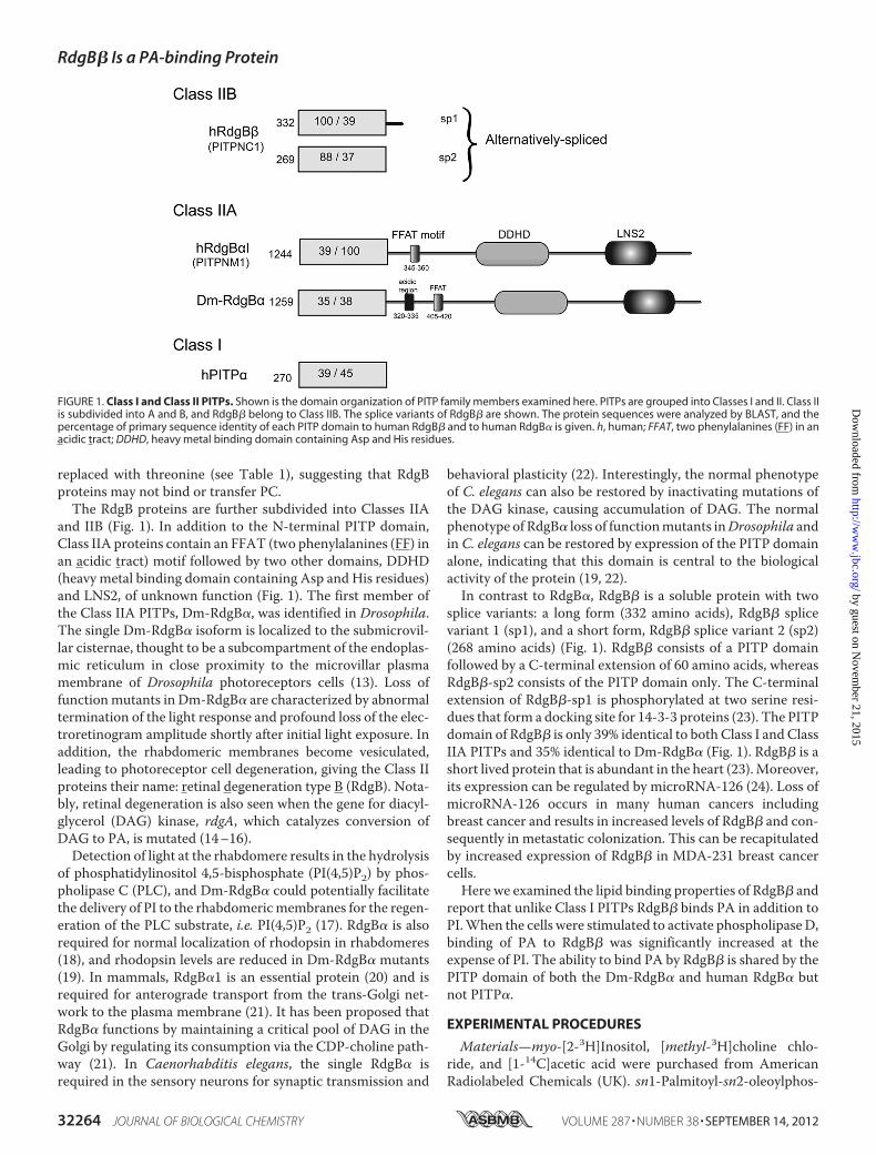
replaced with threonine (see Table 1), suggesting that RdgBproteins may not bind or transfer PC.The RdgB proteins are further subdivided into Classes IIA
and IIB (Fig. 1). In addition to the N-terminal PITP domain,Class IIA proteins contain an FFAT (two phenylalanines (FF) inan acidic tract) motif followed by two other domains, DDHD(heavy metal binding domain containing Asp and His residues)and LNS2, of unknown function (Fig. 1). The first member ofthe Class IIA PITPs, Dm-RdgB�, was identified in Drosophila.The single Dm-RdgB� isoform is localized to the submicrovil-lar cisternae, thought to be a subcompartment of the endoplas-mic reticulum in close proximity to the microvillar plasmamembrane of Drosophila photoreceptors cells (13). Loss offunctionmutants in Dm-RdgB� are characterized by abnormaltermination of the light response and profound loss of the elec-troretinogram amplitude shortly after initial light exposure. Inaddition, the rhabdomeric membranes become vesiculated,leading to photoreceptor cell degeneration, giving the Class IIproteins their name: retinal degeneration type B (RdgB). Nota-bly, retinal degeneration is also seen when the gene for diacyl-glycerol (DAG) kinase, rdgA, which catalyzes conversion ofDAG to PA, is mutated (14–16).Detection of light at the rhabdomere results in the hydrolysis
of phosphatidylinositol 4,5-bisphosphate (PI(4,5)P2) by phos-pholipase C (PLC), and Dm-RdgB� could potentially facilitatethe delivery of PI to the rhabdomericmembranes for the regen-eration of the PLC substrate, i.e. PI(4,5)P2 (17). RdgB� is alsorequired for normal localization of rhodopsin in rhabdomeres(18), and rhodopsin levels are reduced in Dm-RdgB� mutants(19). In mammals, RdgB�1 is an essential protein (20) and isrequired for anterograde transport from the trans-Golgi net-work to the plasma membrane (21). It has been proposed thatRdgB� functions by maintaining a critical pool of DAG in theGolgi by regulating its consumption via the CDP-choline path-way (21). In Caenorhabditis elegans, the single RdgB� isrequired in the sensory neurons for synaptic transmission and
behavioral plasticity (22). Interestingly, the normal phenotypeof C. elegans can also be restored by inactivating mutations ofthe DAG kinase, causing accumulation of DAG. The normalphenotype of RdgB� loss of functionmutants inDrosophila andin C. elegans can be restored by expression of the PITP domainalone, indicating that this domain is central to the biologicalactivity of the protein (19, 22).In contrast to RdgB�, RdgB� is a soluble protein with two
splice variants: a long form (332 amino acids), RdgB� splicevariant 1 (sp1), and a short form, RdgB� splice variant 2 (sp2)(268 amino acids) (Fig. 1). RdgB� consists of a PITP domainfollowed by a C-terminal extension of 60 amino acids, whereasRdgB�-sp2 consists of the PITP domain only. The C-terminalextension of RdgB�-sp1 is phosphorylated at two serine resi-dues that form a docking site for 14-3-3 proteins (23). The PITPdomain of RdgB� is only 39% identical to both Class I and ClassIIA PITPs and 35% identical to Dm-RdgB� (Fig. 1). RdgB� is ashort lived protein that is abundant in the heart (23).Moreover,its expression can be regulated by microRNA-126 (24). Loss ofmicroRNA-126 occurs in many human cancers includingbreast cancer and results in increased levels of RdgB� and con-sequently in metastatic colonization. This can be recapitulatedby increased expression of RdgB� in MDA-231 breast cancercells.Here we examined the lipid binding properties of RdgB� and
report that unlike Class I PITPs RdgB� binds PA in addition toPI.When the cells were stimulated to activate phospholipaseD,binding of PA to RdgB� was significantly increased at theexpense of PI. The ability to bind PA by RdgB� is shared by thePITP domain of both the Dm-RdgB� and human RdgB� butnot PITP�.
EXPERIMENTAL PROCEDURES
Materials—myo-[2-3H]Inositol, [methyl-3H]choline chlo-ride, and [1-14C]acetic acid were purchased from AmericanRadiolabeled Chemicals (UK). sn1-Palmitoyl-sn2-oleoylphos-
FIGURE 1. Class I and Class II PITPs. Shown is the domain organization of PITP family members examined here. PITPs are grouped into Classes I and II. Class IIis subdivided into A and B, and RdgB� belong to Class IIB. The splice variants of RdgB� are shown. The protein sequences were analyzed by BLAST, and thepercentage of primary sequence identity of each PITP domain to human RdgB� and to human RdgB� is given. h, human; FFAT, two phenylalanines (FF) in anacidic tract; DDHD, heavy metal binding domain containing Asp and His residues.
RdgB� Is a PA-binding Protein
32264 JOURNAL OF BIOLOGICAL CHEMISTRY VOLUME 287 • NUMBER 38 • SEPTEMBER 14, 2012
by guest on Novem
ber 21, 2015http://w
ww
.jbc.org/D
ownloaded from

phatidylcholine and sn1-palmitoyl-sn2-oleoyl phosphatidicacid were obtained from Avanti Polar Lipids (Alabaster, AL).N-Trinitrophenyl phosphatidylethanolamine (PE), sn1-palmi-toyl-sn2-pyrenedecanoyl-PC, -PA, and sn1-octadecanoyl-sn2-pyrenedecanoyl-PI were synthesized as described previously(25, 26). Bacterial phospholipase D (Streptomyces sp. (P4912))was obtained from Sigma.Recombinant PITP� and RdgB� (human) were expressed
from pRSET vectors and purified as described previously (4).The Dm-RdgB�-PITP cDNA coding for the N-terminal PITPdomain (amino acids 1–281) was cloned into the pRSET-Cplasmid vector, and the human RdgB� PITP domain (aminoacids 1–277) was cloned into the pET21a-His plasmid (a giftfrom Dr. Sima Lev). For site-directed mutagenesis, primerswere designed, and pointmutations were introduced accordingto the Stratagene QuikChange protocol. The oligonucleotideswere manufactured by Sigma-Aldrich. The mutated sequenceswere verified by DNA sequencing (Eurofins MWG Operon).The His-tagged proteins were expressed in BL21(DE3)pLysScompetent Escherichia coli and purified using nitrilotriaceticacid-agarose resin (HIS-Select nickel affinity gel, Sigma) asdescribed previously (4). The purity and the yield of the pro-teins varied between constructs. PITP� and Dm-RdgB�(1–281) proteins were expressed very well, whereas the yields ofRdgB� and RdgB�(1–277) were much lower. Protein concen-tration was assessed using the bicinchoninic acid (BCA) assay.This was readjusted after SDS-PAGE analysis according to theamount of protein of the correct molecular weight presentusing AIDA software with comparison to 1 �g of PITP�. TheHis-tagged proteins were stored in aliquots in PIPES buffer (20mM PIPES, 137 mM NaCl, 3 mM KCl, pH 6.8) at �80 °C.Transfer of Radiolabeled PI and PC Using Microsomes as a
Donor—100 �l of [3H]inositol-labeled or [3H]choline-labeledmicrosomes (6mg/ml) were incubated with an equal volume ofliposomes (PC/PI, 98:2 molar ratio; 80 nmol of phospholipid)together with 50 �l of recombinant proteins at 25 °C for 20min(27). [3H]Inositol-labeled microsomes were prepared from ratliver, whereas [3H]choline-labeled microsomes were obtainedfromHL60 cells. The reaction was quenched by the addition of50 �l of ice-cold 0.2 M sodium acetate, 0.25 M sucrose, pH 5.0.The tubes were vortexed and left on ice for 10 min. Followingcentrifugation at 12,000 � g for 10 min at 4 °C, 150 �l of super-natant from each reaction was counted in a liquid scintillationcounter. Results are expressed as a percentage of the total inputcount. Error bars indicate mean � S.D. or S.E. as indicated.Transfer of Radiolabeled PI and PC Using Permeabilized
HL60 Cells as a Donor—The phospholipid transfer assays werecarried out as described previously (6). In brief, HL60 cells wereprelabeled with [3H]inositol or [3H]choline for 48 h. The cellswere permeabilized with streptolysin O, centrifuged to removethe cytosolic components, and incubated with liposomes (PC/PI, 98:2, mol/mol) together with 50 �l of recombinant proteinsat 37 °C for 20 min. The samples were processed as describedabove.Binding of Cellular Lipids by PITPs—Association of cellular
lipids with the PITPswas analyzed exactly as described (4, 6). Inbrief, HL60 cells were labeled with 1 �Ci/ml [14C]acetate inRPMI 1640medium for 48 h. The cells were permeabilizedwith
streptolysin O, and the leaked cytosol was removed by centri-fugation. Permeabilized cells (�107 cells) were incubated with120 �g of PITP� or RdgB� protein (100 �l) for 20 min at 37 °Cin the presence of 2 mM Mg2�-ATP and 100 nM Ca2� bufferedwith 3mMEGTA. A sample of the protein was retained and runon SDS-PAGE. At the end of the incubation, the cells wereremoved by centrifugation, and the recombinant proteins inthe supernatant were captured on nickel beads. An aliquot ofthe recovered proteins was run on SDS-PAGE to assess theirrecovery, and the rest of the sample was used for lipid extrac-tion. The lipids were resolved by thin layer chromatographyusing a Whatman silica gel 60 TLC plate using chloroform/methanol/acetic acid/water (75:45:3:1, v/v) as the eluent. Lipidsextracted from the permeabilized HL60 cells (approximately100,000 dpm) were analyzed alongside for comparison. TheTLC plates were exposed to Fuji phosphorimaging screens andanalyzed using a Fuji BAS1000 phosphorimaging system. Boththe SDS-PAGE and TLC images were analyzed using AIDAsoftware.Mass Spectrometric Analysis of Phospholipids—For the mass
spectrometric analyses, the binding assay was scaled up 5-fold;i.e. 5 � 107 HL60 cells (unlabeled) were incubated with 600 �gof a purified transfer protein. Lipidswere also directly extractedfrom the E. coli-expressed PITPs (600 �g/sample). Disposableborosilicate glassware was used for all lipid extractions.Phospholipids were analyzed using a Quatro Ultima triple qua-drupole mass spectrometer (Micromass, Wythenshawe, UK)equipped with direct infusion or nanoflow interfaces, depend-ing upon sample size, as described previously (28). An initial fullscreening for glycerophospholipid species was undertakenunder both positive and negative ionization form/z values up to1300. More detailed selective precursor ion scanning of allphospholipids of potential interest was then pursued includingfor PA, PC, PE, PI, phosphatidylserine, and related lysophos-pholipids. Bacterial lipids including phosphatidylglycerol (PG)were absent from all samples. The PA species were identified byscanning for the precursors of the glycerophospholipid frag-ment (m/z �153), and their identities and dominant acyl com-positions were confirmed by specific peak fragmentations,whereas the PI species were detected by scanning for the pre-cursor of inositol phosphate (m/z �241) as described previ-ously (28).Assay of Pyrene-Lipid Binding by PITPs—To study the bind-
ing of pyrene-labeled phospholipids by the transfer proteins, weused a previously described dequenching assay (25). Briefly,donor vesicles in which the pyrene fluorescence is effectivelyquenched byN-trinitrophenyl PEwere prepared by injecting 10�l of an ethanol solution of pyrenyl phospholipid/sn1-palmi-toyl-sn2-oleylphosphatidylcholine/N-trinitrophenyl PE (2:88:10, mol/mol) mixture (4 nmol of total phospholipid) into 2 mlof a buffer (20 mM Tris-Cl, 100 mM NaCl, 5 mM EDTA) in acuvette. After a 1-min equilibration period, aliquots of a trans-fer protein were added, and the fluorescence intensity (withexcitation at 348 nm and emission at 395 nm) was recordedafter each addition. If the pyrenyl phospholipid incorporatesinto the transfer protein, pyrene fluorescence increases becausethe labeled lipid is removed from the quenching environment,i.e. the donor vesicle. Fluorescence intensity versus protein con-
RdgB� Is a PA-binding Protein
SEPTEMBER 14, 2012 • VOLUME 287 • NUMBER 38 JOURNAL OF BIOLOGICAL CHEMISTRY 32265
by guest on Novem
ber 21, 2015http://w
ww
.jbc.org/D
ownloaded from
centration plots were constructed to assess the relative affinityof a pyrene phospholipid for the transfer protein. When trans-fer was monitored, acceptor vesicles (80 nmol of phospholipid)consisting of sn1-palmitoyl-sn2-oleylphosphatidylcholine/sn1-palmitoyl-sn2-oleoyl phosphatidic acid (98:2, mol/mol) andprepared by the ethanol injection method were also added.Lipid Binding following Incubation with PMA, GTP�S, or
Bacterial Phospholipase D—HL60 cells were labeled with 1�Ci/ml [14C]acetate in RPMI 1640 medium for 48 h. Afterwashing twice in PIPES buffer, the cells were added to amixtureof GTP�S (100 �M final), PMA (100 nM final), 0.2 unit of bac-terial phospholipase D, MgATP (2 mM final), Ca2� bufferedwith 3 mM EGTA at 100 nM (pCa 7) or 10 �M (pCa 5), strepto-lysin O (0.4 IU/ml final), and a recombinant protein (120 �g) as
indicated in a final incubation volume of 600 �l. The inhibitorsFIPI (750 nM) and U73122 (10 �M) were also added where indi-cated. After incubation for 20 min at 37 °C, the cells were cen-trifuged, and 500 �l of the supernatant was used to recapturethe proteins as described above (6). The residual cells werequenched with 375 �l of chloroform/methanol (1:2, v/v), andthe lipids were extracted and analyzed by TLC as described (6).
RESULTS
Comparison of the PI and PC Transfer Properties of the TwoSplice Variants of RdgB� with PITP�—We have recentlyreported that the PI transfer activity of RdgB�-sp1 is low whencompared with PITP� (23). RdgB�-sp1 requires 100-foldhigher concentrations and furthermore does not achieve the
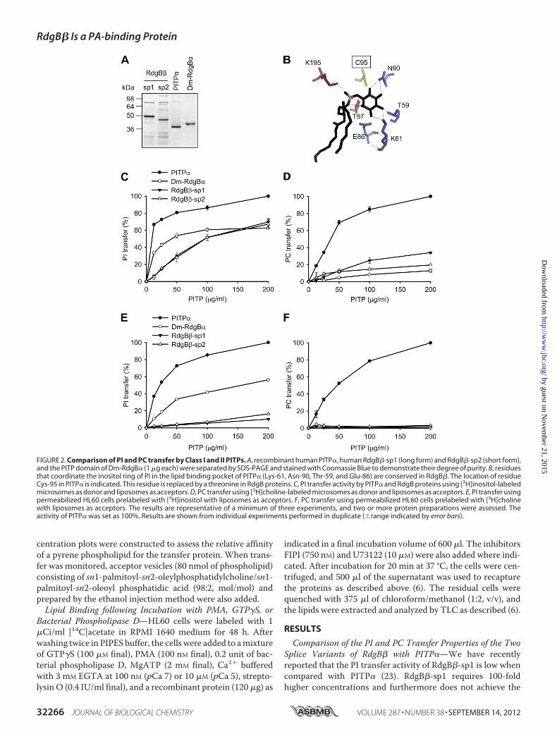
FIGURE 2. Comparison of PI and PC transfer by Class I and II PITPs. A, recombinant human PITP�, human RdgB�-sp1 (long form) and RdgB�-sp2 (short form),and the PITP domain of Dm-RdgB� (1 �g each) were separated by SDS-PAGE and stained with Coomassie Blue to demonstrate their degree of purity. B, residuesthat coordinate the inositol ring of PI in the lipid binding pocket of PITP� (Lys-61, Asn-90, Thr-59, and Glu-86) are conserved in RdgB�. The location of residueCys-95 in PITP� is indicated. This residue is replaced by a threonine in RdgB proteins. C, PI transfer activity by PITP� and RdgB proteins using [3H]inositol-labeledmicrosomes as donor and liposomes as acceptors. D, PC transfer using [3H]choline-labeled microsomes as donor and liposomes as acceptors. E, PI transfer usingpermeabilized HL60 cells prelabeled with [3H]inositol with liposomes as acceptors. F, PC transfer using permeabilized HL60 cells prelabeled with [3H]cholinewith liposomes as acceptors. The results are representative of a minimum of three experiments, and two or more protein preparations were assessed. Theactivity of PITP� was set as 100%. Results are shown from individual experiments performed in duplicate (�range indicated by error bars).
RdgB� Is a PA-binding Protein
32266 JOURNAL OF BIOLOGICAL CHEMISTRY VOLUME 287 • NUMBER 38 • SEPTEMBER 14, 2012
by guest on Novem
ber 21, 2015http://w
ww
.jbc.org/D
ownloaded from
same level of transfer as PITP�. Two splice variants of RdgB�have been identified that differ at their C termini. The canonicalRdgB� (332 amino acids) has a 60-amino acid C-terminalextension compared with RdgB�-sp2 (268 amino acids), whichhas 9 amino acids. The C-terminal extension precedes theG-helix, which in PITP� swings outward, dislodging the C-ter-minal extension; this exposes the hydrophobic cavity requiredfor lipid exchange to take place at themembrane (29). Thus, it ispossible that the C-terminal extension of RdgB�-sp1 is the
main cause for the difference in activity between PITP� andRdgB�-sp1. Alternatively, the difference could lie in the PITPdomain itself as RdgB� is only �40% identical to the Class IPITPs. To distinguish between these possibilities, we examinedthe lipid transfer activity of RdgB�-sp2, which is of similarlength to PITP� (270 amino acids).
To determine PI transfer activity, we used two differentassays: a microsome assay (30) and a cytosol-depleted permea-bilizedHL60 cell assay (6), both containing [3H]inositol-labeled
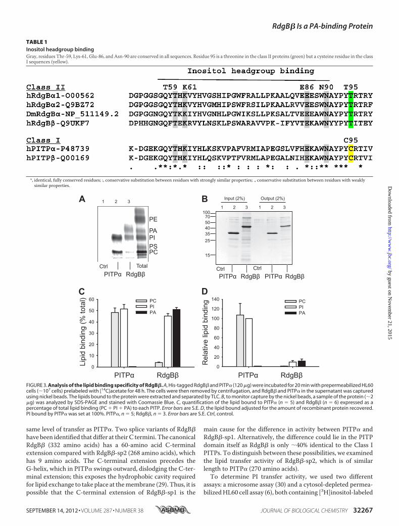
FIGURE 3. Analysis of the lipid binding specificity of RdgB�. A, His-tagged RdgB� and PITP� (120 �g) were incubated for 20 min with prepermeabilized HL60cells (�107 cells) prelabeled with [14C]acetate for 48 h. The cells were then removed by centrifugation, and RdgB� and PITP� in the supernatant was capturedusing nickel beads. The lipids bound to the protein were extracted and separated by TLC. B, to monitor capture by the nickel beads, a sample of the protein (�2�g) was analyzed by SDS-PAGE and stained with Coomassie Blue. C, quantification of the lipid bound to PITP� (n � 5) and RdgB� (n � 6) expressed as apercentage of total lipid binding (PC � PI � PA) to each PITP. Error bars are S.E. D, the lipid bound adjusted for the amount of recombinant protein recovered.PI bound by PITP� was set at 100%. PITP�, n � 5; RdgB�, n � 3. Error bars are S.E. Ctrl, control.
TABLE 1Inositol headgroup bindingGray, residues Thr-59, Lys-61, Glu-86, and Asn-90 are conserved in all sequences. Residue 95 is a threonine in the class II proteins (green) but a cysteine residue in the classI sequences (yellow).
*, identical, fully conserved residues; :, conservative substitution between residues with strongly similar properties; ., conservative substitution between residues with weaklysimilar properties.
RdgB� Is a PA-binding Protein
SEPTEMBER 14, 2012 • VOLUME 287 • NUMBER 38 JOURNAL OF BIOLOGICAL CHEMISTRY 32267
by guest on Novem
ber 21, 2015http://w
ww
.jbc.org/D
ownloaded from
PI. Vesicles composed of PC/PI (98:2) were used as the accep-tors. The His-tagged proteins were expressed in E. coli, and theconcentration of the purified proteins was adjusted accordingto the degree of purity of the recombinant proteins (Fig. 2A).The two splice variants of RdgB� showed very similar PI trans-fer activity regardless of the assay used (Fig. 2, C and E). Com-pared with PITP�, both RdgB� proteins required a muchhigher concentration and did not achieve the same degree ofactivity. Furthermore, the RdgB� proteins showed greateractivity in the microsome-liposome assay compared with thepermeabilized HL60 cell assay, whereas PITP� was equallyeffective in either assay.In addition to transferring PI, Class I PITPs can also transfer
PC. We therefore compared the PC transfer activity of RdgB�splice variants with PITP� using both assays. For PI transfer,four residues (Thr-59, Lys-61, Glu-86, and Asn-90; mousePITP� numbering) that coordinate the inositol ring are essen-tial (Fig. 2B); these residues are conserved in RdgB�. RegardingPC transfer, we have recently shown that mutation of cysteine95 to threonine in PITP� reduces the transfer of this lipid (12).This residue is replaced with threonine in the Class II PITPs(Table 1). Cys-95 resides in the head group binding pocket (Fig.2B) and is not essential for PI transfer activity (12). UnlikePITP�, RdgB proteins showed very low PC transfer in eitherassay (Fig. 2, D and F). The PC transfer activity was 20% of thatof PITP� in the microsome-liposome assay (Fig. 2D), whereasno activity was detected when permeabilized cells were used asdonors (Fig. 2F). The low level of PC transfer activity exhibitedby RdgB� was also observed for another Class II RdgB protein,the PITP domain of Dm-RdgB�. The Dm-RdgB� PITP domainwas active for PI transfer in either assay, but the maximal activ-ity attained was �50% of PITP� (Fig. 2, C and E).Lipid Binding Properties of the Soluble Class II PITP, RdgB�—
The low level of PC transfer exhibited byRdgB� prompted us toexamine its lipid binding properties. This was measured byexposing the His-tagged recombinant proteins to pre-permea-bilized HL60 cells prelabeled with [14C]acetate (6). The recom-binant proteins were recaptured using nickel beads, and thelipids were extracted from the eluted proteins and analyzed byTLC and mass spectrometry. Compared with PITP�, whichbinds PI and PC, RdgB� mainly bound PI and PA (Fig. 3A). Thelipid binding of the individual species is expressed as a percent-age of total lipid bound to a protein (Fig. 3C). PITP� boundnearequal proportions of PI andPC.NoPAbindingwas observed. Incomparison, RdgB� bound PI and PA in near equal propor-tions, whereas PC binding was low (�5%) (Fig. 3C).It is well documented that isolated PITP� contains one mol-
ecule of PI or PC, which undergoes constant exchange withmembrane lipids within minutes; the apo form is present onlytransiently at a membrane after the release of bound lipid (29).Thus, theamountof radioactivity found inRdgB�after incubationwith the labeled HL60 cells provides a measure of lipid exchange(Fig. 3D). Relative to PITP�, RdgB� contained less radioactivityper pmol of protein, implying that a large fraction of the RdgB�molecules may not have undergone lipid exchange.Recombinant PITP� when purified from E. coli comes pre-
loadedwithPG,which is immediately exchangedwhenexposed toPI or to PC (28). To identify which lipids were associated with
recombinant RdgB� when purified from E. coli, the extracted lip-ids were analyzed by mass spectrometry (electrospray ionization-MS/MS). Precursor scans of them/z�153 fragment revealed that
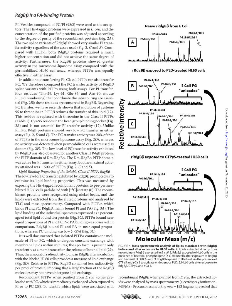
FIGURE 4. Mass spectrometric analysis of lipids associated with RdgB�before and after exposure to HL60 cells. A, lipids extracted directly fromrecombinant RdgB� expressed in E. coli. B, RdgB� exposed to HL60 cells in thepresence of bacterial phospholipase D. C, HL60 cells after exposure to RdgB�and bacterial PLD (0.2 unit). D, RdgB� exposed to HL60 cells in the presence ofGTP�S and pCa 5 to activate endogenous PLD. E, HL60 cells after exposure toRdgB�, GTP�S, and pCa 5.
RdgB� Is a PA-binding Protein
32268 JOURNAL OF BIOLOGICAL CHEMISTRY VOLUME 287 • NUMBER 38 • SEPTEMBER 14, 2012
by guest on Novem
ber 21, 2015http://w
ww
.jbc.org/D
ownloaded from
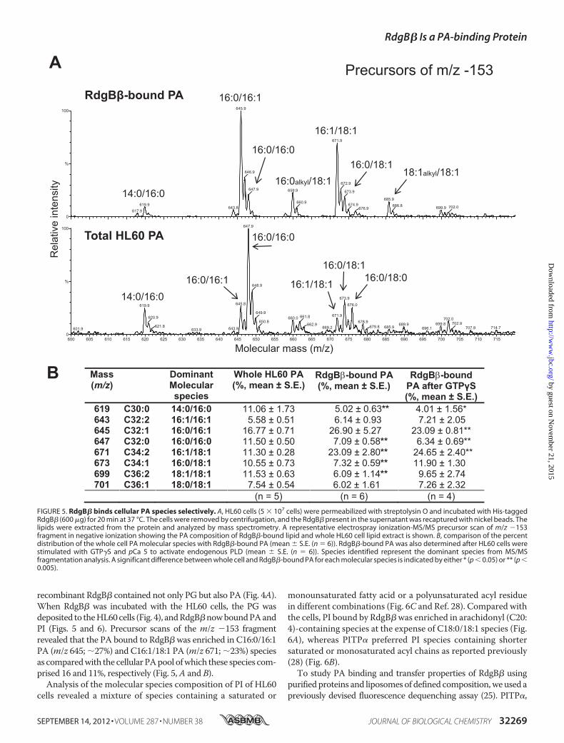
recombinant RdgB� contained not only PG but also PA (Fig. 4A).When RdgB� was incubated with the HL60 cells, the PG wasdeposited to theHL60cells (Fig. 4), andRdgB�nowboundPAandPI (Figs. 5 and 6). Precursor scans of the m/z �153 fragmentrevealed that the PA bound to RdgB� was enriched in C16:0/16:1PA (m/z 645; �27%) and C16:1/18:1 PA (m/z 671; �23%) speciesas comparedwith the cellular PApool ofwhich these species com-prised 16 and 11%, respectively (Fig. 5,A and B).Analysis of the molecular species composition of PI of HL60
cells revealed a mixture of species containing a saturated or
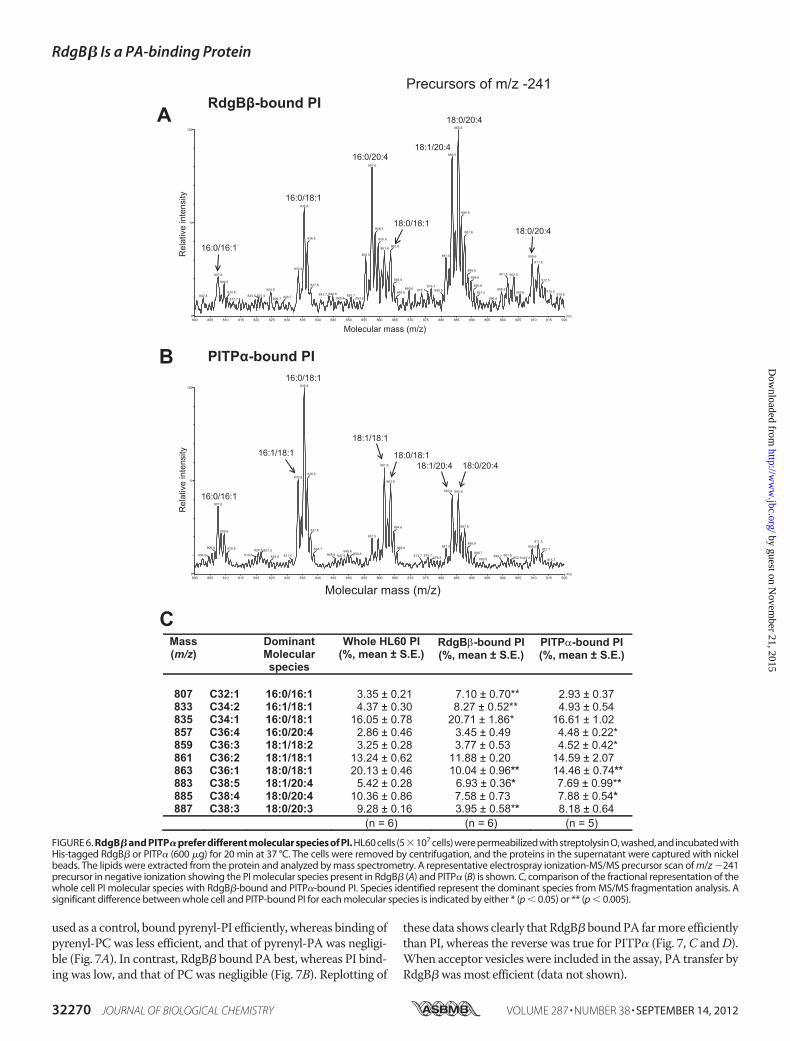
monounsaturated fatty acid or a polyunsaturated acyl residuein different combinations (Fig. 6C and Ref. 28). Compared withthe cells, PI bound by RdgB�was enriched in arachidonyl (C20:4)-containing species at the expense of C18:0/18:1 species (Fig.6A), whereas PITP� preferred PI species containing shortersaturated or monosaturated acyl chains as reported previously(28) (Fig. 6B).To study PA binding and transfer properties of RdgB� using
purified proteins and liposomes of defined composition,weused apreviously devised fluorescence dequenching assay (25). PITP�,
FIGURE 5. RdgB� binds cellular PA species selectively. A, HL60 cells (5 � 107 cells) were permeabilized with streptolysin O and incubated with His-taggedRdgB� (600 �g) for 20 min at 37 °C. The cells were removed by centrifugation, and the RdgB� present in the supernatant was recaptured with nickel beads. Thelipids were extracted from the protein and analyzed by mass spectrometry. A representative electrospray ionization-MS/MS precursor scan of m/z �153fragment in negative ionization showing the PA composition of RdgB�-bound lipid and whole HL60 cell lipid extract is shown. B, comparison of the percentdistribution of the whole cell PA molecular species with RdgB�-bound PA (mean � S.E. (n � 6)). RdgB�-bound PA was also determined after HL60 cells werestimulated with GTP�S and pCa 5 to activate endogenous PLD (mean � S.E. (n � 6)). Species identified represent the dominant species from MS/MSfragmentation analysis. A significant difference between whole cell and RdgB�-bound PA for each molecular species is indicated by either * (p � 0.05) or ** (p �0.005).
RdgB� Is a PA-binding Protein
SEPTEMBER 14, 2012 • VOLUME 287 • NUMBER 38 JOURNAL OF BIOLOGICAL CHEMISTRY 32269
by guest on Novem
ber 21, 2015http://w
ww
.jbc.org/D
ownloaded from
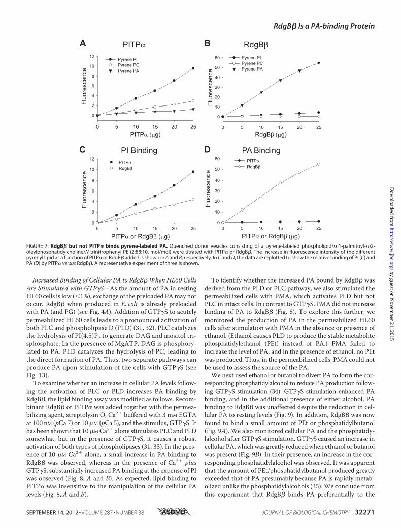
used as a control, bound pyrenyl-PI efficiently, whereas binding ofpyrenyl-PC was less efficient, and that of pyrenyl-PA was negligi-ble (Fig. 7A). In contrast, RdgB� bound PA best, whereas PI bind-ing was low, and that of PC was negligible (Fig. 7B). Replotting of
these data shows clearly that RdgB�boundPA farmore efficientlythan PI, whereas the reverse was true for PITP� (Fig. 7, C andD).When acceptor vesicles were included in the assay, PA transfer byRdgB� wasmost efficient (data not shown).
FIGURE 6. RdgB� and PITP� prefer different molecular species of PI. HL60 cells (5�107 cells) were permeabilized with streptolysin O, washed, and incubated withHis-tagged RdgB� or PITP� (600 �g) for 20 min at 37 °C. The cells were removed by centrifugation, and the proteins in the supernatant were captured with nickelbeads. The lipids were extracted from the protein and analyzed by mass spectrometry. A representative electrospray ionization-MS/MS precursor scan of m/z �241precursor in negative ionization showing the PI molecular species present in RdgB� (A) and PITP� (B) is shown. C, comparison of the fractional representation of thewhole cell PI molecular species with RdgB�-bound and PITP�-bound PI. Species identified represent the dominant species from MS/MS fragmentation analysis. Asignificant difference between whole cell and PITP-bound PI for each molecular species is indicated by either * (p � 0.05) or ** (p � 0.005).
RdgB� Is a PA-binding Protein
32270 JOURNAL OF BIOLOGICAL CHEMISTRY VOLUME 287 • NUMBER 38 • SEPTEMBER 14, 2012
by guest on Novem
ber 21, 2015http://w
ww
.jbc.org/D
ownloaded from
Increased Binding of Cellular PA to RdgB� When HL60 CellsAre Stimulated with GTP�S—As the amount of PA in restingHL60 cells is low (�1%), exchange of the preloaded PAmay notoccur. RdgB� when produced in E. coli is already preloadedwith PA (and PG) (see Fig. 4A). Addition of GTP�S to acutelypermeabilized HL60 cells leads to a pronounced activation ofboth PLC and phospholipase D (PLD) (31, 32). PLC catalyzesthe hydrolysis of PI(4,5)P2 to generate DAG and inositol tri-sphosphate. In the presence of MgATP, DAG is phosphory-lated to PA. PLD catalyzes the hydrolysis of PC, leading tothe direct formation of PA. Thus, two separate pathways canproduce PA upon stimulation of the cells with GTP�S (seeFig. 13).To examine whether an increase in cellular PA levels follow-
ing the activation of PLC or PLD increases PA binding byRdgB�, the lipid binding assay wasmodified as follows. Recom-binant RdgB� or PITP� was added together with the permea-bilizing agent, streptolysin O, Ca2� buffered with 3 mM EGTAat 100 nM (pCa 7) or 10�M (pCa 5), and the stimulus, GTP�S. Ithas been shown that 10�MCa2� alone stimulates PLC andPLDsomewhat, but in the presence of GTP�S, it causes a robustactivation of both types of phospholipases (31, 33). In the pres-ence of 10 �M Ca2� alone, a small increase in PA binding toRdgB� was observed, whereas in the presence of Ca2� plusGTP�S, substantially increased PA binding at the expense of PIwas observed (Fig. 8, A and B). As expected, lipid binding toPITP� was insensitive to the manipulation of the cellular PAlevels (Fig. 8, A and B).
To identify whether the increased PA bound by RdgB� wasderived from the PLD or PLC pathway, we also stimulated thepermeabilized cells with PMA, which activates PLD but notPLC in intact cells. In contrast toGTP�S, PMAdid not increasebinding of PA to RdgB� (Fig. 8). To explore this further, wemonitored the production of PA in the permeabilized HL60cells after stimulation with PMA in the absence or presence ofethanol. (Ethanol causes PLD to produce the stable metabolitephosphatidylethanol (PEt) instead of PA.) PMA failed toincrease the level of PA, and in the presence of ethanol, no PEtwas produced. Thus, in the permeabilized cells, PMA could notbe used to assess the source of the PA.We next used ethanol or butanol to divert PA to form the cor-
responding phosphatidylalcohol to reduce PA production follow-ing GTP�S stimulation (34). GTP�S stimulation enhanced PAbinding, and in the additional presence of either alcohol, PAbinding to RdgB� was unaffected despite the reduction in cel-lular PA to resting levels (Fig. 9). In addition, RdgB� was nowfound to bind a small amount of PEt or phosphatidylbutanol(Fig. 9A). We also monitored cellular PA and the phosphatidy-lalcohol after GTP�S stimulation. GTP�S caused an increase incellular PA,whichwas greatly reducedwhen ethanol or butanolwas present (Fig. 9B). In their presence, an increase in the cor-responding phosphatidylalcohol was observed. It was apparentthat the amount of PEt/phosphatidylbutanol produced greatlyexceeded that of PA presumably because PA is rapidly metab-olized unlike the phosphatidylalcohols (35). We conclude fromthis experiment that RdgB� binds PA preferentially to the
FIGURE 7. RdgB� but not PITP� binds pyrene-labeled PA. Quenched donor vesicles consisting of a pyrene-labeled phospholipid/sn1-palmitoyl-sn2-oleylphosphatidylcholine/N-trinitrophenyl PE (2:88:10, mol/mol) were titrated with PITP� or RdgB�. The increase in fluorescence intensity of the differentpyrenyl lipid as a function of PITP� or RdgB� added is shown in A and B, respectively. In C and D, the data are replotted to show the relative binding of PI (C) andPA (D) by PITP� versus RdgB�. A representative experiment of three is shown.
RdgB� Is a PA-binding Protein
SEPTEMBER 14, 2012 • VOLUME 287 • NUMBER 38 JOURNAL OF BIOLOGICAL CHEMISTRY 32271
by guest on Novem
ber 21, 2015http://w
ww
.jbc.org/D
ownloaded from

phosphatidylalcohols, andmoreover, PA binding to RdgB�wasstill evident even when alcohols were used to divert the cellularproduction of PA to phosphatidylalcohols. Because PA is stillproduced from the phospholipase C pathway, these results donot clarify whether PA from the phospholipase C pathwaymakes a contribution.As an alternative approach to establish the source of the PA
bound to RdgB�, we used inhibitors of PLC (U73122) and PLD(FIPI) (36). We first confirmed that FIPI is able to inhibitGTP�S-stimulated PLD activity by monitoring the productionof PEt in permeabilized cells (data not shown). Next we studiedPA binding to RdgB� by incubatingHL60 cells with a phospho-lipase inhibitor and GTP�S. FIPI but not U73122 inhibited theGTP�S-stimulated increase in RdgB�-boundPA (Fig. 10,A andB). We conclude that RdgB� mainly binds PA derived from thePLD pathway. This conclusion is also supported by the analysisof the molecular species of PA bound by RdgB� after GTP�Sstimulation (Figs. 4B and 5B). The dominant PA species presentare C16:0/16:1 and C16:1/18:1, which are characteristic of the
PC lipids but not PI lipids (Fig. 6). During stimulation of HL60cells with GTP�S, increases in the molecular species of C18:1/18:1 PA and C18:0/18:1 PA were observed, but these specieswere not found in RdgB�.To confirm that increased PA binding to RdgB� was
entirely due to the increased levels of PA and not some sec-ondary effect of G-protein activation, we used the bacterialStreptomyces sp. phospholipase D to produce PA in the per-meabilized cells (37). Bacterial phospholipase D hydrolyzedPC and produced PA in the permeabilized cells (Fig. 4E), andthis was accompanied by an increase in PA binding to RdgB�(Fig. 10C). The dominant molecular species bound to RdgB�were C16:0/16:1 and C16:1/18:1 PA (Fig. 4D). When com-pared with GTP�S, radiolabeled PA bound to RdgB� wasmuch greater, suggesting that the amount of PA present isthe limiting factor (Fig. 10).RdgB� Proteins Are Also PA-binding Proteins—Above we
showed RdgB� as a PI/PA- rather than a PI/PC-binding andtransfer protein. Similar to RdgB�, the PITP domain of Dm-
FIGURE 8. Increased PA binding by RdgB� after stimulation of HL60 cells with GTP�S. [14C]Acetate-labeled HL60 cells were incubated with RdgB� or PITP�in the presence of GTP�S (100 �M) or PMA (100 nM) and calcium-buffered at pCa 5 (10 �M) or pCa 7 (100 nM) together with the permeabilizing agent streptolysinO. After 20 min, the cells were removed by centrifugation, and the PITPs were captured from the supernatant using nickel beads. A, the lipids were extractedand separated by TLC, and the plate was imaged using a phosphorimaging screen. A sample of the captured proteins was also analyzed by SDS-PAGE. B, lipidbinding was quantitated from two independent experiments. Error bars are S.D. (n � 2).
RdgB� Is a PA-binding Protein
32272 JOURNAL OF BIOLOGICAL CHEMISTRY VOLUME 287 • NUMBER 38 • SEPTEMBER 14, 2012
by guest on Novem
ber 21, 2015http://w
ww
.jbc.org/D
ownloaded from

RdgB� exhibited low PC transfer activity (Fig. 2, D and F),implying that itmight also bind PA.We therefore examined thelipid binding properties of the PITP domains of humanRdgB�1and Dm-RdgB� before and after stimulation of HL60 cells withGTP�S. Both proteins were found to bind PA, and the bindingincreased when the cells were stimulated with GTP�S (Fig. 11,A and B). However, PA binding was not as prominent as seenfor RdgB�, and nearly 70% of the lipid bound was PI. In addi-tion, the human RdgB�1 PITP domain bound more PC thanPA.Transfer of PI and PC by the RdgB� Proteins—Residues that
are important in binding the inositol ring in PITP� are con-served in all the Class II PITPs (Table 1), and we demonstrateabove that the PITP domain of RdgB� proteins can transferPI. The PITP domains of human RdgB� and Dm-RdgB�hardly bound and transferred PC (Figs. 12 and 2). However,the PITP domain of human RdgB� did bind substantiallymore PC than human RdgB� (Fig. 11). We therefore ana-lyzed the PC transfer activity of human RdgB�(1–277) andfound that, like the Dm-RdgB� PITP domain, it did nottransfer PC (Fig. 12).Cysteine 95 is important for PC transfer by PITP� because its
replacement by threonine decreases PC transfer significantly(12). Cysteine 95 is not conserved in the Class II family of RdgBproteins but is replaced with threonine (Table 1). To test theimportance of this threonine residue for PC transfer by the
Dm-RdgB� PITP domain, it was replaced by cysteine by site-directedmutagenesis.Neither PCnor PI transfer activitieswereaffected by the replacement (Fig. 12). Binding of PC was alsounchanged in Dm-RdgB�(T95C) (Fig. 11). However, thereplacement increased binding of PA to the protein at theexpense of PI, particularly upon stimulation of the cells withGTP�S (Fig. 11).
FIGURE 9. RdgB� binds phosphatidylalcohols poorly compared with PA.HL60 cells prelabeled with [14C]acetate were incubated with RdgB� in thepresence of pCa 7 (100 nM) (control), GTP�S (100 �M) and pCa 5 (10 �M), andethanol (2%) or butanol (BtOH) (0.5%) as indicated in the presence of strep-tolysin O for 20 min. A, TLC of radiolabeled lipids bound to RdgB� and theirquantitation. B, TLC of the cellular lipids and their quantitation. PBut, phos-phatidylbutanol; PSL, photostimulated luminescence; PAlc, phosphatidylal-cohol. The results are quantitated from four independent experiments. Errorbars are S.E. (n � 4).
FIGURE 10. Increased PA binding to RdgB� is dependent on endogenousphospholipase D activation and is mimicked by exogenous bacterialphospholipase D. [14C]Acetate-labeled HL60 cells were incubated withRdgB� (120 �g) in the presence of GTP�S (100 �M), FIPI (750 nM), or U73122(10 �M) and calcium-buffered at pCa 5 (10 �M) or pCa 7 (100 nM) as indicatedtogether with the permeabilizing agent streptolysin O. After 20 min, the cellswere removed by centrifugation and retained, and the PITPs were cap-tured from the supernatant using nickel beads. A, the lipids wereextracted and analyzed by TLC, and a sample of the captured proteins wasalso analyzed by SDS-PAGE. B, lipid binding to RdgB� was quantified fromthree independent experiments. C, [14C]acetate-labeled HL60 cells wereincubated with RdgB� (120 �g) in the presence of streptomycin sp. PLD(0.2 unit) or GTP�S (100 �M) plus pCa 5 (10 �M) or pCa 7 (100 nM) asindicated together with the permeabilizing agent streptolysin O. After 20min, the cells were removed by centrifugation and retained, and the PITPswere captured from the supernatant using nickel beads. Radiolabeled PAbinding to RdgB� was quantitated from three independent experiments.Error bars are S.E. (n � 3). Ctrl, control.
RdgB� Is a PA-binding Protein
SEPTEMBER 14, 2012 • VOLUME 287 • NUMBER 38 JOURNAL OF BIOLOGICAL CHEMISTRY 32273
by guest on Novem
ber 21, 2015http://w
ww
.jbc.org/D
ownloaded from

DISCUSSION
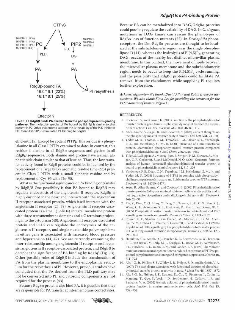
In this study, we provide strong evidence that the PITPdomain of Class II PITPs can bind PA in addition to binding PIand PC. Thus, the lipid binding properties of Class II PITPsdiffer from those of Class I PITPs. This in itself is not surprisinggiven that these PITP domains only share �40% identity intheir amino acid sequence. Residues that are essential for bind-ing the inositol headgroup of PI are conserved in all PITPs, andtherefore PI binding and transfer are shared by all PITPs. Asresidues important for PC binding and transfer are less obviousdespite the availability of the structures of both PITP� andPITP� loaded with PC (3, 5), it has not been possible to predictwhether RdgB� will also be competent for PC binding andtransfer. In this study, we examined the lipid binding and trans-fer properties of RdgB�. Our results show that although allmembers of the PITP family can bind and transfer PI the ClassIIB PITP, RdgB�, has a drastically reduced PC binding andtransfer activity. Using HL60 cells prelabeled with [14C]acetateas lipid donors, RdgB� was found to bind mainly PI and PA. Incontrast, PITP� bound only PI and PC. PA binding by RdgB�was confirmed by mass spectrometry, which also showed thatamong the cellular PA species RdgB� preferably binds the C16:0/16:1 and C16:1/18:1 PA species (Fig. 13). These PA speciesare typically produced fromPCvia the PLDpathway (38). Usinginhibitors specific for PLC or PLD, we confirmed that the PAbound by RdgB� is mainly derived from the PLD pathway. Theability to bind PA was conserved in all members of the Class IIPITPs. However, RdgB� bound the highest level of PA.A striking feature of human RdgB� and the Drosophila
RdgB� is their inability to bind substantial amounts of PC (Fig.11). Residues binding the PC headgroup have been difficult todetermine even from the crystal structures of PITP� andPITP�loadedwith PC (3, 5). Cysteine 95 lies in the lipid-binding cavityand forms a hydrogen bondwith the headgroup of PC indirectlyvia a water molecule, and mutation of this residue to eitherthreonine or alanine eliminates PC transfer, leaving PI transferunaffected (12). In RdgB proteins, cysteine 95 is replaced with athreonine (Table 1). Another residue important for PC transferis phenylalanine 225.When it is replaced with a leucine residueas is the case with mouse or rat PITP�, PC is transferred less
FIGURE 11. The PITP domain of Class II RdgB� proteins also binds PA.[14C]Acetate-labeled HL60 cells were incubated with the PITP domain ofhuman (h) RdgB�, Dm-RdgB�, Dm-RdgB�(T95C), or human RdgB� (120 �g)in the presence of GTP�S (100 �M) and calcium-buffered at pCa 5 (10 �M) orpCa 7 (100 nM) as indicated together with the permeabilizing agent strepto-lysin O. After 20 min, the cells were removed by centrifugation and retained,and the PITPs were captured from the supernatant using nickel beads. A, theradiolabeled lipids were extracted, separated by TLC, and imaged using aphosphorimaging screen. A sample of the captured proteins was also ana-lyzed by SDS-PAGE. B, lipid binding was quantitated using AIDA software. Thedata are means of three independent experiments �S.E. (error bars).
FIGURE 12. Comparison of the PI and PC transfer activity of PITP domains of RdgB� and PITP�. PI and PC transfer activity of PITP� and the PITP domainsof Dm-RdgB� wild type and mutant proteins was measured using appropriately radiolabeled microsomes as donor and liposomes as acceptors. The results arerepresentative of four independent experiments, and two or more protein preparations were assessed. The activity of PITP� was set as 100%. Error bars are S.E.hRdgB�, human RdgB�.
RdgB� Is a PA-binding Protein
32274 JOURNAL OF BIOLOGICAL CHEMISTRY VOLUME 287 • NUMBER 38 • SEPTEMBER 14, 2012
by guest on Novem
ber 21, 2015http://w
ww
.jbc.org/D
ownloaded from
efficiently (5). Except for rodent PITP�, this residue is a pheny-lalanine in all Class I PITPs examined to date. In contrast, thisresidue is alanine in all RdgB� sequences and glycine in allRdgB� sequences. Both alanine and glycine have a small ali-phatic side chain similar to that of leucine. Thus, the low trans-fer activity found in RdgB proteins could be influenced by thereplacement of a large bulky aromatic residue (Phe-225) pres-ent in Class I PITPs with a small aliphatic residue and byreplacement of Cys-95 with Thr-95.What is the functional significance of PA binding or transfer
by RdgB�? One possibility is that PA bound to RdgB� mayregulate endocytosis of the angiotensin II receptor. RdgB� ishighly enriched in the heart and interacts with the angiotensinII receptor-associated protein, which itself interacts with theangiotensin II receptor (23, 39). Angiotensin II receptor-asso-ciated protein is a small (17-kDa) integral membrane proteinwith three transmembrane domains and a C terminus project-ing into the cytoplasm (40). Angiotensin II receptor-associatedprotein and PLD2 can regulate the endocytosis of the an-giotensin II receptor, and single nucleotide polymorphismsin either gene is associated with increased blood pressureand hypertension (41, 42). We are currently examining theinter-relationship among angiotensin II receptor endocyto-sis, angiotensin II receptor-associated protein, and RdgB� todecipher the significance of PA binding by RdgB� (Fig. 13).Other possible roles of RdgB� include the translocation ofPA from the plasma membrane to the endoplasmic reticu-lum for the resynthesis of PI. However, previous studies haveconcluded that the PA derived from the PLD pathway maynot be converted into PI, and cytosolic components are notrequired for the process (43).Because RdgB� proteins also bind PA, it is possible that they
are responsible for PA transfer at intermembrane contact sites.
Because PA can be metabolized into DAG, RdgB� proteinscould possibly regulate the availability of DAG. In C. elegans,mutations in DAG kinase can rescue the phenotypes ofRdgB� loss of function mutants (22). In Drosophila photo-receptors, the Dm-RdgB� proteins are thought to be local-ized at the subrhabdomeric region as is the single phospho-lipase D (44), whereas the hydrolysis of PI(4,5)P2, generatingDAG, occurs at the nearby but distinct microvillar plasmamembrane. In this context, the movement of lipids betweenthe microvillar plasma membrane and the subrhabdomericregion needs to occur to keep the PI(4,5)P2 cycle running,and the possibility that RdgB� proteins could facilitate PAremoval from the rhabdomere while supplying PI requiresfurther exploration.
Acknowledgments—We thanks David Allan and Robin Irvine for dis-cussions. We also thank Sima Lev for providing the construct for thePITP domain of human RdgB�1.
REFERENCES1. Cockcroft, S., and Garner, K. (2011) Function of the phosphatidylinositol
transfer protein gene family: is phosphatidylinositol transfer the mecha-nism of action? Crit. Rev. Biochem. Mol. Biol. 46, 89–117
2. Allen-Baume, V., Segui, B., and Cockcroft, S. (2002) Current thoughts onthe phosphatidylinositol transfer protein family. FEBS Lett. 531, 74–80
3. Yoder, M. D., Thomas, L. M., Tremblay, J. M., Oliver, R. L., Yarbrough,L. R., and Helmkamp, G. M., Jr. (2001) Structure of a multifunctionalprotein. Mammalian phosphatidylinositol transfer protein complexedwith phosphatidylcholine. J. Biol. Chem. 276, 9246–9252
4. Tilley, S. J., Skippen, A., Murray-Rust, J., Swigart, P. M., Stewart, A., Mor-gan, C. P., Cockcroft, S., and McDonald, N. Q. (2004) Structure-functionanalysis of human [corrected] phosphatidylinositol transfer protein �bound to phosphatidylinositol. Structure 12, 317–326
5. Vordtriede, P. B., Doan, C. N., Tremblay, J. M., Helmkamp, G. M., Jr., andYoder, M. D. (2005) Structure of PITP� in complex with phosphatidyl-choline: comparison of structure and lipid transfer to other PITP isoforms.Biochemistry 44, 14760–14771
6. Segui, B., Allen-Baume, V., and Cockcroft, S. (2002) Phosphatidylinositoltransfer protein � displays minimal sphingomyelin transfer activity and isnot required for biosynthesis and trafficking of sphingomyelin. Biochem. J.366, 23–34
7. Xie, Y., Ding, Y. Q., Hong, Y., Feng, Z., Navarre, S., Xi, C. X., Zhu, X. J.,Wang, C. L., Ackerman, S. L., Kozlowski, D., Mei, L., and Xiong, W. C.(2005) Phosphatidylinositol transfer protein-� in netrin-1-induced PLCsignalling and neurite outgrowth. Nature Cell Biol. 7, 1124–1132
8. Cosker, K. E., Shadan, S., van Diepen, M., Morgan, C., Li, M., Allen-Baume, V., Hobbs, C., Doherty, P., Cockcroft, S., and Eickholt, B. J. (2008)Regulation of PI3K signalling by the phosphatidylinositol transfer proteinPITP� during axonal extension in hippocampal neurons. J. Cell Sci. 121,796–803
9. Hamilton, B. A., Smith, D. J., Mueller, K. L., Kerrebrock, A. W., Bronson,R. T., van Berkel, V., Daly, M. J., Kruglyak, L., Reeve, M. P., Nemhauser,J. L., Hawkins, T. L., Rubin, E. M., and Lander, E. S. (1997) The vibratormutation causes neurodegeneration via reduced expression of PITP�: po-sitional complementation cloning and extragenic suppression.Neuron 18,711–722
10. Alb, J. G., Jr., Phillips, S. E.,Wilfley, L. R., Philpot, B. D., and Bankaitis, V. A.(2007) The pathologies associated with functional titration of phosphati-dylinositol transfer protein � activity in mice. J. Lipid Res. 48, 1857–1872
11. Alb, J. G., Jr., Phillips, S. E., Rostand, K., Cui, X., Pinxteren, J., Cotlin, L.,Manning, T., Guo, S., York, J. D., Sontheimer, H., Collawn, J. F., andBankaitis, V. A. (2002) Genetic ablation of phosphatidylinositol transferprotein function in murine embryonic stem cells. Mol. Biol. Cell 13,739–754
FIGURE 13. RdgB� binds PA derived from the phospholipase D signalingpathway. The molecular species of PA bound by RdgB� is similar to thatpresent in PC. Other evidence to support this is the ability of the PLD inhibitorFIPI to inhibit GTP�S-stimulated PA binding to RdgB�.
RdgB� Is a PA-binding Protein
SEPTEMBER 14, 2012 • VOLUME 287 • NUMBER 38 JOURNAL OF BIOLOGICAL CHEMISTRY 32275
by guest on Novem
ber 21, 2015http://w
ww
.jbc.org/D
ownloaded from
12. Carvou, N., Holic, R., Li, M., Futter, C., Skippen, A., and Cockcroft, S.(2010) Phosphatidylinositol- and phosphatidylcholine-transfer activity ofPITP� is essential for COPI-mediated retrograde transport from theGolgito the endoplasmic reticulum. J. Cell Sci. 123, 1262–1273
13. Vihtelic, T. S., Goebl,M.,Milligan, S.,O’Tousa, J. E., andHyde,D. R. (1993)Localization ofDrosophila retinal degenerationB, amembrane-associatedphosphatidylinositol transfer protein. J. Cell Biol. 122, 1013–1022
14. Masai, I., Okazaki, A., Hosoya, T., and Hotta, Y. (1993) Drosophila retinaldegeneration A gene encodes an eye-specific diacylglycerol kinase withcysteine-rich zinc-finger motifs and ankyrin repeats. Proc. Natl. Acad. Sci.U.S.A. 90, 11157–11161
15. Raghu, P., Usher, K., Jonas, S., Chyb, S., Polyanovsky, A., and Hardie, R. C.(2000) Constitutive activity of the light-sensitive channels TRP and TRPLin the Drosophila diacylglycerol kinase mutant, rdgA. Neuron 26,169–179
16. Harris, W. A., and Stark, W. S. (1977) Hereditary retinal degeneration inDrosophila melanogaster. A mutant defect associated with the photo-transduction process. J. Gen. Physiol. 69, 261–291
17. Hardie, R. C., Raghu, P.,Moore, S., Juusola,M., Baines, R. A., and Sweeney,S. T. (2001) Calcium influx via TRP channels is required to maintain PIP2levels in Drosophila photoreceptors. Neuron 30, 149–159
18. Lee, S. J., Xu, H., Kang, L. W., Amzel, L. M., and Montell, C. (2003) Lightadaptation through phosphoinositide-regulated translocation of Dro-sophila visual arrestin. Neuron 39, 121–132
19. Milligan, S. C., Alb, J. G., Jr., Elagina, R. B., Bankaitis, V. A., andHyde, D. R.(1997) The phosphatidylinositol transfer protein domain of Drosophilaretinal degeneration B protein is essential for photoreceptor cell survivaland recovery from light stimulation. J. Cell Biol. 139, 351–363
20. Lu, C., Peng, Y. W., Shang, J., Pawlyk, B. S., Yu, F., and Li, T. (2001) Themammalian retinal degeneration B2 gene is not required for photorecep-tor function and survival. Neuroscience 107, 35–41
21. Litvak, V., Dahan, N., Ramachandran, S., Sabanay, H., and Lev, S. (2005)Maintenance of the diacylglycerol level in the Golgi apparatus by the Nir2protein is critical for Golgi secretory function. Nat. Cell Biol. 7, 225–234
22. Iwata, R., Oda, S., Kunitomo, H., and Iino, Y. (2011) Roles for class IIAphosphatidylinositol transfer protein in neurotransmission and behav-ioral plasticity at the sensory neuron synapses of Caenorhabditis elegans.Proc. Natl. Acad. Sci. U.S.A. 108, 7589–7594
23. Garner, K., Li, M., Ugwuanya, N., and Cockcroft, S. (2011) The phosphati-dylinositol transfer protein RdgB� binds 14-3-3 via its unstructuredC-ter-minus, whereas its lipid-binding domain interacts with the integral mem-brane protein ATRAP (angiotensin II type I receptor-associated protein).Biochem. J. 439, 97–111
24. Png, K. J., Halberg, N., Yoshida, M., and Tavazoie, S. F. (2012) A mi-croRNA regulon that mediates endothelial recruitment and metastasis bycancer cells. Nature 481, 190–194
25. Somerharju, P. J., van Loon, D., andWirtz, K.W. (1987) Determination ofthe acyl chain specificity of the bovine liver phosphatidylcholine transferprotein. Application of pyrene-labeled phosphatidylcholine species. Bio-chemistry 26, 7193–7199
26. van Paridon, P. A., Gadella, T. W., Jr., Somerharju, P. J., and Wirtz, K. W.(1988) Properties of the binding sites for the sn-1 and sn-2 acyl chains onthe phosphatidylinositol transfer protein from bovine brain. Biochemistry27, 6208–6214
27. Cockcroft, S. (2009)Measurement of phosphatidylinositol and phosphati-dylcholine binding and transfer activity of the lipid transport proteinPITP.Methods Mol. Biol. 462, 363–377
28. Hunt, A. N., Skippen, A. J., Koster, G., Postle, A. D., and Cockcroft, S.(2004) Acyl chain-based molecular selectivity for HL60 cellular phos-phatidylinositol and of phosphatidylcholine by phosphatidylinositoltransfer protein �. Biochim. Biophys. Acta 1686, 50–60
29. Shadan, S., Holic, R., Carvou, N., Ee, P., Li, M., Murray-Rust, J., and Cock-croft, S. (2008) Dynamics of lipid transfer by phosphatidylinositol transferproteins in cells. Traffic 9, 1743–1756
30. Thomas, G. M., Cunningham, E., Fensome, A., Ball, A., Totty, N. F.,Truong, O., Hsuan, J. J., and Cockcroft, S. (1993) An essential role forphosphatidylinositol transfer protein in phospholipase C-mediated inosi-tol lipid signaling. Cell 74, 919–928
31. Stutchfield, J., and Cockcroft, S. (1988) Guanine nucleotides stimulatepolyphosphoinositide phosphodiesterase and exocytotic secretion fromHL60 cells permeabilized with streptolysin O. Biochem. J. 250, 375–382
32. Stutchfield, J., and Cockcroft, S. (1993) Correlation between secretion andphospholipase D activation in differentiated HL60 cells. Biochem. J. 293,649–655
33. Geny, B., and Cockcroft, S. (1992) Synergistic activation of phospholipaseD by protein kinase C- and G-protein-mediated pathways in streptolysinO-permeabilized HL60 cells. Biochem. J. 284, 531–538
34. Skippen, A., Jones, D. H., Morgan, C. P., Li, M., and Cockcroft, S. (2002)Mechanism of ADP ribosylation factor-stimulated phosphatidylinositol4,5-bisphosphate synthesis in HL60 cells. J. Biol. Chem. 277, 5823–5831
35. O’Luanaigh, N., Pardo, R., Fensome, A., Allen-Baume, V., Jones, D., Holt,M. R., and Cockcroft, S. (2002) Continual production of phosphatidic acidby phospholipase D is essential for antigen-stimulated membrane rufflingin cultured mast cells.Mol. Biol. Cell 13, 3730–3746
36. Yanase, Y., Carvou, N., Frohman, M. A., and Cockcroft, S. (2010) Revers-ible bleb formation in mast cells stimulated with antigen is Ca2�/calmod-ulin-dependent and bleb size is regulated by ARF6. Biochem. J. 425,179–193
37. Delon, C.,Manifava,M.,Wood, E., Thompson, D., Krugmann, S., Pyne, S.,and Ktistakis, N. T. (2004) Sphingosine kinase 1 is an intracellular effectorof phosphatidic acid. J. Biol. Chem. 279, 44763–44774
38. Holbrook, P. G., Pannell, L. K., Murata, Y., and Daly, J. W. (1992) Molec-ular species analysis of a product of phospholipase D activation. Phos-phatidylethanol is formed fromphosphatidylcholine in phorbol ester- andbradykinin-stimulated PC12 cells. J. Biol. Chem. 267, 16834–16840
39. Daviet, L., Lehtonen, J. Y., Tamura, K., Griese, D. P., Horiuchi, M., andDzau, V. J. (1999) Cloning and characterization of ATRAP, a novel proteinthat interacts with the angiotensin II type 1 receptor. J. Biol. Chem. 274,17058–17062
40. Lopez-Ilasaca, M., Liu, X., Tamura, K., and Dzau, V. J. (2003) The angio-tensin II type I receptor-associated protein, ATRAP, is a transmembraneprotein and a modulator of angiotensin II signaling. Mol. Biol. Cell 14,5038–5050
41. Hong, K. W., Jin, H. S., Lim, J. E., Cho, Y. S., Go, M. J., Jung, J., Lee, J. E.,Choi, J., Shin, C., Hwang, S. Y., Lee, S. H., Park, H. K., and Oh, B. (2010)Non-synonymous single-nucleotide polymorphisms associated withblood pressure and hypertension. J. Hum. Hypertens. 24, 763–774
42. Nakada, T. A., Russell, J. A., Boyd, J. H., McLaughlin, L., Nakada, E., Thair,S. A., Hirasawa, H., Oda, S., and Walley, K. R. (2011) Association of ang-iotensin II type 1 receptor-associated protein gene polymorphism withincreased mortality in septic shock. Crit. Care Med. 39, 1641–1648
43. Whatmore, J.,Wiedemann, C., Somerharju, P., Swigart, P., and Cockcroft,S. (1999) Resynthesis of phosphatidylinositol in permeabilized neutrophilsfollowing phospholipase C� activation: transport of the intermediate,phosphatidic acid, from the plasma membrane to the endoplasmic retic-ulum for phosphatidylinositol resynthesis is not dependent on solublelipid carriers or vesicular transport. Biochem. J. 341, 435–444
44. Raghu, P., Coessens, E., Manifava, M., Georgiev, P., Pettitt, T., Wood, E.,Garcia-Murillas, I., Okkenhaug, H., Trivedi, D., Zhang, Q., Razzaq, A.,Zaid,O.,Wakelam,M.,O’Kane, C. J., andKtistakis, N. (2009) Rhabdomerebiogenesis in Drosophila photoreceptors is acutely sensitive to phospha-tidic acid levels. J. Cell Biol. 185, 129–145
RdgB� Is a PA-binding Protein
32276 JOURNAL OF BIOLOGICAL CHEMISTRY VOLUME 287 • NUMBER 38 • SEPTEMBER 14, 2012
by guest on Novem
ber 21, 2015http://w
ww
.jbc.org/D
ownloaded from
and Shamshad CockcroftMichelle Li, Padinjat Raghu, Roman HolicKoster, Pentti Somerharju, Emily Groves, Kathryn Garner, Alan N. Hunt, Grielof Transfers Phosphatidic AcidCytoplasmic 1 (PITPNC1) Binds and Phosphatidylinositol Transfer Protein,Lipids:
doi: 10.1074/jbc.M112.375840 originally published online July 21, 20122012, 287:32263-32276.J. Biol. Chem.
10.1074/jbc.M112.375840Access the most updated version of this article at doi:
.JBC Affinity SitesFind articles, minireviews, Reflections and Classics on similar topics on the
Alerts:
When a correction for this article is posted•
When this article is cited•
to choose from all of JBC's e-mail alertsClick here
http://www.jbc.org/content/287/38/32263.full.html#ref-list-1
This article cites 44 references, 22 of which can be accessed free at
by guest on Novem
ber 21, 2015http://w
ww
.jbc.org/D
ownloaded from
Related Documents