Yale West Campus Materials Characterization Core (MCC) ywcmatsci.yale.edu PHI VersaProbe II Scanning XPS Microprobe

Welcome message from author
This document is posted to help you gain knowledge. Please leave a comment to let me know what you think about it! Share it to your friends and learn new things together.
Transcript

Yale West Campus Materials Characterization Core (MCC)
ywcmatsci.yale.edu
PHI VersaProbe II Scanning XPS Microprobe

Materials Characterization Core (MCC)
ywcmatsci.yale.edu2/20Yale West Campus
Core Policies
• DO NOT let other people use the facility under your account.
• DO NOT try to fix parts or software issues by yourself!
• DO NOT surf web using instrument computer!
• Follow checklist and SOP! DO NOT explore program!
• Facility usage time at least twice a month, OR receive training
again (two practice sessions within one week).
• No trainings on monthly users
Materials Characterization Core (MCC)
ywcmatsci.yale.edu3/20Yale West Campus
What is XPS? X-ray Photoelectron Spectroscopy
• X-ray tube• UV lamp• Synchrotron
detector
electronoptics
Vacuum orAmbient pressure
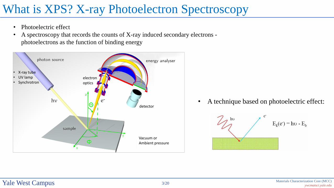
• Photoelectric effect
• A spectroscopy that records the counts of X-ray induced secondary electrons -
photoelectrons as the function of binding energy
• A technique based on photoelectric effect:

Materials Characterization Core (MCC)
ywcmatsci.yale.edu4/20Yale West Campus
What is XPS? X-ray Photoelectron Spectroscopy
• X-ray tube• UV lamp• Synchrotron
detector
electronoptics
Vacuum orAmbient pressure
• Photoelectric effect
• A spectroscopy that records the counts of X-ray induced secondary electrons -
photoelectrons as the function of binding energy
• A technique based on photoelectric effect:

Materials Characterization Core (MCC)
ywcmatsci.yale.edu5/20Yale West Campus
What kinds of samples for XPS?
• Vacuum compatible: low vapor pressure under 10-8 Pascal
• Conductive or insulating
Freezing

Materials Characterization Core (MCC)
ywcmatsci.yale.edu6/20Yale West Campus
How XPS works?
• XPS detects the number of photoelectrons at different kinetic energies (KE)
• The photoelectron binding energy can then be calculated, characteristic of elements
within the sample volume
KE (measured) = hν - BE – Φspec
BE = hν - KE - Φspec
KE (KLL) = BE(K) – BE(L2) – BE(L3)
Ionization (initial state) Relaxation and Emission (final state)
Auger Electron
Φ
BE
L3
L1
L2
X-ray
FluorescenceK
UV
Photoelectron
Vacuum
VB
2p3/22p
1s
X-ray
Photoelectron
EFΦ
hν
2s
2p1/2
hν
e-
Materials Characterization Core (MCC)
ywcmatsci.yale.edu7/20Yale West Campus
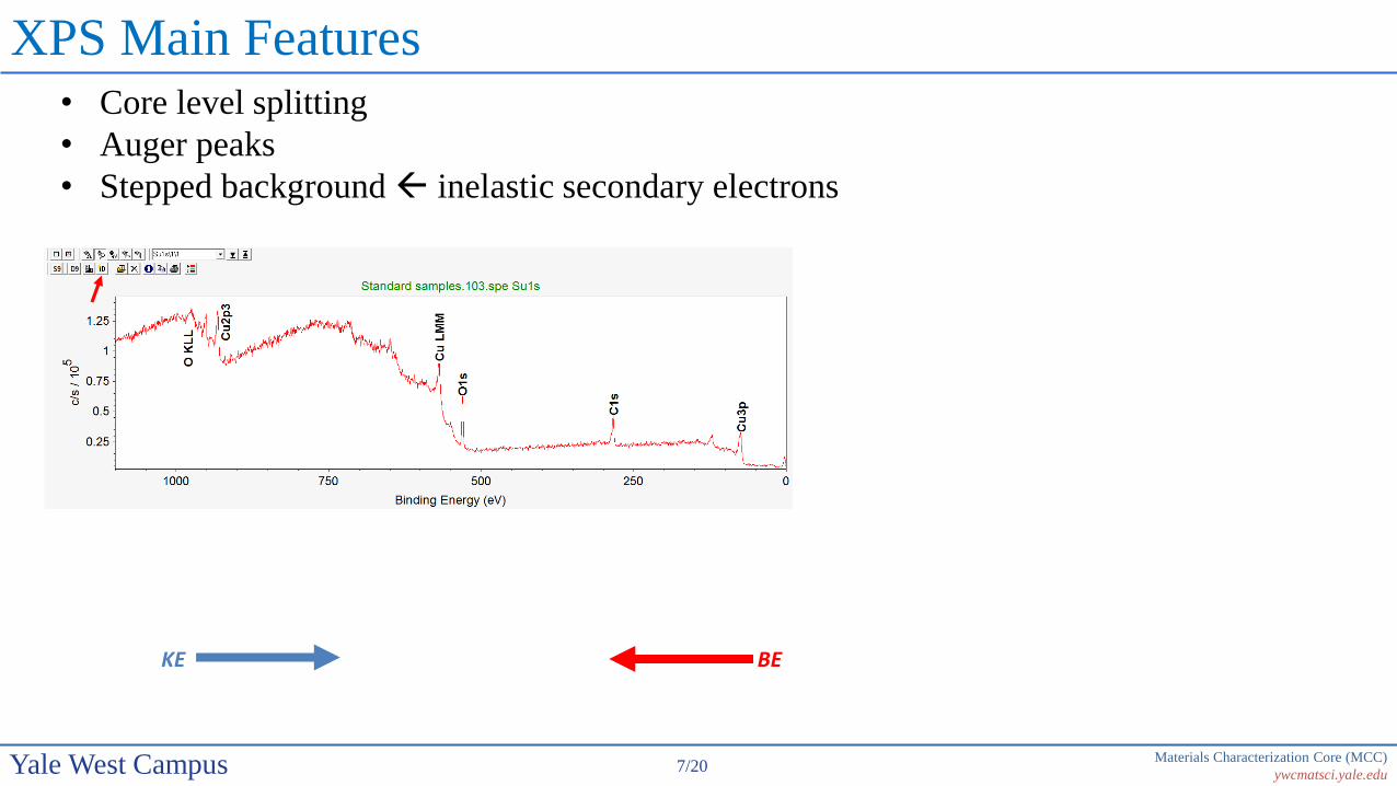
XPS Main Features• Core level splitting
• Auger peaks
• Stepped background inelastic secondary electrons
KE BE

Materials Characterization Core (MCC)
ywcmatsci.yale.edu8/20Yale West Campus
XPS Peak Notation
4f7/2
n
l = 0 s1 p2 d3 f
j = l ± s, s = 1/2
Spin-orbital splitting with l > 0
Orbital l j Degeneracy (2j + 1) Peak area ratio Electron level
s 0 1/2 1 - 1s
p 1 1/2, 3/2 2, 4 1 : 2 2p1/2, 2p3/2
d 2 3/2, 5/2 4, 6 2 : 3 3d3/2, 3d5/2
f 3 5/2, 7/2 6, 8 3 : 4 4f5/2, 4f7/2
Materials Characterization Core (MCC)
ywcmatsci.yale.edu9/20Yale West Campus
XPS Instrumentation
UV lamp
Hemisphericalanalyzer
X-ray source
Flood gun
Sample
UHV chamber(low 10-7 – 5x10⁻⁸ Pa
Ion gun
e-
e-
Ar+
Detector Lens
Pumps
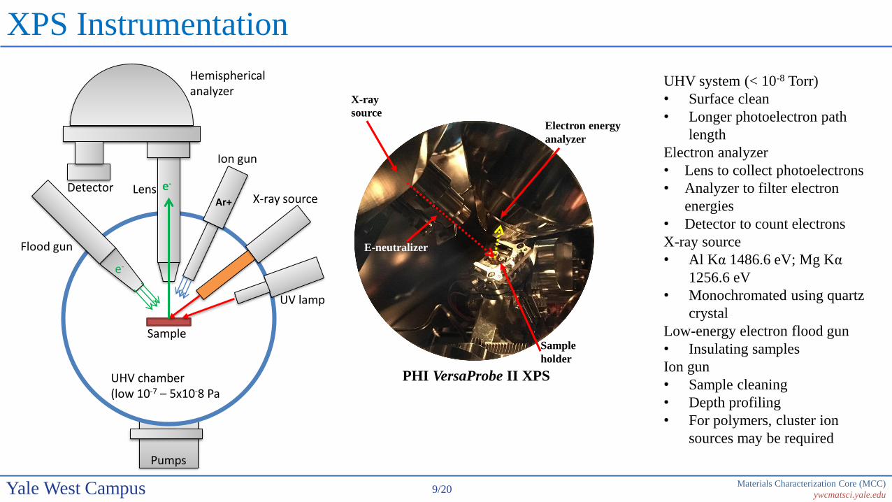
UHV system (< 10-8 Torr)
• Surface clean
• Longer photoelectron path
length
Electron analyzer
• Lens to collect photoelectrons
• Analyzer to filter electron
energies
• Detector to count electrons
X-ray source
• Al Kα 1486.6 eV; Mg Kα
1256.6 eV
• Monochromated using quartz
crystal
Low-energy electron flood gun
• Insulating samples
Ion gun
• Sample cleaning
• Depth profiling
• For polymers, cluster ion
sources may be required
Sample
holder
Electron energy
analyzer
X-ray
source
PHI VersaProbe II XPS
E-neutralizer
Materials Characterization Core (MCC)
ywcmatsci.yale.edu10/20Yale West Campus
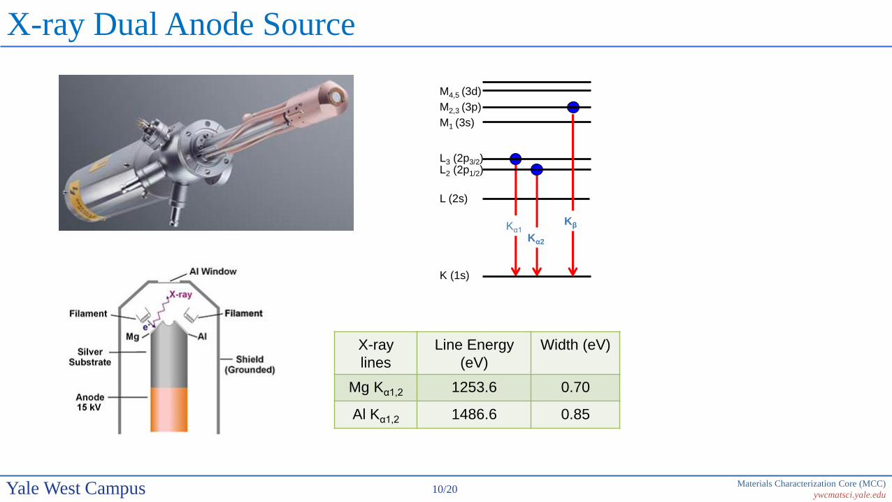
X-ray Dual Anode Source
X-ray
lines
Line Energy
(eV)
Width (eV)
Mg Kα1,2 1253.6 0.70
Al Kα1,2 1486.6 0.85
K (1s)
L (2s)
L2 (2p1/2)L3 (2p3/2)
M1 (3s)
M2,3 (3p)
M4,5 (3d)
Kα1
Kα2
Kβ

Materials Characterization Core (MCC)
ywcmatsci.yale.edu11/20Yale West Campus
X-ray monochromator
• Narrow peak width
• Reduced background
• No satellite & Ghost peaks
n λ = 2dsinθ
For quartz (1010) surface:
n = diffraction order
d = 0.42 nm (lattice constant)
θ = 78.5º
λ = 0.83 nm for Al Kα

Materials Characterization Core (MCC)
ywcmatsci.yale.edu12/20Yale West Campus
Spherical Capacitor Analyzer (SCA)
Pass energy:
Analyzer Resolution:
V0: the median equipotential surface of radius r
V: the potential applied between inner (radius b) and outer (radios a) shells
w: entrance and exit slit widths
𝛿𝛼: angular deviation of the electron trajectories at the entrance with
respect to the center line
r =a+b
2
Where the mean radius
𝐸0 = 𝑒𝑉0 =𝑉
𝑏𝑎−𝑎𝑏
a
b
r
𝜹𝜶
V2<0
w wV
∆𝐸 = 𝐸0𝑤
𝑎 + 𝑏+𝛿𝛼2
4
For the PHI SCA : 𝐸0 = 0.56𝑉 ∆𝐸 = 0.015𝐸0and
Typical 𝐸0 = 100 eV ∆𝐸 = 1.5 eV

Materials Characterization Core (MCC)
ywcmatsci.yale.edu13/20Yale West Campus
Why are we interested in XPS?
http://www.eag.com/mc
• Surface sensitive technique
• Chemical shift detection XPS is also named as Electron Spectroscopy
for Chemical Analysis (ESCA)
Typical Analysis Depths for Techniques
XPS detects electron signals in the near surface region (0 ~ 10 nm)
Materials Characterization Core (MCC)
ywcmatsci.yale.edu14/20Yale West Campus
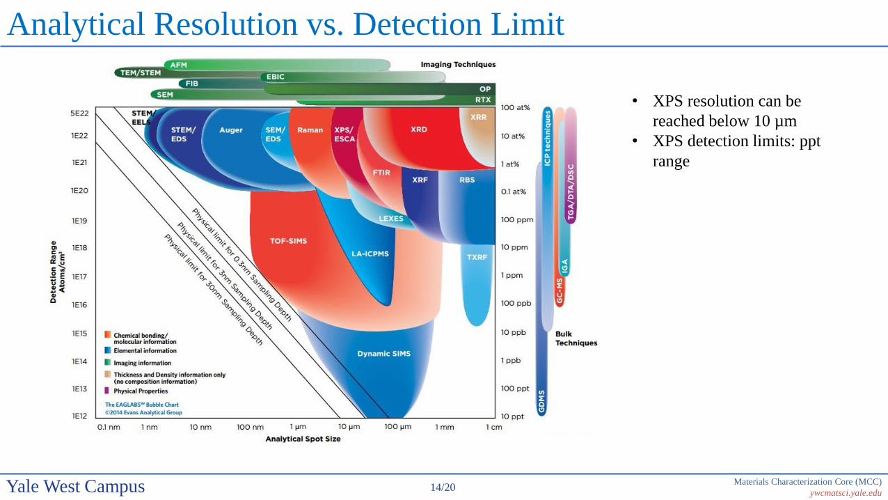
Analytical Resolution vs. Detection Limit
http://www.eag.com/mc
• XPS resolution can be
reached below 10 µm
• XPS detection limits: ppt
range

Materials Characterization Core (MCC)
ywcmatsci.yale.edu15/20Yale West Campus
Why XPS is Surface Sensitive?
• Inelastic scattering of photoelectrons

Materials Characterization Core (MCC)
ywcmatsci.yale.edu16/20Yale West Campus
Electron Inelastic Mean Free Path (IMFP)
“Universal Curve” - λ (IMFP) vs kinetic
energy
λ = 1 ~ 3.5 nm for X-ray photoelectrons
• The average distance an electron travels through a solid before losing energy through
inelastic collisions.
Related Documents