The role of cytokines and pattern-recognition receptors in inflammatory gastrointestinal diseases: clinical and in vitro investigations Ph.D. Thesis Péter Hofner Supervisor: Prof.Yvette Mándi M.D. D.Sc. Department of Medical Microbiology and Immunobiology Faculty of medicine University of Szeged Szeged 2008

Welcome message from author
This document is posted to help you gain knowledge. Please leave a comment to let me know what you think about it! Share it to your friends and learn new things together.
Transcript

The role of cytokines and pattern-recognition receptors in inflammatory
gastrointestinal diseases: clinical and in vitro investigations
Ph.D. Thesis
Péter Hofner
Supervisor: Prof.Yvette Mándi M.D. D.Sc.
Department of Medical Microbiology and Immunobiology
Faculty of medicine
University of Szeged
Szeged
2008
2
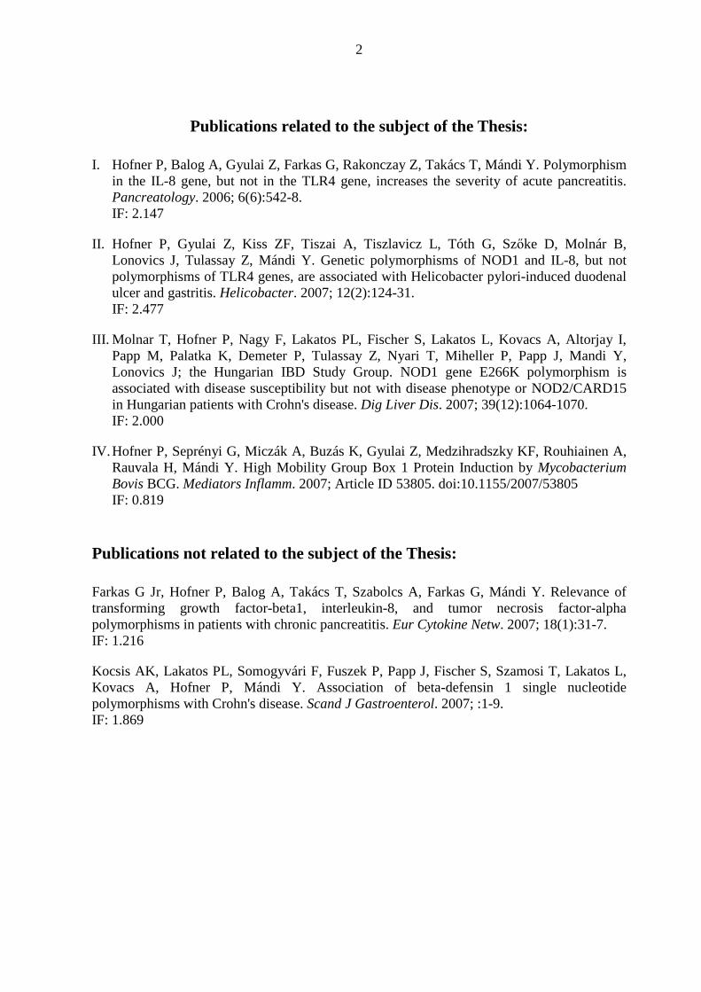
Publications related to the subject of the Thesis:
I. Hofner P, Balog A, Gyulai Z, Farkas G, Rakonczay Z, Takács T, Mándi Y. Polymorphism in the IL-8 gene, but not in the TLR4 gene, increases the severity of acute pancreatitis. Pancreatology. 2006; 6(6):542-8. IF: 2.147
II. Hofner P, Gyulai Z, Kiss ZF, Tiszai A, Tiszlavicz L, Tóth G, Szıke D, Molnár B, Lonovics J, Tulassay Z, Mándi Y. Genetic polymorphisms of NOD1 and IL-8, but not polymorphisms of TLR4 genes, are associated with Helicobacter pylori-induced duodenal ulcer and gastritis. Helicobacter. 2007; 12(2):124-31. IF: 2.477
III. Molnar T, Hofner P, Nagy F, Lakatos PL, Fischer S, Lakatos L, Kovacs A, Altorjay I, Papp M, Palatka K, Demeter P, Tulassay Z, Nyari T, Miheller P, Papp J, Mandi Y, Lonovics J; the Hungarian IBD Study Group. NOD1 gene E266K polymorphism is associated with disease susceptibility but not with disease phenotype or NOD2/CARD15 in Hungarian patients with Crohn's disease. Dig Liver Dis. 2007; 39(12):1064-1070. IF: 2.000
IV. Hofner P, Seprényi G, Miczák A, Buzás K, Gyulai Z, Medzihradszky KF, Rouhiainen A, Rauvala H, Mándi Y. High Mobility Group Box 1 Protein Induction by Mycobacterium Bovis BCG. Mediators Inflamm. 2007; Article ID 53805. doi:10.1155/2007/53805 IF: 0.819
Publications not related to the subject of the Thesis:
Farkas G Jr, Hofner P, Balog A, Takács T, Szabolcs A, Farkas G, Mándi Y. Relevance of transforming growth factor-beta1, interleukin-8, and tumor necrosis factor-alpha polymorphisms in patients with chronic pancreatitis. Eur Cytokine Netw. 2007; 18(1):31-7. IF: 1.216
Kocsis AK, Lakatos PL, Somogyvári F, Fuszek P, Papp J, Fischer S, Szamosi T, Lakatos L, Kovacs A, Hofner P, Mándi Y. Association of beta-defensin 1 single nucleotide polymorphisms with Crohn's disease. Scand J Gastroenterol. 2007; :1-9. IF: 1.869

3
CONTENTS
CONTENTS......................................................................................................... 3
8. ANNEX.......................................................................................................... 5
ABBREVIATIONS ............................................................................................. 6
1. INTRODUCTION........................................................................................ 8
1.1. Pattern-recognition receptors (PRRs) .........................................................................8
1.1.1. Toll-like receptors (TLRs) ..................................................................................8
1.1.2. NOD-like receptors (NLRs).............................................................................10
1.1.3. Cooperation between NOD containing proteins and TLRs ..............................11
1.2. Helicobacter pylori infection....................................................................................13
1.2.1. General features of H. pylori infection ............................................................13
1.2.2. The cag pathogenecity island (cag PAI)...........................................................13
1.2.3. Host responses to H. pylori..............................................................................14
1.3. The role of cytokines and PRRs in acute pancreatitis...............................................15
1.4. The role of cytokines and NOD1 in Crohn’s disease ...............................................16
1.5. Single nucleotide polymorphisms (SNPs) ................................................................17
1.5.1. SNPs of CARD4/Nod1 and CARD15/Nod2 ......................................................18
1.5.2. SNPs of TLR4 ...................................................................................................19
1.5.3. SNPs of IL-8 .....................................................................................................20
1.6. High-mobility group box 1 protein (HMGB-1) ........................................................21
AIMS................................................................................................................... 24
2. PATIENTS AND METHODS................................................................... 25
2.1. Patients and controls .................................................................................................25
2.1.1. Patient groups with duodenal ulcer or chronic active gastritis .........................25
2.1.2. Patient group with acute pancreatitis ................................................................25
2.1.3. Patient group with Crohn’s disease...................................................................26
4
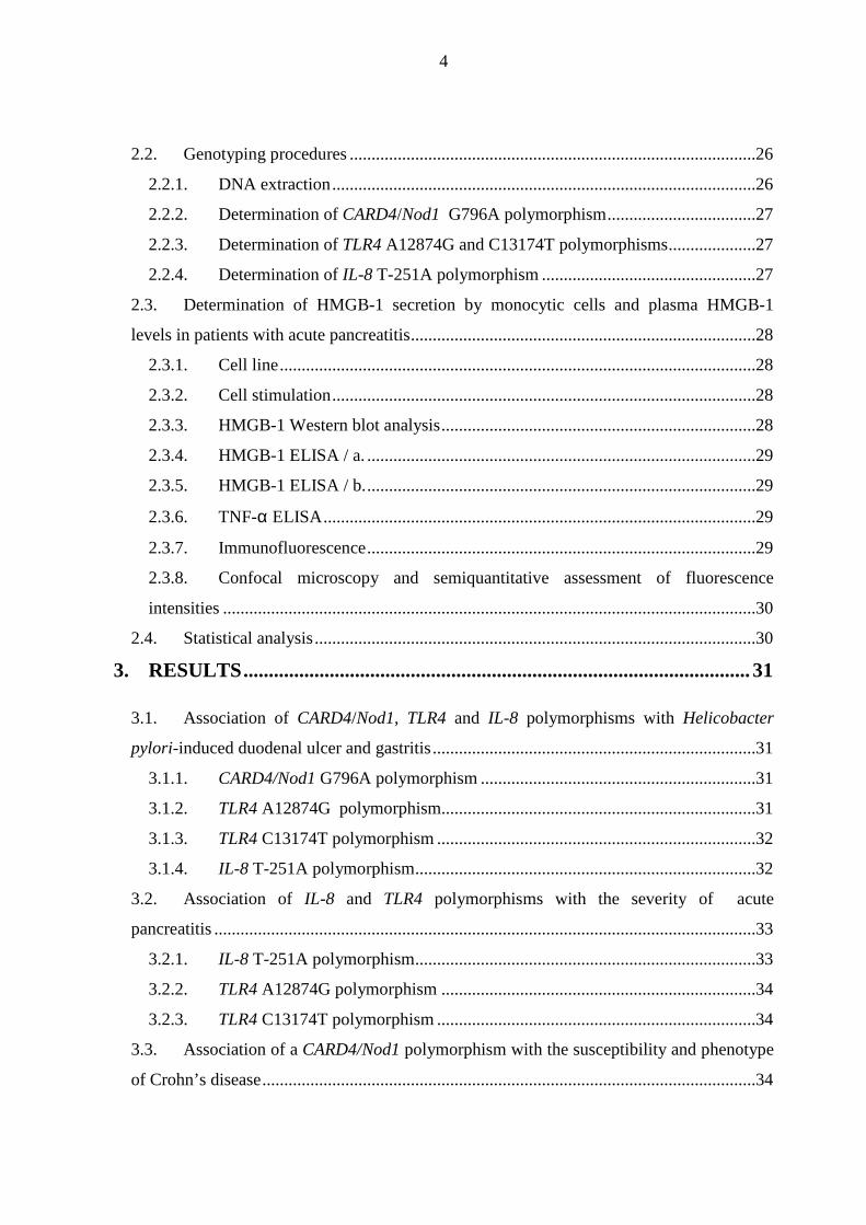
2.2. Genotyping procedures .............................................................................................26
2.2.1. DNA extraction.................................................................................................26
2.2.2. Determination of CARD4/Nod1 G796A polymorphism..................................27
2.2.3. Determination of TLR4 A12874G and C13174T polymorphisms....................27
2.2.4. Determination of IL-8 T-251A polymorphism .................................................27
2.3. Determination of HMGB-1 secretion by monocytic cells and plasma HMGB-1
levels in patients with acute pancreatitis...............................................................................28
2.3.1. Cell line.............................................................................................................28
2.3.2. Cell stimulation.................................................................................................28
2.3.3. HMGB-1 Western blot analysis........................................................................28
2.3.4. HMGB-1 ELISA / a. .........................................................................................29
2.3.5. HMGB-1 ELISA / b..........................................................................................29
2.3.6. TNF-α ELISA...................................................................................................29
2.3.7. Immunofluorescence.........................................................................................29
2.3.8. Confocal microscopy and semiquantitative assessment of fluorescence
intensities ..........................................................................................................................30
2.4. Statistical analysis.....................................................................................................30
3. RESULTS.................................................................................................... 31
3.1. Association of CARD4/Nod1, TLR4 and IL-8 polymorphisms with Helicobacter
pylori-induced duodenal ulcer and gastritis..........................................................................31
3.1.1. CARD4/Nod1 G796A polymorphism ...............................................................31
3.1.2. TLR4 A12874G polymorphism........................................................................31
3.1.3. TLR4 C13174T polymorphism .........................................................................32
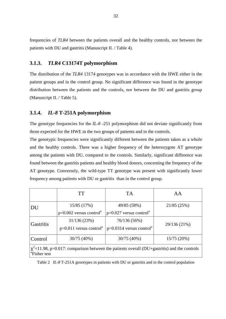
3.1.4. IL-8 T-251A polymorphism..............................................................................32
3.2. Association of IL-8 and TLR4 polymorphisms with the severity of acute
pancreatitis ............................................................................................................................33
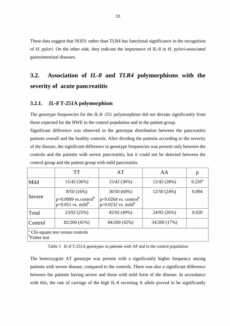
3.2.1. IL-8 T-251A polymorphism..............................................................................33
3.2.2. TLR4 A12874G polymorphism ........................................................................34
3.2.3. TLR4 C13174T polymorphism .........................................................................34
3.3. Association of a CARD4/Nod1 polymorphism with the susceptibility and phenotype
of Crohn’s disease.................................................................................................................34
5
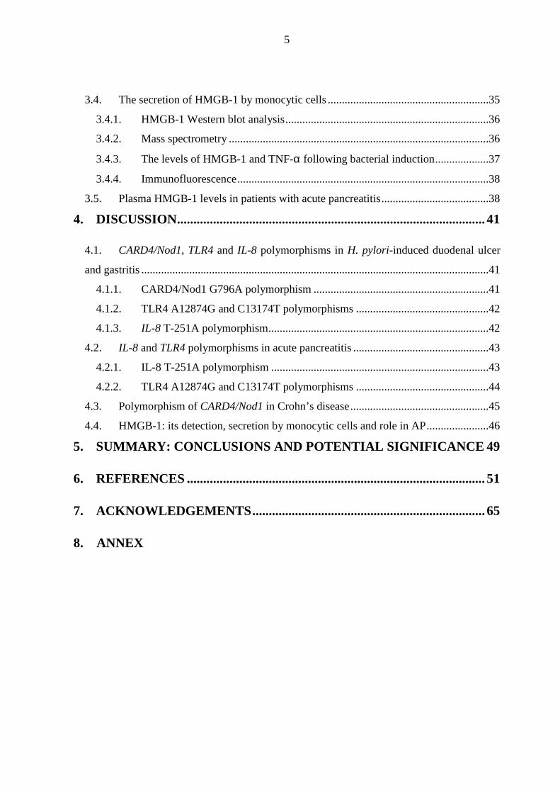
3.4. The secretion of HMGB-1 by monocytic cells .........................................................35
3.4.1. HMGB-1 Western blot analysis........................................................................36
3.4.2. Mass spectrometry ............................................................................................36
3.4.3. The levels of HMGB-1 and TNF-α following bacterial induction...................37
3.4.4. Immunofluorescence.........................................................................................38
3.5. Plasma HMGB-1 levels in patients with acute pancreatitis......................................38
4. DISCUSSION.............................................................................................. 41
4.1. CARD4/Nod1, TLR4 and IL-8 polymorphisms in H. pylori-induced duodenal ulcer
and gastritis ...........................................................................................................................41
4.1.1. CARD4/Nod1 G796A polymorphism ..............................................................41
4.1.2. TLR4 A12874G and C13174T polymorphisms ...............................................42
4.1.3. IL-8 T-251A polymorphism..............................................................................42
4.2. IL-8 and TLR4 polymorphisms in acute pancreatitis ................................................43
4.2.1. IL-8 T-251A polymorphism .............................................................................43
4.2.2. TLR4 A12874G and C13174T polymorphisms ...............................................44
4.3. Polymorphism of CARD4/Nod1 in Crohn’s disease.................................................45
4.4. HMGB-1: its detection, secretion by monocytic cells and role in AP......................46
5. SUMMARY: CONCLUSIONS AND POTENTIAL SIGNIFICANCE 49
6. REFERENCES ........................................................................................... 51
7. ACKNOWLEDGEMENTS....................................................................... 65
8. ANNEX

6
ABBREVIATIONS
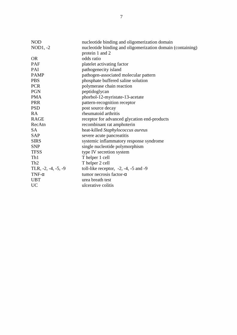
ABC ATP-binding cassette AP acute pancreatitis ATP adenosine triphosphate BCG Bacillus Calmette-Guérin BSA bovine serum albumin CagA cytotoxin-associated protein A (the product of the cagA gene) CARD caspase-recruitment domain CARD4, -15 caspase-recruitment domain 4 and 15 genes CATERPILLER protein family, named after CARD, transcription enhancer, R
(purine)-binding, pyrin, lots of leucine repeats CD Crohn’s disease CHO chinese hamster ovary CI confidence interval DAP diaminopimelic acid DC dendritic cell DU duodenal ulcer EAEC enteroaggregative Escherichia coli ELISA enzyme-linked immunosorbent assay ERK extracellular-signal-regulated kinase HMG high-mobility group HMG-1 high-mobility group 1 protein HMGB high-mobility group box HMGB-1, -2, -3 high-mobility group box -1,-2 and -3 proteins HWE Hardy-Weinberg equilibrium IBD inflammatory bowel disease iE-DAP γ-D-glutamyl-meso-diaminopimelic acid IFN-γ interferon-γ IgG,-Y immunoglobulin G and Y IL-1α, -1β, -2, -4, -6, -8, -10, -12, -14
interleukin-1α, -1β, -2, -4, -6, -8, -10, -12, -14
LPS lipopolysaccharide LRR leucine-rich repeat MALT mucosa-associated lymphoid tissue MAPK mitogen-activated protein kinase MCP, -1 monocyte chemoattractant protein, -1 MDP muramyl dipeptide MIP-1α, -1β macrophage inflammatory protein-1α and -1β MOF multiple organ failure NACHT-LRR protein family, named after neuronal apoptosis inhibitory
protein (NAIP), CIITA, HET-E and TP1 – leucine-rich repeat NF-κB nuclear factor-κB NLS, -1, -2 nuclear localization signal, -1 and -2 NLR NOD-like receptor
7
NOD nucleotide binding and oligomerization domain NOD1, -2 nucleotide binding and oligomerization domain (containing)
protein 1 and 2 OR odds ratio PAF platelet activating factor PAI pathogenecity island PAMP pathogen-associated molecular pattern PBS phosphate buffered saline solution PCR polymerase chain reaction PGN peptidoglycan PMA phorbol-12-myristate-13-acetate PRR pattern-recognition receptor PSD post source decay RA rheumatoid arthritis RAGE receptor for advanced glycation end-products RecAtn recombinant rat amphoterin SA heat-killed Staphylococcus aureus SAP severe acute pancreatitis SIRS systemic inflammatory response syndrome SNP single nucleotide polymorphism TFSS type IV secretion system Th1 T helper 1 cell Th2 T helper 2 cell TLR, -2, -4, -5, -9 toll-like receptor, -2, -4, -5 and -9 TNF-α tumor necrosis factor-α UBT urea breath test UC ulcerative colitis

8
1. INTRODUCTION
In order to counteract events that disturb homeostasis such as infections or tissue injury,
multicellular organisms have developed a mechanism called inflammatory response. Invading
pathogens come into contact first with the mucosal epithelium and with cells of the innate
immune system. These cells recognize endogenous danger signals, for example the high-
mobility group box 1 protein (HMGB-1) [1, 2], and pathogen-associated molecular patterns
(PAMPs) by their germ-line encoded pattern-recognition receptors (PRRs). PAMPs are
evolutionary conserved, often structural motifs found in wide range of different microbes [3].
In mammals PRRs include Toll-like receptors (TLRs) and NOD (nucleotide binding and
oligomerization domain)-like receptors (NLRs) [4, 5]. The receptor signaling leads to the
production of inflammatory mediators such as tumor necrosis factor-α (TNF-α) and
interleukin-8 (IL-8) and to the recruitment of inflammatory cells.
Individual differences in the receptor signaling and in the cytokine production can be
genetically determined, which further influences the host response in inflammatory diseases.
Therefore the aims of our study were to investigate the genetic polymorphisms of IL-8, TLR4
and caspase-recruitment domain 4 gene (CARD4/Nod1) in such gastrointestinal diseases,
where the intensity of host response definitely determines the consequences of infection or
tissue necrosis, i.e. in Helicobacter pylori-induced gastritis and duodenal ulcer (DU), in acute
pancreatitis (AP) and in Crohn’s disease (CD). In addition, we investigated the production of
the „newly” recognized danger signal and late cytokine HMGB-1 following in vitro bacterial
infection and in pancreatitis.
1.1. Pattern-recognition receptors (PRRs)
1.1.1. Toll-like receptors (TLRs)
Toll was originally described as a receptor involved in the embryonic development [6] and in
the defence mechanism against fungal infection in Drosophila melanogaster [7]. Since then,
ten homologues of the Toll protein (TLRs) have been identified in humans. TLRs are
transmembrane proteins that are located on the cell-surface or act as endosomal receptors,

9
recognizing a diverse group of microbial and endogenous ligands. TLR4 recognizes
lipopolysaccharide (LPS), TLR5 detects flagellin, while TLR9 senses viral and bacterial DNA
among others. Similarly to NOD containing proteins, TLRs possess LRR (leucine-rich repeat)
domains for the recognition of their ligands. The downstream signaling pathways lead to
nuclear factor-κB (NF-κB) activation and expression of genes that are involved in the pro-
inflammatory responses [4, 8] .
Data on the role of different TLRs in the recognition of H. pylori are numerous but
controversial. Bäckhed et al. demonstrated that primary gastric antral epithelial cells do not
express TLR4, while the gastric epithelial cell lines AGS, MKN45 and NCI-N87 express the
nonsignaling form of the receptor. Both primary gastric epithelial cells and cell lines were
induced to secrete IL-8 following infection with H. pylori in a cytotoxin-associated gene
pathogenecity island (cag PAI)-dependent manner, indicating that the recognition of H. pylori
is independent of the TLR4-mediated LPS signaling [9]. In contrast, Su et al. found that AGS
cells constitutively express TLR4 mRNA and H. pylori infection induces increase in
transcription, translation and expression of TLR4 independently of cagA [10]. TLR4 was
demonstrated to be expressed at the apical as well as at the basolateral pole of gastric surface
epithelium in vivo, both in noninflamed gastric mucosa and in chronic active gastritis by
Schmausser et al. The expression of TLR5 and TLR9 was identical to that of TLR4 in
noninflamed gastric mucosa, but changed to an exclusive basolateral localization in H. pylori
gastritis. H. pylori bacteria became directly attached to the apically expressed TLR4 receptors
in patients with gastritis, supporting their role as binding sites for the bacteria in vivo [11].
This correlates with the finding that TLR4 expression promotes increased adherence of H.
pylori to the surface of chinese hamster ovary (CHO) fibroblast cells [10].
TLR4 was shown to display an early, transient upregulation in experimental rat pancreatitis,
suggesting its functional role in the development of the disease [12]. In addition, Johnson et
al. described an endogenous pathway mediated by TLR4 which leads to systemic
inflammatory response syndrome (SIRS)-like syndrome, the triggering factor of which is
released into the blood and tissues during both pancreatitis and systemic inflammation [13].
This finding further supports that TLR4 is potentially associated with AP.
10
1.1.2. NOD-like receptors (NLRs)
The NOD (nucleotide binding and oligomerization domain)-like receptor (NLR) family, also
described as NACHT-LRR receptors or CATERPILLER, is involved in bacterial sensing in
the cytoplasm. The NLR family has more than 20 members, including NOD1 and NOD2 [14].
NOD1 is encoded by the caspase-recruitment domain 4 gene (CARD4/Nod1), which is located
on chromosome 7p14-15, extends over 55 kb of genomic DNA and is composed of 14 exons.
The protein is built of 965 amino acids [15]. NOD2, which is encoded by CARD15/Nod2 [16],
is composed of 1040 amino acids [17].
Members of the NLR family, including NOD1 have tripartite domain structure. The carboxy-
terminal LRR domain participates in the recognition and detection of ligands, while there is a
NOD (or NACHT) in central position of the molecule, which posesses ATP-ase activity and
facilitates self-oligomerization. The amino-terminal effector domain is built from protein-
protein interaction cassettes and can be represented either by CARD or pyrin (e.g. in case of
cryopyrin) domain. NOD1 has one, while NOD2 has two CARDs. Following the interaction
between the ligand and LRR domain, a complex conformational change of the protein is
thought to be initiated: binding of ATP to an ATP-binding cassette (ABC) and self-
oligomerization of the molecules are proposed to be the initial steps of binding and activating
downstream effector molecules through the amino-terminal domain(s) [14, 18].
NOD1 and NOD2 are expressed in the cytosol of epithelial [19, 20] and antigen presenting
cells [21], human gingival fibroblasts [22], myofibroblasts [23], astrocytes [24] and microglia
[25]. The baseline expression of NOD1 is constitutive but variable, which is enhanced by
IFN-γ, but not by TNF in epithelial cells [26]. NOD2 posesses low baseline expression, the
upregulation of which can be exerted by TNF and further augmented by IFN-γ [27].
The minimal peptidoglycan (PGN) moieties recognized by NOD1 and NOD2 are γ-D-
glutamyl-meso-diaminopimelic acid (iE-DAP) [28, 29] and muramyl dipeptide (MDP) [30,
31], respectively. There is species specifity in the recognition of ligands by NOD1 receptors:
human NOD1 requires a tripeptide (L-Ala-D-Glu-meso-DAP), while the murine receptor
needs a tetrapeptide (L-Ala-D-Glu-meso-DAP-D-Ala) for optimal sensing of PGN [32]. PGN
in both Gram-negative and Gram-positive bacteria contains MDP, but the presence of iE-DAP
is restricted only to the PGN of Gram-negatives (except for Listeria spp, Bacillus spp. and

11
Gram-positive bacteria in the soil). That is why NOD2 should be considered as a general
sensor for most bacteria and NOD1 for mainly Gram-negatives [14]. The intracellular
pathogen Shigella flexneri [33], the enteroinvasive Escherichia coli (EIEC) [19], Chlamydia
pneumoniae [34] and Pseudomonas aeruginosa [35] have been shown to be detected by
NOD1. In particular, NOD1 was shown to have a crucial role in the containment of H. pylori.
After injected by the type IV secretion system (TFSS) of H. pylori into gastric epithelial cells,
PGN fragments are specifically recognized by NOD1 [36]. Following activation of NOD1
and -2 by their ligands, they recruit a downstream effector molecule, the receptor-interacting
serine/threonine kinase (RICK or RIP2), which triggers a cascade leading to the translocation
of NF-κB to the nucleus [14], controlled by centaurin β1 [37], erbin [38] or by the
transforming growth factor-β (TGF-β)-activated kinase 1 [39] among others. The activation
of the mitogen-associated protein kinase (MAPK) pathways can be exerted through NOD1
and -2. Extracellular signal-regulated kinase (ERK) [40] and p38MAPK [41] pathways are
involved in signaling processes by NOD2, while the activation of NOD1 may result in the
activation of JUN amino-terminal kinase (JNK) [33]. NOD1 is thought to mediate a restricted
pattern of immune responses, the primary role of which is to induce the recruitment and
interaction of immune cells by enhancing the expression of IL-8, CXCL1/Groα,
CXCL2/Groβ, monocyte chemoattractant protein 1 (MCP-1/ CCL2) and CD83 [42].
1.1.3. Cooperation between NOD containing proteins and TLRs
Due to the synergistic effects of the TLR- and NOD containing protein-mediated signaling
pathways, the cellular response to whole microbes is not only recognizing their individual
PAMPs.
NOD1 and NOD2 agonists act cooperatively with the TLR4 agonist LPS to induce the release
of proinflammatory and anti-inflammatory cytokines from CD14+ monocytes and CD1a+
immature DCs, and synergize with sub-active doses of LPS to induce DC maturation [43].
Yang et al. reported that MDP alone is hardly able to elicit cytokine production in human
monocytic cell lines, but exerted priming effects on IL-8 secretion by THP-1 cells upon
stimulation with LPS, which may be partially explained by the up-regulation of MyD88 by
MDP [44]. In addition, Uehara et al. demonstrated that synthetic MDP and DAP-containing
12
desmuramylpeptides, signaling through NOD2 and NOD1 respectively, exhibited synergism
with TLR2 (synthetic E. coli-type triacyl lipopeptide), TLR4 (synthetic E. coli-type lipid A)
and TLR9 agonits (bacterial CpG DNA) to induce the production of IL-8 in THP-1 cells [45].
Bordatella pertussis tracheal cytotoxin (N-acetylglucosaminyl-1,6-anhydro-N-acetylmuramyl-
(L)-alanyl-γ-(D)-glutamyl-meso-diaminopimelyl-(D)-alanine) and endotoxin were synergistic
in the induction of IL-1α mRNA and protein, production of nitric oxide and inhibition of
DNA synthesis in hamster trachea epithelial cells [46], in accordance with the results of
Dokter et al., where the N-acetylglucosaminyl-1,6-anhydro-N-acetylmuramyl-(L)-alanyl-γ-
(D)-glutamyl-meso-diaminopimelyl-(D)-alanine induced IL-1β and IL-6 mRNA expression
was further enhanced by costimulation with LPS or MDP in human monocytes [47]. MDP
had also strong synergistic effects with the mycobacterial TLR2 ligand 19-kDa lipoprotein on
the production of TNF, IL-1β and IL-6 in mononuclear cells [48].
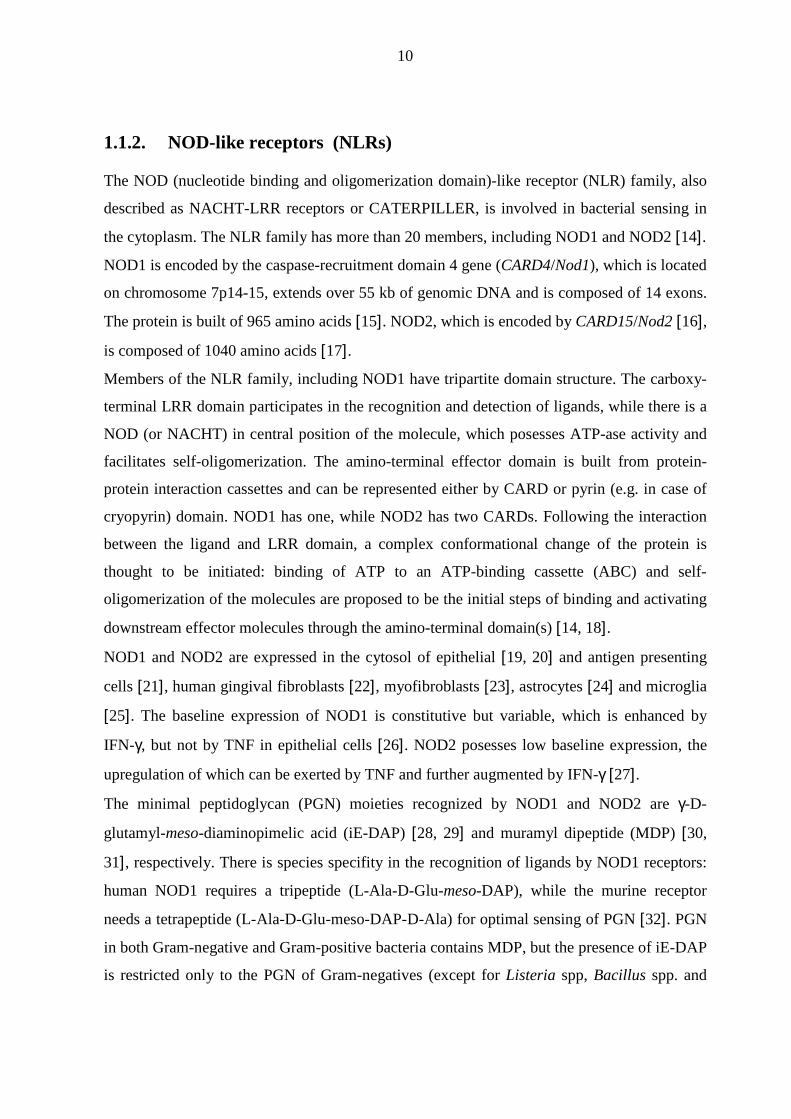
Figure 1 The structure and main signaling pathways of PRR families [49]

13
1.2. Helicobacter pylori infection
1.2.1. General features of H. pylori infection
Helicobacter pylori is a Gram-negative, microaerophilic, neutrophilic, spiral-shaped,
flagellated [50, 51] bacterium that colonizes the human gastric mucosa and induces chronic
inflammation. Since its isolation from human stomach biopsies by Marshall and Warren in
1983, H. pylori was shown to be an etiological agent in the development of gastritis, peptic
ulcer [52], mucosa-associated lymphoid tissue (MALT) lymphoma [53], gastric atrophy
with/without intestinal metaplasia [54] and gastric cancer [55, 56, 57]. H. pylori was declared
to be the first bacterial class1 carcinogen by the World Health Organisation/ International
Agency for Research on Cancer (WHO/IARC) in 1994 [58]. H. pylori infection is one of the
most common bacterial infections worldwide affecting over half of the whole human
population [59]. The infection is mostly acquired during childhood and is connected with low
socioeconomic status. The transmission routes are fecal-oral, oral-oral and gastric-oral. If the
infection is acquired, it can persist for the entire life of the host unless the eradication therapy
is successful [60 61].
1.2.2. The cag pathogenecity island (cag PAI)
The cytotoxin-associated gene pathogenecity island (cag PAI) of H. pylori is a 40 kb cluster
of 31 genes that encodes components of the TFSS [62, 63] among others, and was suggested
to modulate the TLR2-agonist activity of H. pylori [64]. The presence of cag PAI is
associated with increased mucosal inflammation and confers elevated risk for the
development of peptic ulcer disease [65] and gastric cancer [66]. The TFSS can be depicted as
a syringe that enables bacteria to deliver the CagA protein and bacterial breakdown products
into the host cells. Knockout mutants for the virD4 or cagG lose the ability to translocate
CagA and cause temporally retarded or abolished gastric inflammation, respectively. Once
CagA is injected into the gastric epithelial cells, it causes tight junction dysfunction and
becomes phosphorylated by Src kinases on the tyrosine residues of the carboxy-terminal Glu-
Pro-Ile-Tyr-Ala (EPIYA) motifs, which enables it to participate in processes leading to host
cell elongation (hummingbird phenotype) and changes in cell motility [63].
14
In gastric epithelial cells, the activation of the transcription factors NF-κB and activating
protein-1 (AP-1), which leads to the expression of proinflammatory cytokines and exerts
apoptotic and antiapoptotic effects, is cag PAI-dependent. These comprize the upregulation of
the antiapoptotic gene that encodes the cellular inhibitor of apoptosis protein 2 (c-IAP2) in
gastric cancer cells [67], apoptotic processes exerted mainly through the mitochondrial
pathway [68] and the transcriptional regulation of IL-8 [69], among others. In addition, the
cag PAI-positive H. pylori causes inflammation by a synergistic mechanism that activates IL-
8 and its receptors IL-8RA (CXCR1) and IL-8RB (CXCR2) by independent pathways [70].
The levels of TNF-α, IL-1β and IL-6, the genes of which are also NF-κB-responsive, are also
elevated in the gastric mucosa of H. pylori infected patients [71].
1.2.3. Host responses to H. pylori
H. pylori infection alternates and evades the host responses, which enables the bacteria to
perform a long-term colonization up to several decades even though local immune responses
are present [72]. These responses are not only ineffective in eliminating the bacterium from
its preferred niche, but actually contribute to the development of the gastric mucosal lesions
observed in H. pylori-infected individuals.
The function of NOD1 in the host defence against H. pylori seems to be important, because
Nod1-deficient mice have higher bacterial loads after H. pylori infection than wild-type mice
[36]. NOD1 stimulation results in the production of other chemokines, and not merely IL-8.
NOD1 induction stimulates human intestinal epithelial cells to produce CXCL1 (Groα) and
CD83 as well [42]. These molecules are important in the recruitment and interaction of
immune cells and this very probably holds true in the case of H. pylori-induced diseases. The
role of TLR2, TLR4 and TLR5 is controversial in the recognition of H. pylori [73].
The local inflammation in H. pylori infection is characterized by the infiltration of neutrophils
and lymphocytes into the gastric mucosa and by increased production of the proinflammatory
cytokines TNF-α [74], IL-1β [75], IL-6 [76], IL-8 [77] and interferon-γ (IFN-γ) [78].
Lymphocytic derived IL-2 has also been detected [79]. IL-8, as a potent neutrophil
chemotactic and activating factor secreted by epithelial cells, has been suggested to play a
central role in H. pylori-associated diseases [80]. H. pylori also elicits the production of the
15
proinflammatory IL-12 by dendritic cells (DCs) in the gastric mucosa, which further induces
the production of IFN-γ [81]. The effects of proinflammatory cytokines might be counteracted
by the locally produced IL-10, a Th2 cytokine with anti-inflammatory effects [82].
1.3. The role of cytokines and PRRs in acute pancreatitis
AP remains a significant clinical challenge, because it has a complex and poorly understood
pathophysiology, an unpredictable outcome and no specific treatment [83]. It has an annual
incidence of 10-20 cases per 100000 people in the Western world [84]. The etiology is mostly
alcoholic [85] or related to obstruction by gallstones [86]. Although the clinical course of AP
is often mild with only minimal organ dysfunction, a significant proportion of these patients
develop severe disease often associated with multiple organ failure (MOF) and infections
[87]. The initial prediction about the mild or severe outcome of an attack has important
implications for management, prognostication and use of health care resources. Different
methods of prognostic classification have been developed, but so far, none has shown optimal
overall predictive accuracy and usefulness for prediction in individual patients [88].
MOF and septic complications in AP do not differ from the systemic complications of other
diseases such as sepsis itself, trauma or burn, which are included in SIRS. The systemic
manifestations are responsible for the majority of pancreatitis-associated morbidity and
mortality, and are due to the actions of the proinflammatory cytokines TNF-α, IL-1 and IL-8
[89, 90]. The levels of cytokines depend on a number of factors, including genetic
background, particularly polymorphisms of cytokine genes. Genetic polymorphisms within
the promoter region of inflammation-related cytokine genes are considered to influence the
expression of the encoded cytokines [91]. IL-8 is an important neutrophil-activating cytokine
and a potent chemoattractant, the increased levels of which have been demonstrated in AP
[92, 93]. Other factors, including TLR4, that regulate the innate immune responses [94], may
also be involved in the complications of AP. In view of the inflammatory nature of AP, we
postulated that single nucleotide polymorphisms (SNPs) in the promoter region of IL-8 and in
the third exon of TLR4 might have some association with the development of AP.
16
1.4. The role of cytokines and NOD1 in Crohn’s disease
Inflammatory bowel disease (IBD) is a collective term for at least three heterogenous
gastrointestinal disorders including Crohn’s disease (CD), ulcerative colitis (UC) and
indeterminate colitis, which result in several distinct clinical phenotypes and lead to persistent
and sometimes irreversible impairment of the gastrointestinal structure and function. IBD is
thought to be a multifactorial disease, triggered by the inappropriate and exaggerated mucosal
immune response to the normal microflora, which is partly determined by genetic and
environmental factors. The importance of the commensal flora and its communication
strategies with the host is illustrated by several transgenic mouse models and innate
immunodeficiency syndromes. Innate immunity is much more complex than a mere
mechanism which is fully capable to distinguish self from non-self. CARD4/Nod1 is a perfect
candidate as a susceptibility gene for CD. The latest microbiological studies failed to find a
potential pathogen microorganism which leads to development of IBD, perhaps because there
is no such a common microbe. As each bacterial sensor triggers the activation signal of
different bacteria, theoretically the altered function of any of them may result in defective
recognition of at least one part of the commensal flora. This theory might explain how IBD
can develop in those patients who do not have NOD2 polimorphism: other receptors of
PAMPs are responsible for the causative defect, and this receptor can be the NOD1 in
individual cases [95].
In CD, manifestations can occur in any part of the gastrointestinal tract, but most commonly
the terminal ileum, the cecum, the peri-anal area and the colon are affected, where the
alteration of linear ulcers with islands of normal or edematous mucosa produces cobblestone-
like appearance. By the progression of the disease, the bowel narrows, which leads to bowel
obstruction, abscess formation and fistulization. Extraintestinal manifestations of CD affect
the joints, the skin, the eyes, the mouth and the liver [96, 97].
The inflammation observed is dominated by an excessive Th1-mediated effector cell
response. Increased amounts of the Th1 polarizing cytokine IL-12 were shown to be produced
by DCs and macrophages in CD [98]. The mucosal expression of IL-18 - which is considered
to be an activator of Th1 responses - becomes also upregulated in patients with CD [99]. The
TNF superfamily member TL1A protein, the expression of which correlated positively with
17
the intensity of inflammation in CD, was suggested to maintain and perpetuate the IFN-γ
mediated Th1 immune responses in the intestinal mucosa [100]. The production of IFN-γ
provokes downstream effector cells such as macrophages to induce further production of the
proinflammatory cytokines TNF-α, IL-1β and IL-6 [101]. Supporting the significant role of
TNF-α in the pathogenesis of CD, infliximab, a chimeric monoclonal immunoglobulin G1
(IgG1) antibody proved to be effective in inducing and maintaining the remission of CD in
60% of the patients, by binding and neutralizing soluble and membrane-bound TNF-α [102,
103]. Although IL-10 was shown to downregulate the elevated secretion and mRNA levels of
proinflammatory cytokines by CD mononuclear cells in vitro [104, 105], clinical trials with
recombinant human IL-10 yielded only modest results [106].
1.5. Single nucleotide polymorphisms (SNPs)
DNA sequence polymorphisms are variations present at greater than 1% frequency in the
population, while the frequency of mutations is less than 1%. By comparing two DNA
sequences, wherever found different nucleotides at the same position, that is a SNP. SNPs are
responsible for over 80% of the 0.1 % difference that is present between the DNA sequences
of two individuals. They can occur anywhere in the genom: both in coding and noncoding
regions of genes or in intergenic spacers [107]. Non-conservative mutations within the coding
region of genes can result in loss, abrogation or change of function in the expressed protein as
a result of change in protein structure. Although conservative mutations do not affect the
amino acid sequence, they may influence protein expression in a variety of other ways: they
can alter mRNA splicing, mRNA stability and levels of gene expression. Polymorphisms
within the 5’- and 3’-regulatory sequences or introns of genes may have a significant effect on
the transcription, since they may alter the structure of transcription factor binding sites within
gene promoters or the structure of enhancers and silencers within introns [108]. SNPs are
estimated to occur as frequently as every 100-300 bases [109]. More than 4 million SNPs
have been identified to date with the leader participation of The SNP Consortium (TSC).
The clinical outcome of many infectious, autoimmune or malignant diseases appears to be
influenced by the overall balance of production of proinflammatory and anti-inflammatory
cytokines. A significant number of studies have addressed whether genetic polymorphisms

18
within the genes encoding these cytokines might influence the levels of expression, and
therefore the overall immune response [108].
One potential application of SNPs is to develop individualised medicine by
pharmacogenomics, in order to understand adverse drug responses, with respect to SNPs in
the genes of drug-metabolizing enzymes [110]. SNPs represent useful tool in DNA
fingerprinting for criminal investigations [111] and paternity testing [112]. With the help of
SNPs, we can also gain insight into the steps of evolution: DNA sequence variants that
provided benefit for the individual for succesful reproduction can even be selected by natural
selection for retention. SNPs profiles characteristish for a disease trait can help to identify the
relevant genes associated with polygenic diseases [113, 114].
1.5.1. SNPs of CARD4/Nod1 and CARD15/Nod2
The deletion allele of the complex insertion-deletion polymorphism (ND1+32656) of CARD4
was significantly associated with susceptibility to inflammatory bowel disease [115], while
the insertion allele was found to be associated with the presence of asthma and elevated IgE
levels [15]. Focusing on five non-synonymous mutations of CARD4, Zouali et al. could not
reveal any contribution to the genetic predisposition to IBD [116]. Polymorphisms with
database accession numbers rs2736726 and rs2075817 of CARD4 showed weak associations
with atopic eczema [117]. Rosenstiel et al. reported that no mutations of CARD4 or CARD15
contribute to the development of gastritis or gastric ulcer in H. pylori-infected patients. There
was no association found between SNPs of CARD4 and the development of MALT
lymphoma, while the R702W polymorphism of CARD15 was significantly associated with the
development of gastric lymphoma: carriers of the rare T allele had more than doubled risk to
develop the disease than controls [118].
Mutations of CARD15/Nod2 confer genetic predisposition for CD [31, 119], Blau syndrome
[120], early-onset sarcoidosis [121], graft-versus-host disease [122], allergic rhinitis and
atopic dermatitis [123].
19
1.5.2. SNPs of TLR4
The human gene encoding TLR4 is located on chromosome 9q32-q33 and contains four
exons [124]. The A→G substitution at nucleotide 896 from the start codon of the human
TLR4 gene (in the fourth exon of TLR4) leads to the replacement of a conserved aspartic acid
residue with glycine at position 299 (Asp299Gly; NCBI SNP cluster ID: rs4986790; also
referred to as A12874G according to GenBank acc. no. AF177765 [125]). The C→T
conversion at 1196 position results in the replacement of a nonconserved threonine with an
isoleucine at position 399 (Thr399Ile; NCBI SNP cluster ID: rs4986791; also referred to as
C1196T according to GenBank acc. no. AF177765 [125]). Both SNPs affect the extracellular
domain of the receptor [126], presumably by altering its structure, which may cause altered
ligand binding or protein interactions, resulting in impaired LPS signaling.
Peripheral monocytes from donors homozygous for the TLR4 896 G allele were LPS
hyporesponsive, exhibiting fourfold higher median effective concentrations for TNF-α, IL-1β
and IL-6 production compared to monocytes from patients with heterozygous or wild-type
genotypes [127]. Upon in vitro stimulation with LPS, Kroner et al. detected significantly
lower proliferation indices of peripherial blood mononuclear cells (PBMCs) from individuals
heterozygous for A896G, in comparison with cells from individuals with the wild-type
genotype [128]. Surgical intensive care unit patients who were heterozygous for both the
A896G and C1196T polymorphisms (no homozygous mutant was found) had higher risk for
subsequent Gram negative infections [129]. These two TLR4 polymorphisms were shown to
be associated with decreased airway responsiveness to inhaled LPS in humans by Arbour et
al. [130] These generally accepted results and even studies relied on their citation were
questioned by Erridge et al., who found that monocytes from individuals heterozygous for
both polymorphisms of TLR4 do not exhibit deficit in the recognition of LPS [131].
The frequency of the TLR4 896 G allele was significantly higher in patients with CD and UC
than in their respective control populations [132]. CD patients with the A896G SNP had more
often ileal envolvement or fistulizing disease, compared to wild-type patients [133].
The presence of the TLR4 C1196T SNP was shown to be associated with UC and its increased
frequency was observed in patients with CD, without reaching significance [134].
20
In contrast to the defective TLR4-mediated signal transduction in the murine experimental
model of disseminated candidiasis [135], the TLR4 Asp299Gly polymorphism was shown not
to play a role in susceptibility to and severity of human urogenital Candida albicans infection
[136].
1.5.3. SNPs of IL-8
The human gene encoding IL-8 is localized in the CXC chemokine locus on chromosome
4q12-q21 and consists of 4 exons and 3 introns [137]. The SNP at -251 nucleotide relative to
the transcription start site (T-251A), in the promoter region of IL-8 was identified by Hull et
al. The presence of the mutant A allele was associated with increased IL-8 production by LPS
stimulated whole blood, with the rising severity of respiratory syncytial virus acute
bronchiolitis [138] and with increased risk for human tuberculosis [139]. Matheson et al.
found no association between the T-251A SNP and respiratory symptoms or lung function in
middle-aged and older adults [140].
The chance of having enteroaggregative E. coli (EAEC)-associated diarrhea significantly
increased among those harbouring the -251 A allele, compared to the individuals with TT
genotype [141]. Jiang et al. found significantly higher occurence of the homozygous genotype
for the -251 A allele among patients with Clostridium difficile toxin-induced diarrhea,
compared to the control groups of C. difficile-negative diarrhea and nondiarrhea [142]. Fecal
IL-8 levels were significantly higher in subjects with C. difficile toxin-induced or EAEC
diarrhea who were homozygous for the -251 A allele, compared to patients with heterozygous
or wild-type genotype [141].
Suggesting the involvement of IL-8 in the development and progression of human
malignancies, the -251 A allele was found to be associated with higher risk for
nasopharyngeal carcinoma [143], prostate cancer [144], oral squamous cell carcinoma [145]
and breast carcinoma [146]. Individuals homozygous for the A allele at -251 or for the G
allele at +396 on IL-8 were at twofold increased risk of cardia adenocarcinoma compared to
those harbouring the wild-types, furthermore the risk for the AGT/AGC haplotype of IL-8 -
251/+396/+781 was fourfold in a chinese population (T-251A, T+396G and C+781T SNPs on
IL-8) [147].
21
Renzoni et al. identified three polymorphisms in the noncoding areas of IL-8 (A-353T in the
promoter region, G+293T and T+678C in the first intron) that were shown not to be
associated with cryptogenic fibrosing alveolitis and fibrosing alveolitis associated with
systemic sclerosis [148]. Rovin et al. identified the IL-8 T-738A and T-845C biallelic
polymorphisms and demonstrated that the frequency of the -845 C allele was significantly
higher in African American patients with severe, inflammatory systemic lupus erythematosus
nephritis compared to healthy individuals, indicating that the T-845C polymorphism might
influence the expression of IL-8 [149].
1.6. High-mobility group box 1 protein (HMGB-1)
High-mobility group (HMG) chromosomal proteins were described as abundant components
of the chromatin by Goodwin et al. [150] . Their name indicates the rapid electrophoretic
mobility on polyacrilamide gel. The HMG proteins are grouped into 3 families. The HMGB
family has 3 members: HMGB-1, -2 and -3 [151].
HMGB-1 (named also HMG-1 or amphoterin) is a highly conserved protein featuring >98 %
identity among mammals. It has two DNA-binding domains (named A- and B-box) and a
negatively charged, acidic, repetitive carboxy-terminal tail. The nuclear translocation of the
protein is under the control of two nuclear localization signals (NLS) on the molecule: NLS1
is localized to the A-box, while NLS2 can be found between the B-box and the acidic tail
[152].
HMGB-1 has different properties depending on its localization. The intranuclear form binds
to the minor groove of DNA without sequence specificity, and acts as an architectural protein
stabilizing the nucleosome formation, facilitating DNA bending and taking part in the
interaction between transcription factors and DNA [153, 154, 155, 156, 157].
HMGB-1 can get out of the cells by two mechanisms: it can be secreted actively by
monocytes [158], macrophages [159], pituicytes [160], human umbilical vein endothelial cells
(HUVEC) [161] and murine erythroleukemia cells [162] following appropriate stimulation, or
released passively by damaged or necrotic cells [163]. Rouhiainen et al. demonstrated that
HMGB-1 present in human platelets is exported to the cell surface upon activation [164].

22
Proinflammatory mediators can co-operate in order to promote HMGB-1 secretion: IFN-γ
induces HMGB-1 release partly through a TNF-dependent mechanism in murine
macrophages [165]. HMGB-1 released in response to proinflammatory stimuli also induces
the release of proinflammatory mediators by macrophages and neutrophils such as TNF-α,
IL-1α, IL-1β, IL-6, IL-8, macrophage inflammatory proteins MIP-1α and MIP-1β and
HMGB-1 [166, 167, 168], thus prolonging and sustaining inflammatory processes. When
released passively, HMGB-1 acts as an endogenous danger signal which triggers
inflammation and reparative processes [163] .
HMGB-1 lacks a secretory signal peptide (similarly to IL-1β [169]), so it cannot be secreted
via the Golgi-endoplasmic reticulum (ER) pathway [162]. In resting monocytes HMGB-1
shuttles through the nuclear membrane to the cytosol or into the nucleus. Triggered by an
activation signal, lysine residues within the two NLS sites of the HMGB-1 molecule become
acethylated, which is thought to inhibit the relocalization of the protein into the nucleus. This
hyper-acethylated form will accumulate in the cytosol and will be packed into secretory
lysosomes that fuse with the cell membrane and release their content into the extracellular
space [152, 170].
HMGB-1 enhances its own degradation by accelerating the tissue plasminogen activator (t-
PA)-catalysed plasminogen activation: the protein is suggested to be a target of the lysine-
specific serine protease plasmin [171].
HMGB-1 was reported to play an important role in the pathomechanism of atherosclerosis
[167, 172] and rheumatoid arthritis (RA) [173], and its elevated serum levels were measured
in patients with hemorrhagic shock [174], community-acquired pneumonia [175] and Churg-
Strauss syndrome [176]. HMGB-1 was demonstrated to be a late mediator of endotoxin
lethality in mice and of sepsis in humans by Wang et. al [159]. Sundén-Cullberg et al. found
that serum levels of HMGB-1 remained high in the majority of patients with severe sepsis and
septic shock up to one week after admission [177]. In the pathomechanism of SAP, the
release of HMGB-1 by injured pancreas or other damaged organs as well as its secretion by
activated monocytes/ macrophages can be taken into consideration. [178].
The receptor for advanced glycation end-products (RAGE) was shown to be the major
functional receptor responsible for the proinflammatory effects of HMGB-1 in rhodent

23
macrophages [179]. HMGB-1 enhances the maturation of DCs [180], induces the extensive
spreading and mediates the transendothelial migration of monocytes also with the
involvement of RAGE [158]. In addition, HMGB-1 enhances RAGE expression in human
microvascular endothelial cells [181] and in synovial fluid macrophages [166]. TLR4
mediates the early inflammatory effects of HMGB-1 during hepatic ischemia/reperfusion
injury [182], and together with TLR2 might serve as an alternative HMGB-1 receptor [183,
184].
24
AIMS
The present study was designed to address the following aims:
1. To determine the association between CARD4/Nod1, TLR4 and IL-8 polymorphisms and
the development of gastritis and DU in H. pylori-infected patients.
2. To determine the association between IL-8 and TLR4 polymorphisms and the development
of AP.
3. To determine the association between CARD4/Nod1 polymorphism and the development
of CD.
4. To evaluate the role of HMGB-1 protein in AP, which required the elaboration of reliable
in vitro methods for the detection of HMGB-1.
25
2. PATIENTS AND METHODS
2.1. Patients and controls
2.1.1. Patient groups with duodenal ulcer or chronic active gastritis
85 H. pylori-positive patients with DU and 136 with chronic active gastritis were enrolled in
the study. Biopsies were taken during upper gastrointestinal endoscopy from adjacent sites of
the gastric antrum and corpus for histology. In addition, 13C-Urea Breath Test (UBT) was
carried out. Patients with H. pylori infection confirmed by histology and 13C-UBT were
considered to be eligible for the study. The presence of H. pylori and the severity of gastritis
were graded with the updated Syney Classification System [185].
The 75 members of the control population were serologically H. pylori-positive, without any
gastrointestinal symptoms. Their status of H. pylori infection was determined by serology,
with an enzyme-linked immunosorbent assay (ELISA) kit [HP IgG ELISA (Dia.Pro, Milan,
Italy)]. They were recruited from the Blood Transfusion Center at the University of Szeged.
Sera either from patients and control people were tested for CagA by serology, with a
commercial ELISA kit (Dia.Pro, Milan, Italy). Only H. pylori-positive and CagA-positive
patients and controls were considered to be eligible for the study.
All cases and controls were of Hungarian ethnic origin and resident in Hungary. Informed
consent was obtained from all patients. The project was approved by the Clinical Ethical
Committee of the Medical Faculty of the University of Szeged and by the Clinical Ethical
Committee of Semmelweis University.
2.1.2. Patient group with acute pancreatitis
92 patients with AP were enrolled. Diagnostic criteria of AP included clinical history
consistent with the disease, radiological evidence and serum amylase level greater than 660
U/L. According to the original criteria of Ranson [186], patients were classified into groups
with mild or severe pancreatitis: patients with fewer than three positive prognostic signs
(n=42) were considered to have mild pancreatitis; while those with three or more positive
prognostic signs (n=50) were classified into the severe pancreatitis group. Patients with severe

26
acute necrotizing pancreatitis were divided into aseptic (n=28) or infected (n=22) groups on
the basis of the results of bacterial cultures of the necrotic pancreatic tissue sampled during
surgery or ultrasound- or computed tomography (CT)-guided biopsies.
The patients entered this prospective study at the Department of Surgery and at the First
Department of Internal Medicine of Albert Szent-Györgyi Medical Center, University of
Szeged between March 2003 and January 2005. The control cohort consisted of a random,
unrelated population sample of 200 healthy blood donors. All cases and controls were of
Hungarian ethnic origin and resident in Hungary.
2.1.3. Patient group with Crohn’s disease
434 CD patients were investigated. The diagnosis was set up on the basis of Lennard-Jones
criteria [187]. The age at onset, duration of the disease, presence of extraintestinal
manifestations, frequency of flare-ups, effectiveness of therapy, need for surgery, occurence
of familial IBD, smoking habits and peri-anal involvement were investigated by the review of
the medical charts and questionnaries. The disease phenotype was determined according to
the Vienna classification [188].
The control group consisted of 200 age- and gender-matched healthy blood donors without
gastrointestinal or liver diseases. They were selected from consecutive blood donors in
Szeged. The second, non-IBD control group included 136 patients with chronic active
gastritis.
2.2. Genotyping procedures
2.2.1. DNA extraction
For the examination of CARD4/Nod1, TLR-4 and IL-8 polymorphisms, leukocyte DNA from
peripheral blood was isolated using the High Pure PCR Template Preparation Kit according to
the manufacturer’s instructions (Roche Diagnostics GmbH). The genomic DNA was stored at
–20 °C until further use.
27
2.2.2. Determination of CARD4/Nod1 G796A polymorphism
The G→A SNP on CARD4 at position 796 (NCBI SNP cluster ID: rs2075820) was analysed
by PCR-restriction fragment length polymorphism (PCR-RFLP). The 379 bp amplified
product was digested with AvaI restriction endonuclease enzyme and analysed on 2% agarose
gel under UV illumination. The restriction site was present only together with the G allele and
was indicated by the cleavage of the 379 bp product, giving fragments of 209 bp and 170 bp.
The CARD4 A allele gives a single 379 bp fragment due to the absence of restriction [116].
2.2.3. Determination of TLR4 A12874G and C13174T polymorphisms
The TLR4 12874 A→G (according to GenBank acc. no. AF177765, NCBI SNP cluster ID:
rs4986790; also referred to as A896G) and 13174 C→T (according to GenBank acc. no.
AF177765, NCBI SNP cluster ID: 4986791; also referred to as C1196T) polymorphisms were
genotyped simultaneously by a real-time PCR (RT PCR) assay using specific fluorescence-
labelled hybridization probes, followed by melting point analysis with the help of a
LightCycler instrument. By displaying the negative first derivative of the melting curve data
versus temperature [-(dF/dT) vs T], wild-type samples showed a peak at 64 °C (A12874G) or
at 67 °C (C13174T), while heterozygous samples were characterized by an additional peak at
61 °C (A12874G and C13174T) [125].
2.2.4. Determination of IL-8 T-251A polymorphism
The T→A SNP on IL-8 at position -251 (relative to the transcription start site) was genotyped
by amplification refractory mutation system (ARMS). The allele-specific primers were: 5’-
CCACAATTTGGTGAATTATCAAT-3’ (-251A) and 5’-
CCACAATTTGGTGAATTATCAAA-3’ (-251T). The consensus primer was: 5’-
TGCCCCTTCACTCTGTTAAC-3’. The amplified PCR product consisted 336 bp. In each
reaction, a second set of primers for exon 3 of the HLA-DRB1 gene was applied (forward: 5’-
TGCCAAGTGGAGCACCCAA-3’, reverse: 5’-GCATCTTGCTCTGTGCAGAT-3’, product
size: 796 bp) as control for the PCR efficiency [138].

28
2.3. Determination of HMGB-1 secretion by monocytic cells
and plasma HMGB-1 levels in patients with acute pancreatitis
2.3.1. Cell line
Human monocytic U-937 cells were propagated in RPMI 1640 medium (GIBCO)
supplemented with 100 µg/ml ampicillin, 100 µg/ml streptomycin and 10% heat-inactivated
fetal calf serum at 37 °C in a humidified CO2 incubator.
2.3.2. Cell stimulation
Before analysing HMGB-1 secretion by U-937 cells, the culture media were replaced by
OptiMEM (GIBCO) medium, and the number of the cells was adjusted to 106 cells/ml. The
cells were stimulated for 24 hours with 5 ng/ml phorbol-12-myristate-13-acetate (PMA,
Sigma-Aldrich), 10 µg/ml LPS E. coli O111:B4 (Sigma-Aldrich), heat-killed Staphylococcus
aureus (SA, 108/ml) or with Mycobacterium bovis BCG (107/ml, Pasteur strain of M. bovis
BCG, provided by David G. Russel, Department of Microbiology and Immunology, Cornell
University, Ithaca, NY, USA).
2.3.3. HMGB-1 Western blot analysis
The supernatants of stimulated U-937 cells were concentrated 10-fold on Centricon 10 filters
(Millipore Corporation), with subsequent processing in Laemmli buffer. Recombinant human
HMG-1 (Sigma-Aldrich) and recombinant rat amphoterin (RecAtn, provided by Ari
Rouhiainen, Neuroscience Center, University of Helsinki, Helsinki, Finland) served as
standard antigens. The standards and the concentrated supernatants were resolved in 12.5 %
sodium dodecyl sulfate polyacrylamide gel electrophoresis (SDS-PAGE) under reducing
conditions. The proteins were transferred to nitrocellulose blotting membrane that were then
blocked with 5% nonfat dry milk. The filters were stained firstly with chicken polyclonal anti-
RecAtn antibodies (provided by Ari Rouhiainen, Neuroscience Center, University of
Helsinki, Helsinki, Finland), secondly by horseradish peroxidase-conjugated goat anti-
chicken antibodies (Zymed Laboratories), developed with ECL Plus reagents (Amersham
29
Biosciences) and exposed to X-ray film. The densitometric analysis of the immunoreactive
bands was performed by ImageQuant 5.2 software (Molecular Dynamics).
2.3.4. HMGB-1 ELISA / a.
The wells of the 96-well MaxiSorp plate (Nunc) were coated with mouse monoclonal anti-
human HMG-1 antibodies (R&D Systems) at 10 µg/ml, then blocked with 2% nonfat milk.
The recombinant human HMG-1 (Sigma-Aldrich) was used as a standard substance, serially
diluted from 200 to 1.6 ng/ml. After keeping the standard dilutions and the samples in the
wells, the steps separated by washing procedures were as follows: the addition of rabbit anti-
HMG-1 polyclonal antibodies (BD Biosciences), horseradish peroxidase-labelled goat anti-
rabbit IgG antibodies (Bio-Rad Laboratories) and then TMB substrate (BD Biosciences) to
the microtitration plates. After the reaction was stopped by adding 2N sulphuric acid, the
absorbance was measured at 450 nm (650 nm reference). HMGB-1 concentrations of the
samples were calculated from the standard curve.
2.3.5. HMGB-1 ELISA / b.
Plasma HMGB-1 concentrations were determined by the HMGB-1 ELISA kit (Shino-Test
Corporation), according to the instructions of the manufacturer [189, 190].
2.3.6. TNF-αααα ELISA
TNF-α concentrations in the cell culture supernatants were quantified by the use of a TNF-α
ELISA kit (BioSource), according to the instructions of the manufacturer.
2.3.7. Immunofluorescence
Either stimulated and control cells were fixed in 4% paraformaldehide and permeabilized with
0.3% Triton X-100 on glass coverslips. The coverslips were saturated with 10% bovine serum
albumin (BSA) and the cells were stained with chicken polyclonal anti-RecAtn antibodies
(provided by Ari Rouhiainen, Neuroscience Center, University of Helsinki, Helsinki,
Finland). Fluorescein isothiocyanate (FITC)-conjugated donkey anti-chicken IgY (Jackson
Immunoresearch Laboratories) was applied for the detection of the bound antibodies.
30
Between all incubation step, the cells were washed three times with PBS containing 0.2%
BSA. Fluorescence signals were detected by confocal microscopy.
2.3.8. Confocal microscopy and semiquantitative assessment of
fluorescence intensities
Serial images of the immunostained samples were captured by Olympus FV1000 confocal
laser scanning microscope with standard parameter settings. The fluorescent signal of control,
M. bovis BCG and PMA treated cells were quantitatively analysed by ImageQuant software
(Molecular Dynamics). The mean and standard deviation of these data were calculated by the
Microsoft Excel program. The level of statistical significance was determined by two-tailed
T-probe.
2.4. Statistical analysis
In order to analyse the level of significance for genotype and allele frequencies between
groups, chi-square test or Fisher exact test was applied. The genotype frequencies for each
polymorphisms were tested for deviation from the HWE by the χ2 test, with 1 degree of
freedom. An α level of p<0.05 was taken as an indication of statistical significance. The
relationship between genotypes and disease severity is presented as the odds ratio (OR), with
a 95% confidence interval (CI).
The Mann-Whitney test was used to evaluate the differences in HMGB-1 levels between the
group of patients with acute pancreatitis and the control groups. An α level of p<0.05 was
taken as an indication of statistical significance.
All statistical analyses were performed using GraphPad Prism 4 statistical program
(GraphPad Software Inc.).

31
3. RESULTS
3.1. Association of CARD4/Nod1, TLR4 and IL-8
polymorphisms with Helicobacter pylori-induced duodenal ulcer
and gastritis
3.1.1. CARD4/Nod1 G796A polymorphism
The distribution of CARD4 796 genotypes was in accordance with the HWE both in the
control population and in the patient group with gastritis, but not in the group of patients with
DU. Significant difference was found in the genotype distribution between the patients overall
and the controls. There was a higher frequency of the homozygote mutant AA genotype
among patients with DU, compared to the controls or to the gastritis group. Conversely, the
prevalence of the wild-type GG genotype proved to be significantly lower in the group of DU
patients, either in comparison with the control group or patients with gastritis. However, no
significant difference was observed between the genotypes of controls and gastritis patients.
GG GA AA
DU 38/85 (45%)
p=0.017 versus controla p=0.012 versus gastritisa
30/85 (35%)
p=0.020 versus controla p=0.010 versus gastritisa
17/85 (20%)
Gastritis 85/136 (63%) 41/136 (30%) 10/136 (7%)
Control 48/75 (64%) 22/75 (29%) 5/75 (7%)
χ2=13.46, p=0.009: comparison between the patients overall (DU+gastritis) and the controls aFisher test
Table 1 CARD4 G796A genotypes in patients with DU or gastritis and in the control population
3.1.2. TLR4 A12874G polymorphism
The distribution of the TLR4 12874 genotypes was in accordance with the HWE in the two
groups of patients and in the controls. There were no significant differences in the genotype
32
frequencies of TLR4 between the patients overall and the healthy controls, nor between the
patients with DU and gastritis (Manuscript II. / Table 4).
3.1.3. TLR4 C13174T polymorphism
The distribution of the TLR4 13174 genotypes was in accordance with the HWE either in the
patient groups and in the control group. No significant difference was found in the genotype
distribution between the patients and the controls, nor between the DU and gastritis group
(Manuscript II. / Table 5).
3.1.4. IL-8 T-251A polymorphism
The genotype frequencies for the IL-8 -251 polymorphism did not deviate significantly from
those expected for the HWE in the two groups of patients and in the controls.
The genotypic frequencies were significantly different between the patients taken as a whole
and the healthy controls. There was a higher frequency of the heterozygote AT genotype
among the patients with DU, compared to the controls. Similarly, significant difference was
found between the gastritis patients and healthy blood donors, concerning the frequency of the
AT genotype. Conversely, the wild-type TT genotype was present with significantly lower
frequency among patients with DU or gastritis than in the control group.
TT TA AA
DU 15/85 (17%)
p=0.002 versus controla
49/85 (58%)
p=0.027 versus controla
21/85 (25%)
Gastritis 31/136 (23%)
p=0.011 versus controla
76/136 (56%)
p=0.0314 versus controla 29/136 (21%)
Control 30/75 (40%) 30/75 (40%) 15/75 (20%)
χ2=11.98, p=0.017: comparison between the patients overall (DU+gastritis) and the controls aFisher test
Table 2 IL-8 T-251A genotypes in patients with DU or gastritis and in the control population
33
These data suggest that NOD1 rather than TLR4 has functional significance in the recognition
of H. pylori. On the other side, they indicate the importance of IL-8 in H. pylori-associated
gastrointestinal diseases.
3.2. Association of IL-8 and TLR4 polymorphisms with the
severity of acute pancreatitis
3.2.1. IL-8 T-251A polymorphism
The genotype frequencies for the IL-8 -251 polymorphism did not deviate significantly from
those expected for the HWE in the control population and in the patient group.
Significant difference was observed in the genotype distribution between the pancreatitis
patients overall and the healthy controls. After dividing the patients according to the severity
of the disease, the significant difference in genotype frequencies was present only between the
controls and the patients with severe pancreatitis, but it could not be detected between the
control group and the patient group with mild pancreatitis.
TT AT AA p
Mild 15/42 (36%) 15/42 (36%) 12/42 (28%) 0.220a
Severe 8/50 (16%)
p=0.0009 vs.controlb p=0.051 vs. mildb
30/50 (60%)
p=0.0264 vs. controlb p=0.0232 vs. mildb
12/50 (24%) 0.004
Total 23/92 (25%) 45/92 (49%) 24/92 (26%) 0.020
Control 82/200 (41%) 84/200 (42%) 34/200 (17%)
a Chi-square test versus controls bFisher test
Table 3 IL-8 T-251A genotypes in patients with AP and in the control population
The heterozygote AT genotype was present with a significantly higher frequency among
patients with severe disease, compared to the controls. There was also a significant difference
between the patients having severe and those with mild form of the disease. In accordance
with this, the rate of carriage of the high IL-8 secreting A allele proved to be significantly
34
higher in patients with severe pancreatitis than in the control population. Conversely, the
prevalence of the wild-type TT genotype was significantly lower among patients with the
severe disease form than in the control population or in the patient group with mild
pancreatitis.
3.2.2. TLR4 A12874G polymorphism
The distribution of the TLR4 12874 genotypes was in accordance with the HWE neither in the
control nor in the patient group. No significant difference was found in the genotype
distribution between the pancreatitis patients overall and the healthy controls, nor between the
patient group with severe pancreatitis and the control group. It is worth mentioning that six of
the seven AG heterozygote patients were found in the severe pancreatitis group (six of fifty),
in contrast to the only one heterozygote among the 42 patients with mild pancreatitis
(Manuscript I. / Table 2).
3.2.3. TLR4 C13174T polymorphism
The distribution of the TLR4 13174 genotypes both in the control population and in the
patient group was in accordance with the HWE. There were no significant differences in the
genotype frequencies of the TLR4 13174 polymorphism either between the pancreatitis
patients overall and the healthy control population, or between the patients with severe
pancreatitis and the controls. In spite of the relatively low number of CT heterozygotes, it is
striking that six of the seven heterozygotes were found in the severe pancreatitis group, while
there was only one among the 42 patients with mild pancreatitis (Manuscript I. / Table 3). So
we can assume that the activation of TLR4 might not have importance, while the IL-8
producing capacity has a definite role in the pathomechanism of AP.
3.3. Association of a CARD4/Nod1 polymorphism with the
susceptibility and phenotype of Crohn’s disease
The distribution of CARD4 796 genotypes was in accordance with the HWE in the patient
group with CD and in the control groups of healthy blood donors. An additional group of

35
patients without IBD (n=136) was included in the study, whose genotypes were determined
previously, during the course of H. pylori-related diseases.
Significant difference in the genotype distribution was found between the CD patients and the
healthy controls, as well as between CD patients and non-IBD controls. When frequencies of
the homo- and heterozygous mutations were examined together, the carriage of CARD4
G796A polymorphism proved to be a highly significant risk factor for CD. In addition, the A
allele was significantly more frequent among patients with CD, compared to both control
populations.
No significant associations were found between the different genotypes and the demographic
data of the patients or the clinical characteristics of CD (Manuscript III. / Table 4).
Genotype∗ Allele frequency#
GG AG AA G A
CD 215/434 (49.5%) 180/434 (41.5%) 39/434 (9%) 610 (70.3%) 258 (29.7%)
Controls 134/200 (67%) 56/200 (28%) 10/200 (5%) 324 (81%) 76 (19%)
Non-IBD GI 85/136 (62.5%) 41/136 (30%) 10/136 (7.5%) 211 (77.6%) 61 (22.4%)
∗CD vs. controls: χ2<0.0001, p<0.0001, ORhetero/homozygous vs. wild-type: 2.07, 95% CI: 1.46-2.93. CD vs. Non-IBD GI controls: χ2=6.977, p=0.008, ORhetero/homozygous vs. wild-type:1.70, 95% CI: 1.14-2.52.
#CD vs. controls: χ2=16.229, p<0.0001, ORA vs. G: 1.80, 95% CI: 1.35-2.41. CD vs. Non-IBD GI controls: χ2=5.472, p<0.019, ORA vs. G: 1.46, 95% CI: 1.06-2.01.
Table 4 CARD4 G796A genotypes in patients with CD and controls
3.4. The secretion of HMGB-1 by monocytic cells
In order to investigate the role of HMGB-1 in pancreatitis, it was necessary to elaborate a
reliable in vitro model to detect HMGB-1. Therefore at first we set up experiments to measure
HMGB-1 levels in the supernatants of U-937 cells, following stimulation of the cells with
PMA, LPS, SA or with M. bovis BCG.

36
3.4.1. HMGB-1 Western blot analysis
Western blotting of the 10-fold concentrated cell culture supernatants revealed 25 kDa bands
that migrated together with the recombinant rat amphoterin (RecAtn, kindly provided by Ari
Rouhiainen, Neuroscience Center, University of Helsinki, Helsinki, Finland). The E. coli-
derived recombinant human HMG-1 gave also a considerable signal, featuring different
molecular weight at 30 kDa. Chicken polyclonal anti-RecAtn antibodies were applied,
because of their exclusive potential to recognize HMGB-1 secreted by the monocytic cells in
Western blot experiments. These chicken antibodies were raised against recombinant rat
amphoterin, and the rat protein differs only in two amino acids (Asp189Glu and Glu201Asp)
from the human one. The commercial affinity purified rabbit anti-HMG1 polyclonal (BD
Biosciences), mouse anti-human HMG-1 monoclonal (R&D Systems) and mouse monoclonal
(clone 4C3) anti-HMGB-1 antibodies failed to form considerable bands. The densitometric
analysis of the immunoreactive bands revealed higher concentrations of HMGB-1 in the
supernatants of cell cultures stimulated either with PMA or Mycobacterium bovis BCG
bacteria than in those of non-stimulated, control cells (Manuscript IV. / Figure 1).
3.4.2. Mass spectrometry
Because of the two standard antigens featured different molecular weights in the Western
blotting experiments, mass spectrometry analysis was performed in order to verify the identity
of these proteins and to explain the differences in their molecular weights.
Matrix assisted laser desorption/ionization − time of flight (MALDI-TOF) analysis of the
unfractionated tryptic digest of the appropriate gel band identified 79% of the masses detected
as predicted tryptic cleavage products of the recombinant human HMGB-1. These peptides
represented approximately 56% of the protein sequence. The identity of four peptides was
further confirmed by collision-induced dissociation (CID) analysis. Masses corresponding to
predicted His-tag tryptic peptides were also detected, and their identity was confirmed by post
source decay (PSD) analysis (Manuscript IV. / Figure 2).
82% of the masses detected in the RecAtn matched predicted tryptic peptides of the rat
HMGB-1 protein. The identity of m/z 1520.76 as predicted Ile113-Lys127 was further
37
confirmed by PSD analysis. These peptides represented approximately 47.6% of the protein
sequence (Manuscript IV. / Figure 3).
These experiments were performed by the Proteomics Research Group, Biological Research
Center of the Hungarian Academy of Sciences.
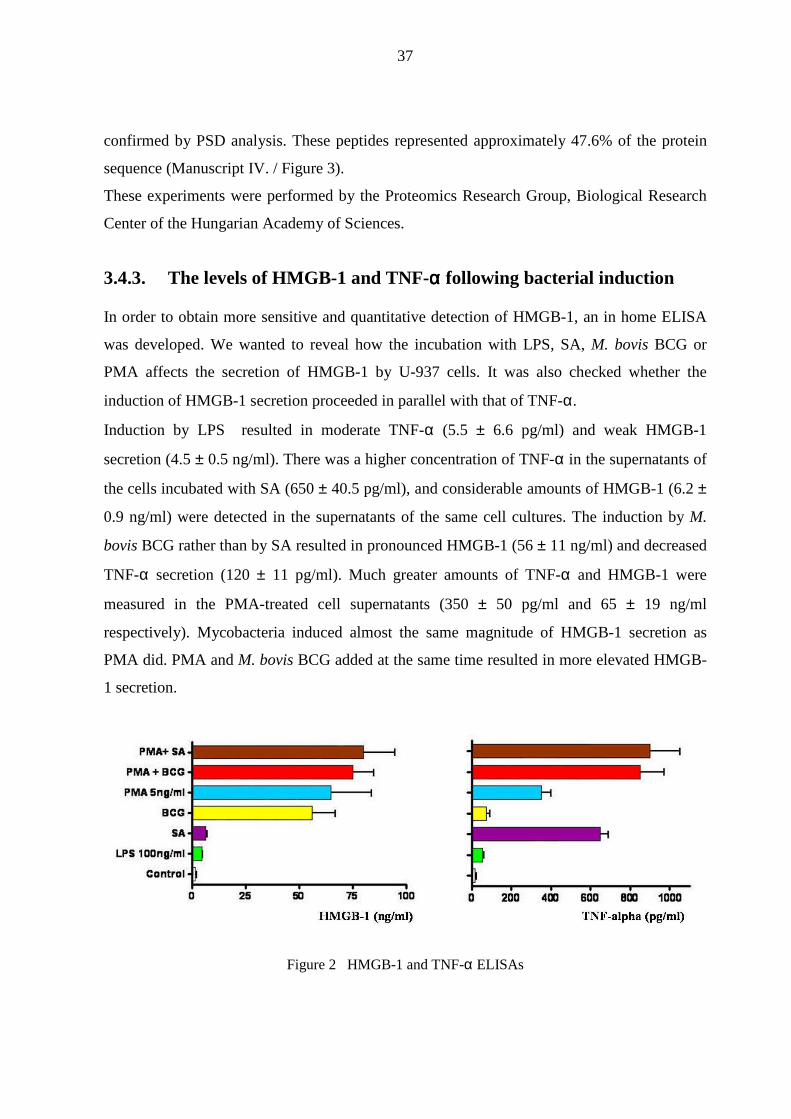
3.4.3. The levels of HMGB-1 and TNF-αααα following bacterial induction
In order to obtain more sensitive and quantitative detection of HMGB-1, an in home ELISA
was developed. We wanted to reveal how the incubation with LPS, SA, M. bovis BCG or
PMA affects the secretion of HMGB-1 by U-937 cells. It was also checked whether the
induction of HMGB-1 secretion proceeded in parallel with that of TNF-α.
Induction by LPS resulted in moderate TNF-α (5.5 ± 6.6 pg/ml) and weak HMGB-1
secretion (4.5 ± 0.5 ng/ml). There was a higher concentration of TNF-α in the supernatants of
the cells incubated with SA (650 ± 40.5 pg/ml), and considerable amounts of HMGB-1 (6.2 ±
0.9 ng/ml) were detected in the supernatants of the same cell cultures. The induction by M.
bovis BCG rather than by SA resulted in pronounced HMGB-1 (56 ± 11 ng/ml) and decreased
TNF-α secretion (120 ± 11 pg/ml). Much greater amounts of TNF-α and HMGB-1 were
measured in the PMA-treated cell supernatants (350 ± 50 pg/ml and 65 ± 19 ng/ml
respectively). Mycobacteria induced almost the same magnitude of HMGB-1 secretion as
PMA did. PMA and M. bovis BCG added at the same time resulted in more elevated HMGB-
1 secretion.
Figure 2 HMGB-1 and TNF-α ELISAs

38
3.4.4. Immunofluorescence
U-937 cells were incubated with M. bovis BCG, the impact of which on the HMGB-1
secretion was examined in the preliminary experiments. Non-stimulated cells displayed
strong staining for HMGB-1 mostly restricted to the nucleus. Eighteen hours after stimulation
by M. bovis BCG, the HMGB-1 protein appeared to be translocated from the nucleus (it was
still partly positive) to the periphery of the cells, displaying patchy staining in the cytoplasm.
Due to the dispersity of the fluorescence, its intensity was significantly lower in the cells
stimulated with M. bovis BCG (Manuscript IV. / Figure 4).
3.5. Plasma HMGB-1 levels in patients with acute pancreatitis
Though we elaborated an in home HMGB-1 ELISA, meanwhile a commercial ELISA kit
became available in the market developed by Shino-Test Corporation. The sensitivity proved
to be higher, therefore when measuring plasma samples, we applied this commercial one in
spite of its expense. The mean value of plasma HMGB-1 levels was significantly higher
(P=0.0038) in patients with AP (2.889±1.976 ng/ml) than in healthy subjects (0,039±0.014
ng/ml).
AP (n=21) Controls (n=9)0
1
2
3
4
5
Pla
sma
HM
GB
-1 (
ng/m
l)
Figure 3 Plasma HMGB-1 levels in patients with AP and in the control population
It was also higher than that measured in a second control group of patients with sepsis
(2.583±0.874 ng/ml) without reaching statistical significance (P=0.4724). The mean value of
plasma HMGB-1 levels was also significantly higher (P=0.0007) in patients with sepsis than

39
in the control group of healthy subjects. The mean value of plasma HMGB-1 concentrations
was higher in the group of patients with SAP (6.014±5.177 ng/ml) than among patients with
the mild form of the disease (0.966±0,295 ng/ml), but the difference was not statistically
significant (P=0.7170). In the SAP group, the mean value of HMGB-1 concentrations was
higher than that measured among healthy control people (P=0.0055) or in the control group
with sepsis (P=0.9058). In the patient group with mild AP, the mean value of HMGB-1
concentrations was higher than the value obtained in the control group of healthy people
(P=0.0161), but was lower than that measured in the group of septic patients (P=0.3738).
AP (n=21) Sepsis (n=32)0
1
2
3
4
5
Pla
sma
HM
GB
-1 (
ng/m
l)
Figure 4 Plasma HMGB-1 levels in patients with AP and with sepsis
SAP (n=8) Mild AP (n=13)0123456789
101112
Pla
sma
HM
GB
-1 (
ng/m
l)
Figure 5 Plasma HMGB-1 levels in patients with SAP and mild AP

40
To our knowledge, this is the first study in Europe investigating HMGB-1 levels in AP.

41
4. DISCUSSION
4.1. CARD4/Nod1, TLR4 and IL-8 polymorphisms in H. pylori-
induced duodenal ulcer and gastritis
The outcome of gastrointestinal diseases in H. pylori-infected persons is determined by the
variations in the immunological responses of the host, environmental influences and genetic
composition of the infecting strains. That is why many infected individuals have few if any
gastrointestinal symptoms, and among H. pylori-infected patients, the incidence of ulcer
formation is 15-20% [191].
4.1.1. CARD4/Nod1 G796A polymorphism
The homozygote mutant AA genotype was present with significantly higher frequency among
patients with DU, either in comparison with the group of patients with gastritis or with the
control population.
There are not any exact data in the literature demonstrating how this polymorphism alters the
function of the receptor. The Chou-Fasman algorithm indicated a slight decrease in the helix-
forming potential by the replacement of the glutamic acid residue to lysine, which may
influence the ability of the NOD to facilitate the self-oligomerization of the molecule. The
fact that 15 of 17 biopsy samples obtained from DU patients with the AA genotype were
scored between 10 and 19 on the updated Sydney system scale, suggest that the SNP studied
may contribute to enhanced inflammatory response.
The lack of significant difference between the genotypes of controls and patients with gastritis
indicates that the presence of this polymorphism is not associated with the onset of gastritis.
This is in good accordance with the findings of Rosenstiel et al. that no mutations of CARD4
or CARD15 contribute to the development of gastritis or gastric ulcer in H. pylori-infected
patients. [118 ].
42
4.1.2. TLR4 A12874G and C13174T polymorphisms
There was no significant difference in the TLR4 A12874G and C13174T genotypes between
the patients overall and the controls.
In accordance with our results, Kato et al. did not reveal significant association between the
Asp299Gly polymorphism and various stages of gastric premalignant lesions [192]. Similarly,
there was not found any association between the Asp299Gly and Thr399Ile polymorphisms
and distal gastric cancer [193].
Since these two SNPs of TLR4 bring about diminished LPS-responsiveness [130], our results
support that TLR4 does not play an important role in the recognition of H. pylori, neither in
the outcome of gastrointestinal diseases in H. pylori-infected individuals. Besides, it is worth
mentioning that the LPS of H. pylori posesses lower immunological activity than that of other
intestinal bacteria such as E. coli or Campylobacter jejuni [194, 195]. This can be explained
by the absence of ester-bound 4’-phosphate and by the presence of tetraacyl lipid A with fatty
acids of 16 to 18 carbons in lenght, which differentiate the lipid A of H. pylori from those of
other bacterial species [196].
These data support the observations that NOD1 rather than TLR4 is of functional significance
in sensing H. pylori. Our results emphasize the importance of the CARD4 G796A
polymorphism in the genetic susceptibility to a more severe disease manifestation that is DU
following H. pylori infection.
4.1.3. IL-8 T-251A polymorphism
There was a significantly higher frequency of the heterozygote AT genotype and a
significantly lower frequency of the wild-type TT genotype in the patient group with DU or
with gastritis than in the control population. The genotype frequencies in the patient group
with gastritis differ significantly (P=0.0213, χ2=7.696) from those reported by Kamali-
Sarvestani et al. in an iranian population [197].
The presence of the -251 mutant A allele was associated with increased IL-8 production by
LPS stimulated whole blood [138], with higher mucosal IL-8 levels in gastric biopsy
specimens from H. pylori-infected Japanese patients with gastric cancer [198], and with
43
higher IL-8 promoter activity in transfected human gastric AGS cells upon stimulation by IL-
1β or TNF-α [199]. These are consistent with the findings of Hamajima et al, who concluded
that the wild type TT genotype might display milder inflammatory reaction [200]. These
findings and our observations are connected by the work of Klausz et al., who detected
higher inducible IL-8 levels in patients with DU than in H. pylori-positive healthy subjects
[201].
Consistently with the higher IL-8 secretion associated with the A allele, the AA genotype was
shown to be associated with significantly elevated risk of atrophic gastritis and gastric cancer
[197, 202]. There was a significant decrease in the neutrophil infiltration score among the
AA, AT and TT genotypes [198]. Similarly, there were associations between the IL-8 -251 A
allele and the development of gastric cancer in Mexican [193], Iranian [197] and in Japanese
populations [198, 199], and increased risk for having gastric ulcer among Japanese patients
[199]. Leung et. al did not find any association between gastric intestinal metaplasia and IL-8
-251 polymorphism among subjects with the mean age of 51 from a Chinese region with high
gastric cancer incidence [203].
These data together with our results suggest that the -251 TT genotype is associated with a
relative protection from a spectra of gastrointestinal diseases associated with H. pylori
infection. The fact that the IL-8 –251 SNP was observed in higher frequency in both the
gastritis and the DU groups draws attention to the possibility that the higher IL-8 producing
ability might result not only in ulcerative processes, but also in chronic gastritis. In our
previous work, a close connection was also obsereved between DU and the IL-8 –251 SNP
[204].
4.2. IL-8 and TLR4 polymorphisms in acute pancreatitis
4.2.1. IL-8 T-251A polymorphism
Although the importance of IL-8 production and neutrophil activation in acute pancreatitis
have been well demonstrated, it is indicated at the first time that a polymorphism in the
promoter region of the IL-8 contributes to the development of the disease. Kim et al. did not
44
find any difference in the genotype and allele frequencies of IL-8 polymorphisms between
patients with alcoholic pancreatitis and the group of healthy blood donors [205].
Because of the -251 T →A conversion was shown to be accompanied with increased IL-8
production, the significantly higher frequencies of the heterozygote AT genotype and A allele
among patients with SAP suggest that the exaggerated IL-8 response might serve as a
predisposing factor to the severe complications of pancreatitis. In accordance with this, the -
251 TT genotype occured significantly more frequently among the healthy control population
and even in the group of patients with mild pancreatitis. It is noteworthy that only one of the
patients with TT genotype in the severe pancreatitis group showed the symptoms of SIRS.
These are consistent with the results of Pooran et al., who found significantly higher serum
IL-8 concentrations in the patient group with SAP than among patients with mild pancreatitis
or in the control population. They did not reveal statistical difference between patients with
mild pancreatitis and control persons [206]. Similarly, Stimac et al. reported that patients with
severe pancreatitis had statistically higher median values of IL-8 than those with mild
pancreatitis on clinical admisson [207]. This difference was demonstrated to remain
statistically significant up to one day after admission by Berney et al., but peak IL-8 levels
measured in the mild group did not reach the values of the severe group [208].
There are relatively few subjects with the AA genotype, which may be explained by the
comparatively small number of patients examined.
It is very likely that merely one polymorphism cannot determine the final outcome of the
disease. Due to other predisposing factors, patients carrying the -251 A allele may have higher
risk for the development of the severe form of acute pancreatitis, and once it has developed,
the elevated IL-8 production exacerbates the inflammatory processes.
4.2.2. TLR4 A12874G and C13174T polymorphisms
Genetic predisposition of LPS immunity is a potent candidate to explain why certain patients
with AP develop infection while others do not. These infections are likely to originate from
the blood or from the gastrointestinal tract, due to the incresed intestinal permeability during
AP [209]. The findings of Li et al. and Johnson et al. further support that TLR4 is potentially
associated with AP [12, 13].
45
Although there was not found statistical difference in the TLR4 A12874G and C13174T
genotypes between the patients and the healthy control population in any form of AP
investigated, the 12874 AG and 13174 CT heterozygote genotypes were present with higher
frequency among patients with severe pancreatitis, compared to the patients with mild
pancreatitis or to the control group. In spite of the low frequency of the heterozygote mutants,
six out of seven were detected among severely ill patients who suffered from the severe,
infected form of acute necrotizing pancreatitis with MOF. These results suggest that the
carrier state of either TLR4 12874 or 13174 heterozygotes might be a risk factor for the
development of severe septic necrotizing pancreatitis. This is supported by Gao et al., who
reported that the incidence of Gram-negative infection was significantly higher in the 12874
AG heterozygote patients with severe AP than in the wild-type population [210].
4.3. Polymorphism of CARD4/Nod1 in Crohn’s disease
Although the 7p14 region, which encompasses the CARD4 gene, has been linked to IBD by
genome-wide scans involving patients from the United Kingdom [211] and North-America
[212, 213], the prevalence of the CARD4 G796A polymorphism is reported for the first time
in patients with IBD from an Eastern European country. According to our study, the
heterozygous/ homozygous carriage of the variant SNP as well as the Nod1 796 A allele is
considered to be possible determinant of the susceptibility for CD in Hungarian patients
compared to both the healthy and non-IBD controls.
However, due to the multifactorial nature of the disease, the literature is conflicting. Therefore
it seems to be hard to reach an unanimous decision upon the possible role of any mutation of
CARD4/Nod1 in the development of CD at all. A multicenter Western European study
involving 381 IBD families did not reveal association between the susceptibility of IBD and
the E266K polymorphism, but when 63 unrelated index patients were subjected to mutation
screening, the E266K variant was the only one out of other polymorphisms detected that
encodes a changed protein, suggesting a potential functional effect of the mutation [116]. By
examining seventy Turkish CD patients, Ozen et al. found that the genotype distribution of
CARD4 G796A and of three CARD15 polymorphisms were similar in the groups of CD
patients and healthy control subjects [214]. The relatively small number of patients and the
46
highly heterogenous population might explain why their results differ from ours. In the study
of McGovern et al., the frequency of the G796A polymorphism proved to be less than 1% in a
British population, and was not tested further. On the other hand, it was demonstrated that the
deletion allele of the complex insertion-deletion polymorphism (ND1 + 32656) of CARD4
was significantly associated with the susceptibility to IBD [115]. A similar positive result was
ruled out in a British population by Tremelling et al. [215] and in a Scottish and Swedish IBD
population by Van Limbergen et al. [216].
There was not found any significant association between the different genotypes and the
demographic data of the patients or the clinical characteristics of CD. Similarly, when 235 CD
patients were subdivided into three groups on the grounds of the number of K variants by
Zouali et al., no difference could be observed between these groups as regards sex, age at
onset, family history, disease location at onset and at maximal severity, behaviour, granuloma
formation, extraintestinal symptoms and therapeutic management [116].
4.4. HMGB-1: its detection, secretion by monocytic cells and
role in AP
Three years ago far less details were revealed about HMGB-1 protein than today, and there
was a lack of commercial kits for the routine screening of HMGB-1 levels in biological
samples. Since then, the second ELISA kit of the Shino-Test Corporation with improved
sensitivity has been released. HMGB-1 is now in the focus of various research groups, but
many reports on the role, secretion and structure of the protein have not been so far
confirmed. In consequence, they are either not taken into consideration or are excessively
generalized. However the primary structure of HMGB-1 proteins of different origin are (on
the whole) known, hardly any information do we have about their post-translational
modifications. These structural changes – which may also be cell- and species-specific –
might influence the translocation of HMGB-1 from the nucleus into the cytoplasm and from
the cytoplasm to the extracellular space.
Even in the dipterous insects Chironomus and Drosophila, HMG1 proteins are constitutively
phosphorylated within the carboxy-terminal tail of the molecules by casein kinase II, which
alters their conformation, thermal stability and DNA-binding properties and stabilizes them

47
against digestion by some proteinases [217]. In macrophages, the hyperacethylation of the
NLS regions causes the relocalization of the molecule to the cytosol due to the inhibition of
the nuclear import [152]. Secretory lysosomes which are responsible for the HMGB-1
secretion by activated monocytes have not been described yet for all cell types secreting this
protein, so there must exist yet uncharacterized mechanisms concerning the externalisation of
HMGB-1 by living cells [218]. The results of Rouhiainen et al. suggest that the secretion of
amphoterin and IL-1β requires the multidrug resistance protein ABC-1 [158]. It is worth
mentioning that besides active secretion, passive release of the protein from necrotic cells has
to be taken into account. On the basis of all these, it might be explained why certain anti-
HMGB-1 antibodies tested by us – in spite of the manufacturer’s recommendation – did not
work in ELISA and Western blot. In accordance with this, Ito et al. reported that the same
monoclonal antibody recognizes the epitope region of certain HMGB-1 residues in
lymphocytes, while it does not in neutrophils, suggesting that the epitope or its peripherial
structure is conformationally changed in the different cell-types [219]. Besides, HMGB-1
molecules of different origin can exert diverse proinflammatory activities. As demonstrated
by Rouhiainen et al., the rat brain-derived and the recombinant rat HMGB-1 showed different
effects on the secretion of TNF-α, IL-6 and MCP and on the release of nitric oxide from
macrophages [220]. Zimmermann et al. reported that the native HMGB-1 proteins, purified
from calf thymus and secreted by a CHO cell line, exert less pronounced biological activity
than the recombinant ones [221].
Our pilot experiments draw attention to the HMGB-1-inducing ability of M. bovis,
demonstrated in both Western blot and ELISA experiments. To date, this is supported only by
Grover et al. who reported that mycobacterial infection resulted in HMGB-1 secretion in both
macrophage and monocytic cell cultures, and the secretion of TNF-α and IL-1β increased
significantly when murine macrophage cell cultures were incubated with HMGB-1 during
infection with M. bovis BCG. Thus, HMGB-1 may enhance and perpetuate the immune
response in tuberculosis [222]. Assessment of the pathophysiological role of this late cytokine
in mycobacterial infections demands further in vitro and in vivo experiments.
Our findings suggest that HMGB-1 might be important in the pathogenesis of AP. Produced
by activated monocytes/macrophages, HMGB-1 can amplify the inflammation and may
contribute to the tissue injury and organ failure during AP. In the severe, necrotizing form of
48
pancreatitis, HMGB-1 may also be produced by the injured pancreas and other damaged
organs. This is consistent with the results of Yasuda et al.: the mean value of serum HMGB-1
levels was significantly higher in patients with SAP than in healthy volunteers, and serum
HMGB-1 concentrations were in significant positive correlation with the Japanese severity
score and Glasgow score [223]. Further supporting the importance of HMGB-1 in SAP, Sawa
et al. reported that anti-HMGB-1 neutralizing antibodies significantly reduced the elevation of
serum amylase level, ameliorated significantly the elevations of serum alanine
aminotransferase and creatinine and improved the histological alterations of pancreas and
lung in experimental murine SAP [224]. Similarly, HMGB-1 was found to be a key mediator
of inflammatory response and organ injury in the rat model of SAP [178].
In our pilot study, a significant elevation of HMGB-1 concentration was observed in plasma
of patients with AP compared to healthy subjects. The HMGB-1 concentration was the
highest in the plasma of patients with SAP. To the best of our knowledge, this is the first
report in Europe in which the role of HMGB-1 is investigated in AP. However, it is necessary
to bear in mind that it is a preliminary result and increase in the number of cases in the study
group is therefore mandatory in the future. Additionally, studying SNPs of the gene of
HMGB-1 would be intriguing, but until now no such mutations have been found in the
databases.
49
5. SUMMARY: CONCLUSIONS AND POTENTIAL
SIGNIFICANCE
1. Association between CARD4/NOD1, TLR4 and IL-8 polymorphisms and the
development of gastritis and duodenal ulcer in H. pylori-infected patients
• Patients carrying the CARD4/Nod1 796 AA genotype are at an increased risk of the
development of H. pylori-induced duodenal ulcer.
• Polymorphisms in TLR4 do not play an important role in the outcome of diseases in H.
pylori-infected individuals.
• In conclusion, NOD1 rather than TLR4 is important in the recognition of H. pylori by
gastric epithelial cells.
• The IL-8 –251 AT genotype and possibly the presence of the A allele contribute to the
genetic predisposition to H. pylori-induced gastritis and duodenal ulcer.
2. Association between CARD4/Nod1 polymorphism and Crohn’s disease
• The CARD4/Nod1 796 A allele is a significant risk factor for Crohn’s disease in the
Hungarian population.
3. Association between IL-8 and TLR4 polymorphisms and acute pancreatitis
• Patients carrying the IL-8 –251 A allele are at an increased risk of severe
complications of acute pancreatitis.
4. HMGB-1 and acute pancreatitis
• A reliable in vitro method was elaborated to detect HMGB-1 in cell supernatants.
• Induction of HMGB-1 secretion was also observed in vitro by Mycobacterium bovis
BCG.

50
• Elevated levels of HMGB-1 were observed in plasma of patients with AP, which
suggests that HMGB-1 as a danger signal and as a “late cytokine” is an important
inflammatory mediator in the pathogenesis of acute pancreatitis
General conclusion: Single nucleotide polymorphisms of genes of IL-8 and pattern-
recognition receptors might be responsible for different host responses, which further
determine the severity of gastrointestinal inflammation.

51
6. REFERENCES
1. Oppenheim JJ, Yang D. Alarmins: chemotactic activators of immune responses. Curr Opin Immunol. 2005; 17(4):359-65.
2. Harris HE, Raucci A. Alarmin(g) news about danger: workshop on innate danger signals and HMGB1. EMBO Rep. 2006; 7(8):774-8.
3. Bianchi ME. DAMPs, PAMPs and alarmins: all we need to know about danger. J Leukoc Biol. 2007; 81(1):1-5.
4. Philpott DJ, Girardin SE. The role of Toll-like receptors and Nod proteins in bacterial infection. Mol Immunol. 2004; 41(11):1099-108.
5. Sansonetti PJ. The innate signaling of dangers and the dangers of innate signaling. Nat Immunol. 2006; 7(12):1237-42.
6. Anderson KV, Bokla L, Nüsslein-Volhard C. Establishment of dorsal-ventral polarity in the Drosophila embryo: the induction of polarity by the Toll gene product. Cell. 1985; 42(3):791-8.
7. Lemaitre B, Nicolas E, Michaut L, Reichhart JM, Hoffmann JA. The dorsoventral regulatory gene cassette spätzle/Toll/cactus controls the potent antifungal response in Drosophila adults. Cell. 1996; 86(6):973-83.
8. Arancibia SA, Beltrán CJ, Aguirre IM, Silva P, Peralta AL et al. Toll-like Receptors are Key Participants in Innate Immune Responses. Biol Res. 2007; 40(2):97-112.
9. Bäckhed F, Rokbi B, Torstensson E, Zhao Y, Nilsson C et al. Gastric mucosal recognition of Helicobacter pylori is independent of Toll-like receptor 4. J Infect Dis. 2003; 187(5):829-36.
10. Su B, Ceponis PJ, Lebel S, Huynh H, Sherman PM. Helicobacter pylori activates Toll-like receptor 4 expression in gastrointestinal epithelial cells. Infect Immun. 2003; 71(6):3496-502.
11. Schmausser B, Andrulis M, Endrich S, Lee SK, Josenhans C et al. Expression and subcellular distribution of toll-like receptors TLR4, TLR5 and TLR9 on the gastric epithelium in Helicobacter pylori infection. Clin Exp Immunol. 2004; 136(3):521-6.
12. Li Y, Zhou ZG, Xia QJ, Zhang J, Li HG et al. Toll-like receptor 4 detected in exocrine pancreas and the change of expression in cerulein-induced pancreatitis. Pancreas. 2005; 30(4):375-81.
13. Johnson GB, Brunn GJ, Platt JL. Cutting edge: an endogenous pathway to systemic inflammatory response syndrome (SIRS)-like reactions through Toll-like receptor 4. J Immunol. 2004; 172(1):20-4.
14. Strober W, Murray PJ, Kitani A, Watanabe T. Signalling pathways and molecular interactions of NOD1 and NOD2. Nat Rev Immunol. 2006; 6(1):9-20.
15. Hysi P, Kabesch M, Moffatt MF, Schedel M, Carr D et al. NOD1 variation, immunoglobulin E and asthma. Hum Mol Genet. 2005; 14(7):935-41.
16. Fritz JH, Ferrero RL, Philpott DJ, Girardin SE. Nod-like proteins in immunity, inflammation and disease. Nat Immunol. 2006; 7(12):1250-7.
17. Tanabe T, Chamaillard M, Ogura Y, Zhu L, Qiu S et al. Regulatory regions and critical residues of NOD2 involved in muramyl dipeptide recognition. EMBO J. 2004; 23(7):1587-97.
18. Inohara N, Chamaillard M, McDonald C, Nuñez G. NOD-LRR proteins: role in host-microbial interactions and inflammatory disease. Annu Rev Biochem. 2005; 74:355-83.
52
19. Kim JG, Lee SJ, Kagnoff MF. Nod1 is an essential signal transducer in intestinal epithelial cells infected with bacteria that avoid recognition by toll-like receptors. Infect Immun. 2004; 72(3):1487-95.
20. Hisamatsu T, Suzuki M, Reinecker HC, Nadeau WJ, McCormick BA et al. CARD15/NOD2 functions as an antibacterial factor in human intestinal epithelial cells. Gastroenterology. 2003; 124(4):993-1000.
21. Ogura Y, Inohara N, Benito A, Chen FF, Yamaoka S et al. Nod2, a Nod1/Apaf-1 family member that is restricted to monocytes and activates NF-kappaB. J Biol Chem. 2001; 276(7):4812-8.
22. Uehara A, Takada H. Functional TLRs and NODs in human gingival fibroblasts. J Dent Res. 2007; 86(3):249-54.
23. Otte JM, Rosenberg IM, Podolsky DK. Intestinal myofibroblasts in innate immune responses of the intestine. Gastroenterology. 2003; 124(7):1866-78.
24. Sterka D Jr, Rati DM, Marriott I. Functional expression of NOD2, a novel pattern recognition receptor for bacterial motifs, in primary murine astrocytes. Glia. 2006; 53(3):322-30.
25. Sterka D Jr, Marriott I. Characterization of nucleotide-binding oligomerization domain (NOD) protein expression in primary murine microglia. J Neuroimmunol. 2006; 179(1-2):65-75.
26. Hisamatsu T, Suzuki M, Podolsky DK. Interferon-gamma augments CARD4/NOD1 gene and protein expression through interferon regulatory factor-1 in intestinal epithelial cells. J Biol Chem. 2003; 278(35):32962-8.
27. Rosenstiel P, Fantini M, Bräutigam K, Kühbacher T, Waetzig GH et al. TNF-alpha and IFN-gamma regulate the expression of the NOD2 (CARD15) gene in human intestinal epithelial cells. Gastroenterology. 2003; 124(4):1001-9.
28. Chamaillard M, Hashimoto M, Horie Y, Masumoto J, Qiu S et al. An essential role for NOD1 in host recognition of bacterial peptidoglycan containing diaminopimelic acid. Nat Immunol. 2003; 4(7):702-7.
29. Girardin SE, Boneca IG, Carneiro LA, Antignac A, Jéhanno M et al. Nod1 detects a unique muropeptide from gram-negative bacterial peptidoglycan. Science. 2003; 300(5625):1584-7.
30. Girardin SE, Boneca IG, Viala J, Chamaillard M, Labigne A et al. Nod2 is a general sensor of peptidoglycan through muramyl dipeptide (MDP) detection. J Biol Chem. 2003; 278(11):8869-72.
31. Inohara N, Ogura Y, Fontalba A, Gutierrez O, Pons F et al. Host recognition of bacterial muramyl dipeptide mediated through NOD2. Implications for Crohn's disease. J Biol Chem. 2003; 278(8):5509-12.
32. Magalhaes JG, Philpott DJ, Nahori MA, Jéhanno M, Fritz J et al. Murine Nod1 but not its human orthologue mediates innate immune detection of tracheal cytotoxin. EMBO Rep. 2005; 6(12):1201-7.
33. Girardin SE, Tournebize R, Mavris M, Page AL, Li X et al. CARD4/Nod1 mediates NF-kappaB and JNK activation by invasive Shigella flexneri. EMBO Rep. 2001; 2(8):736-42.
34. Opitz B, Förster S, Hocke AC, Maass M, Schmeck B et al. Nod1-mediated endothelial cell activation by Chlamydophila pneumoniae. Circ Res. 2005; 96(3):319-26.
35. Travassos LH, Carneiro LA, Girardin SE, Boneca IG, Lemos R et al. Nod1 participates in the innate immune response to Pseudomonas aeruginosa. J Biol Chem. 2005; 280(44):36714-8.

53
36. Viala J, Chaput C, Boneca IG, Cardona A, Girardin SE et al. Nod1 responds to peptidoglycan delivered by the Helicobacter pylori cag pathogenicity island. Nat Immunol. 2004; 5(11):1166-74.
37. Yamamoto-Furusho JK, Barnich N, Xavier R, Hisamatsu T, Podolsky DK. Centaurin beta1 down-regulates nucleotide-binding oligomerization domains 1- and 2-dependent NF-kappaB activation. J Biol Chem. 2006; 281(47):36060-70.
38. Kufer TA, Kremmer E, Banks DJ, Philpott DJ. Role for erbin in bacterial activation of Nod2. Infect Immun. 2006; 74(6):3115-24.
39. Chen CM, Gong Y, Zhang M, Chen JJ. Reciprocal cross-talk between Nod2 and TAK1 signaling pathways. J Biol Chem. 2004; 279(24):25876-82.
40. Kobayashi KS, Chamaillard M, Ogura Y, Henegariu O, Inohara N et al. Nod2-dependent regulation of innate and adaptive immunity in the intestinal tract. Science. 2005; 307(5710):731-4.
41. Pauleau AL, Murray PJ. Role of nod2 in the response of macrophages to toll-like receptor agonists. Mol Cell Biol. 2003; 23(21):7531-9.
42. Masumoto J, Yang K, Varambally S, Hasegawa M, Tomlins SA et al. Nod1 acts as an intracellular receptor to stimulate chemokine production and neutrophil recruitment in vivo. J Exp Med. 2006; 203(1):203-13.
43. Fritz JH, Girardin SE, Fitting C, Werts C, Mengin-Lecreulx D et al. Synergistic stimulation of human monocytes and dendritic cells by Toll-like receptor 4 and NOD1- and NOD2-activating agonists. Eur J Immunol. 2005; 35(8):2459-70.
44. Yang S, Tamai R, Akashi S, Takeuchi O, Akira S et al. Synergistic effect of muramyldipeptide with lipopolysaccharide or lipoteichoic acid to induce inflammatory cytokines in human monocytic cells in culture. Infect Immun. 2001; 69(4):2045-53.
45. Uehara A, Yang S, Fujimoto Y, Fukase K, Kusumoto S et al. Muramyldipeptide and diaminopimelic acid-containing desmuramylpeptides in combination with chemically synthesized Toll-like receptor agonists synergistically induced production of interleukin-8 in a NOD2- and NOD1-dependent manner, respectively, in human monocytic cells in culture. Cell Microbiol. 2005; 7(1):53-61.
46. Flak TA, Heiss LN, Engle JT, Goldman WE. Synergistic epithelial responses to endotoxin and a naturally occurring muramyl peptide. Infect Immun. 2000; 68(3):1235-42.
47. Dokter WH, Dijkstra AJ, Koopmans SB, Stulp BK, Keck W et al. G(Anh)MTetra, a natural bacterial cell wall breakdown product, induces interleukin-1 beta and interleukin-6 expression in human monocytes. A study of the molecular mechanisms involved in inflammatory cytokine expression. J Biol Chem. 1994; 269(6):4201-6.
48. Ferwerda G, Girardin SE, Kullberg BJ, Le Bourhis L, de Jong DJ et al. NOD2 and toll-like receptors are nonredundant recognition systems of Mycobacterium tuberculosis. PLoS Pathog. 2005; 1(3):279-85.
49. Trinchieri G, Sher A. Cooperation of Toll-like receptor signals in innate immune defence. Nat Rev Immunol. 2007; 7(3):179-90.
50. Dunn BE, Cohen H, Blaser MJ. Helicobacter pylori. Clin Microbiol Rev. 1997; 10(4):720-41.
51. O'Toole PW, Lane MC, Porwollik S. Helicobacter pylori motility. Microbes Infect. 2000; 2(10):1207-14.

54
52. Marshall BJ, Warren JR. Unidentified curved bacilli in the stomach of patients with gastritis and peptic ulceration. Lancet. 1984; 1(8390):1311-5.
53. Wotherspoon AC. Helicobacter pylori infection and gastric lymphoma. Br Med Bull. 1998; 54(1):79-85.
54. Leung WK, Lin SR, Ching JY, To KF, Ng EK et al. Factors predicting progression of gastric intestinal metaplasia: results of a randomised trial on Helicobacter pylori eradication. Gut. 2004; 53(9):1244-9.
55. Watanabe T, Tada M, Nagai H, Sasaki S, Nakao M. Helicobacter pylori infection induces gastric cancer in mongolian gerbils. Gastroenterology. 1998; 115(3):642-8.
56. Parsonnet J, Friedman GD, Vandersteen DP, Chang Y, Vogelman JH et al. Helicobacter pylori infection and the risk of gastric carcinoma. N Engl J Med. 1991; 325(16):1127-31.
57. Forman D, Newell DG, Fullerton F, Yarnell JW, Stacey AR et al. Association between infection with Helicobacter pylori and risk of gastric cancer: evidence from a prospective investigation. BMJ. 1991; 302(6788):1302-5.
58. Anonymous. Schistosomes, liver flukes and Helicobacter pylori. International Agency for Research on Cancer (IARC) Working Group on the Evaluation of Carcinogenic Risks to Humans. Lyon, 7-14 June 1994. IARC Monogr Eval Carcinog Risks Hum. 1994; 61:1-241.
59. Höcker M, Hohenberger P. Helicobacter pylori virulence factors--one part of a big picture. Lancet. 2003; 362(9391):1231-3.
60. Czinn SJ. Helicobacter pylori infection: detection, investigation, and management. J Pediatr. 2005; 146(3 Suppl):S21-6.
61. Oderda G. Transmission of Helicobacter pylori infection. Can J Gastroenterol. 1999; 13(7):595-7.
62. Covacci A, Telford JL, Del Giudice G, Parsonnet J, Rappuoli R. Helicobacter pylori virulence and genetic geography. Science. 1999; 284(5418):1328-33.
63. Bourzac KM, Guillemin K. Helicobacter pylori-host cell interactions mediated by type IV secretion. Cell Microbiol. 2005; 7(7):911-9.
64. Mandell L, Moran AP, Cocchiarella A, Houghton J, Taylor N et al. Intact gram-negative Helicobacter pylori, Helicobacter felis, and Helicobacter hepaticus bacteria activate innate immunity via toll-like receptor 2 but not toll-like receptor 4. Infect Immun. 2004; 72(11):6446-54.
65. Ali M, Khan AA, Tiwari SK, Ahmed N, Rao LV et al. Association between cag-pathogenicity island in Helicobacter pylori isolates from peptic ulcer, gastric carcinoma, and non-ulcer dyspepsia subjects with histological changes. World J Gastroenterol. 2005; 11(43):6815-22.
66. Blaser MJ, Perez-Perez GI, Kleanthous H, Cover TL, Peek RM et al. Infection with Helicobacter pylori strains possessing cagA is associated with an increased risk of developing adenocarcinoma of the stomach. Cancer Res. 1995; 55(10):2111-5.
67. Yanai A, Hirata Y, Mitsuno Y, Maeda S, Shibata W et al. Helicobacter pylori induces antiapoptosis through buclear factor-kappaB activation. J Infect Dis. 2003; 188(11):1741-51.
68. Maeda S, Yoshida H, Mitsuno Y, Hirata Y, Ogura K et al. Analysis of apoptotic and antiapoptotic signalling pathways induced by Helicobacter pylori. Gut. 2002; 50(6):771-8.
69. Meyer-ter-Vehn T, Covacci A, Kist M, Pahl HL. Helicobacter pylori activates mitogen-activated protein kinase cascades and induces expression of the proto-oncogenes c-fos and c-jun. J Biol Chem. 2000; 275(21):16064-72.
55
70. Bäckhed F, Torstensson E, Seguin D, Richter-Dahlfors A, Rokbi B. Helicobacter pylori infection induces interleukin-8 receptor expression in the human gastric epithelium. Infect Immun. 2003; 71(6):3357-60.
71. Noach LA, Bosma NB, Jansen J, Hoek FJ, van Deventer SJ et al. Mucosal tumor necrosis factor-alpha, interleukin-1 beta, and interleukin-8 production in patients with Helicobacter pylori infection. Scand J Gastroenterol. 1994; 29(5):425-9.
72. Dubreuil JD, Giudice GD, Rappuoli R. Helicobacter pylori interactions with host serum and extracellular matrix proteins: potential role in the infectious process. Microbiol Mol Biol Rev. 2002; 66(4):617-29.
73. Ferrero RL. Innate immune recognition of the extracellular mucosal pathogen, Helicobacter pylori. Mol Immunol. 2005; 42(8):879-85.
74. Lindholm C, Quiding-Järbrink M, Lönroth H, Hamlet A, Svennerholm AM. Local cytokine response in Helicobacter pylori-infected subjects. Infect Immun. 1998; 66(12):5964-71.
75. Yamaoka Y, Kita M, Kodama T, Sawai N, Kashima K et al. Induction of various cytokines and development of severe mucosal inflammation by cagA gene positive Helicobacter pylori strains. Gut. 1997; 41(4):442-51.
76. Crabtree JE, Shallcross TM, Heatley RV, Wyatt JI. Mucosal tumour necrosis factor alpha and interleukin-6 in patients with Helicobacter pylori associated gastritis. Gut. 1991; 32(12):1473-7.
77. Crabtree JE, Wyatt JI, Trejdosiewicz LK, Peichl P, Nichols PH et al. Interleukin-8 expression in Helicobacter pylori infected, normal, and neoplastic gastroduodenal mucosa. J Clin Pathol. 1994; 47(1):61-6.
78. Sawai N, Kita M, Kodama T, Tanahashi T, Yamaoka Y et al. Role of gamma interferon in Helicobacter pylori-induced gastric inflammatory responses in a mouse model. Infect Immun. 1999; 67(1):279-85.
79. Basso D, Scrigner M, Toma A, Navaglia F, Di Mario F et al. Helicobacter pylori infection enhances mucosal interleukin-1 beta, interleukin-6, and the soluble receptor of interleukin-2. Int J Clin Lab Res. 1996; 26(3):207-10.
80. Crabtree JE, Lindley IJ. Mucosal interleukin-8 and Helicobacter pylori-associated gastroduodenal disease. Eur J Gastroenterol Hepatol. 1994; 6 Suppl 1:S33-8.
81. Guiney DG, Hasegawa P, Cole SP. Helicobacter pylori preferentially induces interleukin 12 (IL-12) rather than IL-6 or IL-10 in human dendritic cells. Infect Immun. 2003; 71(7):4163-6.
82. Neurath MF, Finotto S, Glimcher LH. The role of Th1/Th2 polarization in mucosal immunity. Nat Med. 2002; 8(6):567-73.
83. Windsor JA, Hammodat H. Metabolic management of severe acute pancreatitis. World J Surg. 2000; 24(6):664-72.
84. Smithies AM, Sargen K, Demaine AG, Kingsnorth AN. Investigation of the interleukin 1 gene cluster and its association with acute pancreatitis. Pancreas. 2000; 20(3):234-40.
85. Papachristou GI, Papachristou DJ, Morinville VD, Slivka A, Whitcomb DC. Chronic alcohol consumption is a major risk factor for pancreatic necrosis in acute pancreatitis. Am J Gastroenterol. 2006; 101(11):2605-10.
86. Venneman NG, Buskens E, Besselink MG, Stads S, Go PM et al. Small gallstones are associated with increased risk of acute pancreatitis: potential benefits of prophylactic cholecystectomy? Am J Gastroenterol. 2005; 100(11):2540-50.
56
87. Beger HG, Rau BM. Severe acute pancreatitis: Clinical course and management. World J Gastroenterol. 2007; 13(38):5043-51.
88. Losanoff JE, Asparouhov OK, Jones JW. Multiple factor scoring system for risk assessment of acute pancreatitis. J Surg Res. 2001; 101(1):73-8.
89. Makhija R, Kingsnorth AN. Cytokine storm in acute pancreatitis. J Hepatobiliary Pancreat Surg. 2002; 9(4):401-10.
90. Granger J, Remick D. Acute pancreatitis: models, markers, and mediators. Shock. 2005; 24 Suppl 1:45-51.
91. Bidwell J, Keen L, Gallagher G, Kimberly R, Huizinga T et al. Cytokine gene polymorphism in human disease: on-line databases, supplement 1. Genes Immun. 2001; 2(2):61-70.
92. Rau B, Steinbach G, Gansauge F, Mayer JM, Grünert A et al. The potential role of procalcitonin and interleukin 8 in the prediction of infected necrosis in acute pancreatitis. Gut. 1997; 41(6):832-40.
93. Gross V, Leser HG, Heinisch A, Schölmerich J. Inflammatory mediators and cytokines--new aspects of the pathophysiology and assessment of severity of acute pancreatitis? Hepatogastroenterology. 1993; 40(6):522-30.
94. Takeda K, Akira S. Toll-like receptors in innate immunity. Int Immunol. 2005; 17(1):1-14.
95. Van Limbergen J, Russell RK, Nimmo ER, Satsangi J. The genetics of inflammatory bowel disease. Am J Gastroenterol. 2007; 102(12):2820-31.
96. Baumgart DC, Sandborn WJ. Inflammatory bowel disease: clinical aspects and established and evolving therapies. Lancet. 2007; 369(9573):1641-57.
97. Head K, Jurenka JS. Inflammatory bowel disease. Part II: Crohn's disease − pathophysiology and conventional and alternative treatment options. Altern Med Rev. 2004; 9(4):360-401.
98. Monteleone G, Biancone L, Marasco R, Morrone G, Marasco O et al. Interleukin 12 is expressed and actively released by Crohn's disease intestinal lamina propria mononuclear cells. Gastroenterology. 1997; 112(4):1169-78.
99. Monteleone G, Trapasso F, Parrello T, Biancone L, Stella A et al. Bioactive IL-18 expression is up-regulated in Crohn's disease. J Immunol. 1999; 163(1):143-7.
100. Bamias G, Martin C 3rd, Marini M, Hoang S, Mishina M et al. Expression, localization, and functional activity of TL1A, a novel Th1-polarizing cytokine in inflammatory bowel disease. J Immunol. 2003; 171(9):4868-74.
101. Schreiber S, Nikolaus S, Hampe J, Hämling J, Koop I et al. Tumour necrosis factor alpha and interleukin 1beta in relapse of Crohn's disease. Lancet. 1999; 353(9151):459-61.
102. Hanauer SB, Feagan BG, Lichtenstein GR, Mayer LF, Schreiber S et al. Maintenance infliximab for Crohn's disease: the ACCENT I randomised trial. Lancet. 2002; 359(9317):1541-9.
103. Balog A, Klausz G, Gál J, Molnár T, Nagy F et al. Investigation of the prognostic value of TNF-alpha gene polymorphism among patients treated with infliximab, and the effects of infliximab therapy on TNF-alpha production and apoptosis. Pathobiology. 2004; 71(5):274-80.
104. Schreiber S, Heinig T, Thiele HG, Raedler A. Immunoregulatory role of interleukin 10 in patients with inflammatory bowel disease. Gastroenterology. 1995; 108(5):1434-44.
105. Schmit A, Carol M, Robert F, Bontems P, Houben JJ et al. Dose-effect of interleukin-10 and its immunoregulatory role in Crohn's disease. Eur Cytokine Netw. 2002; 13(3):298-305.
57
106. Schreiber S, Fedorak RN, Nielsen OH, Wild G, Williams CN et al. Safety and efficacy of recombinant human interleukin 10 in chronic active Crohn's disease. Crohn's Disease IL-10 Cooperative Study Group. Gastroenterology. 2000; 119(6):1461-72.
107. Knight JC. Regulatory polymorphisms underlying complex disease traits. J Mol Med. 2005; 83(2):97-109.
108. Bidwell J, Keen L, Gallagher G, Kimberly R, Huizinga T et al. Cytokine gene polymorphism in human disease: on-line databases. Genes Immun. 1999; 1(1):3-19.
109. SNP Scoring Home Page http://las.perkinelmer.com
110. Giacomini KM, Brett CM, Altman RB, Benowitz NL, Dolan ME et al. The pharmacogenetics research network: from SNP discovery to clinical drug response. Clin Pharmacol Ther. 2007; 81(3):328-45.
111. Gill P. An assessment of the utility of single nucleotide polymorphisms (SNPs) for forensic purposes. Int J Legal Med. 2001; 114(4-5):204-10.
112. Ellonen P, Levander M, Ulmanen I, Lukka M. Development of SNP microarray for supplementary paternity testing. International Congress Series. 2004; 1261:12-4.
113. Shastry BS. SNP alleles in human disease and evolution. J Hum Genet. 2002; 47(11):561-6.
114. Shastry BS. SNPs in disease gene mapping, medicinal drug development and evolution. J Hum Genet. 2007; 52(11):871-80.
115. McGovern DP, Hysi P, Ahmad T, van Heel DA, Moffatt MF et al. Association between a complex insertion/deletion polymorphism in NOD1 (CARD4) and susceptibility to inflammatory bowel disease. Hum Mol Genet. 2005; 14(10):1245-50.
116. Zouali H, Lesage S, Merlin F, Cézard JP, Colombel JF et al. CARD4/NOD1 is not involved in inflammatory bowel disease. Gut. 2003; 52(1):71-4.
117. Weidinger S, Klopp N, Rummler L, Wagenpfeil S, Novak N et al. Association of NOD1 polymorphisms with atopic eczema and related phenotypes. J Allergy Clin Immunol. 2005; 116(1):177-84.
118. Rosenstiel P, Hellmig S, Hampe J, Ott S, Till A et al. Influence of polymorphisms in the NOD1/CARD4 and NOD2/CARD15 genes on the clinical outcome of Helicobacter pylori infection. Cell Microbiol. 2006; 8(7):1188-98.
119. Hugot JP, Chamaillard M, Zouali H, Lesage S, Cézard JP et al. Association of NOD2 leucine-rich repeat variants with susceptibility to Crohn's disease. Nature. 2001; 411(6837):599-603.
120. Miceli-Richard C, Lesage S, Rybojad M, Prieur AM, Manouvrier-Hanu S et al. CARD15 mutations in Blau syndrome. Nat Genet. 2001; 29(1):19-20.
121. Kanazawa N, Okafuji I, Kambe N, Nishikomori R, Nakata-Hizume M et al. Early-onset sarcoidosis and CARD15 mutations with constitutive nuclear factor-kappaB activation: common genetic etiology with Blau syndrome. Blood. 2005; 105(3):1195-7.
122. Holler E, Rogler G, Herfarth H, Brenmoehl J, Wild PJ et al. Both donor and recipient NOD2/CARD15 mutations associate with transplant-related mortality and GvHD following allogeneic stem cell transplantation. Blood. 2004; 104(3):889-94.
123. Kabesch M, Peters W, Carr D, Leupold W, Weiland SK et al. Association between polymorphisms in caspase recruitment domain containing protein 15 and allergy in two German populations. J Allergy Clin Immunol. 2003; 111(4):813-7.
58
124. Jaresová I, Rozková D, Spísek R, Janda A, Brázová J et al. Kinetics of Toll-like receptor-4 splice variants expression in lipopolysaccharide-stimulated antigen presenting cells of healthy donors and patients with cystic fibrosis. Microbes Infect. 2007; 9(11):1359-67.
125. Hamann L, Hamprecht A, Gomma A, Schumann RR. Rapid and inexpensive real-time PCR for genotyping functional polymorphisms within the Toll-like receptor -2, -4, and -9 genes. J Immunol Methods. 2004; 285(2):281-91.
126. Schröder NW, Schumann RR. Single nucleotide polymorphisms of Toll-like receptors and susceptibility to infectious disease. Lancet Infect Dis. 2005; 5(3):156-64.
127. Schmitt C, Humeny A, Becker CM, Brune K, Pahl A. Polymorphisms of TLR4: rapid genotyping and reduced response to lipopolysaccharide of TLR4 mutant alleles. Clin Chem. 2002; 48(10):1661-7.
128. Kroner A, Vogel F, Kolb-Mäurer A, Kruse N, Toyka KV et al. Impact of the Asp299Gly polymorphism in the toll-like receptor 4 (tlr-4) gene on disease course of multiple sclerosis. J Neuroimmunol. 2005; 165(1-2):161-5.
129. Agnese DM, Calvano JE, Hahm SJ, Coyle SM, Corbett SA et al. Human toll-like receptor 4 mutations but not CD14 polymorphisms are associated with an increased risk of gram-negative infections. J Infect Dis. 2002; 186(10):1522-5.
130. Arbour NC, Lorenz E, Schutte BC, Zabner J, Kline JN et al. TLR4 mutations are associated with endotoxin hyporesponsiveness in humans. Nat Genet. 2000; 25(2):187-91.
131. Erridge C, Stewart J, Poxton IR. Monocytes heterozygous for the Asp299Gly and Thr399Ile mutations in the Toll-like receptor 4 gene show no deficit in lipopolysaccharide signalling. J Exp Med. 2003; 197(12):1787-91.
132. Franchimont D, Vermeire S, El Housni H, Pierik M, Van Steen K et al. Deficient host-bacteria interactions in inflammatory bowel disease? The toll-like receptor (TLR)-4 Asp299gly polymorphism is associated with Crohn's disease and ulcerative colitis. Gut. 2004; 53(7):987-92.
133. Braat H, Stokkers P, Hommes T, Cohn D, Vogels E et al. Consequence of functional Nod2 and Tlr4 mutations on gene transcription in Crohn's disease patients. J Mol Med. 2005; 83(8):601-9.
134. Török HP, Glas J, Tonenchi L, Mussack T, Folwaczny C. Polymorphisms of the lipopolysaccharide-signaling complex in inflammatory bowel disease: association of a mutation in the Toll-like receptor 4 gene with ulcerative colitis. Clin Immunol. 2004; 112(1):85-91.
135. Netea MG, Van Der Graaf CA, Vonk AG, Verschueren I, Van Der Meer JW et al. The role of toll-like receptor (TLR) 2 and TLR4 in the host defense against disseminated candidiasis. J Infect Dis. 2002; 185(10):1483-9.
136. Morré SA, Murillo LS, Spaargaren J, Fennema HS, Peña AS. Role of the toll-like receptor 4 Asp299Gly polymorphism in susceptibility to Candida albicans infection. J Infect Dis. 2002; 186(9):1377-9.
137. Mukaida N, Shiroo M, Matsushima K. Genomic structure of the human monocyte-derived neutrophil chemotactic factor IL-8. J Immunol. 1989; 143(4):1366-71.
138. Hull J, Thomson A, Kwiatkowski D. Association of respiratory syncytial virus bronchiolitis with the interleukin 8 gene region in UK families. Thorax. 2000; 55(12):1023-7.
139. Ma X, Reich RA, Wright JA, Tooker HR, Teeter LD et al. Association between interleukin-8 gene alleles and human susceptibility to tuberculosis disease. J Infect Dis. 2003; 188(3):349-55.
59
140. Matheson MC, Ellis JA, Raven J, Walters EH, Abramson MJ. Association of IL8, CXCR2 and TNF-alpha polymorphisms and airway disease. J Hum Genet. 2006; 51(3):196-203.
141. Jiang ZD, Okhuysen PC, Guo DC, He R, King TM et al. Genetic susceptibility to enteroaggregative Escherichia coli diarrhea: polymorphism in the interleukin-8 promotor region. J Infect Dis. 2003; 188(4):506-11.
142. Jiang ZD, DuPont HL, Garey K, Price M, Graham G et al. A common polymorphism in the interleukin 8 gene promoter is associated with Clostridium difficile diarrhea. Am J Gastroenterol. 2006; 101(5):1112-6.
143. Ben Nasr H, Chahed K, Mestiri S, Bouaouina N, Snoussi K et al. Association of IL-8 (-251)T/A polymorphism with susceptibility to and aggressiveness of nasopharyngeal carcinoma. Hum Immunol. 2007; 68(9):761-9.
144. McCarron SL, Edwards S, Evans PR, Gibbs R, Dearnaley DP et al. Influence of cytokine gene polymorphisms on the development of prostate cancer. Cancer Res. 2002; 62(12):3369-72.
145. Vairaktaris E, Yapijakis C, Serefoglou Z, Derka S, Vassiliou S et al. The interleukin-8 (-251A/T) polymorphism is associated with increased risk for oral squamous cell carcinoma. Eur J Surg Oncol. 2007; 33(4):504-7.
146. Snoussi K, Mahfoudh W, Bouaouina N, Ahmed SB, Helal AN et al. Genetic variation in IL-8 associated with increased risk and poor prognosis of breast carcinoma. Hum Immunol. 2006; 67(1-2):13-21.
147. Savage SA, Abnet CC, Mark SD, Qiao YL, Dong ZW et al. Variants of the IL8 and IL8RB genes and risk for gastric cardia adenocarcinoma and esophageal squamous cell carcinoma. Cancer Epidemiol Biomarkers Prev. 2004; 13(12):2251-7.
148. Renzoni E, Lympany P, Sestini P, Pantelidis P, Wells A et al. Distribution of novel polymorphisms of the interleukin-8 and CXC receptor 1 and 2 genes in systemic sclerosis and cryptogenic fibrosing alveolitis. Arthritis Rheum. 2000; 43(7):1633-40.
149. Rovin BH, Lu L, Zhang X. A novel interleukin-8 polymorphism is associated with severe systemic lupus erythematosus nephritis. Kidney Int. 2002; 62(1):261-5.
150. Goodwin GH, Sanders C, Johns EW. A new group of chromatin-associated proteins with a high content of acidic and basic amino acids. Eur J Biochem. 1973; 38(1):14-9.
151. Bustin M. Revised nomenclature for high mobility group (HMG) chromosomal proteins. Trends Biochem Sci. 2001; 26(3):152-3.
152. Bonaldi T, Talamo F, Scaffidi P, Ferrera D, Porto A et al. Monocytic cells hyperacetylate chromatin protein HMGB1 to redirect it towards secretion. EMBO J. 2003; 22(20):5551-60.
153. Agresti A, Bianchi ME. HMGB proteins and gene expression. Curr Opin Genet Dev. 2003; 13(2):170-8.
154. Thomas JO, Travers AA. HMG1 and 2, and related 'architectural' DNA-binding proteins. Trends Biochem Sci. 2001; 26(3):167-74.
155. Jayaraman L, Moorthy NC, Murthy KG, Manley JL, Bustin M. High mobility group protein-1 (HMG-1) is a unique activator of p53. Genes Dev. 1998; 12(4):462-72.
156. McKinney K, Prives C. Efficient specific DNA binding by p53 requires both its central and C-terminal domains as revealed by studies with high-mobility group 1 protein. Mol Cell Biol. 2002; 22(19):6797-808.
60
157. Melvin VS, Edwards DP. Coregulatory proteins in steroid hormone receptor action: the role of chromatin high mobility group proteins HMG-1 and -2. Steroids. 1999; 64(9):576-86
158. Rouhiainen A, Kuja-Panula J, Wilkman E, Pakkanen J, Stenfors J et al. Regulation of monocyte migration by amphoterin (HMGB1). Blood. 2004; 104(4):1174-82.
159. Wang H, Bloom O, Zhang M, Vishnubhakat JM, Ombrellino M et al. HMG-1 as a late mediator of endotoxin lethality in mice. Science. 1999; 285(5425):248-51.
160. Wang H, Vishnubhakat JM, Bloom O, Zhang M, Ombrellino M et al. Proinflammatory cytokines (tumor necrosis factor and interleukin 1) stimulate release of high mobility group protein-1 by pituicytes. Surgery. 1999; 126(2):389-92.
161. Mullins GE, Sunden-Cullberg J, Johansson AS, Rouhiainen A, Erlandsson-Harris H et al. Activation of human umbilical vein endothelial cells leads to relocation and release of high-mobility group box chromosomal protein 1. Scand J Immunol. 2004; 60(6):566-73.
162. Passalacqua M, Zicca A, Sparatore B, Patrone M, Melloni E et al. Secretion and binding of HMG1 protein to the external surface of the membrane are required for murine erythroleukemia cell differentiation. FEBS Lett. 1997; 400(3):275-9.
163. Scaffidi P, Misteli T, Bianchi ME. Release of chromatin protein HMGB1 by necrotic cells triggers inflammation. Nature. 2002; 418(6894):191-5.
164. Rouhiainen A, Imai S, Rauvala H, Parkkinen J. Occurrence of amphoterin (HMG1) as an endogenous protein of human platelets that is exported to the cell surface upon platelet activation. Thromb Haemost. 2000; 84(6):1087-94.
165. Rendon-Mitchell B, Ochani M, Li J, Han J, Wang H et al. IFN-gamma induces high mobility group box 1 protein release partly through a TNF-dependent mechanism. J Immunol. 2003; 170(7):3890-7.
166. Taniguchi N, Kawahara K, Yone K, Hashiguchi T, Yamakuchi M et al. High mobility group box chromosomal protein 1 plays a role in the pathogenesis of rheumatoid arthritis as a novel cytokine. Arthritis Rheum. 2003; 48(4):971-81.
167. Kalinina N, Agrotis A, Antropova Y, DiVitto G, Kanellakis P et al. Increased expression of the DNA-binding cytokine HMGB1 in human atherosclerotic lesions: role of activated macrophages and cytokines. Arterioscler Thromb Vasc Biol. 2004; 24(12):2320-5.
168. Maeda S, Hikiba Y, Shibata W, Ohmae T, Yanai A et al. Essential roles of high-mobility group box 1 in the development of murine colitis and colitis-associated cancer. Biochem Biophys Res Commun. 2007; 360(2):394-400.
169. Andrei C, Dazzi C, Lotti L, Torrisi MR, Chimini G et al. The secretory route of the leaderless protein interleukin 1beta involves exocytosis of endolysosome-related vesicles. Mol Biol Cell. 1999; 10(5):1463-75.
170. Gardella S, Andrei C, Ferrera D, Lotti LV, Torrisi MR et al. The nuclear protein HMGB1 is secreted by monocytes via a non-classical, vesicle-mediated secretory pathway. EMBO Rep. 2002; 3(10):995-1001.
171. Parkkinen J, Rauvala H. Interactions of plasminogen and tissue plasminogen activator (t-PA) with amphoterin. Enhancement of t-PA-catalyzed plasminogen activation by amphoterin. J Biol Chem. 1991; 266(25):16730-5.
172. Li W, Sama AE, Wang H. Role of HMGB1 in cardiovascular diseases. Curr Opin Pharmacol. 2006; 6(2):130-5.
61
173. Kokkola R, Sundberg E, Ulfgren AK, Palmblad K, Li J et al. High mobility group box chromosomal protein 1: a novel proinflammatory mediator in synovitis. Arthritis Rheum. 2002; 46(10):2598-603.
174. Ombrellino M, Wang H, Ajemian MS, Talhouk A, Scher LA et al. Increased serum concentrations of high-mobility-group protein 1 in haemorrhagic shock. Lancet. 1999; 354(9188):1446-7.
175. Angus DC, Yang L, Kong L, Kellum JA, Delude RL et al. Circulating high-mobility group box 1 (HMGB1) concentrations are elevated in both uncomplicated pneumonia and pneumonia with severe sepsis. Crit Care Med. 2007; 35(4):1061-7.
176. Taira T, Matsuyama W, Mitsuyama H, Kawahara KI, Higashimoto I et al. Increased serum high mobility group box-1 level in Churg-Strauss syndrome. Clin Exp Immunol. 2007; 148(2):241-7.
177. Sundén-Cullberg J, Norrby-Teglund A, Rouhiainen A, Rauvala H, Herman G et al. Persistent elevation of high mobility group box-1 protein (HMGB1) in patients with severe sepsis and septic shock. Crit Care Med. 2005; 33(3):564-73.
178. Yasuda T, Ueda T, Shinzeki M, Sawa H, Nakajima T et al. Increase of high-mobility group box chromosomal protein 1 in blood and injured organs in experimental severe acute pancreatitis. Pancreas. 2007; 34(4):487-8.
179. Kokkola R, Andersson A, Mullins G, Ostberg T, Treutiger CJ et al. RAGE is the major receptor for the proinflammatory activity of HMGB1 in rodent macrophages. Scand J Immunol. 2005; 61(1):1-9.
180. Messmer D, Yang H, Telusma G, Knoll F, Li J et al. High mobility group box protein 1: an endogenous signal for dendritic cell maturation and Th1 polarization. J Immunol. 2004; 173(1):307-13.
181. Fiuza C, Bustin M, Talwar S, Tropea M, Gerstenberger E et al. Inflammation-promoting activity of HMGB1 on human microvascular endothelial cells. Blood. 2003; 101(7):2652-60.
182. Tsung A, Sahai R, Tanaka H, Nakao A, Fink MP et al. The nuclear factor HMGB1 mediates hepatic injury after murine liver ischemia-reperfusion. J Exp Med. 2005; 201(7):1135-43.
183. Park JS, Svetkauskaite D, He Q, Kim JY, Strassheim D et al. Involvement of toll-like receptors 2 and 4 in cellular activation by high mobility group box 1 protein. J Biol Chem. 2004; 279(9):7370-7.
184. Park JS, Gamboni-Robertson F, He Q, Svetkauskaite D, Kim JY et al. High mobility group box 1 protein interacts with multiple Toll-like receptors. Am J Physiol Cell Physiol. 2006; 290(3):C917-24.
185. Genta RM. Helicobacter pylori, inflammation, mucosal damage, and apoptosis: pathogenesis and definition of gastric atrophy. Gastroenterology. 1997; 113(6 Suppl):S51-5.
186. Ranson JH. Etiological and prognostic factors in human acute pancreatitis: a review. Am J Gastroenterol. 1982; 77(9):633-8.
187. Lennard-Jones JE. Classification of inflammatory bowel disease. Scand J Gastroenterol Suppl. 1989; 170:2-6; discussion 16-9.
188. Gasche C, Scholmerich J, Brynskov J, D'Haens G, Hanauer SB et al. A simple classification of Crohn's disease: report of the Working Party for the World Congresses of Gastroenterology, Vienna 1998. Inflamm Bowel Dis. 2000; 6(1):8-15.
62
189. Yamada S, Inoue K, Yakabe K, Imaizumi H, Maruyama I. High mobility group protein 1 (HMGB1) quantified by ELISA with a monoclonal antibody that does not cross-react with HMGB2. Clin Chem. 2003; 49(9):1535-7.
190. Yamada S, Yakabe K, Ishii J, Imaizumi H, Maruyama I. New high mobility group box 1 assay system. Clin Chim Acta. 2006; 372(1-2):173-8.
191. Kusters JG, van Vliet AH, Kuipers EJ. Pathogenesis of Helicobacter pylori infection. Clin Microbiol Rev. 2006; 19(3):449-90.
192. Kato I, Canzian F, Plummer M, Franceschi S, van Doorn LJ et al. Polymorphisms in genes related to bacterial lipopolysaccharide/peptidoglycan signaling and gastric precancerous lesions in a population at high risk for gastric cancer. Dig Dis Sci. 2007; 52(1):254-61.
193. Garza-Gonzalez E, Bosques-Padilla FJ, Mendoza-Ibarra SI, Flores-Gutierrez JP, Maldonado-Garza HJ et al. Assessment of the toll-like receptor 4 Asp299Gly, Thr399Ile and interleukin-8 -251 polymorphisms in the risk for the development of distal gastric cancer. BMC Cancer. 2007; 7:70.
194. Birkholz S, Knipp U, Nietzki C, Adamek RJ, Opferkuch W. Immunological activity of lipopolysaccharide of Helicobacter pylori on human peripheral mononuclear blood cells in comparison to lipopolysaccharides of other intestinal bacteria. FEMS Immunol Med Microbiol. 1993; 6(4):317-24.
195. Pérez-Pérez GI, Shepherd VL, Morrow JD, Blaser MJ. Activation of human THP-1 cells and rat bone marrow-derived macrophages by Helicobacter pylori lipopolysaccharide. Infect Immun. 1995; 63(4):1183-7.
196. Moran AP, Lindner B, Walsh EJ. Structural characterization of the lipid A component of Helicobacter pylori rough- and smooth-form lipopolysaccharides. J Bacteriol. 1997; 179(20):6453-63.
197. Kamali-Sarvestani E, Bazargani A, Masoudian M, Lankarani K, Taghavi AR et al. Association of H pylori cagA and vacA genotypes and IL-8 gene polymorphisms with clinical outcome of infection in Iranian patients with gastrointestinal diseases. World J Gastroenterol. 2006; 12(32):5205-10.
198. Taguchi A, Ohmiya N, Shirai K, Mabuchi N, Itoh A et al. Interleukin-8 promoter polymorphism increases the risk of atrophic gastritis and gastric cancer in Japan. Cancer Epidemiol Biomarkers Prev. 2005; 14(11 Pt 1):2487-93.
199. Ohyauchi M, Imatani A, Yonechi M, Asano N, Miura A et al. The polymorphism interleukin 8 -251 A/T influences the susceptibility of Helicobacter pylori related gastric diseases in the Japanese population. Gut. 2005; 54(3):330-5.
200. Hamajima N, Katsuda N, Matsuo K, Saito T, Hirose K et al. High anti-Helicobacter pylori antibody seropositivity associated with the combination of IL-8-251TT and IL-10-819TT genotypes. Helicobacter. 2003; 8(2):105-10.
201. Klausz G, Tiszai A, Tiszlavicz L, Gyulai Z, Lénárt Z et al. Local and peripheral cytokine response and CagA status of Helicobacter pylori-positive patients with duodenal ulcer. Eur Cytokine Netw. 2003; 14(3):143-8.
202. Lu W, Pan K, Zhang L, Lin D, Miao X et al. Genetic polymorphisms of interleukin (IL)-1B, IL-1RN, IL-8, IL-10 and tumor necrosis factor {alpha} and risk of gastric cancer in a Chinese population. Carcinogenesis. 2005; 26(3):631-6.
63
203. Leung WK, Chan MC, To KF, Man EP, Ng EK et al. H. pylori genotypes and cytokine gene polymorphisms influence the development of gastric intestinal metaplasia in a Chinese population. Am J Gastroenterol. 2006; 101(4):714-20.
204. Gyulai Z, Klausz G, Tiszai A, Lénárt Z, Kása IT et al. Genetic polymorphism of interleukin-8 (IL-8) is associated with Helicobacter pylori-induced duodenal ulcer. Eur Cytokine Netw. 2004; 15(4):353-8.
205. Kim MS, Lee DH, Kang HS, Park HS, Jung S et al. Genetic polymorphisms of alcohol-metabolizing enzymes and cytokines in patients with alcohol induced pancreatitis and alcoholic liver cirrhosis] Korean J Gastroenterol. 2004; 3(6):355-63.
206. Pooran N, Indaram A, Singh P, Bank S. Cytokines (IL-6, IL-8, TNF): early and reliable predictors of severe acute pancreatitis. J Clin Gastroenterol. 2003; 37(3):263-6.
207. Stimac D, Fisić E, Milić S, Bilić-Zulle L, Perić R. Prognostic values of IL-6, IL-8, and IL-10 in acute pancreatitis. J Clin Gastroenterol. 2006; 40(3):209-12.
208. Berney T, Gasche Y, Robert J, Jenny A, Mensi N et al. Serum profiles of interleukin-6, interleukin-8, and interleukin-10 in patients with severe and mild acute pancreatitis. Pancreas. 1999; 18(4):371-7.
209. Pastor CM, Pugin J, Kwak B, Chanson M, Mach F et al. Role of Toll-like receptor 4 on pancreatic and pulmonary injury in a mice model of acute pancreatitis associated with endotoxemia. Crit Care Med. 2004; 32(8):1759-63.
210. Gao HK, Zhou ZG, Li Y, Chen YQ. Toll-like receptor 4 Asp299Gly polymorphism is associated with an increased risk of pancreatic necrotic infection in acute pancreatitis: a study in the Chinese population. Pancreas. 2007; 34(3):295-8.
211. Satsangi J, Parkes M, Louis E, Hashimoto L, Kato N et al. Two stage genome-wide search in inflammatory bowel disease provides evidence for susceptibility loci on chromosomes 3, 7 and 12. Nat Genet. 1996 Oct;14(2):199-202.
212. Cho JH, Nicolae DL, Gold LH, Fields CT, LaBuda MC et al. Identification of novel susceptibility loci for inflammatory bowel disease on chromosomes 1p, 3q, and 4q: evidence for epistasis between 1p and IBD1. Proc Natl Acad Sci U S A. 1998; 95(13):7502-7.
213. Rioux JD, Silverberg MS, Daly MJ, Steinhart AH, McLeod RS et al. Genomewide search in Canadian families with inflammatory bowel disease reveals two novel susceptibility loci. Am J Hum Genet. 2000; 66(6):1863-70.
214. Ozen SC, Dagli U, Kiliç MY, Törüner M, Celik Y et al. NOD2/CARD15, NOD1/CARD4, and ICAM-1 gene polymorphisms in Turkish patients with inflammatory bowel disease. J Gastroenterol. 2006; 41(4):304-10.
215. Tremelling M, Hancock L, Bredin F, Sharpstone D, Bingham SA et al. Complex insertion/deletion polymorphism in NOD1 (CARD4) is not associated with inflammatory bowel disease susceptibility in East Anglia panel. Inflamm Bowel Dis. 2006; 12(10):967-71.
216. Van Limbergen J, Russell RK, Nimmo ER, Törkvist L, Lees CW et al Contribution of the NOD1/CARD4 insertion/deletion polymorphism +32656 to inflammatory bowel disease in Northern Europe. Inflamm Bowel Dis. 2007; 13(7):882-9.
217. Wiśniewski JR, Szewczuk Z, Petry I, Schwanbeck R, Renner U. Constitutive phosphorylation of the acidic tails of the high mobility group 1 proteins by casein kinase II alters their conformation, stability, and DNA binding specificity. J Biol Chem. 1999 ; 274(29):20116-22.

64
218. Lotze MT, Tracey KJ. High-mobility group box 1 protein (HMGB1): nuclear weapon in the immune arsenal. Nat Rev Immunol. 2005; 5(4):331-42.
219. Ito I, Mitsuoka N, Sobajima J, Uesugi H, Ozaki S, Ohya K, Yoshida M. Conformational difference in HMGB1 proteins of human neutrophils and lymphocytes revealed by epitope mapping of a monoclonal antibody. J Biochem (Tokyo). 2004;136(2):155-62.
220. Rouhiainen A, Tumova S, Valmu L, Kalkkinen N, Rauvala H. Pivotal advance: analysis of proinflammatory activity of highly purified eukaryotic recombinant HMGB1 (amphoterin). J Leukoc Biol. 2007;81(1):49-58.
221. Zimmermann K, Völkel D, Pable S, Lindner T, Kramberger F et al. Native versus recombinant high-mobility group B1 proteins: functional activity in vitro. Inflammation. 2004 Aug;28(4):221-9.
222. Grover A, Taylor J, Troudt J, Keyser A and Sommersted K. Mycobacterial Infection induces the secretion of High mobility group box 1 (HMGB1) protein. The Journal of Immunology, 2007, 178: 43.20.
223. Yasuda T, Ueda T, Takeyama Y, Shinzeki M, Sawa H et al. Significant increase of serum high-mobility group box chromosomal protein 1 levels in patients with severe acute pancreatitis. Pancreas. 2006; 33(4):359-63.
224. Sawa H, Ueda T, Takeyama Y, Yasuda T, Shinzeki M et al. Blockade of high mobility group box-1 protein attenuates experimental severe acute pancreatitis. World J Gastroenterol. 2006; 12(47):7666-70.

65
7. ACKNOWLEDGEMENTS
I would like to express my most sincere gratitude to Professor Yvette Mándi for introducing
me to immunobiology and scientific research and supporting me throughout my Ph.D. studies.
I thank all the members of the Department of Medical Microbiology and Immunobiology,
Faculty of Medicine, University of Szeged for their relentness support during my work. I am
thankful for the help and advice I received from dr. Zsófia Gyulai and for the excellent
technical assistance from Györgyi Deák Müller.
I thank my clinical partners, particularly Professor Gyula Farkas (Department of Surgery), dr.
Tamás Molnár (1st Department of Internal Medicine), dr. Imre Ocsovszky (Department of
Biochemistry), dr. György Seprényi (Department of Medical Biology), dr. Andrea Tiszai (1st
Department of Internal Medicine), dr. László Tiszlavicz (Department of Pathology, Faculty of
Medicine, University of Szeged), dr. Katalin F. Medzihradszky (Department of
Pharmaceutical Chemistry, University of California, San Francisco, CA, USA), dr. Zsuzsanna
F. Kiss (Polyclinic of Szeged).
I am grateful to Professor Heikki Rauvala and Ari Rouhiainen for their help in the detection
of HMGB-1 and allowing me to spend the time of my fellowship at the Neuroscience Center,
University of Helsinki, Helsinki, Finland.
I thank all the love, patience and continuous encouragement of my family.
Related Documents