Phasic-to-tonic shift in trunk muscle activity relative to walking during low-impact weight bearing exercise Nick Caplan a,n , Karl Gibbon a , Angela Hibbs a , Simon Evetts b , Dorothée Debuse a a Faculty of Health and Life Sciences, Northumbria University, Newcastle upon Tyne, United Kingdom b Wyle, Albin-Ko bis Strasse 4, 51147 Cologne, Germany article info Article history: Received 17 December 2013 Received in revised form 11 April 2014 Accepted 10 May 2014 Available online 17 May 2014 Keywords: Lumbopelvic muscles Exercise Phasic-to-tonic shift Low back pain Preferential recruitment of spinal extensors over flexors abstract The aim of this study was to investigate the influence of an exercise device, designed to improve the function of lumbopelvic muscles via low-impact weight-bearing exercise, on electromyographic (EMG) activity of lumbopelvic, including abdominal muscles. Surface EMG activity was collected from lumbar multifidus (LM), erector spinae (ES), internal oblique (IO), external oblique (EO) and rectus abdominis (RA) during overground walking (OW) and exercise device (EX) conditions. During walking, most muscles showed peaks in activity which were not seen during EX. Spinal extensors (LM, ES) were more active in EX. Internal oblique and RA were less active in EX. In EX, LM and ES were active for longer than during OW. Conversely, EO and RAwere active for a shorter duration in EX than OW. The exercise device showed a phasic-to-tonic shift in activation of both local and global lumbopelvic muscles and promoted increased activation of spinal extensors in relation to walking. These features could make the exercise device a useful rehabilitative tool for populations with lumbopelvic muscle atrophy and dysfunction, including those recover- ing from deconditioning due to long-term bed rest and microgravity in astronauts. & 2014 IAA. Published by Elsevier Ltd. All rights reserved. 1. Introduction In vivo spinal loadings can range from 6 kN whilst performing typical everyday tasks [1] to more than 36 kN during intense physical activities [2,3]. The lumbo- pelvic musculature makes a significant contribution to the maintenance of trunk stability against these loads [3,4]. Specifically, the local muscles of the lumbopelvic region, such as lumbar multifidus (LM) and transversus abdominis (TrA), play a vital role in the maintenance of lumbar spine [5,6] and sacro-iliac joint [7] inter-segmental stability. When working optimally, local muscles are activated at much lower levels than global muscles, and local muscles work in a much more tonic fashion [8], as well as in anticipation of loads or movement, rather than in response to them [5]. If local muscles become dysfunctional, global muscles try to compensate. However, the action of global muscles exerts considerable loads on the spine which result in shearing strains at inter-vertebral level, unless the local muscles work effectively to achieve inter-segmental stabi- lity [4]. Table 1 summarises the key differences between local and global muscles [5,8,9]. In people with low back pain (LBP), the local lumbo- pelvic muscles, i.e. the muscles that are responsible for inter-segmental stability, are known to be atrophied [10,11] and show altered motor control [5]. Exposure to microgravity and long-term bed rest (LTBR), an analogue to exposure to micro-gravity, too, are known to lead to atrophy and dysfunction of the local spinal muscles [12–14], and it has been suggested that this dysfunction is Contents lists available at ScienceDirect journal homepage: www.elsevier.com/locate/actaastro Acta Astronautica http://dx.doi.org/10.1016/j.actaastro.2014.05.009 0094-5765/& 2014 IAA. Published by Elsevier Ltd. All rights reserved. n Correspondence to: Faculty of Health and Life Sciences, Northumbria University, Northumberland Building, Newcastle upon Tyne NE1 8ST, United Kingdom. Tel: þ44 191 243 7382. E-mail address: [email protected] (N. Caplan). Acta Astronautica 104 (2014) 388–395

Welcome message from author
This document is posted to help you gain knowledge. Please leave a comment to let me know what you think about it! Share it to your friends and learn new things together.
Transcript
Contents lists available at ScienceDirect
Acta Astronautica
Acta Astronautica 104 (2014) 388–395
http://d0094-57
n CorrUniversUnited
E-m
journal homepage: www.elsevier.com/locate/actaastro
Phasic-to-tonic shift in trunk muscle activity relativeto walking during low-impact weight bearing exercise
Nick Caplan a,n, Karl Gibbon a, Angela Hibbs a, Simon Evetts b, Dorothée Debuse a
a Faculty of Health and Life Sciences, Northumbria University, Newcastle upon Tyne, United Kingdomb Wyle, Albin-Kob̈is Strasse 4, 51147 Cologne, Germany
a r t i c l e i n f o
Article history:Received 17 December 2013Received in revised form11 April 2014Accepted 10 May 2014Available online 17 May 2014
Keywords:Lumbopelvic musclesExercisePhasic-to-tonic shiftLow back painPreferential recruitment of spinal extensorsover flexors
x.doi.org/10.1016/j.actaastro.2014.05.00965/& 2014 IAA. Published by Elsevier Ltd. A
espondence to: Faculty of Health and Life Scity, Northumberland Building, Newcastle upKingdom. Tel: þ44 191 243 7382.ail address: [email protected] (
a b s t r a c t
The aim of this study was to investigate the influence of an exercise device, designed toimprove the function of lumbopelvic muscles via low-impact weight-bearing exercise, onelectromyographic (EMG) activity of lumbopelvic, including abdominal muscles. SurfaceEMG activity was collected from lumbar multifidus (LM), erector spinae (ES), internaloblique (IO), external oblique (EO) and rectus abdominis (RA) during overground walking(OW) and exercise device (EX) conditions. During walking, most muscles showed peaks inactivity which were not seen during EX. Spinal extensors (LM, ES) were more active in EX.Internal oblique and RA were less active in EX. In EX, LM and ES were active for longerthan during OW. Conversely, EO and RA were active for a shorter duration in EX than OW.The exercise device showed a phasic-to-tonic shift in activation of both local and globallumbopelvic muscles and promoted increased activation of spinal extensors in relation towalking. These features could make the exercise device a useful rehabilitative tool forpopulations with lumbopelvic muscle atrophy and dysfunction, including those recover-ing from deconditioning due to long-term bed rest and microgravity in astronauts.
& 2014 IAA. Published by Elsevier Ltd. All rights reserved.
1. Introduction
In vivo spinal loadings can range from 6 kN whilstperforming typical everyday tasks [1] to more than36 kN during intense physical activities [2,3]. The lumbo-pelvic musculature makes a significant contribution to themaintenance of trunk stability against these loads [3,4].Specifically, the local muscles of the lumbopelvic region,such as lumbar multifidus (LM) and transversus abdominis(TrA), play a vital role in the maintenance of lumbar spine[5,6] and sacro-iliac joint [7] inter-segmental stability.When working optimally, local muscles are activated atmuch lower levels than global muscles, and local muscles
ll rights reserved.
iences, Northumbriaon Tyne NE1 8ST,
N. Caplan).
work in a much more tonic fashion [8], as well as inanticipation of loads or movement, rather than in responseto them [5]. If local muscles become dysfunctional, globalmusclestry to compensate. However, the action of global musclesexerts considerable loads on the spine which result inshearing strains at inter-vertebral level, unless the localmuscles work effectively to achieve inter-segmental stabi-lity [4]. Table 1 summarises the key differences betweenlocal and global muscles [5,8,9].
In people with low back pain (LBP), the local lumbo-pelvic muscles, i.e. the muscles that are responsible forinter-segmental stability, are known to be atrophied[10,11] and show altered motor control [5]. Exposure tomicrogravity and long-term bed rest (LTBR), an analogueto exposure to micro-gravity, too, are known to leadto atrophy and dysfunction of the local spinal muscles[12–14], and it has been suggested that this dysfunction is
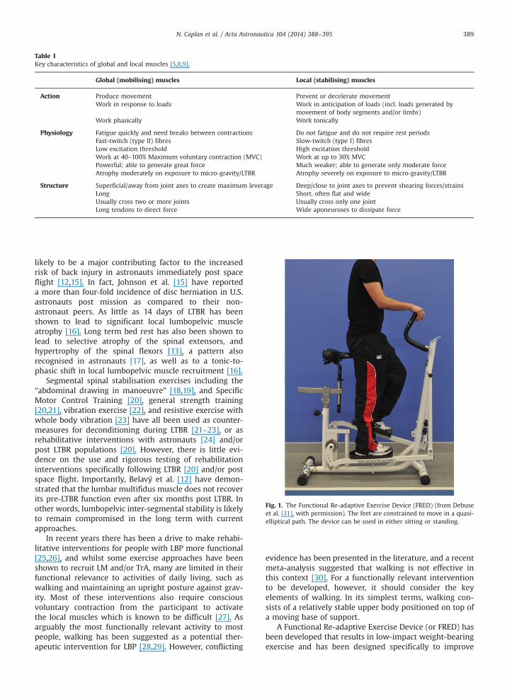
Table 1Key characteristics of global and local muscles [5,8,9].
Global (mobilising) muscles Local (stabilising) muscles
Action Produce movement Prevent or decelerate movementWork in response to loads Work in anticipation of loads (incl. loads generated by
movement of body segments and/or limbs)Work phasically Work tonically
Physiology Fatigue quickly and need breaks between contractions Do not fatigue and do not require rest periodsFast-twitch (type II) fibres Slow-twitch (type I) fibresLow excitation threshold High excitation thresholdWork at 40–100% Maximum voluntary contraction (MVC) Work at up to 30% MVCPowerful; able to generate great force Much weaker; able to generate only moderate forceAtrophy moderately on exposure to micro-gravity/LTBR Atrophy severely on exposure to micro-gravity/LTBR
Structure Superficial/away from joint axes to create maximum leverage Deep/close to joint axes to prevent shearing forces/strainsLong Short, often flat and wideUsually cross two or more joints Usually cross only one jointLong tendons to direct force Wide aponeuroses to dissipate force
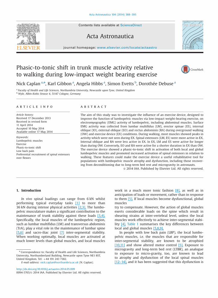
Fig. 1. The Functional Re-adaptive Exercise Device (FRED) (from Debuseet al. [31], with permission). The feet are constrained to move in a quasi-elliptical path. The device can be used in either sitting or standing.
N. Caplan et al. / Acta Astronautica 104 (2014) 388–395 389
likely to be a major contributing factor to the increasedrisk of back injury in astronauts immediately post spaceflight [12,15]. In fact, Johnson et al. [15] have reporteda more than four-fold incidence of disc herniation in U.S.astronauts post mission as compared to their non-astronaut peers. As little as 14 days of LTBR has beenshown to lead to significant local lumbopelvic muscleatrophy [16]. Long term bed rest has also been shown tolead to selective atrophy of the spinal extensors, andhypertrophy of the spinal flexors [13], a pattern alsorecognised in astronauts [17], as well as to a tonic-to-phasic shift in local lumbopelvic muscle recruitment [16].
Segmental spinal stabilisation exercises including the“abdominal drawing in manoeuvre” [18,19], and SpecificMotor Control Training [20], general strength training[20,21], vibration exercise [22], and resistive exercise withwhole body vibration [23] have all been used as counter-measures for deconditioning during LTBR [21–23], or asrehabilitative interventions with astronauts [24] and/orpost LTBR populations [20]. However, there is little evi-dence on the use and rigorous testing of rehabilitationinterventions specifically following LTBR [20] and/or postspace flight. Importantly, Belavý et al. [12] have demon-strated that the lumbar multifidus muscle does not recoverits pre-LTBR function even after six months post LTBR. Inother words, lumbopelvic inter-segmental stability is likelyto remain compromised in the long term with currentapproaches.
In recent years there has been a drive to make rehabi-litative interventions for people with LBP more functional[25,26], and whilst some exercise approaches have beenshown to recruit LM and/or TrA, many are limited in theirfunctional relevance to activities of daily living, such aswalking and maintaining an upright posture against grav-ity. Most of these interventions also require consciousvoluntary contraction from the participant to activatethe local muscles which is known to be difficult [27]. Asarguably the most functionally relevant activity to mostpeople, walking has been suggested as a potential ther-apeutic intervention for LBP [28,29]. However, conflicting
evidence has been presented in the literature, and a recentmeta-analysis suggested that walking is not effective inthis context [30]. For a functionally relevant interventionto be developed, however, it should consider the keyelements of walking. In its simplest terms, walking con-sists of a relatively stable upper body positioned on top ofa moving base of support.
A Functional Re-adaptive Exercise Device (or FRED) hasbeen developed that results in low-impact weight-bearingexercise and has been designed specifically to improve
N. Caplan et al. / Acta Astronautica 104 (2014) 388–395390
the function of the local lumbo-pelvic and lower limbanti-gravity muscles (see Fig. 1). The absence of externalresistance to foot motion, and therefore, an unstable baseof support, places a much greater demand on balance,coordination and motor control than conventional exercisedevices. As a result, the local lumbo-pelvic muscles arerecruited during FRED exercise to stabilise the spineduring exercise [31]. The primary aim of FRED exercise isnot to strengthen muscles, but to improve users' motorcontrol.
Debuse et al. [31] demonstrated that exercising on thedevice recruited both LM and TrA automatically, i.e. with-out a conscious/voluntary contraction. The exercise devicewas also compared to overground walking using threedimensional motion analysis and was shown to achieve amore stable lumbopelvic region, through reduced axialrotation of the spine, and to increase anterior pelvic tilt[32]. This more stable lumbopelvic region could be indi-cative of more tonic muscle activity which has been shownpreviously to be a key characteristic for training localmuscles [8]. However, the influence of the exercise deviceon lumbopelvic muscle activity throughout a completefoot movement cycle, or in comparison to walking, has notyet been determined.
The aims of this investigation were to determine (1)lumbopelvic muscle recruitment over a complete footmovement cycle, and (2) differences in lumbopelvic mus-cle activity between overground walking and when usingthe exercise device.
2. Methods
2.1. Participants
Fifteen male participants took part in this study. Theyhad a mean (7SD) age, height and mass of 24.93 (73.92)years, 1.78 (70.05) m, and 83.03 (77.21) kg, respectively. Allparticipants were free from injury and non-symptomaticat the time of the study. The study was approved by theInstitutional Review Board and all participants gave writteninformed consent to take part. The surface EMG data werecollected during the same study as the kinematic datapresented by Gibbon et al. [32].
2.2. Protocol
Participants attended a single session, during whichthey completed trials in both overground walking (OW)and exercise device (EX) conditions. The order of condi-tions was randomised to avoid any systematic effects onthe data. For OW, participants were asked to walk at theirpreferred speed along a walkway. Sufficient trials werecompleted to ensure that six successful trials were avail-able for analysis. A trial was considered successful whenparticipants landed with their right foot striking the first oftwo force platforms without evidence of targeting.
In EX, participants were given a five minute period tofamiliarise themselves with the exercise device. They wereinstructed to keep their feet in contact with the foot platesat all times, and to move their feet at their preferred speed,ensuring that foot movement was slow and controlled.
Participants were instructed to keep their upper body asstill as possible and to remain in an upright posturethroughout. Each participant completed a single 30 s EXtrial in order to obtain six complete movement cycles. Amovement cycle was defined as the feet completing awhole revolution of the movement cycle starting andending with their right foot at the furthest point forward.
2.3. Equipment
For OW trials, identification of heel strike and toe offevents was aided by two force platforms (OR6–7, AMTI,Watertown) within the walkway in the centre of thelaboratory. Signals were amplified (gain¼1000, MSA-6,AMTI, Watertown), and sampled at 2000 Hz by a dataacquisition card (MX, Vicon Motion Systems, Oxford).
For EX trials, 3D trajectories of retro-reflective markers(∅¼14 mm) placed on the side of the right foot plate weretracked and sampled at 200 Hz using a 12 camera motioncapture system (MX T20, Vicon Motion Systems, Oxford,UK) to aid the demarcation of key cycle events. The startand end of each movement cycle were determined by themost anterior and posterior positions of these markers.Rearward movement of the foot plate (i.e. when the footmoved backwards under the body) was considered com-parable to the stance phase of the walking gait cycle,whereas forward movement of the foot plate (i.e. when thefoot moved forwards under the body) was consideredcomparable to the swing phase of the walking gait cycle.
Surface EMG data were collected using surface electro-des from lumbar multifidus (LM), erector spinae (ES),internal oblique (IO), external oblique (EO) and lowerrectus abdominis (RA), placed unilaterally on the rightside of the body. Electrode sites were shaved to removebody hair, rubbed using abrasive gel, and cleaned withalcohol swabs. Pre-gelled self-adhesive circular electrodes(Ag/AgCl, diameter¼34 mm, sensing area¼13.2 mm2, mea-surement area¼154 mm2, Blue Sensor S, Ambu, Ballerup,Denmark) were placed in a bipolar configuration in accor-dance with existing protocols [33,34] with an inter-electrodedistance of 20 mm. Although potential for cross talk exists ineach EMG signal, previous investigations using the sameelectrode placement have shown this to be insignificant forthe anterior abdominal muscles [3,35–37] and between thespinal extensors [35].
Raw signals from each muscle were pre-amplified(gain¼1000, common mode rejection ratio 4100 dB) andpassed telemetrically to a data receiver (Myon RFTD-E16,Myon AG, Baar, Switzerland) before being amplified andsampled at 2000 Hz by a data acquisition card (MX, ViconMotion Systems, Oxford, UK). All data were collected simul-taneously and stored in specialist software (Nexus 1.7, ViconMotion Systems, Oxford) for subsequent analysis.
2.4. Data analysis
Myoelectric data for each muscle were processedwithin Nexus (1.7, Vicon Motion Systems, Oxford), usingan EMG plugin (ProEMG, Pro Physics AG, Zurich). Allchannels were band-pass filtered (Butterworth 2nd order,10–350 Hz), smoothed using root mean square (RMS)
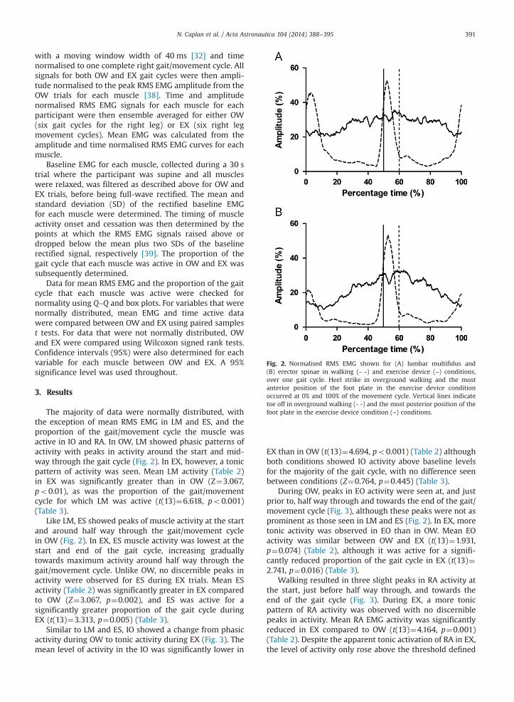
Fig. 2. Normalised RMS EMG shown for (A) lumbar multifidus and(B) erector spinae in walking (- -) and exercise device (–) conditions,over one gait cycle. Heel strike in overground walking and the mostanterior position of the foot plate in the exercise device conditionoccurred at 0% and 100% of the movement cycle. Vertical lines indicatetoe off in overground walking (- -) and the most posterior position of thefoot plate in the exercise device condition (–) conditions.
N. Caplan et al. / Acta Astronautica 104 (2014) 388–395 391
with a moving window width of 40 ms [32] and timenormalised to one complete right gait/movement cycle. Allsignals for both OW and EX gait cycles were then ampli-tude normalised to the peak RMS EMG amplitude from theOW trials for each muscle [38]. Time and amplitudenormalised RMS EMG signals for each muscle for eachparticipant were then ensemble averaged for either OW(six gait cycles for the right leg) or EX (six right legmovement cycles). Mean EMG was calculated from theamplitude and time normalised RMS EMG curves for eachmuscle.
Baseline EMG for each muscle, collected during a 30 strial where the participant was supine and all muscleswere relaxed, was filtered as described above for OW andEX trials, before being full-wave rectified. The mean andstandard deviation (SD) of the rectified baseline EMGfor each muscle were determined. The timing of muscleactivity onset and cessation was then determined by thepoints at which the RMS EMG signals raised above ordropped below the mean plus two SDs of the baselinerectified signal, respectively [39]. The proportion of thegait cycle that each muscle was active in OW and EX wassubsequently determined.
Data for mean RMS EMG and the proportion of the gaitcycle that each muscle was active were checked fornormality using Q–Q and box plots. For variables that werenormally distributed, mean EMG and time active datawere compared between OW and EX using paired samplest tests. For data that were not normally distributed, OWand EX were compared using Wilcoxon signed rank tests.Confidence intervals (95%) were also determined for eachvariable for each muscle between OW and EX. A 95%significance level was used throughout.
3. Results
The majority of data were normally distributed, withthe exception of mean RMS EMG in LM and ES, and theproportion of the gait/movement cycle the muscle wasactive in IO and RA. In OW, LM showed phasic patterns ofactivity with peaks in activity around the start and mid-way through the gait cycle (Fig. 2). In EX, however, a tonicpattern of activity was seen. Mean LM activity (Table 2)in EX was significantly greater than in OW (Z¼3.067,po0.01), as was the proportion of the gait/movementcycle for which LM was active (t(13)¼6.618, po0.001)(Table 3).
Like LM, ES showed peaks of muscle activity at the startand around half way through the gait/movement cyclein OW (Fig. 2). In EX, ES muscle activity was lowest at thestart and end of the gait cycle, increasing graduallytowards maximum activity around half way through thegait/movement cycle. Unlike OW, no discernible peaks inactivity were observed for ES during EX trials. Mean ESactivity (Table 2) was significantly greater in EX comparedto OW (Z¼3.067, p¼0.002), and ES was active for asignificantly greater proportion of the gait cycle duringEX (t(13)¼3.313, p¼0.005) (Table 3).
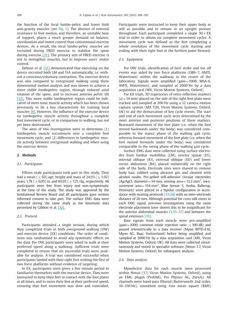
Similar to LM and ES, IO showed a change from phasicactivity during OW to tonic activity during EX (Fig. 3). Themean level of activity in the IO was significantly lower in
EX than in OW (t(13)¼4.694, po0.001) (Table 2) althoughboth conditions showed IO activity above baseline levelsfor the majority of the gait cycle, with no difference seenbetween conditions (Z¼0.764, p¼0.445) (Table 3).
During OW, peaks in EO activity were seen at, and justprior to, half way through and towards the end of the gait/movement cycle (Fig. 3), although these peaks were not asprominent as those seen in LM and ES (Fig. 2). In EX, moretonic activity was observed in EO than in OW. Mean EOactivity was similar between OW and EX (t(13)¼1.931,p¼0.074) (Table 2), although it was active for a signifi-cantly reduced proportion of the gait cycle in EX (t(13)¼2.741, p¼0.016) (Table 3).
Walking resulted in three slight peaks in RA activity atthe start, just before half way through, and towards theend of the gait cycle (Fig. 3). During EX, a more tonicpattern of RA activity was observed with no discerniblepeaks in activity. Mean RA EMG activity was significantlyreduced in EX compared to OW (t(13)¼4.164, p¼0.001)(Table 2). Despite the apparent tonic activation of RA in EX,the level of activity only rose above the threshold defined
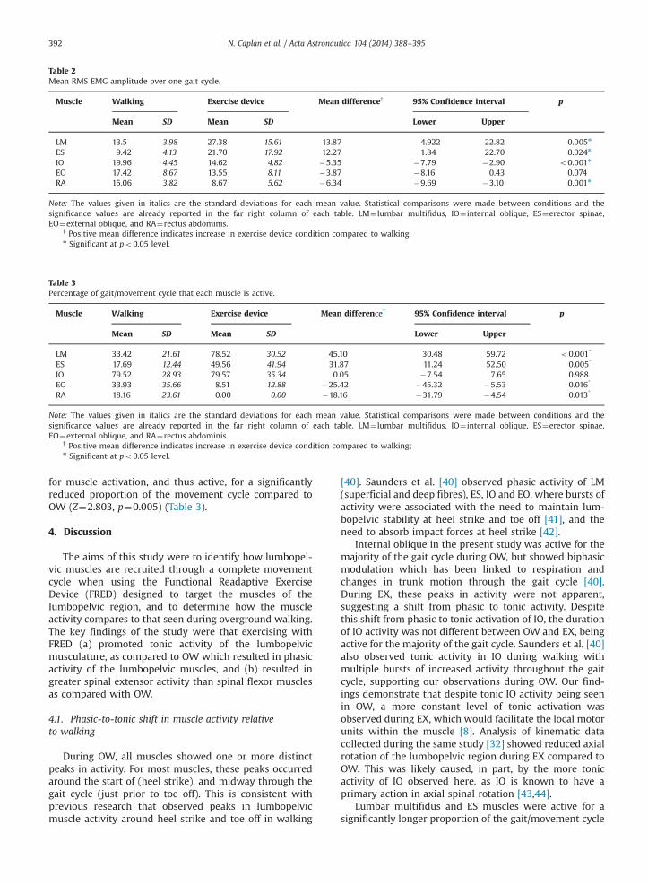
Table 2Mean RMS EMG amplitude over one gait cycle.
Muscle Walking Exercise device Mean difference† 95% Confidence interval p
Mean SD Mean SD Lower Upper
LM 13.5 3.98 27.38 15.61 13.87 4.922 22.82 0.005n
ES 9.42 4.13 21.70 17.92 12.27 1.84 22.70 0.024n
IO 19.96 4.45 14.62 4.82 �5.35 �7.79 �2.90 o0.001n
EO 17.42 8.67 13.55 8.11 �3.87 �8.16 0.43 0.074RA 15.06 3.82 8.67 5.62 �6.34 �9.69 �3.10 0.001n
Note: The values given in italics are the standard deviations for each mean value. Statistical comparisons were made between conditions and thesignificance values are already reported in the far right column of each table. LM¼ lumbar multifidus, IO¼ internal oblique, ES¼erector spinae,EO¼external oblique, and RA¼rectus abdominis.
† Positive mean difference indicates increase in exercise device condition compared to walking.n Significant at po0.05 level.
Table 3Percentage of gait/movement cycle that each muscle is active.
Muscle Walking Exercise device Mean difference† 95% Confidence interval p
Mean SD Mean SD Lower Upper
LM 33.42 21.61 78.52 30.52 45.10 30.48 59.72 o0.001*
ES 17.69 12.44 49.56 41.94 31.87 11.24 52.50 0.005*
IO 79.52 28.93 79.57 35.34 0.05 �7.54 7.65 0.988EO 33.93 35.66 8.51 12.88 �25.42 �45.32 �5.53 0.016*
RA 18.16 23.61 0.00 0.00 �18.16 �31.79 �4.54 0.013*
Note: The values given in italics are the standard deviations for each mean value. Statistical comparisons were made between conditions and thesignificance values are already reported in the far right column of each table. LM¼ lumbar multifidus, IO¼ internal oblique, ES¼erector spinae,EO¼external oblique, and RA¼rectus abdominis.
† Positive mean difference indicates increase in exercise device condition compared to walking;n Significant at po0.05 level.
N. Caplan et al. / Acta Astronautica 104 (2014) 388–395392
for muscle activation, and thus active, for a significantlyreduced proportion of the movement cycle compared toOW (Z¼2.803, p¼0.005) (Table 3).
4. Discussion
The aims of this study were to identify how lumbopel-vic muscles are recruited through a complete movementcycle when using the Functional Readaptive ExerciseDevice (FRED) designed to target the muscles of thelumbopelvic region, and to determine how the muscleactivity compares to that seen during overground walking.The key findings of the study were that exercising withFRED (a) promoted tonic activity of the lumbopelvicmusculature, as compared to OW which resulted in phasicactivity of the lumbopelvic muscles, and (b) resulted ingreater spinal extensor activity than spinal flexor musclesas compared with OW.
4.1. Phasic-to-tonic shift in muscle activity relativeto walking
During OW, all muscles showed one or more distinctpeaks in activity. For most muscles, these peaks occurredaround the start of (heel strike), and midway through thegait cycle (just prior to toe off). This is consistent withprevious research that observed peaks in lumbopelvicmuscle activity around heel strike and toe off in walking
[40]. Saunders et al. [40] observed phasic activity of LM(superficial and deep fibres), ES, IO and EO, where bursts ofactivity were associated with the need to maintain lum-bopelvic stability at heel strike and toe off [41], and theneed to absorb impact forces at heel strike [42].
Internal oblique in the present study was active for themajority of the gait cycle during OW, but showed biphasicmodulation which has been linked to respiration andchanges in trunk motion through the gait cycle [40].During EX, these peaks in activity were not apparent,suggesting a shift from phasic to tonic activity. Despitethis shift from phasic to tonic activation of IO, the durationof IO activity was not different between OW and EX, beingactive for the majority of the gait cycle. Saunders et al. [40]also observed tonic activity in IO during walking withmultiple bursts of increased activity throughout the gaitcycle, supporting our observations during OW. Our find-ings demonstrate that despite tonic IO activity being seenin OW, a more constant level of tonic activation wasobserved during EX, which would facilitate the local motorunits within the muscle [8]. Analysis of kinematic datacollected during the same study [32] showed reduced axialrotation of the lumbopelvic region during EX compared toOW. This was likely caused, in part, by the more tonicactivity of IO observed here, as IO is known to have aprimary action in axial spinal rotation [43,44].
Lumbar multifidus and ES muscles were active for asignificantly longer proportion of the gait/movement cycle
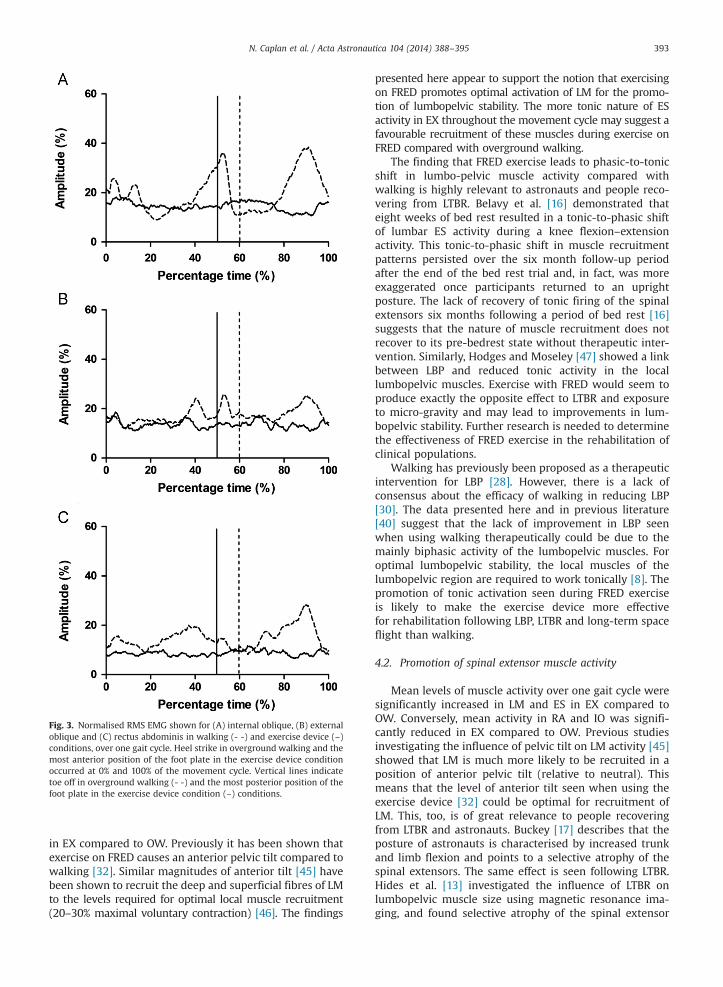
Fig. 3. Normalised RMS EMG shown for (A) internal oblique, (B) externaloblique and (C) rectus abdominis in walking (- -) and exercise device (–)conditions, over one gait cycle. Heel strike in overground walking and themost anterior position of the foot plate in the exercise device conditionoccurred at 0% and 100% of the movement cycle. Vertical lines indicatetoe off in overground walking (- -) and the most posterior position of thefoot plate in the exercise device condition (–) conditions.
N. Caplan et al. / Acta Astronautica 104 (2014) 388–395 393
in EX compared to OW. Previously it has been shown thatexercise on FRED causes an anterior pelvic tilt compared towalking [32]. Similar magnitudes of anterior tilt [45] havebeen shown to recruit the deep and superficial fibres of LMto the levels required for optimal local muscle recruitment(20–30% maximal voluntary contraction) [46]. The findings
presented here appear to support the notion that exercisingon FRED promotes optimal activation of LM for the promo-tion of lumbopelvic stability. The more tonic nature of ESactivity in EX throughout the movement cycle may suggest afavourable recruitment of these muscles during exercise onFRED compared with overground walking.
The finding that FRED exercise leads to phasic-to-tonicshift in lumbo-pelvic muscle activity compared withwalking is highly relevant to astronauts and people reco-vering from LTBR. Belavy et al. [16] demonstrated thateight weeks of bed rest resulted in a tonic-to-phasic shiftof lumbar ES activity during a knee flexion–extensionactivity. This tonic-to-phasic shift in muscle recruitmentpatterns persisted over the six month follow-up periodafter the end of the bed rest trial and, in fact, was moreexaggerated once participants returned to an uprightposture. The lack of recovery of tonic firing of the spinalextensors six months following a period of bed rest [16]suggests that the nature of muscle recruitment does notrecover to its pre-bedrest state without therapeutic inter-vention. Similarly, Hodges and Moseley [47] showed a linkbetween LBP and reduced tonic activity in the locallumbopelvic muscles. Exercise with FRED would seem toproduce exactly the opposite effect to LTBR and exposureto micro-gravity and may lead to improvements in lum-bopelvic stability. Further research is needed to determinethe effectiveness of FRED exercise in the rehabilitation ofclinical populations.
Walking has previously been proposed as a therapeuticintervention for LBP [28]. However, there is a lack ofconsensus about the efficacy of walking in reducing LBP[30]. The data presented here and in previous literature[40] suggest that the lack of improvement in LBP seenwhen using walking therapeutically could be due to themainly biphasic activity of the lumbopelvic muscles. Foroptimal lumbopelvic stability, the local muscles of thelumbopelvic region are required to work tonically [8]. Thepromotion of tonic activation seen during FRED exerciseis likely to make the exercise device more effectivefor rehabilitation following LBP, LTBR and long-term spaceflight than walking.
4.2. Promotion of spinal extensor muscle activity
Mean levels of muscle activity over one gait cycle weresignificantly increased in LM and ES in EX compared toOW. Conversely, mean activity in RA and IO was signifi-cantly reduced in EX compared to OW. Previous studiesinvestigating the influence of pelvic tilt on LM activity [45]showed that LM is much more likely to be recruited in aposition of anterior pelvic tilt (relative to neutral). Thismeans that the level of anterior tilt seen when using theexercise device [32] could be optimal for recruitment ofLM. This, too, is of great relevance to people recoveringfrom LTBR and astronauts. Buckey [17] describes that theposture of astronauts is characterised by increased trunkand limb flexion and points to a selective atrophy of thespinal extensors. The same effect is seen following LTBR.Hides et al. [13] investigated the influence of LTBR onlumbopelvic muscle size using magnetic resonance ima-ging, and found selective atrophy of the spinal extensor
N. Caplan et al. / Acta Astronautica 104 (2014) 388–395394
muscles, in particular LM. In fact, the reduction in LM crosssectional area seen after LTBR is similar to its responseto LBP [48,49]. The spinal flexor muscles including psoas,external oblique and rectus abdominis, however, werefound to increase their cross sectional area following LTBR[13]. The subsequent imbalance between flexor and exten-sor size and function could impact upon the ability of thelumbopelvic muscles to maintain spinal stability, which islikely to contribute to an increased risk of low back painfollowing LTBR [16]. What is likely to compound theproblem further is that LM is not only atrophied followingLTBR and long-term space flight [24], it is also in astretched position [23] which inhibits it from beingrecruited at all [9]. The fact that exercise on FRED resultsin a lumbo-pelvic position that is particularly favourableto effective LM recruitment and activity [45] in non-symptomatic volunteers, may offer an advantage in thisrespect.
In an attempt to address the clear need to prevent thespinal extensor muscle atrophy seen in LTBR, Belavý et al.[23] investigated the use of a combined vibration andresistive exercise countermeasure. Changes in lumbopelvicmuscle cross sectional area were assessed during 8 weeksof LTBR and for six months following return to an uprightposture. Significant atrophy of LM was observed whichwas reduced, but not eradicated, through the use ofresistive vibration exercise. Importantly, ES had recoveredand, in fact, improved on, its pre-LTBR state by 28 daysfollowing the end of LTBR, while LM did not recover itspre-LTBR cross sectional area even after six months [23].This points to very different rehabilitation requirementsbetween the two muscles which have not been addressedadequately in the literature to date.
The extensor–flexor imbalance of the lumbopelvicmusculature reported in both people following LTBR, andpeople with LBP highlights the need for a rehabilitativetool that is able to address the atrophy of the spinalextensors in order to counteract any increase in the sizeof the spinal flexors. Currently, most therapeutic interven-tions evaluated during LTBR and LBP studies lack func-tional relevance to activities of daily living. Also, to date,muscle cross sectional area and/or thickness determinedby ultrasound imaging (e.g. [10,11]) or MRI (e.g. [10,18])has been studied, but not the type of muscle activity (i.e.tonic or phasic), or whether the local lumbo-pelvic mus-cles have regained their anticipatory action.
4.3. Greater increase in LM than ES activity during EXcompared to OW
Our findings also show that the mean differencebetween EX and OW for LM was slightly greater for RMSEMG amplitude compared to ES (13.87% vs. 12.27%, respec-tively), and was notably greater for LM compared to ES forthe percentage of movement cycle the muscle was active(45.10% vs. 31.87%, respectively). While it was not possibleto investigate Transversus Abdominis (TrA) activity usingsurface EMG, Debuse et al. [31] previously showed sig-nificantly greater TrA activity during FRED exercise thanduring a range of control conditions. Together with thefindings for LM in this study, this may indicate that
exercise on FRED results in greater recruitment of localthan global lumbo-pelvic muscles, in general. Local musclesare responsible for segmental spinal stability [4–7], and thefact that there is greater atrophy of the local than globalmuscles following LTBR and exposure to micro-gravity[12–14] has been suggested as a reason for the four-foldincidence in disc prolapse in astronauts as compared to theirpeers. Particularly if a similar pattern of activation duringFRED exercise was to be found for TrA, this could point toFRED exercise being more effective at addressing local muscleatrophy in people with LBP, following LTBR, and astronautsthan conventional exercise approaches [50].
4.4. Limitations
In populations with LBP, following LTBR and followingexposure to micro-gravity, the local lumbopelvic musclesare atrophied and dysfunctional. Transversus abdominisand lumbar multifidus are the most widely studied, andlikely the most important muscles in this context. How-ever, as TrA is situated deep within the anterolateralabdominal wall, it cannot be studied with surface EMG,and its activity could, therefore, not be examined inthis study.
Despite previous reports of insignificant electrical crosstalk affecting the EMG signals measured here [3,35–37],there is still the possibility that the signals could have beeninfluenced by some cross talk. Research using indwellingEMG electrodes is warranted in order to fully validate thefindings presented here, as well as to identify the con-tribution TrA makes to the maintenance of spinal stabilitywhen using the Functional Readaptive Exercise Device.
5. Conclusion
This study has demonstrated that using the FunctionalReadaptive Exercise Device (FRED) leads to more tonicactivation of lumbopelvic muscles, in general, comparedto walking. In addition, levels of muscle activity duringexercise on FRED were increased in the spinal extensormuscles and were reduced in the spinal flexors. The factthat immediate exposure to FRED exercise results in aphasic-to-tonic shift in overall muscle activation whencompared to overground walking and in a preferentialactivation of spinal extensors over the spinal flexors, ascompared to overground walking suggests that the newFRED exercise device could be a highly effective tool foruse in rehabilitation of people following LTBR, in thosewith LBP and potentially in astronauts returning fromlong-term space missions. Further research is needed toevaluate the effectiveness of FRED exercise in restoring theextensor–flexor imbalance of the lumbo-pelvic muscula-ture in these populations.
Sources of funding
No funding was received in support of this study.
N. Caplan et al. / Acta Astronautica 104 (2014) 388–395 395
References
[1] S.M. McGill, R.W. Norman, Partitioning of the L4–L5 dynamicmoment into disc, ligamentous, and muscular components duringlifting, Spine 11 (1986) 666–678.
[2] H. Granhed, R. Jonson, T. Hansson, The loads on the lumbar spineduring extreme weight lifting, Spine 12 (1987) 146–149.
[3] J. Cholewicki, S.M. McGill, Mechanical stability of the in vivo lumbarspine: implications for injury and chronic low back pain, Clin.Biomech. 11 (1996) 1–15.
[4] M.M. Panjabi, The stabilizing system of the spine. Part I. Function,dysfunction, adaptation, and enhancement, J. Spinal Disord. 5 (1992)383–389.
[5] P.W. Hodges, C.A. Richardson, Inefficient muscular stabilizationof the lumbar spine associated with low back pain. A motor controlevaluation of transversus abdominis, Spine 21 (1996) 2640–2650.
[6] P.W. Hodges, Is there a role for transversus abdominis in lumbo-pelvic stability? Man. Ther. 4 (1999) 74–86.
[7] C.A. Richardson, C.J. Snijders, J.A. Hides, L. Damen, M.S. Pas,J. Storm, The relation between the transversus abdominis muscles,sacroiliac joint mechanics, and low back pain, Spine 27 (2002) 399–405.
[8] C.A. Richardson, G. Jull, Muscle control – pain control. Whatexercises would you prescribe? Man. Ther. 1 (1995) 2–10.
[9] M.J. Comerford, S.L. Mottram, Movement and stability dysfunction –
contemporary developments, Man. Ther. 6 (2001) 15–26.[10] J.A. Hides, W.R. Stanton, S. McMahon, K. Sims, C.A. Richardson, Effect
of stabilization training on multifidus muscle cross-sectional areaamong young elite cricketers with low back pain, J. Orthop. SportsPhys. Ther. 38 (2008) 101–108.
[11] T.L. Wallwork, W.R. Stanton, M. Freke, J.A. Hides, The effect of chroniclow back pain on size and contraction of the lumbar multifidusmuscle, Man. Ther. 14 (2009) 496–500.
[12] D.L. Belavý, G. Armbrecht, C.A. Richardson, D. Felsenberg, J.A. Hides,Muscle atrophy and changes in spinal morphology: is the lumbarspine vulnerable after prolonged bed-rest? Spine 36 (2011) 137–145.
[13] J.A. Hides, D.L. Belavý, W. Stanton, S.J. Wilson, J. Rittweger,D. Felsenberg, et al., Magnetic resonance imaging assessment of trunkmuscles during prolonged bed rest, Spine 32 (2007) 1687–1692.
[14] J.V. Sayson, J. Lotz, S. Parazynski, A.R. Hargens, Back pain in spaceand post-flight spine injury: mechanisms and countermeasuredevelopment, Acta Astronaut. 86 (2013) 24–38.
[15] S.L. Johnson, M. Campbell, R. Scheuring, A.H. Feiveson, Risk ofherniated nucleus pulposus among U.S. astronauts, Aviat. SpaceEnviron. Med. 81 (2010) 566–574.
[16] D.L. Belavy, C.A. Richardson, S.J. Wilson, D. Felsenberg, Tonic-to-phasic shift of lumbo-pelvic muscle activity during 8 weeks of bedrest and 6-months follow up, J. Appl. Physiol. 103 (2007) 48–54.
[17] J.C. Buckey, Space Physiology, Oxford University Press, Oxford, 2006.[18] J.A. Hides, S. Wilson, W. Stanton, S. McMahon, H. Keto, K. McMahon,
et al., An MRI investigation into the function of the transversusabdominis muscle during “drawing-in” of the abdominal wall, Spine31 (2006) E175–E178.
[19] J.A. Hides, T. Miokovic, D. Belavy, W.R. Stanton, C.A. Richardson,Ultrasound imaging assessment of abdominal muscle functionduring drawing-in of the abdominal wall: an intrarater reliabilitystudy, J. Orthop. Sports Phys. Ther. 37 (2007) 480–486.
[20] J.A. Hides, G. Lambrecht, C.A. Richardson, W.R. Stanton, G. Armbrecht,C. Pruett, et al., The effects of rehabilitation on the muscles of the trunkfollowing prolonged bed rest, Eur. Spine J. 20 (2011) 808–818.
[21] D.L. Belavý, H. Ohshima, M.-P. Bareille, J. Rittweger, D. Felsenberg,Limited effect of fly-wheel and spinal mobilization exercise counter-measures on lumbar spine deconditioning during 90 d bed-restin the Toulouse LTBR study, Acta Astronaut. 69 (2011) 406–419.
[22] J. Rittweger, K. Just, K. Kautzsch, P. Reeg, D. Felsenberg, Treatment ofchronic lower back pain with lumbar extension and whole-bodyvibration exercise: a randomized controlled trial, Spine 27 (2002)1829–1834.
[23] D.L. Belavý, J.A. Hides, S.J. Wilson, W. Stanton, F.C. Dimeo,J. Rittweger, et al., Resistive simulated weightbearing exercisewith whole body vibration reduces lumbar spine deconditioningin bed-rest, Spine 33 (2008) E121–E131.
[24] G. Lambrecht, Space-related neuromuscular control, Space ExerciseCountermeasures and Post-mission Rehabilitation Work, UK SpaceBiomedicine Association, Newcastle upon Tyne, UK, 2010.
[25] P.W. Hodges, J. Cholewicki, Functional control of the spine, in:R.S. Vleeming, V. Mooney (Eds.), Movement, Stability and Lumbo-pelvic Pain, Churchill Livingstone, Edinburgh, 2007.
[26] C.A. Richardson, J.A. Hides, Closed chain segmental control, in:C. Richardson, P. Hodges, J. Hides (Eds.), Therapeutic Exercise
for Lumbopelvic Stabilization, Churchill Livingstone, Edinburgh,2004.
[27] K. Van, J.A. Hides, C.A. Richardson, The use of real-time ultra-sound imaging for biofeedback of lumbar multifidus muscle con-traction in healthy subjects, J. Orthop. Sports Phys. Ther. 36 (2006)920–925.
[28] N.F. Taylor, O.M. Evans, P.A. Goldie, The effect of walking faster onpeople with acute low back pain, Eur. Spine J. 12 (2003) 166–172.
[29] D. Joffe, M. Watkins, L. Steiner, B.A. Pfeifer, Treadmill ambulationwith partial body weight support for the treatment of low back andleg pain, J. Orthop. Sports Phys. Ther. 32 (2002) 202–213.
[30] P. Hendrick, A.M. Te Wake, A.S. Tikkisetty, L. Wulff, C. Yap,S. Milosavljevic, The effectiveness of walking as an intervention forlow back pain: a systematic review, Eur. Spine J. 19 (2010) 1613–1620.
[31] D. Debuse, O. Birch, A. St Clair Gibson, N. Caplan, Low impactweight-bearing exercise in an upright posture increases the activa-tion of two key local muscles of the lumbo-pelvic region, Physiother.Theory Pract. 29 (2013) 51–60.
[32] K.C. Gibbon, D. Debuse, N. Caplan, Low impact weight-bearingexercise in an upright posture achieves greater lumbopelvic stabilitythan overground walking, J. Bodyw. Mov. Ther. 17 (2013) 462–468.
[33] H.J. Hermens, B. Freriks, C. Disselhorst-Klug, G. Rau, Development ofrecommendations for SEMG sensors and sensor placement proce-dures, J. Electromyogr. Kinesiol. 10 (2000) 361–374.
[34] K.J. Ng, V. Kippers, C.A. Richardson, Muscle fibre orientation ofabdominal muscles and suggested surface EMG electrode positions,Electromyogr. Clin. Neurophysiol. 38 (1998) 51–58.
[35] P.E. Vink, A. ven der Velde, A.J. Verbout, A functional subdivision ofthe lumbar extensor musculature: recruitment patterns and force–RA–EMG relationships under isometric conditions, Electromyogr.Clin. Neurophysiol. 28 (1987) 517–525.
[36] W.F. Floyd, P.H. Silver, Electromyographic study of patterns ofactivity of the anterior abdominal wall muscles in man, J. Anat. 84(1950) 132–145.
[37] J. Cholewicki, J.J. Van Vliet, Relative contribution of trunk muscles tothe stability of the lumbar spine during isometric exertions, Clin.Biomech. 17 (2002) 99–105.
[38] C. Stackhouse, P.A. Shewokis, S.R. Pierce, B. Smith, J. McCarthy,C. Tucker, Gait initiation in childrenwith cerebral palsy, Gait Posture26 (2007) 301–308.
[39] G. Morey-Klapsing, A. Arampatzis, G.P. Brüggemann, Choosing EMGparameters: comparison of different onset determination algo-rithms and EMG integrals in a joint stability study, Clin. Biomech.19 (2004) 196–201.
[40] S.W. Saunders, D. Rath, P.W. Hodges, Postural and respiratoryactivation of the trunk muscles changes with mode and speed oflocomotion, Gait Posture 20 (2004) 280–290.
[41] A. Thorstensson, H. Carlson, M.R. Zomlefer, J. Nilsson, Lumbar backmuscle activity in relation to trunk movements during locomotionin man, Acta Physiol. Scand. 116 (1982) 13–20.
[42] B. Jonsson, The functions of individual muscles in the lumbar part ofthe spinae muscle, Electromyography 10 (1970) 5–21.
[43] G.A. Dumas, M.J. Poulin, B. Roy, M. Gagnon, M. Jovanovic, Orientationand moment arms of some trunk muscles, Spine 16 (1991) 293–303.
[44] K.J. Ng, C.A. Richardson, M. Parnianpour, V. Kippers, EMG activity oftrunk muscles and torque output during isometric axial rotationexertion: a comparison between back pain patients and matchedcontrols, J. Orthop. Res. 20 (2002) 112–121.
[45] P.B. O’Sullivan, W. Dankaerts, A.F. Burnett, G.T. Farrell, E. Jefford,C.S. Naylor, et al., Effect of different upright sitting postures onspinal–pelvic curvature and trunk muscle activation in a pain-freepopulation, Spine 31 (2006) E707–E712.
[46] W.D. McArdle, F.I. Katch, V.L. Katch, Exercise Physiology: Energy,Nutrition, and Human Performance, 3rd ed. Lea & Febiger, Philadelphia,1991.
[47] P.W. Hodges, G.L. Moseley, Pain and motor control of the lumbo-pelvic region: effect and possible mechanisms, J. Electromyogr.Kinesiol. 13 (2003) 361–370.
[48] J.A. Hides, C.A. Richardson, G. Jull, Multifidus muscle recovery is notautomatic after resolution of acute first-episode low back pain,Spine (1996) 2763–2769.
[49] J.A. Hides, M.J. Stokes, M. Saide, G. Jull, D.H. Cooper, Evidence of lumbarmultifidus muscle wasting ipsilateral to symptoms in patients withacute/subacute low back pain, Spine 19 (1994) 165–172.
[50] S. Evetts, N. Caplan, D. Debuse, G. Lambrecht, V. Damann,N. Petersen, J.A. Hides, Post space mission lumbo-pelvic neuromus-cular rehabilitation: a European perspective, Aviat. Space Environ.Med. 85 (2014), http://dx.doi.org/10.3357/ASEM.3943.2014.
Related Documents