JOURNAL OF VIROLOGY, Feb. 2008, p. 1448–1457 Vol. 82, No. 3 0022-538X/08/$08.000 doi:10.1128/JVI.01409-07 Copyright © 2008, American Society for Microbiology. All Rights Reserved. Phase I Trial of a CD8 T-Cell Peptide Epitope-Based Vaccine for Infectious Mononucleosis Suzanne L. Elliott, 1 Andreas Suhrbier, 1 * John J. Miles, 1 Greg Lawrence, 1 Stephanie J. Pye, 1 Thuy T. Le, 1 Andrew Rosenstengel, 1 Tam Nguyen, 1 Anthony Allworth, 2 Scott R. Burrows, 1 John Cox, 3 David Pye, 3 Denis J. Moss, 1 and Mandvi Bharadwaj 1 * Australian Centre for Vaccine Development, Queensland Institute of Medical Research, Brisbane, Australia 1 ; Infectious Disease Unit, Royal Brisbane Hospital, Brisbane, Australia 2 ; and CSL Limited, Melbourne, Australia 3 Received 28 June 2007/Accepted 17 October 2007 A single blind, randomized, placebo-controlled, single-center phase I clinical trial of a CD8 T-cell peptide epitope vaccine against infectious mononucleosis was conducted with 14 HLA B*0801-positive, Epstein-Barr virus (EBV)-seronegative adults. The vaccine comprised the HLA B*0801-restricted peptide epitope FLRGR AYGL and tetanus toxoid formulated in a water-in-oil adjuvant, Montanide ISA 720. FLRGRAYGL-specific responses were detected in 8/9 peptide-vaccine recipients and 0/4 placebo vaccine recipients by gamma interferon enzyme-linked immunospot assay and/or limiting-dilution analysis. The same T-cell receptor V CDR3 sequence that is found in FLRGRAYGL-specific T cells from most EBV-seropositive individuals could also be detected in the peripheral blood of vaccine recipients. The vaccine was well tolerated, with the main side effect being mild to moderate injection site reactions. After a 2- to 12-year follow-up, 1/2 placebo vaccinees who acquired EBV developed infectious mononucleosis, whereas 4/4 vaccinees who acquired EBV after completing peptide vaccination seroconverted asymptomatically. Single-epitope vaccination did not predispose individuals to disease, nor did it significantly influence development of a normal repertoire of EBV-specific CD8 T-cell responses following seroconversion. Epstein-Barr Virus (EBV) is a human gammaherpesvirus that is usually transmitted orally during childhood and infects more than 90% of the world’s population. The virus establishes a lifelong latent infection in B cells and is controlled in healthy seropositive individuals by CD8 T lymphocytes that rec- ognize latent and lytic EBV antigens (38, 44). When infection with EBV is delayed until adolescence or adulthood, as is common in many Western countries, individuals have a 26 to 74% chance of developing glandular fever or infectious mono- nucleosis (IM) (19). IM is an acute, self-limiting disorder as- sociated with fatigue, fever, sore throat, and generalized lymphadenopathy and is characterized by a pronounced blood monocytosis. The severity of symptoms varies from mild flu- like symptoms for a few weeks to prolonged and debilitating disease lasting several months (19), and in rare cases the dis- ease can be fatal (59). IM caused by EBV has also been associated with increased risk of multiple sclerosis and Hodgkin’s lymphoma (26, 41). In Western countries the inci- dence of IM has been estimated to be 45/100,000 per annum in the general community. However, the incidence is much higher among adolescents (15 to 19 years), at 320 to 370/100,000 per annum (18), and thus often affects young people at a critical time in their studies. The disease is less common in developing countries, where 90% of people generally seroconvert asymptomatically to EBV during childhood. In Western coun- tries the number of children entering adolescence without be- ing infected with EBV has been estimated at 10 to 20%. How- ever, this figure may be increasing with improvements in living standards. In the Tokyo region this percentage has recently been estimated to have reached 50% by 2006 (57). Although ganciclovir has been used in severe cases and in immunocom- promised patients (1) and corticosteroids are sometimes used (15), there are currently no treatments recommended for rou- tine use in IM. The ability of many individuals’ immune systems to control primary EBV infections without IM has prompted efforts to- ward the design of an IM vaccine (38, 39). Two approaches have been taken. The first involves a vaccine based on the major surface glycoprotein of EBV, gp350. This vaccine was originally conceived to induce neutralizing antibodies; how- ever, antibody responses do not appear to correlate with pro- tection, with some evidence suggesting that gp350-specific CD4 T-cell responses might mediate protection (40). The second approach seeks to generate EBV-specific CD8 T cells that control the expansion of EBV-infected B cells after infection, thereby promoting asymptomatic seroconversion rather than preventing infection (38, 39). Such T cells are not only strongly implicated in controlling EBV in healthy individuals but have also been successfully used to treat EBV-associated posttrans- plant lymphoproliferative disease (PTLD) by adoptive transfer (29, 47, 49). Many EBV-specific CD8 T cells recognize epitopes from the EBV nuclear antigens (EBNAs) (44). How- ever, the association of these proteins with B-cell transforma- tion precludes, and their large size complicates, their use in recombinant protein-based vaccines (54). A CD8 T-cell epitope-based approach was thus pursued (39). Here we de- * Corresponding author. Mailing address for A. Suhrbier: Queens- land Institute of Medical Research, P.O. Royal Brisbane Hospital, Queensland 4029, Australia. Phone: 61-7-33620415. Fax: 61-7- 33620107. E-mail: [email protected]. Mailing address for M. Bharadwaj: Dept. of Microbiology and Immunology, University of Melbourne, Parkville, Victoria 3052, Australia. Phone: 61-3-83449911. Fax: 61-3-93471540. E-mail: [email protected]. Published ahead of print on 21 November 2007. 1448

Welcome message from author
This document is posted to help you gain knowledge. Please leave a comment to let me know what you think about it! Share it to your friends and learn new things together.
Transcript
JOURNAL OF VIROLOGY, Feb. 2008, p. 1448–1457 Vol. 82, No. 30022-538X/08/$08.00�0 doi:10.1128/JVI.01409-07Copyright © 2008, American Society for Microbiology. All Rights Reserved.
Phase I Trial of a CD8� T-Cell Peptide Epitope-Based Vaccine forInfectious Mononucleosis�
Suzanne L. Elliott,1 Andreas Suhrbier,1* John J. Miles,1 Greg Lawrence,1 Stephanie J. Pye,1Thuy T. Le,1 Andrew Rosenstengel,1 Tam Nguyen,1 Anthony Allworth,2 Scott R. Burrows,1
John Cox,3 David Pye,3 Denis J. Moss,1 and Mandvi Bharadwaj1*Australian Centre for Vaccine Development, Queensland Institute of Medical Research, Brisbane, Australia1; Infectious Disease Unit,
Royal Brisbane Hospital, Brisbane, Australia2; and CSL Limited, Melbourne, Australia3
Received 28 June 2007/Accepted 17 October 2007
A single blind, randomized, placebo-controlled, single-center phase I clinical trial of a CD8� T-cell peptideepitope vaccine against infectious mononucleosis was conducted with 14 HLA B*0801-positive, Epstein-Barrvirus (EBV)-seronegative adults. The vaccine comprised the HLA B*0801-restricted peptide epitope FLRGRAYGL and tetanus toxoid formulated in a water-in-oil adjuvant, Montanide ISA 720. FLRGRAYGL-specificresponses were detected in 8/9 peptide-vaccine recipients and 0/4 placebo vaccine recipients by gammainterferon enzyme-linked immunospot assay and/or limiting-dilution analysis. The same T-cell receptor V�CDR3 sequence that is found in FLRGRAYGL-specific T cells from most EBV-seropositive individuals couldalso be detected in the peripheral blood of vaccine recipients. The vaccine was well tolerated, with the main sideeffect being mild to moderate injection site reactions. After a 2- to 12-year follow-up, 1/2 placebo vaccinees whoacquired EBV developed infectious mononucleosis, whereas 4/4 vaccinees who acquired EBV after completingpeptide vaccination seroconverted asymptomatically. Single-epitope vaccination did not predispose individualsto disease, nor did it significantly influence development of a normal repertoire of EBV-specific CD8� T-cellresponses following seroconversion.
Epstein-Barr Virus (EBV) is a human gammaherpesvirusthat is usually transmitted orally during childhood and infectsmore than 90% of the world’s population. The virus establishesa lifelong latent infection in B cells and is controlled in healthyseropositive individuals by CD8� �� T lymphocytes that rec-ognize latent and lytic EBV antigens (38, 44). When infectionwith EBV is delayed until adolescence or adulthood, as iscommon in many Western countries, individuals have a 26 to74% chance of developing glandular fever or infectious mono-nucleosis (IM) (19). IM is an acute, self-limiting disorder as-sociated with fatigue, fever, sore throat, and generalizedlymphadenopathy and is characterized by a pronounced bloodmonocytosis. The severity of symptoms varies from mild flu-like symptoms for a few weeks to prolonged and debilitatingdisease lasting several months (19), and in rare cases the dis-ease can be fatal (59). IM caused by EBV has also beenassociated with increased risk of multiple sclerosis andHodgkin’s lymphoma (26, 41). In Western countries the inci-dence of IM has been estimated to be 45/100,000 per annum inthe general community. However, the incidence is much higheramong adolescents (15 to 19 years), at 320 to 370/100,000 perannum (18), and thus often affects young people at a criticaltime in their studies. The disease is less common in developingcountries, where �90% of people generally seroconvert
asymptomatically to EBV during childhood. In Western coun-tries the number of children entering adolescence without be-ing infected with EBV has been estimated at 10 to 20%. How-ever, this figure may be increasing with improvements in livingstandards. In the Tokyo region this percentage has recentlybeen estimated to have reached �50% by 2006 (57). Althoughganciclovir has been used in severe cases and in immunocom-promised patients (1) and corticosteroids are sometimes used(15), there are currently no treatments recommended for rou-tine use in IM.
The ability of many individuals’ immune systems to controlprimary EBV infections without IM has prompted efforts to-ward the design of an IM vaccine (38, 39). Two approacheshave been taken. The first involves a vaccine based on themajor surface glycoprotein of EBV, gp350. This vaccine wasoriginally conceived to induce neutralizing antibodies; how-ever, antibody responses do not appear to correlate with pro-tection, with some evidence suggesting that gp350-specific CD4T-cell responses might mediate protection (40). The secondapproach seeks to generate EBV-specific CD8� T cells thatcontrol the expansion of EBV-infected B cells after infection,thereby promoting asymptomatic seroconversion rather thanpreventing infection (38, 39). Such T cells are not only stronglyimplicated in controlling EBV in healthy individuals but havealso been successfully used to treat EBV-associated posttrans-plant lymphoproliferative disease (PTLD) by adoptive transfer(29, 47, 49). Many EBV-specific CD8� T cells recognizeepitopes from the EBV nuclear antigens (EBNAs) (44). How-ever, the association of these proteins with B-cell transforma-tion precludes, and their large size complicates, their use inrecombinant protein-based vaccines (54). A CD8� T-cellepitope-based approach was thus pursued (39). Here we de-
* Corresponding author. Mailing address for A. Suhrbier: Queens-land Institute of Medical Research, P.O. Royal Brisbane Hospital,Queensland 4029, Australia. Phone: 61-7-33620415. Fax: 61-7-33620107. E-mail: [email protected]. Mailing address for M.Bharadwaj: Dept. of Microbiology and Immunology, University ofMelbourne, Parkville, Victoria 3052, Australia. Phone: 61-3-83449911.Fax: 61-3-93471540. E-mail: [email protected].
� Published ahead of print on 21 November 2007.
1448
scribe the results of the first phase I CD8� T-cell epitope-based EBV vaccine trial. The vaccine comprised the HLAB*0801-restricted CD8� T-cell epitope FLRGRAYGL (FLR)from the latent antigen EBNA3 (9, 10) and tetanus toxoid (TT)as a source of CD4� T-cell help formulated in the water-in-oiladjuvant, Montanide ISA 720. This adjuvant has been success-fully used to induce peptide-epitope-specific CD8� T-cell re-sponses in mice (20, 48) and has been used in several humanvaccine trials (23, 36, 46, 62).
The primary aim of this trial was to establish that CD8�
T-cell epitope-based vaccination of EBV seronegative individ-uals was safe in the context of a primary EBV infection. IM ischaracterized by a high viral load and a pronounced lympho-cytosis dominated by EBV-specific T cells (13, 14, 24). Asymp-tomatic seroconversion does not appear to be associated witha lymphocytosis, despite significant viral loads, suggesting thatIM arises from an overreaction of T-cell responses (53).Whether a vaccine-induced EBV-specific memory T-cell re-sponse would subsequently expand following a primary infec-tion and thereby promote lymphocytosis and IM remains animportant question. In addition, since an FLR-based vaccinewould induce CD8� T-cell responses only to this singleepitope, it also remains to be established whether immu-nodominance and/or immunodomination effects (17, 50)would narrow the immune response following a subsequentprimary EBV infection to this epitope. Rapid recovery fromIM symptoms has been correlated with broad T-cell reactivityto multiple CD8� T-cell epitopes within lytic and latent anti-gens, whereas protracted illness was associated with a narrowlyfocused response (4). It has also been suggested that epitope-based vaccines might induce tolerance against the vaccineepitope and thus prevent a normal response to that epitopefrom developing (2, 61). This trial illustrates that single-epitope vaccination does not promote disease or predisposeindividuals to aberrant EBV-specific CD8� T-cell responseafter seroconversion, indicating that an epitope-based ap-proach for IM or PTLD vaccines is both feasible and safe.
MATERIALS AND METHODS
Study design. The single-blind, randomized, placebo-controlled phase I (safetyand immunogenicity) EBV vaccine trial was conducted at the Queensland Insti-tute of Medical Research (QIMR), Australia and was approved by The BancroftCentre Research Ethics Committee and conducted under the Australian Ther-apeutic Goods Administration CTN scheme. Volunteers were recruited fromQIMR and the University of Queensland and were screened for HLA B*0801status and EBV infection by serology. The initial HLA screening was performedby fluorescence-activated cell sorter (FACS) analysis using the HLA B*0801monoclonal antibody (clone 59HA-1; One Lambda Inc.) and was later confirmedby full class I tissue typing (Princess Alexandra Hospital tissue typing facility,Brisbane, Australia). Serological testing for EBV capsid antibody (VCA) wasinitially done by immunofluorescence microscopy (37) and later confirmed byenzyme-linked immunosorbent assay specific for VCA immunoglobulin G (IgG)and IgM and EBNA IgG (Queensland Medical Laboratory, Brisbane, Queens-land, Australia). The inclusion criteria for the trial were EBV-seronegative andHLA B*0801-positive status. To avoid adverse reactions to TT (27, 33), volun-teers were excluded if they had anti-TT titers of above 5 IU/ml (QueenslandMedical Laboratory) or a history of marked reaction to previous TT injections.Volunteers were also excluded if 2 weeks prior to vaccination (i) either full anddifferential blood counts, CD4/CD8 lymphocyte count, full blood biochemistry(including aspartate transaminase, alanine aminotransferase, gamma glutamyltransferase, urea, and creatinine), antinuclear factor, direct Coombs test, orurine protein was outside the normal range; (ii) females tested positive for serumor urine human chorionic gonadotropin; (iii) volunteers were seropositive forEBV, human immunodeficiency virus, or hepatitis B virus (tests performed by
Queensland Health Pathology Services, Royal Brisbane Hospital); or (iv) themedical history or physical examination revealed clinical abnormalities. Separateinformation sessions and consent forms were provided for screening and enroll-ment into the trial.
Fourteen healthy, EBV-seronegative, HLA B*0801-positive, 18- to 50-year-old volunteers (seven females and seven males) were enrolled in the trial. Theywere randomly assigned by a computer-generated list into vaccine (n � 10, with8 to be immunized with 5 �g and 2 with 50 �g peptide) and placebo (n � 4)groups (Table 1). Vaccine recipients were observed for 6 h following eachimmunization and contacted by telephone after 1 week. Clinical assessment wasperformed and blood was collected for immunological screening (including EBVserology and T-cell responses) before each injection and at 2, 4, 8, 10, and 12weeks and 6, 12, and 24 months after each vaccination. Two vaccinations wereoffered, the first at day 0 and the second at week 8. Where possible, volunteerswere also followed up after 8 to 12 years to test for EBV seroconversion andinquire whether they had suffered from IM since vaccination.
The EBV vaccine. The vaccine consisted of the synthetic peptide, FLR (9),mixed with TT in 5 mM phosphate buffer-isotonic saline and emulsified (3:7[wt/wt]) with the water-in-oil adjuvant Montanide ISA 720 (Seppic, Paris,France) (20). The FLR peptide was chemically synthesized under GMP condi-tions (Auspep Ltd.). Two peptide doses were tested, 5 and 50 �g per vaccinedose, with the placebo vaccine containing no peptide. The peptide and placebovaccines also contained 1.35 Lyme factor/ml (0.675 Lyme factor/dose) TT (CSLLtd.) and thiomersal (0.01% [wt/vol]) (CSL Ltd.). Stoppered glass vials werefilled with the vaccines under nitrogen by CSL Ltd. Volunteers were given 0.5 mlof vaccine per injection subcutaneously into the thigh.
Formal toxicology was undertaken by Pharmatox. Two 500-�l subcutaneous(day 1 and 15) injections of the vaccine (with 50 �g of peptide) given to five maleand five female rats and guinea pigs failed to show clinical signs of toxicity over29 days. Upon necroscopy, organ histology and blood hematology and biochem-istry were not clinically different from those of animals injected with saline. Thesame dose also passed pyrogenicity testing in three rabbits. The vaccine showedno activity in the Ames test or sister chromatid exchange assay. The peptide wasstable in the vaccine for 2 years at 4°C and was extracted from the vaccine byvigorous shaking with chloroform–0.1% trifluoroacetic acid in distilled water(1.5:1, vol/vol). The recovered peptide (in the aqueous phase) was analyzed byhigh-pressure liquid chromatography and was fully active in chromium releaseassays (9).
LCLs and PHA blasts. EBV-transformed lymphoblastoid cell lines (LCLs)were established from each vaccinee’s peripheral blood mononuclear cells(PBMC) by exogenous virus transformation of peripheral B cells using theQIMR Wil strain of virus (43). LCLs were maintained in medium comprisingRPMI 1640 (Gibco), 2 mM glutamine (ICN Biomed. Aust. Pty Ltd., Seven Hills,Australia), 100 IU/ml penicillin and 100 mg/ml streptomycin (CSL Ltd., Mel-bourne, Australia), and 10% fetal calf serum (QIMR). Phytohemagglutinin(PHA) blasts were generated and maintained as described previously (31).
Ex vivo ELISPOT assay. PBMC were separated by Ficoll density gradientcentrifugation and frozen in 10% dimethyl sulfoxide and 90% fetal calf serumusing the Cryo 1° freezing container (Nalgene). Gamma interferon (IFN-�)enzyme-linked immunospot (ELISPOT) assays were performed as describedbefore (5) using the FLR peptide. T-cell responses to the HLA B8-restrictedepitopes QAKWRLQTL (QAK) and RAKFKQLL (RAK), and the HLA A2-restricted epitopes LLDFVRFMGV, CLGGLLTMV, YLLEMLWRL, YLQQNWWTL, GLCTLVAML, and ILIYNGWYA were also assessed for the indicatedindividuals. Peptides for in vitro assays were synthesized by Chiron Mimotopes(Melbourne, Australia).
LDA. The limiting-dilution analysis (LDA) was performed as described pre-viously (30). Briefly, PBMC from vaccinees were distributed in twofold dilutionsfrom 6.25 103 to 5 104 cells per well in round-bottom microtiter plates andstimulated with irradiated (2,000 rads) FLR-sensitized autologous PBMC orirradiated (8,000 rads) autologous LCLs. On day 10, each of the 24 replicatemicrocultures for each dilution was split into two replicates and used as effectorsin a standard 5-h 51Cr release assay against autologous PHA blasts sensitizedwith and without peptide epitope. Wells were scored as positive when the percentspecific chromium release for peptide-sensitized target cells exceeded the meanrelease from control wells by 3 standard deviations. T-cell frequency was calcu-lated by the method of maximum-likelihood estimation.
Bulk cultures. Polyclonal T-cell effectors were generated from 2 106 PBMCstimulated (on day 0 and day 7) either with irradiated (8,000 rads) autologousLCLs (responder/stimulator ratio of 20:1) or with PBMC sensitized with syn-thetic peptides (10 �g/ml) (responder/stimulator ratio of 4:1). On day 10, thesebulk effectors were used in a standard 51Cr release assay against peptide-sensi-
VOL. 82, 2008 PHASE I TRIAL OF AN IM VACCINE 1449
tized autologous PHA blasts at a 20:1 effector-to-target ratio as described pre-viously (8).
T-cell receptor (TCR) sequencing. Total RNA was extracted from frozenPBMC or frozen bulk culture pellets containing 5 million cells by using TRIzolreagent (Invitrogen). The RNA pellets were resuspending in 20 �l of diethylpyrocarbonate-treated double-distilled water (ICN) and were each added to asingle-tube one-step reverse transcription-PCR (RT-PCR) system (Invitogen),with the addition of the TRBV7-8 family-specific forward primer (5-TGAAGCTCAAACTAGACAAATCG-3) and the FLR-specific reverse CDR3-2-7 prim-ers (5-CTGGTAGGCCTGBCCTAAGCTGCTGGC-3 and 5-AGTACTGCTCGTAGGCCTGBCC-3). The RT-PCR cycling was performed at 50°C for 30min and 94°C for 2 min. The PCR cycling was performed at 94°C for 15 s, 55°Cfor 30 s, and 68°C for 1 min for 40 cycles and at 68°C for 5 min for 1 cycle. ThePCR products were cloned into the pGEM-T vector system (Promega) andtransformed into DH5 cells (Invitrogen). White colonies (six to eight) werepicked and placed into a M13 miniscreen PCR instrument. The PCR productswere then sequenced with the ABI Prism Big Dye Terminator reaction kit(Applied Biosystems).
TCR RNA viability was evaluated in all samples with a total TCR constant-region RT-PCR with the addition of the TCR constant-region forward primerC�5 (5-CGTGTTCCCACCCGAGGTCGC-3) and the TCR constant-regionreverse primer C�B (5-ATTCACCCACCAGCTCAGCTCCACG-3) into theone-step RT-PCR system (Invitogen). The PCR cycling conditions were identicalto those for the FLR CDR3-specific PCR described above.
Major histocompatibility complex (MHC)-peptide multimer analysis. PBMCor T-cell lines were incubated for 30 min at 4°C with an HLA B*0801-FLR orHLA B*0801-RAK phycoerythrin-labeled pentamer (ProImmune, Oxford,United Kingdom). Cells were then washed and labeled for 30 min at 4°C withTri-color-labeled anti-human CD8 (Caltag, Burlingame, CA), allophycocyanin-labeled anti-human CD3 (BD Pharmingen, San Diego, CA), and one of fluores-cein isothiocyanate-labeled anti-human-CD27 (Caltag, Burlingame, CA), anti-human CD28 (BD Pharmingen, San Diego, CA), anti-human CD45RA(Beckman-Coulter, Fullerton, CA), anti-human CD45RO (Beckman-Coulter,Fullerton, CA), or anti-human CD62L (Caltag, Burlingame, CA). Cells werethen washed twice and analyzed on a four-color FACSCalibur using CellQuestsoftware (Becton Dickinson, Mountain View, CA).
RESULTS
Volunteer selection. A total of 1,075 resident students fromthe University of Queensland and staff members from QIMRwere screened within a span of 5 years (1994 to 1999), and 25%were shown to be EBV seronegative. Of these, 72 (6.7%) werefound to be EBV seronegative and HLA B*0801 positive dur-ing the initial screening. Ultimately 14 volunteers (1.5%) wereenrolled in the trial. The main reasons for exclusion wereanti-TT antibody titers of �5 IU/ml, EBV seroconversion priorto final enrollment, and subjects declining to continue.
Vaccination, adverse events, and seroconversion. Fourteenvolunteers were vaccinated, including eight with 5 �g peptide(volunteers designated #01, #02, #04, #05, #06, #07, #08,and #09), two with 50 �g peptide (#013 and #014), and fourwith placebo (#03, #010, #011, and #012). Three subjects(#07, #08, and #011) were not given the second vaccinationbecause of an increase in anti-TT titers beyond 5 IU/ml. Onesubject (#014) was not given the second vaccination due toEBV seroconversion. One subject (#09) declined the seconddose of vaccine because of the discomfort following the firstdose (Table 1). The vaccine was well tolerated in most volun-teers, and no serious adverse events were seen throughout thetrial period. Side effects were restricted to self-limiting, mild tomoderate injection site reactions, which are summarized inTable 2.
Two vaccinees (#04 and #08) were EBV seronegative at 52weeks but had seroconverted by week 104, and one vaccinee(#09) was seronegative at 12 weeks but had seroconverted byweek 26 (Table 1). These seroconversion events were asymp-tomatic, and no adverse events were experienced during these
TABLE 1. Vaccination summary for vaccine recipients, EBV seroconversion, and IM
Vaccine Vaccinerecipient
EBVseroconversion
(wk of test)IM or asymptomatic
EBV serology
VCAEBNA IgG
IgM IgG
Peptide5 �g #01 No (412) Negative Negative Negative
#02 Yes (628) Asymptomatic Negative Positive Positive#04 Yes (104) Asymptomaticd Low positive Moderate Negative#05 No (542) Negative Negative Negative#06 No (523) Negative Negative Negative#07a No (520) Negative Negative Negative#08a Yes (104) Asymptomaticd Negative Moderate Positive#09b Yes (26) Asymptomaticd Negative Moderate Negative
50 �g #13 No (421) Negative Negative Negative#14c Yes (8) ?d,e Positive Borderlinef Negative
Placebo #03 No (585) Negative Negative Negative#10 No (494) Negative Negative Negative#11a Yes (438) Asymptomatic Negative Borderlineg Not tested#12 Yes (392) IM, treating doctor notified
a Second immunization not given due to high anti-TT antibody titers.b Declined second immunization.c Second immunization not given due to EBV seroconversion.d Lymphocyte count within normal range at time of serology test.e Diagnosis unclear, possible mild IM or tonsillitis (see text).f Borderline. VCA IgM usually lasts for 1 to 2 months. Anti-EBNA IgG responses usually appear after 2 to 6 months.g FLR- and RAK-specific T-cell expansions seen by FACS (data not shown), confirming EBV-positive status.
1450 ELLIOTT ET AL. J. VIROL.
periods. A further vaccinee, #02, seroconverted betweenweeks 104 and 628 with no history of IM. One of the placebo-vaccinated volunteers (#12) was diagnosed with IM by theirgeneral practitioner using symptoms and serology at week 392(Table 1).
Vaccinee #14 was found to be EBV seropositive at 8 weeks(Table 1) and at that time had significant responses againstFLR (20/106 lymphocytes), RAK (480/106), and QAK 72/106)as measured by LDA. No responses were detected at weeks 2and 4 (data not shown). The incubation period for EBV hasbeen reported to be 38 days for one individual (56), whichsuggests that vaccinee 14 became infected within a few weeksof the first immunization. This vaccinee complained of a severesore throat and fatigue, which lasted about 2 to 3 weeks andbegan at about week 7. No time off work was taken, biochem-istry was normal, and no significant lymphocytosis was presentat week 4 or 8. At week 4 the CD4 and CD8 counts were 1.64 109 and 0.45 109/liter, respectively, and this changed to 0.89 109 and 1.77 109/liter at week 8. This clinical picture mayrepresent mild IM; however, this vaccinee had a history ofrepeated tonsillitis and was prescribed penicillin by her generalpractitioner. To try to determine whether vaccinee 14 had IM,interleukin-15 (IL-15) receptor status was determined, as thisreceptor has been reported to be permanently down regulatedin T cells from individuals who have had IM (45). However, incontrast to the findings of Sauce et al. (45), we found nodifferences in IL-15 receptor status between several EBV-se-
ronegative individuals, healthy EBV-seropositive individuals,and individuals recovered from IM (data not shown). Usingmonoclonal antibodies 151303 and 151307 (R&D Systems), 0.1to 4% of CD4� T cells and 3 to 15% of CD8� T cells wereIL-15 receptor positive in all groups and in vaccinee #14 (datanot shown). The IL-15 receptor profile thus does not appear tobe a reliable indicator of past IM.
Induction of FLR-specific T-cell responses postvaccination.Ex vivo IFN-� ELISPOT assays illustrated that the vaccine hadgenerated detectable FLR-specific responses in 8/9 vaccinerecipients. Responses were detected in 7/8 of the vaccinees(#01, #02, #04, #05, #06, #07, and #08) who received 5 �gof peptide and in one vaccinee (#13) who received 50 �g ofpeptide (Fig. 1A). T-cell data from vaccinee #14 are not pre-sented (see above). Vaccinees #09 and #14 were the onlyvaccinees in whom FLR-specific responses could not be dem-onstrated postvaccination by ELISPOT assay. However,PBMC from #08, #10, and #12 weeks were not available fortesting for vaccinee #09, and PBMC from only two time points(weeks 2 and 4) were available for vaccinee 14 before serocon-version. Appropriate V� TCR sequences were neverthelessidentified in PBMC from vaccinee #09 at week 8 (see below).No responses were observed in any of the placebo vaccinerecipients (#03 and #10 [Fig. 1] and #011 and #12 [data notshown]). When the mean responses for the six vaccinees whoreceived two immunizations were plotted over time, clear
TABLE 2. Injection site reactions and TT titers
Vaccine Vaccinerecipient
Injection site reactiona after: TT titer, IU/ml (wk aftervaccinationb)First vaccination Second vaccination
Peptide5 �g #01 11- by 6.5-cm red warm swelling at day 9,
resolved at day 282- to 3-cm mild swelling 0.25 (P), 0.91 (4), 1.1 (8),
1.2 (104)#02 6-cm swelling at day 5–6, mild discomfort,
resolving at day 14Slight thickening 1.0 (P), 0.74 (4), 0.73 (8),
0.86 (12)#04 Slight pain for 24 h, 5- by 4-cm induration at
day 14, itchingNo significant reaction 1.6 (P), 1.5 (4), 1.2 (8),
1.3 (12), 1.1 (52)#05 10- by 8-cm firm warm raised swelling, resolved
at day 7No significant reaction 0.7 (P), 1.5 (4), 1.5 (8),
1.5 (10), 1.9 (12)#06 No significant reaction 9-cm swelling at day 3, slightly
tender, resolved at day 52.6 (P), 1.5 (4), 2.40 (8),
3.0 (26)#07 11- by 8-cm raised erythema at day 2, pain,
resolving at day 14NAc 4.9 (P), 7.0 (4), 5.3 (8),
7.0 (14)#08 7-cm raised pink warm itchy area at day 3 NA 0.35 (P), 5.3 (4)#09 10-cm tender raised red swelling at day 2, 2 cm
by day 4NA 1.7 (P), 1.5 (4), 2.8 (26)
50 �g #13 No significant reaction No significant reaction 0.5 (P), 1.1 (4), 1.1 (8),1.8 (10)
#14 No significant reaction NA 3.0 (P), 3.9 (4), 3.9 (8)
Placebo #03 2- to 3-cm induration, slightly tender for 3 days 5-cm red tender warm swelling atday 1, 3-cm induration at day3, resolving at day 18
3.9 (P), 4.5 (4), 2.6 (8),2.8 (10), 2.2 (12)
#10 Slight tender swelling at day 5 6-cm warm swelling at day 5 0.55 (P), 3.3 (4), 3.6 (52)#11 2-cm induration, slightly tender at day 14 NA 1.1 (P), 5.9 (4)#12 No significant reaction 8-cm erythema, itchy, resolved
after day 83.7 (P), 3.2 (4), 2.9 (8),
4.6 (156)
a Measurements are diameters.b P, prevaccination. Boldface indicates TT values above the 5-IU/ml cutoff.c NA, not applicable because no second vaccination was given.
VOL. 82, 2008 PHASE I TRIAL OF AN IM VACCINE 1451
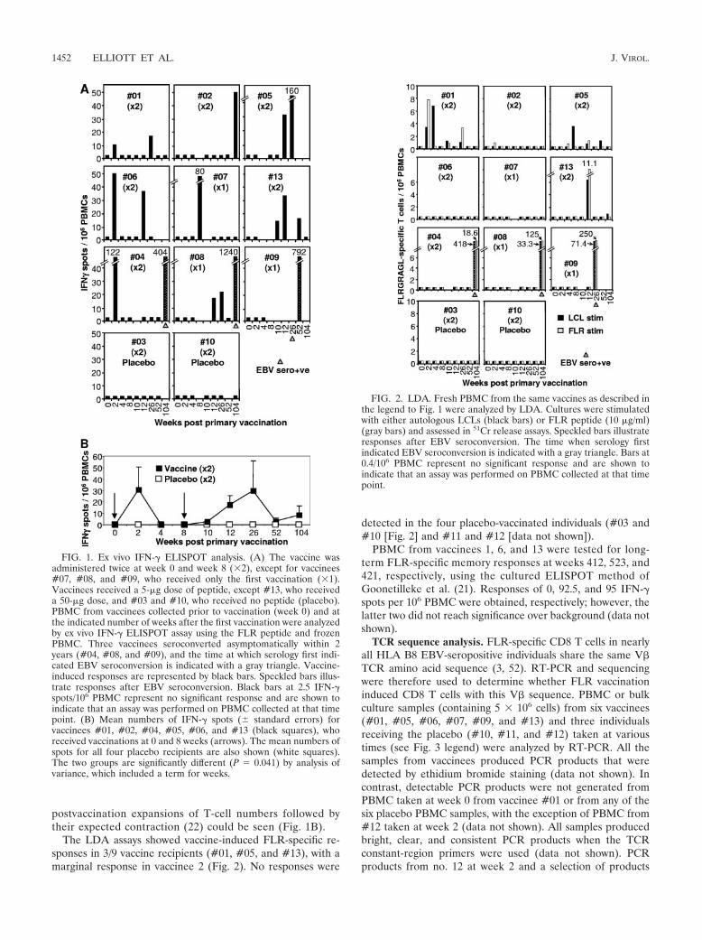
postvaccination expansions of T-cell numbers followed bytheir expected contraction (22) could be seen (Fig. 1B).
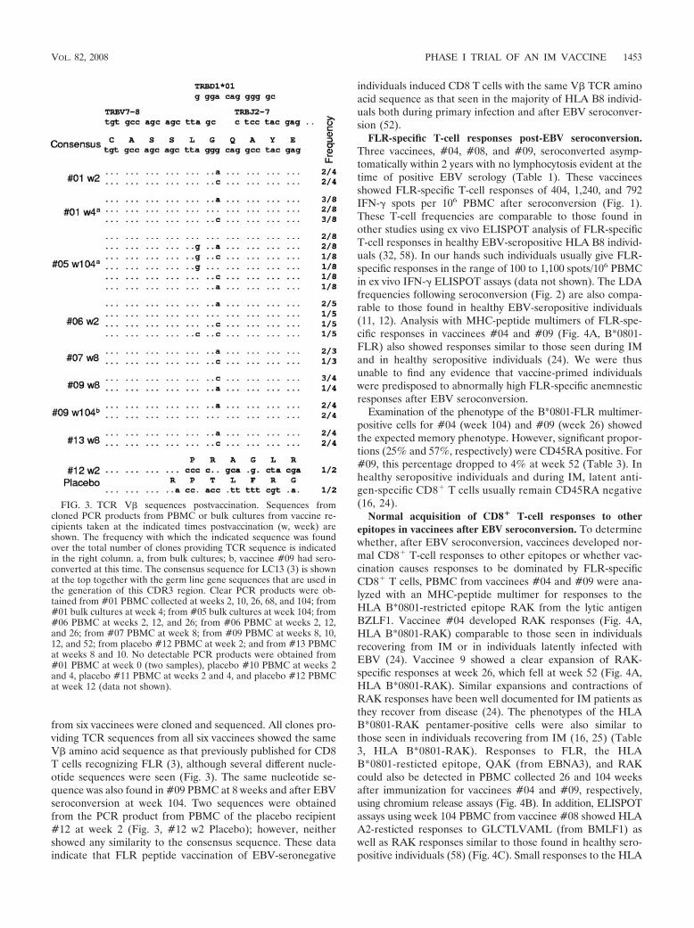
The LDA assays showed vaccine-induced FLR-specific re-sponses in 3/9 vaccine recipients (#01, #05, and #13), with amarginal response in vaccinee 2 (Fig. 2). No responses were
detected in the four placebo-vaccinated individuals (#03 and#10 [Fig. 2] and #11 and #12 [data not shown]).
PBMC from vaccinees 1, 6, and 13 were tested for long-term FLR-specific memory responses at weeks 412, 523, and421, respectively, using the cultured ELISPOT method ofGoonetilleke et al. (21). Responses of 0, 92.5, and 95 IFN-�spots per 106 PBMC were obtained, respectively; however, thelatter two did not reach significance over background (data notshown).
TCR sequence analysis. FLR-specific CD8 T cells in nearlyall HLA B8 EBV-seropositive individuals share the same V�TCR amino acid sequence (3, 52). RT-PCR and sequencingwere therefore used to determine whether FLR vaccinationinduced CD8 T cells with this V� sequence. PBMC or bulkculture samples (containing 5 106 cells) from six vaccinees(#01, #05, #06, #07, #09, and #13) and three individualsreceiving the placebo (#10, #11, and #12) taken at varioustimes (see Fig. 3 legend) were analyzed by RT-PCR. All thesamples from vaccinees produced PCR products that weredetected by ethidium bromide staining (data not shown). Incontrast, detectable PCR products were not generated fromPBMC taken at week 0 from vaccinee #01 or from any of thesix placebo PBMC samples, with the exception of PBMC from#12 taken at week 2 (data not shown). All samples producedbright, clear, and consistent PCR products when the TCRconstant-region primers were used (data not shown). PCRproducts from no. 12 at week 2 and a selection of products
FIG. 1. Ex vivo IFN-� ELISPOT analysis. (A) The vaccine wasadministered twice at week 0 and week 8 (2), except for vaccinees#07, #08, and #09, who received only the first vaccination (1).Vaccinees received a 5-�g dose of peptide, except #13, who receiveda 50-�g dose, and #03 and #10, who received no peptide (placebo).PBMC from vaccinees collected prior to vaccination (week 0) and atthe indicated number of weeks after the first vaccination were analyzedby ex vivo IFN-� ELISPOT assay using the FLR peptide and frozenPBMC. Three vaccinees seroconverted asymptomatically within 2years (#04, #08, and #09), and the time at which serology first indi-cated EBV seroconversion is indicated with a gray triangle. Vaccine-induced responses are represented by black bars. Speckled bars illus-trate responses after EBV seroconversion. Black bars at 2.5 IFN-�spots/106 PBMC represent no significant response and are shown toindicate that an assay was performed on PBMC collected at that timepoint. (B) Mean numbers of IFN-� spots (� standard errors) forvaccinees #01, #02, #04, #05, #06, and #13 (black squares), whoreceived vaccinations at 0 and 8 weeks (arrows). The mean numbers ofspots for all four placebo recipients are also shown (white squares).The two groups are significantly different (P � 0.041) by analysis ofvariance, which included a term for weeks.
FIG. 2. LDA. Fresh PBMC from the same vaccines as described inthe legend to Fig. 1 were analyzed by LDA. Cultures were stimulatedwith either autologous LCLs (black bars) or FLR peptide (10 �g/ml)(gray bars) and assessed in 51Cr release assays. Speckled bars illustrateresponses after EBV seroconversion. The time when serology firstindicated EBV seroconversion is indicated with a gray triangle. Bars at0.4/106 PBMC represent no significant response and are shown toindicate that an assay was performed on PBMC collected at that timepoint.
1452 ELLIOTT ET AL. J. VIROL.
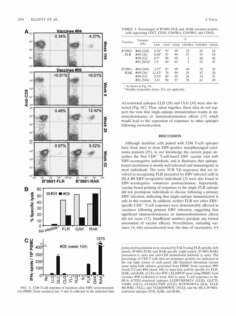
from six vaccinees were cloned and sequenced. All clones pro-viding TCR sequences from all six vaccinees showed the sameV� amino acid sequence as that previously published for CD8T cells recognizing FLR (3), although several different nucle-otide sequences were seen (Fig. 3). The same nucleotide se-quence was also found in #09 PBMC at 8 weeks and after EBVseroconversion at week 104. Two sequences were obtainedfrom the PCR product from PBMC of the placebo recipient#12 at week 2 (Fig. 3, #12 w2 Placebo); however, neithershowed any similarity to the consensus sequence. These dataindicate that FLR peptide vaccination of EBV-seronegative
individuals induced CD8 T cells with the same V� TCR aminoacid sequence as that seen in the majority of HLA B8 individ-uals both during primary infection and after EBV seroconver-sion (52).
FLR-specific T-cell responses post-EBV seroconversion.Three vaccinees, #04, #08, and #09, seroconverted asymp-tomatically within 2 years with no lymphocytosis evident at thetime of positive EBV serology (Table 1). These vaccineesshowed FLR-specific T-cell responses of 404, 1,240, and 792IFN-� spots per 106 PBMC after seroconversion (Fig. 1).These T-cell frequencies are comparable to those found inother studies using ex vivo ELISPOT analysis of FLR-specificT-cell responses in healthy EBV-seropositive HLA B8 individ-uals (32, 58). In our hands such individuals usually give FLR-specific responses in the range of 100 to 1,100 spots/106 PBMCin ex vivo IFN-� ELISPOT assays (data not shown). The LDAfrequencies following seroconversion (Fig. 2) are also compa-rable to those found in healthy EBV-seropositive individuals(11, 12). Analysis with MHC-peptide multimers of FLR-spe-cific responses in vaccinees #04 and #09 (Fig. 4A, B*0801-FLR) also showed responses similar to those seen during IMand in healthy seropositive individuals (24). We were thusunable to find any evidence that vaccine-primed individualswere predisposed to abnormally high FLR-specific anemnesticresponses after EBV seroconversion.
Examination of the phenotype of the B*0801-FLR multimer-positive cells for #04 (week 104) and #09 (week 26) showedthe expected memory phenotype. However, significant propor-tions (25% and 57%, respectively) were CD45RA positive. For#09, this percentage dropped to 4% at week 52 (Table 3). Inhealthy seropositive individuals and during IM, latent anti-gen-specific CD8� T cells usually remain CD45RA negative(16, 24).
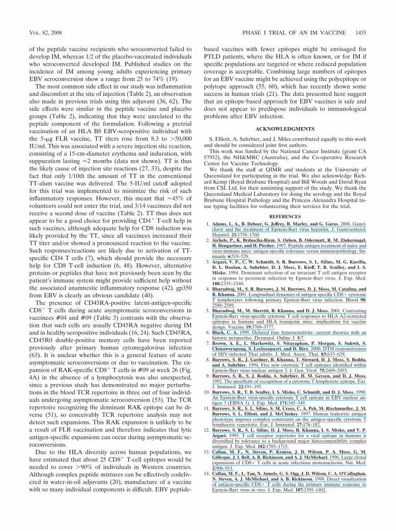
Normal acquisition of CD8� T-cell responses to otherepitopes in vaccinees after EBV seroconversion. To determinewhether, after EBV seroconversion, vaccinees developed nor-mal CD8� T-cell responses to other epitopes or whether vac-cination causes responses to be dominated by FLR-specificCD8� T cells, PBMC from vaccinees #04 and #09 were ana-lyzed with an MHC-peptide multimer for responses to theHLA B*0801-restricted epitope RAK from the lytic antigenBZLF1. Vaccinee #04 developed RAK responses (Fig. 4A,HLA B*0801-RAK) comparable to those seen in individualsrecovering from IM or in individuals latently infected withEBV (24). Vaccinee 9 showed a clear expansion of RAK-specific responses at week 26, which fell at week 52 (Fig. 4A,HLA B*0801-RAK). Similar expansions and contractions ofRAK responses have been well documented for IM patients asthey recover from disease (24). The phenotypes of the HLAB*0801-RAK pentamer-positive cells were also similar tothose seen in individuals recovering from IM (16, 25) (Table3, HLA B*0801-RAK). Responses to FLR, the HLAB*0801-resticted epitope, QAK (from EBNA3), and RAKcould also be detected in PBMC collected 26 and 104 weeksafter immunization for vaccinees #04 and #09, respectively,using chromium release assays (Fig. 4B). In addition, ELISPOTassays using week 104 PBMC from vaccinee #08 showed HLAA2-resticted responses to GLCTLVAML (from BMLF1) aswell as RAK responses similar to those found in healthy sero-positive individuals (58) (Fig. 4C). Small responses to the HLA
FIG. 3. TCR V� sequences postvaccination. Sequences fromcloned PCR products from PBMC or bulk cultures from vaccine re-cipients taken at the indicated times postvaccination (w, week) areshown. The frequency with which the indicated sequence was foundover the total number of clones providing TCR sequence is indicatedin the right column. a, from bulk cultures; b, vaccinee #09 had sero-converted at this time. The consensus sequence for LC13 (3) is shownat the top together with the germ line gene sequences that are used inthe generation of this CDR3 region. Clear PCR products were ob-tained from #01 PBMC collected at weeks 2, 10, 26, 68, and 104; from#01 bulk cultures at week 4; from #05 bulk cultures at week 104; from#06 PBMC at weeks 2, 12, and 26; from #06 PBMC at weeks 2, 12,and 26; from #07 PBMC at week 8; from #09 PBMC at weeks 8, 10,12, and 52; from placebo #12 PBMC at week 2; and from #13 PBMCat weeks 8 and 10. No detectable PCR products were obtained from#01 PBMC at week 0 (two samples), placebo #10 PBMC at weeks 2and 4, placebo #11 PBMC at weeks 2 and 4, and placebo #12 PBMCat week 12 (data not shown).
VOL. 82, 2008 PHASE I TRIAL OF AN IM VACCINE 1453
A2-restricted epitopes LLD (28) and CLG (34) were also de-tected (Fig. 4C). Thus, taken together, these data do not sup-port the view that single-epitope immunization results in im-munodominance or immunodomination effects (17) whichwould lead to the repression of responses to other epitopesfollowing seroconversion.
DISCUSSION
Although dendritic cells pulsed with CD8 T-cell epitopeshave been used to treat EBV-positive nasopharyngeal carci-noma patients (35), to our knowledge the current paper de-scribes the first CD8� T-cell-based EBV vaccine trial withEBV-seronegative individuals, and it illustrates that epitope-based vaccination is mostly well tolerated and immunogenic inmost individuals. The same TCR V� sequences that are in-volved in recognizing FLR presented by EBV-infected cells inHLA B8 EBV-seropositive individuals (3) were also found inEBV-seronegative volunteers postvaccination. Importantly,vaccine-based priming of responses to the single FLR epitopedid not predispose individuals to disease following a primaryEBV infection, indicating that single-epitope immunization issafe in this context. In addition, neither FLR nor other EBV-specific CD8� T-cell responses were demonstrably affected invaccinees following primary EBV infection, suggesting thatsignificant immunodominance or immunodomination effectsdid not occur (17). Insufficient numbers preclude any formalassessment of vaccine efficacy. Nevertheless, excluding vac-cinee 14, who seroconverted near the time of vaccination, 4/4
FIG. 4. CD8 T-cell response of vaccinees after EBV seroconversion.(A) PBMC from vaccinees (no. 9 and 4) collected at the indicated time
points postvaccination were assessed by FACS using FLR-specific (leftpanels, B*0801-FLR) and RAK-specific (right panels, B*0801-RAK)pentamers (x axis) and anti-CD8 monoclonal antibody (y axis). Thepercentage of CD8 T cells that are pentamer positive are indicated inthe top right corner of each panel. (B) Standard chromium releaseassay using bulk cultures generated from PBMC from vaccinees #09(week 52) and #04 (week 104) to asses lytic activity specific for FLR,QAK, and RAK. (C) Ex vivo IFN-� ELISPOT assay using PBMC fromvaccinee #08 (collected at week 104) to asses T-cell responses to theHLA A*0201-restricted epitopes LLDFVRFMGV (LLD), GLCTLVAML (GLC), CLGGLLTMV (CLG), ILIYNGWYA (ILI), YLLEMLWRL (YLL), and YLQQNWWTL (YLQ) and the HLA B*0801-restricted epitopes FLR, QAK, and RAK.
TABLE 3. Percentages of B*0801-FLR and -RAK tetramer-positivecells expressing CD27, CD28, CD45RA, CD45RO, and CD62L
Vaccinee Tetramer(wk)
%
CD8 CD27 CD28 CD45RA CD45RO CD62L
B*0801- #04 (104) 0.34a 97 99 25 82 24FLR #09 (26) 0.48a 97 99 57 91 58
#09 (52) 0.7a 98 99 4 86 48#01 (NA)b 1.3 89 83 5 92 47
B*0801- #04 (104) 4.37a 87 99 40 42 24RAK #09 (26) 12.42a 79 99 26 47 28
#09 (52) 8.52a 89 99 28 34 31#01 (NA) 3.41 90 97 38 61 40
a As shown in Fig. 3A.b Healthy seropositive donor. NA, not applicable.
1454 ELLIOTT ET AL. J. VIROL.
of the peptide vaccine recipients who seroconverted failed todevelop IM, whereas 1/2 of the placebo-vaccinated individualswho seroconverted developed IM. Published studies on theincidence of IM among young adults experiencing primaryEBV seroconversion show a range from 25 to 74% (19).
The most common side effect in our study was inflammationand discomfort at the site of injection (Table 2), an observationalso made in previous trials using this adjuvant (36, 62). Theside effects were similar in the peptide vaccine and placebogroups (Table 2), indicating that they were unrelated to thepeptide component of the formulation. Following a pretrialvaccination of an HLA B8 EBV-seropositive individual withthe 5-�g FLR vaccine, TT titers rose from 8.3 to �50,000IU/ml. This was associated with a severe injection site reaction,consisting of a 15-cm-diameter erythema and induration, withsuppuration lasting �2 months (data not shown). TT is thusthe likely cause of injection site reactions (27, 33), despite thefact that only 1/10th the amount of TT in the conventionalTT-alum vaccine was delivered. The 5-IU/ml cutoff adoptedfor this trial was implemented to minimize the risk of suchinflammatory responses. However, this meant that �45% ofvolunteers could not enter the trial, and 3/14 vaccinees did notreceive a second dose of vaccine (Table 2). TT thus does notappear to be a good choice for providing CD4� T-cell help insuch vaccines, although adequate help for CD8 induction waslikely provided by the TT, since all vaccinees increased theirTT titer and/or showed a pronounced reaction to the vaccine.Such responses/reactions are likely due to activation of TT-specific CD4 T cells (7), which should provide the necessaryhelp for CD8 T-cell induction (6, 48). However, alternativeproteins or peptides that have not previously been seen by thepatient’s immune system might provide sufficient help withoutthe associated anamnestic inflammatory response (42). gp350from EBV is clearly an obvious candidate (40).
The presence of CD45RA-positive latent-antigen-specificCD8� T cells during acute asymptomatic seroconversions invaccinees #04 and #09 (Table 3) contrasts with the observa-tion that such cells are usually CD45RA negative during IMand in healthy seropositive individuals (16, 24). Such CD45RA,CD45R0 double-positive memory cells have been reportedpreviously after primary human cytomegalovirus infection(63). It is unclear whether this is a general feature of acuteasymptomatic seroconversions or due to vaccination. The ex-pansion of RAK-specific CD8� T cells in #09 at week 26 (Fig.4A) in the absence of a lymphocytosis was also unexpected,since a previous study has demonstrated no major perturba-tions in the blood TCR repertoire in three out of four individ-uals undergoing asymptomatic seroconversion (53). The TCRrepertoire recognizing the dominant RAK epitope can be di-verse (51), so conceivably TCR repertoire analysis may notdetect such expansions. This RAK expansion is unlikely to bea result of FLR vaccination and therefore indicates that lyticantigen-specific expansions can occur during asymptomatic se-roconversions.
Due to the HLA diversity across human populations, wehave estimated that about 25 CD8� T-cell epitopes would beneeded to cover �90% of individuals in Western countries.Although complex peptide mixtures can be effectively codeliv-ered in water-in-oil adjuvants (20), manufacture of a vaccinewith so many individual components is difficult. EBV peptide-
based vaccines with fewer epitopes might be envisaged forPTLD patients, where the HLA is often known, or for IM ifspecific populations are targeted or where reduced populationcoverage is acceptable. Combining large numbers of epitopesfor an EBV vaccine might be achieved using the polyepitope orpolytope approach (55, 60), which has recently shown somesuccess in human trials (21). The data presented here suggestthat an epitope-based approach for EBV vaccines is safe anddoes not appear to predispose individuals to immunologicalproblems after EBV infection.
ACKNOWLEDGMENTS
S. Elliott, A. Suhrbier, and J. Miles contributed equally to this workand should be considered joint first authors.
This work was funded by the National Cancer Institute (grant CA57952), the NH&MRC (Australia), and the Co-operative ResearchCentre for Vaccine Technology.
We thank the staff at QIMR and students at the University ofQueensland for participating in the trial. We also acknowledge Rich-ard Kemp (Royal Brisbane Hospital) and Bill Woods and David Ryanfrom CSL Ltd. for their unstinting support of the study. We thank theQueensland Medical Laboratory for doing the serology and the RoyalBrisbane Hospital Pathology and the Princess Alexandra Hospital tis-sue typing facilities for volunteering their services for the trial.
REFERENCES
1. Adams, L. A., B. Deboer, G. Jeffrey, R. Marley, and G. Garas. 2006. Ganci-clovir and the treatment of Epstein-Barr virus hepatitis. J. Gastroenterol.Hepatol. 21:1758–1760.
2. Aichele, P., K. Brduscha-Riem, S. Oehen, B. Odermatt, R. M. Zinkernagel,H. Hengartner, and H. Pircher. 1997. Peptide antigen treatment of naive andvirus-immune mice: antigen-specific tolerance versus immunopathology. Im-munity 6:519–529.
3. Argaet, V. P., C. W. Schmidt, S. R. Burrows, S. L. Silins, M. G. Kurilla,D. L. Doolan, A. Suhrbier, D. J. Moss, E. Kieff, T. B. Sculley, and I. S.Misko. 1994. Dominant selection of an invariant T cell antigen receptorin response to persistent infection by Epstein-Barr virus. J. Exp. Med.180:2335–2340.
4. Bharadwaj, M., S. R. Burrows, J. M. Burrows, D. J. Moss, M. Catalina, andR. Khanna. 2001. Longitudinal dynamics of antigen-specific CD8� cytotoxicT lymphocytes following primary Epstein-Barr virus infection. Blood 98:2588–2589.
5. Bharadwaj, M., M. Sherritt, R. Khanna, and D. J. Moss. 2001. ContrastingEpstein-Barr virus-specific cytotoxic T cell responses to HLA A2-restrictedepitopes in humans and HLA transgenic mice: implications for vaccinedesign. Vaccine 19:3769–3777.
6. Black, C. A. 1999. Delayed type hypersensitivity: current theories with anhistoric perspective. Dermatol. Online J. 5:7.
7. Brown, A. E., L. Markowitz, S. Nitayaphan, P. Morgan, S. Sukwit, S.Chinaworapong, S. Leelasupasri, and D. Birx. 2000. DTH responsivenessof HIV-infected Thai adults. J. Med. Assoc. Thai. 83:633–639.
8. Burrows, S. R., J. Gardner, R. Khanna, T. Steward, D. J. Moss, S. Rodda,and A. Suhrbier. 1994. Five new cytotoxic T cell epitopes identified withinEpstein-Barr virus nuclear antigen 3. J. Gen. Virol. 75:2489–2493.
9. Burrows, S. R., S. J. Rodda, A. Suhrbier, H. M. Geysen, and D. J. Moss.1992. The specificity of recognition of a cytotoxic T lymphocyte epitope. Eur.J. Immunol. 22:191–195.
10. Burrows, S. R., T. B. Sculley, I. S. Misko, C. Schmidt, and D. J. Moss. 1990.An Epstein-Barr virus-specific cytotoxic T cell epitope in EBV nuclear an-tigen 3 (EBNA 3). J. Exp. Med. 171:345–349.
11. Burrows, S. R., S. L. Silins, S. M. Cross, C. A. Peh, M. Rischmueller, J. M.Burrows, S. L. Elliott, and J. McCluskey. 1997. Human leukocyte antigenphenotype imposes complex constraints on the antigen-specific cytotoxic Tlymphocyte repertoire. Eur. J. Immunol. 27:178–182.
12. Burrows, S. R., S. L. Silins, D. J. Moss, R. Khanna, I. S. Misko, and V. P.Argaet. 1995. T cell receptor repertoire for a viral epitope in humans isdiversified by tolerance to a background major histocompatibility complexantigen. J. Exp. Med. 182:1703–1715.
13. Callan, M. F., N. Steven, P. Krausa, J. D. Wilson, P. A. Moss, G. M.Gillespie, J. I. Bell, A. B. Rickinson, and A. J. McMichael. 1996. Large clonalexpansions of CD8� T cells in acute infectious mononucleosis. Nat. Med.2:906–911.
14. Callan, M. F., L. Tan, N. Annels, G. S. Ogg, J. D. Wilson, C. A. O’Callaghan,N. Steven, A. J. McMichael, and A. B. Rickinson. 1998. Direct visualizationof antigen-specific CD8� T cells during the primary immune response toEpstein-Barr virus in vivo. J. Exp. Med. 187:1395–1402.
VOL. 82, 2008 PHASE I TRIAL OF AN IM VACCINE 1455
15. Candy, B., and M. Hotopf. 2006. Steroids for symptom control in infectiousmononucleosis. Cochrane Database Syst. Rev. 3:CD004402.
16. Catalina, M. D., J. L. Sullivan, R. M. Brody, and K. Luzuriaga. 2002.Phenotypic and functional heterogeneity of EBV epitope-specific CD8� Tcells. J. Immunol. 168:4184–4191.
17. Chen, W., and J. McCluskey. 2006. Immunodominance and immunodomi-nation: critical factors in developing effective CD8� T-cell-based cancervaccines. Adv. Cancer Res. 95:203–247.
18. Chetham, M. M., and K. B. Roberts. 1991. Infectious mononucleosis inadolescents. Pediatr. Ann. 20:206–213.
19. Crawford, D. H., K. F. Macsween, C. D. Higgins, R. Thomas, K. McAulay, H.Williams, N. Harrison, S. Reid, M. Conacher, J. Douglas, and A. J. Swerd-low. 2006. A cohort study among university students: identification of riskfactors for Epstein-Barr virus seroconversion and infectious mononucleosis.Clin. Infect. Dis. 43:276–282.
20. Elliott, S. L., S. Pye, T. Le, L. Mateo, J. Cox, L. Macdonald, A. A. Scalzo,C. A. Forbes, and A. Suhrbier. 1999. Peptide based cytotoxic T-cell vaccines;delivery of multiple epitopes, help, memory and problems. Vaccine 17:2009–2019.
21. Goonetilleke, N., S. Moore, L. Dally, N. Winstone, I. Cebere, A. Mahmoud,S. Pinheiro, G. Gillespie, D. Brown, V. Loach, J. Roberts, A. Guimaraes-Walker, P. Hayes, K. Loughran, C. Smith, J. De Bont, C. Verlinde, D. Vooijs,C. Schmidt, M. Boaz, J. Gilmour, P. Fast, L. Dorrell, T. Hanke, and A. J.McMichael. 2006. Induction of multifunctional human immunodeficiencyvirus type 1 (HIV-1)-specific T cells capable of proliferation in healthysubjects by using a prime-boost regimen of DNA- and modified vacciniavirus Ankara-vectored vaccines expressing HIV-1 Gag coupled to CD8�
T-cell epitopes. J. Virol. 80:4717–4728.22. Haring, J. S., V. P. Badovinac, and J. T. Harty. 2006. Inflaming the CD8�
T cell response. Immunity 25:19–29.23. Hersey, P., S. W. Menzies, B. Coventry, T. Nguyen, M. Farrelly, S. Collins,
D. Hirst, and H. Johnson. 2005. Phase I/II study of immunotherapy withT-cell peptide epitopes in patients with stage IV melanoma. Cancer Immu-nol. Immunother. 54:208–218.
24. Hislop, A. D., N. E. Annels, N. H. Gudgeon, A. M. Leese, and A. B. Rickinson.2002. Epitope-specific evolution of human CD8(�) T cell responses fromprimary to persistent phases of Epstein-Barr virus infection. J. Exp. Med.195:893–905.
25. Hislop, A. D., N. H. Gudgeon, M. F. Callan, C. Fazou, H. Hasegawa, M.Salmon, and A. B. Rickinson. 2001. EBV-specific CD8� T cell memory:relationships between epitope specificity, cell phenotype, and immediateeffector function. J. Immunol. 167:2019–2029.
26. Hjalgrim, H., K. E. Smedby, K. Rostgaard, D. Molin, S. Hamilton-Dutoit,E. T. Chang, E. Ralfkiaer, C. Sundstrom, H. O. Adami, B. Glimelius, and M.Melbye. 2007. Infectious mononucleosis, childhood social environment, andrisk of Hodgkin lymphoma. Cancer Res. 67:2382–2388.
27. Jacobs, R. L., R. S. Lowe, and B. Q. Lanier. 1982. Adverse reactions totetanus toxoid. JAMA 247:40–42.
28. Kerr, B. M., N. Kienzle, J. M. Burrows, S. Cross, S. L. Silins, M. Buck, E. M.Benson, B. Coupar, D. J. Moss, and T. B. Sculley. 1996. Identification of typeB-specific and cross-reactive cytotoxic T-lymphocyte responses to Epstein-Barr virus. J. Virol. 70:8858–8864.
29. Khanna, R., S. Bell, M. Sherritt, A. Galbraith, S. R. Burrows, L. Rafter, B.Clarke, R. Slaughter, M. C. Falk, J. Douglass, T. Williams, S. L. Elliott, andD. J. Moss. 1999. Activation and adoptive transfer of Epstein-Barr virus-specific cytotoxic T cells in solid organ transplant patients with posttrans-plant lymphoproliferative disease. Proc. Natl. Acad. Sci. USA 96:10391–10396.
30. Khanna, R., S. R. Burrows, J. Nicholls, and L. M. Poulsen. 1998. Identificationof cytotoxic T cell epitopes within Epstein-Barr virus (EBV) oncogene latentmembrane protein 1 (LMP1): evidence for HLA A2 supertype-restricted im-mune recognition of EBV-infected cells by LMP1-specific cytotoxic T lympho-cytes. Eur. J. Immunol. 28:451–458.
31. Khanna, R., S. R. Burrows, A. Suhrbier, C. A. Jacob, H. Griffin, I. S. Misko,T. B. Sculley, M. Rowe, A. B. Rickinson, and D. J. Moss. 1993. EBV peptideepitope sensitization restores human cytotoxic T cell recognition of Burkitt’slymphoma cells. Evidence for a critical role for ICAM-2. J. Immunol. 150:5154–5162.
32. Klatt, T., Q. Ouyang, T. Flad, I. Koetter, H. J. Buhring, H. Kalbacher, G.Pawelec, and C. A. Muller. 2005. Expansion of peripheral CD8� CD28� Tcells in response to Epstein-Barr virus in patients with rheumatoid arthritis.J. Rheumatol. 32:239–251.
33. Kuhlwein, A., and A. Bleyl. 1985. Tetanus antitoxin levels and reactionsfollowing tetanus vaccination. Hautarzt 36:462–464.
34. Lee, S. P., W. A. Thomas, R. J. Murray, F. Khanim, S. Kaur, L. S. Young, M.Rowe, M. Kurilla, and A. B. Rickinson. 1993. HLA A2.1-restricted cytotoxicT cells recognizing a range of Epstein-Barr virus isolates through a definedepitope in latent membrane protein LMP2. J. Virol. 67:7428–7435.
35. Lin, C. L., W. F. Lo, T. H. Lee, Y. Ren, S. L. Hwang, Y. F. Cheng, C. L.Chen, Y. S. Chang, S. P. Lee, A. B. Rickinson, and P. K. Tam. 2002.Immunization with Epstein-Barr virus (EBV) peptide-pulsed dendriticcells induces functional CD8� T-cell immunity and may lead to tumor
regression in patients with EBV-positive nasopharyngeal carcinoma. Can-cer Res. 62:6952–6958.
36. Lopez, J. A., C. Weilenman, R. Audran, M. A. Roggero, A. Bonelo, J. M.Tiercy, F. Spertini, and G. Corradin. 2001. A synthetic malaria vaccine elicitsa potent CD8(�) and CD4(�) T lymphocyte immune response in humans.Implications for vaccination strategies. Eur. J. Immunol. 31:1989–1998.
37. Moss, D. J., S. R. Burrows, D. J. Castelino, R. G. Kane, J. H. Pope, A. B.Rickinson, M. P. Alpers, and P. F. Heywood. 1983. A comparison of Epstein-Barr virus-specific T-cell immunity in malaria-endemic and -nonendemicregions of Papua New Guinea. Int. J. Cancer 31:727–732.
38. Moss, D. J., C. Schmidt, S. Elliott, A. Suhrbier, S. Burrows, and R. Khanna.1996. Strategies involved in developing an effective vaccine for EBV-associ-ated diseases. Adv. Cancer Res. 69:213–245.
39. Moss, D. J., A. Suhrbier, and S. L. Elliott. 1998. Candidate vaccines forEpstein-Barr virus. BMJ 317:423–424.
40. Moutschen, M., P. Leonard, E. M. Sokal, F. Smets, M. Haumont, P. Mazzu,A. Bollen, F. Denamur, P. Peeters, G. Dubin, and M. Denis. 2007. Phase I/IIstudies to evaluate safety and immunogenicity of a recombinant gp350 Ep-stein-Barr virus vaccine in healthy adults. Vaccine 25:4697–4705.
41. Nielsen, T. R., K. Rostgaard, N. M. Nielsen, N. Koch-Henriksen, S. Haahr,P. S. Sorensen, and H. Hjalgrim. 2007. Multiple sclerosis after infectiousmononucleosis. Arch. Neurol. 64:72–75.
42. Parra-Lopez, C., J. M. Calvo-Calle, T. O. Cameron, L. E. Vargas, L. M.Salazar, M. E. Patarroyo, E. Nardin, and L. J. Stern. 2006. Major histocom-patibility complex and T cell interactions of a universal T cell epitope fromPlasmodium falciparum circumsporozoite protein. J. Biol. Chem. 281:14907–14917.
43. Pope, J. H. 1968. Establishment of cell lines from Australian leukaemicpatients: presence of a herpes-like virus. Aust. J. Exp. Biol. Med. Sci. 46:643–645.
44. Rickinson, A. B., and D. J. Moss. 1997. Human cytotoxic T lymphocyteresponses to Epstein-Barr virus infection. Annu. Rev. Immunol. 15:405–431.
45. Sauce, D., M. Larsen, S. J. Curnow, A. M. Leese, P. A. Moss, A. D. Hislop,M. Salmon, and A. B. Rickinson. 2006. EBV-associated mononucleosisleads to long-term global deficit in T-cell responsiveness to IL-15. Blood108:11–18.
46. Saul, A., G. Lawrence, A. Smillie, C. M. Rzepczyk, C. Reed, D. Taylor, K.Anderson, A. Stowers, R. Kemp, A. Allworth, R. F. Anders, G. V. Brown, D.Pye, P. Schoofs, D. O. Irving, S. L. Dyer, G. C. Woodrow, W. R. Briggs, R.Reber, and D. Sturchler. 1999. Human phase I vaccine trials of 3 recombi-nant asexual stage malaria antigens with Montanide ISA720 adjuvant. Vac-cine 17:3145–3159.
47. Savoldo, B., J. A. Goss, M. M. Hammer, L. Zhang, T. Lopez, A. P. Gee, Y. F.Lin, R. E. Quiros-Tejeira, P. Reinke, S. Schubert, S. Gottschalk, M. J.Finegold, M. K. Brenner, C. M. Rooney, and H. E. Heslop. 2006. Treatmentof solid organ transplant recipients with autologous Epstein Barr virus-specific cytotoxic T lymphocytes (CTLs). Blood 108:2942–2949.
48. Scalzo, A. A., S. L. Elliott, J. Cox, J. Gardner, D. J. Moss, and A. Suhrbier.1995. Induction of protective cytotoxic T cells to murine cytomegalovirus byusing a nonapeptide and a human-compatible adjuvant (Montanide ISA720). J. Virol. 69:1306–1309.
49. Sherritt, M. A., M. Bharadwaj, J. M. Burrows, L. E. Morrison, S. L. Elliott,J. E. Davis, L. M. Kear, R. E. Slaughter, S. C. Bell, A. J. Galbraith, R.Khanna, and D. J. Moss. 2003. Reconstitution of the latent T-lymphocyteresponse to Epstein-Barr virus is coincident with long-term recovery fromposttransplant lymphoma after adoptive immunotherapy. Transplantation75:1556–1560.
50. Sherritt, M. A., J. Gardner, S. L. Elliott, C. Schmidt, D. Purdie, G. Deliy-annis, W. R. Heath, and A. Suhrbier. 2000. Effect of pre-existing cytotoxic Tlymphocytes on therapeutic vaccines. Eur. J. Immunol. 30:671–677.
51. Silins, S. L., S. M. Cross, S. L. Elliott, S. J. Pye, J. M. Burrows, D. J. Moss,and I. S. Misko. 1997. Selection of a diverse TCR repertoire in response toan Epstein-Barr virus-encoded transactivator protein BZLF1 by CD8� cy-totoxic T lymphocytes during primary and persistent infection. Int. Immunol.9:1745–1755.
52. Silins, S. L., S. M. Cross, S. L. Elliott, S. J. Pye, S. R. Burrows, J. M.Burrows, D. J. Moss, V. P. Argaet, and I. S. Misko. 1996. Development ofEpstein-Barr virus-specific memory T cell receptor clonotypes in acute in-fectious mononucleosis. J. Exp. Med. 184:1815–1824.
53. Silins, S. L., M. A. Sherritt, J. M. Silleri, S. M. Cross, S. L. Elliott, M.Bharadwaj, T. T. Le, L. E. Morrison, R. Khanna, D. J. Moss, A. Suhrbier,and I. S. Misko. 2001. Asymptomatic primary Epstein-Barr virus infectionoccurs in the absence of blood T-cell repertoire perturbations despite highlevels of systemic viral load. Blood 98:3739–3744.
54. Suhrbier, A. 1997. Multi-epitope DNA vaccines. Immunol. Cell Biol. 75:402–408.
55. Suhrbier, A. 2002. Polytope vaccines for the codelivery of multiple CD8T-cell epitopes. Expert Rev. Vaccines 1:207–213.
56. Svedmyr, E., I. Ernberg, J. Seeley, O. Weiland, G. Masucci, K. Tsukuda, R.Szigeti, M. G. Masucci, H. Blomogren, and W. Berthold. 1984. Virologic,immunologic, and clinical observations on a patient during the incubation,
1456 ELLIOTT ET AL. J. VIROL.

acute, and convalescent phases of infectious mononucleosis. Clin. Immunol.Immunopathol. 30:437–450.
57. Takeuchi, K., K. Tanaka-Taya, Y. Kazuyama, Y. M. Ito, S. Hashimoto, M.Fukayama, and S. Mori. 2006. Prevalence of Epstein-Barr virus in Japan:trends and future prediction. Pathol. Int. 56:112–116.
58. Tan, L. C., N. Gudgeon, N. E. Annels, P. Hansasuta, C. A. O’Callaghan, S.Rowland-Jones, A. J. McMichael, A. B. Rickinson, and M. F. Callan. 1999.A re-evaluation of the frequency of CD8� T cells specific for EBV in healthyvirus carriers. J. Immunol. 162:1827–1835.
59. Tattevin, P., Y. Le Tulzo, S. Minjolle, A. Person, J. M. Chapplain, C.Arvieux, R. Thomas, and C. Michelet. 2006. Increasing incidence of severeEpstein-Barr virus-related infectious mononucleosis: surveillance study.J. Clin. Microbiol. 44:1873–1874.
60. Thomson, S. A., R. Khanna, J. Gardner, S. R. Burrows, B. Coupar, D. J.Moss, and A. Suhrbier. 1995. Minimal epitopes expressed in a recombinant
polyepitope protein are processed and presented to CD8� cytotoxic T cells:implications for vaccine design. Proc. Natl. Acad. Sci. USA 92:5845–5849.
61. Toes, R. E., R. Offringa, R. J. Blom, C. J. Melief, and W. M. Kast. 1996.Peptide vaccination can lead to enhanced tumor growth through specificT-cell tolerance induction. Proc. Natl. Acad. Sci. USA 93:7855–7860.
62. Toledo, H., A. Baly, O. Castro, S. Resik, J. Laferte, F. Rolo, L. Navea, L.Lobaina, O. Cruz, J. Miguez, T. Serrano, B. Sierra, L. Perez, M. E. Ricardo,M. Dubed, A. L. Lubian, M. Blanco, J. C. Millan, A. Ortega, E. Iglesias, E.Penton, Z. Martin, J. Perez, M. Diaz, and C. A. Duarte. 2001. A phase Iclinical trial of a multiepitope polypeptide TAB9 combined with MontanideISA 720 adjuvant in non-HIV-1 infected human volunteers. Vaccine 19:4328–4336.
63. Wills, M. R., A. J. Carmichael, M. P. Weekes, K. Mynard, G. Okecha, R.Hicks, and J. G. Sissons. 1999. Human virus-specific CD8� CTL clonesrevert from CD45ROhigh to CD45RAhigh in vivo: CD45RAhighCD8� Tcells comprise both naive and memory cells. J. Immunol. 162:7080–7087.
VOL. 82, 2008 PHASE I TRIAL OF AN IM VACCINE 1457
Related Documents


