PHASE I CLINICAL TESTING AND IMMUNE CHARACTERIZATION OF AN ADIPOSE EXTRACELLULAR MATRIX DERIVED BIOMATERIAL FOR SOFT TISSUE RECONSTRUCTION by Alexis Parrillo A thesis submitted to Johns Hopkins University in conformity with the requirements for the degree of Master of Science in Engineering. Baltimore, Maryland May 2017

Welcome message from author
This document is posted to help you gain knowledge. Please leave a comment to let me know what you think about it! Share it to your friends and learn new things together.
Transcript

PHASE I CLINICAL TESTING AND IMMUNE CHARACTERIZATION OF AN
ADIPOSE EXTRACELLULAR MATRIX DERIVED BIOMATERIAL FOR SOFT
TISSUE RECONSTRUCTION
by Alexis Parrillo
A thesis submitted to Johns Hopkins University in conformity with the requirements for the degree of Master of Science in Engineering.
Baltimore, Maryland May 2017

ii
ABSTRACT
An adipose extracellular matrix derived biomaterial, Acellular Adipose Tissue,
can be used as an off-the-shelf alternative to autologous fat transfer for the treatment of
soft tissue deformities and defects. This tissue engineering solution overcomes many
challenges associated with autologous fat transfer and other common methods of soft
tissue reconstruction. Subcutaneous Acellular Adipose Tissue implants display
significant volume retention with minimal inflammatory response in both pre-clinical and
clinical studies. The material triggers cell migration which supports development of new
adipose and has also demonstrated the potential to modulate the immune response to
create a more pro-regenerative microenvironment in the presence of trauma. These results
indicate that Acellular Adipose Tissue could be a promising new therapeutic tool to treat
soft tissue defects and promote wound healing.
Advisor: Jennifer Elisseeff, Ph.D. Readers: Warren Grayson, Ph.D. Alexander Hillel, M.D.

iii
ACKNOWLEDGEMENTS
I would like to acknowledge all of the many individuals who helped make this
work possible. I am extremely thankful for every member of the Elisseeff lab who trained
and guided me throughout my Master's degree, especially Amy Anderson and Matt Wolf
for answering my never-ending questions and always making me feel comfortable going
to them for support. Thank you to Bahar Zarrabi for always having her door open, both
when I needed an administrator and when I needed a friend. I don’t know where I would
be without her endless support! I also would like to acknowledge the people who helped
make the clinical trial possible, including our team of physicians and trial managers. And
of course, thank you to my friends and family for their endless love and support.
My advisor, Dr. Jennifer Elisseeff, has provided so much guidance and helpful
input throughout my Master's program. Her direction of this project and help thinking
through the tough questions has been invaluable. I have also appreciated her willingness
to allow me to work on projects, sit in on phone calls, and pursue topics that aligned with
my interests. Her guidance throughout my Master's has helped me to develop into the
scientist, engineer, and person that I am. I have enjoyed working with her tremendously,
and I appreciate all of the opportunities that she has given me.
Amy Anderson has been my partner and friend throughout my entire program,
and I would not have made it through without her. All of the work in this thesis belongs
to her as much as it does to me. Being able to work with her made me enjoy coming to
the lab every single day (even the early ones). Somehow, we were still laughing together
even at the end of a fifteen-hour day. I have learned so much about being a scientist from
her, and I cannot thank her enough for everything that she has taught me.
iv
TABLE OF CONTENTS
ABSTRACT ........................................................................................................................ ii
ACKNOWLEDGEMENTS ............................................................................................... iii
TABLE OF CONTENTS ................................................................................................... iv
LIST OF FIGURES ............................................................................................................ v
LIST OF TABLES ............................................................................................................. vi
TABLE OF ABBREVIATIONS ...................................................................................... vii
INTRODUCTION .............................................................................................................. 1
METHODS ......................................................................................................................... 7
RESULTS ......................................................................................................................... 17
DISCUSSION ................................................................................................................... 33
CONCLUSION AND FUTURE WORK ......................................................................... 36
REFERENCES ................................................................................................................. 38
CURRICULUM VITAE ................................................................................................... 40
v
LIST OF FIGURES
Figure 1. Migration assay results for the clinical batch of human AAT........................... 18
Figure 2. Assays to test the human clinical AAT batch for residual process chemicals. . 19
Figure 3. Lipid content assay and hydroxyproline assay results. ..................................... 20
Figure 4. Hematoxylin and Eosin staining of subcutaneous implants. ............................. 21
Figure 5. Subcutaneous flow cytometry results at 1 and 3 weeks. ................................... 22
Figure 6. Subcutaneous RT-PCR results at 3 weeks......................................................... 24
Figure 7. Volumetric muscle loss flow cytometry results at 1 week. ............................... 25
Figure 8. Volumetric muscle loss RT-PCR results at 1 week. .......................................... 27
Figure 10. Histological analysis of clinical trial samples. ................................................ 28
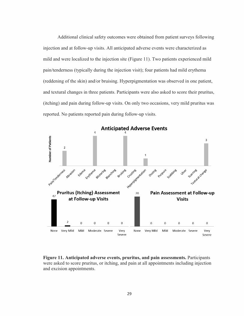
Figure 11. Anticipated adverse events, pruritus, and pain assessments............................ 29
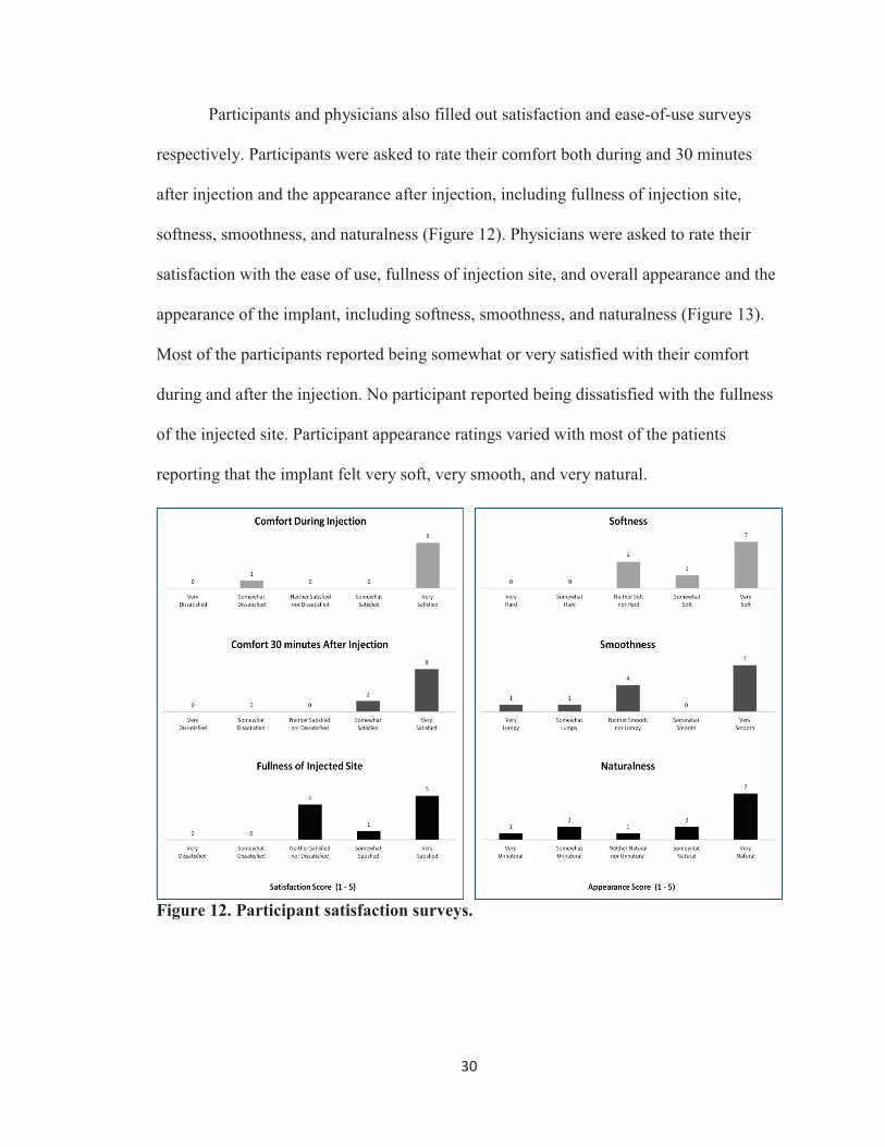
Figure 12. Participant satisfaction surveys. ...................................................................... 30
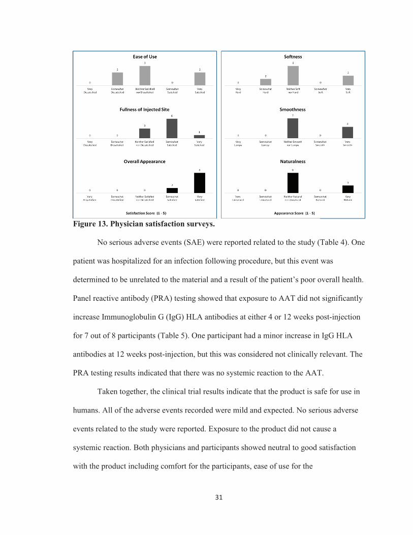
Figure 13. Physician satisfaction surveys. ........................................................................ 31

vi
LIST OF TABLES
Table 1. Mouse flow cytometry panel for subcutaneous injection studies ....................... 10
Table 2. Mouse flow cytometry panel for volumetric muscle loss studies ....................... 10
Table 3. Real time quantitative PCR Primer Sequences ................................................... 11
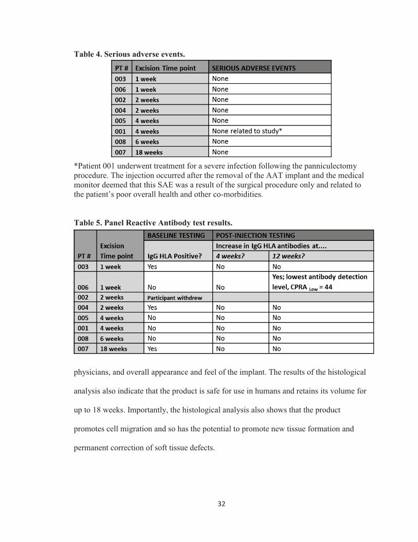
Table 4. Serious adverse events. ....................................................................................... 32
Table 5. Panel Reactive Antibody test results. ................................................................. 32
vii
TABLE OF ABBREVIATIONS
AAT Acellular Adipose Tissue ANOVA Analysis of Variance ASC Adipose-derived stem cell bFGF Basic fibroblast growth factor cDNA Complementary deoxyribonucleic acid DAMP Damage associated molecular patterns DMAB 4-(Dimethylamino)benzaldehyde DPBS Dulbeccos’ phosphate buffered saline ECM Extracellular Matrix EDTA Ethylenediaminetetraacetic acid FBS Fetal bovine serum GMP Good manufacturing practices H&E Hematoxylin and Eosin hAAT Human acellular adipose tissue HLA Human leukocyte antigen HPLC High-performance liquid chromatography IACUC Institutional Care and Use Committee IgG Immunoglobulin G mAAT Mouse acellular adipose tissue pAAT Porcine acellular adipose tissue PRA Panel-reactive antibody PCR Polymerase chain reaction qPCR Quantitative PCR RNA Ribonucleic acid RT Reverse transcriptase SAE Serious adverse event SQ Subcutaneous VML Volumetric muscle loss
1
INTRODUCTION
Soft tissue defects are relatively common and can occur due to trauma, congenital
disease, or surgical inventions. As tissue loss is often permanent, these defects can impact
not only cosmesis but also normal physiological function, including lack of support for
distal extremities and contracture leading to restricted range of motion [1]. The gold
standard for soft tissue reconstruction is autologous adipose transfer (otherwise known as
fat grafting), a procedure first developed by Gustav Neuber over a century ago [2, 3].
However, although these procedures are still commonly used today and many
advancements have been introduced, autologous fat transfer techniques have many
limitations. Adipose grafts behave unpredictably and outcomes can often vary
significantly from patient to patient. The grafted tissue can be resorbed anywhere from
20%-90%, decreasing the total volume of the graft and often requiring multiple surgical
procedures to achieve the desired correction. Like any surgical procedure, autologous fat
grafting carries inherent safety risks. The unpredictability of these procedures can result
in costly surgeries and co-morbidities related to tissue harvest. Harvest procedures often
lead to scarring at the donor site and are limited by the volume of autologous tissue
available in each patient [4]. Transplanted adipocytes are often damaged or subjected to
hypoxic conditions that result in the release of intracellular lipids, a potent pro-
inflammatory signal. These signals combined with a lack of vascularization within the
graft can lead to tissue necrosis and calcifications which impact the quality and durability
of the reconstructed tissue [5].
Decellularized extracellular matrix (ECM) products are a type of biomaterial that
have recently gained popularity in the field of regenerative medicine, although they have
2
been studied and used in various clinical applications since as early as 1995 [6]. Unlike
synthetic biomaterials such as polymers, ECM materials are tissue-derived and are
created using physical, enzymatic, and chemical approaches that remove the living cells
from almost any type of animal or human tissue [7]. These decellularization processes
disrupt cellular membranes and denature key intracellular components such as DNA, but
leave behind the non-cellular component which is present in all tissues and organs. This
non-cellular component is mainly structural in nature and is known as the extracellular
matrix (ECM). Although all ECMs are composed of water, proteins, and polysaccharides,
the physical characteristics and exact composition of any specific ECM depends on its
tissue of origin. ECMs have many physiological roles, including providing a physical
scaffold and initiating cues required for tissue homeostasis and differentiation. ECM also
directs function by binding to growth factors and interacting with receptors on the cell
surface [8].
ECM scaffolds are commonly used in tissue engineering to promote the healing or
regrowth of damaged tissue. Initially, they provide a physical substrate upon which the
cells can be seeded and localized to a specific area. They also provide key biochemical
and physical cues for adhesion, migration, proliferation, and differentiation which help
cells to form fully functional tissues or organs. Implanted ECM scaffolds will eventually
be remodeled and be replaced with the seeded cells’ own secreted matrices.
ECM has many beneficial uses and new applications that are currently being
explored in regenerative medicine. In the past 15 years, many ECM materials have been
brought to market and been successfully used clinically. Alloderm®, an ECM product,
has been used for dentistry, burn therapy, plastic surgery, and hernia repair for over 13

3
years. Many decellularized bone allografts have also been successfully marketed and
used clinically for bone reconstruction. In one case, a complicated scaffold made of
porcine small intestinal sub-mucosa ECM was used to treat a quadriceps defect in a 19-
year-old marine three years post-injury, resulting in remarkable improvement after only 4
months [9]. The complex protein and polysaccharide composition and unique physical
structures of an ECM cannot be mimicked using any synthetic biomaterial currently
available, and thus these biomimetic scaffolds effectively modulate signal transduction
and both directly and indirectly regulate cellular function similar to natural ECM [10].
The remarkable successes of ECM products already on the market and the potential
benefits of ECM materials due to their biomimetic properties indicate that the possible
clinical applications of these materials are vast.
Adipose tissue is where the body stores excess energy and regulates metabolic
homeostasis by synthesizing and secreting various compounds. It is made up mostly of
adipocytes, but also consists of blood cells, endothelial cells, adipose precursor cells,
pericytes, and other cells in the stromal vascular fraction. Adipocytes can increase
primarily in size but also in number in order to store excess energy produced from food
[11]. Adipose is also a robust source of mesenchymal stem cells, called adipose-derived
stem cells (ASCs), which have the potential to differentiate into multiple lineages [12].
As per their role as energy suppliers for the body, adipose cell signaling can have
a significant impact on overall health. It has been noted clinically that transplanting
autologous fat can have a positive impact on surrounding tissues. This includes
improvements in both scarring and aging skin. Perhaps more strikingly, autologous fat
transfers have also improved radiation damage, damaged vocal cords, and chronic

4
ulceration. These improvements may be related to undifferentiated cells, such as ASCs,
in the adipose tissue [13]. These clinical observations indicate an important connection
between adipose tissue and wound healing.
Adipose tissue is a practical raw material source for producing ECM biomaterials
for several reasons. To start, adipose is relatively abundant and easy to harvest, whether
from a deceased tissue donor or in a minimally invasive procedure for autologous use. An
adult human can have a body fat composition of anywhere from less than 10% (a lean
individual) to more than 50% (an obese individual) [14]. Additionally, adipose contains
secreted factors that are beneficial for angiogenesis, anti-inflammation, and anti-
apoptosis [15, 16]. Importantly, adipose tissue can also play a role in immune
modulation. The metabolic processes carried out by adipose tissue, including adipocyte
expansion and thermogenesis, can activate both the adaptive and innate immune system
[17]. All of these qualities of adipose tissue make it an ideal candidate for a
decellularized ECM biomaterial for use in wound healing and reconstruction
applications.
When addressing the challenges of wound healing and reconstruction, it is
important to understand the role of the immune system. Immune cells are important in
every step of the wound healing process, from debridement to new tissue and scar
formation. Neutrophils arrive at the wound site about 24 hours after injury. Their main
role is to debride the wound and decrease the likelihood of infection. Approximately 48-
96 hours after injury, macrophages migrate in and promptly become the predominant cell
population. They contribute to and conclude wound debridement and secrete cytokines
and growth factors that play a key role in cell recruitment and regulation during tissue
5
repair, including both angiogenesis and new matrix deposition. On approximately the
fifth day following injury, T lymphocytes migrate into the wound and regulate the
proliferation phase of tissue repair [18].
As the immune system’s contribution to and regulation of tissue development and
regeneration is becoming better understood [19], tissue engineers are beginning to realize
that it is important to approach regeneration with the immune system in mind. Recent
studies have identified the role of T helper 2 cells in the biomaterial scaffold directed
tissue repair [20] and examined the role of macrophages in the remodeling process after
implantation of a surgical mesh [21]. It is also important to note that macrophage
phenotype plays a key role in wound healing. Macrophages exist on a spectrum ranging
from “M1 macrophages” which are typically described as pro-inflammatory to “M2
macrophages” which are considered regulatory or homeostatic [21]. Macrophage
heterogeneity [22] and its implications for wound healing [23] can have a huge impact on
the design of biomaterials to elicit pro-regenerative responses.
The work in this thesis describes the immunological characterization and Phase I
clinical testing of an adipose extracellular matrix product, called Acellular Adipose
Tissue (AAT). This product was developed at Johns Hopkins University in the laboratory
of Dr. Jennifer Elisseeff and is intended to fill the clinical need for an "off-the-shelf" soft
tissue repair technology for volume augmentation and soft tissue reconstruction. AAT
provides a structure that mimics normal soft tissue and a matrix to promote cell migration
and new fat tissue growth. Extensive preclinical studies have characterized the physical
properties and evaluated compatibility and efficacy in vivo [24, 25], including an
evaluation of new adipose tissue development in athymic mice and biocompatibility in

6
immune competent rodents. The in vivo behavior of the AAT was evaluated in multiple
animal models including mouse, rat, and swine in comparison to the current clinical gold
standard of autologous fat grafting. Overall, these results highlight the biocompatibility
of the AAT implants and their ability to provide soft tissue volume replacement. They
also show an advantage autologous fat grafting which may cause calcification due to an
inflammatory response to released intracellular lipids [3, 26].
Studies done by Dr. Elisseeff and the Biomaterials and Tissue Engineering Lab at
Johns Hopkins investigating the immunological profile of various ECM-derived scaffolds
[20] suggest that the constituents of an ECM scaffold can alter the immune
microenvironment of the tissue. These studies establish a critical role for the local
immunological microenvironment in wound healing and suggest that the immune-
modulating properties of ECM-based biomaterials such as AAT may be used in a
targeted manner to facilitate wound healing. Human clinical studies and animal research
aimed to investigate the immunocomposition of AAT and determine the resulting impact
on wound healing and tissue regeneration.
7
METHODS
ECM production
Human cadaveric adipose tissue was obtained from Donor Network West and shipped to
Johns Hopkins at -20˚C. Porcine adipose tissue was obtained from Wagner Meats and
delivered to Johns Hopkins at 4˚C and stored at -20°C. Tissue was thawed at room
temperature immediately prior to use (porcine tissue was warmed to 37˚C in a water bath
prior to processing). The tissue was dissected into 1 cm3 pieces and mechanically pressed
until all lipids were removed. The tissue was then incubated in 3% peracetic acid for 3
hours at room temperature stirred at 300 RPM. The tissue was rinsed with Dulbecco’s
phosphate buffered saline (DPBS) + HEPES buffer six times and then pH tested. Once
the pH became neutral (7 or greater), tissue was incubated in Triton X-
100/ethylenediaminetetraacetic acid (EDTA) overnight at room temperature with
continuous stirring at 300 RPM. The following day, the treated tissue was rinsed in DPBS
until no bubbles appeared upon agitation. Water was then pressed out of the tissue and
the moisture content was analyzed. The tissue was then knife milled and DPBS was
added until the moisture content was 91% (± 1%). The final ECM product is stored in
capped 5 mL syringes at 4˚C for up to 1 year. All processing is done in a biosafety
cabinet to ensure sterility. The clinical lot was manufactured at the Johns Hopkins Cell
Therapy Laboratory's GMP manufacturing facility in accordance with good
manufacturing practices (GMP).
In preclinical studies, several lots of human adipose-derived AAT (hAAT) and one lot of
porcine adipose-derived AAT (pAAT) were assessed. A single lot of GMP-manufactured
8
hAAT was used in Phase I clinical testing (TS-0680). Biochemical characterization
studies were performed on the GMP-manufactured hAAT clinical lot (TS-0268) and a
second non-GMP hAAT batch that was manufactured sterilely in a biosafety cabinet
according to Good Laboratory Practices (TS-0267). A pAAT batch was also
manufactured sterilely in the lab according to the same protocol (PA1). All in vivo animal
studies were performed using the hAAT lab batch (TS-0267) and the pAAT lab batch
(PA1). Unlike the clinical lot, the two non-GMP batches used in animal testing were not
terminally sterilized by gamma irradiation, though sterility was carefully maintained.
VML Surgeries
Animals were anesthetized using 4% isoflurane and maintained during the surgery using
2.5% isoflurane. Hair was removed at the surgical site, and the area was sterilized with
70% ethanol. An incision was created from just above the knee to the hip. A 3 mm x 3
mm defect was created in the quadricep muscle using surgical scissors. The defect was
filled with 0.05 cc of either ECM material or sterile DPBS (Gibco) as a control. The
incision was closed using 3-5 sterilized wound clips (Roboz Surgical). Immediately
following surgery, animals received carprofen (Rimadyl, Zoetis) subcutaneously at 5
mg/kg for pain. Animals were then monitored until waking. At the desired study end
points (1, 3, or 6 weeks), animals were sacrificed and both quadriceps and all inguinal
(local) lymph nodes, and axillary / brachial (distal) lymph nodes were removed. All
samples designated for gene expression analysis were immediately transferred into
RNAlater (ThermoFisher), stored at 4˚C for 24 hours, and then moved to -80˚C if
ribonucleic acid (RNA) isolations were not to be performed immediately. Quadriceps for

9
flow cytometry analysis were processed immediately after removal. All animal
procedures were performed in accordance with protocols approved by Johns Hopkins
Institutional Care and Use Committee (IACUC).
Subcutaneous Injections
Animals were anesthetized using 4% isoflurane and maintained during the surgery using
2.5% isoflurane. The area was sterilized using 70% ethanol. Animals received two 0.20
cc injections of ECM into the subcutaneous space at superior and inferior positions on the
dorsal side of the animal. Animals were then monitored until waking. At desired study
end points (1 or 3 weeks), animals were sacrificed and samples were collected (implants
and inguinal, axillary, and brachial lymph nodes). For flow cytometry and gene
expression analysis, any skin was cut away from the implant. For histology, skin and
implant were harvested together.
Flow cytometry
Harvested animal or human tissue was finely diced in 1X DPBS on ice. Diced tissue was
then digested in an enzyme solution consisting of 1.67 Wunsch U/ml Liberase TL
(Sigma-Aldrich) and 0.2 mg/ml DNAse I (Roche) in serum-free RPMI 1640. Digested
tissue was then filtered sequentially through 100 μm and 70 µm filters. Cells were then
pelleted at 4°C at 300xg for 10 minutes. In some cases, the cell pellets were enriched for
hematopoietic cells using Lympholyte (Cedarlane) reagent. Remaining cells were then
washed in DPBS (300xg for 5 minutes), then resuspended in a viability dye and stained
for 30 minutes on ice, then washed, then stained 45 minutes on ice with the flow
10
cytometry antibody mixtures (see panel summary in Tables 1 and 2 below). Stained cells
were then washed and fixed using Cytofix (BD Biosciences), then washed and stored in
DPBS for up to 24 hours prior to data acquisition. Data was obtained using an LSR II
flow cytometer (BD Biosciences) and analysis was conducted with FlowJo software.
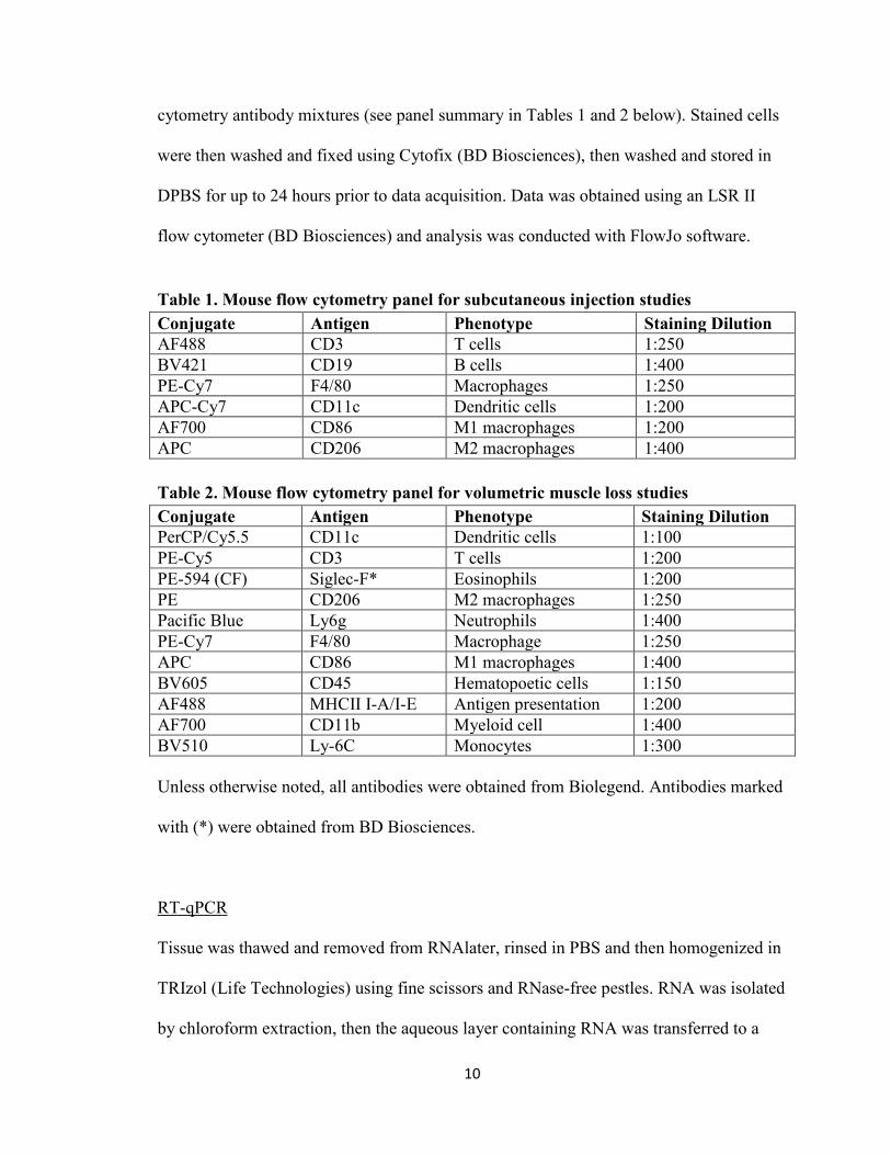
Table 1. Mouse flow cytometry panel for subcutaneous injection studies Conjugate Antigen Phenotype Staining Dilution AF488 CD3 T cells 1:250 BV421 CD19 B cells 1:400 PE-Cy7 F4/80 Macrophages 1:250 APC-Cy7 CD11c Dendritic cells 1:200 AF700 CD86 M1 macrophages 1:200 APC CD206 M2 macrophages 1:400 Table 2. Mouse flow cytometry panel for volumetric muscle loss studies Conjugate Antigen Phenotype Staining Dilution PerCP/Cy5.5 CD11c Dendritic cells 1:100 PE-Cy5 CD3 T cells 1:200 PE-594 (CF) Siglec-F* Eosinophils 1:200 PE CD206 M2 macrophages 1:250 Pacific Blue Ly6g Neutrophils 1:400 PE-Cy7 F4/80 Macrophage 1:250 APC CD86 M1 macrophages 1:400 BV605 CD45 Hematopoetic cells 1:150 AF488 MHCII I-A/I-E Antigen presentation 1:200 AF700 CD11b Myeloid cell 1:400 BV510 Ly-6C Monocytes 1:300 Unless otherwise noted, all antibodies were obtained from Biolegend. Antibodies marked
with (*) were obtained from BD Biosciences.
RT-qPCR
Tissue was thawed and removed from RNAlater, rinsed in PBS and then homogenized in
TRIzol (Life Technologies) using fine scissors and RNase-free pestles. RNA was isolated
by chloroform extraction, then the aqueous layer containing RNA was transferred to a
11
fresh tube containing an equal volume of 70% ethanol. The mixture was then applied to
RNeasy Mini columns (Qiagen) and purified according to the manufacturer's instructions.
RNA was eluted in RNase-free water and quantified using a Qubit 2.0 fluorometer
(Invitrogen). RNA was treated to remove residual DNA using a cocktail containing
DNase I, 10x DNase buffer and RNaseOUT inhibitor according to reagent protocols (Life
Technologies). Complementary deoxyribonucleic acid (cDNA) synthesis was conducted
using Superscript Reverse Transcriptase (RT) III enzyme as per manufacturer's
instructions (Life Technologies). Real time quantitative polymerase chain reaction
(qPCR) was conducted on Applied Biosystems Real Time PCR machines using SYBR
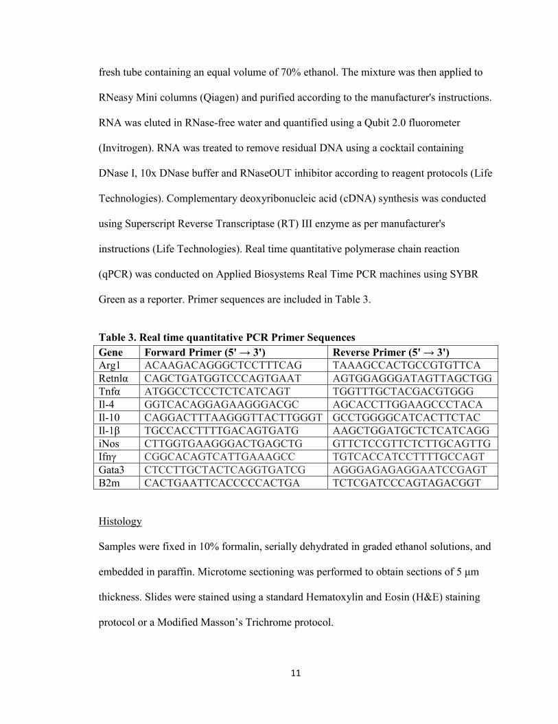
Green as a reporter. Primer sequences are included in Table 3.
Table 3. Real time quantitative PCR Primer Sequences Gene Forward Primer (5' → 3') Reverse Primer (5' → 3') Arg1 ACAAGACAGGGCTCCTTTCAG TAAAGCCACTGCCGTGTTCA Retnlα CAGCTGATGGTCCCAGTGAAT AGTGGAGGGATAGTTAGCTGG Tnfα ATGGCCTCCCTCTCATCAGT TGGTTTGCTACGACGTGGG Il-4 GGTCACAGGAGAAGGGACGC AGCACCTTGGAAGCCCTACA Il-10 CAGGACTTTAAGGGTTACTTGGGT GCCTGGGGCATCACTTCTAC Il-1β TGCCACCTTTTGACAGTGATG AAGCTGGATGCTCTCATCAGG iNos CTTGGTGAAGGGACTGAGCTG GTTCTCCGTTCTCTTGCAGTTG Ifnγ CGGCACAGTCATTGAAAGCC TGTCACCATCCTTTTGCCAGT Gata3 CTCCTTGCTACTCAGGTGATCG AGGGAGAGAGGAATCCGAGT B2m CACTGAATTCACCCCCACTGA TCTCGATCCCAGTAGACGGT
Histology
Samples were fixed in 10% formalin, serially dehydrated in graded ethanol solutions, and
embedded in paraffin. Microtome sectioning was performed to obtain sections of 5 μm
thickness. Slides were stained using a standard Hematoxylin and Eosin (H&E) staining
protocol or a Modified Masson’s Trichrome protocol.

12
Functional Testing
Animals were trained on the treadmill apparatus 48 hours prior to testing. During the
training, the treadmill was set to 5 m/min and increased by 1 m/min every minute for 5
minutes. During testing, mice were run to exhaustion starting at a speed of 5 m/min and
increased by 1 m/min every minute. The mice were considered exhausted when they
remained on the pulsed shock grid for 30 continuous seconds. All treadmill testing was
done at least 48 hours prior to the study end points where tissue samples were collected.
Clinical Trial
Eight healthy volunteers were injected with AAT in redundant tissue previously
scheduled for surgical removal in an elective surgical procedure (i.e. panniculectomy,
abdominoplasty). Each patient received a total of 2 mL of AAT in one (2 mL injection)
or two injection sites (1 mL injection). Participants had follow-up visits at 1, 2, and 4
weeks post-injection (if implant has not yet been excised) and 2 and 6 weeks post-
excision. Pain and itching were assessed at all follow-up visits. Implants were excised
during an elective surgical procedure after 1, 2, 4, 6 or 18 weeks in situ and delivered to
the lab (tissue was transported on 4°C gel packs if travel time exceeded 20 minutes).
Tissue samples were photographed, dissected, weighed, and transferred to the appropriate
storage or processing reagent for downstream analysis. Panel-reactive antibody testing at
4 and 12 weeks post injection (independent of the excision time-point) indicated if the
patient had experienced a systemic human leukocyte antigen (HLA) antibody reaction to
the material.
13
Cell Migration Assay
AAT-triggered cell migration was measured using a transwell assay relative to several
control solutions: 10% fetal bovine serum (FBS) in serum free media for a positive
control, 1% PBS in serum free media for a buffer control, and serum free media alone for
a negative control. Human adipose-derived stem cells (ASCs) were thawed and grown in
basic growth media supplemented with 1 ng/mL basic fibroblastic growth factor (bFGF)
and passaged and split once at approximately 80% confluency, with media changes every
2-3 days. Upon reaching 80% confluency a second time, cells were serum starved for 24
hours prior to performing assay. After starvation, cells were trypsinized and a single cell
suspension was created at 300,000 cells/mL in serum free media. Sample was prepared
by adding AAT to serum free media at a 1% (v/v) concentration and vortexing. In some
assays, samples incubated at room temperature for 15 minutes and centrifuged to remove
large chunks of AAT that might stick to the transwell membrane. Sample, 10% FBS, 1%
buffer and serum free media (600 µl) were added in triplicate wells of a 24 well plate, a
transwell insert was placed on top, and the plate was allowed to acclimate in an
incubator. After acclimation, 100 μL of the cell suspension was added into the upper
chamber of each transwell (300,000 cells per well) and incubated at 37˚C for 6 hours. All
cells remaining on the upper side of the membrane were removed using a cotton swab
and any excess AAT was removed from the lower membrane by rinsing in DPBS.
Transwells were fixed in methanol for 15 minutes at room temperature, stained with
DAPI, and then imaged within 72 hours. Cell nuclei were counted in a 50x field of view.
Cell migration was calculated relative to positive and negative control groups.
14
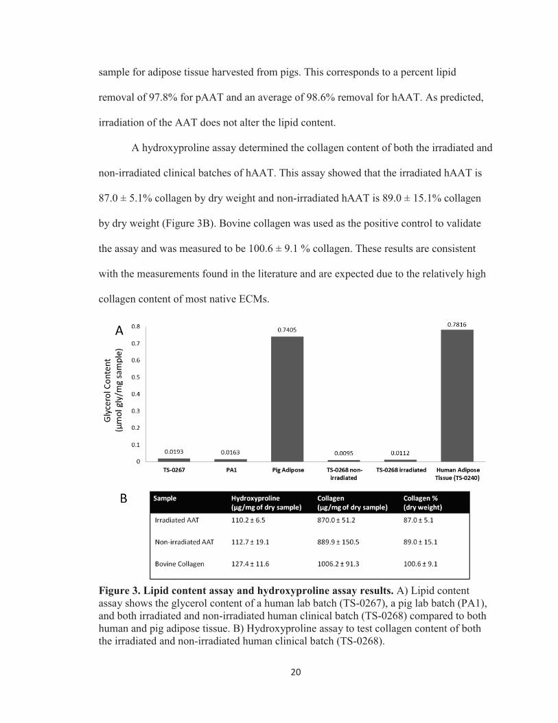
Lipid Content
The total lipid content of biomaterials was quantified using a triglyceride colorimetric
assay. Adipose samples and AAT were minced into 1 mm or smaller pieces to disrupt
physical barriers and release lipids. Organic extraction was conducted by the Schwartz
method and enzymatic reactions were carried out using Infinity TG Reagent according to
the manufacturer's protocol. Absorbance was measured at 540 nm and concentration of
the samples was determined using a glycerol standard curve (samples prepared in water).
Collagen Content
Since hydroxyproline is largely restricted to collagen, the measurement of
hydroxyproline levels can be used as an indicator of collagen content. In this assay,
hydroxyproline concentration is determined by a reaction of oxidized hydroxyproline
with 4-(Dimethylamino)benzaldehyde (DMAB), which results in a colorimetric (560 nm)
product, proportional to the hydroxyproline present. Sample preparation consisted of
lyophilization and hydrochloric acid hydrolysis of 10 mg of sample (dry weight) at 120˚C
for 3 hours. The hydrolyzed samples were diluted 100x and added in triplicate to a 96
well plate in multiple dilutions, along with samples for a hydroxyproline standard curve.
A spiked control was also used to detect any interfering endogenous compounds. All
wells in the plate were evaporated to dryness. The colorimetric reaction was carried out
using reagents provided in the Hydroxyproline Assay Kit (Sigma) according to the
manufacturer's instructions. Absorbance was measured at 560 nm and the standard curve
was used calculate hydroxyproline content of unknown samples.
15
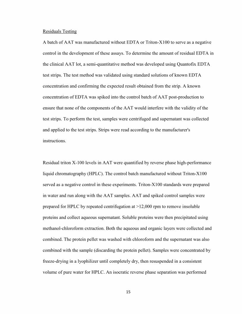
Residuals Testing
A batch of AAT was manufactured without EDTA or Triton-X100 to serve as a negative
control in the development of these assays. To determine the amount of residual EDTA in
the clinical AAT lot, a semi-quantitative method was developed using Quantofix EDTA
test strips. The test method was validated using standard solutions of known EDTA
concentration and confirming the expected result obtained from the strip. A known
concentration of EDTA was spiked into the control batch of AAT post-production to
ensure that none of the components of the AAT would interfere with the validity of the
test strips. To perform the test, samples were centrifuged and supernatant was collected
and applied to the test strips. Strips were read according to the manufacturer's
instructions.
Residual triton X-100 levels in AAT were quantified by reverse phase high-performance
liquid chromatography (HPLC). The control batch manufactured without Triton-X100
served as a negative control in these experiments. Triton-X100 standards were prepared
in water and run along with the AAT samples. AAT and spiked control samples were
prepared for HPLC by repeated centrifugation at >12,000 rpm to remove insoluble
proteins and collect aqueous supernatant. Soluble proteins were then precipitated using
methanol-chloroform extraction. Both the aqueous and organic layers were collected and
combined. The protein pellet was washed with chloroform and the supernatant was also
combined with the sample (discarding the protein pellet). Samples were concentrated by
freeze-drying in a lyophilizer until completely dry, then resuspended in a consistent
volume of pure water for HPLC. An isocratic reverse phase separation was performed

16
using an HC-C18(2) column and two mobile phases: HPLC-grade water and 100%
acetonitrile. The peak corresponding to Triton-X100 was measured and quantified
relative to standards.
Statistical Analysis
Statistical analysis was performed using GraphPad Prism software. In grouped analyses
with a single variable, significance was determined by one-way analysis of variance
(ANOVA) using the Holm-Sidak correction for multiple comparisons where applicable
(α = 0.05). Significance in grouped analyses with two variables was calculated using two-
way ANOVA with Tukey post-hoc testing (α = 0.05). P values less than 0.05 were
considered statistically significant (* < 0.05, ** < 0.01, *** < 0.001, **** < 0.0001).
Plotted values represent the arithmetic or geometric mean (RT-qPCR data only) of the
data set. Error bars represent +/- one standard deviation or geometric standard deviation
(RT-qPCR only).

17
RESULTS
Building on previous studies done in the Elisseeff lab which characterized the
physical properties of AAT, we initially sought to study the biochemical characteristics
of AAT. Biochemical assays performed included an in vitro cell migration assay, residual
chemicals testing, a lipid content assay, and a collagen content assay. There were two
main purposes for collecting this data: to get a better understanding of the biochemical
composition and properties of the material, and to start building a database with the goal
of understanding the batch–to–batch differences in AAT. This information will help
define the expected variability from both tissue donors and from any changes in the
manufacturing process, and will be critical for scaling up the manufacturing protocols for
later stage clinical trials and eventual commercialization. The biochemical
characterizations also allowed us to study how the terminal sterilization process of
gamma irradiation might change the properties of the final product.
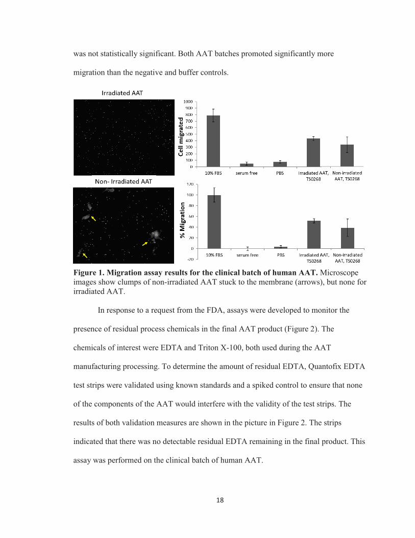
The in vitro cell migration assays were conducted on both irradiated and non-
irradiated samples from the human clinical batch (TS-02568) (Figure 1). The results of
these assays showed that both the irradiated and the non-irradiated samples of human
AAT promote cell migration of ASCs across a transwell membrane. In a pilot
experiment, images taken of the transwell membrane showed that the non-irradiated AAT
was more likely to stick to the membrane, potentially indicating a difference in
mechanical properties. A second assay was performed with the samples centrifuged to
remove large chunks that might stick to membrane, thus ensuring migration would only
be triggered by interaction with soluble factors. In this second assay, the non-irradiated
AAT resulted in slightly less cell migration than the irradiated batch. However, this result
18

19
20

21
pAAT – 1 week hAAT – 1 week
Supe
rior
Infe
rior
pAAT – 3 week hAAT – 3 week
Supe
rior
Infe
rior

22
smoother and more homogenous. Cell infiltration occurs from the surrounding tissues
into the implant, indicating that the material promotes cell migration and corroborating
the results of the in vivo cell migration assay. It is also interesting to note the brown fat
pad adjacent to the implant in the superior position (visible in the pAAT 3-week top
section) which could potentially impact the cellular response to the material.
The immune cell profile of the subcutaneous implants after 1 or 3 weeks in vivo
were assessed using flow cytometry (Figure 5). Overall, the flow analysis showed that
human and porcine AAT had similar immune cell profiles at each time point. A greater
percentage of the cells migrating into the implant were CD206+ macrophages (M2
polarized) than CD86+ (M1 polarized) macrophages, suggesting that the biomaterial
Figure 5. Subcutaneous flow cytometry results at 1 and 3 weeks. Implants were pooled for each animal to ensure adequate cell number for analysis. Statistical significance calculated using two-way ANOVA with TUKEY post-hoc testing for everything except F4/80hi macrophage activation for which a one-way ANOVA with TUKEY post-hoc testing was used. * < 0.05, ** < 0.01, *** < 0.001, **** < 0.0001.

23
skews macrophage polarization towards an M2 phenotype. When considering this result,
it is important to understand that macrophage polarization is spectrum rather than a
binary change, so macrophages could potentially be somewhere between an M1 and an
M2 phenotype. It was also noted that the level of macrophage activation (F4/80hi relative
to F4/80lo) was higher at 1 week than at 3 weeks. The percentage of CD3+ T cells in the
implant is significantly higher at 3 weeks than at 1 week, indicating that the T cell
response begins prior to 1 week and increases to a peak at some later time point.
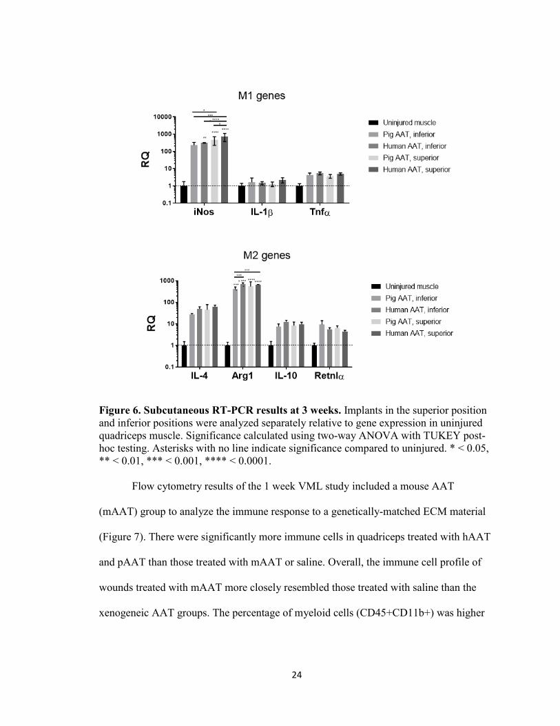
Gene expression analysis performed on the SQ implants at 3 weeks post-injection
showed that iNos, an M1 gene, and Arg1, an M2 gene, were both significantly increased
in almost all of the implants (Figure 6). The increase in iNos gene expression in the
pAAT far implant was not considered statistically significant. This information is
interesting given the higher percentage of CD206+ M2 macrophages than CD86+ M1
macrophages in the implant observed in the flow cytometry data. Other genes including
Il-4 were elevated in the implant relative to normal muscle, but these results were not
statistically significant.
A mouse volumetric muscle wound (VML) model was used to study the response
to both pAAT and hAAT in a wound environment. In this experiment, a critical sized
defect was created in the mouse quadriceps muscle and was filled with a biomaterial or
saline as a control. To explore whether there was an effect related to xenogenic AAT in
the mouse wound model, these experiments also included mouse-derived AAT produced
from C57BL/6 mice as a syngeneic ECM control. Response to the biomaterials was
assessed at 1 week post- injury.
24
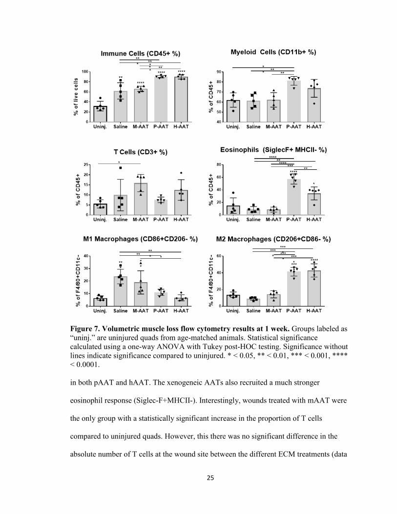
Figure 6. Subcutaneous RT-PCR results at 3 weeks. Implants in the superior position and inferior positions were analyzed separately relative to gene expression in uninjured quadriceps muscle. Significance calculated using two-way ANOVA with TUKEY post-hoc testing. Asterisks with no line indicate significance compared to uninjured. * < 0.05, ** < 0.01, *** < 0.001, **** < 0.0001. Flow cytometry results of the 1 week VML study included a mouse AAT
(mAAT) group to analyze the immune response to a genetically-matched ECM material
(Figure 7). There were significantly more immune cells in quadriceps treated with hAAT
and pAAT than those treated with mAAT or saline. Overall, the immune cell profile of
wounds treated with mAAT more closely resembled those treated with saline than the
xenogeneic AAT groups. The percentage of myeloid cells (CD45+CD11b+) was higher
25
Figure 7. Volumetric muscle loss flow cytometry results at 1 week. Groups labeled as “uninj.” are uninjured quads from age-matched animals. Statistical significance calculated using a one-way ANOVA with Tukey post-HOC testing. Significance without lines indicate significance compared to uninjured. * < 0.05, ** < 0.01, *** < 0.001, **** < 0.0001. in both pAAT and hAAT. The xenogeneic AATs also recruited a much stronger
eosinophil response (Siglec-F+MHCII-). Interestingly, wounds treated with mAAT were
the only group with a statistically significant increase in the proportion of T cells
compared to uninjured quads. However, this there was no significant difference in the
absolute number of T cells at the wound site between the different ECM treatments (data
26
not shown). The data also indicates that pAAT and hAAT promote greater skewing of
polarized macrophages to an M2-like phenotype than mAAT, though overall mAAT is
still somewhat M2-polarizing. Saline treatment promotes a more M1-like phenotype than
any ECM treatment, as determined by the relative proportions of CD206+CD86- and
CD86+CD206- macrophages.
RT-qPCR analysis of the wounded muscle 1 week after treatment showed that
wounds treated with mouse syngeneic AAT were not significantly different than wounds
treated with saline or uninjured muscle in any of the genes tested (Figure 8). Most of
these M2 genes - including Il-4, Arg1, and Retnlα - were significantly increased in pAAT
and hAAT treated wounds compared to saline treated wounds and uninjured muscle.
Increases were also observed in M1 genes relative to saline treatment; though these
increases were generally similar between different ECMs. Most importantly, expression
of Il-4 increased more than 100-fold in pAAT and hAAT relative to control groups,
whereas mAAT also increased but was not significantly different than saline. Taken
together, these results are consistent with flow cytometry analysis of macrophage
polarization and indicate a difference in the profile of immune cells migrating to wounds
treated with syngeneic ECM than those treated with xenogeneic ECMs.
A human clinical trial studied the safety of AAT when implanted subcutaneously
in human participants. The primary outcome measures of this study were histopathology,
safety, and patient and physician satisfaction. On an exploratory basis, the immune cell
populations and cell migration into the implant were also characterized.

27
Figure 8. Volumetric muscle loss RT-PCR results at 1 week. Statistical significance is calculated relative to saline controls. * < 0.05, ** < 0.01, *** < 0.001, **** < 0.0001.
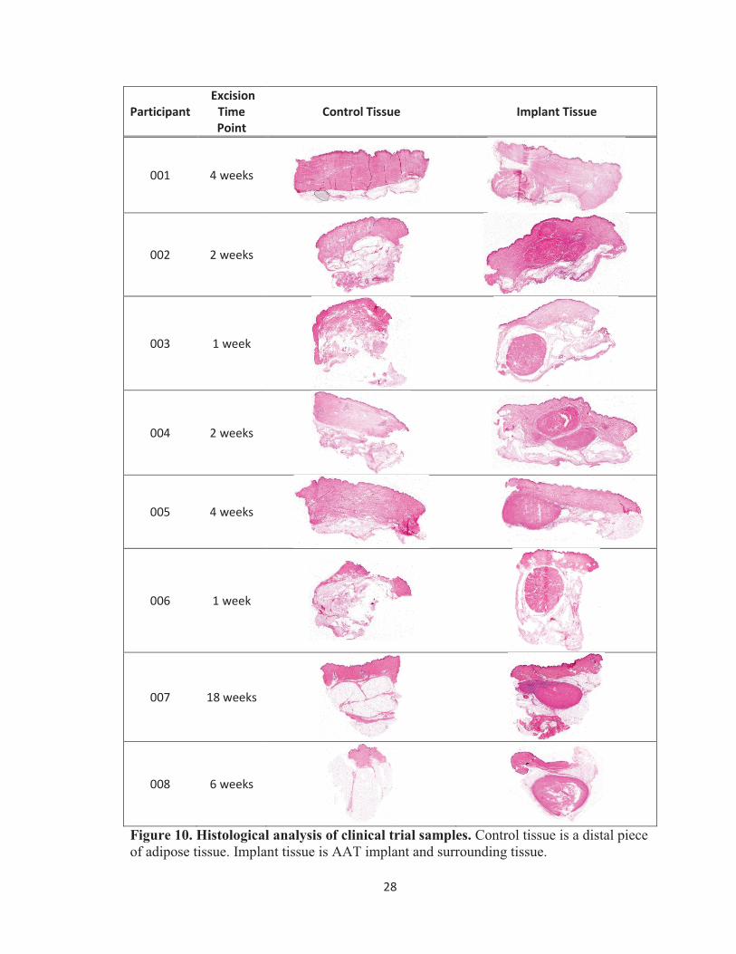
Histological analysis of the clinical trial implants showed minimal negative
inflammatory response with cell migration into the implant (Figure 10), demonstrating
the potential for new tissue formation. The implants also show good volume retention at
up to 18 weeks post-injection, the latest time-point studied. The implants do not show any
indication of capsule or cyst formation or tissue necrosis. All of these histological results
indicate demonstrate the biocompatibility of AAT when implanted into humans and that
the implant may support new tissue formation.
28
Participant Excision
Time Point
Control Tissue Implant Tissue
001 4 weeks
002 2 weeks
003 1 week
004 2 weeks
005 4 weeks
006 1 week
007 18 weeks
008 6 weeks
29
30
31
32

33
DISCUSSION
Preclinical studies showed promising results for the safe and effective use of AAT
for soft tissue reconstruction in humans. In both VML and subcutaneous studies in mice,
AAT demonstrated good tissue integration with cell migration into the implant. No
significant inflammatory response was noted in these experiments. Results obtained from
the subcutaneous implants indicate that macrophages begin migrating in from the
surrounding tissue within 1 week. By three weeks, however, the macrophages are no
longer as dominant and adaptive cells such as T cells have begun to appear. This mimics
the immune response to a wound during which macrophages enter the wound first
followed by lymphocytes [18]. RT-qPCR analysis showed an increase in gene expression
for both iNos, the inducible form of nitric oxide synthase, and Arg1, Arginase 1. The iNos
gene generates nitric oxide and is an important enzyme in the macrophage inflammatory
response [27]. Arg1 is an enzyme that metabolizes arginine and is highly expressed on
M2 macrophages. Resident adipose tissue-associated macrophages typically have M2-
like polarization and express Arg1 and are important for balancing inflammation in fat
tissue and maintaining metabolism. Interestingly, Arg1 and iNos compete to metabolize
arginine when they are co-expressed [28]. The co-expression of iNos and Arg1 likely
indicates that the macrophages present within the AAT at 3 weeks are neither purely M1
nor M2, though they may be skewed towards M2 at a population level.
Flow cytometry analysis of the AAT-treated quadriceps muscles at 1 week after
critical injury showed significant infiltration of immune cells into the wound site. Overall
the flow data suggest similarity between the xenogeneic AATs, including an increased
percentage of recruited immune cells (CD45+), eosinophils (CD45+ CD11b+ SiglecF+
34
MHCII-), and M2 macrophages (CD206+CD86-) relative to saline and uninjured
controls. Although the differences were not significant, syngeneic mouse AAT was also
somewhat more M2-polarizing than saline alone, and recruited greater percentages of
immune cells. However, the macrophages present in mouse AAT tended to be more M1-
polarized (CD86+) than in the other AATs. Gene expression analysis also showed that
significantly more interleukin 4 (Il-4) was present in wounds treated with both pig and
human AAT than those treated with saline or mouse AAT. This indicates that in some
contexts, AAT promotes the migration of immune cells which trigger the release of this
key pro-regenerative cytokine. This increased expression of Il-4 is not correlated with an
increase in the proportion or absolute number of T cells in the wound, but may be due to
the increased activity of myeloid cells orchestrating the upregulation of Il-4. Missing
from these analyses are quantifications of different T cells subsets, particularly TH2
helper T cells, which are essential for creating a pro-regenerative microenvironment.
The combined results of our syngeneic versus xenogeneic AAT studies indicate
that pro-regenerative immune responses to ECM are likely not driven by non-specific
damage-associated molecular patterns (DAMPs) inherent to all ECMs [29]. It is possible
that a response related to foreign antigens in both the pig and human AAT is significantly
driving the observed increases in Il-4 expression, eosinophils, or M2 macrophage
polarization. However, there are likely factors other than species contributing to the
differences observed between AAT treatments. Biochemical characterizations of the
mouse AAT may reveal a key difference that helps to explain the loss of the pro-
regenerative phenotype. More rapid resorption of the mouse AAT compared to the
xenogeneic materials may also be preventing the immune system from mounting a full

35
M2/Th2 response. Additional studies will be needed to confirm the mechanism or
mechanisms driving these phenotypes. Future work will investigate an allogeneic ECM
material in the wound environment in order to better understand the implications of these
results for the translation of AAT to clinical applications.
In Phase I clinical trial studies, AAT proved safe, biocompatible, and well-
tolerated by all outcomes measured. No serious adverse events were reported, and all
anticipated adverse events were mild and localized to the injection site. Both physicians
and participants reported overall satisfaction with the comfort/ease of use and appearance
of the injected area. Importantly, implant volume was retained until the latest measured
time point of 18 weeks, and the material integrated into the surrounding tissue rather than
becoming isolated by fibrosis. Histological analysis of the implants also showed
significant cell migration into the implant from surrounding tissue which generally
increased as time went on. Although immune cells were observed within the implant, the
lack of a systemic immune reaction suggests that any immune-modulation is occurring
locally. All of this data indicates that AAT is safe for use in humans and has the potential
to support cell infiltration and new tissue formation. Future analyses will be conducted to
identify specific types of immune cells present in the AAT. It will be critical to determine
whether these cells are pro-inflammatory immune cells, pro-regenerative immune cells,
or stem cells to fully understand the cellular response to the material.
36
CONCLUSION AND FUTURE WORK
This work describes the development of an adipose-tissue derived ECM material
for soft tissue reconstruction, including many of the material characterizations and
preclinical studies that lead to the first in human study. These preclinical results and
others were critical in obtaining FDA approval for initial clinical testing. Consequently,
the safety results obtained from this first clinical study will be leveraged to advance AAT
to Phase II clinical testing to confirm safety and determine efficacy in patients. Our most
recent animal studies have sought to identify the mechanisms of ECM-mediated
immunomodulation and will be critical to help define clinical indications for AAT and
inform future research.
In these preclinical studies, AAT demonstrated volume retention, significant
tissue integration, and minimal inflammation. Subcutaneous implants attracted large
proportions of macrophages around 1 week, followed later by the clearing of the
macrophages and increased migration of T cells. In a mouse wound environment, pig and
human AAT elicited a strong M2-macrophage response while syngeneic mouse AAT
elicited a more neutrally-polarized macrophage response that was similar to saline treated
wounds. However, CD3+ T cell response was not significantly impacted by ECM tissue
source, which may suggest a combination of factors (both species-specific and non-
species specific properties of ECMs) is responsible for the immune response to these
materials. The ability of AAT to modulate immune response and induce a favorable pro-
regenerative environment could potentially be harnessed to improve wound healing and
reduce scarring after injury. This data indicates that AAT could be a good substitute for
autologous fat transfer in the treatment of soft tissue defects.
37
Future studies will determine the immune response to allogeneic ECM in a wound
model and determine mechanisms contributing to the ECM-associated immune
microenvironment. Biochemical characterizations will continue for each new
manufactured lot to form an understanding of batch-to-batch variability and help
determine which properties of the material are correlated with successful clinical
outcomes. These factors and others will be considered for design and validation of a
scaled-up manufacturing process for future clinical trials involving significantly more
participants. Together, this work will enable Phase II clinical testing, which will be
conducted to test the safety and efficacy of AAT in filling small soft tissue defects in
human patients.
38
REFERENCES
1. Ring, A., et al., Reconstruction of Soft-Tissue Defects at the Foot and Ankle after Oncological Resection. Frontiers in Surgery, 2016. 3(15).
2. Coleman, S.R., Structural fat grafting: more than a permanent filler. Plast Reconstr Surg, 2006. 118(3 Suppl): p. 108S-120S.
3. Ross, R.J., et al., Autologous fat grafting: current state of the art and critical review. Ann Plast Surg, 2014. 73(3): p. 352-7.
4. Rubin, J.P. and K.G. Marra, Soft Tissue Reconstruction, in Adipose-Derived Stem Cells: Methods and Protocols, J.M. Gimble and B.A. Bunnell, Editors. 2011, Humana Press: Totowa, NJ. p. 395-400.
5. Rai, S., A.M. Marsland, and V. Madan, Facial Fat Necrosis Following Autologous Fat Transfer and its Management. Journal of Cutaneous and Aesthetic Surgery, 2014. 7(3): p. 173-175.
6. Wainwright, D.J., Use of an acellular allograft dermal matrix (AlloDerm) in the management of full-thickness burns. Burns, 1995. 21(4): p. 243-248.
7. Gilbert, T.W., T.L. Sellaro, and S.F. Badylak, Decellularization of tissues and organs. Biomaterials, 2006. 27(19): p. 3675-3683.
8. Frantz, C., K.M. Stewart, and V.M. Weaver, The extracellular matrix at a glance. Journal of Cell Science, 2010. 123(24): p. 4195-4200.
9. Hinderer, S., S.L. Layland, and K. Schenke-Layland, ECM and ECM-like materials — Biomaterials for applications in regenerative medicine and cancer therapy. Advanced Drug Delivery Reviews, 2016. 97: p. 260-269.
10. Hoshiba, T., et al., Decellularized matrices for tissue engineering. Expert Opinion on Biological Therapy, 2010. 10(12): p. 1717-1728.
11. Coelho, M., T. Oliveira, and R. Fernandes, Biochemistry of adipose tissue: an endocrine organ. Archives of Medical Science : AMS, 2013. 9(2): p. 191-200.
12. Tran, T.T. and C.R. Kahn, Transplantation of adipose tissue and stem cells: role in metabolism and disease. Nat Rev Endocrinol, 2010. 6(4): p. 195-213.
13. Coleman, S.R., Structural Fat Grafting: More Than a Permanent Filler. Plastic and Reconstructive Surgery, 2006. 118(3S): p. 108S-120S.
14. Gallagher, D., et al., Healthy percentage body fat ranges: an approach for developing guidelines based on body mass index. Am J Clin Nutr, 2000. 72(3): p. 694-701.
15. Rehman, J., et al., Secretion of angiogenic and antiapoptotic factors by human adipose stromal cells. Circulation, 2004. 109(10): p. 1292-8.
16. Ouchi, N. and K. Walsh, Adiponectin as an anti-inflammatory factor. Clinica Chimica Acta, 2007. 380(1–2): p. 24-30.
17. Ferrante, A.W., Jr., The immune cells in adipose tissue. Diabetes Obes Metab, 2013. 15 Suppl 3: p. 34-8.
18. Park, J.E. and A. Barbul, Understanding the role of immune regulation in wound healing. The American Journal of Surgery, 2004. 187(5, Supplement 1): p. S11-S16.
19. Wynn, T.A., A. Chawla, and J.W. Pollard, Macrophage biology in development, homeostasis and disease. Nature, 2013. 496(7446): p. 445-55.
39
20. Sadtler, K., et al., Developing a pro-regenerative biomaterial scaffold microenvironment requires T helper 2 cells. Science, 2016. 352(6283): p. 366-70.
21. Brown, B.N., et al., Macrophage phenotype as a predictor of constructive remodeling following the implantation of biologically derived surgical mesh materials. Acta Biomater, 2012. 8(3): p. 978-87.
22. Gordon, S. and P.R. Taylor, Monocyte and macrophage heterogeneity. Nat Rev Immunol, 2005. 5(12): p. 953-64.
23. Adamson, R., Role of macrophages in normal wound healing: an overview. J Wound Care, 2009. 18(8): p. 349-51.
24. Wu, I., et al., An injectable adipose matrix for soft-tissue reconstruction. Plast Reconstr Surg, 2012. 129(6): p. 1247-57.
25. Wu, I., Design and translation of an adipose-derived soft tissue substitute, in Biomedical Engineering. 2014, Johns Hopkins University: Baltimore.
26. Pulagam, S.R., T. Poulton, and E.P. Mamounas, Long-term clinical and radiologic results with autologous fat transplantation for breast augmentation: case reports and review of the literature. Breast J, 2006. 12(1): p. 63-5.
27. McNeill, E., et al., Regulation of iNOS function and cellular redox state by macrophage Gch1 reveals specific requirements for tetrahydrobiopterin in NRF2 activation. Free Radical Biology and Medicine, 2015. 79: p. 206-216.
28. Murray, P.J., Amino acid auxotrophy as a system of immunological control nodes. Nat Immunol, 2016. 17(2): p. 132-139.
29. Sofat, N., et al., Interaction between extracellular matrix molecules and microbial pathogens: evidence for the missing link in autoimmunity with rheumatoid arthritis as a disease model. Frontiers in Microbiology, 2014. 5: p. 783.
40
CURRICULUM VITAE
Education Johns Hopkins University, Baltimore, MD May 2017 Master of Science in Engineering in Biomedical Engineering Villanova University, Villanova, PA May 2015 Bachelor of Science in Chemical Engineering Minors in Bioengineering and Mathematics Research Experience Graduate student researcher, Laboratory of Dr. Jennifer Elisseeff, Johns Hopkins August 2015-May 2017 Use techniques such as flow cytometry, RT-PCR, cell culture, and histology to test a decellularized adipose extracellular matrix product in various animal models to determine the effectiveness, safety, and immune modulatory properties of the product. Assist in the execution of the Phase I and the planning of the Phase II clinical trial studies for the product, including preparation of regulatory documents. Optimize production protocols and design quality control testing for the product. Analyze and organize data for use in presentations, manuscripts, and regulatory documents. Prepare grant applications. Explore the ability of a biomaterial scaffold to modulate the tumor immune microenvironment. Evaluate the role of the immune system on the growth of cancer in an animal model. Analyze the effect of drug administration on the response to the biomaterial and the growth of cancer. Research Assistant, Invisible Sentinel, Philadelphia, PA May 2015-August 2015 Assisted in the development and validation of a new assay, including testing limit of detection and inclusivity/exclusivity. Tested and validated new technology to be used with a company product. Assisted with cell culture, inventory, and other laboratory projects. Undergraduate student researcher, Biomaterials and Drug Delivery Laboratory, Villanova University August 2014-May 2015 Conducted laboratory experiments to develop effective routes of targeted drug delivery to the lungs using nanoparticles. Synthesized PLA-PEG copolymer micelles in which to encapsulate a drug to treat severe asthma. Optimized polymer synthesis protocols. Research Intern, Invisible Sentinel, Philadelphia, PA May 2013-August 2013 Developed a pure positive control for a company product by sequencing and cloning strands of DNA. Created a database of different strains of bacteria to be used by the company. Assisted with various research projects in a laboratory setting.
41
Teaching Experience Grader, Johns Hopkins University, Baltimore, MD Course: Statistical Mechanics and Thermodynamics Fall 2015, Fall 2016 Course: Models and Simulations Spring 2015, Spring 2016 Teaching Assistant, Villanova University, Villanova, PA Course: Freshman Miniproject: Artificial Kidney October 2013-May 2015 Conference Presentations A.J. Parrillo, BS, A.E. Anderson, BS, I. Wu, PhD, K. Sadtler, PhD, L. Chung, BS, C. Cooney, MPH, D. Cooney, MD, PhD, R.M. Payne, BS, J. Aston, BS, P. Byrne, MD, J.H. Elisseeff, PhD. An Adipose Tissue Extracellular Matrix Derived Biomaterial for Soft Tissue Reconstruction. Northeast Bioengineering Conference. Newark, NJ. March 31, 2017-April 2, 2017
Related Documents


![Phase 1b: Preliminary clinical activity and immune ... · Phase 1b: Preliminary clinical activity and immune activation for NKTR-262 [TLR 7/8 agonist] plus bempegaldesleukin (NKTR-214)](https://static.cupdf.com/doc/110x72/5e2b12f484810b1f5f0a10e6/phase-1b-preliminary-clinical-activity-and-immune-phase-1b-preliminary-clinical.jpg)
