Pharynx Dr. Deepak K Gupta

Welcome message from author
This document is posted to help you gain knowledge. Please leave a comment to let me know what you think about it! Share it to your friends and learn new things together.
Transcript

PharynxDr. Deepak K Gupta

Introduction
• Musculo-fascial half-cylinder that links the oral and nasal cavities in the head to the larynx and esophagus in the neck
• Common pathway for air and food
• Attached above to the base of the skull and is continuous below with the top of them esophagus
• At the level of vertebra C6
www.facebook.com/notesdental
www.facebook.com/notesdental
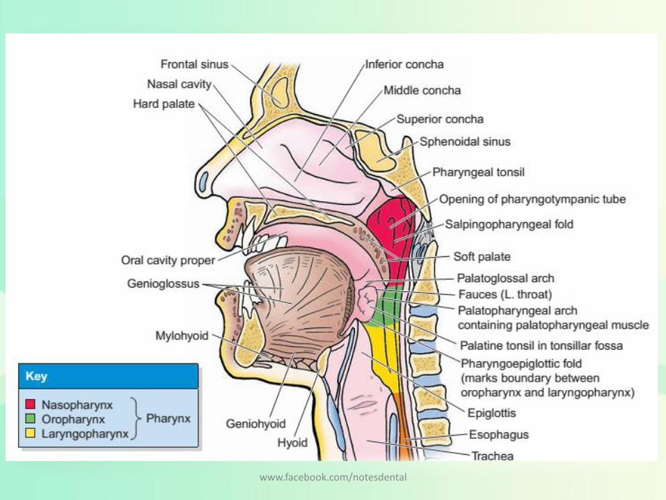
Region of Pharynx
• The walls of the pharynx are attached anteriorlyto the margins of the nasal cavities, oral cavity, and larynx
• Subdivided into three regions having opening of
– Nasopharynx : posterior apertures (choanae) of the nasal cavities
– Oropharynx : posterior opening of the oral cavity (oropharyngeal isthmus)
– Laryngopharynx: superior aperture of the larynx (laryngeal inlet) opens
www.facebook.com/notesdental
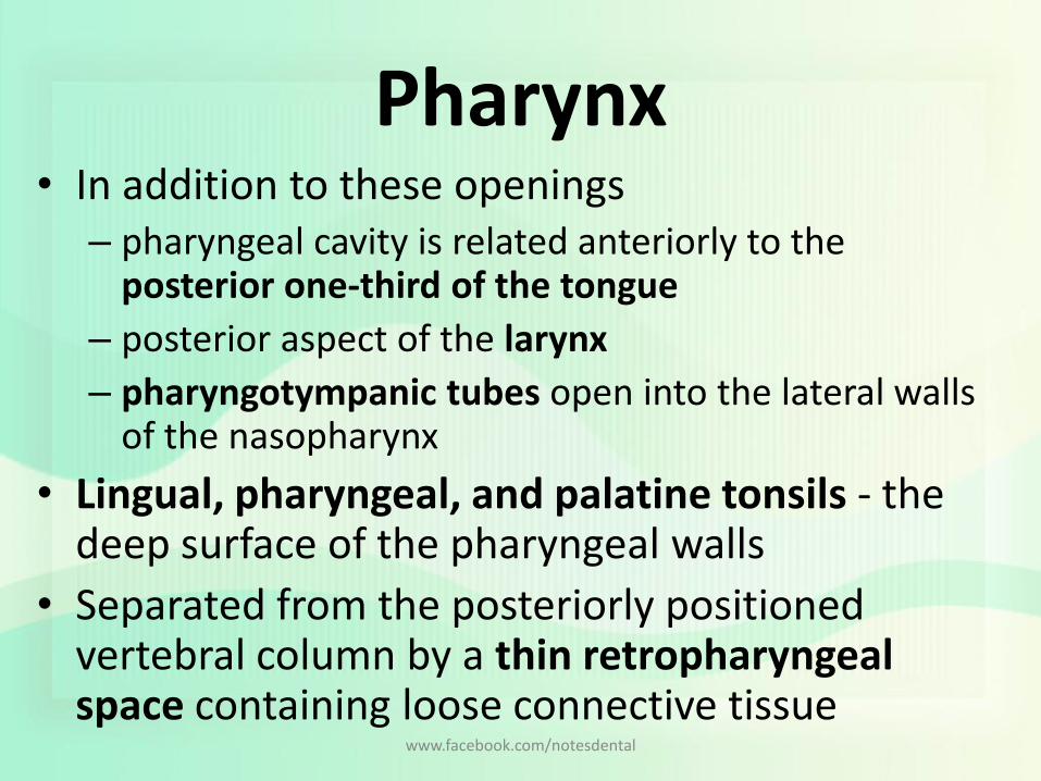
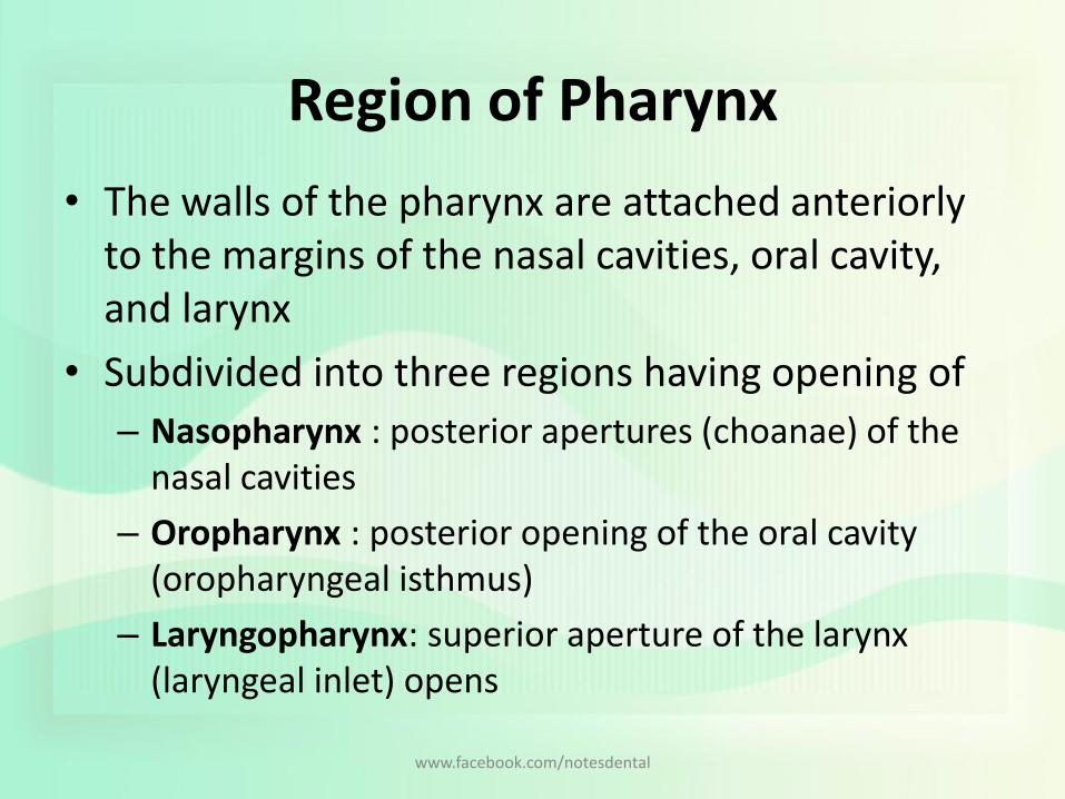
Pharynx• In addition to these openings
– pharyngeal cavity is related anteriorly to the posterior one-third of the tongue
– posterior aspect of the larynx
– pharyngotympanic tubes open into the lateral walls of the nasopharynx
• Lingual, pharyngeal, and palatine tonsils - the deep surface of the pharyngeal walls
• Separated from the posteriorly positioned vertebral column by a thin retropharyngeal space containing loose connective tissue
www.facebook.com/notesdental

www.facebook.com/notesdental

Skeletal framework
• 2 sides of the pharyngeal wall are welded together posteriorly in the midline by a vertically oriented cord-like ligament - pharyngeal raphe– From pharyngeal tubercle on the base of the skull – To the level of cervical vertebra (C6) : blends with connective
tissue in the posterior wall of the esophagus
• Irregular C-shaped line of pharyngeal wall attachment on the base of the skull– Begins at the posterior margin of the medial plate of the
pterygoid process of the sphenoid bone, – Then just inferior to the cartilaginous part of the
pharyngotympanic tube– then passes onto the petrous part of the temporal bone to
reach pharyngeal tubercle
www.facebook.com/notesdental

Bony Attachment for the lateral pharyngeal walls
• Vertical line of attachment for the lateral pharyngeal walls is discontinuous and in three parts.
• First part– Begins superiorly on the posterior edge of the medial pterygoid plate -
inferior to pharyngotympanic tube– onto the pterygoid hamulus– line descends along the pterygomandibular raphe
• Second part– begins on the lower aspect of the stylohyoid ligament– line continues onto the lesser horn of hyoid bone– then turns and runs posteriorly along the entire upper surface of the greater
horn of the hyoid where it terminates
• Third part– begins superiorly on the superior tubercle of the thyroid cartilage– descends along the oblique line to the inferior tubercle– Then over the cricothyroid muscle along a tendinous thickening of fascia to
the cricoid cartilage where it terminateswww.facebook.com/notesdental

www.facebook.com/notesdental
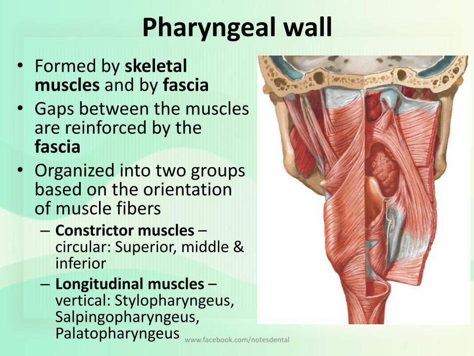
Pharyngeal wall• Formed by skeletal
muscles and by fascia• Gaps between the muscles
are reinforced by the fascia
• Organized into two groups based on the orientation of muscle fibers– Constrictor muscles –
circular: Superior, middle & inferior
– Longitudinal muscles –vertical: Stylopharyngeus, Salpingopharyngeus, Palatopharyngeus www.facebook.com/notesdental
Constrictor muscles
www.facebook.com/notesdental
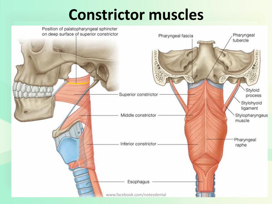
Constrictor muscles
• The 3 constrictor muscle on each side - major contributors to the structure of the pharyngeal wall– Superior, Middle and inferior
• Posteriorly, the muscles from each side are joined together by the pharyngeal raphe
• Anteriorly, these muscles attach to bones and ligaments related to the lateral margins of the nasaland oral cavities and the larynx.
• Walls of three flower pots stacked one on the other• Constrict or narrow the pharyngeal cavity.• Innervated by the pharyngeal branch of the vagus
nerve [X]
www.facebook.com/notesdental
Superior constrictors
• Upper part of the pharyngeal cavity
• Attached anteriorly to the pterygoid hamulus, pterygomandibular raphe, and adjacent bone of the mandible.
• Muscle fans out posteriorly and joins with its partner muscle from the other side at the pharyngeal raphe
• Palatopharyngeal sphincter– Special band of muscle originating from anterolateral
surface of the soft palate
– Circles the inner aspect of the pharyngeal wall
• Functions: constricts during swallowing - prominent ridge on the deep aspect of the pharyngeal wall
www.facebook.com/notesdental
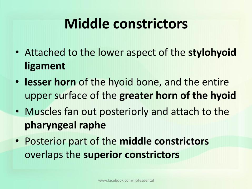
Middle constrictors
• Attached to the lower aspect of the stylohyoidligament
• lesser horn of the hyoid bone, and the entire upper surface of the greater horn of the hyoid
• Muscles fan out posteriorly and attach to the pharyngeal raphe
• Posterior part of the middle constrictors overlaps the superior constrictors
www.facebook.com/notesdental
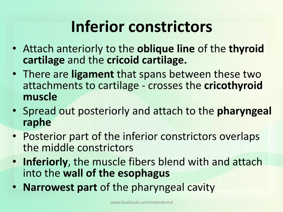
Inferior constrictors• Attach anteriorly to the oblique line of the thyroid
cartilage and the cricoid cartilage.• There are ligament that spans between these two
attachments to cartilage - crosses the cricothyroidmuscle
• Spread out posteriorly and attach to the pharyngeal raphe
• Posterior part of the inferior constrictors overlaps the middle constrictors
• Inferiorly, the muscle fibers blend with and attach into the wall of the esophagus
• Narrowest part of the pharyngeal cavitywww.facebook.com/notesdental

Longitudinal muscles
www.facebook.com/notesdental

Longitudinal muscles
• Named according to their origins– Stylopharyngeus: styloid process of the temporal bone
– Salpingopharyngeus: cartilaginous part of the pharyngotympanic tube
– Palatopharyngeus: soft palate
• Function– elevate the pharyngeal wall, or
– during swallowing, pull the pharyngeal wall up and food bolus into the esophagus
• Nerve Supply: Except stylopharyngeus(Glossopharyngeal nerve ) both the muscle is supplied by Vagus Nerve
www.facebook.com/notesdental

Longitudinal muscles• Stylopharyngeus
– cylindrical stylopharyngeus muscle– medial surface of the styloid process– descends between the superior and middle constrictor muscles– fan out on, and blend with, the deep surface of the pharyngeal wall.
• Salpingopharyngeus– small muscle originating from the inferior aspect of the
pharyngotympanic tube– descending on, and blending into the deep surface of the pharyngeal
wall
• Palatopharyngeus– in addition to being a muscle of the pharynx, is also a muscle of the
soft palate – attached to the upper surface of the palatine aponeurosis– passes posteriorly and inferiorly to blend with the deep surface of
the pharyngeal wall
www.facebook.com/notesdental
Fascia
• Pharyngeal fascia is separated into two layers, which sandwich the pharyngeal muscles between them– Buccopharyngeal fascia
• thin layer coats the outside of the muscular part of the wall• component of the pretracheal layer of cervical fascia
– Pharyngobasilar fascia• thicker layer lines the inner surface
• Fascia reinforces the pharyngeal wall where muscle is deficient– above the level of the superior constrictor -
reinforced externally by muscles of the soft palate
www.facebook.com/notesdental
Artery Supply
www.facebook.com/notesdental

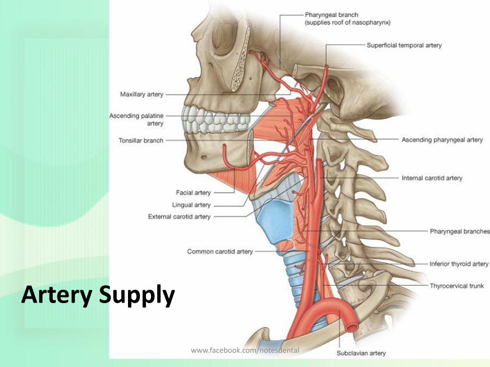
Artery Supply
• Upper parts of the pharynx
– the ascending pharyngeal artery;
– the ascending palatine and tonsillar branches of the facial artery;
– numerous branches of the maxillary and the lingual arteries.
• Lower parts of the pharynx
– pharyngeal branches from the inferior thyroid artery
www.facebook.com/notesdental

Veins and Lymphatic Drainage
www.facebook.com/notesdental

Veins and Lymphatic Drainage
• Veins of the pharynx form a plexus
– Superiorly - pterygoid plexus in the infratemporalfossa
– Inferiorly - facial and internal jugular veins
• Lymphatics
– drain into the deep cervical nodes and include retropharyngeal, paratracheal, and infrahyoidnodes
www.facebook.com/notesdental

Nerves • Motor and most sensory
innervation - branches of the vagus [X] & glossopharyngeal[IX] nerves
• This 2 nerved forms a plexus in the outer fascia of the pharyngeal wall, consisting of– the pharyngeal branch of the
vagus nerve [X]; – branches from the external
laryngeal nerve from the superior laryngeal branch of the vagus nerve [X];
– pharyngeal branches of the glossopharyngeal nerve [IX]
www.facebook.com/notesdental
Refrences
• Grays Anatomy for Students 2nd Edition
• Head and Neck Anatomy for Dental Medicine
• Head, Neck and Dental Anatomy, 4th Edition
• Netter’s Head and Neck Anatomy for Dentistry, 2nd Edition Neil S norton
www.facebook.com/notesdental
Related Documents