Pharmacological and biological evaluation of a series of substituted 1,4-naphthoquinone bioreductive drugs Roger M. Phillips a, * , Mohammed Jaffar b , Derek J. Maitland c , Paul M. Loadman a , Steven D. Shnyder c , Gillian Steans c , Patricia A. Cooper c , Amanda Race c , Adam V. Patterson b , Ian J. Stratford b a Cancer Research Unit, Tom Connors Cancer Research Centre, University of Bradford, Bradford BD71DP, UK b School of Pharmacy and Pharmaceutical Sciences, University of Manchester, Oxford Road, Manchester, UK c School of Pharmacy, University of Bradford, Bradford BD71DP, UK Received 14 April 2004; accepted 3 August 2004 Abstract The indolequinone compound EO9 has good pharmacodynamic properties in terms of bioreductive activation and selectivity for either NAD(P)H:quinone oxidoreductase-1 (NQO1)-rich aerobic or NQO1-deficient hypoxic cells. However, its pharmacokinetic properties are poor and this fact is believed to be a major reason for EO9’s lack of clinical efficacy. The purpose of this study was to develop quinone-based bioreductive drugs that retained EO9’s good properties, in terms of bioreductive activation, but have improved pharmacokinetic properties. Out of 11 naphthoquinone compounds evaluated, 2-aziridinyl-5-hydroxy-1,4-naphthoquinone (com- pound 2), 2,3-bis(aziridinyl)-5-hydroxy-1,4-naphthoquinone (compound 3), and 2-aziridinyl-6-hydroxymethyl-1,4-naphthoquinone (compound 11) were selected for further evaluation based on good substrate specificity for NQO1 and selectivity towards NQO1-rich cells in vitro. Compound 3 was of particular interest as it also demonstrated selectivity for NQO1-rich cells under hypoxic conditions. Compound 3 was not metabolised by murine whole blood in vitro (in contrast to compounds 2, 11 and EO9) and pharmacokinetic studies in non-tumour-bearing mice in vivo (at the maximum soluble dose of 60 mg kg 1 administered intraperitoneally) demon- strated significant improvements in plasma half-life (16.2 min) and AUC values (22.5 mM h) compared to EO9 (T 1/2 = 1.8 min, AUC = 0.184 mM h). Compound 3 also demonstrated significant anti-tumour activity against H460 and HCT-116 human tumour xenografts in vivo, whereas EO9 was inactive against these tumours. In conclusion, compound 3 is a promising lead compound that may target both aerobic and hypoxic fractions of NQO1-rich tumours and further studies to elucidate its mechanism of action and improve solubility are warranted. # 2004 Elsevier Inc. All rights reserved. Keywords: NQO1; Hypoxia; Bioreductive drugs; Naphthoquinones 1. Introduction The indolequinone compound EO9 (3-hydroxy-5-azir- idinyl-1-methyl-2[indole-4,7-dione]-prop-b-en-a-ol) is a bioreductive drug that was selected for clinical evaluation in the early 1990s on the basis of a novel mechanism of action and promising preclinical activity [1,2]. The enzyme NAD(P)H:Quinone oxidoreductase-1 (NQO1, EC 1.6.99.2) plays a central role in bioreductively activat- ing EO9 to DNA-damaging species [3,4] and good corre- lations between NQO1 activity and chemosensitivity in vitro under aerobic conditions have been reported [5–8]. EO9 is also selectively toxic to hypoxic cells, although good hypoxic cytotoxicity ratios (HCR) are only obtained in cells that have low NQO1 activity [5,9,10]. Despite evidence of activity (albeit modest) against a range of solid tumour models in vivo [1], EO9 failed to show activity in phase II clinical trials [11,12]. Several possible explana- tions for EO9’s lack of clinical efficacy have been sug- gested [13], although the major causative factor is likely to be poor drug delivery to tumours as a result of rapid pharmacokinetic elimination and poor penetration through www.elsevier.com/locate/biochempharm Biochemical Pharmacology 68 (2004) 2107–2116 Abbreviations: DMSO, dimethylsulphoxide; NQO1, NAD(P)H:Qui- none oxidoreductase 1; P450R, NADPH cytochrome P450 reductase; NSCLC, non small cell lung cancer; MTD, maximum tolerated dose; MMC, mitomycin C * Corresponding author. Tel.: +44 1274 233226; fax: +44 1274 233234. E-mail address: [email protected] (R.M. Phillips). 0006-2952/$ – see front matter # 2004 Elsevier Inc. All rights reserved. doi:10.1016/j.bcp.2004.08.007

Welcome message from author
This document is posted to help you gain knowledge. Please leave a comment to let me know what you think about it! Share it to your friends and learn new things together.
Transcript
www.elsevier.com/locate/biochempharm
Biochemical Pharmacology 68 (2004) 2107–2116
Pharmacological and biological evaluation of a series of
substituted 1,4-naphthoquinone bioreductive drugs
Roger M. Phillipsa,*, Mohammed Jaffarb, Derek J. Maitlandc, Paul M. Loadmana,Steven D. Shnyderc, Gillian Steansc, Patricia A. Cooperc, Amanda Racec,
Adam V. Pattersonb, Ian J. Stratfordb
aCancer Research Unit, Tom Connors Cancer Research Centre, University of Bradford, Bradford BD71DP, UKbSchool of Pharmacy and Pharmaceutical Sciences, University of Manchester, Oxford Road, Manchester, UK
cSchool of Pharmacy, University of Bradford, Bradford BD71DP, UK
Received 14 April 2004; accepted 3 August 2004
Abstract
The indolequinone compound EO9 has good pharmacodynamic properties in terms of bioreductive activation and selectivity for
either NAD(P)H:quinone oxidoreductase-1 (NQO1)-rich aerobic or NQO1-deficient hypoxic cells. However, its pharmacokinetic
properties are poor and this fact is believed to be a major reason for EO9’s lack of clinical efficacy. The purpose of this study was to
develop quinone-based bioreductive drugs that retained EO9’s good properties, in terms of bioreductive activation, but have improved
pharmacokinetic properties. Out of 11 naphthoquinone compounds evaluated, 2-aziridinyl-5-hydroxy-1,4-naphthoquinone (com-
pound 2), 2,3-bis(aziridinyl)-5-hydroxy-1,4-naphthoquinone (compound 3), and 2-aziridinyl-6-hydroxymethyl-1,4-naphthoquinone
(compound 11) were selected for further evaluation based on good substrate specificity for NQO1 and selectivity towards NQO1-rich
cells in vitro. Compound 3 was of particular interest as it also demonstrated selectivity for NQO1-rich cells under hypoxic conditions.
Compound 3 was not metabolised by murine whole blood in vitro (in contrast to compounds 2, 11 and EO9) and pharmacokinetic
studies in non-tumour-bearing mice in vivo (at the maximum soluble dose of 60 mg kg�1 administered intraperitoneally) demon-
strated significant improvements in plasma half-life (16.2 min) and AUC values (22.5 mM h) compared to EO9 (T1/2 = 1.8 min, AUC =
0.184 mM h). Compound 3 also demonstrated significant anti-tumour activity against H460 and HCT-116 human tumour xenografts
in vivo, whereas EO9 was inactive against these tumours. In conclusion, compound 3 is a promising lead compound that may target
both aerobic and hypoxic fractions of NQO1-rich tumours and further studies to elucidate its mechanism of action and improve
solubility are warranted.
# 2004 Elsevier Inc. All rights reserved.
Keywords: NQO1; Hypoxia; Bioreductive drugs; Naphthoquinones
1. Introduction
The indolequinone compound EO9 (3-hydroxy-5-azir-
idinyl-1-methyl-2[indole-4,7-dione]-prop-b-en-a-ol) is a
bioreductive drug that was selected for clinical evaluation
in the early 1990s on the basis of a novel mechanism
of action and promising preclinical activity [1,2]. The
enzyme NAD(P)H:Quinone oxidoreductase-1 (NQO1,
Abbreviations: DMSO, dimethylsulphoxide; NQO1, NAD(P)H:Qui-
none oxidoreductase 1; P450R, NADPH cytochrome P450 reductase;
NSCLC, non small cell lung cancer; MTD, maximum tolerated dose;
MMC, mitomycin C
* Corresponding author. Tel.: +44 1274 233226; fax: +44 1274 233234.
E-mail address: [email protected] (R.M. Phillips).
0006-2952/$ – see front matter # 2004 Elsevier Inc. All rights reserved.
doi:10.1016/j.bcp.2004.08.007
EC 1.6.99.2) plays a central role in bioreductively activat-
ing EO9 to DNA-damaging species [3,4] and good corre-
lations between NQO1 activity and chemosensitivity in
vitro under aerobic conditions have been reported [5–8].
EO9 is also selectively toxic to hypoxic cells, although
good hypoxic cytotoxicity ratios (HCR) are only obtained
in cells that have low NQO1 activity [5,9,10]. Despite
evidence of activity (albeit modest) against a range of solid
tumour models in vivo [1], EO9 failed to show activity in
phase II clinical trials [11,12]. Several possible explana-
tions for EO9’s lack of clinical efficacy have been sug-
gested [13], although the major causative factor is likely to
be poor drug delivery to tumours as a result of rapid
pharmacokinetic elimination and poor penetration through

R.M. Phillips et al. / Biochemical Pharmacology 68 (2004) 2107–21162108
avascular tissue [14]. The targets for this class of com-
pound (i.e. elevated NQO1 activity and hypoxia) have been
extensively characterised in human tumours [15–17], and
therefore further development of quinone-based bioreduc-
tive drugs is warranted. Based on experience with EO9, it is
clear that future quinone-based bioreductive drugs must
have improved properties in terms of drug delivery if
therapeutic effects are to be obtained.
Numerous factors will determine how much drug is
delivered to tumours following systemic administration
with one of the most important factors being the drug’s
pharmacokinetic characteristics. In both rodents and
humans, EO9 is rapidly cleared from the systemic circula-
tion with plasma half-lives of 1.8 min in mice and between
0.8 and 19 min in humans [18,19]. The reasons for EO9’s
rapid pharmacokinetic elimination are not completely
clear, although extra-hepatic metabolism by red blood
cells is likely to be a significant contributing factor
[18,19]. EO9 is rapidly metabolised by murine whole
blood in vitro (T1/2 = 15.6 � 2.0 min), and recent studies
have suggested that the identification of compounds that
are metabolically stable in murine whole blood could be
used as a filter to select compounds that are likely to have
improved pharmacokinetic properties in vivo [19]. In this
study a series of naphthoquinone compounds have been
evaluated not only in terms of their selective toxicity
towards NQO1-rich cells in vitro but also for metabolic
stability in murine whole blood. Pharmacokinetic studies
and anti-tumour activity against human tumour xenografts
of selected compounds are also reported. Throughout this
study, EO9 was used as a yardstick against which the
relative merits of the naphthoquinones were measured.
2. Materials and methods
2.1. Compounds
A series of 11 substituted naphthoquinones were synthe-
sised by methods described elsewhere [20–22] and their
chemical structures are presented in Table 1. EO9 was
obtained from the Screening and Pharmacology Group of
the European Organisation for Research and Treatment of
Cancer. All compounds were initially dissolved in DMSO,
aliquoted into Eppendorf tubes (100 ml per tube) and stock
solutions at 5 mM were stored at �80 8C prior to biological
and pharmacological evaluation.
2.2. Cell lines and chemosensitivity
A panel of human tumour cell lines was employed which
had previously been characterised in terms of NQO1
enzyme activity. These included: A549 (NSCLC), H460
(NSCLC), HT-29 (colon adenocarcinoma), HCT-116
(colon adenocarcinoma) and BE (colon adenocarcinoma).
A549, H460, HCT-116 and HT-29 cells were obtained
from the American Tissue Type Collection (LGC Promo-
chem) and BE cells were a gift from Dr. T. Ward (Paterson
Institute for Cancer Research). All cell lines were routinely
maintained at 37 8C in a humidified, CO2-enriched (5%)
environment and cultured in RPMI 1640 supplemented
with 10% foetal calf serum, sodium pyruvate (2 mM), L-
glutamine (2 mM), penicillin/streptomycin (50 IU ml�1/
50 mg ml�1) and buffered with HEPES (25 mM). The
NQO1 enzyme activities of A549, H460, HT-29, HCT-
116 and BE cells are 1800 � 122, 1652 � 142, 688 � 52,
565.6 � 108.5 and <0.1 nmol DCPIP reduced/min/mg
protein, respectively [23,24]. The BE cell line is devoid
of functional NQO1 activity due to the presence of the
C609T polymorphism [25]. In vitro chemosensitivity was
determined using the MTT assay, details of which have
been published elsewhere [26]. Briefly, cells in exponential
growth were exposed to a range of drug concentrations for
1 h (at 37 8C) under aerobic conditions. Cells were washed
twice in Hanks Balanced Salt Solution, re-suspended in
RPMI 1640 growth medium and 1–2 � 103 cells plated into
each well of a 96-well plate (8 wells per drug concentration
with a final volume of 200 ml per well). Following in-
cubation at 37 8C for 5 days, medium was removed and
replaced with fresh RPMI 1640 medium prior to the
addition of 20 ml MTT (5 mg ml�1) per well. After a 4-
h incubation at 37 8C, 200 ml of media plus MTT solution
was removed and formazan crystals were dissolved in
150 ml of DMSO. The absorbance of the resulting solution
was read at 550 nm using an ELISA spectrophotometer and
percent cell survival determined as the absorbance of
treated wells divided by the absorbance of the controls.
The final DMSO concentration during drug exposure was
0.1% in all cases and each experiment was repeated in
triplicate.
For analysis of hypoxia selectivity in vitro, T47D
(human breast carcinoma) cell lines transfected with either
NQO1 (DT-1) or P450R (R3) were employed [27,28].
Cells were routinely maintained as monolayer cultures
in DMEM supplemented with 10% foetal calf serum, L-
glutamine (2 mM), non-essential amino acids (1�), peni-
cillin/streptomycin (50 IU ml�1/50 mg ml�1) and buffered
with HEPES (25 mM). Puromycin (4 mg ml�1) was also
included in the media for culturing transfected cell lines.
NQO1 enzyme activity in wild-type, DT-1 and R3 cells
was 15.2, 1,234 and 12.1 nmol DCPIP reduced/min/mg
protein, respectively. P450R activity in wild-type, DT-1
and R3 cells was 13, 11 and 441 nmol cytochrome c
reduced/min/mg protein, respectively [27]. All compounds
evaluated were exposed to cells under standard aerobic
conditions and under nitrogen for 3 h, according to pre-
viously published protocols [28]. Following drug exposure,
chemosensitivity was assessed using the MTT assay as
described above and results were expressed in terms of
IC50 values and hypoxic cytotoxicity ratios (HCR, defined
as the ratio of IC50 values under air to IC50 values under
nitrogen).

R.M. Phillips et al. / Biochemical Pharmacology 68 (2004) 2107–2116 2109
Table 1
Chemical structures and substrate specificity for purified human NQO1 of naphthoquinones (A) and EO9 (B)
,
Compound R1 R2 R3 R4 R5 Substrate specificity (mmol/min/mg)
1 Az CH3 OH H H 41.87 � 7.80
2 H Az OH H H 208.97 � 30.48
3 Az Az OH H H 13.02 � 1.10
4 Az CH3 H H H 61.44 � 0.18
5 NHCH2CH2Cl H H H H 51.15 � 5.95
6 Az CH3 OCH3 H H 2.49 � 0.82
7 H H OCH3 H H 73.06 � 7.48
8 H CH3 OCH3 H H 232.28 � 13.86
9 H H OH H NH2 7.81 � 0.42
10 H H OH H I 137.95 � 34.57
11 H Az H CH2OH H 399.8 � 26.4
EO9 – – – – – 15.39 � 0.61
Values for substrate specificity are the mean � standard deviation for three independent experiments.
2.3. Substrate specificity for NQO1
Purified human recombinant NQO1 was prepared as
described elsewhere [29] and analysis of substrate speci-
ficity was conducted according to previously published
methodology [29]. Briefly, each reaction consisted of
NADH (2 mM), cytochrome c (75 mM), purified NQO1
(77.8 ng), test compounds (25 mM) in a final volume of
1 ml of tris–HCl buffer (50 mM, pH 7.4) containing bovine
serum albumin (0.7% w/v). Reactions were started by the
addition of NADH and the reduction of cytochrome c was
monitored at 550 nm over the initial linear phase of the
reaction curve (30 s). Results were expressed in terms of
specific enzyme activity � standard deviation for three
independent experiments.
2.4. Animals
Two strains of mice aged 6–8 weeks were used: pure
strain female NMRI mice (B and K Universal) and NCR/
Nu (National Cancer Institute, USA). The latter were
housed in isolated cabinets. Mice received CRM diet
(SDS) and water ad libitum. Mice were kept in cages in
an air-conditioned room with regular alternating cycles of
light and darkness. All animal procedures were carried out
under a project licence issued by the UK Home Office, and
UKCCCR guidelines [30] were followed throughout.
2.5. Compound stability in murine whole blood
Blood from non-tumour-bearing NMRI mice was taken
by cardiac puncture under terminal ether anaesthesia and
collected in heparinised tubes. Whole blood (480 ml) was
warmed to 37 8C on a heated reaction block for 30 min
prior to the addition of 20 ml of test compounds (final drug
concentration = 20 mM). Blood and drug solutions were
vortexed and a 50 ml sample was taken (representing t = 0)
to which 100 ml of acetonitrile was added. The mixture was
vortexed and precipitated proteins were removed by cen-
trifugation at 7000 � g for 3 min. The supernatant was
collected and subjected to a further centrifugation step
(7000 � g for 3 min) prior to drug analysis. Further
samples were taken at various time intervals thereafter,
and treated in the same manner as above. All samples were
analysed by reverse phase HPLC using a Beckman system
gold programmable solvent module 126, Beckman auto-
sampler 50Y, diode array detector module 168 and version
gold 711V software. Injection volumes were 50 ml and
chromatographic separation was performed using a
Lichrosorb RP8 column with a mobile phase of either
53% water (containing 1% phosphate buffer [0.5 mM, pH
7.4]):47% methanol (for analysis of compounds 2 and 3) or
69% water (containing 1% phosphate buffer [0.5 mM, pH
7.4]):31% methanol (for analysis of compound 11 and
EO9). Samples were run at a flow rate of 1.4 ml min�1 and
compounds detected at a wavelength of 290 nm. The
chemical stability in aqueous solutions of all compounds
evaluated was determined by replacing murine whole
blood with the same quantity of phosphate-buffered saline
(pH 7.4). Sample preparation and analysis were identical to
that described above.
2.6. Anti-tumour studies
H460 (human NSCLC) and HCT-116 (human colon
carcinoma) human tumour xenografts were established
R.M. Phillips et al. / Biochemical Pharmacology 68 (2004) 2107–21162110
in NCR/Nu mice by subcutaneous inoculation of cell lines
derived from cell cultures. The activity of NQO1 in these
tumours in vivo have been determined elsewhere with
specific activities of 1526 � 42.6 (unpublished data) and
155 � 3.9 nmol [31] DCPIP reduced/(min mg) protein for
H460 and HCT-116 tumours, respectively. P450R activity in
H460 and HCT-116 tumours in vivo were 8.2� 2.5 and 12.4
� 4.8 nmol cytochrome c reduced/(min mg) protein, respec-
tively (unpublished data). Tumours were excised from donor
animals, placed in sterile physiological saline containing
antibiotics and cut into small fragments of approximately
2 mm3. Under brief general anaesthesia, a single fragment
was implanted into the flank of each mouse using a trocar.
Once the tumours could be accurately measured, the mice
were allocated into groups of six by restricted randomisation
to keep group mean tumour size variation to a minimum and
treatment was commenced. To establish the MTD, com-
pound 3 was dissolved in 10% DMSO/arachis oil and mice
received escalating doses of compound (two mice per dose
escalation) administered intraperitoneally. Toxicity was
monitored by measuring body weight at various time inter-
vals after drug administration and the percent maximum
weight loss (relative to initial starting weight) was recorded
with a weight loss of greater than 15% being considered
toxic. The maximum amount of compound 3 that could be
solubilised was 60 mg kg�1 (which was not toxic), and thus
this dose was used for efficacy and pharmacokinetic studies.
For efficacy studies, EO9 was administered in saline at its
MTD of 6 mg kg�1 as a single intraperitoneal (i.p.) injec-
tion. Compound 3 was administered in 10% DMSO/arachis
oil as either a single i.p. injection at 30 or 60 mg kg�1, or as
four consecutive daily i.p. injections of 12 or 15 mg kg�1 per
day. The effects of therapy were assessed three to five times a
week by two-dimensional calliper measurements of the
tumours. Tumour volumes were then calculated using the
formula (a2 � b)/2 where a is the smaller and b the larger
diameter of the tumour. Tumour volume was then normal-
ised relative to the respective volume on day 0 and semi-log
plots of relative tumour volume (RTV) against time were
made. Mann–Whitney U-tests were performed to determine
Table 2
The response of cell lines in vitro following a 1-h exposure to test compounds u
Compound IC50 (mM) A549 cells IC50 (mM) H460 ce
1 7.2 � 3.5 (24.2) N/a
2 1.0 � 0.1 (138.3) 0.6 � 0.2 (230.5
3 0.9 � 0.2 (240.4) 1.5 � 0.7 (144.3
4 27.8 � 4.4 (25.1) 690.4 � 50.6 (1.0)
5 93.8 � 1.8 (2.4) 206.5 � 39.8 (1.1)
6 11.8 � 5.6 (107.2) 29.5 � 3.9 (42.9)
7 10.8 � 2.8 (0.4) 2.5 � 1.9 (1.7)
8 95.3 � 16.1 (0.6) 45.2 � 1.8 (1.3)
9 45.5 � 8.3 (0.7) 54.1 � 4.1 (0.6)
10 6.8 � 1.6 (0.3) 9.1 � 1.2 (0.2)
11 2.5 � 0.1 (61.9) 2.3 � 0.1 (67.3)
EO9 0.1 � 0.03 (475.0) 0.2 � 0.1 (237.5
Each value represents the mean � standard deviation for three independent experim
of IC50 values for the NQO1-deficient BE cell line to NQO1-rich cell lines. N/a
the statistical significance of any differences in growth rate
(based on tumour volume doubling time) between controlled
and treated groups.
2.7. Pharmacokinetic analysis
Pharmacokinetic analysis was conducted in non-
tumour-bearing NCR/Nu mice following the i.p. adminis-
tration of compound 3 at 60 mg kg�1. At 5, 15, 30, 60 and
120 min after administration, blood samples were taken by
cardiac puncture under terminal ether anaesthesia and
collected in heparinised collection tubes. Tubes were
stored on ice during transport to the analytical laboratory.
Three mice per time point were used and plasma was
separated following centrifugation at 3000 � g for 15 min
(at 4 8C). Compound 3 was extracted from plasma and
analysed by HPLC as described above.
3. Results
3.1. Substrate specificity for purified human NQO1
A broad spectrum of substrate specificity for NQO1
existed within the panel of compounds evaluated ranging
from 2.49 � 0.82 to 399.8 � 26.4 mmol/min/mg (Table 1).
Compounds 2, 11 and 8 were particularly good substrates
for NQO1 with specific activities of 208.9 � 30.5, 232.3 �13.9 and 399.8 � 26.4 mmol/min/mg, respectively. In
comparison with EO9, all compounds except 6 and 9 were
better substrates for NQO1 with compound 3 being similar
to EO9 in terms of reduction by NQO1.
3.2. Chemosensitivity studies in vitro under aerobic
conditions
The response of a panel of cell lines with a broad
spectrum of NQO1 activities to the various compounds
tested is presented in Table 2. Whilst all compounds were
less potent in vitro than EO9, preferential activity towards
nder aerobic conditions
lls IC50 (mM) HT-29 cells IC50 (mM) BE cells
3.9 � 0.8 (44.7) 174.2 � 41.2
) 0.6 � 0.1 (230.5) 138.3 � 6.4
) 0.6 � 0.3 (360.7) 216.4 � 70.0
11.4 � 2.0 (61.2) 679.2 � 28.7
98.5 � 4.0 (2.3) 223.1 � 26.5
9.3 � 3.5 (136.1) 1265.5 � 85.0
12.9 � 1.8 (0.3) 4.2 � 0.5
36.4 � 1.9 (1.6) 57.5 � 13.5
15.3 � 3.6 (2.0) 30.8 � 5.5
5.2 � 1.0 (0.4) 2.1 � 0.1
5.3 � 0.2 (29.2) 154.4 � 6.4
) 0.3 � 0.03 (158.2) 47.5 � 9.5
ents. Values in parentheses represent the selectivity ratio defined as the ratio
represents data not available.
R.M. Phillips et al. / Biochemical Pharmacology 68 (2004) 2107–2116 2111
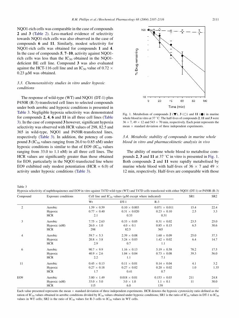
Fig. 1. Metabolism of compounds 2 (!), 3 (*) and 11 (*) in murine
whole blood in vitro at 37 8C. The half-lives of compounds 2, 11 and 3 were
36 � 7, 49 � 12 and 543 � 70 min, respectively. Each point represents the
mean � standard deviation of three independent experiments.
NQO1-rich cells was comparable in the case of compounds
2 and 3 (Table 2). Less-marked evidence of selectivity
towards NQO1-rich cells was also observed in the case of
compounds 6 and 11. Similarly, modest selectivity for
NQO1-rich cells was obtained for compounds 1 and 4.
In the case of compounds 5, 7–10, activity against NQO1-
rich cells was less than the IC50 obtained in the NQO1-
deficient BE cell line. Compound 3 was also evaluated
against the HCT-116 cell line and an IC50 value of 0.72 �0.23 mM was obtained.
3.3. Chemosensitivity studies in vitro under hypoxic
conditions
The response of wild-type (WT) and NQO1 (DT-1) plus
P450R (R-3)-transfected cell lines to selected compounds
under both aerobic and hypoxic conditions is presented in
Table 3. Negligible hypoxia selectivity was demonstrated
for compounds 2, 4, 6 and 11 in all three cell lines (Table
3). In the case of compound 3 however, significant hypoxia
selectivity was observed with HCR values of 298, 82.5 and
365 in wild-type, NQO1 and P450R-transfected lines,
respectively (Table 3). In addition, the potency of com-
pound 3 (IC50 values ranging from 26.0 to 0.85 nM) under
hypoxic conditions is similar to that of EO9 (IC50 values
ranging from 33.0 to 1.1 nM) in all three cell lines. The
HCR values are significantly greater than those obtained
for EO9, particularly in the NQO1-transfected line where
EO9 exhibited only modest potentiation (HCR = 6.0) of
activity under hypoxic conditions (Table 3).
Table 3
Hypoxia selectivity of naphthoquinones and EO9 in vitro against T47D wild-type
Compound Exposure conditions Cell line and IC50 values
Wt D
2 Aerobic 1.59 � 0.59 0
Hypoxic 0.77 � 0.40 0
HCR 2.1
3 Aerobic 7.75 � 2.63 0
Hypoxic (nM) 26.0 � 1.0
HCR 298
4 Aerobic 59.7 � 5.3 2
Hypoxic 20.8 � 3.8 3
HCR 2.9
6 Aerobic 90.7 � 9.9 1
Hypoxic 40.9 � 2.6 1
HCR 2.2
11 Aerobic 0.45 � 0.13 0
Hypoxic 0.27 � 0.18 0
HCR 1.7
EO9 Aerobic 3.80 � 1.49 0.
Hypoxic (nM) 33.0 � 5.0
HCR 115
Each value presented represents the mean � standard deviation of three independ
ration of IC50 values obtained in aerobic conditions divided by IC50 values obtaine
values in WT cells; SR2 is the ratio of IC50 values for R-3 cells to IC50 values
3.4. Metabolic stability of compounds in murine whole
blood in vitro and pharmacokinetic analysis in vivo
The ability of murine whole blood to metabolise com-
pounds 2, 3 and 11 at 37 8C in vitro is presented in Fig. 1.
Both compounds 2 and 11 were rapidly metabolised by
murine whole blood with half-lives of 36 � 7 and 49 �12 min, respectively. Half-lives are comparable with those
(WT) and T47D cells transfected with either NQO1 (DT-1) or P450R (R-3)
(mM except where indicated) SR1 SR2
T-1 R-3
.10 � 0.003 0.071 � 0.011 15.4 22.4
.31 � 0.020 0.23 � 0.10 2.5 3.3
0.33 0.31
.33 � 0.05 0.31 � 0.02 23.5 25.0
4.0 � 0.1 0.85 � 0.15 6.5 30.6
82.5 365
.39 � 0.08 1.60 � 0.09 25.0 37.3
.24 � 0.03 1.42 � 0.02 6.4 14.7
0.7 1.1
.16 � 0.13 5.19 � 0.58 78.2 17.5
.04 � 0.04 0.73 � 0.08 39.3 56.0
1.1 7.1
.11 � 0.01 0.14 � 0.04 4.1 3.2
.27 � 0.02 0.20 � 0.02 1.0 1.35
0.41 0.7
018 � 0.01 0.153 � 0.03 211 24.8
3.0 � 1.0 1.1 � 0.1 11 30.0
6.0 139
ent experiments. HCR denotes the hypoxic cytotoxicity ratio defined as the
d under hypoxic conditions; SR1 is the ratio of IC50 values in DT-1 to IC50
in WT cells.
R.M. Phillips et al. / Biochemical Pharmacology 68 (2004) 2107–21162112
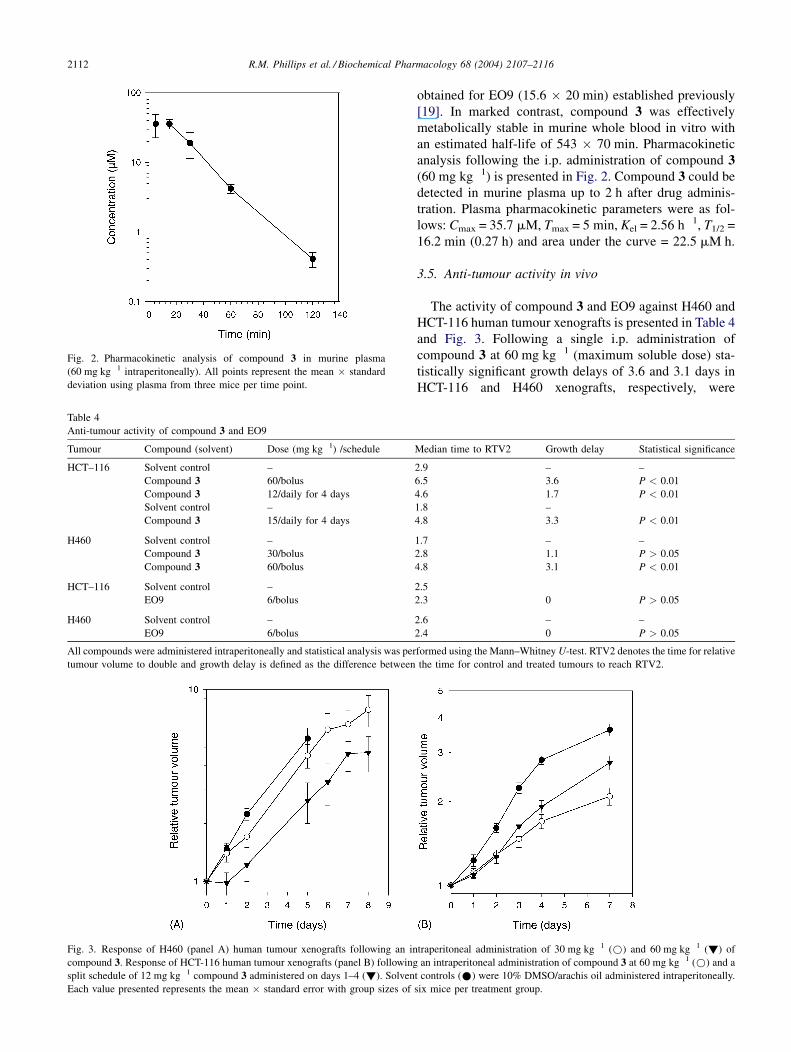
Fig. 2. Pharmacokinetic analysis of compound 3 in murine plasma
(60 mg kg�1 intraperitoneally). All points represent the mean � standard
deviation using plasma from three mice per time point.
Table 4
Anti-tumour activity of compound 3 and EO9
Tumour Compound (solvent) Dose (mg kg�1) /schedule
HCT–116 Solvent control –
Compound 3 60/bolus
Compound 3 12/daily for 4 days
Solvent control –
Compound 3 15/daily for 4 days
H460 Solvent control –
Compound 3 30/bolus
Compound 3 60/bolus
HCT–116 Solvent control –
EO9 6/bolus
H460 Solvent control –
EO9 6/bolus
All compounds were administered intraperitoneally and statistical analysis was per
tumour volume to double and growth delay is defined as the difference between
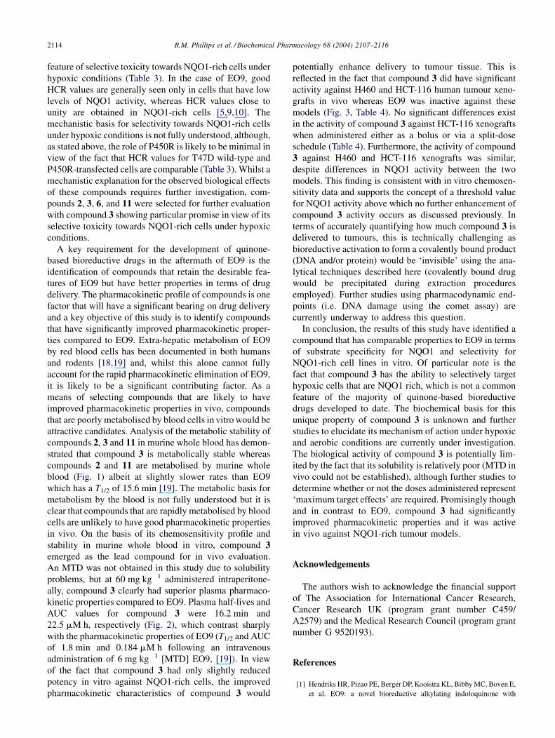
Fig. 3. Response of H460 (panel A) human tumour xenografts following an in
compound 3. Response of HCT-116 human tumour xenografts (panel B) following
split schedule of 12 mg kg�1 compound 3 administered on days 1–4 (!). Solven
Each value presented represents the mean � standard error with group sizes of
obtained for EO9 (15.6 � 20 min) established previously
[19]. In marked contrast, compound 3 was effectively
metabolically stable in murine whole blood in vitro with
an estimated half-life of 543 � 70 min. Pharmacokinetic
analysis following the i.p. administration of compound 3(60 mg kg�1) is presented in Fig. 2. Compound 3 could be
detected in murine plasma up to 2 h after drug adminis-
tration. Plasma pharmacokinetic parameters were as fol-
lows: Cmax = 35.7 mM, Tmax = 5 min, Kel = 2.56 h�1, T1/2 =
16.2 min (0.27 h) and area under the curve = 22.5 mM h.
3.5. Anti-tumour activity in vivo
The activity of compound 3 and EO9 against H460 and
HCT-116 human tumour xenografts is presented in Table 4
and Fig. 3. Following a single i.p. administration of
compound 3 at 60 mg kg�1 (maximum soluble dose) sta-
tistically significant growth delays of 3.6 and 3.1 days in
HCT-116 and H460 xenografts, respectively, were
Median time to RTV2 Growth delay Statistical significance
2.9 – –
6.5 3.6 P < 0.01
4.6 1.7 P < 0.01
1.8 –
4.8 3.3 P < 0.01
1.7 – –
2.8 1.1 P > 0.05
4.8 3.1 P < 0.01
2.5
2.3 0 P > 0.05
2.6 – –
2.4 0 P > 0.05
formed using the Mann–Whitney U-test. RTV2 denotes the time for relative
the time for control and treated tumours to reach RTV2.
traperitoneal administration of 30 mg kg�1 (*) and 60 mg kg�1 (!) of
an intraperitoneal administration of compound 3 at 60 mg kg�1 (*) and a
t controls (*) were 10% DMSO/arachis oil administered intraperitoneally.
six mice per treatment group.

R.M. Phillips et al. / Biochemical Pharmacology 68 (2004) 2107–2116 2113
obtained. Growth delays induced by compound 3 in the
H460 model were dose-dependent with no significant
growth delays seen at 30 mg kg�1. Split-dose scheduling
(15 mg kg�1 administered daily for 4 days) induced sig-
nificant growth delays (3.3 days) in the HCT-116 xeno-
graft. No anti-tumour effects were observed in both HCT-
116 and H460 xenografts treated with EO9 at the MTD of
6 mg kg�1 administered i.p. (Table 4).
4. Discussion
Whilst the failure of EO9 to show efficacy in the clinic
was a major disappointment for the field of quinone-based
bioreductive drug development, the search for novel com-
pounds that can exploit elevated tumour levels of NQO1
and/or tumour hypoxia continues. Medicinal chemistry
approaches have predominantly focused on indolequinone
derivatives of EO9 with comparatively little attention
being paid to naphthoquinone compounds [23,28,32–
34]. Early studies were conducted principally by Sartor-
elli’s group and these demonstrated that 2,3-diaziridinyl-
1,4-naphthoquinone sulphonate derivatives were active
against a variety of tumour models in vivo [20]. It is
interesting to note that good anti-tumour effects were
observed only in compounds that possessed both the
5-O-sulphonyl and the 2,3-disubstituted aziridinyl
groups [20]. Compounds such as 2-aziridinyl-5-hydroxy-
1,4-naphthoquinone and 2,3-diaziridinyl-5-hydroxy-1,4-
naphthoquinone (compounds 2 and 3 in this study) were
classified as inactive against the experimental models
employed (the L1210 murine leukaemia) by Lin et al.
[20]. The results of this study clearly demonstrate, how-
ever, that aziridinyl substituted naphthoquinones such as
compounds 1–3, 6 and 11 are good substrates for human
NQO1 and exhibit selective toxicity towards NQO1-rich
cells in vitro under aerobic conditions (Table 2). The
reasons for the discrepancy between this study and pre-
vious studies is likely to be the fact that haematological
malignancies typically have low levels of NQO1 activity
[6,7]. With the benefit of hindsight, the lack of anti-tumour
activity of compounds 2 and 3, for example, could be
viewed as a positive result and the compounds activated by
NQO1 would not be expected to demonstrate activity
against a tumour model that lacks the appropriate enzy-
mology.
In terms of structural features of naphthoquinones that
determine substrate specificity, potency and selectivity for
NQO1-rich cells, the presence or absence of an aziridinyl
functional group appears to have a profound effect. In
terms of substrate specificity for NQO1, the inclusion of an
aziridine ring at position R1 generally reduces substrate
specificity (compounds 1, 4 and 6, Table 1) that is reduced
further if two aziridinyl groups are present (compare 1 and
3, Table 1). Whilst the inclusion of two aziridinyl groups
reduces substrate specificity, it is important to note that
compound 3 is comparable to EO9 which is widely recog-
nised as a good substrate for NQO1 (Table 1). In marked
contrast, the presence of an aziridinyl group at position R2
does not adversely affect substrate specificity and both
compounds 2 and 11 are excellent substrates for NQO1
(Table 1). In terms of potency, the inclusion of an aziridinyl
group is desirable but not absolutely essential. Compounds
7 and 10, for example, lack aziridine groups but still show
good potency in vitro (Table 2). Whilst substrate specificity
and potency are important issues in the development of
these compounds, the key parameter required is good
selectivity for NQO1-rich cells in vitro. In this case,
inclusion of the aziridinyl functional group is essential
as all compounds that have this group (i.e. compounds 1–3,
4, 6 and 11) are selectively toxic towards NQO1-rich cells
in vitro to a greater or lesser extent (Table 2). These
observations are essentially consistent with previous stu-
dies using indolequinone derivatives of EO9 [23,32].
Similarly, compounds that lack good leaving groups (com-
pounds 7–10) are likely to be detoxified by NQO1 under
aerobic conditions (Table 2), as in the case with menadione
[35,36]. These results suggest that reduction by NQO1
results in the generation of metabolites that can alkylate
DNA and recent studies in our laboratory have shown that
compound 3 induces interstrand cross-links in DNA (data
not shown). It is of interest to note that whilst the aziridinyl
analogues appear to be selectively toxic towards NQO1-
rich cells compared to BE cells, IC50 are similar amongst
NQO1-rich cells despite the fact that there is a broad range
of NQO1 activities (Table 2). This suggests that there may
be a threshold level of NQO1 activity, above which no
further increase in toxicity occurs. Similar findings have
been reported for other quinone-based compounds [37] and
further studies are required to define this value.
One electron reductases such as P450R are known to
reduce naphthoquinones effectively [38] and there is evi-
dence that P450R is also involved in the activation of
aziridinyl naphthoquinones used in this study (Table 3). In
the case of compound 3, T47D cells transfected with
P450R are significantly more sensitive than T47D WT
cells under aerobic conditions (selectivity ratio = 25.0).
Furthermore, transfection of cells with P450R increases the
potency of compound 3 under hypoxic conditions,
although the role of P450R in determining hypoxia selec-
tivity for compound 3 requires further investigation as
HCR values for both T47D WT and P450R-transfected
cells are comparable (298 and 365, respectively, Table 3).
The mechanistic basis to explain these observations is not
fully understood and further studies to characterise the type
of DNA damage induced in cells following drug exposure
are underway to address this question (i.e. single-strand
DNA breaks indicate redox-mediated damage following
one electron reduction or DNA alkylation cross-links
formed following either one or two electron reduction).
Whilst the mechanistic details are unclear, it is of particular
interest to note that compound 3 exhibits the unusual
R.M. Phillips et al. / Biochemical Pharmacology 68 (2004) 2107–21162114
feature of selective toxicity towards NQO1-rich cells under
hypoxic conditions (Table 3). In the case of EO9, good
HCR values are generally seen only in cells that have low
levels of NQO1 activity, whereas HCR values close to
unity are obtained in NQO1-rich cells [5,9,10]. The
mechanistic basis for selectivity towards NQO1-rich cells
under hypoxic conditions is not fully understood, although,
as stated above, the role of P450R is likely to be minimal in
view of the fact that HCR values for T47D wild-type and
P450R-transfected cells are comparable (Table 3). Whilst a
mechanistic explanation for the observed biological effects
of these compounds requires further investigation, com-
pounds 2, 3, 6, and 11 were selected for further evaluation
with compound 3 showing particular promise in view of its
selective toxicity towards NQO1-rich cells under hypoxic
conditions.
A key requirement for the development of quinone-
based bioreductive drugs in the aftermath of EO9 is the
identification of compounds that retain the desirable fea-
tures of EO9 but have better properties in terms of drug
delivery. The pharmacokinetic profile of compounds is one
factor that will have a significant bearing on drug delivery
and a key objective of this study is to identify compounds
that have significantly improved pharmacokinetic proper-
ties compared to EO9. Extra-hepatic metabolism of EO9
by red blood cells has been documented in both humans
and rodents [18,19] and, whilst this alone cannot fully
account for the rapid pharmacokinetic elimination of EO9,
it is likely to be a significant contributing factor. As a
means of selecting compounds that are likely to have
improved pharmacokinetic properties in vivo, compounds
that are poorly metabolised by blood cells in vitro would be
attractive candidates. Analysis of the metabolic stability of
compounds 2, 3 and 11 in murine whole blood has demon-
strated that compound 3 is metabolically stable whereas
compounds 2 and 11 are metabolised by murine whole
blood (Fig. 1) albeit at slightly slower rates than EO9
which has a T1/2 of 15.6 min [19]. The metabolic basis for
metabolism by the blood is not fully understood but it is
clear that compounds that are rapidly metabolised by blood
cells are unlikely to have good pharmacokinetic properties
in vivo. On the basis of its chemosensitivity profile and
stability in murine whole blood in vitro, compound 3emerged as the lead compound for in vivo evaluation.
An MTD was not obtained in this study due to solubility
problems, but at 60 mg kg�1 administered intraperitone-
ally, compound 3 clearly had superior plasma pharmaco-
kinetic properties compared to EO9. Plasma half-lives and
AUC values for compound 3 were 16.2 min and
22.5 mM h, respectively (Fig. 2), which contrast sharply
with the pharmacokinetic properties of EO9 (T1/2 and AUC
of 1.8 min and 0.184 mM h following an intravenous
administration of 6 mg kg�1 [MTD] EO9, [19]). In view
of the fact that compound 3 had only slightly reduced
potency in vitro against NQO1-rich cells, the improved
pharmacokinetic characteristics of compound 3 would
potentially enhance delivery to tumour tissue. This is
reflected in the fact that compound 3 did have significant
activity against H460 and HCT-116 human tumour xeno-
grafts in vivo whereas EO9 was inactive against these
models (Fig. 3, Table 4). No significant differences exist
in the activity of compound 3 against HCT-116 xenografts
when administered either as a bolus or via a split-dose
schedule (Table 4). Furthermore, the activity of compound
3 against H460 and HCT-116 xenografts was similar,
despite differences in NQO1 activity between the two
models. This finding is consistent with in vitro chemosen-
sitivity data and supports the concept of a threshold value
for NQO1 activity above which no further enhancement of
compound 3 activity occurs as discussed previously. In
terms of accurately quantifying how much compound 3 is
delivered to tumours, this is technically challenging as
bioreductive activation to form a covalently bound product
(DNA and/or protein) would be ‘invisible’ using the ana-
lytical techniques described here (covalently bound drug
would be precipitated during extraction procedures
employed). Further studies using pharmacodynamic end-
points (i.e. DNA damage using the comet assay) are
currently underway to address this question.
In conclusion, the results of this study have identified a
compound that has comparable properties to EO9 in terms
of substrate specificity for NQO1 and selectivity for
NQO1-rich cell lines in vitro. Of particular note is the
fact that compound 3 has the ability to selectively target
hypoxic cells that are NQO1 rich, which is not a common
feature of the majority of quinone-based bioreductive
drugs developed to date. The biochemical basis for this
unique property of compound 3 is unknown and further
studies to elucidate its mechanism of action under hypoxic
and aerobic conditions are currently under investigation.
The biological activity of compound 3 is potentially lim-
ited by the fact that its solubility is relatively poor (MTD in
vivo could not be established), although further studies to
determine whether or not the doses administered represent
‘maximum target effects’ are required. Promisingly though
and in contrast to EO9, compound 3 had significantly
improved pharmacokinetic properties and it was active
in vivo against NQO1-rich tumour models.
Acknowledgements
The authors wish to acknowledge the financial support
of The Association for International Cancer Research,
Cancer Research UK (program grant number C459/
A2579) and the Medical Research Council (program grant
number G 9520193).
References
[1] Hendriks HR, Pizao PE, Berger DP, Kooistra KL, Bibby MC, Boven E,
et al. EO9: a novel bioreductive alkylating indoloquinone with
R.M. Phillips et al. / Biochemical Pharmacology 68 (2004) 2107–2116 2115
preferential solid tumour activity and lack of bone marrow toxicity in
preclinical models. Eur J Cancer 1993;29A:897–906.
[2] Workman P. Enzyme directed bioreductive drug development revis-
ited: a commentary on recent progress and future prospects
with emphasis on quinone anti-cancer agents and quinone metabolis-
ing enzymes, particularly DT-diaphorase. Oncol Res 1994;6:
461–75.
[3] Walton MI, Smith PJ, Workman P. The role of NAD(P)H quinone
reductase (EC 1.6.99.2 DT-diaphorase) in the reductive bioactivation
of the novel indoloquinone antitumour agent EO9. Cancer Commun
1991;3:199–206.
[4] Bailey SM, Wyatt MD, Friedlos F, Hartley JA, Knox RJ, Lewis AD, et
al. Involvement of DT-diaphorase (EC 1.6.99.2) in the DNA cross-
linking and sequence selectivity of the bioreductive anti-tumour agent
EO9. Br J Cancer 1997;76:1596–603.
[5] Robertson N, Haigh A, Adams GE, Stratford IJ. Factors affecting
sensitivity to EO9 in rodent and human tumour cells in vitro:
DT-diaphorase activity and hypoxia. Eur J Cancer 1994;30A:
1013–9.
[6] Smitkamp-Wilms E, Peters GF, Pinedo HM, Van Arkotte J, Giaccone
G. Chemosensitivity to the indolequinone EO9 is correlated with DT-
diaphorase activity and gene expression. Biochem Pharmacol
1994;47:1325–32.
[7] Collard J, Matthew AM, Double JA, Bibby MC. EO9: relationship
between DT-diaphorase levels and response in vitro and in vivo. Br J
Cancer 1995;71:1199–203.
[8] Fitzsimmons SA, Workman P, Grever M, Paull K, Camalier R, Lewis
AD. Reductase enzyme expression across the National Cancer Insti-
tute tumour cell line panel: correlation with sensitivity to mitomycin C
and EO9. J Natl Cancer Inst 1996;88:259–69.
[9] Plumb JA, Workman P. Unusually marked hypoxic sensitisation
to indoloquinone EO9 and Mitomycin C in a human colon cell
line that lacks DT-diaphorase activity. Int J Cancer 1994;56:
134–9.
[10] Plumb JA, Gerritsen M, Workman P. DT-diaphorase protects cells
from the hypoxic cytotoxicity of indoloquinone EO9. Br J Cancer
1994;70:1136–43.
[11] Dirix LY, Tonnesen F, Cassidy J, Epelbaum R, ten Bokkel Huinink
WW, Pavlidis N, et al. EO9 phase II study in advanced breast, gastric,
pancreatic and colorectal carcinoma by the EORTC Early Clinical
Studies Group. Eur J Cancer 1996;32A:2019–22.
[12] Pavlidis N, Hanauske AR, Gamucci T, Smyth J, Lehnert M, te Velde A,
et al. A randomized phase II study with two schedules of the novel
indoloquinone EO9 in non-small-cell lung cancer: a study of the
EORTC Early Clinical Studies Group (ECSG). Ann Oncol
1996;7:529–31.
[13] Connors TA. Bioreductive agents, hypoxic cells and therapy. Eur J
Cancer 1996;32A:1833–4.
[14] Phillips RM, Loadman PM, Cronin BP. Evaluation of a novel in vitro
assay for assessing drug penetration into avascular regions of tumours.
Br J Cancer 1998;77:2112–9.
[15] Vaupel P, Kallinowski F, Okunieff P. Blood flow, oxygen and nutrient
supply and metabolic microenvironment of human tumours: a review.
Cancer Res 1989;49:6449–65.
[16] Malkinson AM, Siegel D, Forrest GL, Gazdar AF, Oie HK, Chan DC,
et al. Elevated DT-diaphorase activity and messenger-RNA content
in human non-small-cell lung-carcinoma–relationship to the response
of lung tumor xenografts to mitomycin-C. Cancer Res 1992;52:
4752–7.
[17] Siegel D, Franklin WA, Ross D. Immunohistochemical detection of
NAD(P)H:Quinone oxidoreductase in human lung and lung tumours.
Clin Cancer Res 1999;4:2065–70.
[18] Schellens JHM, Planting AST, van Acker BAC, Loos WJ, de Boer-
Dennert M, van der Burg MEL, et al. Phase I and pharmacological
study of the novel indoloquinone bioreductive alkylating cytotoxic
drug EO9. J Natl Cancer Inst 1994;86:906–12.
[19] Loadman PM, Bibby MC, Phillips RM. Pharmacological approach
towards the development of indolequinone bioreductive drugs based
on the clinically inactive agent EO9. Br J Pharmacol 2002;137:701–9.
[20] Lin TS, Xu SP, Zhu LY, Cosby LA, Sartorelli AC. Synthesis of 2,3-
diaziridinyl-1,4-naphthoquinone sulfonate derivatives as potential
antineoplastic agents. J Med Chem 1989;32:1467–71.
[21] Lin TS, Zhu LY, Xu SP, Divo AA, Sartorelli AC. Synthesis antimalar-
ial activity of 2-aziridinyl- and 2,3-bis(aziridinyl)-1,4-naphthoquino-
nyl sulfonate and acylate derivatives. J Med Chem 1991;34:
1634–9.
[22] Sartorelli AC, Lin TS. 2,3-Bis(azridinyl)-1,4-naphthoquinone sulpho-
nate derivatives having antineoplastic activity. United States Patent.
Patent number 4,806,531 (1989).
[23] Phillips RM, Naylor MA, Jaffar M, Doughty SW, Everett SA, Breen
AG, et al. Bioreductive activation of a series of indolequinones by
human DT-diaphorase: structure activity relationships. J Med Chem
1999;42:4071–80.
[24] Choudry GA, Hamilton-Stewart PA, Double JA, Krul MRL, Naylor B,
Flannigan GM, et al. A novel strategy for NQO1 (NAD(P)H:quinone
oxidoreductase EC 1. 6. 99. 2) mediated therapy of bladder cancer
based on the pharmacological properties of EO9. Br J Cancer
2001;85:1137–46.
[25] Traver RD, Horikoshi T, Dannenberg KD, Stadlbauer THW, Dannen-
berg PV, Ross D, et al. NAD(P)H:Quinone oxidoreductase gene
expression in human colon carcinoma cells: characterisation of a
mutation which modulates DT-diaphorase activity and mitomycin
sensitivity. Cancer Res 1992;52:797–802.
[26] Phillips RM, Hulbert PB, Bibby MC, Sleigh NR, Double JA. In vitro
activity of the novel indoloquinone EO9 and the influence of pH on
cytotoxicity. Br J Cancer 1992;65:359–64.
[27] Cowen RL, Patterson AV, Telfer BA, Airley RE, Hobbs S, Phillips RM,
et al. Viral delivery of P450 reductase recapitulates the ability of
constitutive overexpression of reductase enzymes to potentiate the
activity of mitomycin C in human breast cancer xenografts. Mol
Cancer Ther 2003;2:901–9.
[28] Jaffar M, Phillips RM, Williams KJ, Mrema I, Cole C, Wind NS,
et al. 3-Substituted-5-aziridinyl-1-methylindole-47-diones as NQO1-
directed antitumour agents: mechanism of activation and cytotoxicity
in vitro. Biochem Pharmacol 2003;66:1199–206.
[29] Phillips RM. Bioreductive activation of a series of analogues of 5-
aziridinyl-3-hydroxymethyl-1-methyl-2-[1H-indole-47-dione] prop-
b-en-a-ol (EO9) by human DT-diaphorase. Biochem Pharmacol
1996;52:1711–8.
[30] Workman P, Twentyman P, Balkwill F, Balmain A, Chaplin D, Double
JA, et al. United Kingdom Co-ordinating Committee on Cancer
Research (UKCCCR) guidelines for the welfare of animals in experi-
mental neoplasia. Br J Cancer 1998;77:1–10.
[31] Phillips RM, Burger AM, Loadman PM, Jarret CM, Swaine DJ, Fiebig
HH. Predicting response to mitomycin C on the basis of DT-diaphor-
ase activity or drug metabolism by tumour homogenates: implications
for enzyme directed bioreductive drug development. Cancer Res
2000;60:6384–90.
[32] Naylor MA, Jaffar M, Nolan J, Stephens MA, Butler S, Patel KB, et al.
2-cyclopropylindoloquinones and their analogues as bioreductively
activated antitumor agents: structure-activity in vitro and efficacy in
vivo. J Med Chem 1997;40:2335–46.
[33] Beall HD, Winski S, Swann E, Hudnott AR, Cotterill AS, O’Sullivan
N, et al. Indolequinone antitumor agents: correlation between quinone
structure, rate of metabolism by recombinant human NAD(P)H:qui-
none oxidoreductase, and in vitro cytotoxicity. Med Chem
1998;41:4755–66.
[34] Swann E, Barraja P, Oberlander AM, Gardipee WT, Hudnott AR, Beall
HD, et al. Indolequinone antitumor agents: correlation between
quinone structure and rate of metabolism by recombinant human
NAD(P)H:quinone oxidoreductase. Part 2. J Med Chem 2001;44:
3311–9.
R.M. Phillips et al. / Biochemical Pharmacology 68 (2004) 2107–21162116
[35] Cadenas E. Antioxidant and prooxidant functions of DT-diaphorase in
quinone metabolism. Biochem Pharmacol 1995;49:127–40.
[36] Joseph P, Long II DJ, Klein-Szanto AJP, Jaiswal AK. Role of
NAD(P)H:quinone oxidoreductase 1 (DT-diaphorase) in protection
against quinone toxicity. Biochem Pharmacol 2000;60:207–14.
[37] Winski SL, Swann E, Hargreaves RHJ, Dehn DL, Butler J, Moody CJ,
et al. Relationship between NAD(P)H:Quinone oxidoreductase 1
(NQO1) levels in a series of stably transfected cell lines and
susceptibility to antitumour quinines. Biochem Pharmacol 2001;
61:1509–16.
[38] Giulivi C, Cadenas E. One and two electron reduction of 2-methyl-1,4-
naphthoquinone bioreductive alkylating agents: kinetic studies, free
radical production, thiol oxidation and DNA strand break formation.
Biochem J 1994;301:21–30.
Related Documents