Pharmacokinetics and Brain Uptake of a Genetically Engineered Bi-functional Fusion Antibody Targeting the Mouse Transferrin Receptor Ruben J. Boado 1,2 , Qing-Hui Zhou 2 , Jeff Zhiqiang Lu 1 , Eric Ka-Wai Hui 1 , and William M. Pardridge 2 1 ArmaGen Technologies, Inc., Santa Monica, CA 90401 2 Department of Medicine, UCLA, Los Angeles, CA 90024 Abstract Monoclonal antibodies (MAb) are potential new therapeutics for brain diseases. However, MAb’s do not cross the blood-brain barrier (BBB). The present work describes the genetic engineering of a fusion protein comprised of a therapeutic single chain Fv (ScFv) antibody and a mouse/rat chimeric MAb against the mouse transferrin receptor (TfR). The TfRMAb acts as a molecular Trojan horse to ferry the therapeutic ScFv across the BBB in vivo in the mouse. The ScFv is fused to the carboxyl terminus of the heavy chain of the chimeric TfRMAb, and this fusion protein is designated cTfRMAb-ScFv. Chinese hamster ovary cells were permanently transfected, and a high secreting cell line in serum free medium was cloned. The cTfRMAb-ScFv fusion protein was purified to homogeneity on gels and Western blotting with protein G affinity chromatography. The cTfRMAb-ScFv fusion protein was bi-functional and bound both the target antigen, as determined by ELISA, and the mouse TfR, and as determined with a radio-receptor assay. The cTfRMAb- ScFv fusion protein was radio-iodinated with the Bolton-Hunter reagent, and a pharmacokinetics study in mice showed the fusion protein was rapidly cleared from blood with a median residence time of 175 ± 32 min. The fusion protein was avidly taken up by brain with a % injected dose (ID)/g of 3.5 ± 0.7, as compared to an MAb with no receptor specificity, which was 0.06 ± 0.01 %ID/g. These studies demonstrate that therapeutic MAb’s may be re-engineered as fusion proteins with BBB molecular Trojan horses for targeted delivery across the BBB in vivo. Keywords blood-brain barrier; drug targeting; transferrin receptor; monoclonal antibody Introduction Monoclonal antibodies (MAb) are potential large molecule drugs for the brain. However, MAb’s do not cross the blood-brain barrier (BBB). Recombinant proteins such as MAb drugs can be re-engineered to cross the BBB as fusion proteins with a BBB molecular Trojan horse 1 . The latter is an endogenous peptide or peptidomimetic MAb against an endogenous BBB receptor-mediated transport (RMT) system, such as the insulin receptor or the transferrin receptor (TfR). The most potent BBB Trojan horse is a MAb against the human insulin receptor (HIR)2. However, the HIRMAb is not active in rodents, and there is Address correspondence to: Dr. William M. Pardridge, UCLA Warren Hall 13-164, 900 Veteran Ave., Los Angeles, CA 90024, Ph: 310-825-8858, Fax: 310-206-5163, [email protected]. NIH Public Access Author Manuscript Mol Pharm. Author manuscript; available in PMC 2010 April 22. Published in final edited form as: Mol Pharm. 2010 February 1; 7(1): 237–244. doi:10.1021/mp900235k. NIH-PA Author Manuscript NIH-PA Author Manuscript NIH-PA Author Manuscript

Welcome message from author
This document is posted to help you gain knowledge. Please leave a comment to let me know what you think about it! Share it to your friends and learn new things together.
Transcript
Pharmacokinetics and Brain Uptake of a Genetically EngineeredBi-functional Fusion Antibody Targeting the Mouse TransferrinReceptor
Ruben J. Boado1,2, Qing-Hui Zhou2, Jeff Zhiqiang Lu1, Eric Ka-Wai Hui1, and William M.Pardridge21ArmaGen Technologies, Inc., Santa Monica, CA 904012Department of Medicine, UCLA, Los Angeles, CA 90024
AbstractMonoclonal antibodies (MAb) are potential new therapeutics for brain diseases. However, MAb’sdo not cross the blood-brain barrier (BBB). The present work describes the genetic engineering ofa fusion protein comprised of a therapeutic single chain Fv (ScFv) antibody and a mouse/ratchimeric MAb against the mouse transferrin receptor (TfR). The TfRMAb acts as a molecularTrojan horse to ferry the therapeutic ScFv across the BBB in vivo in the mouse. The ScFv is fusedto the carboxyl terminus of the heavy chain of the chimeric TfRMAb, and this fusion protein isdesignated cTfRMAb-ScFv. Chinese hamster ovary cells were permanently transfected, and a highsecreting cell line in serum free medium was cloned. The cTfRMAb-ScFv fusion protein waspurified to homogeneity on gels and Western blotting with protein G affinity chromatography. ThecTfRMAb-ScFv fusion protein was bi-functional and bound both the target antigen, as determinedby ELISA, and the mouse TfR, and as determined with a radio-receptor assay. The cTfRMAb-ScFv fusion protein was radio-iodinated with the Bolton-Hunter reagent, and a pharmacokineticsstudy in mice showed the fusion protein was rapidly cleared from blood with a median residencetime of 175 ± 32 min. The fusion protein was avidly taken up by brain with a % injected dose(ID)/g of 3.5 ± 0.7, as compared to an MAb with no receptor specificity, which was 0.06 ± 0.01%ID/g. These studies demonstrate that therapeutic MAb’s may be re-engineered as fusion proteinswith BBB molecular Trojan horses for targeted delivery across the BBB in vivo.
Keywordsblood-brain barrier; drug targeting; transferrin receptor; monoclonal antibody
IntroductionMonoclonal antibodies (MAb) are potential large molecule drugs for the brain. However,MAb’s do not cross the blood-brain barrier (BBB). Recombinant proteins such as MAbdrugs can be re-engineered to cross the BBB as fusion proteins with a BBB molecularTrojan horse1. The latter is an endogenous peptide or peptidomimetic MAb against anendogenous BBB receptor-mediated transport (RMT) system, such as the insulin receptor orthe transferrin receptor (TfR). The most potent BBB Trojan horse is a MAb against thehuman insulin receptor (HIR)2. However, the HIRMAb is not active in rodents, and there is
Address correspondence to: Dr. William M. Pardridge, UCLA Warren Hall 13-164, 900 Veteran Ave., Los Angeles, CA 90024, Ph:310-825-8858, Fax: 310-206-5163, [email protected].
NIH Public AccessAuthor ManuscriptMol Pharm. Author manuscript; available in PMC 2010 April 22.
Published in final edited form as:Mol Pharm. 2010 February 1; 7(1): 237–244. doi:10.1021/mp900235k.
NIH
-PA Author Manuscript
NIH
-PA Author Manuscript
NIH
-PA Author Manuscript
no known MAb against the mouse insulin receptor that can be used as a BBB molecularTrojan horse. The BBB TfR is also an RMT system3, and rat MAb’s against the mouse TfRhave been previously shown to undergo rapid transport across the BBB in the mouse4.However, in order to engineer fusion proteins between a therapeutic MAb and a TfRMAb, itis necessary to produce a recombinant form of the molecular Trojan horse. Prior workdescribes the genetic engineering and validation of a recombinant mouse/rat chimericTfRMAb, designated cTfRMAb5. Fusion genes encoding both the heavy chain (HC) and thelight chain (LC) were engineered which allowed for expression of a chimeric MAb, wherethe variable region of the heavy chain (VH) of a rat TfRMAb was fused to the constantregion of mouse IgG1. In parallel, the variable region of the light chain (VL) of the ratTfRMAb was fused to the constant region of the mouse kappa LC. Therefore, the aminoacid sequence of the cTfRMAb is >80% of mouse origin, although the VH and VL werederived from a rat IgG. The presence of the mouse constant region will minimizeimmunogenicity from chronic administration of the cTfRMAb fusion proteins in mousemodels of brain disease.
In the present study a new recombinant fusion protein of the cTfRMAb was engineered,wherein a therapeutic single chain Fv (ScFv) antibody was fused to the carboxyl terminus ofthe HC of the cTfRMAb. The structure of the fusion protein, which is designated cTfRMAb-ScFv, is shown in Figure 1. The model ScFv used in this study was engineered previouslyfollowing cloning of the genes encoding the VH and VL of a MAb against the Aβ amyloidpeptide of Alzheimer’s disease (AD)6. Anti-Aβ antibodies (AAA) cause disaggregation ofthe amyloid plaque of AD, and are potential new therapies for AD7–9. It is likely thatAAA’s must penetrate the BBB to cause plaque disaggregation, since AAAs that bind Aβplaque, as opposed to soluble Aβ, are the most potent at reversal of the AD amyloidosis intransgenic mouse models10. The purpose of the present study was to engineer, express, andvalidate the cTfRMAb-ScFv. In vitro studies assess the bi-functional binding properties ofthe fusion protein using assays that quantitate binding to both the murine TfR and the humanAβ peptide. In vivo investigations were performed with cTfRMAb-ScFv fusion proteinradiolabeled with the [125I]-Bolton-Hunter reagent, and the pharmacokinetics (PK) ofplasma clearance, and a high level of brain uptake in the mouse are described.
Methods and MaterialsProduction of CHO line
A tandem vector (TV) was engineered in which the expression cassettes encoding this fusiongene of the ScFv and the HC of the cTfRMAb, as well as the cTfRMAb light chain (LC),and the murine dihydrofolate reductase (DHFR), are all contained on a single strand of DNAsimilar to a TV described previously11. The cTfRMAb LC expression cassette containingthe cytomegalovirus (CMV) promoter, the cTfRMAb LC open reading frame and the bovinegrowth hormone (BGH) polyadenylation sequence was released from the pCD-LC vectordescribed previously5 with NruI and AfeI and inserted into the NruI site of the cTfRMAbHC expression vector located on the 5’-flanking region of the HC CMV promoter. TheDHFR expression cassette was later inserted at the AfeI site located on the 3’-flankingregion of the cTfRMAb HC expression cassette as previously described11 to form the atandem vector encoding the cTfRMAb. The cDNA encoding the anti-Aβ ScFv wasamplified by PCR as described previously6, and subcloned at the 3’-end of the cTfRMAbHC to form the pcTfRMAb-ScFv tandem vector shown in Figure 2. The TV was linearizedand DG44 CHO cells were electroporated, followed by selection in hypoxanthine-thyminedeficient medium and amplification with graded increases in methotrexate (MTX) up to 80nM in serum free medium (SFM). The CHO line was dilutionally cloned at 1 cell/well, andhigh producing clones were selected by measurement of medium mouse IgG concentrationsby enzyme-linked immunosorbent assay (ELISA). The CHO line was stable through
Boado et al. Page 2
Mol Pharm. Author manuscript; available in PMC 2010 April 22.
NIH
-PA Author Manuscript
NIH
-PA Author Manuscript
NIH
-PA Author Manuscript
multiple generations, and produced medium IgG levels of 5–10 mg/L in shake flasks at acell density of 1–2 million cells/mL.
Protein purificationThe CHO cells were propagated in 1 L bottles, until 2.4L of conditioned serum free mediumwas collected. The medium was supplemented with 0.1% Tween-80, ultra-filtered with a 0.2um Sartopore-2 sterile-filter unit (Sartorius Stedim Biotech, Goettingen, Germany), andapplied to a 25 mL protein G Sepharose 4 Fast Flow (GE Life Sciences, Chicago, IL)column equilibrated in 25 mM Tris/25 mM NaCl/5 mM EDTA/pH=7.1/0.1% Tween-80.Following application of the sample, the column was washed with 25 mM Tris/1 M NaCl/5mM EDTA/pH=7.1/0.1% Tween-80, and the fusion protein was eluted with 0.1 M glycine/pH=2.8/0.2% Tween-80. The acid eluate was pooled and neutralized to pH=6.5 with 1MTris base, and concentrated with an Ultra-15 microconcentrator (Millipore, Bedford, MA)and stored sterile-filtered at 4C.
SDS-PAGE and Western blottingThe homogeneity of the cTfRMAb-ScFv fusion protein was evaluated with sodium dodecylsulfate polyacrylamide gel electrophoresis (SDS-PAGE) under reducing and non-reducingconditions using a 4–12% Bis-Tris gel and the Nu-Page system (Invitrogen, Carlsbad, CA).Western blot analysis was performed with a goat anti-mouse IgG (H+L) antibody (BethylLabs, Montgomery, TX).
Aβ1–40 binding ELISAThe binding of the cTfRMAb-ScFv fusion protein to the Aβ peptide was determined byELISA. Recombinant streptavidin (SA) was obtained from Sigma Chemical Co. (St. Louis,MO), and plated in 96 well plates overnight at 4C in 100 uL/well of 0.1 M NaHCO3/pH=8.3(400 ng/well). The SA was removed by aspiration, and the wells were blocked with 0.25%casein (Sigma) in 0.01 M Tris/0.15 M NaCl/pH=7.4 (TBS) for 30 min at room temperature(RT). The wells were washed with TBS/0.05% Tween-20 (TBST) and 100 uL/well of N-biotinyl Aβ1–40 peptide (Invitrogen, Carlsbad, CA) was applied (1000 ng/well) for 60 min atRT. Following washing in TBST, 100 uL/well of 30–3000 ng/mL of either the cTfRMAb-ScFv fusion protein or mouse IgG1κ (Sigma) was incubated for 90 min at RT. A conjugateof alkaline phosphatase (AP) and a goat anti-mouse (GAM) kappa light chain antibody(Bethyl Labs) was applied in a volume of 100 uL (100 ng)/well, followed by a 45 minincubation at RT. The wells were washed with TBST and the binding was measuredcolorimetrically with p-nitrophenylphosphate (Sigma).
Radio-labeling of proteinThe 8D3 MAb, which was used for the mouse TfR radio-receptor assay, and the cTfRMAb-ScFv fusion protein, which was injected into mice for a pharmacokinetics analysis, wereradiolabeled with [125I]-Bolton-Hunter reagent, which was purchased from AmericanRadiolabeled Chemicals (St. Louis, MO). The labeled protein was purified with a 1×28 cmSephadex G-25 gel filtration column. The elution buffer for the 8D3 MAb was 0.01 MNaH2PO4/0.15 M NaCl/pH=7.4/0.1% Tween-20, and the elution buffer for the cTfRMAb-ScFv fusion protein was 0.05 M sodium acetate/0.15 M NaCl/pH=6.5/0.18% Tween-80. The8D3 MAb was labeled to a specific activity of 2.8 uCi/ug and a trichloroacetic acid (TCA)precipitability of >98%. The TCA precipitability of the labeled 8D3 MAb remained >95%for a month after labeling while stored at −70C in 0.01 M NaH2PO4/0.15 M NaCl/pH=7.4/0.1% Tween-20 (PBST). The cTfRMAb-ScFv fusion protein was labeled to aspecific activity of 0.4 uCi/ug and a TCA precipitability of 96%. The labeled cTfRMAb-ScFv fusion protein was administered to mice within 24 hrs of radio-labeling.
Boado et al. Page 3
Mol Pharm. Author manuscript; available in PMC 2010 April 22.
NIH
-PA Author Manuscript
NIH
-PA Author Manuscript
NIH
-PA Author Manuscript

Mouse transferrin receptor radio-receptor assayBinding of the cTfRMAb-ScFv fusion protein to the mouse TfR was measured with a radio-receptor assay (RRA) described previously5. The assay employs [125I]-8D3 MAb as theligand, and the source of the mouse TfR is the mouse 3T3 fibroblast, which were plated onBD BioCoat 24-well dishes (BD Biosciences, Mississauga, Ontario, Canada). Incubationswere performed in 500 uL volumes for 3 hours at 4C in the presence of 1.2 nM [125I]-8D3MAb, and various concentrations of either unlabeled 8D3 MAb, or unlabeled cTfRMAb-ScFv fusion protein. After washing the plates, cell radioactivity was determined with aPerkin-Elmer liquid scintillation counter and the protein was determined with thebicinchoninic acid (BCA) assay (Pierce Chemical Co., Rockford, IL). Binding wasexpressed as fractional bound per mg protein, by total ligand (pmol) bound per mg protein,and by volume of distribution (VD), uL/mg protein, which is computed from the ratio of thetotal ligand bound per mg protein divided by the ligand concentration (S), nM, in themedium. The half-saturation constant, KD, in nM, the maximal binding, Bmax, in pmol/mgprotein, and the non-saturable binding (NSB), in uL/mg protein, for the binding of the 8D3MAb was determined by non-linear regression analysis as described previously5 with theAR program of the BMDP2007 Statistical software (Statistical Solutions, Dublin, Ireland).The half-saturation constant, KI, of cTfRMAb-ScFv fusion protein inhibition of 8D3 MAbbinding to the TfR was determined by non-linear regression analysis as describedpreviously5.
Pharmacokinetics and brain uptake in the mouseAdult male BALB/c mice, weighing 25–27 grams, were obtained from Charles River(Hollister, CA). All procedures were carried out in accordance with the Guide for the Careand Use of Laboratory Animals as adopted and promulgated by the U.S. National Institutesof Health. Mice were anesthetized with intra-peritoneal (IP) ketamine and xylazine, andinjected intravenously (IV) with 0.1 mL (1.5 uCi) of [125I]-cTfRMAb-ScFv fusion protein inthe tail vein, and small aliquots of blood were collected from the femoral vein at 0.25, 2, 5,15, 30, and 60 min after injection. In separate studies, the [125I]-cTfRMAb-ScFv fusionprotein was administered by IP injection. At 60 min after injection, the mice wereeuthanized and brain and peripheral organs (liver, spleen, lung, heart, and kidney) wereremoved and weighed. Plasma and tissue samples were analyzed for 125I radioactivity with agamma counter (Wizard 1470, Perkin Elmer). Organ uptake data was expressed as a volumeof distribution (VD), which is the ratio of the 60 min organ radioactivity (DPM/gram)divided by the 60 min plasma radioactivity (DPM/uL), or as % of injected dose (ID)/gramtissue.
The plasma radioactivity, DPM/mL, was converted to % injected dose (ID)/mL, and the%ID/mL was fit to a bi-exponential equation,
The intercepts (A1, A2) and the slopes (k1, k2) were used to compute the pharmacokinetics(PK) parameters, including the median residence time (MRT), the central volume ofdistribution (Vc), the steady state volume of distribution (Vss), the area under the plasmaconcentration curve (AUC), and the systemic clearance (CL). Non-linear regression analysisused the AR subroutine of the BMDP Statistical Software. Data were weighted by 1/(%ID/mL)2. PK and organ uptake parameters for the cTfRMAb-ScFv fusion protein werecompared to parameters reported previously in the mouse for (a) the 8D3 rat MAb againstthe mouse TfR, (b) the OX26 mouse MAb against the rat TfR4, and (c) the geneticallyengineered cTfRMAb5.
Boado et al. Page 4
Mol Pharm. Author manuscript; available in PMC 2010 April 22.
NIH
-PA Author Manuscript
NIH
-PA Author Manuscript
NIH
-PA Author Manuscript

The organ clearance (µL/min/g), also called the BBB permeability-surface area (PS)product, is computed from the terminal organ uptake (%ID/g) and the 60 min plasma AUC(%IDmin/mL) as follows:
ResultsA tandem vector was engineered, which contained the expression cassettes for the heavychain fusion gene, the light chain gene, and the DHFR gene on a single strand of DNA. The3 expression cassettes spanned 6,820 nucleotides (nt). The light chain was comprised of 234amino acids (AA), which included a 20 AA signal peptide, a 108 AA variable region of thelight chain (VL) of the cTfRMAb, and a 106 AA mouse kappa light chain constant region.The predicted molecular weight of the light chain is 23,554 Da with a predicted isoelectricpoint (pI) of 5.73.The fusion protein of the cTfRMAb heavy chain and the anti-Aβ ScFv wascomprised of 707 AA, which included a 19 AA signal peptide. The predicted molecularweight of the heavy chain, without glycosylation, is 75,738 Da with a predicted pI of 7.03.The domains of the fusion heavy chain include a 118 AA variable region of the heavy chain(VH) of the cTfRMAb, a 324 AA mouse IgG1 constant region, a 2 AA linker (Ser-Ser), a114 AA VH of the anti-Aβ ScFv, a 17 AA linker, and a 113 AA VL of the anti-Aβ ScFv.Single predicted N-linked glycosylation sequences were present in the secondcomplementarity determining region of the anti-Aβ ScFv VH, and in the mouse IgG1 C-region.
The CHO-derived cTfRMAb-ScFv fusion protein was homogeneous on reducing SDS-PAGE with a ~85 kDa heavy chain (HC) and a ~28 kDa light chain (LC) (Figure 3). Thefusion protein was also homogenous on non-reducing SDS-PAGE (not shown), and theprotein migrated as a single band of approximately 210 kDa. Western blot analysis showedthe size of the HC of the cTfRMAb-ScFv fusion protein was about 25–30 kDa larger thanthe HC of the cTfRMAb, whereas the size of the LC for the cTfRMAb-ScFv fusion proteinand the cTfRMAb was identical (Figure 4).
The 8D3 MAb was radio-iodinated and used as the ligand for the mouse TfR RRA withmouse 3T3 fibroblasts, and a self-inhibition curve was saturable and characterized by a KDof 1.1 ± 0.4 nM, a Bmax of 0.34 ± 0.05 pmol/mg protein (Figure 5A), and a NSB of 40 ± 2uL/mg protein. The KI of inhibition of binding of the 125I-8D3 MAb by unlabeledcTfRMAb-ScFv fusion protein was 1.6 ± 0.4 nM (Figure 5B), which was comparable to theKD of 8D3 binding to the mouse TfR. The design of the ELISA used to measure binding ofthe cTfRMAb-ScFv fusion protein to the Aβ amyloid peptide is shown in Figure 6A. ThecTfRMAb-ScFv, but not mouse IgG1, bound to the Aβ peptide in a dose-dependentmechanism (Figure 6B).
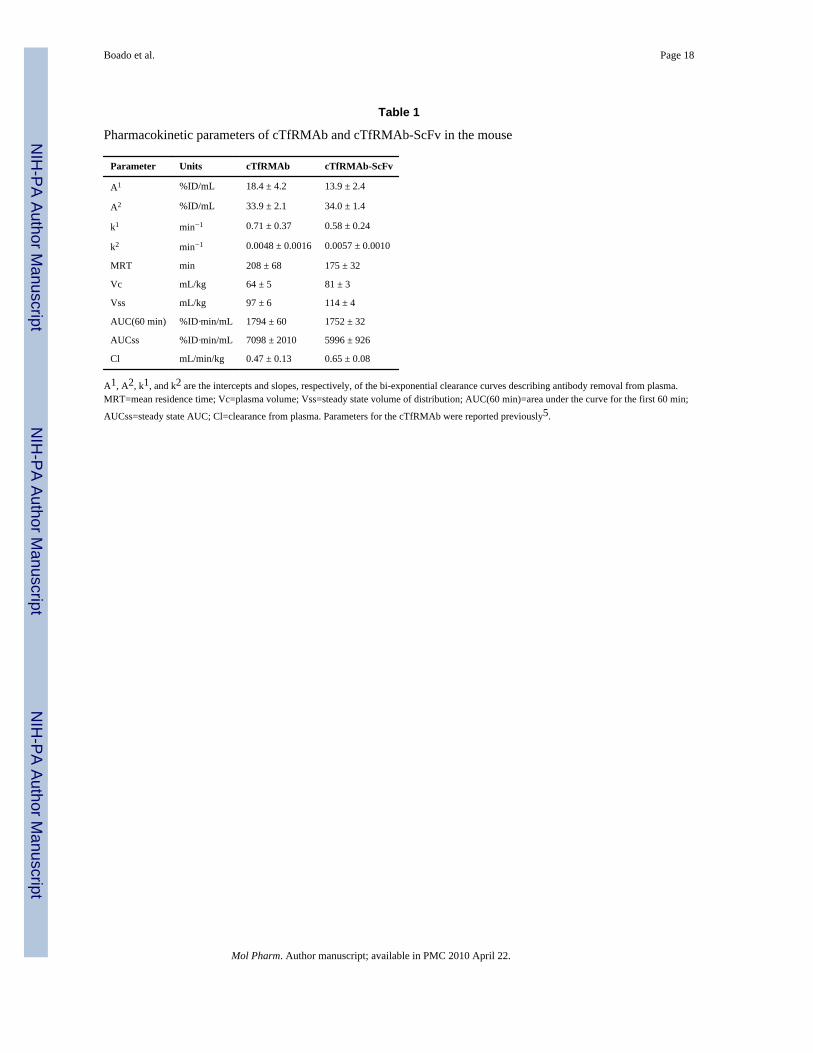
The cTfRMAb-ScFv fusion protein was radiolabeled with the 125I-Bolton-Hunter reagent,and injected into adult BALB/c mice via intravenous (IV) administration. The clearance ofthe [125I]-cTfRMAb-ScFv fusion protein from plasma after IV administration is plotted inFigure 7. The plasma radioactivity decay curve was fit to a bi-exponential equation to yieldthe pharmacokinetics (PK) parameters shown in Table 1, which are compared to the PKparameters for the parent [125I]-cTfRMAb reported previously5.
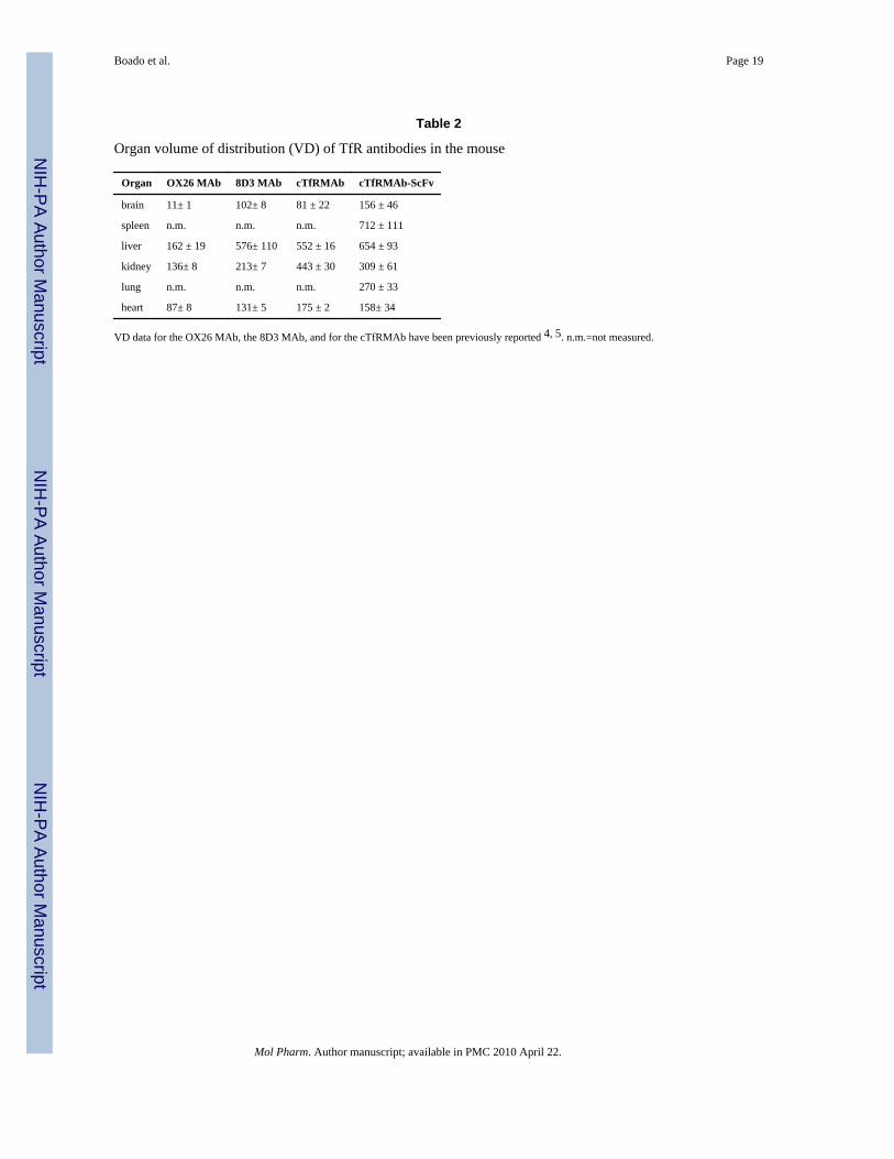
The uptake of the [125I]-cTfRMAb-ScFv fusion protein by brain and peripheral organs wasexpressed as a volume of distribution (VD) at 1 hour after IV injection, and these data areshown in Table 2, in comparison with the 60 min VD values for the OX26 MAb, the 8D3
Boado et al. Page 5
Mol Pharm. Author manuscript; available in PMC 2010 April 22.
NIH
-PA Author Manuscript
NIH
-PA Author Manuscript
NIH
-PA Author Manuscript

MAb, and the cTfRMAb in the mouse reported previously4,5. The brain uptake of theantibodies was also expressed as a %ID/g brain (Figure 8). The BBB permeability-surfacearea (PS) product of the cTfRMAb-ScFv fusion protein is equal to the ratio of the 60 min%ID/g (Figure 8) and the 60 min plasma AUC (Table 1), and is 2.0 uL/min/g.
The [125I]-cTfRMAb-ScFv fusion protein was also administered by IP injection, and theplasma concentration of the fusion protein is plotted in Figure 7. The 60 min plasma AUCfollowing IP administration of the fusion protein, computed with the trapezoidal rule, 436%IDmin/mL, is 25% of the corresponding 60 min plasma AUC for the fusion proteinfollowing IV administration (Table 1). The brain uptake of the fusion protein at 60 minfollowing IP injection was 0.41 ± 0.03 % ID/g, which is 12% of the brain uptake after IVinjection (Figure 8). The plasma radioactivity that was precipitable with TCA wascomparable after IP and IV administration. The % TCA precipitable radioactivity was 95 ±1% and 96 ± 1% at 0.25 min, and 82 ± 1% and 77 ± 6% at 60 min, after IV and IP injection,respectively.
DiscussionThe results of this study are consistent with the following conclusions. First, a cTfRMAb-ScFv fusion protein has been engineered, which is derived from the VH and VL of the rat8D3 MAb, the C-region from mouse IgG1 heavy chain and mouse kappa light chain, and theVH and VL of a mouse MAb against the Aβ peptide of AD (Figure 1). Second, CHO cellshave been permanently transfected with a tandem vector (Figure 2), the cloned CHO linesecretes the cTfRMAb-ScFv fusion protein in serum free medium, and the cTfRMAb-ScFvfusion protein is homogenous on SDS-PAGE (Figure 3) and Western blotting (Figure 4).Third, the cTfRMAb-ScFv fusion protein is bi-functional, and binds both the mouse TfR(Figure 5) and the Aβ peptide of AD (Figure 6). Fourth, the pharmacokinetics (PK) ofcTfRMAb-ScFv fusion protein clearance have been measured following IV and IP injection(Figure 7), and the PK parameters of fusion protein clearance from blood are comparable tothe PK parameters for the cTfRMAb (Table 1). Fifth, the brain uptake of the cTfRMAb-ScFv fusion protein is high, >3% ID/g (Figure 8), and is comparable to the brain uptake ofthe 8D3 MAb or the cTfRMAb, but much greater than the brain uptake of a MAb that has nospecificity for a BBB receptor, such as the OX26 MAb against the rat TfR (Table 2).
The delivery of MAb-based therapeutics to the brain could be applied to the development ofnew drugs for AD and other disorders. MAb therapeutics have been delivered to the brain bycationization12, BBB disruption13, or trans-nasal administration14. In the present approach,the MAb therapeutic is re-engineered as an IgG-ScFv fusion protein, whereby the anti-AβScFv is fused to a chimeric MAb against the mouse TfR (Figure 1). The expression of theTfRMAb-ScFv fusion protein was enabled by the genetic engineering of a tandem vectorencoding the hetero-tetrameric cTfRMAb-ScFv fusion protein (Figure 2). This tandemvector was engineered following the cloning of 6 separate genes, including the VH and VLof the 8D3 MAb against the mouse TfR, the VH and VL of a mouse MAb against the Aβpeptide of AD, the mouse IgG1 HC C-region, and the mouse kappa LC C-region5,6,11.Although the parent TfRMAb, the 8D3 MAb, is a rat IgG, the amino acid sequence of thecTfRMAb-ScFv fusion protein is >80% mouse origin5. Therefore, the fusion protein can beadministered chronically to AD transgenic mice with minimal immune reaction. IgG fusionproteins may be minimally immunogenic, as recent work shows the constant region of IgGcontains amino acid sequences called Tregitopes, which induce immune tolerance15. Recentwork shows that the chronic administration of HIRMAb fusion proteins to primates resultsin minimal immune reactions16. Nevertheless, future chronic administration of the fusionprotein will be required to fully ascertain the extent of immune reactions in mice. In
Boado et al. Page 6
Mol Pharm. Author manuscript; available in PMC 2010 April 22.
NIH
-PA Author Manuscript
NIH
-PA Author Manuscript
NIH
-PA Author Manuscript
addition, future studies can determine the distribution of the fusion protein in brain at timepoints beyond 60 min after administration.
The cTfRMAb-ScFv fusion protein is a tri-functional fusion protein (Figure 1). The ‘head’of the molecule binds the mouse TfR, to trigger receptor-mediated influx from blood tobrain across the BBB on the endogenous TfR. The ‘tail’ of the molecule binds/disaggregatesAβ amyloid plaque. The ‘mid-section’ of the fusion protein, at the CH2–CH3 region, bindsthe neonatal Fc receptor (FcRn), which is expressed at the BBB17, and binding to the BBBFcRn triggers the receptor-mediated efflux of the IgG-ScFv fusion protein from brain toblood across the BBB6. The BBB FcRn is a reverse transcytosis system that operates onlyuni-directionally in the brain to blood direction18. These 3 functionalities have beendocumented previously for the HIRMAb-ScFv fusion protein with in vivo studies in theRhesus monkey, the rat, and the AD transgenic mouse6. The anti-Aβ ScFv part of theHIRMAb-ScFv fusion protein has the same binding affinity for the Aβ peptide as theoriginal murine anti-Aβ MAb, and binds and disaggregates Aβ amyloid fibrils both in vitroand in vivo6. However, the HIRMAb-ScFv does not recognize the murine insulin receptor,and cannot penetrate the BBB of the mouse following peripheral administration. The speciesspecificity of the antibodies against the BBB insulin or transferrin receptor is illustrated bythe very low brain uptake in the mouse of the murine OX26 MAb against the rat TfR (Figure8).
The cTfRMAb-ScFv fusion protein binds the mouse TfR with high affinity (Figure 5), andthis correlates with a high degree of brain uptake in the mouse in vivo (Figure 8). The BBBPS product for the cTfRMAb-ScFv fusion protein is 2.0 uL/min/g, which is comparable tothe previously reported BBB PS product for the 8D3 MAb in the mouse, 3.3 uL/min/g4. Theratio of the PS products for the rat 8D3 MAb, and the cTfRMAb-ScFv fusion protein,parallels the ratio of the KD values of antibody binding to the mouse TfR (Figure 5). Thebrain uptake (%ID/g) of the cTfRMAb-ScFv fusion protein is slightly higher than the brainuptake of the 8D3 MAb (Figure 8), owing to the higher plasma AUC for the cTfRMAb-ScFv fusion protein (Table 1), as compared to the 8D3 MAb4.
The high brain uptake of the cTfRMAb-ScFv fusion protein is to be contrasted with the verylow brain uptake of a MAb that has no specificity for a BBB receptor transport system. TheOX26 MAb is a murine antibody against the rat TfR that does not recognize the mouseTfR4. The brain VD of the OX26, 11 uL/g (Table 2), is equal to the cerebral blood volume4,which indicates the OX26 MAb does not cross the mouse BBB. The VD for the OX26 MAbin peripheral organs in the mouse represents the plasma volume in those organs (Table 2).The ratio of the organ VD for the cTfRMAb-ScFv fusion protein divided by the organ VDfor the OX26 MAb is 14, 4, 2, and 2 for brain, liver, kidney, and heart, respectively (Table2). This analysis shows that the cTfRMAb-ScFv fusion protein is preferentially targeted tobrain, relative to peripheral organs in the mouse.
The brain uptake of the OX26 MAb in the mouse is representative of the brain uptake of anantibody that has no specificity for a BBB receptor. An anti-Aβ MAb has no affinity for aBBB transporter, and prior work as shown an anti-Aβ MAb has the same property as theOX26 MAb in the mouse, in that the brain VD is no different from the cerebral bloodvolume6. Such antibodies do not penetrate the BBB, and cannot access the amyloid plaquein brain behind the BBB, in the absence of BB disruption. In contrast, the cTfRMAb-ScFvfusion protein rapidly penetrates the BBB in the mouse, and 3.5% of the injected dose istaken up per gram brain (Figure 8). At a systemic dose of 1 mg/kg of the cTfRMAb-ScFvfusion protein, the brain concentration of the cTfRMAb-ScFv fusion protein is projected tobe 4 pmol/gram. Previous studies show that brain concentrations of this magnitude are
Boado et al. Page 7
Mol Pharm. Author manuscript; available in PMC 2010 April 22.
NIH
-PA Author Manuscript
NIH
-PA Author Manuscript
NIH
-PA Author Manuscript

sufficient to cause disaggregation and clearance of amyloid plaque in AD transgenic mousebrain6.
In summary, the present studies describe the genetic engineering of an antibody fusionprotein that has been specifically engineered to cross the mouse BBB, as well as bind the Aβamyloid plaque of AD. The BBB-penetrating properties of the cTfRMAb-ScFv fusionprotein enable the antibody to access the amyloid plaque in brain behind the BBB. Inaddition, the antibody is rapidly cleared from blood with a MRT of 3 hours (Table 1), andthis rapid removal from blood will prevent the build-up of high concentrations of the Aβ-binding antibody in plasma. Future treatment studies of AD transgenic mice are required tofully evaluate the therapeutic and toxicologic effects of chronic administration of thecTfRMAb-ScFv fusion protein.
AcknowledgmentsWinnie Tai and Phuong Tram provided technical assistance. This work was supported by NIH grant R01-AG032244.
References1. Pardridge WM. Re-engineering biopharmaceuticals for delivery to brain with molecular Trojan
horses. Bioconjug. Chem 2008;19:1327–1338. [PubMed: 18547095]2. Pardridge WM, Kang Y-S, Buciak JL, Yang J. Human insulin receptor monoclonal antibody
undergoes high affinity binding to human brain capillaries in vitro and rapid transcytosis throughthe blood-brain barrier in vivo in the primate. Pharm. Res 1995;12:807–816. [PubMed: 7667183]
3. Skarlatos S, Yoshikawa T, Pardridge WM. Transport of [125I]transferrin through the rat blood-brainbarrier in vivo. Brain Res 1995;683:164–171. [PubMed: 7552351]
4. Lee HJ, Engelhardt B, Lesley J, Bickel U, Pardridge WM. Targeting rat anti-mouse transferrinreceptor monoclonal antibodies through blood-brain barrier in mouse. J. Pharmacol. Exp. Ther2000;292:1048–1052. [PubMed: 10688622]
5. Boado RJ, Zhang Y, Wang Y, Pardridge WM. Engineering and expression of a chimeric transferrinreceptor monoclonal antibody for blood-brain barrier delivery in the mouse. Biotechnol Bioeng2009;102:1251–1258. [PubMed: 18942151]
6. Boado RJ, Zhang Y, Xia CF, Pardridge WM. Fusion antibody for Alzheimer's disease withbidirectional transport across the blood-brain barrier and abeta fibril disaggregation. Bioconjug.Chem 2007;18:447–455. [PubMed: 17315944]
7. Bard F, Cannon C, Barbour R, Burke RL, Games D, Grajeda H, Guido T, Hu K, Huang J, Johnson-Wood K, Khan K, Kholodenko D, Lee M, Lieberburg I, Motter R, Nguyen M, Soriano F, VasquezN, Weiss K, Welch B, Seubert P, Schenk D, Yednock T. Peripherally administered antibodiesagainst amyloid beta-peptide enter the central nervous system and reduce pathology in a mousemodel of Alzheimer disease. Nat Med 2000;6:916–919. [PubMed: 10932230]
8. DeMattos RB, Bales KR, Cummins DJ, Dodart JC, Paul SM, Holtzman DM. Peripheral anti-A betaantibody alters CNS and plasma A beta clearance and decreases brain A beta burden in a mousemodel of Alzheimer's disease. Proc. Natl. Acad. Sci. U S A 2001;98:8850–8855. [PubMed:11438712]
9. Lombardo JA, Stern EA, McLellan ME, Kajdasz ST, Hickey GA, Bacskai BJ, Hyman BT.Amyloid-beta antibody treatment leads to rapid normalization of plaque-induced neuriticalterations. J. Neurosci 2003;23:10879–10883. [PubMed: 14645482]
10. Seubert P, Barbour R, Khan K, Motter R, Tang P, Kholodenko D, Kling K, Schenk D, Johnson-Woo K, Schroeter S, Gill D, Jacobsen JS, Pangalos M, Basi G, Games D. Antibody capture ofsoluble Abeta does not reduce cortical Abeta amyloidosis in the PDAPP mouse. Neurodegener.Dis 2008;5:65–71. [PubMed: 18182780]
11. Boado RJ, Zhang Y, Pardridge WM. Genetic engineering, expression, and activity of a fusionprotein of a human neurotrophin and a molecular Trojan horse for delivery across the humanblood-brain barrier. Biotechnol. Bioeng 2007;97:1376–1386. [PubMed: 17286273]
Boado et al. Page 8
Mol Pharm. Author manuscript; available in PMC 2010 April 22.
NIH
-PA Author Manuscript
NIH
-PA Author Manuscript
NIH
-PA Author Manuscript

12. Poduslo JF, Ramakrishnan M, Holasek SS, Ramirez-Alvarado M, Kandimalla KK, Gilles EJ,Curran GL, Wengenack TM. In vivo targeting of antibody fragments to the nervous system forAlzheimer’s disease immunotherapy and molecular imaging of amyloid plaques. J. Neurochem2007;102:420–433. [PubMed: 17596213]
13. Raymond SB, Treat LH, Dewey JD, McDannold NJ, Hynynen K, Bacskai BJ. Ultrasoundenhanced delivery of molecular imaging and therapeutic agents in Alzheimer’s disease mousemodels. PLoS ONE 2008;3:e2175. [PubMed: 18478109]
14. Solomon, B. In vivo targeting of amyloid plaques via intranasal administration of phage anti-b-amyloid antibodies. In: Fisher, A.; Memo, M.; Stocchi, F.; Hanin, I., editors. Advances inAlzheimer’s and Parkinson’s Disease. New York: Springer Science; 2008. p. 273-278.
15. De Groot AS, Moise L, McMurry JA, Wambre E, Van Overtvelt L, Moingeon P, Scott DW,Martin W. Activation of natural regulatory T cells by IgG Fc-derived peptide "Tregitopes". Blood2008;112:3303–3311. [PubMed: 18660382]
16. Boado RJ, Hui EK-W, Lu JZ, Pardridge WM. AGT-181: Expression in CHO cells andpharmacokinetics, safety, and plasma iduronidase enzyme activity in Rhesus monkeys. J.Biotechnol 2009;144:135–141. [PubMed: 19735678]
17. Schlachetzki F, Zhu C, Pardridge WM. Expression of the neonatal Fc receptor (FcRn) at the blood-brain barrier. J. Neurochem 2002;81:203–206. [PubMed: 12067234]
18. Zhang Y, Pardridge WM. Rapid transferrin efflux from brain to blood across the blood-brainbarrier. J Neurochem 2001;76:1597–1600. [PubMed: 11238745]
Boado et al. Page 9
Mol Pharm. Author manuscript; available in PMC 2010 April 22.
NIH
-PA Author Manuscript
NIH
-PA Author Manuscript
NIH
-PA Author Manuscript
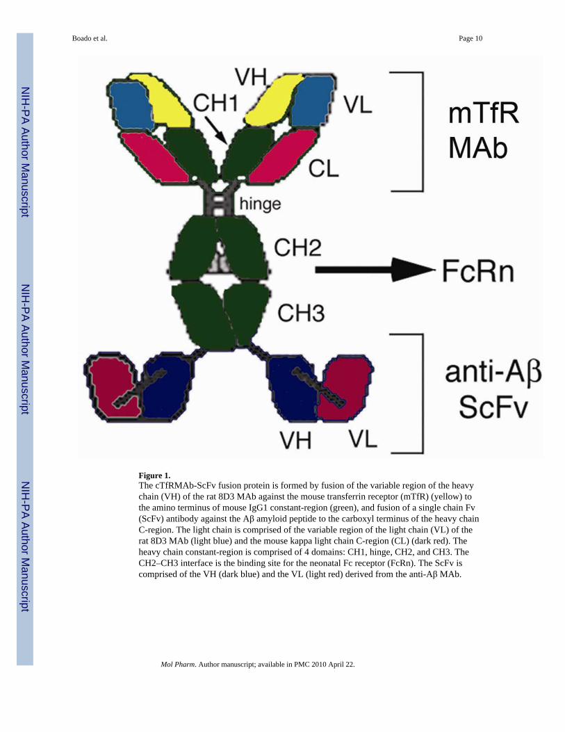
Figure 1.The cTfRMAb-ScFv fusion protein is formed by fusion of the variable region of the heavychain (VH) of the rat 8D3 MAb against the mouse transferrin receptor (mTfR) (yellow) tothe amino terminus of mouse IgG1 constant-region (green), and fusion of a single chain Fv(ScFv) antibody against the Aβ amyloid peptide to the carboxyl terminus of the heavy chainC-region. The light chain is comprised of the variable region of the light chain (VL) of therat 8D3 MAb (light blue) and the mouse kappa light chain C-region (CL) (dark red). Theheavy chain constant-region is comprised of 4 domains: CH1, hinge, CH2, and CH3. TheCH2–CH3 interface is the binding site for the neonatal Fc receptor (FcRn). The ScFv iscomprised of the VH (dark blue) and the VL (light red) derived from the anti-Aβ MAb.
Boado et al. Page 10
Mol Pharm. Author manuscript; available in PMC 2010 April 22.
NIH
-PA Author Manuscript
NIH
-PA Author Manuscript
NIH
-PA Author Manuscript
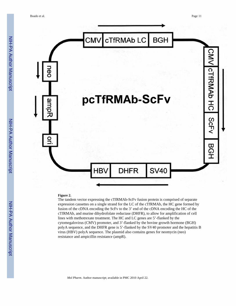
Figure 2.The tandem vector expressing the cTfRMAb-ScFv fusion protein is comprised of separateexpression cassettes on a single strand for the LC of the cTfRMAb, the HC gene formed byfusion of the cDNA encoding the ScFv to the 3’ end of the cDNA encoding the HC of thecTfRMAb, and murine dihydrofolate reductase (DHFR), to allow for amplification of celllines with methotrexate treatment. The HC and LC genes are 5’-flanked by thecytomegalovirus (CMV) promoter, and 3’-flanked by the bovine growth hormone (BGH)polyA sequence, and the DHFR gene is 5’-flanked by the SV40 promoter and the hepatitis Bvirus (HBV) polyA sequence. The plasmid also contains genes for neomycin (neo)resistance and ampicillin resistance (ampR).
Boado et al. Page 11
Mol Pharm. Author manuscript; available in PMC 2010 April 22.
NIH
-PA Author Manuscript
NIH
-PA Author Manuscript
NIH
-PA Author Manuscript

Figure 3.Reducing SDS-PAGE of molecular weight standards (lane 1) and the cTfRMAb-ScFvfusion protein (lane 2). The size of the heavy chain is about 85 kDa and the size of the lightchain is about 26 kDa.
Boado et al. Page 12
Mol Pharm. Author manuscript; available in PMC 2010 April 22.
NIH
-PA Author Manuscript
NIH
-PA Author Manuscript
NIH
-PA Author Manuscript

Figure 4.Western blotting with a primary antibody against mouse IgG and biotinylated molecularweight standards (lane 1), the cTfRMAb (lane 2), and the cTfRMAb-ScFv fusion protein(lane 3). Both the cTfRMAb and the cTfRMAb-ScFv fusion protein share the same lightchain. The size of the heavy chain of the cTfRMAb-ScFv fusion protein is about 28 kDalarger than the size heavy chain of the cTfRMAb owing to fusion of the ScFv.
Boado et al. Page 13
Mol Pharm. Author manuscript; available in PMC 2010 April 22.
NIH
-PA Author Manuscript
NIH
-PA Author Manuscript
NIH
-PA Author Manuscript

Figure 5.Radio-receptor assay of the mouse TfR uses mouse fibroblasts as the source of the mouseTfR and [125I]-8D3 as the binding ligand. Binding is displaced by unlabeled 8D3 MAb (A)or the cTfRMAb-ScFv fusion protein at concentrations up to 30 nM (B). The KD of 8D3self-inhibition and the KI of chimeric TfRMAb cross-inhibition were computed by non-linear regression analysis.
Boado et al. Page 14
Mol Pharm. Author manuscript; available in PMC 2010 April 22.
NIH
-PA Author Manuscript
NIH
-PA Author Manuscript
NIH
-PA Author Manuscript
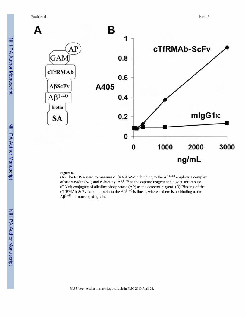
Figure 6.(A) The ELISA used to measure cTfRMAb-ScFv binding to the Aβ1–40 employs a complexof streptavidin (SA) and N-biotinyl Aβ1–40 as the capture reagent and a goat anti-mouse(GAM) conjugate of alkaline phosphatase (AP) as the detector reagent. (B) Binding of thecTfRMAb-ScFv fusion protein to the Aβ1–40 is linear, whereas there is no binding to theAβ1–40 of mouse (m) IgG1κ.
Boado et al. Page 15
Mol Pharm. Author manuscript; available in PMC 2010 April 22.
NIH
-PA Author Manuscript
NIH
-PA Author Manuscript
NIH
-PA Author Manuscript

Figure 7.Plasma concentration, expressed as % of injected dose (I.D.)/mL, of the [125I]-cTfRMAb-ScFv fusion protein after either intravenous or intra-peritoneal injection in the mouse. Dataare mean ± S.E. (n=3 mice/point).
Boado et al. Page 16
Mol Pharm. Author manuscript; available in PMC 2010 April 22.
NIH
-PA Author Manuscript
NIH
-PA Author Manuscript
NIH
-PA Author Manuscript
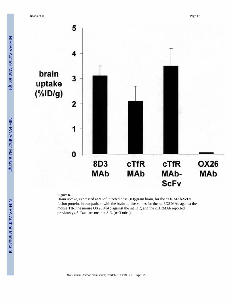
Figure 8.Brain uptake, expressed as % of injected dose (ID)/gram brain, for the cTfRMAb-ScFvfusion protein, in comparison with the brain uptake values for the rat 8D3 MAb against themouse TfR, the mouse OX26 MAb against the rat TfR, and the cTfRMAb reportedpreviously4,5. Data are mean ± S.E. (n=3 mice).
Boado et al. Page 17
Mol Pharm. Author manuscript; available in PMC 2010 April 22.
NIH
-PA Author Manuscript
NIH
-PA Author Manuscript
NIH
-PA Author Manuscript
NIH
-PA Author Manuscript
NIH
-PA Author Manuscript
NIH
-PA Author Manuscript
Boado et al. Page 18
Table 1
Pharmacokinetic parameters of cTfRMAb and cTfRMAb-ScFv in the mouse
Parameter Units cTfRMAb cTfRMAb-ScFv
A1 %ID/mL 18.4 ± 4.2 13.9 ± 2.4
A2 %ID/mL 33.9 ± 2.1 34.0 ± 1.4
k1 min−1 0.71 ± 0.37 0.58 ± 0.24
k2 min−1 0.0048 ± 0.0016 0.0057 ± 0.0010
MRT min 208 ± 68 175 ± 32
Vc mL/kg 64 ± 5 81 ± 3
Vss mL/kg 97 ± 6 114 ± 4
AUC(60 min) %ID·min/mL 1794 ± 60 1752 ± 32
AUCss %ID·min/mL 7098 ± 2010 5996 ± 926
Cl mL/min/kg 0.47 ± 0.13 0.65 ± 0.08
A1, A2, k1, and k2 are the intercepts and slopes, respectively, of the bi-exponential clearance curves describing antibody removal from plasma.MRT=mean residence time; Vc=plasma volume; Vss=steady state volume of distribution; AUC(60 min)=area under the curve for the first 60 min;
AUCss=steady state AUC; Cl=clearance from plasma. Parameters for the cTfRMAb were reported previously5.
Mol Pharm. Author manuscript; available in PMC 2010 April 22.
NIH
-PA Author Manuscript
NIH
-PA Author Manuscript
NIH
-PA Author Manuscript
Boado et al. Page 19
Table 2
Organ volume of distribution (VD) of TfR antibodies in the mouse
Organ OX26 MAb 8D3 MAb cTfRMAb cTfRMAb-ScFv
brain 11± 1 102± 8 81 ± 22 156 ± 46
spleen n.m. n.m. n.m. 712 ± 111
liver 162 ± 19 576± 110 552 ± 16 654 ± 93
kidney 136± 8 213± 7 443 ± 30 309 ± 61
lung n.m. n.m. n.m. 270 ± 33
heart 87± 8 131± 5 175 ± 2 158± 34
VD data for the OX26 MAb, the 8D3 MAb, and for the cTfRMAb have been previously reported 4, 5. n.m.=not measured.
Mol Pharm. Author manuscript; available in PMC 2010 April 22.
Related Documents