PHARMACOGNOSTIC, PHYTOCHEMICAL AND PHARMACOLOGICAL EVALUATION OF THE LEAVES OF Citrullus lanatus (Thunb.) Matsum. & Nakai. (CUCURBITACEAE) Dissertation submitted to The Tamil Nadu Dr. M.G.R. Medical University Chennai‐600 032 In partial fulfilment of the requirements for the award of the degree of MASTER OF PHARMACY IN PHARMACOGNOSY Submitted by 261220709 DEPARTMENT OF PHARMACOGNOSY COLLEGE OF PHARMACY MADURAI MEDICAL COLLEGE MADURAI - 625 020 APRIL 2014

Welcome message from author
This document is posted to help you gain knowledge. Please leave a comment to let me know what you think about it! Share it to your friends and learn new things together.
Transcript
PHARMACOGNOSTIC, PHYTOCHEMICAL AND PHARMACOLOGICAL EVALUATION OF THE LEAVES OF
Citrullus lanatus (Thunb.) Matsum. & Nakai. (CUCURBITACEAE)
Dissertation submitted to
The Tamil Nadu Dr. M.G.R. Medical University
Chennai‐600 032
In partial fulfilment of the requirements
for the award of the degree of
MASTER OF PHARMACY IN
PHARMACOGNOSY
Submitted by
261220709
DEPARTMENT OF PHARMACOGNOSY
COLLEGE OF PHARMACY MADURAI MEDICAL COLLEGE
MADURAI - 625 020
APRIL 2014
Dr.A. ABDUL HASAN SATHALI, M.Pharm., Ph.D., PRINCIPAL (i/c), College of Pharmacy, Madurai Medical College, Madurai-625 020
CERTIFICATE
This is to certify that the dissertation entitled “PHARMACOGNOSTIC,
PHYTOCHEMICAL AND PHARMACOLOGICAL EVALUATION OF THE
LEAVES OF Citrullus lanatus (Thunb.) Matsum. & Nakai, (CUCURBITACEAE)’’
submitted by Miss. K.VIJAYALAKSHMI (Reg. No. 261220709) in partial fulfilment of the
requirements for the award of the degree of MASTER OF PHARMACY in
PHARMACOGNOSY by The Tamil Nadu Dr. M.G.R. Medical University is a bonafied
work done by her during the academic year 2013-2014 under the guidance of
Dr.(Mrs). AJITHADAS ARUNA, M.Pharm., Ph.D., Joint Director of Medical Education
(Pharmacy), in the Department of Pharmacognosy, College of Pharmacy, Madurai Medical
College, Madurai-625 020.
(Dr.A. ABDUL HASAN SATHALI )
ACKNOWLEDGEMENTS
I first and foremost express my revered regard and obeisance to the ALMIGHTY
GOD with whose blessings I was able to complete my project work.
I am grateful to express my sincere thanks to Dr. B. SANTHAKUMAR, M.Sc
(F.Sc)., M.D(F.M)., PGDMLE, Dip.N.B (F.M)., Dean, Madurai Medical College for giving
me an opportunity to carry out my project work.
I sincerely thanks with heartfelt sense of gratitude to
Dr. L. SANTHANALAKSHMI, M.D., D.G.O., M.B.A., Vice Principal, Madurai Medical
College for giving me an opportunity to carry out my project work.
It is my privilege to express a deep and heartfelt sense of gratitude and my regards to
our respected Dr. Mrs. AJITHADAS ARUNA, M. Pharm., Ph.D., Joint Director of
Medical Education (Pharmacy) and former Principal, College of Pharmacy, Madurai Medical
College and for her active guidance, advice, help, support and encouragement. I am very
much obliged for her perseverance, without which my project work would not be completed.
I owe a great debt of gratitude and heartful thanks to Dr. A. ABDUL HASAN
SATHALI, M. Pharm., Ph.D., Principal (in charge) and Head of Department of
Pharmaceutics, College of Pharmacy, Madurai Medical College, Madurai
I express my heartful thanks and regards to Miss R. GOWRI, M.Pharm.,
Dr.K.PERIYANAYAGAM,M.Pharm.,Ph.D., and Mr.T.VENKATARATHINA
KUMAR,M.Pharm.,(Ph.D)., Assistant Readers in Pharmacy, Department of
Pharmacognosy, College of Pharmacy, Madurai Medical College for their support and
valuable suggestions.
I thank Prof. Mrs. R. THARABAI, M. Pharm, Professor and Head of Department
of Pharmaceutical Chemistry, College of Pharmacy, Madurai Medical College for their
guidance during the course of my study.
I thank Mrs. A. SETHURAMANI, M.Pharm.,(Ph.D)., and Dr.Mrs.A.
KRISHNAVENI, M. Pharm., Ph.D., Tutors in Pharmacy, Department of Pharmacognosy,
College of Pharmacy, Madurai Medical College for their help.
I thank Mr. P. SIVAKUMAR M.Sc., DMLT, Lab Supervisor of the Dept. of
Pharmacognosy and Mr. MAGUDESWARAN, DMLT, Lab Technician and
Mrs. P. ELLAYEE for their support during my study of this work.
I extend my thanks to all the teaching staff of College of Pharmacy and of Madurai
Medical College who have rendered their help towards completion of the project.
I place on record my gratitude to Dr. P. JAYARAMAN, M.Sc., Ph.D., Director,
Plant Anatomy Research Centre, Chennai 600 045 who helped me in the microscopic studies
and Dr. D. STEPHEN, Ph.D., Senior Lecturer, Department of Botany, American College,
Madurai who helped me in the identification of my plant.
I thank Dr. Mr. MARIYAPPAN (Scientist), Indian Council of Medical Research,
Madurai for his kind guidance for study of my project work.
I am thankful to Mr. Jones Universal Scientific Supplier for his timely supply of
chemicals utilized during the project work.
I express my gratitude to Mrs. Anithakumari of AVN formulations Madurai for her
support and help in carry out HPTLC analysis of this work.
I also thank my ever loving classmates, Miss. P. Anitha, Miss. P. Bala, Miis. R.
Jancy Gracelet, Mr. S. Jegadeesh, Miss. M.Kalaiyarasi, Mrs. S.R.Nandhini,
Mrs. S.Nathiya, Miss. D.Suganya, Mr.A.Manikkavasagan, Mr.J.Rajesh Kumar,
Mr.Sankar Ganesh, Mr.S.Sudhakar, Mr.M.Ramanathan, Mr.P.Kanniyappan,
Mr.P.Arjun Kumar, Mrs.S.Ponnammal Asmi, Mr.C.Pravin Kumar, Miss.R.Elavarasi,
Miss.S.Karpagam, Miss.E.Ajila, Mr.K.Sasikumar and all my juniors of 2013-2015 batch
and other friends for their constant motivation and help.
Above all, I am forever indebted to my father Mr. M.KANNAPPAN, my mother
Mrs. K. PUSHPAM, my brother Mr. K. MANIKANDAN and Mr. P. MOHAN for their
understanding, endless patience, help and encouragement which made me to complete this
work.
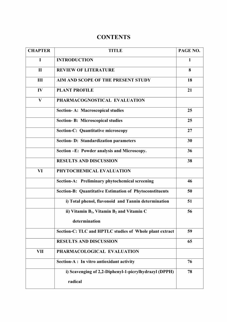
CONTENTS
CHAPTER TITLE PAGE NO.
I INTRODUCTION 1
II REVIEW OF LITERATURE 8
III AIM AND SCOPE OF THE PRESENT STUDY 18
IV PLANT PROFILE 21
V PHARMACOGNOSTICAL EVALUATION
Section- A: Macroscopical studies 25
Section- B: Microscopical studies 25
Section-C: Quantitative microscopy 27
Section- D: Standardization parameters 30
Section –E: Powder analysis and Microscopy. 36
RESULTS AND DISCUSSION 38
VI PHYTOCHEMICAL EVALUATION
Section-A: Preliminary phytochemical screening 46
Section-B: Quantitative Estimation of Phytoconstituents 50
i) Total phenol, flavonoid and Tannin determination 51
ii) Vitamin B1, Vitamin B2 and Vitamin C
determination
56
Section-C: TLC and HPTLC studies of Whole plant extract 59
RESULTS AND DISCUSSION 65
VII PHARMACOLOGICAL EVALUATION
Section-A : In vitro antioxidant activity 76
i) Scavenging of 2,2-Diphenyl-1-picrylhydrazyl (DPPH)
radical
78
ii) Total Antioxidant activity by phosphomolybdenum
method).
79
iii) Reducing power assay. 80
iv) Ferric Reducing Antioxidant Power Assay (TPTZ
method).
81
Section-B: Larvicidal activity of methanolic extract of
Citrullus lanatus.
82
Section-C: Invitro activity of chicken pancreatic lipase
inhibition assay
84
Section-D: Invitro anti-cancer (Breast cancer) activity by
MTT assay.
87
Section-E: Invitro Anti-diabetic activity by various method 90
i) Non-enzymatic glycosylation of haemoglobin Assay.
94
ii) Glucose uptake in yeast cells
a) % inhibition of Glucose uptake in 5mM glucose
concentrations.
b) % inhibition of Glucose uptake in 10mM glucose
concentrations.
96
iii) Alpha amylase inhibition assay. 97
iv) Alpha glucosidase inhibition assay
99
RESULTS AND DISCUSSION 102
VIII SUMMARY & CONCLUSION 123
IX REFERENCES i-xiii
INTRODUCTION
Chapter I Introduction
Department of Pharmacognosy, MMC. 1
CHAPTER I
INTRODUCTION
Medicinal Plants [1-3]
A plant is any plant which, in one or more of its organs, contains substances that can
be used for therapeutic purposes, or which are precursors for semi-synthetic compounds.
When a plant is designated as ‘medicinal’, it is implied that the said plant is useful as a drug
or therapeutic agent or an active ingredient of a medicinal preparation. Medicinal plants may
therefore be defined as a group of plants that possess some special properties or virtues that
qualify them as articles of drugs and therapeutic agents, and are used for medicinal purposes.
History of medicinal plants
Plants have been used for medicinal purposes from 5000 BC with the emergence of
the Indus Valley Civilization. The indigenous system of medicine, viz.-Ayurvedic, Siddha
and Unani, have been in existence for several centuries. The country has 45,000 different
plant species and 15000 medicinal plants that include 2000 plants used in Ayurveda, 700 in
Unani, 600 in Siddha, 450 in Homoeopathy and 30 in modern medicines. The drugs are
derived either from the whole plant or from different parts like leaves, stem, bark, root,
flower, seed etc. Some drugs are prepared from excretory plant product such as gum, resins
and latex.
Significance of medicinal plants to human beings
(1) Many of the modern medicines are produced indirectly from medicinal plants, for
example aspirin.
(2) Plants are directly used as medicines by a majority of cultures around the world,
for example Chinese medicine and Indian medicine.
(3) Many food crops have medicinal effects, for example garlic.
Chapter I Introduction
Department of Pharmacognosy, MMC. 2
(4) Medicinal plants are resources of new drugs. It is estimated there are more than
250, 000 flower plant species.
Hence studying medicinal plants helps to understand plant toxicity and protect human
and animals from natural poisons. Cultivation and preservation of medicinal plants protect
biological diversity, for example metabolic engineering of plants.
Future of medicinal plants
Medicinal plants have a promising future because there are about half million plants
around the world, and most of their medical activities have not been investigated yet, and
their pharmacological activities could be decisive in the treatment of present or future studies.
Characteristics of medicinal plants
Medicinal plants have many characteristics when used as a treatment, as follow:
Synergic medicine - The ingredients of plants all interact simultaneously, so their
uses can complement or damage others or neutralize their possible negative effects.
Support of official medicine - The components of the plants proved to be very
effective in the treatment of complex cases like cancer diseases.
Preventive medicine - It has been proven that the component of the plants also has
the ability to prevent the appearance of some diseases which can help to reduce the
use of the chemical remedies and reduce the side effect of synthetic treatment.
Medicinal plants in India
About 60 percent of the world’s population use herbal medicines. Herbal medicines
are not only used for primary health care not just in rural areas in developing countries, but
also in developed countries as well where modern medicines are predominantly used.

Chapter I Introduction
Department of Pharmacognosy, MMC. 3
There are about 45,000 medicinal plant species in India, concentrated in the region of
Eastern Himalayas, Western Ghats and Andaman & Nicobar Island. The officially
documented plants with medicinal potential are 3000 but traditional practitioners use more
than 6000 plants. India is the largest producer of medicinal herbs and is called the botanical
garden of the world.
Ayurveda and Kabiraji (herbal medicine) are two important forms of alternative
medicine that is widely available in India. Ayurveda form of medicine is believed to be
existent in India for thousands of years.
The codified traditions have about 25,000 plant drug formulations that have emerged
from such studies. In addition to this, over 50,000 formulations are believed to be available in
the folk and tribal traditions. All these point to the deep passion for an exhaustive knowledge
about medicinal plants that have existed in this land from time immemorial.
Importance of Medicinal Plants [4]
The medicinal plants find application in pharmaceutical, cosmetic, agricultural and
food industry. The use of the medicinal herbs for curing disease has been documented in
history of all civilizations. Man in the pre-historic era was probably not aware about the
health hazards associated with irrational therapy. With the onset of research in medicine, it
was concluded that plants contain active principles, which are responsible, for curative action
of the herbs.
Integrating the use of Traditional medicine (TM) in the treatment of incurable disease
such as AIDS to boost immunity is wiser than waiting for the immune system to weaken to
begin antiviral therapy as is the common practice, especially when evidence exists that 11 of
the anti infective herbs in Chinese TM have shown to be anti-HIV.
Chapter I Introduction
Department of Pharmacognosy, MMC. 4
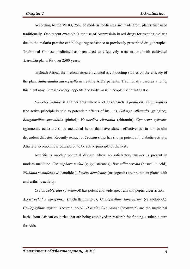
According to the WHO, 25% of modern medicines are made from plants first used
traditionally. One recent example is the use of Artemisinin based drugs for treating malaria
due to the malaria parasite exhibiting drug resistance to previously prescribed drug therapies.
Traditional Chinese medicine has been used to effectively treat malaria with cultivated
Artemisia plants for over 2500 years.
In South Africa, the medical research council is conducting studies on the efficacy of
the plant Sutherlandia microphylla in treating AIDS patients. Traditionally used as a tonic,
this plant may increase energy, appetite and body mass in people living with HIV.
Diabetes mellitus is another area where a lot of research is going on. Ajuga reptens
(the active principle is said to potentiate effects of insulin), Galagea officinalis (galagine),
Bougainvillea spectabilis (pinitol), Momordica charantia (chirantin), Gymnema sylvestre
(gymnemic acid) are some medicinal herbs that have shown effectiveness in non-insulin
dependent diabetes. Recently extract of Tecoma stans has shown potent anti diabetic activity.
Alkaloid tecomonine is considered to be active principle of the herb.
Arthritis is another potential disease where no satisfactory answer is present in
modern medicine. Commiphora mukul (guggulsterones), Boswellia serrata (boswellic acid),
Withania somnifera (withanolides), Ruscus acueleatus (ruscogenin) are prominent plants with
anti-arthritic activity.
Croton sublyratus (plaunoyol) has potent and wide spectrum anti peptic ulcer action.
Ancistrocladus korupensis (michellammine-b), Caulophyllum langigerum (calanolide-A),
Caulophyllum teymani (costatolide-A), Homalanthus natans (prostratin) are the medicinal
herbs from African countries that are being employed in research for finding a suitable cure
for Aids.
Chapter I Introduction
Department of Pharmacognosy, MMC. 5
Some Common Major Diseases [5]
Some common major disease described by the Dirnasa tribe are jaundice, diabetes,
high blood pressure, urinary tract infection, carbuncles, cardiac problem, cancer, hearnaturia.
Malaria, filariasis, Japanese encephalitis, dengue hemorrhagic fever, chikungunya and yellow
fever are transmitted by mosquitoes which cause millions of deaths every year.
Modern medicine discovered from plants [6]
Plants provide biologically active molecules and lead structures for the development
of modified derivatives with enhanced activity and reduced toxicity. The small fraction of
flowering plants that have so far been investigated have yielded about 120 therapeutic agents
of known structure from about 90 species of plants. Some of the useful plant drugs include
vinblastine, vincristine, taxol, podophyllotoxin, camptothecin, digitoxigenin, gitoxigenin,
digoxigenin, tubocurarine, morphine, codeine, aspirin, pilocarpine, capscicine, allicin,
curcumin, artemesinin and ephedrine among others. In some cases, the crude extract of
medicinal plants may be used as medicaments. About 121 (45 tropical and 76 subtropical)
major plant drugs have been identified for which no synthetic one is currently available.
It has been estimated that more than 400 traditional plants or plant-derived products
have been used for the management of type 2 diabetes across geographically. Galegine, a
substance produced by the herb Galega officinalis, provides an excellent example of such a
discovery. Experimental and clinical evaluations of galegine, provided the pharmacological
and chemical basis for the discovery of metformin which is the foundation therapy for type 2
diabetes. Plant derived agents are also being used for the treatment of cancer. Several
anticancer agents including taxol, vinblastine, vincristine, the camptothecin derivatives,
topotecan and irinotecan, and etoposide derived from epipodophyllotoxin are in clinical use
all over the world.
Chapter I Introduction
Department of Pharmacognosy, MMC. 6
The use of herbal products is of global importance because of their low side effects,
accessibility and affordability when compared with conventional medicine.
Advantages of Herbal Medicines compare with alternative therapy [7-8]
Herbal medicine has been used for centuries to treat many different health conditions.
As with most types of complementary or alternative therapy, people may use it to help
themselves feel better or feel more in control of their situation. Herbal medicine is often
promoted as a natural way to help you relax and cope with anxiety, depression and other
conditions such as hay fever, irritable bowel syndrome, menstrual (period) problems and skin
conditions such as eczema.
In India it is proving to be a major health problem, especially in the urban areas. Though
there are various approaches to reduce the ill effects of diabetes and its secondary complications,
herbal formulations are preferred due to lesser side effects, low cost, widely available and less
toxic compared with allopathic drugs.
CITRULLUS LANATUS (Watermelon) [9]
Citrallus lanatus (water melon) produces a fruit that is about 93% water, hence the
name “water” melon. The “melon” part came from the fact that the fruit is large and round
and has a sweet, pulpy flesh. The scientific name of the watermelon is derived from both
Greek and Latin roots. The Citrullus part comes from a Greek word “citrus” which is a
reference to the fruit. The lanatus part is Latin, and has the meaning of being wooly, referring
to the small hairs on the stems and leaves of the plant (Baker, et al., 2012). Watermelon is
thought to have originated in southern Africa because it is found growing wild throughout the
area, and reaches maximum diversity of forms there. It has been cultivated in Africa for over
4,000 years. Citrullus lanatus was brought to America by Spanish and quickly became very
popular crop (Robinson and Decker, 1997).
Chapter I Introduction
Department of Pharmacognosy, MMC. 7
Citrullus lanatus (Water melon) has been reportedly used widely in traditional
herbal medicine. The fruit is used as a febrifuge, diuretic, purgative and used
in treatment of diarrhoea, gonorrhoea, dropsy and renal stones.
The leaves of Citrullus lanatus is analgesic, anti-inflammatory, mosquitocidal,
gonorrhoea and anti microbial property.
The fruit is also diuretic, anti-cancer, high BP, antiviral and is effective in the
treatment of dropsy and renal stones. The seed is also a good vermifuge and
has a hypotensive action. Preliminary research indicates that the consumption
of watermelon may have antihypertensive effects.
The root is purgative and in high dose it can also serve as emetic.
The seed is a good vermifuge and has a hypotensive action. It is a demulcent
and used in the treatment of the urinary tract infections as well as bed wetting.
Fatty oil in the seed, as well as aqueous or alcoholic extracts, had been
reported to paralyze tapeworms and roundworms.
The rind of the fruit is prescribed in cases of alcoholic poisoning and
The plant Citrullus lanatus has been selected (specially the leaves) for the present
investigation on the basis of the ethnomedical information and the review of literature as the
plant is widely cultivated throughout India.
REVIEW OF
LITERATURE
Chapter II Review of Literature
Department of Pharmacognosy, MMC 8
CHAPTER II
REVIEW OF LITERATURE
A literature review is an evaluative report of studies found in the literature related to
the selected area of research. The review describes, summarizes, evaluates and clarifies the
literature available in the present research. It is a step towards further investigation on a
particular work. It denotes with works derived from primary and secondary sources.
Mallavarapu GR and Rao LR (1979) isolated the chemical constituents of some
Cucurbitaceae plants including Citrullus colocynthis. Cucurbitacin B,E and I and
Cucurbiracin-E-2-glycoside were isolated from Citrullus colocynthis.[10]
Tripathi SN et al., (1980) evaluated the hypoglycemic activity of certain indigenous
drugs including Citrullus colocynthis in rabbits. The response of these drugs on glucose
induced hyperglycaemia. [11]
Itoh T et al., (1981) has demonstrated the co-occurrence of the C-24 epimers
spinasterol and chondrillasterol in seeds of Citrullus lanatus (Cucurbitaceae) and seeds of
bottle guard (Langenaria leucantha var. gourda) by 13C NMR spectroscopy method.[12]
Yohora SB and Khan MSY (1981) studied the diuretic activity of Albizzia lebbeck
(seeds saponin), A. odoratissima (seeds, saponin), Annona squamosa (seeds), Cicer arietinum
(seed coat), Citrullus colocynthis (seeds), Lepidium sativum (seeds), Nigella sativa (seed,
seed oil), Ochrocarpus longifolius (flowers, flavonoids), Peucedanum grande (seed, oil),
Solanum xanthocarpum (seeds, saponin), Taxus baccata (leaves) and vitex nigando in rats
when compared to that of urea. The seed coat of Cicer arietinum and T. baccata exhibited
maximum diuretic activity.[13]
Chapter II Review of Literature
Department of Pharmacognosy, MMC 9
Hussein Ayoub SM and Yankov LK (1981) isolated 8 components from peels of
Citrullus colocynthis (Cucurbitaceae) by column chromatography and preparative TLC. The
less polar component represented an alkaline mixture (mp 38-42°C).[14]
Hussein Ayoub SM and Yankov LK (1981) examined the free hydroxyl and
carbonyl components mixture from the petroleum ether fraction of Citrullus colocynthis peels
by GLC.[15]
Nag TN and Harsh ML (1982) reported the presence of steroidal sapogenins viz.,
diosgenin, tryptogenin, lanosterol and beta sitosterol from various parts of Citrullus
colocynthis (Cucurbitaceae).[16]
Hussein ASM and Yankov LK (1983) isolated the two isomers of 11,14 dimethyl
hexadecane -14-ol-2-one (C18H36O8) from petroleum ether extract of Citrullus colocynthis
fruit peels (Cucurbitaceae).[17]
Harsh ML et al., (1983) studied the antimicrobial activity of petroleum ether and
50% ethanolic extract of roots, shoot and fruits of Citrullus colocynthis against
Staphylococcus aureus, E. coli and Candida albicans. The extracts were found to be effective
against the tested organisms. [18]
Pandey P et al., (1985) evaluated the effect of livol (R) a formulation on some
biochemical parameters in relation to improvement of liver function. The composition of
herbal drug include Citrullus colocynthis administered orally exhibited protective hepatotoxic
activity in experimental dogs.[19]
Garg VK and Nes WR (1986) studied the sterol composition of 13 components of 6
Cucurbitaceae seeds including Citrullus lanatus. They were codisterol, 25 (27)-
dihydroporiferasterol, clerosterol, isofucosterol, stigmasterol, campesterol, 22 (27)-
dehydrochondrillasterol, 24-β-ethyl-25(27)-dehydrolathosterol, avenasterol, spinasterol, 24
Chapter II Review of Literature
Department of Pharmacognosy, MMC 10
epsilon methyl lathosterol and 22-dihydrospinasterol, 24-methylene cholesterol (small
quantities). δ-5-sterol were noticed in all the species.[20].
Sushil Kumar et al., (1997) have reported the antibacterial activity of seeds 40 plant
species including Citrullus vulgaris (Cucurbitaceae). The antibacterial activities of the seeds
of 36 plant species were tested against Pseudomonas cichorii, Bacillus substilis, Salmonella
typhimurium and E. coli. Citrullus vulgaris showed larger inhibition zones than the other
tested species.[21]
Ziyyat A et al., (1997) studied antidiabetic activity of 41 plants including Citrullus
colocynthis (Cucurbitaceae). The most used plants included Trigonella foerumgraecum,
Globularia alypum, Artemisia herbaalba, Citrullus colocynthis and Terraclinis articulate. In
the hypertension’s therapy 18 vegetal species were reported, of which the most used were
Allium sativum, Olea europea, Arbutus unedo, Urtica dioica and petroselinum crispum.
Among the 18 species used for hypertension, 14 were also employed for diabetes. Moreover
these two diseases were associated in 41% of hypertensives. These findings suggest that
hypertension observed in this region would be in a large part related to diabetes.[22]
Rizvi MA et al., (1998) discussed the medicinal uses of some poisonous plants
including Citrullus colocynthis (Cucurbitaceae) and also discussed botanical description,
distribution, chemical and poisonous constituents.[23]
ESJ Nidiry (1998) studied the antifungal activity of various extracts of seeds of
Citrullus lanatus. The methanolic extract exhibited higher activity against mycelial growth of
Collectotrichum gloeosporiodes while the petroleum ether extract had higher activity against
spore germination of Cladosporium cucurmerinum.[24]
Bhujbal MM (1999) studied the management of 50 cases of various skin diseases.
They were treated with decoction mixture of Trichosanthus, Citrullus colocynthis, Gentiana
Chapter II Review of Literature
Department of Pharmacognosy, MMC 11
kurroo, Terminalia chebula and Zingiber officinale at a dose of 20-40mL on empty stomach
with hot water and honey for 4-6 weeks. It was useful and no side effects were observed.[25]
Billore KV et al., (2000) studied the ethnobotanical lores in birth control of some
species including Citrullus species, practiced traditionally by the tribals of Rajasthan in
western India. The study may play a vital role in the prospective national birth control
programme of the country.[26]
Ayangarya VS (2000) studied the treatment of all type of skin disease cured by the
fruit juice of Citrullus colocynthis. The boils on the skin have been cured by applying the
fruit juice on the body. A lotion was prepared from the fruits of cucumis and skin care
benefits were also felt by the urbanites. Even, powder can be prepared from the fruits of this
plant for the treatment during unseasonal days.[27]
Anuradha V et al., (2000) examined the highest larvae mortality of petroleum ether
and benzene extract of 6 plants including Citrullus colocynthis. Highest mortality was
observed in seed extract of Citrullus colocynthis. The percentage of adult emergene was 8.3.
Extended larval periods, low fecuding and 100% mortality of second generation larvae were
also observed.[28]
Al-yahya MA (2000) studied the toxicity of 10% of Citrullus colocynthis fruits or
10% of Nerium oleander leaves or their 1:1 mixture in rats. Dullness, ruffled hair, decreased
body weight gains and feed efficiency and entero hepatoneuropathy characterized treatment
with Citrullus colocynthis and Nerium oleander given alone. Diarrhea was a prominent sign
of Citrullus colocynthis. Feeding the mixture of above two drugs caused more effect and
death of rats.[29]
Shantha et al., (2001) have studied the pharmacognostic features of the seeds of
Citrullus lanatus. They have described macroscopy, microscopy, histochemical tests,
Chapter II Review of Literature
Department of Pharmacognosy, MMC 12
solubility, physical constants, extractive values, and test for inorganic and organic
constituents, UV and TLC studies of the seeds of Citrullus lanatus.[30]
Pino et al., (2003) have isolated the volatile oil components of Citrullus lanatus fruit
by simultaneous steam distillation/solvent extraction method. The fruit had 7.6mg/kg of total
volatile compounds.[31]
Bendjeddou D et al., (2003) studied the immuno stimulating activity of the hot water
soluble polysaccharide extract of 3 plants including Citrullus colocynthis. The extract of
Citrullus colocynthis showed weaker immune stimulating activity to Anacyclus pyrethrum
and Alpinia galanga which showed a marked stimulating effect on the reticulo endothelial
system.[32]
Paudel RC et al., (2003) reported the anti-hepatitis activity of 44 plant species
included Citrullus colocynthis and also tested the antiviral activity of this plant in China and
India.[33]
Mukherjee A et al., (2003) evaluated the hepatoprotective activity of Citrullus
colocynthis root against carbon tetrachloride induced toxicity in albino rats. Hepato-
protective activity of different extracts of Citrullus colocynthis L. Sch. (roots)
(Cucurbitaceae) was investigated in albino rats by inducing hepatotoxicity with carbon
tetrachloride. The alcoholic extract of Citrullus colocynthis Sch. 100 mg/kg b.w. has been
shown to posses significant hepatoprotective effect by lowering the serum level of
transaminases (GPT and GOT), alkaline phosphate (ALP) and bilirubin (P < 0.05 to P <
0.001).[34]
Goswami DN (2003) examined the fatty acid composition of seeds of Citrullus
colocynthis by various chromatographic and spectral technique.[35]
Fukushige H and Hilderbrand DF (2005) compared the highest hydroperoxide
lyase enzyme activity of Citrullus lanatus (Cucurbitaceae) leaves with Nicotiana tobaccum
Chapter II Review of Literature
Department of Pharmacognosy, MMC 13
(Solanaceae) in transgenic leaves (50 times higher than endogenous HL activity). The
enzyme is 3 times more active with 13-hydroperoxylinolenic acid than with 13-
hydroperoxylinoleic acid. The activity against 9-hydroperoxides of polyunsaturated fatty acid
is minimal when compared with Arabidopsis HL also expressed in N.tobaccum the highest
HL activity is 10 time higher in watermelon.[36]
Kozan E et al., (2006) have evaluated the in vivo anthelmintic activity of ethanolic
and aqueous extract of 9 plant species including Citrullus lanatus against pinworm, Syphacia
obvelata and Aspiculiris tetraptera in mice. The ethanolic and aqueous extracts of Citrullus
lanatus showed anthelmintic activity.[37]
Perkin S et al., (2006) have determined the carotenoid content (84.97%) of Citrullus
lanatus by HPLC method and lycopene content of Citrullus lanatus by colorimetric assay.
The total lycopene content was used to separate watermelon cultivars into low (more than
50mg/kg fw), average (50-70mg/kg fw), high (70-90mg/kg fw), and very high (less than
90mg/kg fw). Cultivars varied greatly in lycopene content, ranging from 33 to 100mg/kg).[38]
Jabbar A et al., (2006) reported the inventory of the ethnobotanical used as
anthelmintics in southern Punjab (Pakistan). 3 stage process was used to document the plants
being used to treat and/or helminthosis in ruminants. The main plants used were Lamium
amplexicaule, Mallotus philippinensis, Withania somnifera, Azadirachta indica and Citrullus
colocynthis. The study provided a foundation for the scientific study and verification of those
plants used as anthelmintics.[39]
Nayab D et al., (2006) isolated the cucurbitacin glycoside from Citrullus colocynthis
(Cucurbitaceae). A new cucurbitacin glucoside 2-O-β-D-glucopyranosyl-16α-20R-dihydroxy-
cucurbita-1,5,23E,25(26)-tetraen-3,11,22-trione (1) has been isolated from the methanolic
extract of the fruits of Citrullus colocynthis. The structure has been assigned on the basis of
spectral analysis including 1D and 2D NMR techniques. In addition 2-O-β-D-
Chapter II Review of Literature
Department of Pharmacognosy, MMC 14
glucopyranosyl-cucurbitacin B (arvenin I) (2) and 2,25-di-O-β-D-glucopyranosyl-
cucurbitacin L (3) are reported for the first time from this species.[40]
Sharma M and Vats S (2007) collected the ethnobotanical survey of digestive
disorder cure plants. 16 species oncluding Citrullus colocynthis (Cucurbitaceae) were used by
the tribal people of Rajasthan for curing digestive disorders.[41]
Qureshi R and Bhatti GR (2007) studied the Citrullus colocynthis medicinal
properties used in traditional system of medicine i.e. Unani, Ayurvedic and Homeopathic. Its
purgative action was due to the presence of alkaloids. This plant is commonly used for
digestive complaints in human beings and livestock by traditional users in Nara desert,
Pakistan. The medico ethno-botanical survey presented describing phytochemistry, medicinal
properties, description and distribution of plants.[42]
Khowri NA et al., (2007) studied the effect of aqueous extract of Citrullus
colocynthis (Cucurbitaceae) leaves on the lipid profile and other biochemical parameters on
Albino rats. The extract showed decreases in total serum cholesterol level and decrease in
blood level of both serum alanine and serum creatine kinase (p ≤ 0.05) and increase in serum
lactate dehydrogenase (p ≤ 0.01) by oral administration of the extract to albino rats at a dose
500mg/kg for 7 days.[43]
Joshua AJ et al., (2007) examined the lipotropic activity of Natchol, a polyhedral
formulation containing Solanum nigrum (Solanaceae), Citrullus colocynthis
(Scrophulariacea), Sida cardifolia (Malvaceae) and Boerhaavia diffusa (Nyctaginaceae). The
formulation was administered orally at 100 mg/kg b.w. while choline chloride was given at
the dose of 200mg/kg b.w for 7 days prior to carbon tetrachloride treatment in albino wistar
rats. Natchol treatment significantly reduced the hepatic triglyceride level and reduction in
weight gain induced by carbon tetrachloride. The histopathological examination further
confirmed the lipotropic activity.[44]
Chapter II Review of Literature
Department of Pharmacognosy, MMC 15
Aburjai T et al., (2007) studied the ethno-pharmacological survey of medicinal herbs
including Citrullus colocynthis in Jordan, the Ajloun height region. The use of moderately
unsafe or toxic plants by traditional healers was noted. These plants include Ecballium
elaterium, Euphorbia hierosolymitana, Mandragora autumnalis and Citrullus colocynthis.
Kidney problems scored the highest informant concensus factor (ICF) while Cracus heymalis
was the plant of highest use value.[45]
Mukherjee A et al., (2007) evaluated the activity of alcoholic aqueous extract of
roots of Citrullus colocynthis against carbon tetrachloride induced hepatotoxicity in albino
rats. The extract showed a significant hepatoprotective activity.[46]
Yoshikava M et al., (2007) isolated the two new Cucurbitane type triterpene
glycoside colocynthoside A and B with 17 known constituents and Cucurbitacin E2-O-β-D-
glucopyranoside from methanolic extract of Citrullus colocynthis (Cucurbitaceae). The
Cucurbitane type triterpene glycoside, Cucurbitacin E2-O-β–D-glucopyranoside and its
aglycone, Cucurbitacin E exhibited antiallergic activity at 100 and 1.25mg/kg, p.o.,
respectively.[47]
Meena MC and Parni V (2008) isolated and identified the flavonoid “quercetin”
from various solvent extractions of leaf, stem, fruit and root of Citrullus colocynthis
(Cucurbitaceae). The purified material was subjected to IR, HPLC and identified as
quercetin. The Rf value of isolated quercetin and standard quercetin was compared.[48]
Sunil Kumar et al., (2008) evaluated the phytochemical screening of methanolic
extract of fruits of Citrullus colocynthis (Cucurbitaceae). The plant showed higher amount of
phenolic and flavonoids. The phenolic content was 0.74% (calculated at gallic acid) and
0.13% of flavonoid calculated as catechin equivalents per 100g of fresh mass and antioxidant
activity was evaluated and free radical scavenging effect also evaluated. The IC50 of the
extract was found to be 2500µg/mL.[49]
Chapter II Review of Literature
Department of Pharmacognosy, MMC 16
Allali H et al., (2008) studied the most useful hypoglycemic activity of more than 58
plants including Citrullus colocynthis (Cucurbitaceae). The results gathered from 634 injury
forms (435 women and 199 men) were separated into two groups; diabetic using medicinal
plants (62%) and industrial hypoglycemic medicines (38%). The finding also showed non-
insulin dependent patient used more medicinal plants than insulin dependent patients.[50]
Benmehdi H et al., (2008) studied the hypo and anti hyperglycemic effect of the
seeds of Citrullus colocynthis. Intra peritoneal administration of the aqueous extract 1.25g/kg
to streptozotocin induced diabetic rats produced reduction of blood sugar level in long term
while the same extract produced no alteration of glycaemia in normal rats in short term. The
extract has maximal adverse effect and high LD 100 value.[51]
Sangameswaran B et al., (2008) studied the oral hypoglycemic activity of both
aqueous and methanolic extracts of leaves of Citrullus colocynthis (Cucurbitaceae) in
experimental animal (dose 500mg/kg). The standard anti diabetic agent, glibenglamide
(500mg/kg) used to compare in this activity. The results indicate significant anti diabetic
activity by both the test extracts (p ≤ 0.01).[52]
Dineshkumar B et al., (2009) reviewed the anti diabetic activity of common Indian
plants including Citrullus colocynthis. These may act on beta cells of the pancreas and
stimulate the secretion of insulin, inhibits α-cells for the release of hypoglycemic factors,
enhance the effect insulin, inhibit the synthesis of glucose 6-phosphate phosphatase, fructose
diphosphatase, pyruvate carboxylase of phosphoenol pyruvate carboxykinase and stimulate
the synthesis of glucokinase.[53]
Dhanotia R et al., (2011) evaluated the effect of Citrullus colocynthis Schrad fruits
for hair growth in androgen induced alopecia. The petroleum ether extract was applied
topically. Alopecia was induced in albino mice by simultaneous administration of extract
were evaluated using follicular density, anagen/telogen (A/T) ratio and microscopic
Chapter II Review of Literature
Department of Pharmacognosy, MMC 17
observation of skin section. Petroleum ether extract of (Citrullus colocynthis) exhibited
promising hair growth promoting activity, as reflected from follicular density, A/T ratio and
skin sections. The treatment was also successful in bringing a greater number of hair follicles
in anagenic phase than the standard Finasteride. The result of treatment with 2 and 5%
petroleum ether extracts were comparable to the positive control Finasteride.[54]
Upadhyay B et al., (2011) reported the ethno-veterinary uses and informants
consensus factor of medicinal plants of Sariska region, Rajasthan, India. The highest ICF
(0.61) was scored for digestive problem. Citrullus colocynthis used for fever and general
sickness with a highest use value (UV) of 0.62.[55]
AIM AND SCOPE
Chapter III Aim and Scope of Study
Department of Pharmacognosy, MMC. 18
CHAPTER III
AIM AND SCOPE OF THE PRESENT STUDY
The use of herbal products is global importance because of their low side effects,
accessibility and affordability when compared with conventional medicine. Citrallus lanatus
(watermelon) is popular in indigenous system of folk medicine and it is known to contain
bioactive compounds such as cucurbitacin, triterpenes, sterols and alkaloids, vitamins,
minerals.
Citrullus lanatus has been reportedly used widely in traditional herbal medicine. The
leaves of Citrullus lanatus is analgesic, anti-inflammatory, mosquitocidal, gonorrhoea and
anti microbial property. The fruits of Citrullus lanatus are eaten as a febrifuge when fully
ripe or even when almost putrid. The fruit is used as a diuretic, anti-cancer, for treatment of
high BP, antiviral and is effective in the treatment of dropsy and renal stones. The seed is
also a good vermifuge and has a hypotensive action. It is demulcent, pectoral and tonic. It is
sometimes used in the treatment of the urinary tract infections as well as bed wetting. The
root is purgative and in high dose it can also serve as emetic. Fatty oil in the seed, as well
as aqueous or alcoholic extracts, had been reported to paralyze tapeworms and roundworms.
The rind of the fruit is prescribed in cases of alcoholic poisoning and diabetes. Citrullus
lanatus is used in Northern Sudan for burns, swellings, rheumatism, gout and as laxative.
The biological activities reviewed include antimicrobial, antioxidant, anti-plasmodial,
anti-inflammatory, anti-prostatic hyperplasia activity, antigiardial activity, anti-oxidant,
analgesic properties, its effects on the histology of the kidney of adult Wistar rats,
antisecretory, antidiabetic, laxative, antiulcerogenis and hepatoprotective activities. In view
of its wide pharmacological and biological activities, it’s traditionally reported therapeutic
potential such as, antihypertensive, anti diarrhoeal, as well as its in-depth toxicity studies,
among others, are yet to be experimented.
Chapter III Aim and Scope of Study
Department of Pharmacognosy, MMC. 19
The species of Citrullus such as Citrullus colocynthis have already reported anti-
cancer (breast cancer) activity.
Based on the ethnomedical information and studies available, the present research
work has been framed to carry out the following studies on the leaves of Citrullus lanatus.
I. Pharmacognostical Evaluation
Macroscopical evaluation and Microscopical Evaluation.
Microscopical evaluation
Standardization parameters
Quantitative Analytical parameters.
Powder Microscopy and Fluorescence analysis of powder and extracts.
II. Phytochemical Evaluation
Preliminary phytochemical screening.
Quantitative estimation of some secondary metabolites present in the plant.
TLC and HPTLC finger print analysis.
III. Pharmacological evaluation
1. In-vitro antioxidant activity by various methods
Scavenging of 2,2-Diphenyl-1-picrylhydrazyl (DPPH) radical
Total antioxidant activity by Phosphomolybdenum Method.
Reducing power assay
Ferric Reducing Antioxidant Power Assay (TPTZ method).
2. Larvicidal activity of methanolic extract of Citrullus lanatus.
3. In vitro activity of chicken pancreatic lipase inhibition Assay.
4. In vitro anti-cancer (Breast cancer) activity by MTT assay.
Chapter III Aim and Scope of Study
Department of Pharmacognosy, MMC. 20
5. In vitro Anti-diabetic activity by various methods
Non-enzymatic glycosylation of haemoglobin Assay.
Glucose uptake in yeast cells
% inhibition of Glucose uptake in 5mM and 10mM glucose
concentrations
Alpha amylase inhibition assay.
Alpha glucosidase inhibition assay
PLANT PROFILE
Chapter IV Plant Profile
Department of Pharmacognosy, MMC. 21
CHAPTER IV
PLANT PROFILE[56-59]
DESCRIPTION
It is an annual climbing or trailing herb, with hairy stem up to 10m long. Tendrils
divided at the tip into two or three parts. Separate male and female flowers are borne on the
same plant.
Botanical Source: Citrullus lanatus (Thunb). Matsum. & Nakai
Family: Cucurbitaceae
Synonyms: Citrullus vulgaris Schrad., Colocynthis citrullus Linn., Citrullus citrullus (L.),
Cucubertia citrullus L., Anguria citrullus Mill., Momordica lanata Thunb.
Common Names: Watermelon, wild watermelon, sweet melon (English); Egusi melon
(English, Kenya); pastèque, melon d’eau (French).
Vernacular Names
Malaysia : Tembikai
English : Watermelon
India : Karingda
Chinese : Da zi gua zi xi gua.
Tamil : Pitcha.
Sanskrit : Tarambuja.
Hindi : Tarbuj
Chapter IV Plant Profile
Department of Pharmacognosy, MMC. 22
General
Symbol : CILAL
Group : Dicot
Family : Cucurbitaceae
Duration : Annual
Growth habit : Vine Forb/ herb
GEOGRAPHY & DISTRIBUTION: Citrullus lanatus is thought to be native to Africa. It
is found in grassland and bushland, mostly on sandy soils, and often along watercourses or
near water, up to 1,785 m above sea level. It flourishes in dry climates and requires only
limited rainfall. Some say that the Kalahari region (Botswana, Namibia and South Africa) as
the area of origin, whereas others suggest it is native to north eastern Africa.
HABITAT: Grassland and bushland, often along watercourses.
Classification:
Kingdom : Plantae - Plants
Subkingdom : Tracheobionta - Vascular plants
Superdivision : Spermatophyta - Seed plants
Division : Magnoliophyta - Flowering plants
Class : Magnoliopsida – Dicotyledons
Subclass : Dilleniidae
Order : Violales
Family : Cucurbitaceae
Chapter IV Plant Profile
Department of Pharmacognosy, MMC. 23
Genus : Citrullus Schard - watermelon
Species : Citrullus lanatus (Thunb.) Mastum. & Nakai var lanatus
ETHNOMEDICAL USES
(1) Citrullus lanatus has been reportedly used widely in traditional herbal medicine. The
fruits of Citrullus lanatus are eaten as a febrifuge when fully ripe or even when
almost putrid. The fruit is also diuretic and is effective in the treatment of dropsy and
renal stones.
(2) The root is purgative and in high dose it can also serve as emetic.
(3) The seed is demulcent, pectoral and tonic. It is sometimes used in the treatment of
the urinary tract infections as well as bed wetting. The seed is also a good vermifuge
and has a hypotensive action.
(4) Preliminary research indicates that the consumption of watermelon may have
antihypertensive effects
(5) Fatty oil in the seed, as well as aqueous or alcoholic extracts have been reported to
paralyze tapeworms and roundworms.
(6) The rind of the fruit is prescribed in cases of alcoholic poisoning and diabetes
(7) Citrullus lanatus is used in Northern Sudan for burns, swellings, rheumatism, gout
and as laxative.
(8) The fruits are used as a drastic purgative in Senegal; they are also used to treat
diarrhoea and gonorrhoea in Nigeria.
(9) Tar is extracted from the seeds and used for the treatment of scabies and for skin
tanning. The seed oil has an anthelmintic action which is better than that of pumpkin
seed oil.
PHARMACOGNOSTICAL
EVALUATION
Chapter V Pharmacognostical Studies
Department of Pharmacognosy, MMC. 24
CHAPTER V
PHARMACOGNOSTICAL STUDIES
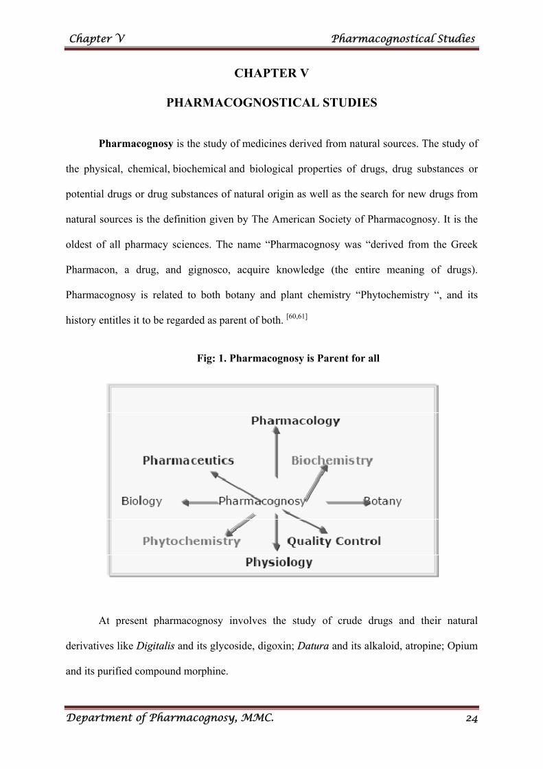
Pharmacognosy is the study of medicines derived from natural sources. The study of
the physical, chemical, biochemical and biological properties of drugs, drug substances or
potential drugs or drug substances of natural origin as well as the search for new drugs from
natural sources is the definition given by The American Society of Pharmacognosy. It is the
oldest of all pharmacy sciences. The name “Pharmacognosy was “derived from the Greek
Pharmacon, a drug, and gignosco, acquire knowledge (the entire meaning of drugs).
Pharmacognosy is related to both botany and plant chemistry “Phytochemistry “, and its
history entitles it to be regarded as parent of both. [60,61]
Fig: 1. Pharmacognosy is Parent for all
At present pharmacognosy involves the study of crude drugs and their natural
derivatives like Digitalis and its glycoside, digoxin; Datura and its alkaloid, atropine; Opium
and its purified compound morphine.
Chapter V Pharmacognostical Studies
Department of Pharmacognosy, MMC. 25
Pharmacognostical evaluation represents valuable information regarding the
morphology, microscopical and physical characters of crude drugs and thus gives the
scientific information regarding the purity and quality of crude drugs.
MATERIALS AND METHODS
SECTION A - MACROSCOPICAL STUDIES [61- 66]
Macroscopical studies include aspects of the outward appearance (shape, structure,
colour and pattern) as well as the form and structure of the internal parts like cells etc. Some
of these gross morphological characters of drugs such as shape, size, margin, apex and
venation are identification features of drugs. these features give valuable information about
the drugs.
Collection of plant material
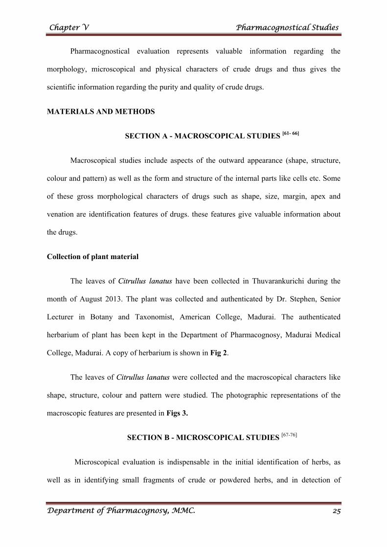
The leaves of Citrullus lanatus have been collected in Thuvarankurichi during the
month of August 2013. The plant was collected and authenticated by Dr. Stephen, Senior
Lecturer in Botany and Taxonomist, American College, Madurai. The authenticated
herbarium of plant has been kept in the Department of Pharmacognosy, Madurai Medical
College, Madurai. A copy of herbarium is shown in Fig 2.
The leaves of Citrullus lanatus were collected and the macroscopical characters like
shape, structure, colour and pattern were studied. The photographic representations of the
macroscopic features are presented in Figs 3.
SECTION B - MICROSCOPICAL STUDIES [67-76]
Microscopical evaluation is indispensable in the initial identification of herbs, as
well as in identifying small fragments of crude or powdered herbs, and in detection of
Fig.2: Herbarium of Citrullus lanatus (Thunb.) Matsum. & Nakai.
Chapter V Pharmacognostical Studies
Department of Pharmacognosy, MMC. 26
adulterants (eg. insects, animal faeces, mould, fungi etc.) as well as identifying the plant by
characteristic tissue features. Every plant possess a characteristic tissue structure, which can
be demonstrated through study of tissue arrangement, cell walls, and configuration when
properly mounted in stains, reagents and media.
The microscopical evaluation allows more detailed examination of the plant material
to identify the organised drug by its histological character. It provides detailed information
about the crude drugs by virtue of its property to magnify the fine structures of minute objects
to be visualised and thereby confirm the structural details of the plant drugs under evaluation.
It can also be used in the determination of the optical as well as micro chemical properties of
the crude drug confirmation study.
Collection of specimens
The plant specimen for the proposed study was collected from Citrullus lanatus leaf.
Care was taken to select healthy plant. The leaf was cut and removed from the plant and fixed
in FAA (Formalin- 5ml + Acetic acid- 5ml + 70% Ethyl alcohol-90 ml) After 24 hrs of
fixing, the specimens were dehydrated with graded series of tertiary-Butyl alcohol as per the
schedule given by Sass, 1940.[67] Infiltration of the specimens was carried by gradual addition
of paraffin wax (melting point 58-60˚C) until TBA solution stained super saturation. The
specimens were cast into paraffin blocks.
Sectioning
The paraffin embedded specimens were sectioned with the help of Rotary
Microtome. The thickness of the sections was 10-12µm. Dewaxing of the sections was by
customary procedure (Johansen, 1940)[68]. The sections were stained with Toluidine blue as
per the method published by O’Brien et al. (1964)[69]. Since Toluidine blue is a
polychromatic stain. The staining results were remarkably good; and some cytochemical
Chapter V Pharmacognostical Studies
Department of Pharmacognosy, MMC. 27
reactions were also obtained. The dye rendered pink colour to the cellulose walls, blue to the
lignified cells, dark green to suberin, violet to the mucilage, blue to the protein bodies etc.
whenever necessary sections were also stained with safranin and Fast green and iodine in
potassium iodide.
For studying the stomatal morphology, venation pattern and trichome distribution,
paradermal sections (sections taken parallel to the surface of leaf) as well as clearing of leaf
with 5% sodium hydroxide or epidermal peeling by partial maceration employing Jaffrey’s
maceration fluid (Sass, 1940) were prepared. Glycerine mounted temporary preparations
were made for macerated/cleared materials. Powdered materials of different parts were
cleared with sodium hydroxide and mounted in glycerine medium after staining. Different
cell component were studied and measured.
Photomicrographs
Microscopic descriptions of tissues are supplemented with micrographs wherever
necessary. Photographs of different magnifications were taken with Nikon labphoto 2
microscopic Unit. For normal observations bright field was used for the study of crystals,
starch grains, and lignified cells, polarized light was employed. Since these structures have
birefringent property, under polarized light they appear bright against dark background.
Magnifications of the figures are indicated by the scale-bars. Descriptive terms of the
anatomical features are as given in the standard Anatomy books (Easu, 1964)[77]. The
microscopic features observed for both plant are represented in Fig. 4 to 7.
SECTION C - QUANTITATIVE MICROSCOPY[76-78]
Quantitative analytical microscopy is useful in measuring the cell contents of the
crude drugs and help in their identification, characterization and standardization. A clear idea
Chapter V Pharmacognostical Studies
Department of Pharmacognosy, MMC. 28
about the identity and characteristic features of the drug can be obtained after several
numbers of determinations. The number so obtained can be compared with a standard value
to find out whether it is within the range. It helps to determinine the purity of the plant
material.
LEAF CONSTANTS
The stomatal number, stomatal index, vein islet number and vein termination number
were determined on fresh leaves using the standard procedures. A number of leaf
measurements are used to distinguish between some closely related species not easily
characterised by general microscopy.
Determination of stomatal number and stomatal index
Stomatal number: Stomatal number is the average number of stomata/sq.mm of
epidermis of the leaf.
Stomatal index: Stomatal index is the percentage which the number of stomata forms
to the total number of epidermal cells.
To study the morphology (type of stomata),stomatal number and stomatal index of leaf, the
leaf was subjected to epidermal peeling.
Procedure: The leaf was cleared by boiling with chloral hydrate solution. The upper
and lower epidermis was peeled out separately by means of forceps. The cleared leaf was
placed on the slide and mounted in glycerin. A camera Lucida and drawing board was placed
and stage micrometer was inserted for making the drawing scale. A square of 1mm was
drawn by means of stage micrometer. The slide with cleared leaf (epidermis) was placed on
the stage of the microscope and examined under 45X objective and 10X eye piece. The
epidermal cell and stomata was traced .The number of stomata present in the area of 1sq.mm
Chapter V Pharmacognostical Studies
Department of Pharmacognosy, MMC. 29
including the cell or at least half of its area within the square was counted. The average
number of stomata per sq.mm was determined and their values are tabulated.
For stomatal index, the glycerin mounted leaf peeling as mentioned above was made
and circle like mark for each stomata and cross like mark for each epidermal cells was
marked on the chart paper. The stomatal index was calculated by using the formula -
Stomatal index I= S/ (E+S) X 100; where I is the stomatal index, S is the number of stomata
in 1sq.mm area of leaf and E is the number of epidermal cells in 1sq.mm area of leaf. The
values are tabulated.
Determination of vein islet and vein termination number
Vein islet number is defined as the number of vein islet per sq.mm of the leaf surface
midway between the midrib and the margin. It is used to denote the minute area of
photosynthetic tissue encircled by the ultimate division of the conducting strands.
Vein termination number is defined as the number of vein termination per sq.mm of
the leaf surface midway between the midrib and margin. A vein termination is the ultimate
free termination of a veinlet or branch of a veinlet.
Procedure: A few leaves were boiled in chloral hydrate solution in a test tube placed
in a boiling water bath until clear. The cleared leaves were stained with saffranin solution and
a temporary mount was prepared with glycerin solution. The stage micrometer placed on the
microscopic stage, examined under 10X objective and 6X eye piece and an area of 1sq.mm
square was drawn. The cleared leaf piece was placed on the microscope stage, the vein islets
and vein terminals included in the square was drawn. The number of vein islets and vein
terminals within the square were counted. The results obtained for the number of vein islets
and vein terminals in 1sq.mm are tabulated in Table 1.
Chapter V Pharmacognostical Studies
Department of Pharmacognosy, MMC. 30
SECTION D - STANDARDIZATION PARAMETERS[77-80]
The determination of the foreign organic matter, loss on drying, ash values and
extractive values etc. gives a clear idea about the specific characteristics of crude drug under
examination, besides its macro-morphological or cyto-morphological, microscopical nature in
both its entire and its powder form. These diagnostic features enable the analyst to know the
nature and characteristic of crude drugs.
FOREIGN ORGANIC MATTER
The part of organ or organs other than those specified in the definition or description
of the crude drugs is defined as foreign organic matter.
Procedure: 500g of the original sample was weighed and spread out in a thin layer and
inspected with the unaided eye or with the use of a 6X lens and the foreign organic matter
was separated manually as completely as possible. The foreign organic matter was weighed
and the percentage of foreign organic matter was determined from the weight of the drug
taken. The results obtained are presented in table 2.
ASH VALUES
The ash values for air dried powdered leaves of Citrullus lanatus were determined as
per official method. The determination of ash is useful for detecting low grade products,
exhausted drug and excess of sandy or earthy matter.
Different types of ash values are used in detection of crude drugs like, total ash, acid
insoluble ash, water soluble ash and sulphated ash.
Chapter V Pharmacognostical Studies
Department of Pharmacognosy, MMC. 31
Determination of total ash
Total ash is useful in detecting the crude drugs that are mixed with various mineral
substances like sand, soil, calcium oxalate, chalk powder or other drug with different
inorganic contents to improve their appearance.
Procedure: 2 to 3g of the air dried crude drug was weighed accurately and taken in a tared
platinum or silica dish and incinerated at a temperature not exceeding 450oC until free from
carbon then cooled and weighed. The percentage of ash with reference to the air dried drug
was calculated. The results obtained are presented in table 2.
Determination of acid insoluble ash
Acid insoluble ash is the residue obtained after boiling the total ash with dilute
hydrochloric acid and igniting the remaining insoluble matter. This measures the amount of
silica present, especially as sand and siliceous earth.
Procedure
The ash obtained from the total ash was boiled for 5min with 25mL of 2M
hydrochloric acid; the insoluble matter was collected in a Gooch crucible or on an ashless
filter paper and washed with hot water and ignited, cooled in a dessicator and weighed. The
percentage of acid insoluble ash was calculated with reference to the air dried drug. The
results obtained are presented in table 2.
Determination of water soluble ash
Water soluble ash is used to detect the presence of material exhausted by water. If
carbon is still present after heating at a moderate temperature, the water- soluble ash may be
separated and the residue again ignited.
Chapter V Pharmacognostical Studies
Department of Pharmacognosy, MMC. 32
Procedure
The ash obtained from the total ash was boiled for 5min with 25mL of water; the
insoluble matter was collected in a Gooch crucible or on an ashless filter paper and washed
with hot water and ignited for 15min at a temperature not exceeding 450oC. The weight of
the insoluble matter was subtracted from the weight of the total ash; the difference in weight
representing the water soluble ash. The percentage of water soluble ash was calculated with
reference to the air dried drug. The results obtained are presented in table 2.
Determination of sulphated ash
The treatment of the drug with sulphuric acid before ignition, whereby all oxides and
carbonates are converted to sulphates is called as sulphated ash.
Procedure
A platinum dish was heated to redness for 10min and allowed to cool in a desiccator
and weighed. 1g of the substance being examined was placed in the dish, moistened with
sulphuric acid, ignited gently, moistened again with sulphuric acid and ignited at about
800°C. It was then cooled and weighed. The percentage of sulphated ash was calculated with
reference to the air dried drug. The results obtained are presented in table 2.
LOSS ON DRYING
The moisture content of a drug should be minimized to prevent decomposition of
plant material due to chemical or microbial contamination. It may be determined by heating a
material at constant temperature to constant weight.
Procedure
About 2g of the powdered crude drug was accurately weighted in a tared dish and
dried in an oven at 100˚-105˚ C. It was cooled in a desiccator and again weighed. The loss on

Chapter V Pharmacognostical Studies
Department of Pharmacognosy, MMC. 33
drying was calculated with reference to the amount of the dried powder taken and the results
obtained are presented in table 2.
EXTRACTIVE VALUES
The extractive values are the important factor to determine the amount of active
principle or phytoconstituents present in the plant materials, when extracted with suitable
solvents. The extraction of crude drug with a particular solvent yields a solution containing
different phyto-constituents. The composition of these phyto constituents in that particular
solvent depends upon the nature of the drug and solvent used. The use of a single solvent can
be the means of providing preliminary information on the quality of a particular drug sample;
for example, in a drug where the extraction procedure for the constituents commences with
water as the solvent, any subsequent aqueous extraction on the re-dried residue will give a
very low yield of soluble matter.
Determination of water soluble extractive
Procedure: 5g of the air dried drug, coarsely powdered have to be macerated with
100mL of water closed flask for 24h, shaking frequently during the first 6h and allowing to
stand for 18h. Thereafter, filter rapidly taking precautions against loss of water, evaporate 25
mL of the filtrate to dryness in a tared flat-bottomed shallow dish, dry at 105°C and weigh.
The percentage of water soluble extractive with reference to the air dried drug was calculated.
Determination of ethanol soluble extractive
Procedure
Macerate 5g of the air dried drug, coarsely powdered, with 100mL of the ethanol of
the specified strength in a closed flask for 24h, shaking frequently during the first 6h and
allowing to stand for 18h. Thereafter, filter rapidly taking precaution against ethanol,
evaporate 25mL of the filtrate to dryness in a tared flat-bottomed shallow dish, dry at 105°C
Chapter V Pharmacognostical Studies
Department of Pharmacognosy, MMC. 34
and weigh. Calculate the percentage of ethanol soluble extractive with reference to the air
dried drug.
Determination of ethanol soluble extractive
Procedure
Macerate 5g of the air dried drug, coarsely powdered, with 100mL of the methanol of
the specified strength in a closed flask for 24h, shaking frequently during the first 6 hours and
allowing to stand for 18h. Thereafter, filter rapidly taking precaution against ethanol,
evaporate 25mL of the filtrate to dryness in a tared flat-bottomed shallow dish, dry at 105°C
and weigh. Calculate the percentage of methanol soluble extractive with reference to the air
dried drug.
Determination of petroleum ether soluble extractive
The procedure adopted under ethanol soluble extractive was followed using petroleum
ether as a solvent.
Determination of hexane soluble extractive
The procedure adopted under ethanol soluble extractive was followed using hexane as
a solvent.
Determination of chloroform, benzene, ethyl acetate and acetone soluble extractive
The procedure adopted under ethanol soluble extractive was followed using the
requisite solvent as the medium of extraction.The results obtained for the various extractive
values are presented in table 2.
Chapter V Pharmacognostical Studies
Department of Pharmacognosy, MMC. 35
FOAMING INDEX
Some plant materials when shaken with water cause persistent foam which may be
attributed to the presence of saponins in that material. The foaming ability of an aqueous
solution of plant materials and their extracts is measured in terms of foaming index.
Procedure
An accurate quantity of about 1g of the coarse plant material was weighed and
transferred into an Erlenmeyer flask containing 100mL of boiling water. The flask was boiled
at moderate heat for 30min. The solution was cooled and filtered into a 100mL volumetric
flask and sufficient distilled water was added to dilute to volume. The solution was poured
into ten stoppered test tubes in successive portions of 1mL, 2mL, etc. upto 10mL, and the
volume of the liquid in each tube was adjusted with water upto 10mL. The tubes were then
stoppered and shaken in a length wise motion for 15sec (two shakes/sec) and allowed to stand
for 15min. The height of foam was measured. If the height of the foam in every tube was less
than 1cm the foaming index was less than 100. If a height of foam of 1cm was measured in
any test tube, the volume of the plant material decoction in this tube (a) was used to
determine the index. If the height of the foam was more than 1cm in every tube, the foaming
index was over 1000. In this case, the determination was repeated using a new series dilution
of the decoction in order to obtain a result. The foaming index was calculated by using the
following formula 1000/A where A was the volume in mL of the decoction used for
preparing the dilution in the tube where foaming to a height of 1cm was observed. The results
obtained are presented in table 2.
Chapter V Pharmacognostical Studies
Department of Pharmacognosy, MMC. 36
SWELLING INDEX
Swelling index is the volume in mL taken up by the swelling of plant material under
specified conditions. The medicinal plant materials like gums, mucilage, and pectin have
swelling property.
Procedure: An accurately weighed 1g of the powdered drug material was taken in
the 25mL glass stoppered measuring cylinder. 25mL of water was added and shaken the
mixture thoroughly every 10min for 1h. Then, allowed to stand for 3h at room temperature.
The volume in mL occupied by the plant material was measured, including sticky mucilage
was observed. The results obtained are presented in table 2.
SECTION E - POWDER ANALYSIS & MICROSCOPY[81-83]
The powdered crude drug analysis was aimed to study and also to assess the quality of
herbal drugs for therapeutic value which are generally studied by classical pharmacognostical
studies. The authenticity of herbal drugs was confirmed by comparison of their powder
characteristics.
Procedure
A) Reaction of chemicals with powdered crude drugs
The raw leaf powder of Citrullus lanatus was treated with different chemical
reagent such as iodine solution, 10% potassium hydroxide solution, glacial acetic acid etc. on
a clean watch glass for the identification of secondary metabolites. The colours obtained with
various reagents are presented in Table 3.
B) Fluorescence analysis
The fluorescence nature of powder drugs was analysed to find out whether any
fluorescent compound was present in the sample and the observations with different
chemicals were also carried out and recorded. The air dried plant materials of both plants
Chapter V Pharmacognostical Studies
Department of Pharmacognosy, MMC. 37
were taken in clean warch glass and subjected to different chemicals such as acids, alkalis
and some reagents are observed under day light and UV light. Detailed fluorescence behavior
of crude drug powder has been shown in Table 4.
C) Powder microscopy
The dried leaf was powdered and the powder was passed through sieve no.60 for the
study of powder microscopy. Chloral hydrate, water, iodine, phloroglucinol and hydrochloric
acid (1:1) etc. were employed as mounting medium. The pictorial representation are
presented in Fig 8.
Chapter V Pharmacognostical Studies
Department of Pharmacognosy, MMC. 38
RESULTS AND DISCUSSION
SECTION A - MACROSCOPICAL EVALUATION
Macroscopical characters of Citrullus lanatus (Thunb.) Matsum. & Nakai
Whole plant: Citrullus lanatus is an annual climbing or trailing herb, with hairy stem up to
10 m long. Tendrils divided at the tip into two or three parts. Separate male and female
flowers are borne on the same plant. (Fig. 3.1)
Leaves: Leaf blades up to about 20 × 20 cm, more or less hairy, usually deeply 3–5-lobed,
the central lobe being the largest. The lobes themselves are further divided. Leaf stalks
(petioles) up to about 19 cm long, more or less hairy. (Fig. 3.2, 6, 7)
Flowers: Solitary, borne in leaf axils. Both male and female flowers are yellow, up to 3 cm in
diameter, and borne on pedicels (flower stalks) up to 45 mm long. Flowers are usually
pollinated by honey bees. (Fig. 3.3)
Fruits: Fruits of wild plants up to about 20 cm in diameter, greenish mottled with darker
green. Fruits of cultivated plants up to about 70 × 30 cm, rounded, oval or oblong, with a
golden-yellow to dark green skin, the skin being uniform, mottled or striped. Flesh usually
red or yellow, sometimes orange, pink or white. (Fig.3.4)
Seeds: Flat, smooth, variable in size and colour (white, tan, brown, black, red, green or
mottled).(Fig. 3.5)
SECTION B - MICROSCOPICAL EVALUATION
Anatomy of the leaf: The leaf has very thick abaxially hanging midrib and thin lamina
(Fig.4.1). The midrib is 1.9mm thick and 1.7mm wide. It has four thick ridges alternatively
deep furrows. (Fig.4.1) The adaxial part of the midrib has short, tick cone. The epidermis has
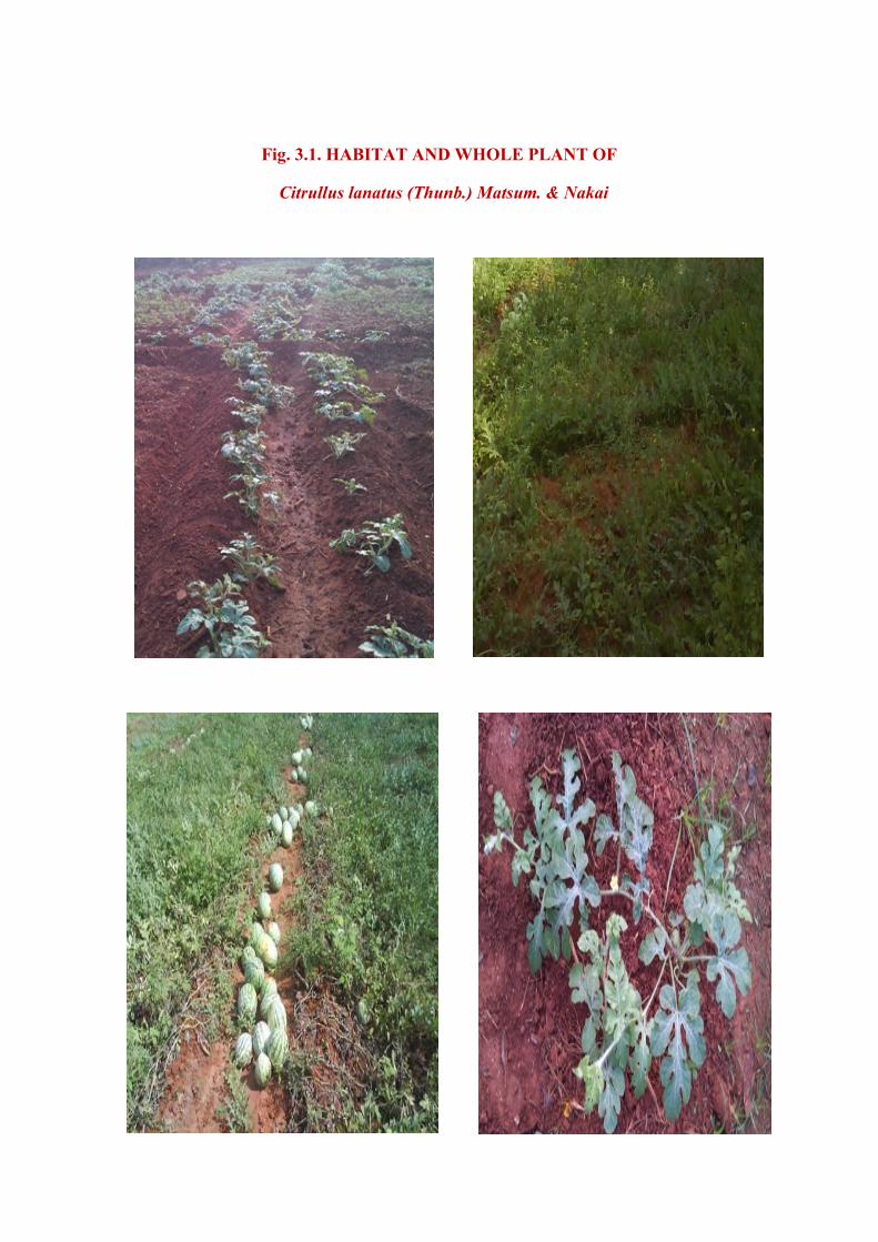
Fig. 3.1. HABITAT AND WHOLE PLANT OF
Citrullus lanatus (Thunb.) Matsum. & Nakai
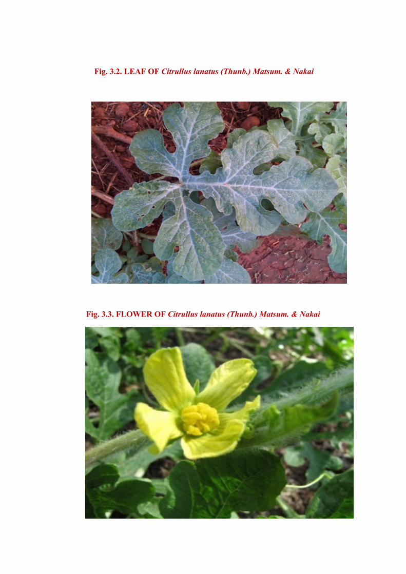
Fig. 3.2. LEAF OF Citrullus lanatus (Thunb.) Matsum. & Nakai
Fig. 3.3. FLOWER OF Citrullus lanatus (Thunb.) Matsum. & Nakai
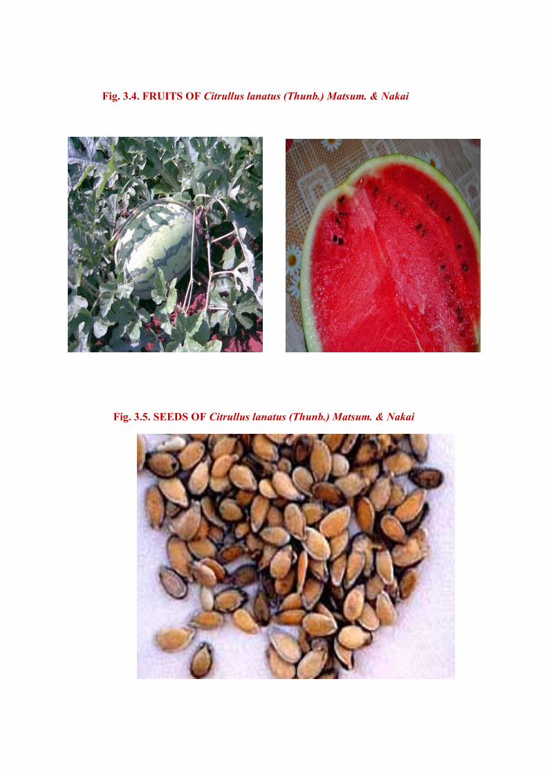
Fig. 3.4. FRUITS OF Citrullus lanatus (Thunb.) Matsum. & Nakai
Fig. 3.5. SEEDS OF Citrullus lanatus (Thunb.) Matsum. & Nakai
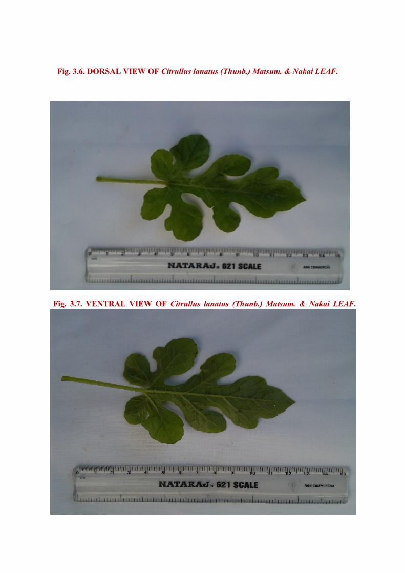
Fig. 3.6. DORSAL VIEW OF Citrullus lanatus (Thunb.) Matsum. & Nakai LEAF.
Fig. 3.7. VENTRAL VIEW OF Citrullus lanatus (Thunb.) Matsum. & Nakai LEAF.
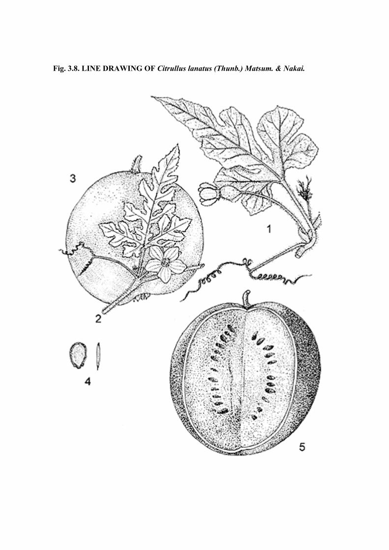
Fig. 3.8. LINE DRAWING OF Citrullus lanatus (Thunb.) Matsum. & Nakai.
Chapter V Pharmacognostical Studies
Department of Pharmacognosy, MMC. 39
thin intact epidermal layer of rectangular cells. Inner to the epidermis is a narrow, two or
three layers of collenchyma and the remaining ground tissue is thin walled compact
parenchyma cells.
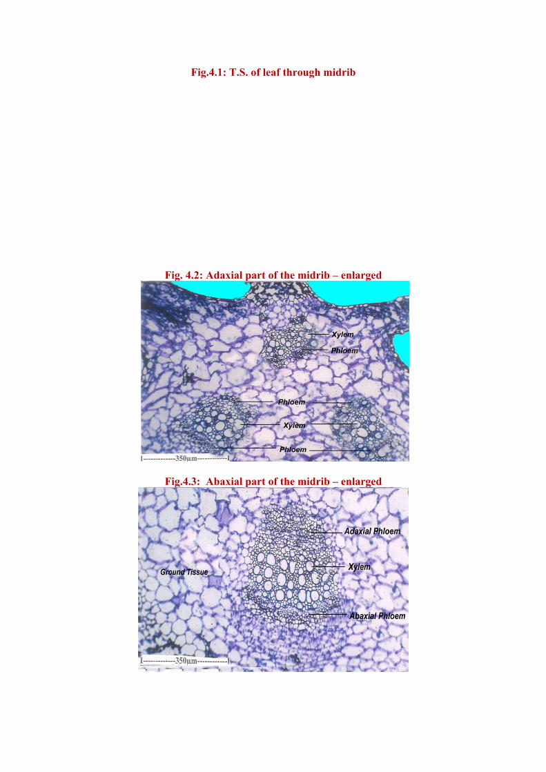
The vascular system of the midrib is multi stranded. There is a large abaxial median
bundle, two adaxial lateral smaller bundles and cone still smaller adaxial median bundles
(Fig. 4.1) All the bundles are bicollateral having phloem strands both on the outer and inner
sides of the xylem. The xylem consists of short radial chains of wide circular elements.
Phloem strands are composed of small groups of sieve elements which are small and darkly
stained. (Fig.4.2, 3).
The lateral vein is simple in structure. It projects slightly on the lower side and the
upper side is flat. (Fig. 5.1) The epidermal cells of the lateral veins are large and thick; they
are circular or cylindrical and thin walled. The vascular bundle is small, collateral and
includes adaxial group of angular xylem elements and small cluster of abaxial phloem
elements.
Lamina (Fig. 5.2): The lamina is 90-100µm thick. It is heterofacial with distinct adaxial
sides. It is heterofacial with distinct adaxial and abaxial side. It is amphistomatic with stomata
located on the adaxial and abaxial side.The epidermal cells are small elliptical or rectangular
and thin walled. The palisade zone consists of single layer of cylindrical cells which are
loosely arranged. The sponge parenchyma cells small and spherical. They form wide air
chambers separated by partition filaments. (Fig. 5.2)
Stomata (Fig.6.1, 2, 3): The stomata epidermal cells were studied in paradermal sections of
the lamina. The epidermal cells small and vary in shape and size. Their anticlinal walls are
thick and wavy. The stomata are diffuse in distribution. (Fig.6.1,2) The stomatal type is
actinocytic. (Fig.6.2, 3) The stoma is surrounded by three to six subsidiary cells which
F
Fig. 4.2
Fig.4.3
ig.4.1: T.S.
2: Adaxial p
: Abaxial p
. of leaf thr
part of the
part of the
rough midr
midrib – e
midrib – e
rib
enlarged
enlarged
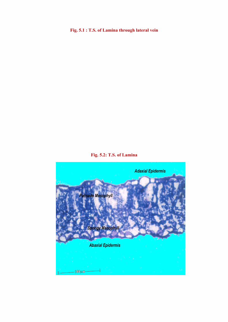
Fig. 5.1 : T.S. of L
Fig. 5.
Lamina thr
2: T.S. of L
rough later
Lamina
ral vein
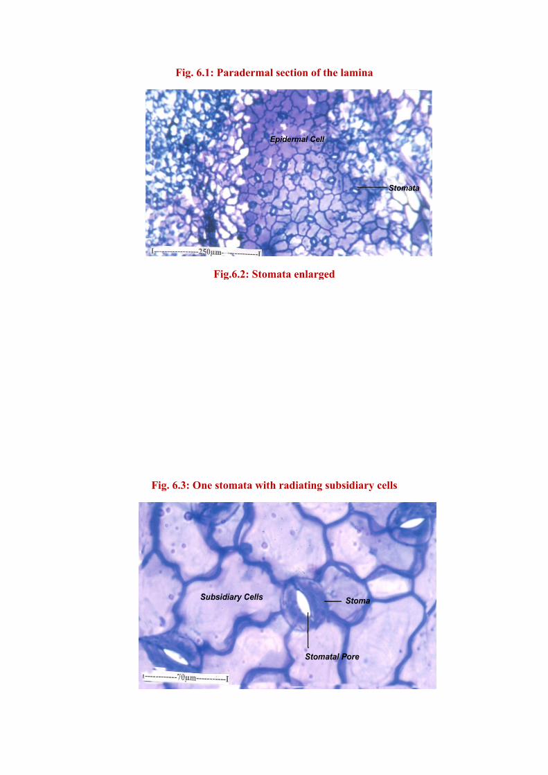
Fig. 6
Fig. 6.3: O
6.1: Parade
Fig.6.2:
One stomata
ermal sectio
: Stomata e
a with radi
on of the la
enlarged
iating subsi
amina
idiary cells
s
Fi
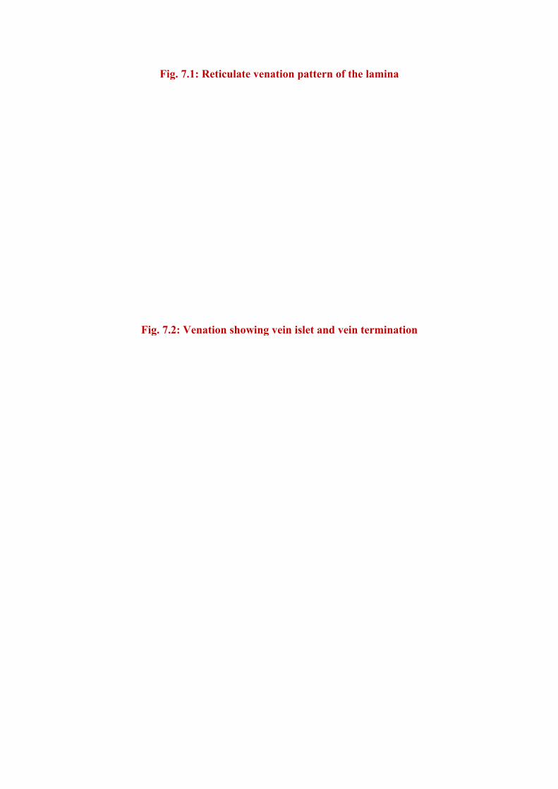
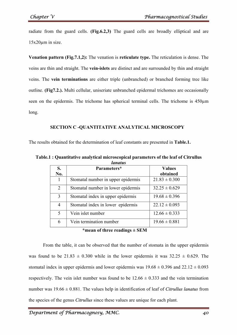
Fig. 7.1:
g. 7.2: Ven
Reticulate
nation show
venation p
wing vein isl
pattern of th
let and vein
he lamina
n terminati
ion
Chapter V Pharmacognostical Studies
Department of Pharmacognosy, MMC. 40
radiate from the guard cells. (Fig.6.2,3) The guard cells are broadly elliptical and are
15x20µm in size.
Venation pattern (Fig.7.1,2): The venation is reticulate type. The reticulation is dense. The
veins are thin and straight. The vein-islets are distinct and are surrounded by thin and straight
veins. The vein terminations are either triple (unbranched) or branched forming tree like
outline. (Fig7.2.). Multi cellular, uniseriate unbranched epidermal trichomes are occasionally
seen on the epidermis. The trichome has spherical terminal cells. The trichome is 450µm
long.
SECTION C -QUANTITATIVE ANALYTICAL MICROSCOPY
The results obtained for the determination of leaf constants are presented in Table.1.
Table.1 : Quantitative analytical microscopical parameters of the leaf of Citrullus lanatus
S. No.
Parameters* Values obtained
1 Stomatal number in upper epidermis 21.83 ± 0.300
2 Stomatal number in lower epidermis 32.25 ± 0.629
3 Stomatal index in upper epidermis 19.68 ± 0.396
4 Stomatal index in lower epidermis 22.12 ± 0.093
5 Vein islet number 12.66 ± 0.333
6 Vein termination number 19.66 ± 0.881
*mean of three readings ± SEM
From the table, it can be observed that the number of stomata in the upper epidermis
was found to be 21.83 ± 0.300 while in the lower epidermis it was 32.25 ± 0.629. The
stomatal index in upper epidermis and lower epidermis was 19.68 ± 0.396 and 22.12 ± 0.093
respectively. The vein islet number was found to be 12.66 ± 0.333 and the vein termination
number was 19.66 ± 0.881. The values help in identification of leaf of Citrullus lanatus from
the species of the genus Citrullus since these values are unique for each plant.
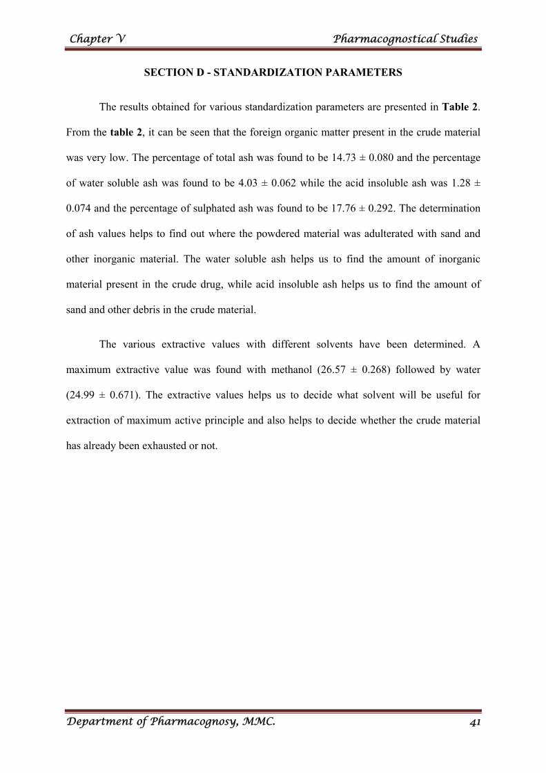
Chapter V Pharmacognostical Studies
Department of Pharmacognosy, MMC. 41
SECTION D - STANDARDIZATION PARAMETERS
The results obtained for various standardization parameters are presented in Table 2.
From the table 2, it can be seen that the foreign organic matter present in the crude material
was very low. The percentage of total ash was found to be 14.73 ± 0.080 and the percentage
of water soluble ash was found to be 4.03 ± 0.062 while the acid insoluble ash was 1.28 ±
0.074 and the percentage of sulphated ash was found to be 17.76 ± 0.292. The determination
of ash values helps to find out where the powdered material was adulterated with sand and
other inorganic material. The water soluble ash helps us to find the amount of inorganic
material present in the crude drug, while acid insoluble ash helps us to find the amount of
sand and other debris in the crude material.
The various extractive values with different solvents have been determined. A
maximum extractive value was found with methanol (26.57 ± 0.268) followed by water
(24.99 ± 0.671). The extractive values helps us to decide what solvent will be useful for
extraction of maximum active principle and also helps to decide whether the crude material
has already been exhausted or not.
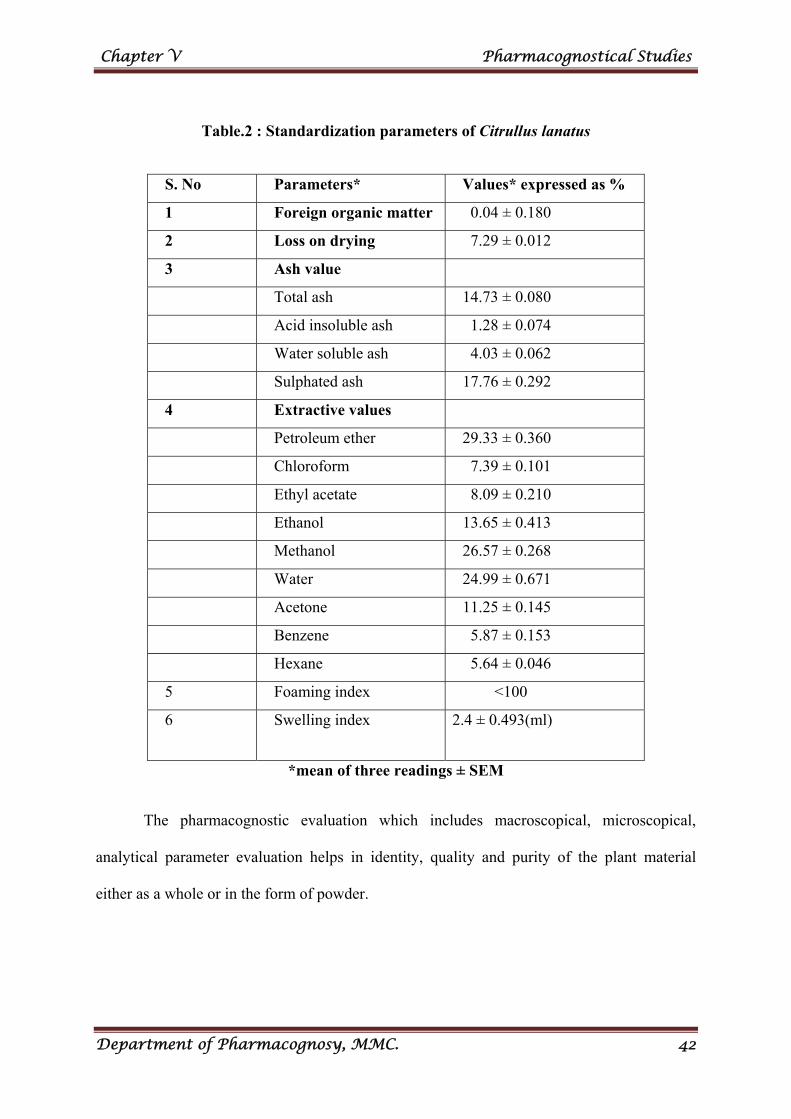
Chapter V Pharmacognostical Studies
Department of Pharmacognosy, MMC. 42
Table.2 : Standardization parameters of Citrullus lanatus
S. No Parameters* Values* expressed as %
1 Foreign organic matter 0.04 ± 0.180
2 Loss on drying 7.29 ± 0.012
3 Ash value
Total ash 14.73 ± 0.080
Acid insoluble ash 1.28 ± 0.074
Water soluble ash 4.03 ± 0.062
Sulphated ash 17.76 ± 0.292
4 Extractive values
Petroleum ether 29.33 ± 0.360
Chloroform 7.39 ± 0.101
Ethyl acetate 8.09 ± 0.210
Ethanol 13.65 ± 0.413
Methanol 26.57 ± 0.268
Water 24.99 ± 0.671
Acetone 11.25 ± 0.145
Benzene 5.87 ± 0.153
Hexane 5.64 ± 0.046
5 Foaming index <100
6 Swelling index 2.4 ± 0.493(ml)
*mean of three readings ± SEM
The pharmacognostic evaluation which includes macroscopical, microscopical,
analytical parameter evaluation helps in identity, quality and purity of the plant material
either as a whole or in the form of powder.
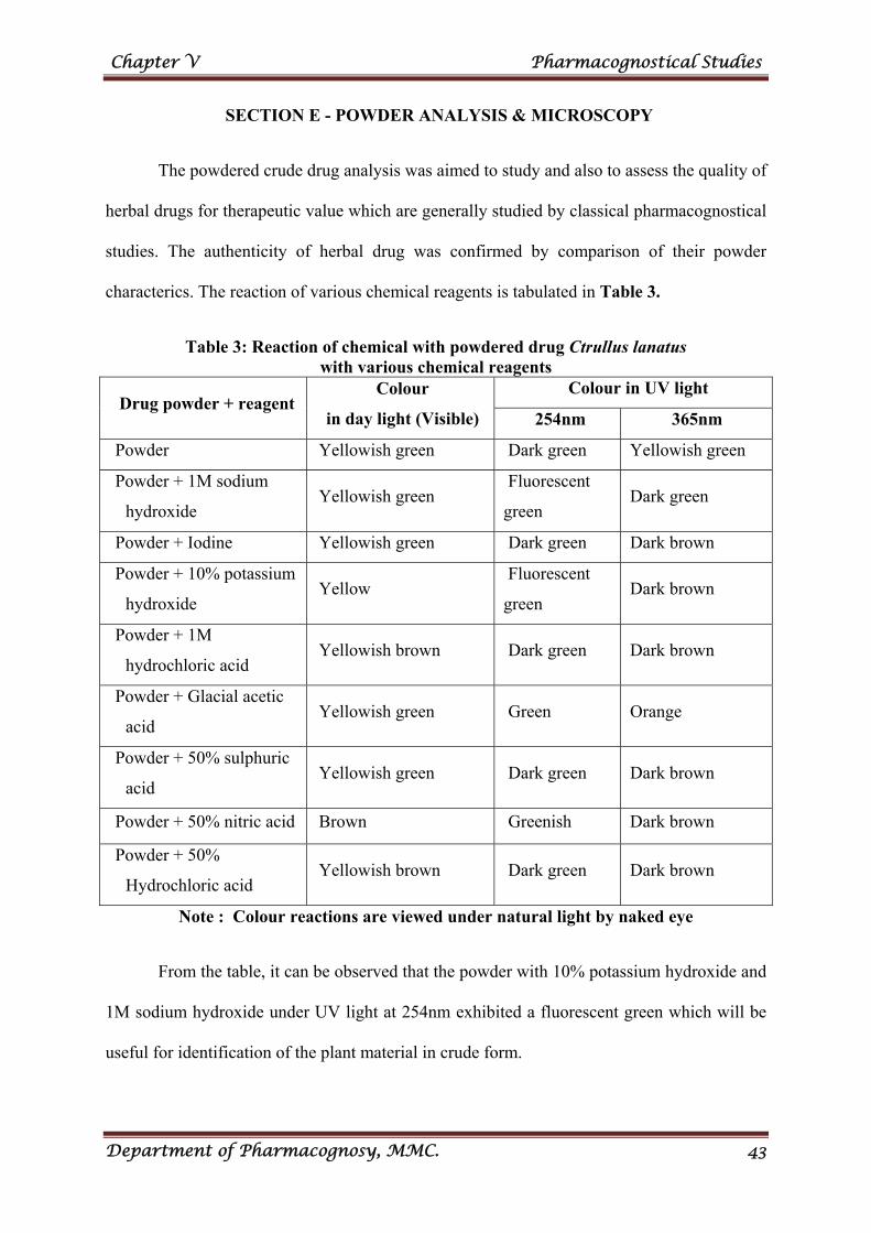
Chapter V Pharmacognostical Studies
Department of Pharmacognosy, MMC. 43
SECTION E - POWDER ANALYSIS & MICROSCOPY
The powdered crude drug analysis was aimed to study and also to assess the quality of
herbal drugs for therapeutic value which are generally studied by classical pharmacognostical
studies. The authenticity of herbal drug was confirmed by comparison of their powder
characterics. The reaction of various chemical reagents is tabulated in Table 3.
Table 3: Reaction of chemical with powdered drug Ctrullus lanatus with various chemical reagents
Drug powder + reagent Colour
in day light (Visible)
Colour in UV light
254nm 365nm
Powder Yellowish green Dark green Yellowish green
Powder + 1M sodium
hydroxide Yellowish green
Fluorescent
green Dark green
Powder + Iodine Yellowish green Dark green Dark brown
Powder + 10% potassium
hydroxide Yellow
Fluorescent
green Dark brown
Powder + 1M
hydrochloric acid Yellowish brown Dark green Dark brown
Powder + Glacial acetic
acid Yellowish green Green Orange
Powder + 50% sulphuric
acid Yellowish green Dark green Dark brown
Powder + 50% nitric acid Brown Greenish Dark brown
Powder + 50%
Hydrochloric acid Yellowish brown Dark green Dark brown
Note : Colour reactions are viewed under natural light by naked eye
From the table, it can be observed that the powder with 10% potassium hydroxide and
1M sodium hydroxide under UV light at 254nm exhibited a fluorescent green which will be
useful for identification of the plant material in crude form.
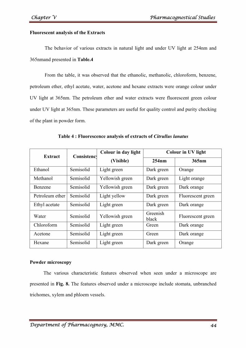
Chapter V Pharmacognostical Studies
Department of Pharmacognosy, MMC. 44
Fluorescent analysis of the Extracts
The behavior of various extracts in natural light and under UV light at 254nm and
365nmand presented in Table.4
From the table, it was observed that the ethanolic, methanolic, chloroform, benzene,
petroleum ether, ethyl acetate, water, acetone and hexane extracts were orange colour under
UV light at 365nm. The petroleum ether and water extracts were fluorescent green colour
under UV light at 365nm. These parameters are useful for quality control and purity checking
of the plant in powder form.
Table 4 : Fluorescence analysis of extracts of Citrullus lanatus
Extract ConsistencyColour in day light
(Visible)
Colour in UV light
254nm 365nm
Ethanol Semisolid Light green Dark green Orange
Methanol Semisolid Yellowish green Dark green Light orange
Benzene Semisolid Yellowish green Dark green Dark orange
Petroleum ether Semisolid Light yellow Dark green Fluorescent green
Ethyl acetate Semisolid Light green Dark green Dark orange
Water Semisolid Yellowish green Greenish black
Fluorescent green
Chloroform Semisolid Light green Green Dark orange
Acetone Semisolid Light green Green Dark orange
Hexane Semisolid Light green Dark green Orange
Powder microscopy
The various characteristic features observed when seen under a microscope are
presented in Fig. 8. The features observed under a microscope include stomata, unbranched
trichomes, xylem and phloem vessels.
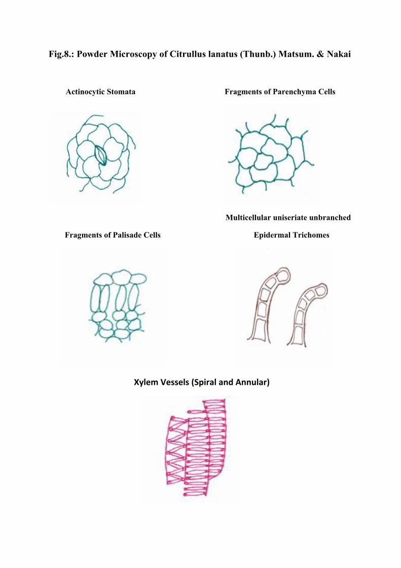
Fig.8.: Powder Microscopy of Citrullus lanatus (Thunb.) Matsum. & Nakai
Actinocytic Stomata Fragments of Parenchyma Cells
Multicellular uniseriate unbranched
Fragments of Palisade Cells Epidermal Trichomes
Xylem Vessels (Spiral and Annular)
PHYTOCHEMICAL
EVALUATION
Chapter VI Phytochemical Evaluation
Department of Pharmacognosy, MMC. 45
CHAPTER VI
PHYTOCHEMICAL EVALUATION
Phytochemicals means plant chemicals. They are naturally occuring in plants. They
give plants its colour, flavour, smell and are part of a plant’s natural defence system (disease
resistance). The phytochemicals are bioactive, non-nutrient plant compounds in fruits,
vegetables, grains and other plant foods that have been linked to reducing the risk of major
degenerative diseases. [84]
In plants phytochemicals attract beneficial and repel harmful organisms, serve as
photoprotectants, and respond to environmental changes. They work together with nutrients
and dietary fibres to protect the body against diseases, slow the aging process and reduce the
risk of many diseases such as cancer, heart disease, stroke, high blood pressure, cataracts,
osteoporosis and urinary tract infection. Alkaloids have analgesic effects, glycosides are used
in cardiac diseases, tannins prevent urinary tract infection by preventing bacteria from
adhering to the walls. Tannins, along with vitamin C help build and strengthen collagen.
Saponins serve as natural antibiotics, which help body to fight infections and microbial
invasions. They also enhance the effectiveness of certain vaccines, lower cholesterol and
knock out some tumor cells. Flavonoids have antioxidant activity in biological systems and
protect the body against allergies, inflammation, free radicals, platelet aggregation, microbes,
ulcers, hepatoxins, viruses and tumors The flavonoid quercetin is known for its ability to
relieve hey fever, eszema, sinusitis and asthma while certain flavonoids also can protect low-
density-lipoproteins from being oxidized, thereby playing an important role in
atherosclerosis.[85]
Chapter VI Phytochemical Evaluation
Department of Pharmacognosy, MMC. 46
MATERIALS AND METHODS
COLLECTION AND PREPARATION OF EXTRACT
The leaves of Citrullus lanatus were collected in Thuvarankurichi during the month of
August 2013. The leaves were washed thoroughly and dried in shade. The shade dried leaves
were powdered and used for further studies.
Extraction of leaves of Citrullus lanatus was carried out by washing the plants and
drying at room temperature in 14 days. After that, they were filtered with sieve analyzer to
get homogeneous particles and defatted with 2.5L of petroleum ether (60-80˚C) by cold
maceration method for 72h. The solvent was then removed by filtration and the marc was
dried. The dried marc was re-soaked with 2.5L of methanol. The steps were performed three
times and the combined filtrates were evaporated to a cohesive mass using rota vapour.
SECTION A - PRELIMINARY PHYTOCHEMICAL SCREENING [86-90]
The preliminary phytochemical screening helps us in identifying the type of
secondary metabolites present in plants. The screening was carried out Aqueous and
methanolic extract of citrullus lanatus leaf powder. The various chemical tests carried out are
described.
1. Test for carbohydrates
a. Molisch’s Test: The aqueous extract of the powdered leaf when treated with
alcoholic solution of α-naphthol in the presence of sulphuric acid. Purple colour indicates the
presence of carbohydrates.
b. Benedict’s test: The aqueous extract of the powdered was treated with Benedict’s
reagent and boiled on water bath and cooled. An orange colour precipitate indicates the
presence of carbohydrates.
Chapter VI Phytochemical Evaluation
Department of Pharmacognosy, MMC. 47
c. Fehling’s Test: The aqueous extract of the powdered leaf was treated with
Fehling’s solution I and II and heated on a boiling water bath for half an hour. A red
precipitate indicates the presence of free reducing sugars.
2. Test for Alkaloids
About 2g of the powdered material was mixed with 1g of calcium hydroxide and 5mL
of water into a smooth paste and set aside for 5min. It was then evaporated to dryness in a
porcelain dish on a water bath. To the residue, 20mL of chloroform was added, mixed well
and refluxed for half an hour on a water bath. Then it was filtered and the chloroform was
evaporated. To the residue, 5mL of dilute hydrochloric acid was added. The solution was
divided into four parts and 2mL of each of the following reagents were added and the colour
noted below indicates the presence of alkaloids.
a) Mayer’s Reagent - Cream precipitate
b) Dragendorff’s Reagent - Reddish brown precipitate
c) Hager’s Reagent - Yellow precipitate
d) Wagner’s Reagent - Reddish brown precipitate
3. Test for phytosterols
The powdered leaf was first extracted with petroleum ether and evaporated to a
residue. Then the residue was dissolved in chloroform and tested for phytosterols.
a. Salkowski’s Test: A few drops of concentrated sulphuric acid were added to the
above solution, shaken well and set aside. The lower chloroform layer of the solution turning
red indicates the presence of phytosterols.
b. Liebermann – Burchard’s Test: To the chloroform solution a few drops of acetic
anhydride and 1mL of concentrated sulphuric acid were added through the sides of the test
tube and set aside for a while. At the junction of two layers a brown ring and the upper layer
turning green indicates the presence of phytosterols.
Chapter VI Phytochemical Evaluation
Department of Pharmacognosy, MMC. 48
4. Test for glycosides
The substance was mixed with a little anthrone on a watch glass. One drop of
concentrated sulphuric acid was added, made into a paste and warmed gently over a water
bath. A dark green coloration indicates the presence of glycosides.
Test for Cardiac Glycosides
a. Keller Killiani Test: The substance was boiled with 10% alcohol for 2min, cooled
and filtered. To the filtrate, lead sub acetate was added and filtered. The filtrate was then
extracted with chloroform. The chloroform layer was separated and evaporated to dryness.
The residue was dissolved in glacial acetic acid with traces of ferric chloride. To this few
drops of sulphuric acid was added slowly along the sides of the test tube. A reddish brown
layer changes to green colour on standing.
b. Legal test: The substance was dissolved in pyridine, sodium nitroprusside solution
was added to it and made alkaline. A pink or red colour indicates the presence of cardiac
glycosides.
c. Baljet test: To the substance sodium picrate solution is added. A yellow to orange
colour indicates the presence of cardiac glycosides.
5. Test for Proteins and free amino acids
a. Millon’s Test: A small quantity of acidulous-methanolic extract of the powdered
drug was heated with Millon’s reagent. A white precipitate turning red on heating indicates
the presence of proteins.
b. Biuret Test: To the methanolic extract of powdered drug, one ml of dilute sodium
hydroxide(10%) solution was added followed by this one drop of very dilute copper sulphate
solution was added. A violet colour indicates the presence of proteins.
Chapter VI Phytochemical Evaluation
Department of Pharmacognosy, MMC. 49
c. Ninhydrin test: To the methanolic extract of powdered drug, two drops of
ninhudrin solution (10mg of ninhydrin in 200mL of acetone) were added to 2mL of aqueous
filtrate. A characteristic purple colour indicated the presence of amino acid.
6. Test for Mucilage
A few mL of aqueous extract was prepared from the powdered drug was treated with
ruthenium red. Pinkish red colour indicates the presence of mucilage.
7. Test for Flavonoids
a. Magnesium turning- con HCl test: A little of the powdered drug was heated with
alcohol and filtered. To the test solution magnesium turnings and few drops of concentrated
hydrochloric acid were added and Boiled for five minutes. A red colour indicates the
presence of flavonoids.
b. Alkali Test: To the small quantity of test solution 10% aqueous sodium
hydroxide Solution was added. A yellow orange color indicates the presence of flavonoids.
c. Acid Test: To a small quantity of test solution, few drops of concentrated sulphuric
acid was added. A yellow orange color indicates the presence of flavonols.
8. Test for terpenoids
The powdered leaf was shaken with petroleum ether and filtered. The filtrate was
evaporated and residue obtained was dissolved in small amount of chloroform and the
chloroform solution tin and thionyl chloride were added. A pink color indicates the presence
of terpenoids.
9. Test for phenolic compounds
Small quantity of powdered sample was tested with the following reagents and the
colour produced indicates the presence of phenolic compounds.
a. 5% Ferric chloride solution --- Deep bluish black colour.
b. Lead acetate solution --- White precipitate.
Chapter VI Phytochemical Evaluation
Department of Pharmacognosy, MMC. 50
c. Bromine water --- Decolouration of bromine water.
d. Acetic acid solution --- Red colour.
e. Dilute iodine solution --- Transient red colour.
f. Tannic acid --- Precipitate.
10. Test for Tannins
A small quantity of the powdered drug was extracted with water. To the aqueous
extract, few drops of ferric chloride solution were added. A bluish black color indicates the
presence of tannins.
11. Test for saponins
About 0.5g of the powdered drug was boiled gently for 2min with 20mL of water and
filtered while hot and allowed to cool. 5mL of the filtrate was then diluted with water and
shaken vigorously. A frothing indicates the presence of saponins.
12. Test for volatile oil
About 250gm of fresh leaves of Citrullus lanatus was weighed and subjected to
hydrodistillation separately using volatile oil estimation apparatus (BP 1980). Absence of
volatile oil.
The above chemical tests were carried out on aqueous and methanolic extract of
Citrullus lanatus leaf powder and the results were tabulated in Table 5.
SECTION B - QUANTITATIVE ESTIMATION OF PHYTOCONSTITUENTS
Phytochemical constituents such as tannins, flavonoids, phenols, alkaloids and
several other aromatic compounds or secondary metabolites of plants serve as a defence
mechanism against predation by many microorganism, insects and herbivores. The curative
properties of medicinal plants are perhaps due to the presence of these secondary metabolites
Medicinal plants may be used to cure some common and other various diseases.[91]
Chapter VI Phytochemical Evaluation
Department of Pharmacognosy, MMC. 51
Estimation of total phenol content [92-93]
Natural bioactive compounds like phenols and flavonoids are important secondary
metabolites in plants having intrinsic properties that affect appearance, taste, odour and
oxidative stability of plant based foods. These compounds also posses biological properties
like antioxidant, anti-aging, anti-carcinogen, protection from cardiovascular, immune and
autoimmune diseases and brain dysfunctions viz. Parkinson’s, Alzheimer’s, Huntington’s
diseases, etc
Principle [94-98]
The total phenolic content of methanolic extracts of Citrullus lanatus were
determined by Folin Ciocalteu reagent method. All the phenolic compounds are oxidized by
the Folin-Ciocalteu Reagent and the reaction was neutralized with sodium carbonate, which
is reduced during oxidation of the phenolic substances, into a mixture of blue molybdenum
and tungsten oxides. The blue colour produced has a maximum absorption at about 760nm.
The absorption is proportional to the quantity of oxidized phenolic compounds. The
absorbance of the resulting blue colour was measured at 760nm, using gallic acid as standard.
Instrument
Shimadzu UV Visible spectrophotometer, Model 1800
Reagents
a) Folin Ciocalteu Reagent (1N)
Commercially available Folin Ciocalteu Reagent (2N) was diluted with an equal
volume of distilled water. The resultant solution was kept in a brown color bottle and
stored in refrigerator at 4ºC.
b) Sodium carbonate solution (10%)
Chapter VI Phytochemical Evaluation
Department of Pharmacognosy, MMC. 52
Procedure
Gallic acid was accurately weighed and diluted in water to concentration of 1mg/mL.
This solution was suitably diluted to get concentrations ranging from 2, 4, 6, 8, 10µg/mL.
0.5mL of Folin Ciocalteu reagent was added and allowed to stand for 15min. Then 1mL of
10% sodium carbonate solution was added. Finally the mixtures were mixed with distilled
water and made upto10mL, allowed to stand for 30min at room temperature and total phenols
were determined by spectrophotometrically at 760nm using the reagent as blank.
The methanolic extract of Citrullus lanatus was weighed and diluted to get a solution
of 1mg/mL. Different concentrations of the solution were taken in separate test tubes. 0.5mL
of Folin Ciocalteu reagent was added and allowed to stand for 15min. Then 1mL of 10%
sodium carbonate solution was added. Finally the mixtures were mixed with distilled water
and made upto10mL, allowed to stand for 30min at room temperature and total phenols were
determined by spectrophotometrically at 760nm using the reagent as blank.
A calibration curve was generated by plotting concentration of gallic acid versus
absorbance (Fig.9). A linear regression equation was determined using regression analysis.
The total phenol content was calculated using the linear regression equation and expressed in
terms mg of gallic acid equivalent per gm of extract (mg GAE/g). The results obtained are
presented in Table 6.
Estimation of total flavonoids content [99-101]
Principle
The aluminum chloride colorimetric technique was used for estimation of total
flavonoid estimation .Aluminum ions form stable complexes with C4 keto group and either to
C3 or C5 hydroxyl groups of flavones and flavonols in acidic medium. It also forms acid
labile complexes with ortho hydroxyl groups in the A or B rings of flavonoids. These
Chapter VI Phytochemical Evaluation
Department of Pharmacognosy, MMC. 53
complexes showed a strong absorption at 415nm which is used for the estimation of
flavonoids.
Instrument
Shimadzu UV Visible spectrophotometer, Model 1800
Reagents
10% aluminum chloride
1M potassium acetate
Procedure
An aliquot quantity of quercetin was dissolved in ethanol to get a stock solution of
1mg/mL. Further dilutions were made to get concentrations ranging from 20-100µg/mL. 1mL
of the above standard solutions were taken in different volumetric flasks, 0.1mL of aluminum
chloride solution, 0.1mL of potassium acetate solution and 2.8mL of ethanol were added and
the final volume was then made up to 5mL with distilled water. After 20min the absorbance
was measured at 415nm. A sample without aluminium chloride was used as a blank. From
the absorbance obtained, a calibration curve was constructed by plotting concentration versus
absorbance of quercetin (Fig.10). 1mL of methanolic extract at concentrations 50µg/mL and
100µg/mL were taken and the reaction was carried out as above and the absorbance was
measured at 415nm after 20min and the readings were tabulated in Table 7. The amount of
flavonoids present can be determined by linear regression analysis. The total flavonoid
content was expressed as mg querecetin equivalents /g of extract.
Estimation of total tannin content [102-103]
Tannins are naturally occurring polyphenolic compounds of varying structure.
Tannins are having antioxidant and microbial activities and also used as antiseptic and
astringents. They are divided into two main groups namely hydrolysable and condensed.
Chapter VI Phytochemical Evaluation
Department of Pharmacognosy, MMC. 54
Hydrolysable and tannins contain a polyhydric alcohol and condensed tannins are mostly
flavonols.
Principle
The tannins are estimated by Folin-Denis Method. This is based on the non
stoichiometric oxidation of the molecules containing a phenolic hydroxyl group. Tannins
reduce phosphotungstomolybdic acid in alkaline solution to produce a highly colored blue
solution. The intensity is directly proportional to the amount of tannins and measured in a
spectrophotometer at 700nm.
Instrument
Shimadzu UV Visible spectrophotometer, Model 1800
Reagents
Folin-Denis reagent
10% Sodium carbonate solution
Procedure
About 0.2mL of methanolic extract of C. lanatus was pipetted into test tubes. To this,
0.5ml of Folin-Denis reagent and 0.8mL of distilled water was added. The tubes were kept
aside for 15min. To this, 1mL of sodium carbonate solution was added and the remaining
volume was made up with 7.5mL of distilled water. Then the tubes were shaken and the
absorbance was recorded at 700nm after 30min. Tannic acid, used as a standard was taken at
different concentration i.e 2, 4, 8, 12, 16, 20µg/mL in different test tubes and the procedure
adopted above was followed. The calibration curve for tannic acid was plotted using
concentration versus absorbance (Fig.11). A linear regression equation was calculated and
the equation was used to calculate the amount of total tannins as tannic acid equivalent. The
amount of tannin content is expressed in mg/g of extract. The results obtained are presented
in table 8.
Chapter VI Phytochemical Evaluation
Department of Pharmacognosy, MMC. 55
Estimation of vitamins[104-108]
Nutrients are building blocks of the human body that regulate essential body functions
as well as furnish them with the energy for their work. Nutrients are divided into macro-
nutrients (proteins, fats, carbohydrates) and micronutrients (vitamins and minerals).
Vitamins are class of micro nutrients which play an essential role in human health and
are classified into water soluble (niacin, riboflavin, and thiamine) and fat soluble (retinol and
tocopherol). Plants synthesize most of the vitamins and serve as primary sources of these
dietary essentials.
Riboflavin is involved in the regulatory functions of some hormones that are
connected with carbohydrate metabolism.
Niacin (Vitamin B3) is essential for the normal functioning of the skin, intestinal tract
and the nervous system.
Tocopherol as a lipophilic vitamin is the most powerful antioxidant. Tocopherol
protects the red blood cell from hemolysis, boosts the immune response, and reduces the risk
of myocardial infarction by reducing the oxidation of LDL as well as acting as an anti-
mutagen. It also functions synergistically with other antioxidants like vitamin A and C and
selenium.
Vitamin C functions as a water soluble antioxidant.
Carotenoids are antioxidant compounds found in plants that can enhance the human
health immune response by playing preventive roles against degenerative diseases such as:
cancer, carcinogenesis and cardiovascular diseases, vision related abnormalities,
Parkinsonism, infertility, etc. This source of vitamin A from vegetables and fruits is the main
Chapter VI Phytochemical Evaluation
Department of Pharmacognosy, MMC. 56
source for people living in developing countries and makes up about 70-90% of their dietary
Vitamin A intake.
The composition of the water-insoluble vitamins, riboflavin and thiamine were
determined by the method of Okwu, D.E and C.Joshi, and ascorbic acid content was
determined by the method of Sarojini et al.
Estimation of vitamin B1 [109-112]
Principle
The estimation of Vitamin B1 was carried out using the method of Okwu, D.E and C.
Joshi with slight modification. The reaction of thiamine hydrochloride with potassium
dichromate (K2Cr2O7) has been studied in alcoholic solution at room temperature. The
compound obtained is ionic 1:1 in which thiamine is present as a cation and Cr2O72- as an
anion. The chemical formula of the salt, (C12H18N4OS) (Cr2O7) (symbolically ThHCr2O7),
has been established by UV/Vis spectra absorbance of about 360nm.
Instrument
Shimadzu UV Visible spectrophotometer, Model 1800
Reagent
0.01% potassium dichromate solution
Sample Preparation
10g of powdered plant material was homogenized with ethanolic sodium hydroxide
(100mlL. The plant extract was filtered and used as a sample.
Procedure
Thiamine was weighed and dissolved in water to get stock solution of 2mg/mL.
Further dilutions were made to get the concentrations ranging from 100-500µg/mL. To 10mL

Chapter VI Phytochemical Evaluation
Department of Pharmacognosy, MMC. 57
of sample, 10mL of potassium dichromate solution was added and the colour produced was
measured at 360nm. A calibration curve was constructed by plotting concentration versus
absorbance of thiamine (Fig.12). The above procedure was repeated for the plant extract and
the absorbance was measured at 360nm and the readings were tabulated in Table 9. The
amount of vitamin B1 present can be determined by linear regression analysis. The vitamin B1
content was expressed as mg/g of extract.
Estimation of vitamin B2
Principle
The estimation of vitamin B2 was carried out using the method of Okwu, D.E and
C.Joshi with slight modification. Riboflavin was treated with potassium permanganate
(KMnO4) and hydrogen peroxide (H2O2). Mixing of hydrogen peroxide solution, whereupon
the permanganate colour is destroyed, excess oxygen is expelled and then sodium sulphate
was added and an yellow colour was obtained. The absorbance of the colour was measured at
550nm by UV/Vis spectrophotometer.
Instrument
Shimadzu UV Visible spectrophotometer, Model 1800
Reagent
0.5% potassium permanganate.
30% hydrogen peroxide
40% sodium sulphate
Sample Preparation
10g of powdered plant material was extracted with 50% ethanol solution and shaken
for 1h. The plant extract was filtered and used as a sample.
Chapter VI Phytochemical Evaluation
Department of Pharmacognosy, MMC. 58
Procedure
Riboflavin was weighed and dissolved in water to get stock solution of 20mg/mL.
Further dilutions were made to get the concentrations ranging from 2-10mg/mL. To 15mL of
sample and 10mL of 0.5% potassium permanganate and 1mL of 30% hydrogen peroxide
were added and allowed to stand over a hot water bath for about 30min. 2mL of 40% sodium
sulphate was added. This was made up to 5mL. The absorbance of the chromogen was
measured at 550nm in a UV visible spectrophotometer. A calibration curve was constructed
by plotting concentration versus absorbance of riboflavin (Fig. 13). The above procedure was
repeated for the plant extract and the absorbance was measured at 550nm and the readings
were tabulated in Table 10. The amount of vitamin B2 present can be determined by linear
regression analysis. The vitamin B1 content was expressed as mg/g of extract.
Estimation of vitamin C [113-114]
Principle
The estimation of vitamin C was carried out using the method of Sarojini et al., with
slight modifications. The keto group of ascorbic acid undergoes a condensation reaction with
2,4 dinitro phenyl hydrazine to form a hydrazone which is orange yellow and has an
absorbance of about 520nm.
Instrument
Shimadzu UV Visible spectrophotometer, Model 1800
Reagents
0.2% dinitro phenyl hydrazine
85% sulphuric acid
Sample Preparation
Methanolic extract of Citrullus lanatus plant extract was and used as a sample.
Chapter VI Phytochemical Evaluation
Department of Pharmacognosy, MMC. 59
Procedure
Ascorbic acid was weighed and dissolved in water to get stock solution of 1mg/mL.
Further dilutions were made to get the concentrations ranging from 40-200µg/mL. To 1mL of
sample 0.5mL of di nitro phenyl hydrazine solution was added and incubated for 3h at 37ºC.
After 3h, 2.5mL of 85% sulphuric acid was added and the absorbance was measured after
30min at 520nm. A calibration curve was constructed by plotting concentration versus
absorbance of ascorbic acid (Fig.14). The procedure was repeated for the plant extract as
above and the absorbance was measured at 520nm after 3h and the readings were tabulated in
Table 11. The amount of vitamin C present can be determined by linear regression analysis.
The vitamin C content was expressed as mg/g of extract.
SECTION C - CHROMATOGRAPHY
Chromatography is a non-destructive procedure for resolving a complex mixture into
its individual fractions or compounds. "Chromato" "graphy" derives its name from two words
as chromo= colour and graphy= writing. i.e colour bands are formed in the procedure which
are measured or analysed. It is defined as the process of separation of the individual
components of a mixture based on their relative affinities towards stationary and mobile
phases. These two phases can be can be solid-liquid, liquid-liquid or gas-liquid.
Principle
The samples are subjected to flow by mobile liquid onto or through the stable
stationary phase. The sample components are separated into fractions based on their relative
affinity towards the two phases during their travel. The fraction with greater affinity to
stationary layer travels slower and shorter distance while that with less affinity travels faster
and longer. Overall available chromatography techniques for regular analysis include,
Chapter VI Phytochemical Evaluation
Department of Pharmacognosy, MMC. 60
a) Column chromatography.
b) High performance liquid chromatography.
c) Gas chromatography.
d) Ion-exchange chromatography.
e) Size exclusion chromatography.
f) Thin layer chromatography.
g) High performance thin layer liquid chromatography.
h) Paper chromatography.
i) Affinity chromatography.
THIN LAYER CHROMATOGRAPHY[115]
The term “thin-layer chromatography”, introduced by E. Stahl in 1956, means a
chromatographic separation process in which the stationary phase consists of a thin layer
applied to a solid substrate or “support”. Thin layer chromatography (TLC) and high
performance thin layer chromatography (HPTLC) – now also called planar chromatography.
Thin-layer chromatography or TLC, is a solid-liquid form of chromatography where
the stationary phase is normally a polar absorbent and the mobile phase can be a single
solvent or combination of solvents. TLC is a quick, inexpensive microscale technique that
can be used to:
• determine the number of components in a mixture
• verify a substance’s identity
• monitor the progress of a reaction
• determine appropriate conditions for column chromatography.
• analyze the fractions obtained from column chromatography.
Chapter VI Phytochemical Evaluation
Department of Pharmacognosy, MMC. 61
Principle
TLC is based on the principle of adsorption. The separation depends on the relative
affinity of compounds towards stationary and mobile phase. The compounds under the
influence of mobile phase (driven by capillary action) travel over the surface of stationary
phase and during this movement, the compounds with higher affinity to stationary phase
travel slowly while the others travel faster. Thus separation of components in the mixture is
achieved. [122-126]
Preparation of TLC Plates
The adsorbent (silica gel G) slurry was prepared in water in the ratio of (1: 2). The
glass plates (20cm x 5cm) were cleaned and laid in a row as a template, the suspension was
poured into Stahl TLC spreader, which was adjusted to 0.25mm thickness and coated in a
single passage of the spreader over them. These plates were air dried and activated in hot air
oven at 105ºC for 30min and kept in a dessicator. The plates were used as the stationary
phase or Pre-coated aluminum plates coated with silica gel G F254 (Merck) were also used for
analysis.
Sample application
The extracts were dissolved in mobile phase and the spot was applied on the TLC
plates using capillary tube.
Development of the chromatogram
After drying of the spot, the plates were developed in a chromatographic tank
containing the solvent system. After one third of the plate was developed the plates were
taken outside and dried. The TLC plate was examined visually or under UV light
Chapter VI Phytochemical Evaluation
Department of Pharmacognosy, MMC. 62
Solvent system I
Stationary phase - Silica gel G
Mobile phase - Toluene : Ethyl Acetate : Methanol (7:2:1)
Detecting agent - visual & UV light
Solvent system II
Stationary phase - Silica gel G
Mobile phase - Chloroform : Methanol (9.5:1)
Detecting agent - visual & UV light
The Rf values were calculated using the formula [Distance travelled by solute/ Distance
travelled by solvent]. The phytochemical evaluation of methanolic extract of Citrullus
lanatus was carried out by using TLC studies. The results are presented in Table 12 and Fig
15 & 16.
HIGH PERFORMANCE THIN LAYER CHROMATOGRAPHY
High performance thin layer chromatography (HPTLC) is an enhanced form
of thin layer chromatography (TLC). A number of enhancements can be made to the basic
method of thin layer chromatography to automate the different steps, to increase the
resolution achieved and to allow more accurate quantitative measurements. [116]
Analysis of pharmaceutical and natural compounds and newer drugs is commonly
used in all the stages of drug discovery and development process. High-performance thin
layer chromatography is one of the sophisticated instrumental techniques based on the full
capabilities of thin layer chromatography. The advantages of automation, scanning, full
optimization, selective detection principle, minimum sample preparation, hyphenation, and so
on enable it to be a powerful analytical tool for chromatographic information of complex
mixtures of pharmaceuticals, natural products, clinical samples, food stuffs, and so on. [117]
Chapter VI Phytochemical Evaluation
Department of Pharmacognosy, MMC. 63
Principle
HPTLC take place in high speed capillary flow range of the mobile phase. There are
three main steps in HPTLC
1] sample to analyzed to chromatogram layer volume precision and suitable position are
achieved by use of suitable instrument.
2]solvent (mobile phase) migrates the planned distance in layer(stationary) by capillary
action in this process sample separated in its components.
3] separation tracks are scanned in densitometer with light beam in visible or uv region.[118]
The phytochemical evaluation of methanolic extract of Citrullus lanatus was carried
out by using HPTLC studies. The results are presented in Table 13and Fig. 17 to 20.
HPTLC fingerprint Development
Instrument used
CAMAG TLC Scanner 3 "Scanner3-070408"S/N 070408(1.41.21) was used for
detection and CAMAG Linomat 5 sample applicator was used for the application of the track.
Twin trough plate development chamber was used for development of chromatogram.
Software used was winCATS 1.4.3
Sample
The Methanolic extract of Citrullus lanatus was dissolved in mobile phase and 2l
sample is applied as 8mm band was used for taking HPTLC fingerprint.
Stationary Phase
HPTLC plates silica gel 60 F 254
Chapter VI Phytochemical Evaluation
Department of Pharmacognosy, MMC. 64
Mobile phase
Toluene: ethyl acetate: methanol (7: 2: 1) was used as the mobile phase for
development of chromatogram. The mobile phase was taken in a CAMAG twin trough glass
chamber.
Detection wavelength
The developed plates were examined at wavelength 254 and 366nm and @520nm in
Densitometry TLC Scanner 3. The TLC visualization, 3D display of the finger print profile
and peak display at 254 and 366nm are presented in Table 13 and Fig 17 to 20.
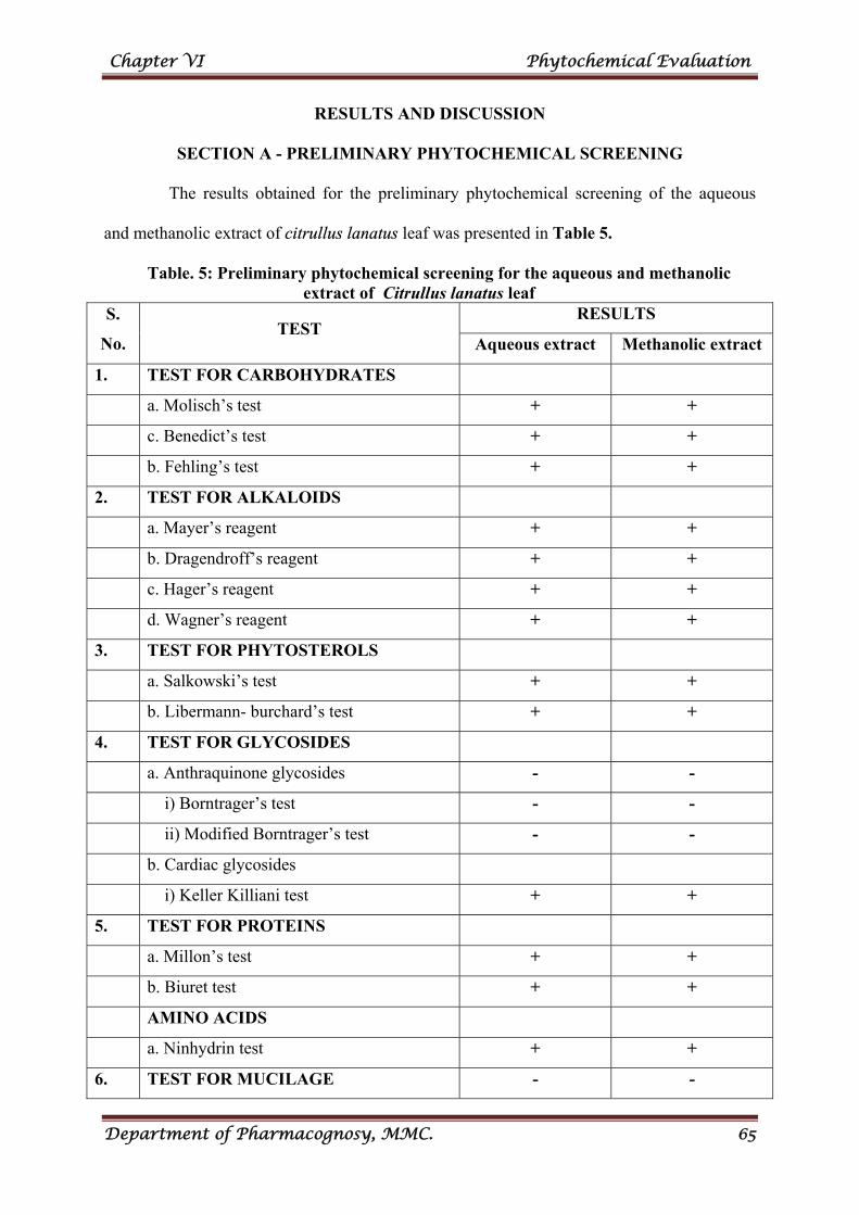
Chapter VI Phytochemical Evaluation
Department of Pharmacognosy, MMC. 65
RESULTS AND DISCUSSION
SECTION A - PRELIMINARY PHYTOCHEMICAL SCREENING
The results obtained for the preliminary phytochemical screening of the aqueous
and methanolic extract of citrullus lanatus leaf was presented in Table 5.
Table. 5: Preliminary phytochemical screening for the aqueous and methanolic extract of Citrullus lanatus leaf S.
No. TEST
RESULTS
Aqueous extract Methanolic extract
1. TEST FOR CARBOHYDRATES
a. Molisch’s test + +
c. Benedict’s test + +
b. Fehling’s test + +
2. TEST FOR ALKALOIDS
a. Mayer’s reagent + +
b. Dragendroff’s reagent + +
c. Hager’s reagent + +
d. Wagner’s reagent + +
3. TEST FOR PHYTOSTEROLS
a. Salkowski’s test + +
b. Libermann- burchard’s test + +
4. TEST FOR GLYCOSIDES
a. Anthraquinone glycosides - -
i) Borntrager’s test - -
ii) Modified Borntrager’s test - -
b. Cardiac glycosides
i) Keller Killiani test + +
5. TEST FOR PROTEINS
a. Millon’s test + +
b. Biuret test + +
AMINO ACIDS
a. Ninhydrin test + +
6. TEST FOR MUCILAGE - -
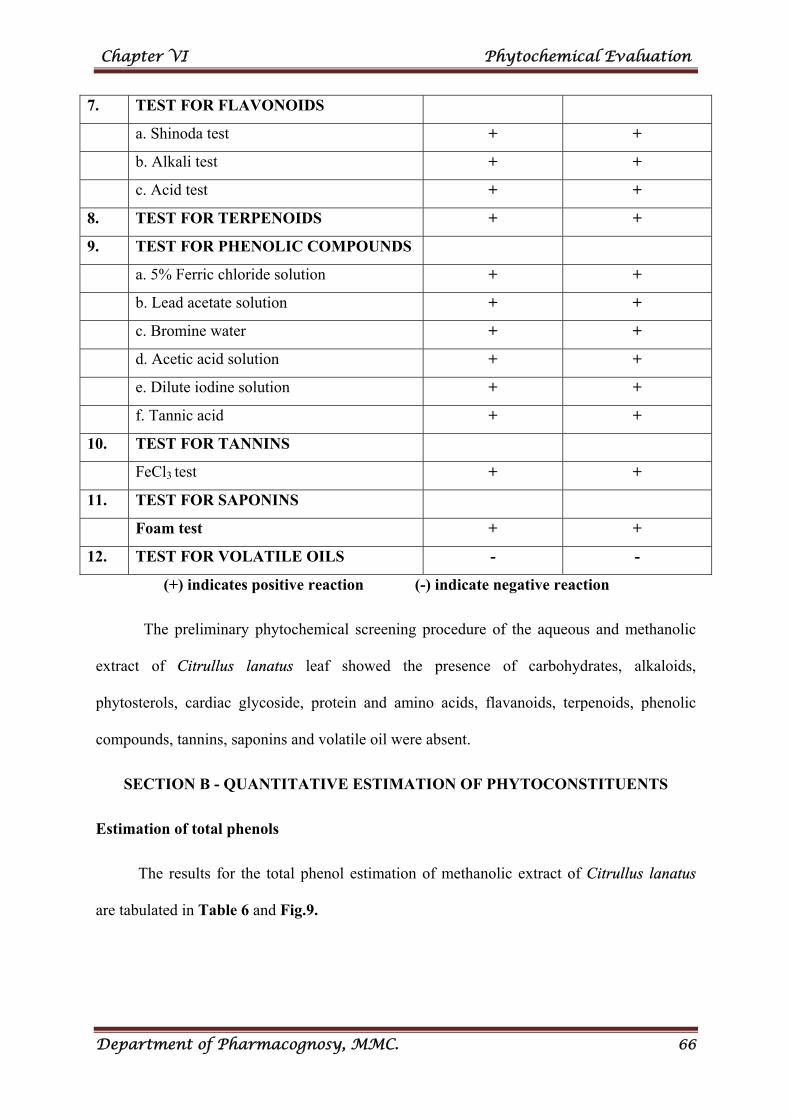
Chapter VI Phytochemical Evaluation
Department of Pharmacognosy, MMC. 66
7. TEST FOR FLAVONOIDS
a. Shinoda test + +
b. Alkali test + +
c. Acid test + +
8. TEST FOR TERPENOIDS + +
9. TEST FOR PHENOLIC COMPOUNDS
a. 5% Ferric chloride solution + +
b. Lead acetate solution + +
c. Bromine water + +
d. Acetic acid solution + +
e. Dilute iodine solution + +
f. Tannic acid + +
10. TEST FOR TANNINS
FeCl3 test + +
11. TEST FOR SAPONINS
Foam test + +
12. TEST FOR VOLATILE OILS - -
(+) indicates positive reaction (-) indicate negative reaction
The preliminary phytochemical screening procedure of the aqueous and methanolic
extract of Citrullus lanatus leaf showed the presence of carbohydrates, alkaloids,
phytosterols, cardiac glycoside, protein and amino acids, flavanoids, terpenoids, phenolic
compounds, tannins, saponins and volatile oil were absent.
SECTION B - QUANTITATIVE ESTIMATION OF PHYTOCONSTITUENTS
Estimation of total phenols
The results for the total phenol estimation of methanolic extract of Citrullus lanatus
are tabulated in Table 6 and Fig.9.
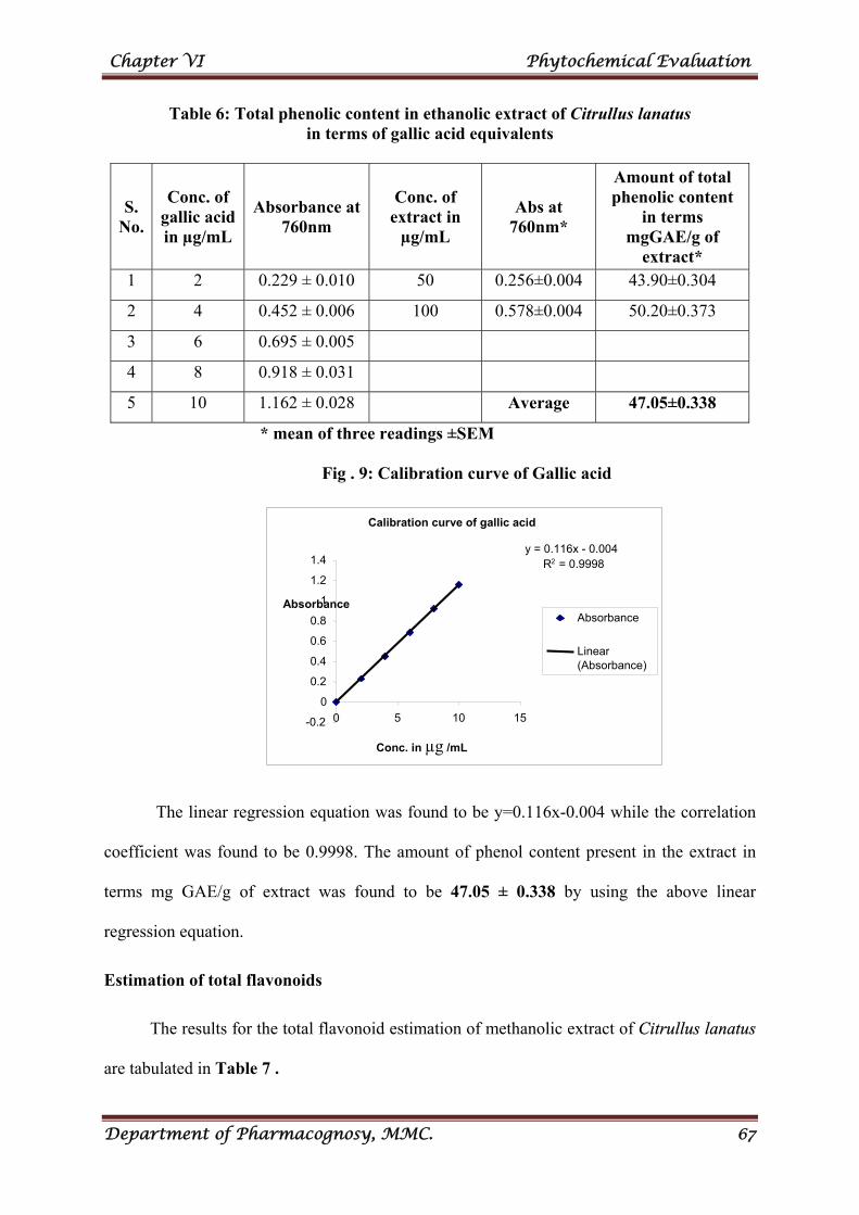
Chapter VI Phytochemical Evaluation
Department of Pharmacognosy, MMC. 67
Table 6: Total phenolic content in ethanolic extract of Citrullus lanatus in terms of gallic acid equivalents
S. No.
Conc. of gallic acid in µg/mL
Absorbance at 760nm
Conc. of extract in
µg/mL
Abs at 760nm*
Amount of total phenolic content
in terms mgGAE/g of
extract* 1 2 0.229 ± 0.010 50 0.256±0.004 43.90±0.304
2 4 0.452 ± 0.006 100 0.578±0.004 50.20±0.373
3 6 0.695 ± 0.005
4 8 0.918 ± 0.031
5 10 1.162 ± 0.028 Average 47.05±0.338
* mean of three readings ±SEM
Fig . 9: Calibration curve of Gallic acid
The linear regression equation was found to be y=0.116x-0.004 while the correlation
coefficient was found to be 0.9998. The amount of phenol content present in the extract in
terms mg GAE/g of extract was found to be 47.05 ± 0.338 by using the above linear
regression equation.
Estimation of total flavonoids
The results for the total flavonoid estimation of methanolic extract of Citrullus lanatus
are tabulated in Table 7 .
Calibration curve of gallic acid
y = 0.116x - 0.004 R2 = 0.9998
-0.2
0
0.2
0.4
0.6
0.8
1
1.2
1.4
0 5 10 15
Conc. in µg /mL
AbsorbanceAbsorbance
Linear(Absorbance)
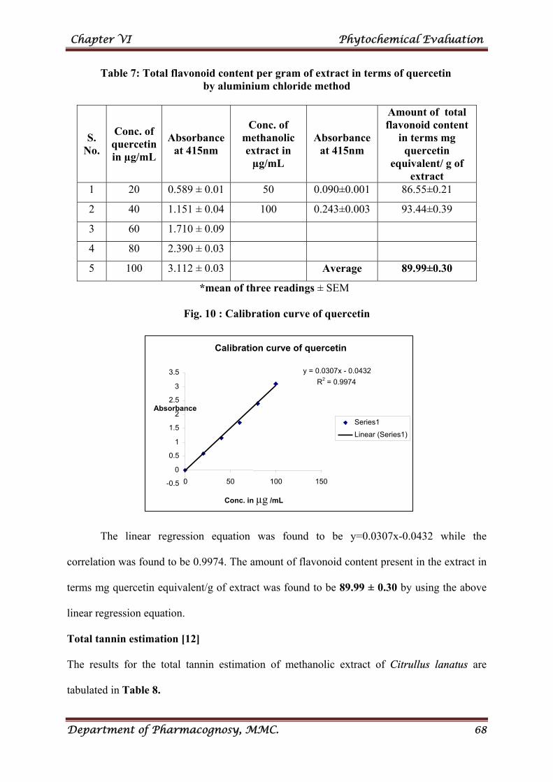
Chapter VI Phytochemical Evaluation
Department of Pharmacognosy, MMC. 68
Table 7: Total flavonoid content per gram of extract in terms of quercetin by aluminium chloride method
S. No.
Conc. of quercetin in µg/mL
Absorbance at 415nm
Conc. of methanolic extract in
µg/mL
Absorbance at 415nm
Amount of total flavonoid content
in terms mg quercetin
equivalent/ g of extract
1 20 0.589 ± 0.01 50 0.090±0.001 86.55±0.21
2 40 1.151 ± 0.04 100 0.243±0.003 93.44±0.39
3 60 1.710 ± 0.09
4 80 2.390 ± 0.03
5 100 3.112 ± 0.03 Average 89.99±0.30
*mean of three readings ± SEM
Fig. 10 : Calibration curve of quercetin
The linear regression equation was found to be y=0.0307x-0.0432 while the
correlation was found to be 0.9974. The amount of flavonoid content present in the extract in
terms mg quercetin equivalent/g of extract was found to be 89.99 ± 0.30 by using the above
linear regression equation.
Total tannin estimation [12]
The results for the total tannin estimation of methanolic extract of Citrullus lanatus are
tabulated in Table 8.
Calibration curve of quercetin
y = 0.0307x - 0.0432
R2 = 0.9974
-0.5
0
0.5
1
1.5
2
2.5
3
3.5
0 50 100 150
Conc. in µg /mL
Absorbance
Series1
Linear (Series1)
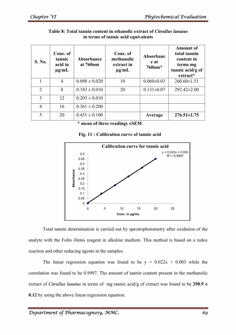
Chapter VI Phytochemical Evaluation
Department of Pharmacognosy, MMC. 69
Table 8: Total tannin content in ethanolic extract of Citrullus lanatus in terms of tannic acid equivalents
S. No.
Conc. of tannic acid in µg/mL
Absorbance at 760nm
Conc. of methanolic extract in
µg/mL
Absorbance at
760nm*
Amount of total tannin content in terms mg
tannic acid/g of extract*
1 4 0.098 ± 0.020 10 0.060±0.03 260.60±1.51
2 8 0.183 ± 0.010 20 0.131±0.07 292.42±2.00
3 12 0.203 ± 0.010
4 16 0.361 ± 0.200
5 20 0.451 ± 0.100 Average 276.51±1.75
* mean of three readings ±SEM
Fig. 11 : Calibration curve of tannic acid
Total tannin determination is carried out by spectrophotometry after oxidation of the
analyte with the Folin–Denis reagent in alkaline medium. This method is based on a redox
reaction and other reducing agents in the samples.
The linear regression equation was found to be y = 0.022x + 0.003 while the
correlation was found to be 0.9997. The amount of tannin content present in the methanolic
extract of Citrullus lanatus in terms of mg tannic acid/g of extract was found to be 290.9 ±
0.12 by using the above linear regression equation.
y = 0.022x + 0.003R² = 0.9997
0
0.05
0.1
0.15
0.2
0.25
0.3
0.35
0.4
0.45
0.5
0 5 10 15 20 25
Ab
sorb
anc
e
Conc. in µg/mL
Calibration curve for tannic acid
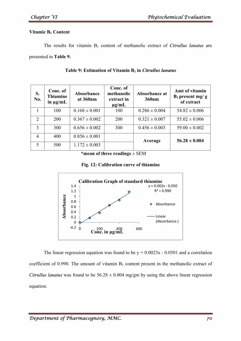
Chapter VI Phytochemical Evaluation
Department of Pharmacognosy, MMC. 70
Vitamic B1 Content
The results for vitamin B1 content of methanolic extract of Citrullus lanatus are
presented in Table 9.
Table 9: Estimation of Vitamin B1 in Citrullus lanatus
*mean of three readings ± SEM
Fig. 12: Calibration curve of thiamine
The linear regression equation was found to be y = 0.0023x - 0.0501 and a correlation
coefficient of 0.990. The amount of vitamin B1 content present in the methanolic extract of
Citrullus lanatus was found to be 56.28 ± 0.004 mg/gm by using the above linear regression
equation.
y = 0.002x ‐ 0.050R² = 0.990
‐0.2
0
0.2
0.4
0.6
0.8
1
1.2
1.4
0 200 400 600
Ab
sorb
ance
Conc. in µg/mL
Calibration Graph of standard thiamine
Absorbance
Linear (Absorbance )
S. No.
Conc. of Thiamine in µg/mL
Absorbance at 360nm
Conc. of methanolic extract in
µg/mL
Absorbance at 360nm
Amt of vitamin B1 present mg/ g
of extract
1 100 0.168 ± 0.001 100 0.286 ± 0.004 54.82 ± 0.006
2 200 0.367 ± 0.002 200 0.321 ± 0.007 55.02 ± 0.006
3 300 0.656 ± 0.002 300 0.456 ± 0.003 59.00 ± 0.002
4 400 0.856 ± 0.001 Average 56.28 ± 0.004
5 500 1.172 ± 0.003
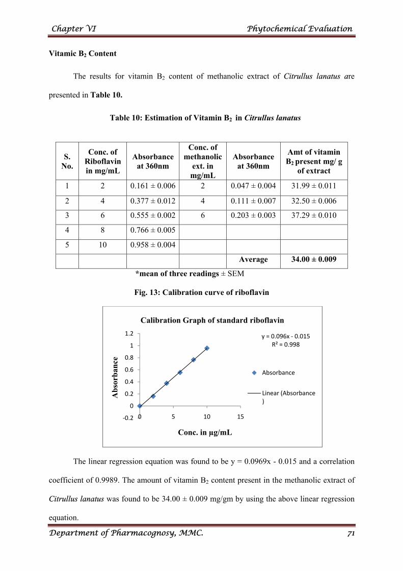
Chapter VI Phytochemical Evaluation
Department of Pharmacognosy, MMC. 71
Vitamic B2 Content
The results for vitamin B2 content of methanolic extract of Citrullus lanatus are
presented in Table 10.
Table 10: Estimation of Vitamin B2 in Citrullus lanatus
*mean of three readings ± SEM
Fig. 13: Calibration curve of riboflavin
The linear regression equation was found to be y = 0.0969x - 0.015 and a correlation
coefficient of 0.9989. The amount of vitamin B2 content present in the methanolic extract of
Citrullus lanatus was found to be 34.00 ± 0.009 mg/gm by using the above linear regression
equation.
y = 0.096x ‐ 0.015R² = 0.998
‐0.2
0
0.2
0.4
0.6
0.8
1
1.2
0 5 10 15
Ab
sorb
ance
Conc. in µg/mL
Calibration Graph of standard riboflavin
Absorbance
Linear (Absorbance )
S. No.
Conc. of Riboflavin in mg/mL
Absorbance at 360nm
Conc. of methanolic
ext. in mg/mL
Absorbance at 360nm
Amt of vitamin B2 present mg/ g
of extract
1 2 0.161 ± 0.006 2 0.047 ± 0.004 31.99 ± 0.011
2 4 0.377 ± 0.012 4 0.111 ± 0.007 32.50 ± 0.006
3 6 0.555 ± 0.002 6 0.203 ± 0.003 37.29 ± 0.010
4 8 0.766 ± 0.005
5 10 0.958 ± 0.004
Average 34.00 ± 0.009
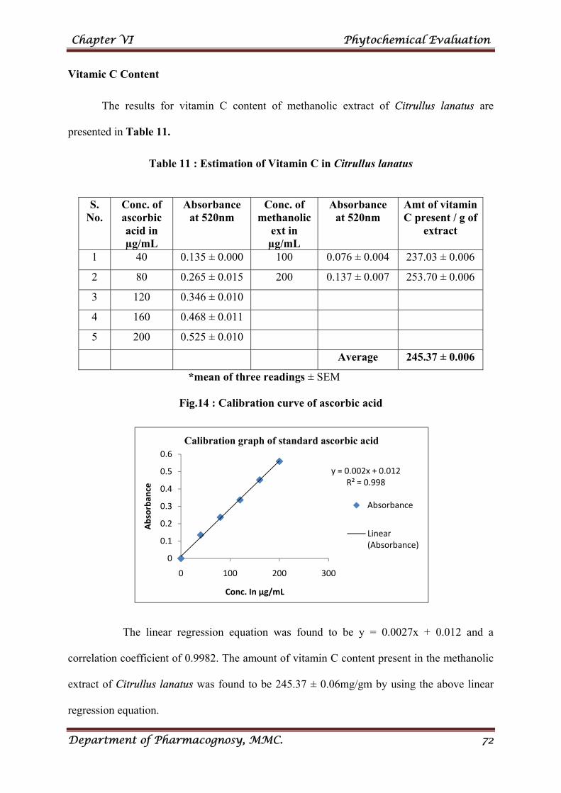
Chapter VI Phytochemical Evaluation
Department of Pharmacognosy, MMC. 72
y = 0.002x + 0.012R² = 0.998
0
0.1
0.2
0.3
0.4
0.5
0.6
0 100 200 300
Absorban
ce
Conc. In µg/mL
Calibration graph of standard ascorbic acid
Absorbance
Linear (Absorbance)
Vitamic C Content
The results for vitamin C content of methanolic extract of Citrullus lanatus are
presented in Table 11.
Table 11 : Estimation of Vitamin C in Citrullus lanatus
*mean of three readings ± SEM
Fig.14 : Calibration curve of ascorbic acid
The linear regression equation was found to be y = 0.0027x + 0.012 and a
correlation coefficient of 0.9982. The amount of vitamin C content present in the methanolic
extract of Citrullus lanatus was found to be 245.37 ± 0.06mg/gm by using the above linear
regression equation.
S. No.
Conc. of ascorbic acid in µg/mL
Absorbance at 520nm
Conc. of methanolic
ext in µg/mL
Absorbance at 520nm
Amt of vitamin C present / g of
extract
1 40 0.135 ± 0.000 100 0.076 ± 0.004 237.03 ± 0.006
2 80 0.265 ± 0.015 200 0.137 ± 0.007 253.70 ± 0.006
3 120 0.346 ± 0.010
4 160 0.468 ± 0.011
5 200 0.525 ± 0.010
Average 245.37 ± 0.006
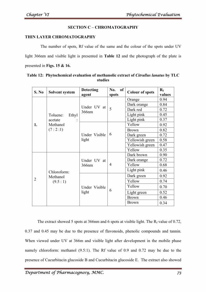
Chapter VI Phytochemical Evaluation
Department of Pharmacognosy, MMC. 73
SECTION C – CHROMATOGRAPHY
THIN LAYER CHROMATOGRAPHY
The number of spots, Rf value of the same and the colour of the spots under UV
light 366nm and visible light is presented in Table 12 and the photograph of the plate is
presented in Figs. 15 & 16.
Table 12: Phytochemical evaluation of methanolic extract of Citrullus lanatus by TLC studies
S. No Solvent system Detecting agent
No. of spots
Colour of spots Rf
values
1.
Toluene: Ethyl acetate : Methanol (7 : 2 :1)
Under UV at 366nm
5
Orange 0.94 Dark orange 0.84 Dark red 0.72 Light pink 0.45 Light pink 0.37
Under Visible light
6
Yellow 0.92 Brown 0.82 Dark green 0.72 Yellowish green 0.58 Yellowish green 0.47 Yellow 0.35
2
Chloroform: Methanol (9.5 : 1)
Under UV at 366nm
4
Dark brown 0.90 Dark orange 0.72 Yellow 0.68 Light pink 0.46
Under Visible light
6
Dark green 0.92 Yellow 0.74 Yellow 0.70 Light green 0.52 Brown 0.46 Brown 0.34
The extract showed 5 spots at 366nm and 6 spots at visible light. The Rf value of 0.72,
0.37 and 0.45 may be due to the presence of flavonoids, phenolic compounds and tannin.
When viewed under UV at 366m and visible light after development in the mobile phase
namely chloroform: methanol (9.5:1). The Rf value of 0.9 and 0.72 may be due to the
presence of Cucurbitacin glucoside B and Cucurbitacin glucoside E. The extract also showed
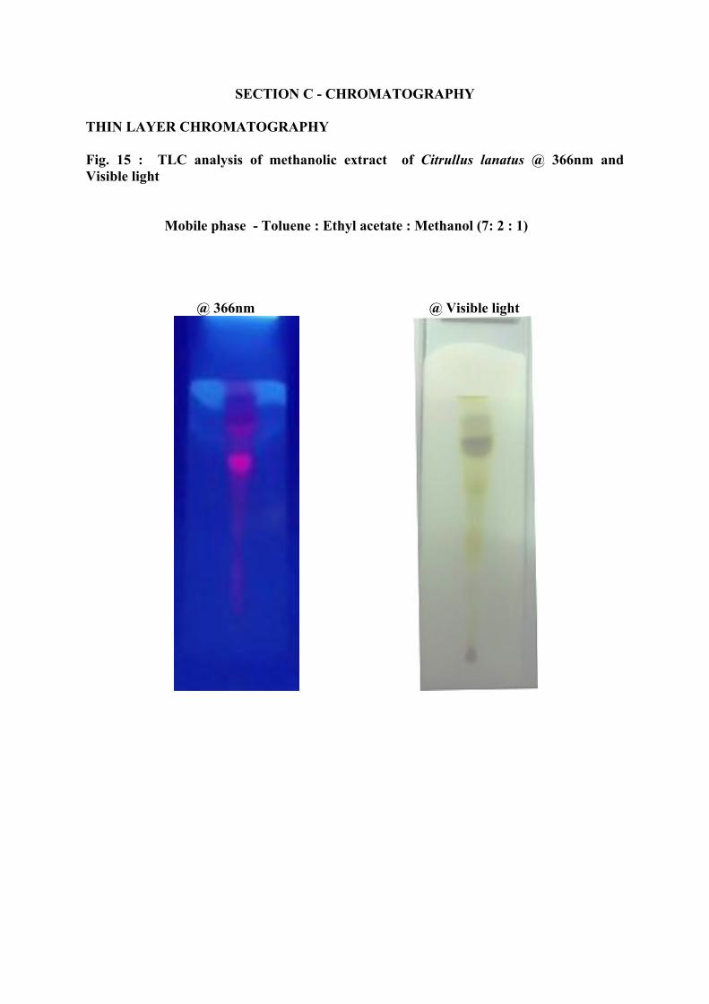
SECTION C - CHROMATOGRAPHY
THIN LAYER CHROMATOGRAPHY
Fig. 15 : TLC analysis of methanolic extract of Citrullus lanatus @ 366nm and Visible light Mobile phase - Toluene : Ethyl acetate : Methanol (7: 2 : 1)
@ 366nm @ Visible light
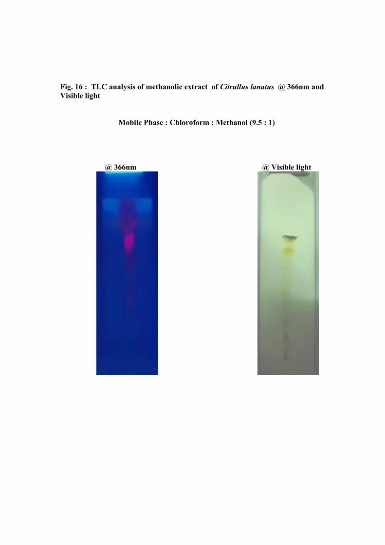
Fig. 16 : TLC analysis of methanolic extract of Citrullus lanatus @ 366nm and Visible light Mobile Phase : Chloroform : Methanol (9.5 : 1)
@ 366nm @ Visible light
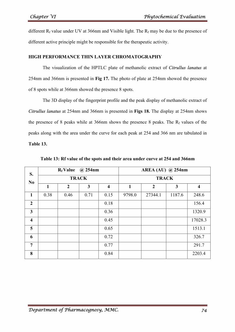
Chapter VI Phytochemical Evaluation
Department of Pharmacognosy, MMC. 74
different Rf value under UV at 366nm and Visible light. The Rf may be due to the presence of
different active principle might be responsible for the therapeutic activity.
HIGH PERFORMANCE THIN LAYER CHROMATOGRAPHY
The visualization of the HPTLC plate of methanolic extract of Citrullus lanatus at
254nm and 366nm is presented in Fig 17. The photo of plate at 254nm showed the presence
of 8 spots while at 366nm showed the presence 8 spots.
The 3D display of the fingerprint profile and the peak display of methanolic extract of
Citrullus lanatus at 254nm and 366nm is presented in Figs 18. The display at 254nm shows
the presence of 8 peaks while at 366nm shows the presence 8 peaks. The Rf values of the
peaks along with the area under the curve for each peak at 254 and 366 nm are tabulated in
Table 13.
Table 13: Rf value of the spots and their area under curve at 254 and 366nm
S.
No
Rf Value @ 254nm AREA (AU) @ 254nm
TRACK TRACK
1 2 3 4 1 2 3 4
1 0.38 0.46 0.71 0.15 9798.0 27344.1 1187.6 248.6
2 0.18 156.4
3 0.36 1320.9
4 0.45 17028.3
5 0.65 1513.1
6 0.72 326.7
7 0.77 291.7
8 0.84 2203.4
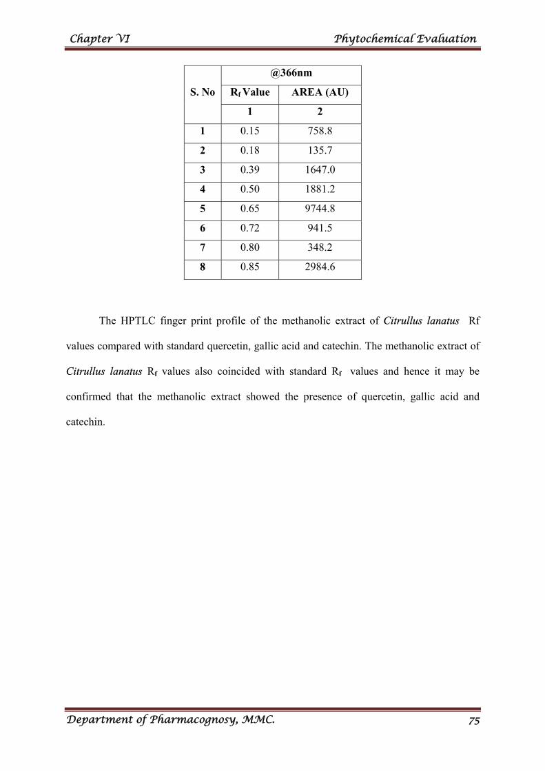
Chapter VI Phytochemical Evaluation
Department of Pharmacognosy, MMC. 75
S. No
@366nm
Rf Value AREA (AU)
1 2
1 0.15 758.8
2 0.18 135.7
3 0.39 1647.0
4 0.50 1881.2
5 0.65 9744.8
6 0.72 941.5
7 0.80 348.2
8 0.85 2984.6
The HPTLC finger print profile of the methanolic extract of Citrullus lanatus Rf
values compared with standard quercetin, gallic acid and catechin. The methanolic extract of
Citrullus lanatus Rf values also coincided with standard Rf values and hence it may be
confirmed that the methanolic extract showed the presence of quercetin, gallic acid and
catechin.
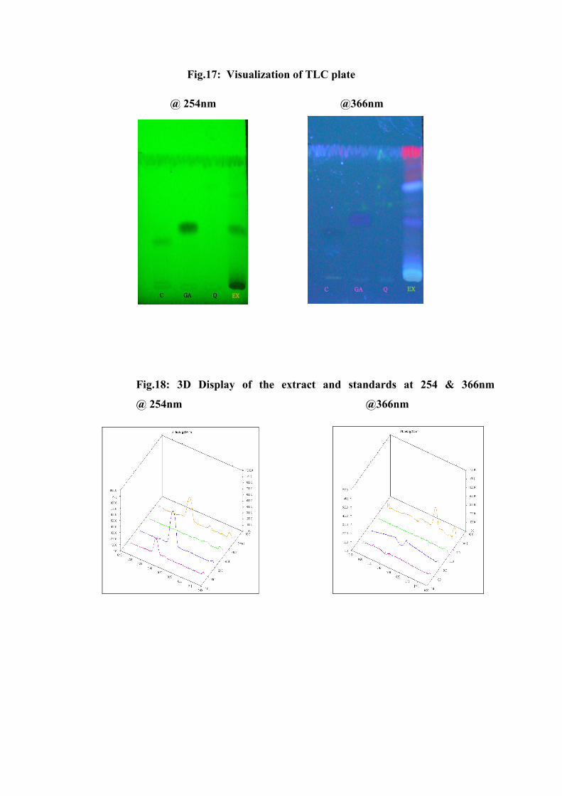
Fig.17: Visualization of TLC plate
@ 254nm @366nm
Fig.18: 3D Display of the extract and standards at 254 & 366nm
@ 254nm @366nm
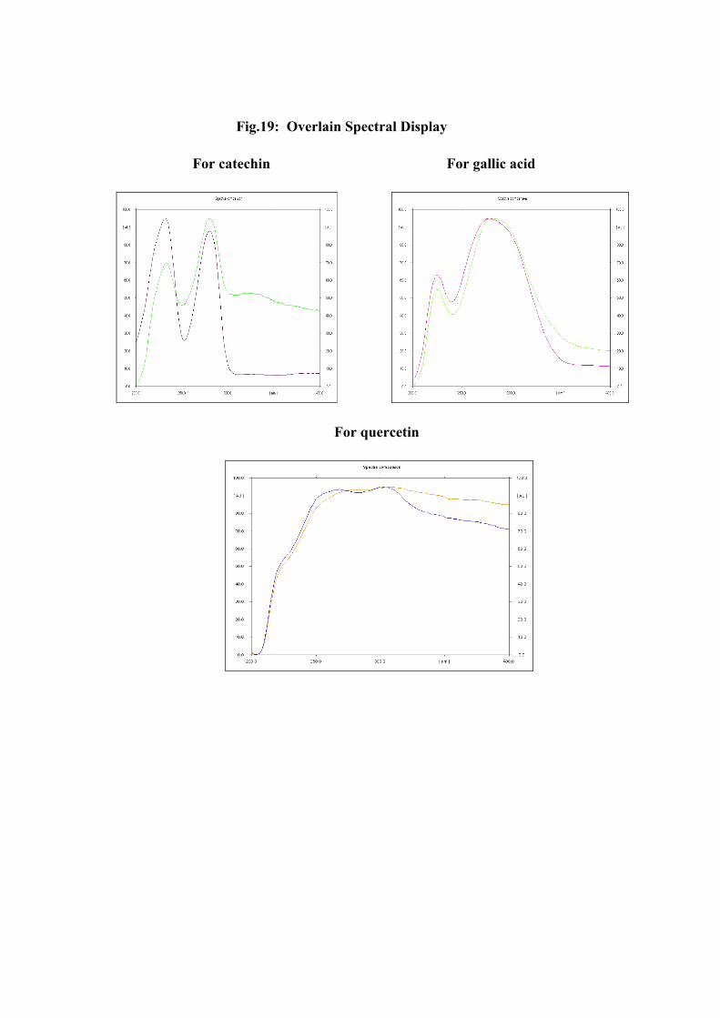
Fig.19: Overlain Spectral Display
For catechin For gallic acid
For quercetin
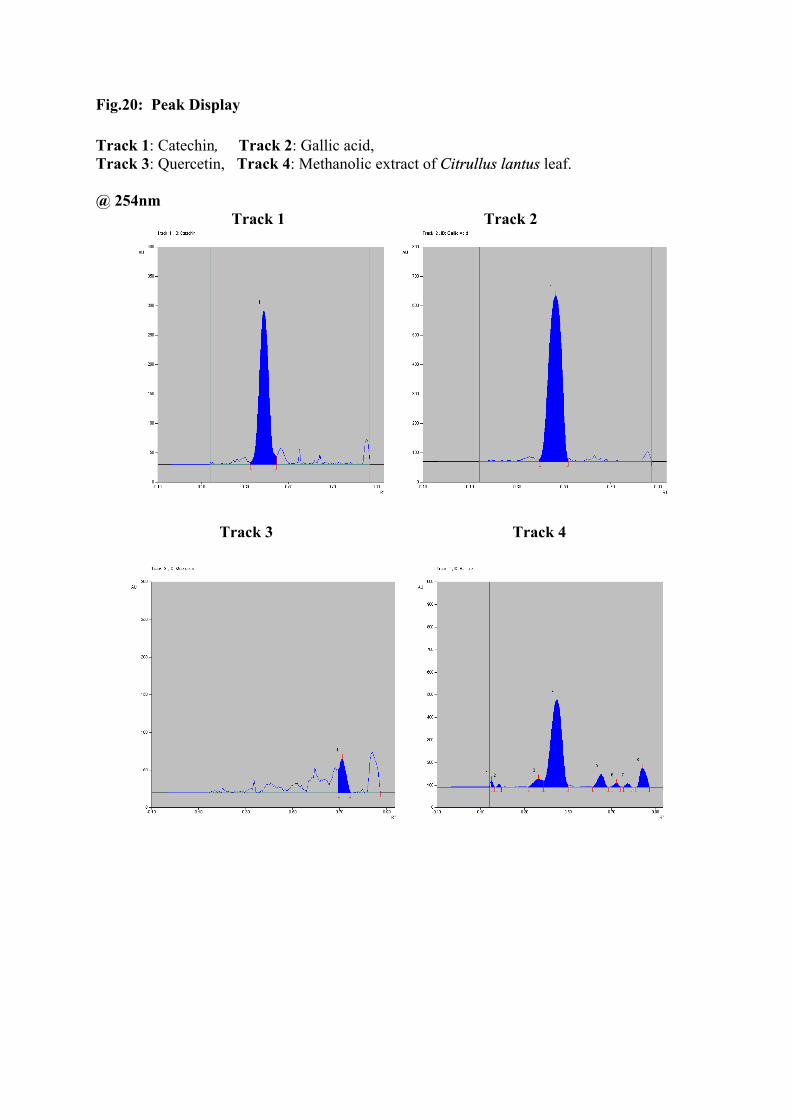
Fig.20: Peak Display
Track 1: Catechin, Track 2: Gallic acid, Track 3: Quercetin, Track 4: Methanolic extract of Citrullus lantus leaf. @ 254nm Track 1 Track 2
Track 3 Track 4
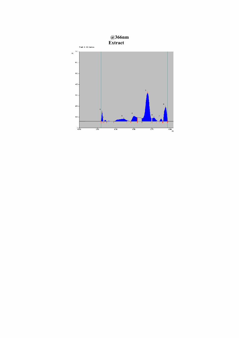
@366nm
Extract
PHARMACOLOGICAL
EVALUATION
Chapter VII Pharmacological Evaluation
Department of Pharmacognosy, MMC. 76
CHAPTER VII
PHARMACOLOGICAL EVALUATION[119-120]
The word pharmacology is derived from Greek words pharmacon (an active principle)
and logos (a discourse or treatise). It is the science that deals with drugs. It consists of
detailed study of drug, particularly their actions on living animal’s organ and tissue. The
object of pharmacology is mainly to provide such scientific data, using which one can choose
a drug treatment of proven efficacy and safety from various options available, so suit the
patient.
For thousand of years most drugs were crude natural products of unknown
composition and limited efficacy. The overall effect of these substances on the body was
rather imprecisely known, but how the same were produced was entirely unknown. The drugs
have been purified, chemically characterised and a vast variety of highly potent and selective
new drugs have been developed.
SECTION A – IN VITRO ANTIOXIDANT ACTIVITY
An antioxidant is a chemical that prevents the oxidation of other chemicals. They
protect the key cell components by neutralizing the damaging effects of free radicals, which
are natural by- products of cell metabolism.[121-122].
Free radicals may be either oxygen derived (ROS, reactive oxygen species) or
nitrogen derived (RNS, reactive nitrogen species). The oxygen derived molecules are O2-
Superoxide], HO [hydroxyl] ,HO2 [hydroperoxyl], ROO [peroxyl], RO [alkoxyl] as free
radical and H2O2 oxygen as non-radical. Nitrogen derived oxidant species are mainly NO
[nitric oxide], ONOO [peroxy nitrate], NO2 [nitrogen dioxide] and N2O3 [dinitrogen trioxide].
[123-124]
Chapter VII Pharmacological Evaluation
Department of Pharmacognosy, MMC. 77
In a normal cell, there is appropriate oxidant: antioxidant balance. However, this
balance can be shifted, when production species is increased or when levels of antioxidants
are diminished. This stage is called oxidative stress. Oxidative stress results in the damage of
biopolymers including nucleic acids, proteins, polyunsaturated fatty acids and carbohydrates.
Oxidative stress causes serious cell damage leading to a variety of human diseases
like Alzheimer’s disease, Parkinson’s disease, atheroscleorosis, cancer, arthritis,
immunological incompetence and neurodegenerative disorders, etc. [125-127].
Antioxidants and Health
Antioxidants are man-made or natural substances that may prevent or delay
some types of cell damage. Diets high in vegetables and fruits, which are good
sources of antioxidants, have been found to be healthy; however, research has
not shown antioxidant supplements to be beneficial in preventing diseases.
Examples of antioxidants include vitamins C and E, selenium, and
carotenoids, such as beta-carotene, lycopene, lutein, and zeaxanthin.
Rigorous scientific studies involving more than 100,000 people combined
have tested whether antioxidant supplements can help prevent chronic
diseases, such as cardiovascular diseases, cancer, and cataracts. In most
instances, antioxidants did not reduce the risks of developing
these diseases.[128]
Medicinal herb as Antioxidants:
Indian medicinal herbs were extensively investigated as vital sources of antioxidants.
Allium cepa (Onion), Allium sativum (Garlic), Aloe vera (Indian aloe), Amomum subulatum
(greater cardamom, Bari elachi), Andrographis paniculata (The creat), Asparagus racemosus
(Shatavari), Azadirachta indica (Neem, Nimba), Bacopa monniera (Brahmi), Camellia
sinensis (Green tea), Cinnamomum verum (Cinnamon), Curcuma longa (Turmeric), Emblica
Chapter VII Pharmacological Evaluation
Department of Pharmacognosy, MMC. 78
officinalis (Indian gooseberry), Glycyrrhiza glabra (Yashtimadhu), Withania somnifera
(Ashwagandha), and Zingiber officinalis (Ginger). [129]
MATERIALS AND METHODS
Method 1: Free radical Scavenging activity using diphenyl picryl hydrazyl (DPPH) free radical
The free radical scavenging activity of the extracts is evaluated by assessing their
ability to reduce the colour of DPPH in ethanol according to Brand Williams [130]. DPPH
stable free radical method is an easy, rapid and sensitive way to survey the antioxidant
activity of specific compound or plant extracts.
Principle
A simple method that has been developed to determine the antioxidant activity of
plants utilizes the stable 2,2-diphenyl-1-picrylhydrazyl (DPPH) radical. The odd electron in
the DPPH free radical gives a strong absorption maximum at 517nm and is purple in colour.
The colour turns from purple to yellow as the molar absorptivity of the DPPH radical at
520nm reduces when the odd electron of DPPH radical becomes paired with hydrogen from a
free radical scavenging antioxidant to form the reduced DPPH-H. The resulting
decolorization is stoichiometric with respect to number of electrons captured [130-131].
DPPH. + AH DPPH – H + A.
Instrument
Shimadzu UV Visible spectrometer, Model 1800
Reagents
0.1mM Diphenyl Picryl Hydrazyl Radical in ethanol
Procedure [132-133]
A stock solution of 0.5mg/mL concentration of methanolic extract of C. lanatus was
prepared. To 1mL of various concentrations of test samples, 4mL of DPPH was added.
Chapter VII Pharmacological Evaluation
Department of Pharmacognosy, MMC. 79
Control was prepared without sample in an identical manner. DPPH was replaced by ethanol
in case of blank. The reaction was allowed to be completed in the dark for about 30min. Then
the absorbance was measured at 517nm. Vitamin C was used as standard. The percentage
scavenging was calculated using the formula [(Control-Test)/Control] x 100. A graph was
constructed by plotting concentration versus percentage inhibition and a linear regression
equation calculated. The concentration of the sample required for 50% reduction in
absorbance (IC50) was calculated using linear regression analysis. A triplicate reading was
taken and average was calculated. The results obtained are presented in Table 14 and Fig.21.
Method 2: Total antioxidant activity by Phosphomolybdenum Method. [134-135]
Principle
Total antioxidant capacity was measured by spectrophotometric method of Prieto et
al. Phosphomolybdenum method assay is based on the reduction of Mo (VI) to Mo (V) by the
sample analyte and the subsequent formation of green phosphate Mo (V) complex at acidic
pH and the absorbance was measured at 695nm. This method is used to determine the total
antioxidant activity of samples.
Mo6+ → Mo5+
Reagents
0.6M sulphuric acid
28mM sodium phosphate
4mM ammonium molybdate
Instruments
Shimadzu UV Visible spectrophotometer, Model 1800
Procedure
An aliquot of 0.3mL of different concentrations of sample solution was combined
with 2.7mL of the reagent solution (H2SO4, sodium phosphate and ammonium molybdate). In
Chapter VII Pharmacological Evaluation
Department of Pharmacognosy, MMC. 80
case of blank, 0.3mL of msethanol was used in place of sample. The tubes were incubated for
95°C for 90min. After the mixture was cooled to room temperature, the absorbance was
measured at 695nm against blank. Ascorbic acid was used as a standard and was treated in a
similar manner. The total antioxidant activity is expressed as the number of equivalents of
ascorbic acid (µg/g). The results were tabulated in table 15 and the graphical representation
is presented in Fig. 22.
Method 3: Reducing power assay [134]
Principle:
This is a spectrophotometric method and is based on the principle that an increase in
absorbance of the reaction mixture as concentration increase indicates an increased
antioxidant activity.
The assay is based on the reduction of ferric in potassium ferricyanide to ferrous to
form potassium ferrocyanide by the sample and the formation of Prussian blue colour
complex when treated with ferric chloride. The absorbance of the blue complex is measured
at 700nm.
Instrument
Shimadzu UV Visible spectrophotometer. Model 1800
Reagents
1% potassium ferricyanide
10% trichloro acetic acid.
0.2M, phosphate buffer (pH 6.6)
0.1% ferric chloride.
Procedure
Various concentration of methanolic extracts of Citrullus lanatus was mixed with
0.75mL phosphate buffer and 0.75mL potassium ferricyanide [K3 Fe(CN6)], then the mixture
Chapter VII Pharmacological Evaluation
Department of Pharmacognosy, MMC. 81
was incubated at 50°C for 20 min. 0.75mL of trichloro acetic acid was added to the mixture,
which was then centrifuged at 3000rpm for 10min. Finally 1.5mL of the supernatant solution
was mixed with 1.5mL of distilled water and 0.1mL of ferric chloride (FeCl3) and absorbance
was measured at 700nm in a UV-Visible Spectrophotometer. Ascorbic acid was used as
standard and phosphate buffer used as blank. The absorbance of the final reaction mixture of
three parallel experiments was expressed as mean ± standard error of mean. Increased
absorbance of the reaction mixture indicates stronger reducing power. The results obtained
are tabulated in table 16 and the graphical representation is presented in Fig. 23.
Method 4: Ferric Reducing Antioxidant Power (FRAP) Assay [135]
Total antioxidant activity is measured by FRAP assay of Benzie et al., 1999. The
ferric reducing antioxidant power assay measures the potential of antioxidants to reduce the
Fe3+ and 2,4,6 tripyridyl-s-triazyne (TPTZ) complex present in stoichiometric excess to the
blue coloured Fe2+ complex which increases the absorbance at 593nm.
Principle
At low pH, reduction of ferric tripyridyl triazine (Fe III TPTZ) complex to ferrous
form (which has an intense blue colour) can be monitored by measuring the change in
absorption at 593nm. The reaction is non-specific, in that any half reaction that has lower
redox potential, under reaction conditions, than that of ferric ferrous half reaction, will drive
the ferrous (Fe III to Fe II) ion formation. The change in absorbance is therefore, directly
related to the combined or “total” reducing power of the electron donating antioxidants
present in the reaction mixture.
Instrument
Shimadzu UV Visible spectrophotometer, Model 1800
Reagents
FRAP Reagent
Chapter VII Pharmacological Evaluation
Department of Pharmacognosy, MMC. 82
a) Acetate buffer 30mM pH 3.6: Weigh 3.1g sodium acetate trihydrate and add 16
ml of glacial acetic acid and make the volume to 1 L with distilled water.
b) TPTZ (2, 4, 6-tripyridyl-s- triazine) (M.W. 312.34) 10mM in 40mM HCl
c) FeCl3. 6H2O (M.W. 270.30) 20mM
The working FRAP reagent was prepared freshly by mixing a, b & c in
the ratio of 10:1:1 at the time of use.
Procedure
The methanolic extract of Citrullus lanatus was dissolved in methanol to get a stock
solution containing 1mg/mL. Varying quantities of the stock solution were added to 3mL of
working FRAP reagent and absorbance was measured at 0min after vortexing at 593nm.
Thereafter samples were placed at 37ºC in water bath and absorption was again measured
after 4min. Ascorbic acid was used as standard. The result obtained for the FRAP assay are
presented in the table 17 and Fig.24.
SECTION B - LARVICIDAL ACTIVITY OF METHANOLIC EXTRACT OF
CITRULLUS LANATUS[136-147]
Malaria and other vector-borne diseases contribute to the major disease burden in
India. One of the methods to control these diseases is to control the vectors for the
interruption of disease transmission. In the past, synthetic organic chemical insecticides based
intervention measures for the control of insect pests and disease vectors have resulted in
development of insecticide resistance in some medically important vectors of malaria,
filariasis, Japanese encephalitis, dengue, hemorrhagic fever, chikungunya and yellow fever
transmitted by mosquitoes which cause millions of death every year. Hence destroying
mosquitoes is one method for preventing the above infections [147-148]. During the last decade,
Chapter VII Pharmacological Evaluation
Department of Pharmacognosy, MMC. 83
various studies on natural plant products against mosquito vectors indicate them as possible
alternatives to synthetic chemical insecticides.
The present study has attempted to study mosquito larvicidal property of C. lanatus
against three mosquito species - Anopheles stephensi, Culex quinquefasciatus and Aedes
aegypti (Diptera: Culicidae).
Preparation of stock solution of methanolic extract
A stock solution of 100mg/mL of the methanolic extract of the plant Citrullus lanatus
was prepared by dissolving the required quantity of the extract in distilled water.
Procedure for extract
The method adopted by Arivoli S and Samuel Tennyson 2012 was followed. The
larvicidal activity of plant extract was carried out on late 3rd and early 4th instar larvae of
Anopheles stephensi, a primary vector of urban malaria, Culex quinquefaciatus, a common
vector of filariasis, Aedes aegypti, common vector of dengue and yellow fever. The mosquito
larvae were obtained from ICMR, Madurai. Twenty larvae were released in 500mL beaker
containing 200mL distilled water with varying concentrations of plant extract. The larvae
were provided with dog biscuit and yeast powder in a ratio of 3:2 as nutrients. The
experiments were carried out a room temperature (26°C ± 2°C). Three replicates of each
concentration were run under the same microclimate conditions along with untreated control.
The mortality was monitored for 24h. The results obtained are presented in Table 18 to 20
and Fig. 25 to 27.[148].
Data analysis[149-150]
LC50 (lethal concentration to cause 50% mortality in the population) and LC90
(lethal concentration to cause 90% mortality in the population) were determined by plotting
the regression line as described by Finney. The percentage mortality was calculated using
Abbot’s formula and the data so obtained was analyzed by probit analysis (Finney 1989) by
Chapter VII Pharmacological Evaluation
Department of Pharmacognosy, MMC. 84
using the software Minitab-15 for dose and time mortality regression lines. The purpose of
the probit transformation is to straighten the line so we can estimate LC 50 more easily.
Percentage Mortality = %Mortality in treated- %Mortality in control X 100 100 - % Mortality in control
SECTION C - PANCREATIC LIPASE INHIBITION ASSAY[151-153]
Obesity is a medical complication caused by an imbalance between energy intake and
expenditure and is broadly recognized as a major public health problem. Obesity can lead to
variety serious diseases, including hypertension, hyperlipidemia, atherosclerosis, and type II
diabetes and thus indirectly leading to aging. The inhibitors of digestive lipases reduce
dietary fat absorption and hence act as anti-obesity agents. New Pancreatic lipase inhibitors
derived from natural sources especially from medicinal herbs are used for the treatment of
obesity.
Pancreatic lipase
Pancreatic lipase (PL) is an enzyme, secreted from the pancreas and plays an excellent
role in the absorption of triglyceride in the small intestine. Dietary fats are composed of about
95% triacylglycerols (TG). Pancreatic lipase hydrolyses the water insoluble triacylglycerols
in the intestinal lumen and thereby used for the dietary fat absorption.
Pancreatic lipase inhibitors
Pancreatic lipase inhibitors are considered to be a valuable therapeutic agent for
treating diet-induced obesity in humans as anti-obesity agents. Grape seed extracts, carnosic
acid from the ethanolic extract of leaves of Salvia officinalis L (sage), flavan dimmers
isolated from fruits of Cassia nomame (Leguminosae), the methanol extract of Dioscorea
nipponica,5-hydroxy-7-(41-hrdroxy-31-methoxyphenyl)-l-phenyl-3-heptone (HPH) and 3-
methylethergalangin from the rhizome of Alpinia officinarum are examples of pancreatic
lipase inhibitors of plant sources
Chapter VII Pharmacological Evaluation
Department of Pharmacognosy, MMC. 85
Principle Lipases have certain roles in human pathogenesis and are a key enzyme catalyzes
the hydrolysis of emulsified esters of glycerol and long chain fatty acids. Short chain fatty
acids can be directly absorbed into the blood, while long-chain fatty acids and
monoglycerides combine with bile salts to form water soluble micelles. The micelles are
absorbed into the mucosal cells of the intestine and the fatty acids and monoglycerides are
resynthesized into triglycerides. Dietary triglyceride is usually stored in the adipose tissue.
Pharmacological agents that reduce the absorption of dietary triglycerides, reduce the
probability of the formation of atherosclerotic plaque. Triglyceride hydrolysis by pancreatic
lipase is inhibited by physiological concentrations of bile salts.
An anti-obesity drug Orlistat, inhibits pancreatic lipase in the lumen of the
gastrointestinal tract to decrease systemic absorption of dietary fat.
Requirements
Chicken (Gallus domesticus) pancreas
Sucrose solution (0.01M)
Ammonium sulphate (50% saturation)
Phosphate buffer (pH7)
Olive oil
Pancreatic lipase
Orlistat (60mg)
Procedure [153]
Extraction of Lipase from Chicken (Gallus domesticus) pancreas
Pancreas of freshly slaughtered chicken was collected with the guidance of a
veterinary surgeon. It was washed thoroughly and pancreatic lipase was placed in ice cold
Chapter VII Pharmacological Evaluation
Department of Pharmacognosy, MMC. 86
sucrose solution (0.01M). The Pancreas was homogenized in 0.01M sucrose and centrifuged.
The supernatant solution was separated and subjected to ammonium sulphate precipitation
(50% saturation). The obtained white pellets after centrifugation was dissolved in sucrose
solution and again saturated with 50% ammonium sulphate and centrifuged. Finally pellets
were used as enzyme source by dissolving in phosphate buffer (pH 7).
Determination of chicken pancreatic lipase activity
The chicken pancreatic lipase activity was determined by incubating an emulsion
containing 8mL of olive oil (Dietary fat), 0.4mL of phosphate buffer and 1 mL of chicken
pancreatic lipase for an hour. The reaction was stopped by addition of 1.5mL of a mixture
containing acetone and 95% ethanol (1:1). The amount of liberated fatty acid was
determined by titrating the emulsion against 0.02M NaOH (standardized by potassium
hydrogen phthalate) using phenolphthalein as an indicator. The end point is the appearance of
pink colour. The volume of sodium hydroxide consumed was taken as (A).
Pancreatic lipase inhibitory activity
Pancreatic lipase inhibitory activity has been studied using Prashith Kekuda T R et al.
method with slight modification. The plant extract of Citrullus lanatus was prepared in
different concentrations such as 1.0mg/mL, 2.0mg/mL, 3.0mg/mL, 4.0mg/mL, 5.0mg/mL. A
100µL of each concentration of sample was mixed with 8 mL of olive oil, 0.4 mL phosphate
buffer and 1 mL of Chicken pancreatic lipase and it was incubated for 60mins. The reaction
was stopped by the addition of 1.5 mL of a mixture containing acetone and 95% ethanol
(1:1). Appearance of pink colour from yellow colour shows the liberated fatty acids, which
was determined by titrating the solution against 0.02M NaOH (standardized by potassium
hydrogen phthalate) using phenolphthalein as an indicator and the percentage inhibition of
lipase activity was calculated using the following formula , Lipase inhibition = [A-B/ B] ×
Chapter VII Pharmacological Evaluation
Department of Pharmacognosy, MMC. 87
100, where A - Lipase activity, B - Activity of lipase when incubated with the standard and
test compounds and the results are tabulated in Table 21 and the pictorial representations are
presented in Fig.28.
SECTION D - IN VITRO ANTI CANCER ACTIVITY (BREAST CANCER
ACTIVITY BY USING HUMAN BREAST CANCER CELL LINES) [154-156]
Cancer is a group of many different diseases that have some important things in
common. They all arise in cells, the body’s unit of life. The body is made up of many types of
cells. Normally, cells grow and divide to produce more cells only when the body needs them.
Sometimes cells keep dividing when new cells are not needed. These cells may form a mass
of extra tissue called a growth or tumor. Tumors can be benign or malignant.
Benign tumors are not cancer. They can usually be removed, and in most cases, they
don’t come back. Most important, the cells in benign tumors do not invade other tissues and
do not spread to other parts of the body. Benign tumors are not a threat to life.
Malignant tumors are cancer. Cells in these tumors can invade and damage nearby
tissues and organs. Also cancer cells can break away from a malignant tumor and enter the
bloodstream or lymphatic system. That is how breast cancer spreads and forms secondary
tumors in other parts of the body. The spread of cancer is called metastasis.
Breast cancer
The most common type of breast cancer begins in the lining of the ducts and is called
ductal carcinoma. Another type, called lobular carcinoma, arises in the lobules.The risk of
breast cancer increases gradually as a woman gets older. The risk factors for breast cancer
include age, personal and family health history, genetic changes, prior radiation therapy,
reproductive and menstrual history, race, breast density, overweight and obesity, physical
inactivity, and alcohol consumption. The various symptoms include a lump or thickening in
Chapter VII Pharmacological Evaluation
Department of Pharmacognosy, MMC. 88
or near the breast or underarm area, change in the size or shape of the breast, fluid discharge
from the nipple, especially if it's bloody
Treatment
Surgery is the most common treatment for breast cancer. An operation to remove the
breast (or as much of the breast as possible) is a mastectomy. An operation to remove the
cancer but not the breast is called breast-sparing surgery or breast-conserving surgery.
Lumpectomy and segmental mastectomy (also called partial mastectomy) are types of breast-
sparing surgery. They usually are followed by radiation therapy to destroy any cancer cells
that may remain in the area. In most cases, the surgeon also removes lymph nodes under the
arm to help determine whether cancer cells have entered the lymphatic system.
Radiation therapy is the use of high-energy rays to kill cancer cells and stop them
from growing. The therapy may be by external source or implant radiation. Some women
receive both kinds of radiation therapy.
Chemotherapy is the use of drugs to kill cancer cells. It is generally a combination of
drugs. The drugs may be given by mouth or by injection. It is a systemic therapy because the
drugs enter the bloodstream and travel throughout the body.
Hormonal therapy is used to keep cancer cells from getting the hormones they need
to grow. This treatment may include the use of drugs that change the way hormones work or
surgery to remove the ovaries, which make female hormones. It is also a systemic
treatmentand hence can affect cancer cells throughout the body.
Advantages of Herbal Medicines compare with alternative therapy [157]
Herbal medicine is one of the most commonly used complementary and alternative
therapies (CAM) by people with cancer. Some studies have shown that as many as 6 out of
every 10 people with cancer (60%) use herbal remedies alongside conventional cancer
treatments.
Chapter VII Pharmacological Evaluation
Department of Pharmacognosy, MMC. 89
Cell line
The human breast cancer cell line (MCF-7) was obtained from National Centre for
Cell Science (NCCS), Pune and grown in Eagles Minimum Essential Medium containing
10% fetal bovine serum (FBS). The cells were maintained at 370C, 5% CO2, 95% air and
100% relative humidity. Maintenance cultures were passaged weekly, and the culture
medium was changed twice a week.
Cell treatment procedure
The monolayer cells were detached with trypsin-ethylene diamine tetraacetic acid
(EDTA) to make single cell suspension and viable cells were counted using a hemocytometer
and diluted with medium containing 5% FBS to give final density of 1x10⁵ cells/ml. One
hundred microlitres per well of cell suspension were seeded into 96-well plates at plating
density of 10,000 cells/well and incubated to allow for cell attachment at 370C, 5% CO2, 95%
air and 100% relative humidity. After 24h the cells were treated with serial concentrations of
the test samples. They were initially dissolved in neat dimethyl sulfoxide (DMSO) and an
aliquot of the sample solution was diluted to twice the desired final maximum test
concentration with serum free medium. Additional four serial dilutions were made to provide
a total of five sample concentrations. Aliquots of 100 µl of these different sample dilutions
were added to the appropriate wells already containing 100 µl of medium, resulting in the
required final sample concentrations. Following sample addition, the plates were incubated
for an additional 48 h at 370C, 5% CO2, 95% air and 100% relative humidity. The medium
containing without samples were served as control and triplicate was maintained for all
concentrations.
Chapter VII Pharmacological Evaluation
Department of Pharmacognosy, MMC. 90
MTT assay[158]
3-[4,5-dimethylthiazol-2-yl]2,5-diphenyltetrazolium bromide (MTT) is a yellow water
soluble tetrazolium salt. A mitochondrial enzyme in living cells, succinate-dehydrogenase,
cleaves the tetrazolium ring, converting the MTT to an insoluble purple formazan. Therefore,
the amount of formazan produced is directly proportional to the number of viable cells.
After 48h of incubation, 15µL of MTT (5mg/mL) in phosphate buffered saline (PBS)
was added to each well and incubated at 370C for 4h. The medium with MTT was then
flicked off and the formed formazan crystals were solubilized in 100µl of DMSO and then
measured the absorbance at 570nm using micro plate reader. The percentage cell viability
was then calculated with respect to control as follows - % Cell viability = [A] Test /
[A]control x 100. The results are tabulated in Table 22 & 23 and Photomicrograph of
human breast cancer cell lines (MCF-7) presented in Fig. 29 & 30.
SECTION E - IN VITRO ANTI DIABETIC ACTIVITY [159-163]
Diabetes mellitus is a group of metabolic diseases in which a person has high blood
sugar, either because the pancreas does not produce enough insulin, or because cells do not
respond to the insulin that is produced. This high blood sugar produces the classical
symptoms of polyuria (frequent urination), polydispsia (increased thirst) and polyphagia
(increased hunger). Therefore a therapeutic approach to treat diabetes is to decrease
postprandial hyperglycemia. This can be achieved by the inhibition of carbohydrate
hydrolyzing enzymes like alpha amylase and alpha Glucosidase.
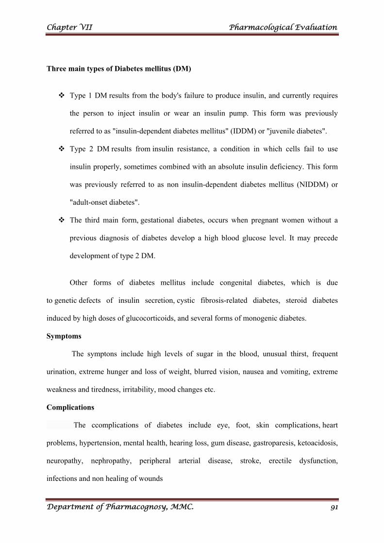
Chapter VII Pharmacological Evaluation
Department of Pharmacognosy, MMC. 91
Three main types of Diabetes mellitus (DM)
Type 1 DM results from the body's failure to produce insulin, and currently requires
the person to inject insulin or wear an insulin pump. This form was previously
referred to as "insulin-dependent diabetes mellitus" (IDDM) or "juvenile diabetes".
Type 2 DM results from insulin resistance, a condition in which cells fail to use
insulin properly, sometimes combined with an absolute insulin deficiency. This form
was previously referred to as non insulin-dependent diabetes mellitus (NIDDM) or
"adult-onset diabetes".
The third main form, gestational diabetes, occurs when pregnant women without a
previous diagnosis of diabetes develop a high blood glucose level. It may precede
development of type 2 DM.
Other forms of diabetes mellitus include congenital diabetes, which is due
to genetic defects of insulin secretion, cystic fibrosis-related diabetes, steroid diabetes
induced by high doses of glucocorticoids, and several forms of monogenic diabetes.
Symptoms
The symptons include high levels of sugar in the blood, unusual thirst, frequent
urination, extreme hunger and loss of weight, blurred vision, nausea and vomiting, extreme
weakness and tiredness, irritability, mood changes etc.
Complications
The ccomplications of diabetes include eye, foot, skin complications, heart
problems, hypertension, mental health, hearing loss, gum disease, gastroparesis, ketoacidosis,
neuropathy, nephropathy, peripheral arterial disease, stroke, erectile dysfunction,
infections and non healing of wounds
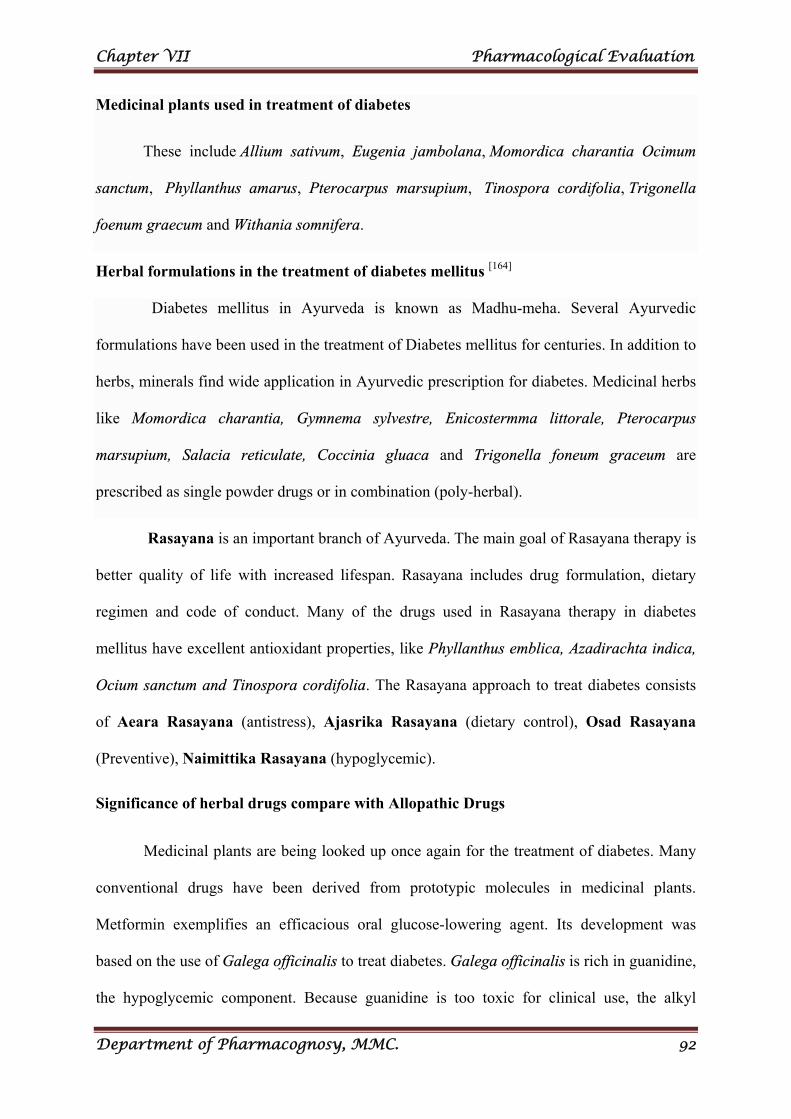
Chapter VII Pharmacological Evaluation
Department of Pharmacognosy, MMC. 92
Medicinal plants used in treatment of diabetes
These include Allium sativum, Eugenia jambolana, Momordica charantia Ocimum
sanctum, Phyllanthus amarus, Pterocarpus marsupium, Tinospora cordifolia, Trigonella
foenum graecum and Withania somnifera.
Herbal formulations in the treatment of diabetes mellitus [164]
Diabetes mellitus in Ayurveda is known as Madhu-meha. Several Ayurvedic
formulations have been used in the treatment of Diabetes mellitus for centuries. In addition to
herbs, minerals find wide application in Ayurvedic prescription for diabetes. Medicinal herbs
like Momordica charantia, Gymnema sylvestre, Enicostermma littorale, Pterocarpus
marsupium, Salacia reticulate, Coccinia gluaca and Trigonella foneum graceum are
prescribed as single powder drugs or in combination (poly-herbal).
Rasayana is an important branch of Ayurveda. The main goal of Rasayana therapy is
better quality of life with increased lifespan. Rasayana includes drug formulation, dietary
regimen and code of conduct. Many of the drugs used in Rasayana therapy in diabetes
mellitus have excellent antioxidant properties, like Phyllanthus emblica, Azadirachta indica,
Ocium sanctum and Tinospora cordifolia. The Rasayana approach to treat diabetes consists
of Aeara Rasayana (antistress), Ajasrika Rasayana (dietary control), Osad Rasayana
(Preventive), Naimittika Rasayana (hypoglycemic).
Significance of herbal drugs compare with Allopathic Drugs
Medicinal plants are being looked up once again for the treatment of diabetes. Many
conventional drugs have been derived from prototypic molecules in medicinal plants.
Metformin exemplifies an efficacious oral glucose-lowering agent. Its development was
based on the use of Galega officinalis to treat diabetes. Galega officinalis is rich in guanidine,
the hypoglycemic component. Because guanidine is too toxic for clinical use, the alkyl
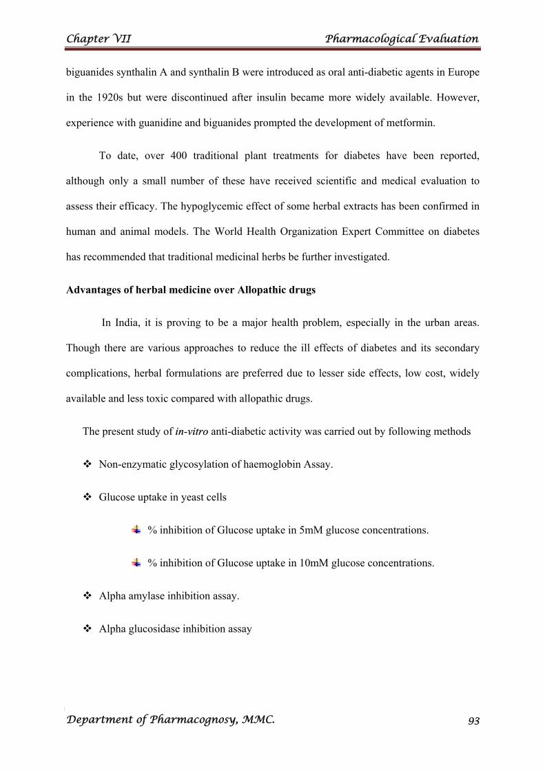
Chapter VII Pharmacological Evaluation
Department of Pharmacognosy, MMC. 93
biguanides synthalin A and synthalin B were introduced as oral anti-diabetic agents in Europe
in the 1920s but were discontinued after insulin became more widely available. However,
experience with guanidine and biguanides prompted the development of metformin.
To date, over 400 traditional plant treatments for diabetes have been reported,
although only a small number of these have received scientific and medical evaluation to
assess their efficacy. The hypoglycemic effect of some herbal extracts has been confirmed in
human and animal models. The World Health Organization Expert Committee on diabetes
has recommended that traditional medicinal herbs be further investigated.
Advantages of herbal medicine over Allopathic drugs
In India, it is proving to be a major health problem, especially in the urban areas.
Though there are various approaches to reduce the ill effects of diabetes and its secondary
complications, herbal formulations are preferred due to lesser side effects, low cost, widely
available and less toxic compared with allopathic drugs.
The present study of in-vitro anti-diabetic activity was carried out by following methods
Non-enzymatic glycosylation of haemoglobin Assay.
Glucose uptake in yeast cells
% inhibition of Glucose uptake in 5mM glucose concentrations.
% inhibition of Glucose uptake in 10mM glucose concentrations.
Alpha amylase inhibition assay.
Alpha glucosidase inhibition assay
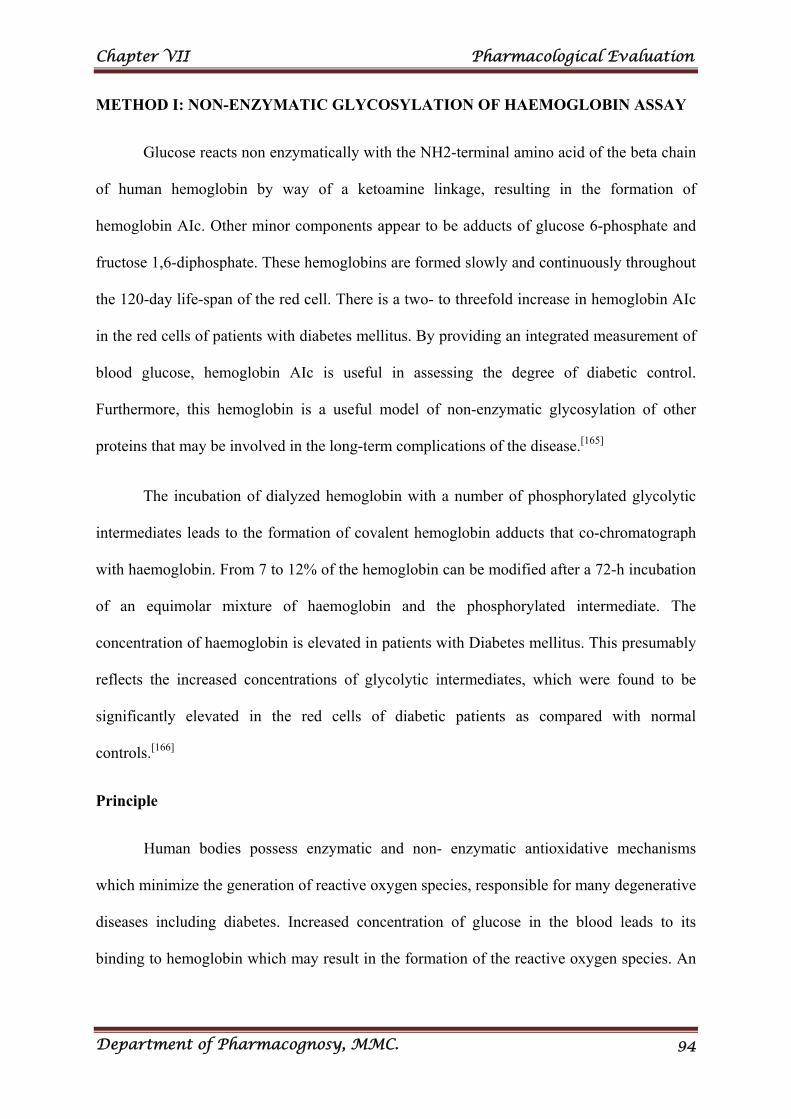
Chapter VII Pharmacological Evaluation
Department of Pharmacognosy, MMC. 94
METHOD I: NON-ENZYMATIC GLYCOSYLATION OF HAEMOGLOBIN ASSAY
Glucose reacts non enzymatically with the NH2-terminal amino acid of the beta chain
of human hemoglobin by way of a ketoamine linkage, resulting in the formation of
hemoglobin AIc. Other minor components appear to be adducts of glucose 6-phosphate and
fructose 1,6-diphosphate. These hemoglobins are formed slowly and continuously throughout
the 120-day life-span of the red cell. There is a two- to threefold increase in hemoglobin AIc
in the red cells of patients with diabetes mellitus. By providing an integrated measurement of
blood glucose, hemoglobin AIc is useful in assessing the degree of diabetic control.
Furthermore, this hemoglobin is a useful model of non-enzymatic glycosylation of other
proteins that may be involved in the long-term complications of the disease.[165]
The incubation of dialyzed hemoglobin with a number of phosphorylated glycolytic
intermediates leads to the formation of covalent hemoglobin adducts that co-chromatograph
with haemoglobin. From 7 to 12% of the hemoglobin can be modified after a 72-h incubation
of an equimolar mixture of haemoglobin and the phosphorylated intermediate. The
concentration of haemoglobin is elevated in patients with Diabetes mellitus. This presumably
reflects the increased concentrations of glycolytic intermediates, which were found to be
significantly elevated in the red cells of diabetic patients as compared with normal
controls.[166]
Principle
Human bodies possess enzymatic and non- enzymatic antioxidative mechanisms
which minimize the generation of reactive oxygen species, responsible for many degenerative
diseases including diabetes. Increased concentration of glucose in the blood leads to its
binding to hemoglobin which may result in the formation of the reactive oxygen species. An
Chapter VII Pharmacological Evaluation
Department of Pharmacognosy, MMC. 95
increase in the glycosylation was observed on incubation of hemoglobin with the increasing
concentration of the glucose over a period of 72h. [167-168]
Reagents
2% glucose solution.
0.06% haemoglobin solution.
0.02% gentamycin solutions.
0.01 M phosphate buffer (pH 7.4)
Procedure
Anti diabetic activity of methanolic extract of the leaves of Citrullus lanatus were
investigated by estimating degree of non-enzymatic haemoglobin glycosylation, measured
spectrophotometrically at 520nm. Glucose (2%), haemoglobin (0.06%) and Gentamycin
(0.02%) solutions were prepared in 0.01M phosphate buffer (pH 7.4). 1mL each of above
solution was mixed with 1mL of various concentrations of the extract. The mixture was
incubated in dark at room temperature for 72h. The degree of glycosylation of haemoglobin
was measured spectrophotometrically at 520nm. α-tocopherol (Trolax) was used as a
standard drug for assay. The percentage inhibition was calculated. All the tests were
performed in triplicate. [169-170]
The % inhibition was calculated using the following formula,
% inhibition = Abs sample – Abs control X 100 Abs sample Where Abs control is the absorbance of the control and Abs sample is the absorbance of the
test sample. The results are tabulated in Table 24 and Fig. 33.
Chapter VII Pharmacological Evaluation
Department of Pharmacognosy, MMC. 96
METHOD II: IN VITRO GLUCOSE UPTAKE IN YEAST CELLS
The rate of glucose transport across cell membrane in yeast cells system is presented.
Regulation of glucose level in the blood of the diabetic patient can prevent the various
complications associated with the disease. The maintenance of plasma glucose concentration
for a long term under a variety of dietary conditions is one of the most important and closely
regulated processes observed in the mammalian species. In Yeast glucose transport takes
place through facilitated diffusion. After the treatment of the yeast cells with extract, the
glucose uptake was found to increase in a dose dependent manner. The methanolic extract of
Citrullus lanatus glucose uptake by yeast cells as compared to standard drug acarbose. [171-172]
Principle
In the glucose uptake in yeast cells method the glucose transport across the yeast cell
membrane has been receiving attention as in vitro screening method for hypoglycaemic effect
of various compounds/ medicinal plants. The transport of non metabolizable sugars and
certain metabolizable glycosides suggest that sugar transport across the yeast cell membrane
is mediated by stereospecific membrane carriers. It is reported that in yeast cells glucose
transport is extremely complex and it is generally agreed that glucose is transported in yeast
is by a facilitated diffusion process. Facilitated carriers are specific carriers that transport
solutes down the concentration gradient. This means that effective transport is only attained if
there is removal of intracellular glucose.
Reagents [171]
Baker’s yeast.
Glucose solution (5 and 10mM)
GOD solution
Chapter VII Pharmacological Evaluation
Department of Pharmacognosy, MMC. 97
Procedure
Yeast cells were prepared by commercial baker’s yeast was washed repeated
centrifugation (3,000 rpm; 5min) in distilled water until the supernatant fluids were clear and
a 10% (v/v) suspension was prepared in distilled water. 1mL of various concentrations of
methanolic extract of Citrullus lanatus were added to 1mL of glucose solution (5 and 10mM)
and incubated together for 10min at 37°C. Reaction was started by adding 100μL of yeast
suspension, vortexed and further incubated at 37°C for 60min. After 60min, the tubes were
centrifuged (2,500 rpm, 5min) and glucose was estimated in the supernatant by glucose
oxidase method. The optical density 520 nm was measured. Acarbose was taken as standard
drug. The percentage increase in glucose uptake by yeast cells was calculated using the
following formula,
Increase in glucose uptake (%) = Abs sample – Abs control X 100 Abs sample Where, Abs control is the absorbance of the control and Abs sample is the absorbance of the
test sample. All the experiments were carried out in triplicates. The results are tabulated in
Table 25,26 and Figs. 34, 35.
METHOD III: IN VITRO ALPHA AMYLASE INHIBITION ASSAY
The α-amylase is the one of the main enzymes in human that catalyses the hydrolysis
of 1,4-glucosidic linkage of complex carbohydrates like starch into simple sugars namely,
maltose. Inhibition of the α-amylase activity is one of the possible six mechanisms that can
be potentially used for controlling diabetes.
Controlling the glucose production from complex carbohydrates is considered to be
effective in controlling diabetes. Although the inhibitory drug like acarbose is which inhibits
α-amylase and α-glucosidase used in controlling glucose level in type 2 diabetic patients, the
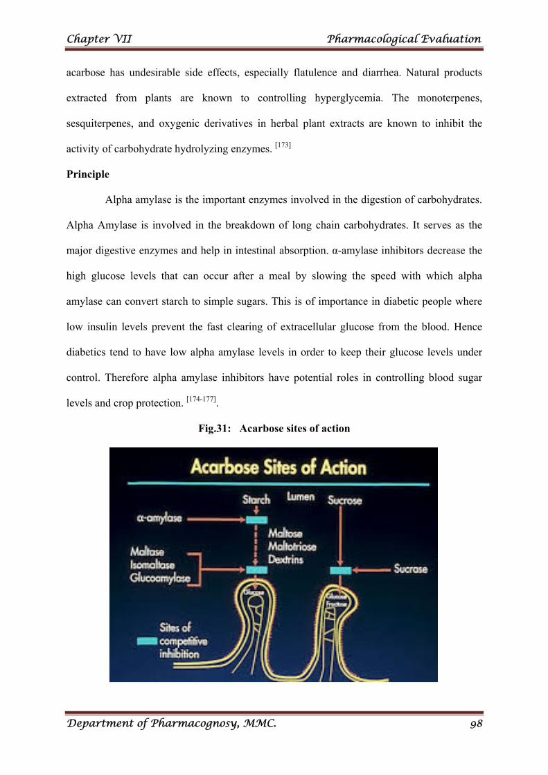
Chapter VII Pharmacological Evaluation
Department of Pharmacognosy, MMC. 98
acarbose has undesirable side effects, especially flatulence and diarrhea. Natural products
extracted from plants are known to controlling hyperglycemia. The monoterpenes,
sesquiterpenes, and oxygenic derivatives in herbal plant extracts are known to inhibit the
activity of carbohydrate hydrolyzing enzymes. [173]
Principle
Alpha amylase is the important enzymes involved in the digestion of carbohydrates.
Alpha Amylase is involved in the breakdown of long chain carbohydrates. It serves as the
major digestive enzymes and help in intestinal absorption. α-amylase inhibitors decrease the
high glucose levels that can occur after a meal by slowing the speed with which alpha
amylase can convert starch to simple sugars. This is of importance in diabetic people where
low insulin levels prevent the fast clearing of extracellular glucose from the blood. Hence
diabetics tend to have low alpha amylase levels in order to keep their glucose levels under
control. Therefore alpha amylase inhibitors have potential roles in controlling blood sugar
levels and crop protection. [174-177].
Fig.31: Acarbose sites of action
Chapter VII Pharmacological Evaluation
Department of Pharmacognosy, MMC. 99
Reagents
0.02M sodium phosphate buffer (pH 6.9 containing 6mM sodium chloride)
α-amylase solution (27.5mg of α-amylase in 100ml 0f water)
1%, w/v soluble starch
1M HCl
Iodine reagent (5mM I2 and 5mM KI)
Procedure
Various concentrations of methanolic extract of Citrullus lanatus 500μL were added
to 500μL of 0.02M sodium phosphate buffer (pH 6.9 containing 6mM sodium chloride)
containing 500μL of α-amylase solution and were incubated at 37°C for 10min. Then 500μL
soluble starch (1%, w/v) was added to each reaction well and incubated at 37°C for 15min.
1M HCl (20μL) was added to stop the enzymatic reaction, followed by the addition of 100μL
of iodine reagent. The colour change was noted and the absorbance was read at 620nm.
Acarbose was used as reference. Inhibition of enzyme activity was calculated as: Inhibition
of enzyme activity (%) = (C-S) / C × 100, where S is the absorbance of the sample and C is
the absorbance of blank (no extract).[190] . The results are tabulated in Table 27 and Fig. 36.
METHOD IV: IN VITRO ALPHA GLUCOSIDASE INHIBITION ASSAY
α-glucosidase catalyzes the final step in the digestive process of carbohydrates. Its
inhibitors can retard the uptake of dietary carbohydrates and suppress postprandial
hyperglycemia and could be useful for treating diabetic and/or obese patients (Toeller, 1994).
α-Glucosidase inhibitors such as acarbose, miglitol, and voglibose are known to reduce
postprandial hyperglycemia primarily by interfering with the carbohydrate digestive enzymes
and by delaying glucose absorption.
Acarbose a carbohydrate inhibitor, when administered showed delayed in glucose
absorption Acarbose, an α-Glucosidase inhibitors, reduce intestinal absorption of
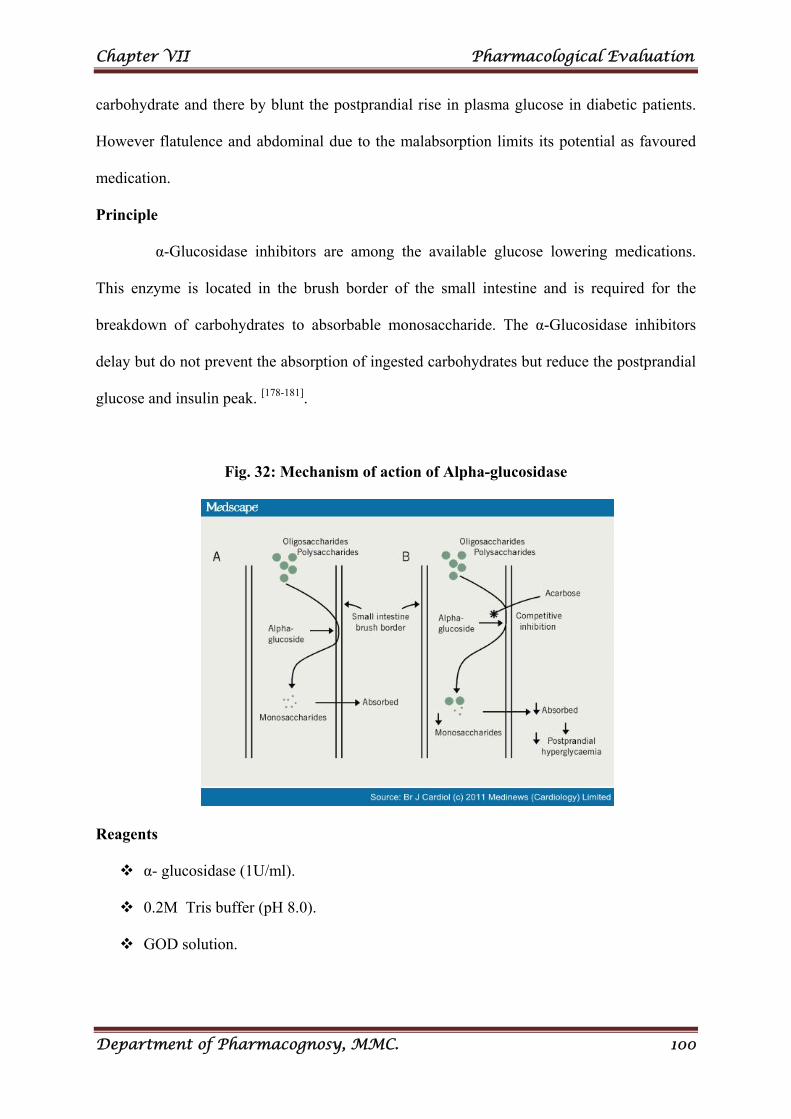
Chapter VII Pharmacological Evaluation
Department of Pharmacognosy, MMC. 100
carbohydrate and there by blunt the postprandial rise in plasma glucose in diabetic patients.
However flatulence and abdominal due to the malabsorption limits its potential as favoured
medication.
Principle
α-Glucosidase inhibitors are among the available glucose lowering medications.
This enzyme is located in the brush border of the small intestine and is required for the
breakdown of carbohydrates to absorbable monosaccharide. The α-Glucosidase inhibitors
delay but do not prevent the absorption of ingested carbohydrates but reduce the postprandial
glucose and insulin peak. [178-181].
Fig. 32: Mechanism of action of Alpha-glucosidase
Reagents
α- glucosidase (1U/ml).
0.2M Tris buffer (pH 8.0).
GOD solution.
Chapter VII Pharmacological Evaluation
Department of Pharmacognosy, MMC. 101
Procedure
The enzyme α- glucosidase inhibitory activity is determined by incubating solution
(0.1 ml) of an enzyme preparation with 0.2 M Tris buffer, pH 8.0 (1.0ml) containing 1mL of
various concentrations of extract at 37 ºC for 60 minutes. The reaction mixture is heated for
two minutes in boiling water bath to stop the reaction. The amount of liberated glucose is
measured by glucose oxidation method. Acarbose was used as reference. [182-183]. The results
are tabulated in Table 28 and Fig. 37.
(Enzyme activity of control – Enzyme activity of extract) % inhibition = ---------------------------------------------------------------------------- × 100
Enzyme activity of control
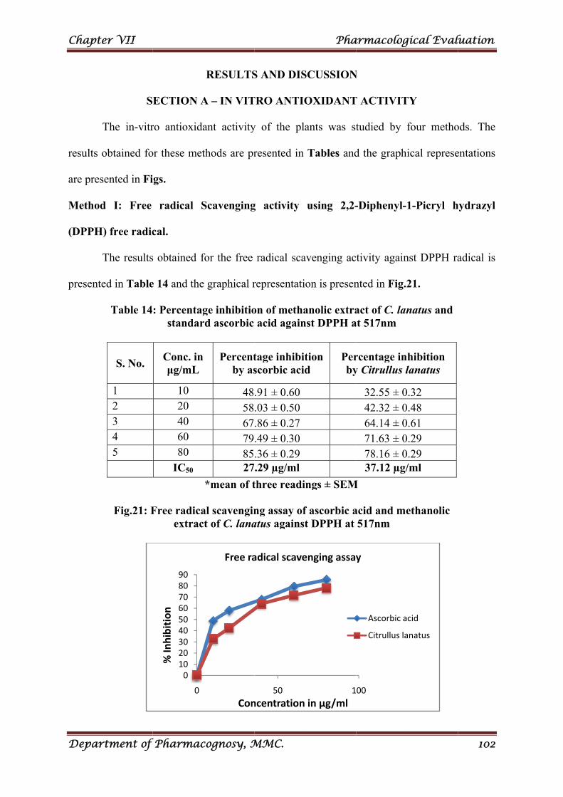
Chapt
Depar
results
are pres
Method
(DPPH
presente
er VII
rtment of
S
The in-vitr
obtained fo
sented in Fi
d I: Free
H) free radic
The results
ed in Table
Table 14
S. No.
1 2 3 4 5
Fig.21: F
f Pharmac
ECTION A
ro antioxida
or these met
igs.
radical S
cal.
s obtained f
e 14 and the
: Percentagstandar
Conc. in μg/mL
10 20 40 60 80
IC50
Free radicaextrac
0102030405060708090
0
% In
hibition
cognosy, M
RESULTS
A – IN VIT
ant activity
thods are pr
Scavenging
for the free
e graphical r
ge inhibitiord ascorbic
Percentby as
485867798527
*mean of t
al scavenginct of C. lana
Con
Free ra
MMC.
S AND DIS
TRO ANTIO
y of the pla
resented in
g activity
radical sca
representati
on of methaacid again
tage inhibitscorbic acid
.91 ± 0.60
.03 ± 0.50
.86 ± 0.27
.49 ± 0.30
.36 ± 0.29
.29 µg/ml
three readi
ng assay ofatus against
50
ncentration
adical scave
Pha
SCUSSION
OXIDANT
ants was st
Tables and
using 2,2-
avenging ac
on is presen
anolic extranst DPPH a
tion d
Perby
ngs ± SEM
f ascorbic at DPPH at
1
in µg/ml
enging assay
armacolog
N
T ACTIVIT
tudied by f
d the graphi
-Diphenyl-1
tivity again
nted in Fig.2
act of C. lanat 517nm
rcentage iny Citrullus l
32.55 ± 042.32 ± 064.14 ± 071.63 ± 078.16 ± 037.12 µg/
M
acid and me517nm
00
y
Ascorbic a
Citrullus la
gical Eval
TY
four metho
ical represe
1-Picryl h
nst DPPH ra
21.
natus and
hibition lanatus
0.32 0.48 0.61 0.29 0.29 /ml
ethanolic
cid
anatus
luation
102
ods. The
entations
hydrazyl
adical is
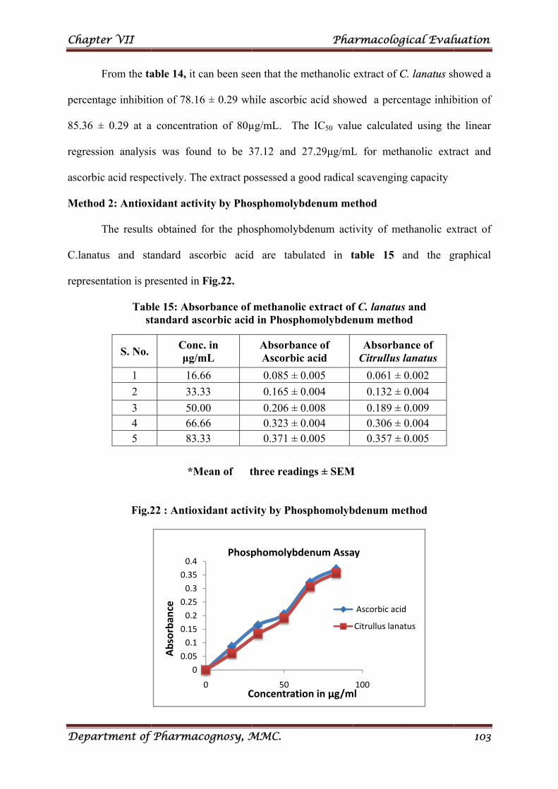
Chapt
Depar
percent
85.36 ±
regressi
ascorbic
Method
C.lanatu
represen
er VII
rtment of
From the ta
age inhibiti
± 0.29 at a
ion analysi
c acid respe
d 2: Antiox
The results
us and sta
ntation is pr
Tabst
Fig.2
S. No.
1
2
3 4 5
f Pharmac
able 14, it c
ion of 78.16
a concentrat
is was foun
ectively. Th
xidant activ
s obtained f
andard asc
resented in
le 15: Absotandard as
*M
22 : Antiox
0
0.05
0.1
0.15
0.2
0.25
0.3
0.35
0.4
Absorban
ce
Concμg/m
16.6
33.3
50.066.683.3
cognosy, M
can been see
6 ± 0.29 wh
tion of 80µ
nd to be 3
he extract po
vity by Pho
for the pho
corbic acid
Fig.22.
orbance of corbic acid
Mean of t
xidant activ
0C
Phosp
c. in mL
66
33
00 66 33
MMC.
en that the m
hile ascorbic
µg/mL. Th
37.12 and 2
ossessed a g
sphomolyb
osphomolyb
d are tabu
methanolicd in Phosph
three readi
vity by Pho
50Concentratio
phomolybd
AbsorbanAscorbic
0.085 ± 0
0.165 ± 0
0.206 ± 00.323 ± 00.371 ± 0
Pha
methanolic
c acid show
he IC50 valu
27.29μg/mL
good radical
bdenum me
bdenum acti
ulated in t
c extract ofhomolybden
ngs ± SEM
sphomolyb
on in µg/m
denum Assa
nce of c acid
0.005
0.004
0.008 0.004 0.005
armacolog
extract of C
wed a perce
ue calculate
L for meth
l scavenging
ethod
ivity of me
table 15 a
f C. lanatusnum metho
M
bdenum me
100ml
ay
Ascorbic acid
Citrullus lana
AbsorbaCitrullus l
0.061 ±
0.132 ±
0.189 ± 0.306 ± 0.357 ±
gical Eval
C. lanatus sh
entage inhib
ed using th
hanolic extr
g capacity
ethanolic ex
and the g
s and od
ethod
d
tus
ance of lanatus
0.002
0.004
0.009 0.004 0.005
luation
103
howed a
bition of
he linear
ract and
xtract of
graphical
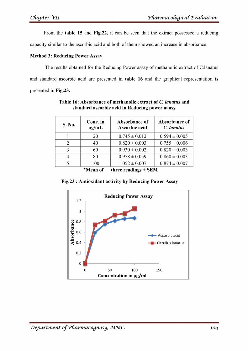
Chapt
Depar
F
capacity
Method
and sta
presente
er VII
rtment of
From the ta
y similar to
d 3: Reduci
The results
andard asco
ed in Fig.23
Tab
S.
12345
Fig
f Pharmac
able 15 and
the ascorbi
ing Power
obtained fo
orbic acid a
3.
le 16: Absostandard
No. Cμ
1 2 3 4 5
*M
g.23 : Antio
0
0.2
0.4
0.6
0.8
1
1.2
0
Ab
sorb
ance
cognosy, M
d Fig.22, it
ic acid and b
Assay
for the Redu
are presente
orbance of d ascorbic
Conc. in μg/mL
20 40 60 80
100 Mean of t
oxidant acti
50
Concen
Red
MMC.
t can be se
both of them
ucing Power
ed in table
methanolicacid in Red
AbsorbaAscorbi
0.745 ± 0.820 ± 0.930 ± 0.958 ± 1.052 ±
three readi
ivity by Re
10
ntration in µ
ducing Pow
Pha
een that the
m showed a
r assay of m
e 16 and th
c extract ofducing pow
ance of ic acid
0.012 0.003 0.002 0.059 0.007 ngs ± SEM
educing Pow
00 1
µg/ml
wer Assay
armacolog
e extract po
an increase i
methanolic e
he graphica
f C. lanatuswer aaasy
AbsorbanC. lanat
0.594 ± 0.0.755 ± 0.0.820 ± 0.0.860 ± 0.0.874 ± 0.
M
wer Assay
150
Ascorbic acid
Citrullus lanat
gical Eval
ossessed a r
in absorban
extract of C
al represent
s and
nce of tus
.005
.006
.003
.003
.007
d
tus
luation
104
reducing
nce.
C.lanatus
tation is
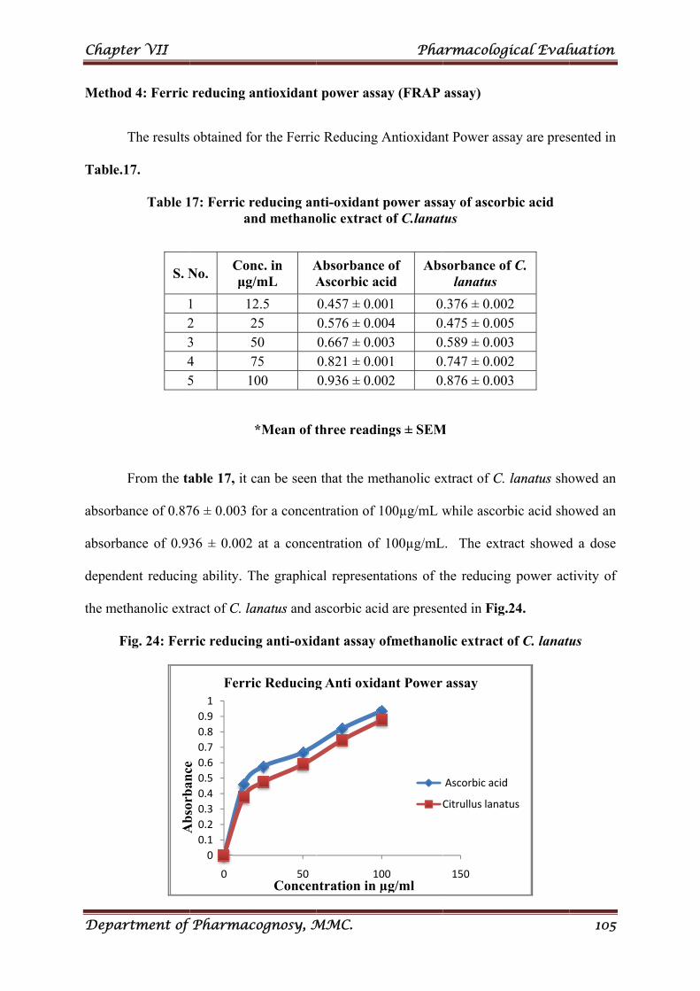
Chapt
Depar
Method
Table.1
absorba
absorba
depende
the met
F
er VII
rtment of
d 4: Ferric
The results
17.
Table 1
From the ta
ance of 0.87
ance of 0.93
ent reducin
thanolic extr
Fig. 24: Ferr
Ab
sorb
ance
S. N
12345
f Pharmac
reducing a
obtained fo
17: Ferric ran
able 17, it c
76 ± 0.003 f
36 ± 0.002
ng ability. T
ract of C. la
ric reducin
0
0.1
0.2
0.3
0.4
0.5
0.6
0.7
0.8
0.9
1
0
Ab
sorb
ance
Ferri
No. Conμg/
1 122 23 54 75 1
cognosy, M
antioxidant
or the Ferric
reducing annd methano
*Mean of t
can be seen
for a concen
at a conce
The graphic
anatus and a
ng anti-oxid
50Concen
ic Reducing
nc. in /mL
AA
2.5 25 50 75 00
MMC.
t power ass
c Reducing
nti-oxidant olic extract
three readi
n that the me
ntration of
entration of
al represen
ascorbic aci
dant assay
10ntration in
g Anti oxid
AbsorbanceAscorbic a
0.457 ± 0.00.576 ± 0.00.667 ± 0.00.821 ± 0.00.936 ± 0.0
Pha
say (FRAP
Antioxidan
power assat of C.lanat
ings ± SEM
ethanolic ex
100µg/mL w
f 100µg/mL
ntations of t
id are presen
ofmethano
00µg/ml
dant Power
e of cid
Abs
001 0004 0003 0001 0002 0
armacolog
assay)
nt Power ass
ay of ascortus
M
xtract of C.
while ascor
L. The extr
the reducing
nted in Fig.
lic extract
150
assay
Ascorbic acid
Citrullus lanat
sorbance olanatus
.376 ± 0.00
.475 ± 0.00
.589 ± 0.00
.747 ± 0.00
.876 ± 0.00
gical Eval
say are pres
rbic acid
lanatus sho
rbic acid sho
ract showed
g power act
.24.
of C. lanatu
d
tus
f C.
02 05 03 02 03
luation
105
sented in
owed an
owed an
d a dose
tivity of
us
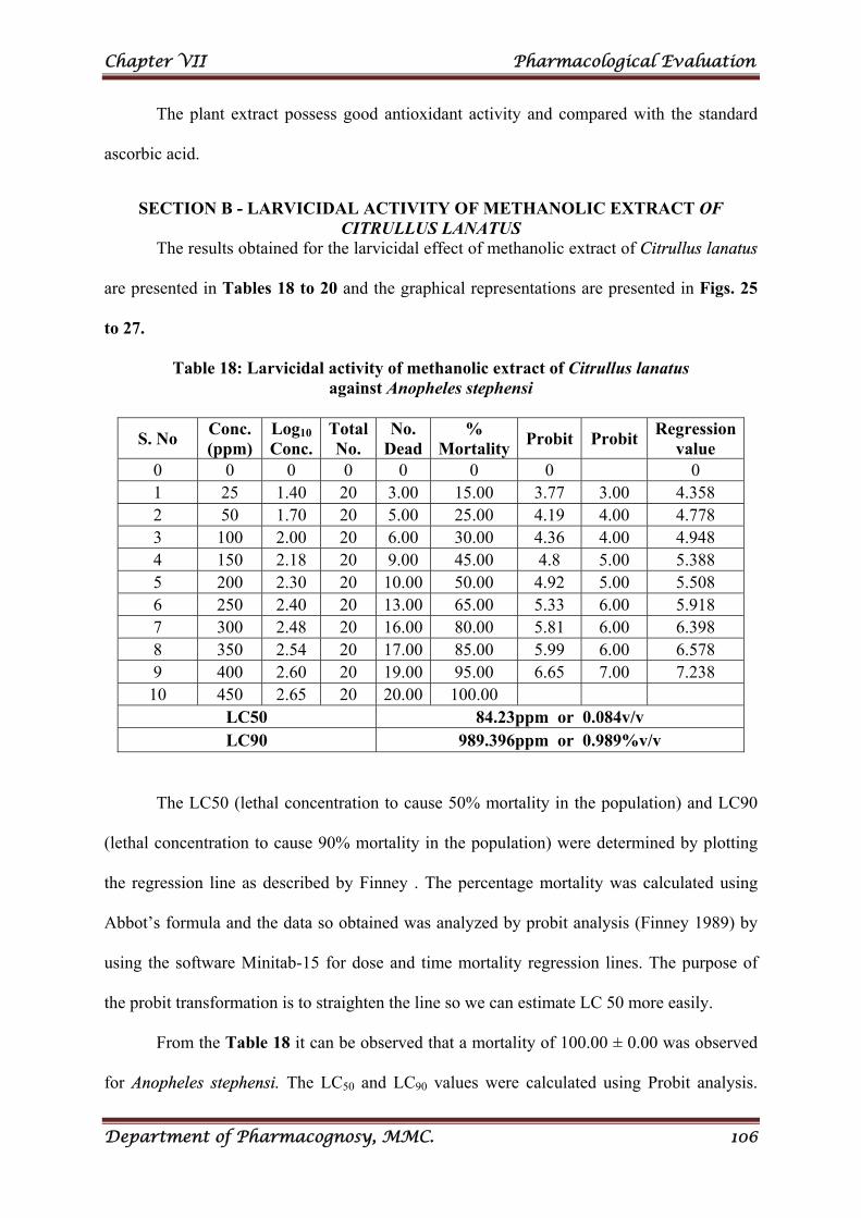
Chapter VII Pharmacological Evaluation
Department of Pharmacognosy, MMC. 106
The plant extract possess good antioxidant activity and compared with the standard
ascorbic acid.
SECTION B - LARVICIDAL ACTIVITY OF METHANOLIC EXTRACT OF CITRULLUS LANATUS
The results obtained for the larvicidal effect of methanolic extract of Citrullus lanatus
are presented in Tables 18 to 20 and the graphical representations are presented in Figs. 25
to 27.
Table 18: Larvicidal activity of methanolic extract of Citrullus lanatus against Anopheles stephensi
S. No Conc. (ppm)
Log10 Conc.
Total No.
No. Dead
% Mortality
Probit Probit Regression
value 0 0 0 0 0 0 0 0 1 25 1.40 20 3.00 15.00 3.77 3.00 4.358 2 50 1.70 20 5.00 25.00 4.19 4.00 4.778 3 100 2.00 20 6.00 30.00 4.36 4.00 4.948 4 150 2.18 20 9.00 45.00 4.8 5.00 5.388 5 200 2.30 20 10.00 50.00 4.92 5.00 5.508 6 250 2.40 20 13.00 65.00 5.33 6.00 5.918 7 300 2.48 20 16.00 80.00 5.81 6.00 6.398 8 350 2.54 20 17.00 85.00 5.99 6.00 6.578 9 400 2.60 20 19.00 95.00 6.65 7.00 7.238 10 450 2.65 20 20.00 100.00
LC50 84.23ppm or 0.084v/v LC90 989.396ppm or 0.989%v/v
The LC50 (lethal concentration to cause 50% mortality in the population) and LC90
(lethal concentration to cause 90% mortality in the population) were determined by plotting
the regression line as described by Finney . The percentage mortality was calculated using
Abbot’s formula and the data so obtained was analyzed by probit analysis (Finney 1989) by
using the software Minitab-15 for dose and time mortality regression lines. The purpose of
the probit transformation is to straighten the line so we can estimate LC 50 more easily.
From the Table 18 it can be observed that a mortality of 100.00 ± 0.00 was observed
for Anopheles stephensi. The LC50 and LC90 values were calculated using Probit analysis.
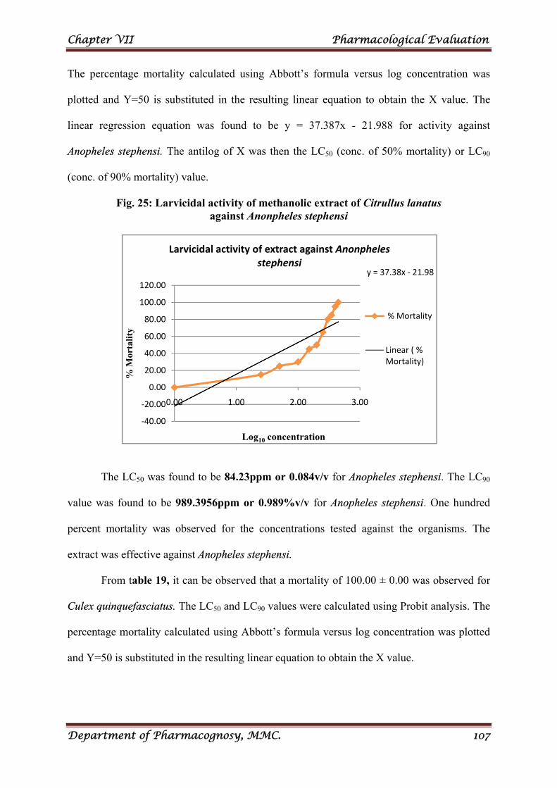
Chapter VII Pharmacological Evaluation
Department of Pharmacognosy, MMC. 107
The percentage mortality calculated using Abbott’s formula versus log concentration was
plotted and Y=50 is substituted in the resulting linear equation to obtain the X value. The
linear regression equation was found to be y = 37.387x - 21.988 for activity against
Anopheles stephensi. The antilog of X was then the LC50 (conc. of 50% mortality) or LC90
(conc. of 90% mortality) value.
Fig. 25: Larvicidal activity of methanolic extract of Citrullus lanatus against Anonpheles stephensi
The LC50 was found to be 84.23ppm or 0.084v/v for Anopheles stephensi. The LC90
value was found to be 989.3956ppm or 0.989%v/v for Anopheles stephensi. One hundred
percent mortality was observed for the concentrations tested against the organisms. The
extract was effective against Anopheles stephensi.
From table 19, it can be observed that a mortality of 100.00 ± 0.00 was observed for
Culex quinquefasciatus. The LC50 and LC90 values were calculated using Probit analysis. The
percentage mortality calculated using Abbott’s formula versus log concentration was plotted
and Y=50 is substituted in the resulting linear equation to obtain the X value.
y = 37.38x ‐ 21.98
‐40.00
‐20.00
0.00
20.00
40.00
60.00
80.00
100.00
120.00
0.00 1.00 2.00 3.00
% M
orta
lity
Log10 concentration
Larvicidal activity of extract against Anonpheles stephensi
% Mortality
Linear ( % Mortality)
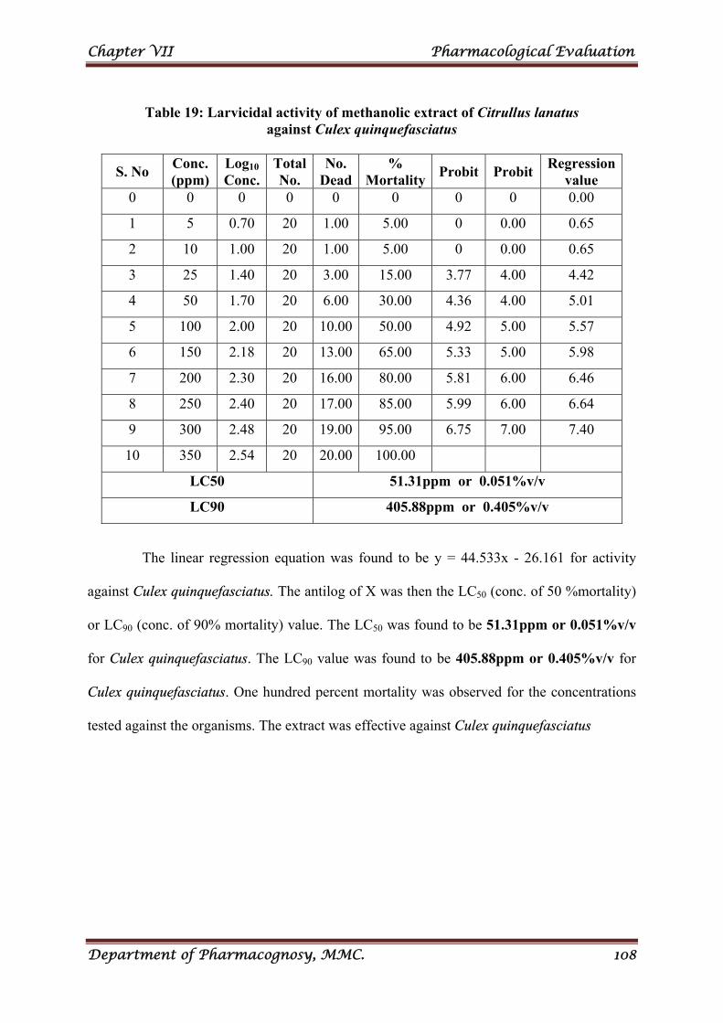
Chapter VII Pharmacological Evaluation
Department of Pharmacognosy, MMC. 108
Table 19: Larvicidal activity of methanolic extract of Citrullus lanatus
against Culex quinquefasciatus
S. No Conc. (ppm)
Log10 Conc.
Total No.
No. Dead
% Mortality
Probit Probit Regression
value 0 0 0 0 0 0 0 0 0.00
1 5 0.70 20 1.00 5.00 0 0.00 0.65
2 10 1.00 20 1.00 5.00 0 0.00 0.65
3 25 1.40 20 3.00 15.00 3.77 4.00 4.42
4 50 1.70 20 6.00 30.00 4.36 4.00 5.01
5 100 2.00 20 10.00 50.00 4.92 5.00 5.57
6 150 2.18 20 13.00 65.00 5.33 5.00 5.98
7 200 2.30 20 16.00 80.00 5.81 6.00 6.46
8 250 2.40 20 17.00 85.00 5.99 6.00 6.64
9 300 2.48 20 19.00 95.00 6.75 7.00 7.40
10 350 2.54 20 20.00 100.00
LC50 51.31ppm or 0.051%v/v
LC90 405.88ppm or 0.405%v/v
The linear regression equation was found to be y = 44.533x - 26.161 for activity
against Culex quinquefasciatus. The antilog of X was then the LC50 (conc. of 50 %mortality)
or LC90 (conc. of 90% mortality) value. The LC50 was found to be 51.31ppm or 0.051%v/v
for Culex quinquefasciatus. The LC90 value was found to be 405.88ppm or 0.405%v/v for
Culex quinquefasciatus. One hundred percent mortality was observed for the concentrations
tested against the organisms. The extract was effective against Culex quinquefasciatus
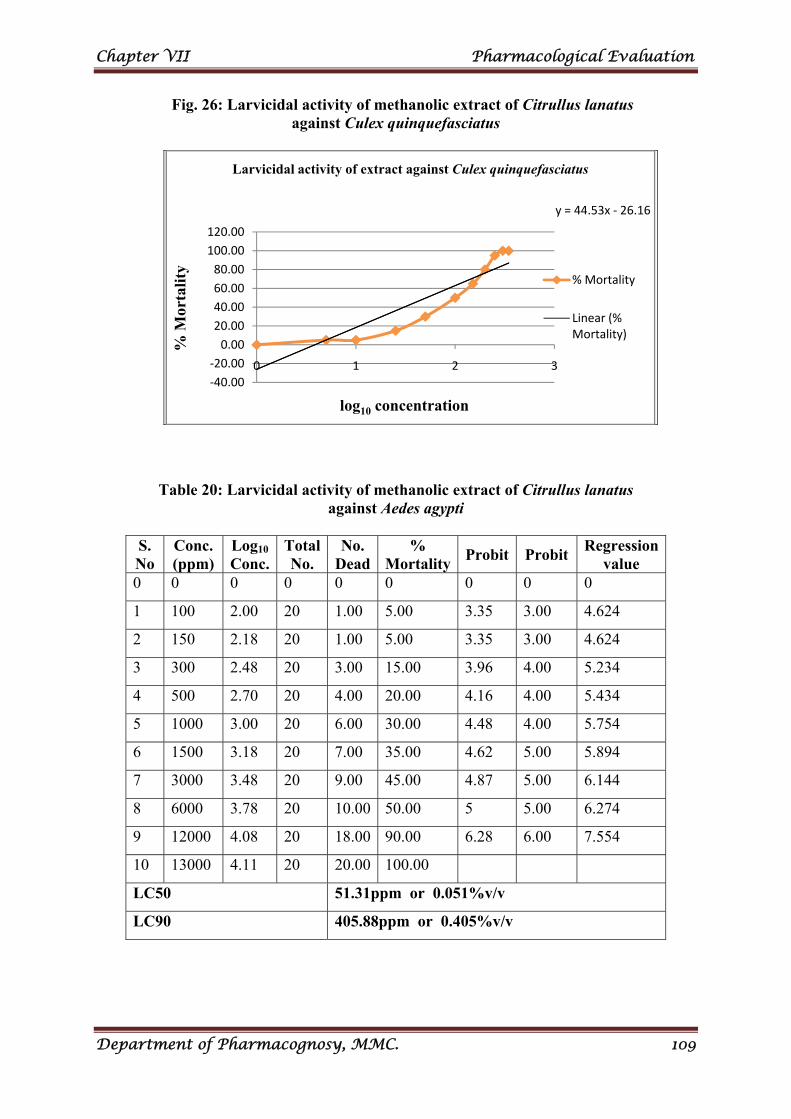
Chapter VII Pharmacological Evaluation
Department of Pharmacognosy, MMC. 109
Fig. 26: Larvicidal activity of methanolic extract of Citrullus lanatus against Culex quinquefasciatus
Table 20: Larvicidal activity of methanolic extract of Citrullus lanatus against Aedes agypti
S. No
Conc. (ppm)
Log10 Conc.
Total No.
No. Dead
% Mortality
Probit Probit Regression
value 0 0 0 0 0 0 0 0 0
1 100 2.00 20 1.00 5.00 3.35 3.00 4.624
2 150 2.18 20 1.00 5.00 3.35 3.00 4.624
3 300 2.48 20 3.00 15.00 3.96 4.00 5.234
4 500 2.70 20 4.00 20.00 4.16 4.00 5.434
5 1000 3.00 20 6.00 30.00 4.48 4.00 5.754
6 1500 3.18 20 7.00 35.00 4.62 5.00 5.894
7 3000 3.48 20 9.00 45.00 4.87 5.00 6.144
8 6000 3.78 20 10.00 50.00 5 5.00 6.274
9 12000 4.08 20 18.00 90.00 6.28 6.00 7.554
10 13000 4.11 20 20.00 100.00
LC50 51.31ppm or 0.051%v/v
LC90 405.88ppm or 0.405%v/v
y = 44.53x ‐ 26.16
‐40.00
‐20.00
0.00
20.00
40.00
60.00
80.00
100.00
120.00
0 1 2 3
% M
orta
lity
log10 concentration
Larvicidal activity of extract against Culex quinquefasciatus
% Mortality
Linear (% Mortality)
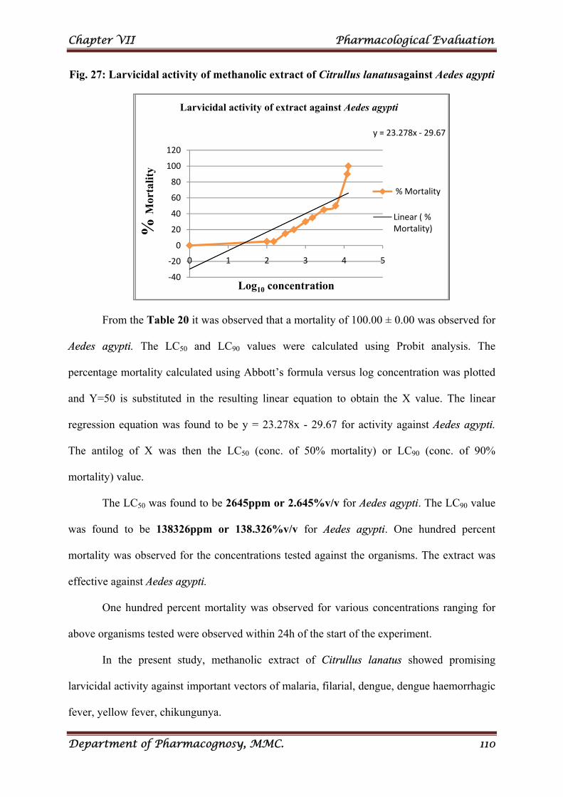
Chapter VII Pharmacological Evaluation
Department of Pharmacognosy, MMC. 110
Fig. 27: Larvicidal activity of methanolic extract of Citrullus lanatusagainst Aedes agypti
From the Table 20 it was observed that a mortality of 100.00 ± 0.00 was observed for
Aedes agypti. The LC50 and LC90 values were calculated using Probit analysis. The
percentage mortality calculated using Abbott’s formula versus log concentration was plotted
and Y=50 is substituted in the resulting linear equation to obtain the X value. The linear
regression equation was found to be y = 23.278x - 29.67 for activity against Aedes agypti.
The antilog of X was then the LC50 (conc. of 50% mortality) or LC90 (conc. of 90%
mortality) value.
The LC50 was found to be 2645ppm or 2.645%v/v for Aedes agypti. The LC90 value
was found to be 138326ppm or 138.326%v/v for Aedes agypti. One hundred percent
mortality was observed for the concentrations tested against the organisms. The extract was
effective against Aedes agypti.
One hundred percent mortality was observed for various concentrations ranging for
above organisms tested were observed within 24h of the start of the experiment.
In the present study, methanolic extract of Citrullus lanatus showed promising
larvicidal activity against important vectors of malaria, filarial, dengue, dengue haemorrhagic
fever, yellow fever, chikungunya.
y = 23.278x ‐ 29.67
‐40
‐20
0
20
40
60
80
100
120
0 1 2 3 4 5
% M
orta
lity
Log10 concentration
Larvicidal activity of extract against Aedes agypti
% Mortality
Linear ( % Mortality)
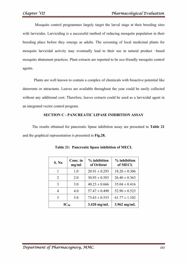
Chapter VII Pharmacological Evaluation
Department of Pharmacognosy, MMC. 111
Mosquito control programmes largely target the larval stage at their breeding sites
with larvicides. Larviciding is a successful method of reducing mosquito population in their
breeding place before they emerge as adults. The screening of local medicinal plants for
mosquito larvicidal activity may eventually lead to their use in natural product –based
mosquito abatement practices. Plant extracts are reported to be eco-friendly mosquito control
agents.
Plants are well known to contain a complex of chemicals with bioactive potential like
deterrents or attractants. Leaves are available throughout the year could be easily collected
without any additional cost. Therefore, leaves extracts could be used as a larvicidal agent in
an integrated vector control program.
SECTION C - PANCREATIC LIPASE INHIBITION ASSAY
The results obtained for pancreatic lipase inhibition assay are presented in Table 21
and the graphical representation is presented in Fig.28.
Table 21: Pancreatic lipase inhibition of MECL
S. No Conc. in mg/ml
% inhibition of Orlistat
% inhibition of MECL
1 1.0 20.91 ± 0.293 18.20 ± 0.306
2 2.0 30.93 ± 0.583 26.40 ± 0.363
3 3.0 40.23 ± 0.666 35.04 ± 0.416
4 4.0 57.47 ± 0.490 52.90 ± 0.523
5 5.0 73.63 ± 0.553 61.77 ± 1.102
IC50 3.420 mg/mL 3.962 mg/mL
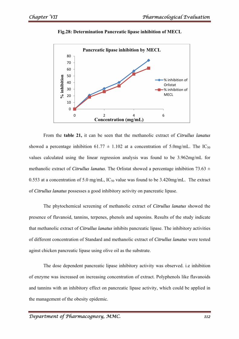
Chapter VII Pharmacological Evaluation
Department of Pharmacognosy, MMC. 112
Fig.28: Determination Pancreatic lipase inhibition of MECL
From the table 21, it can be seen that the methanolic extract of Citrullus lanatus
showed a percentage inhibition 61.77 ± 1.102 at a concentration of 5.0mg/mL. The IC50
values calculated using the linear regression analysis was found to be 3.962mg/mL for
methanolic extract of Citrullus lanatus. The Orlistat showed a percentage inhibition 73.63 ±
0.553 at a concentration of 5.0 mg/mL, IC50 value was found to be 3.420mg/mL. The extract
of Citrullus lanatus possesses a good inhibitory activity on pancreatic lipase.
The phytochemical screening of methanolic extract of Citrullus lanatus showed the
presence of flavanoid, tannins, terpenes, phenols and saponins. Results of the study indicate
that methanolic extract of Citrullus lanatus inhibits pancreatic lipase. The inhibitory activities
of different concentration of Standard and methanolic extract of Citrullus lanatus were tested
aginst chicken pancreatic lipase using olive oil as the substrate.
The dose dependent pancreatic lipase inhibitory activity was observed. i.e inhibition
of enzyme was increased on increasing concentration of extract. Polyphenols like flavanoids
and tannins with an inhibitory effect on pancreatic lipase activity, which could be applied in
the management of the obesity epidemic.
0
10
20
30
40
50
60
70
80
0 2 4 6
% in
hib
itio
n
Concentration (mg/mL)
Pancreatic lipase inhibition by MECL
% inhibition of Orlistat% inhibition of MECL
Chapter VII Pharmacological Evaluation
Department of Pharmacognosy, MMC. 113
Overweight, obesity and weight change: Overweight (BMI ≥25 and < 30kg/m2) or
obese (BMI ≥ 30kg/m2 ) individual have a higher risk for many types of cancer compared
with individuals whose BMI is considered within the normal range (18.5 to < 25 kg/m2 ). The
cancers most consistently associated with overweight and obesity are breast, colon/rectum,
endometrium, pancreas, adenocarcinoma of the esophagus, kidney, gallbladder, and liver.
Obesity may also increase risk of mortality from some cancers, such as prostate. A
growing body of evidence suggests that weight gain is associated with an increased risk of
some cancers, breast cancer in particular. Increases in body weight during adulthood largely
reflect increases in adipose tissue rather than lean mass. The total body fat may be a better
measure of the risk for cancer than BMI.
Studies over decades have consistently shown a strong association between obesity
and both insulin resistance and type 2 diabetes incidence, with risk of diabetes and earlier age
at onset directly linked to obesity severity. For type 2 diabetes as well as certain cancers,
some studies suggest that waist circumference, waist –to-hip ratio, or direct measures of
visceral adiposity with risk independently of BMI.
The case for a causal relationship between obesity and disease is strengthened by
evidence that weight loss lowers disease risk. In the case of diabetes, numerous studies have
shown that weight loss decreases diabetes incidence and restores euglycemia in a significant
fraction of individuals with type 2 diabetes. In the randomised, prospective, multicenter
Diabetes prevention program trial, an intensive lifestyle intervention of diet and physical
activity was associated with a 58% reduction in diabetes incidence in high risk individuals,
and weight loss accounted for most of the effect.
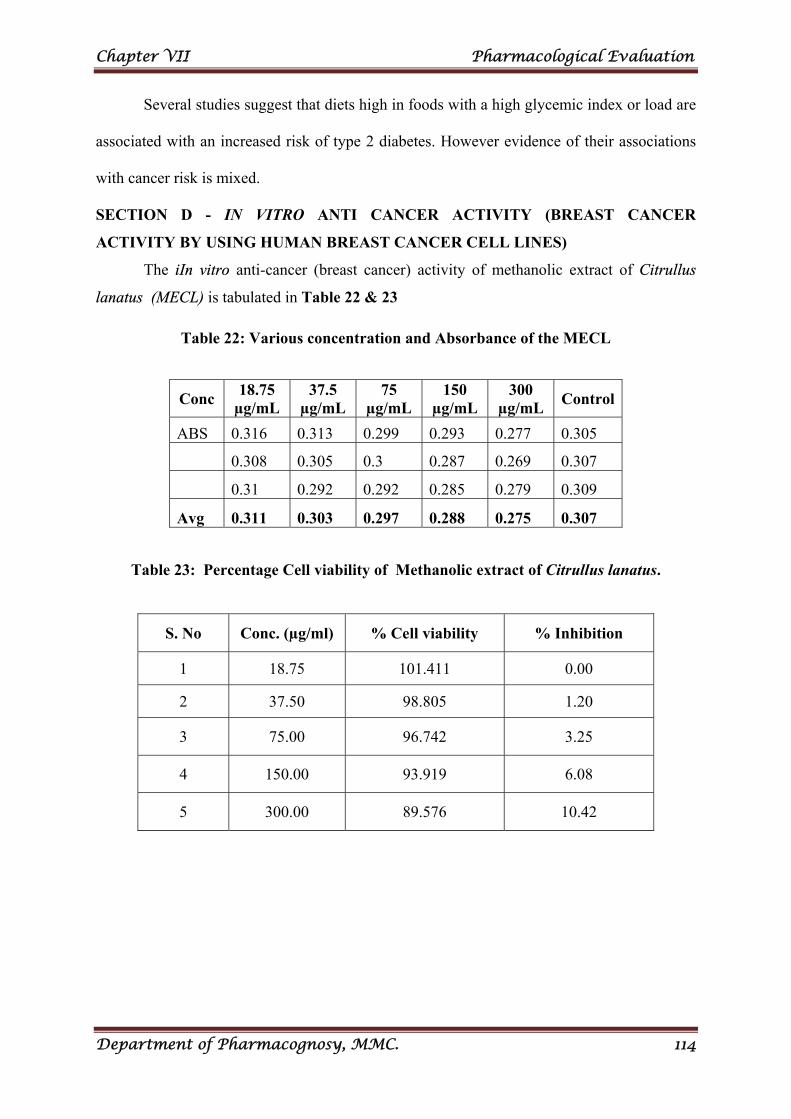
Chapter VII Pharmacological Evaluation
Department of Pharmacognosy, MMC. 114
Several studies suggest that diets high in foods with a high glycemic index or load are
associated with an increased risk of type 2 diabetes. However evidence of their associations
with cancer risk is mixed.
SECTION D - IN VITRO ANTI CANCER ACTIVITY (BREAST CANCER
ACTIVITY BY USING HUMAN BREAST CANCER CELL LINES)
The iIn vitro anti-cancer (breast cancer) activity of methanolic extract of Citrullus
lanatus (MECL) is tabulated in Table 22 & 23
Table 22: Various concentration and Absorbance of the MECL
Table 23: Percentage Cell viability of Methanolic extract of Citrullus lanatus.
Conc 18.75
µg/mL 37.5
µg/mL 75
µg/mL 150
µg/mL 300
µg/mL Control
ABS 0.316 0.313 0.299 0.293 0.277 0.305
0.308 0.305 0.3 0.287 0.269 0.307
0.31 0.292 0.292 0.285 0.279 0.309
Avg 0.311 0.303 0.297 0.288 0.275 0.307
S. No Conc. (µg/ml) % Cell viability % Inhibition
1 18.75 101.411 0.00
2 37.50 98.805 1.20
3 75.00 96.742 3.25
4 150.00 93.919 6.08
5 300.00 89.576 10.42
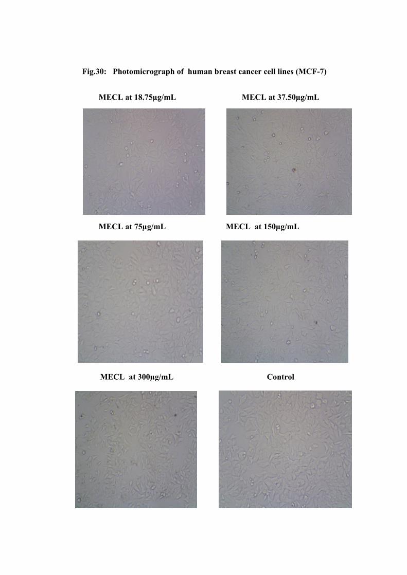
Fig.30: Photomicrograph of human breast cancer cell lines (MCF-7)
MECL at 18.75µg/mL MECL at 37.50µg/mL
MECL at 75µg/mL MECL at 150µg/mL
MECL at 300µg/mL Control
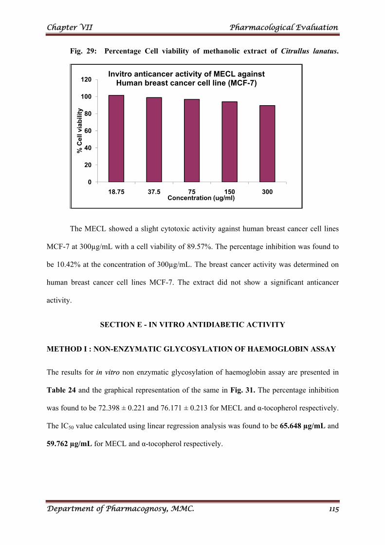
Chapter VII Pharmacological Evaluation
Department of Pharmacognosy, MMC. 115
Fig. 29: Percentage Cell viability of methanolic extract of Citrullus lanatus.
The MECL showed a slight cytotoxic activity against human breast cancer cell lines
MCF-7 at 300µg/mL with a cell viability of 89.57%. The percentage inhibition was found to
be 10.42% at the concentration of 300µg/mL. The breast cancer activity was determined on
human breast cancer cell lines MCF-7. The extract did not show a significant anticancer
activity.
SECTION E - IN VITRO ANTIDIABETIC ACTIVITY
METHOD I : NON-ENZYMATIC GLYCOSYLATION OF HAEMOGLOBIN ASSAY
The results for in vitro non enzymatic glycosylation of haemoglobin assay are presented in
Table 24 and the graphical representation of the same in Fig. 31. The percentage inhibition
was found to be 72.398 ± 0.221 and 76.171 ± 0.213 for MECL and α-tocopherol respectively.
The IC50 value calculated using linear regression analysis was found to be 65.648 µg/mL and
59.762 µg/mL for MECL and α-tocopherol respectively.
0
20
40
60
80
100
120
18.75 37.5 75 150 300
% C
ell v
iab
ility
Concentration (ug/ml)
Invitro anticancer activity of MECL against Human breast cancer cell line (MCF-7)
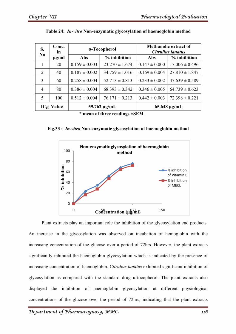
Chapter VII Pharmacological Evaluation
Department of Pharmacognosy, MMC. 116
Table 24: In-vitro Non-enzymatic glycosylation of haemoglobin method
S. No
Conc. in
µg/ml
α-Tocopherol Methanolic extract of
Citrullus lanatus Abs % inhibition Abs % inhibition
1 20 0.159 ± 0.003 23.270 ± 1.674 0.147 ± 0.000 17.006 ± 0.496
2 40 0.187 ± 0.002 34.759 ± 1.016 0.169 ± 0.004 27.810 ± 1.847
3 60 0.258 ± 0.004 52.713 ± 0.813 0.233 ± 0.002 47.639 ± 0.589
4 80 0.386 ± 0.004 68.393 ± 0.342 0.346 ± 0.005 64.739 ± 0.623
5 100 0.512 ± 0.004 76.171 ± 0.213 0.442 ± 0.003 72.398 ± 0.221
IC50 Value 59.762 µg/mL 65.648 µg/mL
* mean of three readings ±SEM
Fig.33 : In-vitro Non-enzymatic glycosylation of haemoglobin method
Plant extracts play an important role the inhibition of the glycosylation end products.
An increase in the glycosylation was observed on incubation of hemoglobin with the
increasing concentration of the glucose over a period of 72hrs. However, the plant extracts
significantly inhibited the haemoglobin glycosylation which is indicated by the presence of
increasing concentration of haemoglobin. Citrullus lanatus exhibited significant inhibition of
glycosylation as compared with the standard drug α-tocopherol. The plant extracts also
displayed the inhibition of haemoglobin glycosylation at different physiological
concentrations of the glucose over the period of 72hrs, indicating that the plant extracts
0
20
40
60
80
100
0 50 100 150
% in
hib
itio
n
Concentration (µg/ml)
Non‐enzymatic glycosylation of haemoglobin method
% inhibition of Vitamin E
% inhibition 0f MECL
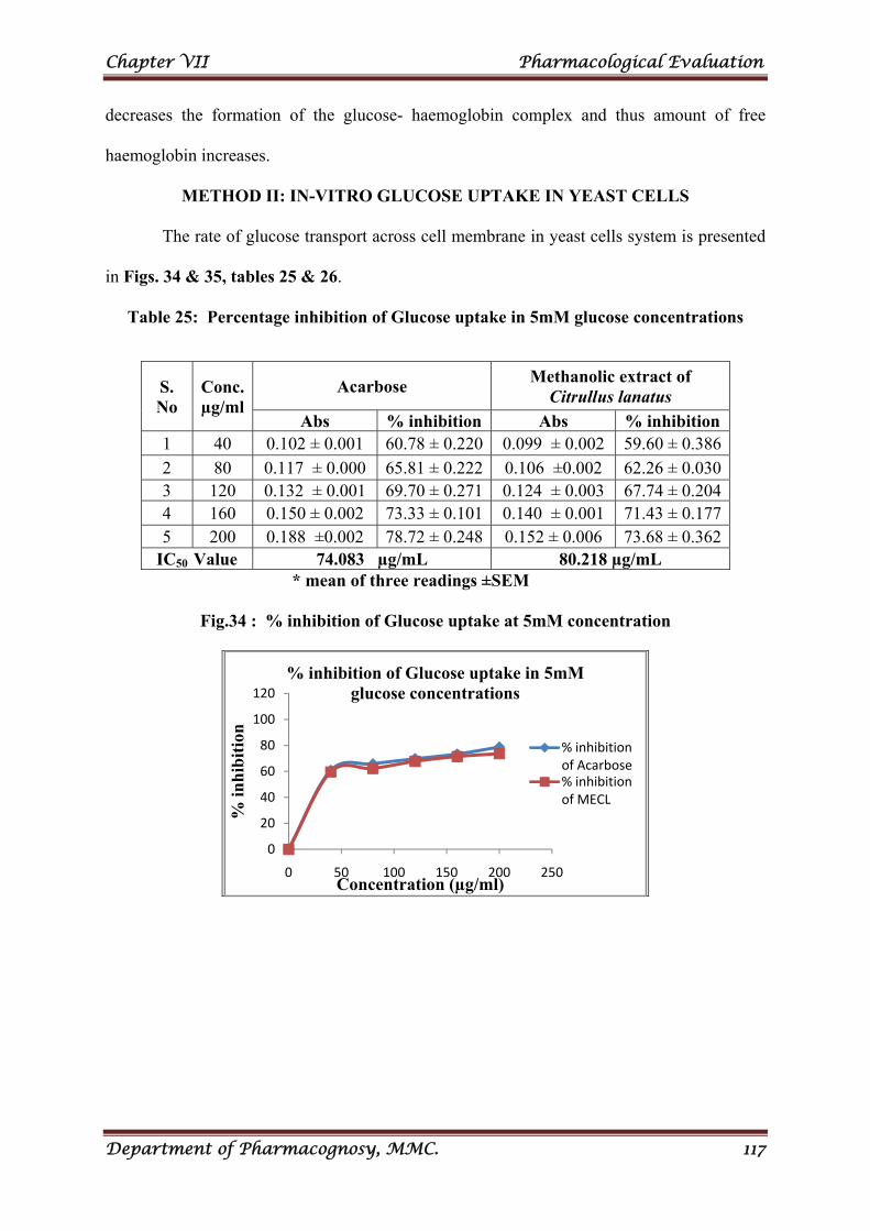
Chapter VII Pharmacological Evaluation
Department of Pharmacognosy, MMC. 117
decreases the formation of the glucose- haemoglobin complex and thus amount of free
haemoglobin increases.
METHOD II: IN-VITRO GLUCOSE UPTAKE IN YEAST CELLS
The rate of glucose transport across cell membrane in yeast cells system is presented
in Figs. 34 & 35, tables 25 & 26.
Table 25: Percentage inhibition of Glucose uptake in 5mM glucose concentrations
S. No
Conc. µg/ml
Acarbose Methanolic extract of
Citrullus lanatus
Abs % inhibition Abs % inhibition 1 40 0.102 ± 0.001 60.78 ± 0.220 0.099 ± 0.002 59.60 ± 0.386
2 80 0.117 ± 0.000 65.81 ± 0.222 0.106 ±0.002 62.26 ± 0.0303 120 0.132 ± 0.001 69.70 ± 0.271 0.124 ± 0.003 67.74 ± 0.2044 160 0.150 ± 0.002 73.33 ± 0.101 0.140 ± 0.001 71.43 ± 0.1775 200 0.188 ±0.002 78.72 ± 0.248 0.152 ± 0.006 73.68 ± 0.362
IC50 Value 74.083 µg/mL 80.218 µg/mL
* mean of three readings ±SEM
Fig.34 : % inhibition of Glucose uptake at 5mM concentration
0
20
40
60
80
100
120
0 50 100 150 200 250
% in
hib
itio
n
Concentration (µg/ml)
% inhibition of Glucose uptake in 5mM glucose concentrations
% inhibition of Acarbose% inhibition of MECL
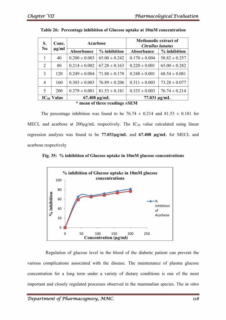
Chapter VII Pharmacological Evaluation
Department of Pharmacognosy, MMC. 118
Table 26: Percentage inhibition of Glucose uptake at 10mM concentration
S. No
Conc. µg/ml
Acarbose Methanolic extract of
Citrullus lanatus Absorbance % inhibition Absorbance % inhibition
1 40 0.200 ± 0.003 65.00 ± 0.242 0.170 ± 0.004 58.82 ± 0.257
2 80 0.214 ± 0.002 67.28 ± 0.163 0.220 ± 0.001 65.00 ± 0.282
3 120 0.249 ± 0.004 71.88 ± 0.178 0.248 ± 0.001 68.54 ± 0.081
4 160 0.303 ± 0.003 76.89 ± 0.206 0.311 ± 0.003 73.28 ± 0.077
5 200 0.379 ± 0.001 81.53 ± 0.181 0.335 ± 0.003 76.74 ± 0.214
IC50 Value 67.408 µg/mL 77.031 µg/mL
* mean of three readings ±SEM
The percentage inhibition was found to be 76.74 ± 0.214 and 81.53 ± 0.181 for
MECL and acarbose at 200µg/mL respectively. The IC50 value calculated using linear
regression analysis was found to be 77.031µg/mL and 67.408 µg/mL for MECL and
acarbose respectively
Fig. 35: % inhibition of Glucose uptake in 10mM glucose concentrations
Regulation of glucose level in the blood of the diabetic patient can prevent the
various complications associated with the disease. The maintenance of plasma glucose
concentration for a long term under a variety of dietary conditions is one of the most
important and closely regulated processes observed in the mammalian species. The in vitro
0
20
40
60
80
100
0 50 100 150 200 250
% in
hib
itio
n
Concentration (µg/ml)
% inhibition of Glucose uptake in 10mM glucose concentrations
% inhibition of Acarbose
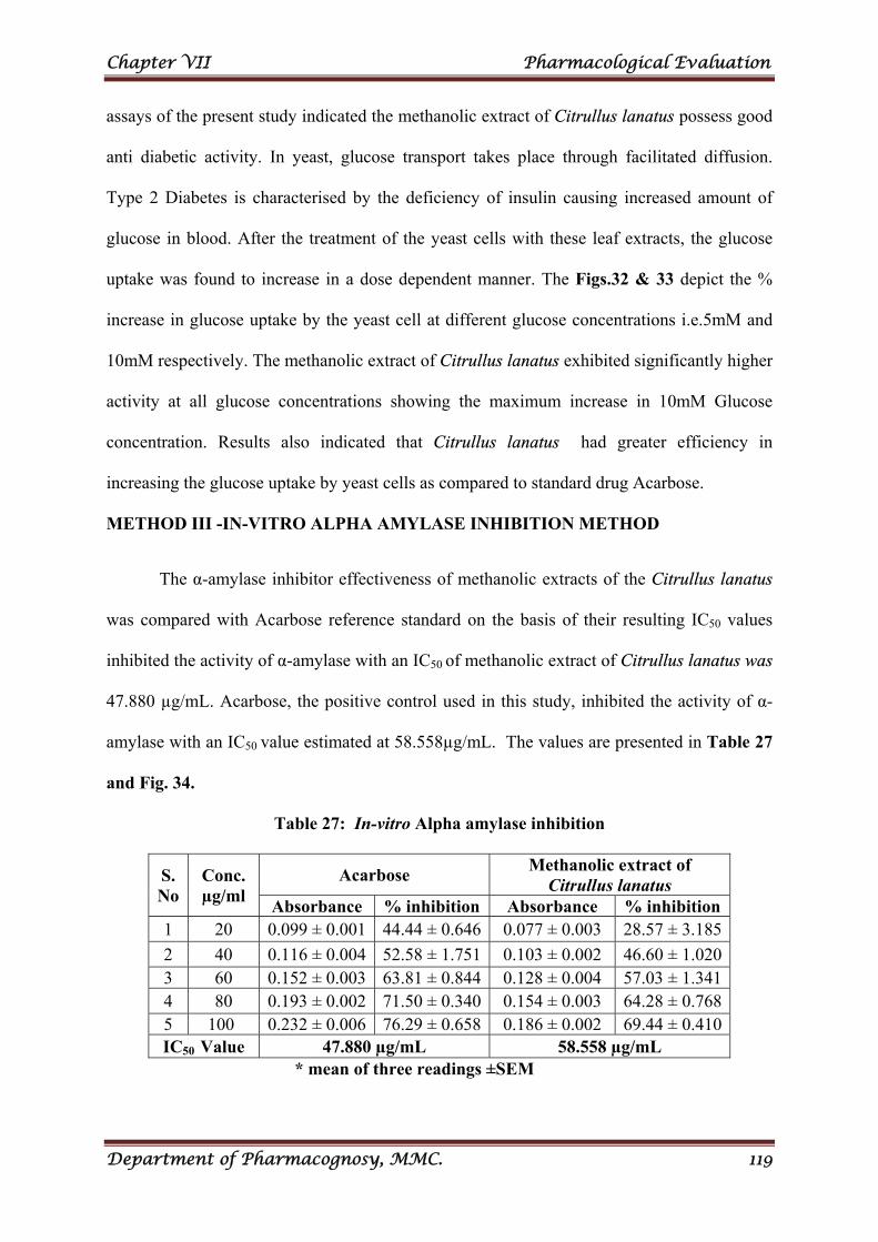
Chapter VII Pharmacological Evaluation
Department of Pharmacognosy, MMC. 119
assays of the present study indicated the methanolic extract of Citrullus lanatus possess good
anti diabetic activity. In yeast, glucose transport takes place through facilitated diffusion.
Type 2 Diabetes is characterised by the deficiency of insulin causing increased amount of
glucose in blood. After the treatment of the yeast cells with these leaf extracts, the glucose
uptake was found to increase in a dose dependent manner. The Figs.32 & 33 depict the %
increase in glucose uptake by the yeast cell at different glucose concentrations i.e.5mM and
10mM respectively. The methanolic extract of Citrullus lanatus exhibited significantly higher
activity at all glucose concentrations showing the maximum increase in 10mM Glucose
concentration. Results also indicated that Citrullus lanatus had greater efficiency in
increasing the glucose uptake by yeast cells as compared to standard drug Acarbose.
METHOD III -IN-VITRO ALPHA AMYLASE INHIBITION METHOD
The α-amylase inhibitor effectiveness of methanolic extracts of the Citrullus lanatus
was compared with Acarbose reference standard on the basis of their resulting IC50 values
inhibited the activity of α-amylase with an IC50 of methanolic extract of Citrullus lanatus was
47.880 µg/mL. Acarbose, the positive control used in this study, inhibited the activity of α-
amylase with an IC50 value estimated at 58.558µg/mL. The values are presented in Table 27
and Fig. 34.
Table 27: In-vitro Alpha amylase inhibition
S. No
Conc. µg/ml
Acarbose Methanolic extract of
Citrullus lanatus Absorbance % inhibition Absorbance % inhibition
1 20 0.099 ± 0.001 44.44 ± 0.646 0.077 ± 0.003 28.57 ± 3.185
2 40 0.116 ± 0.004 52.58 ± 1.751 0.103 ± 0.002 46.60 ± 1.020 3 60 0.152 ± 0.003 63.81 ± 0.844 0.128 ± 0.004 57.03 ± 1.341 4 80 0.193 ± 0.002 71.50 ± 0.340 0.154 ± 0.003 64.28 ± 0.768 5 100 0.232 ± 0.006 76.29 ± 0.658 0.186 ± 0.002 69.44 ± 0.410 IC50
Value 47.880 µg/mL 58.558 µg/mL * mean of three readings ±SEM
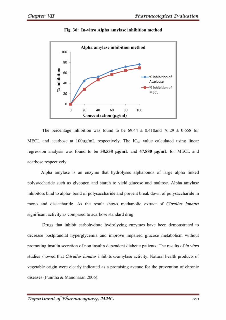
Chapter VII Pharmacological Evaluation
Department of Pharmacognosy, MMC. 120
Fig. 36: In-vitro Alpha amylase inhibition method
The percentage inhibition was found to be 69.44 ± 0.410and 76.29 ± 0.658 for
MECL and acarbose at 100µg/mL respectively. The IC50 value calculated using linear
regression analysis was found to be 58.558 µg/mL and 47.880 µg/mL for MECL and
acarbose respectively
Alpha amylase is an enzyme that hydrolyses alphabonds of large alpha linked
polysaccharide such as glycogen and starch to yield glucose and maltose. Alpha amylase
inhibitors bind to alpha- bond of polysaccharide and prevent break down of polysaccharide in
mono and disaccharide. As the result shows methanolic extract of Citrullus lanatus
significant activity as compared to acarbose standard drug.
Drugs that inhibit carbohydrate hydrolyzing enzymes have been demonstrated to
decrease postprandial hyperglycemia and improve impaired glucose metabolism without
promoting insulin secretion of non insulin dependent diabetic patients. The results of in vitro
studies showed that Citrullus lanatus inhibits α-amylase activity. Natural health products of
vegetable origin were clearly indicated as a promising avenue for the prevention of chronic
diseases (Punitha & Manoharan 2006).
0
20
40
60
80
100
0 20 40 60 80 100
% in
hib
itio
n
Concentration (µg/ml)
Alpha amylase inhibition method
% inhibition of Acarbose
% inhibition of MECL
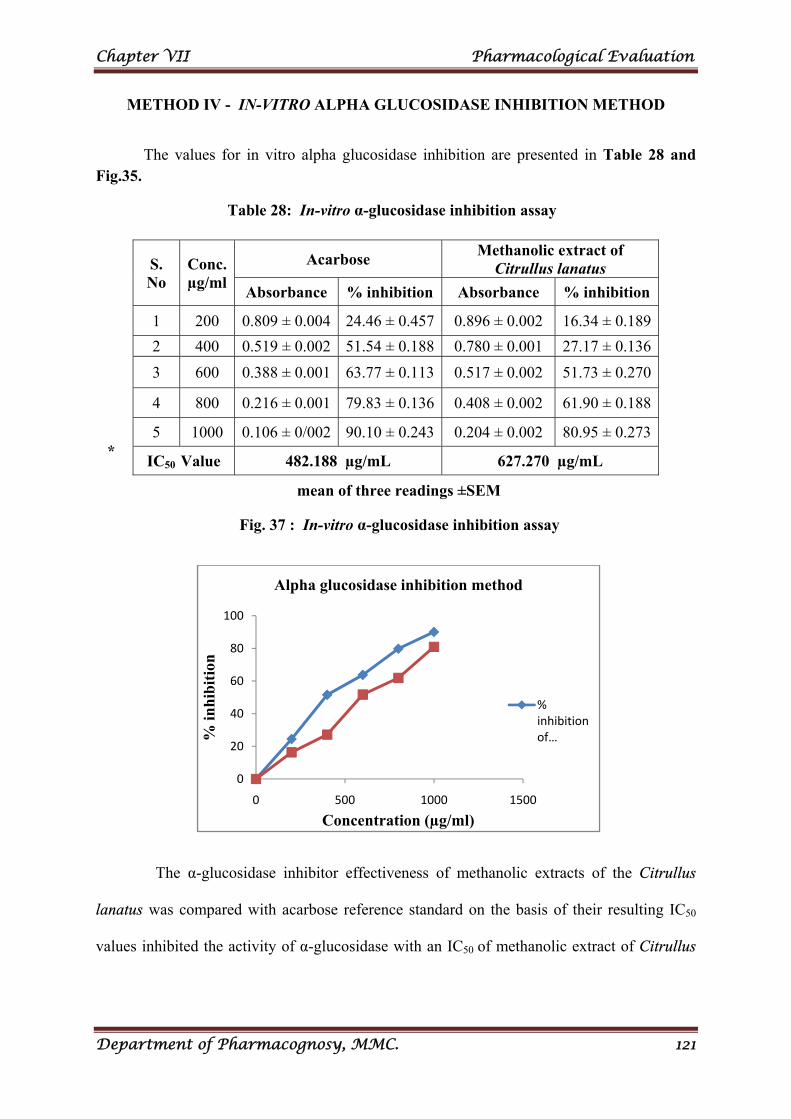
Chapter VII Pharmacological Evaluation
Department of Pharmacognosy, MMC. 121
METHOD IV - IN-VITRO ALPHA GLUCOSIDASE INHIBITION METHOD
The values for in vitro alpha glucosidase inhibition are presented in Table 28 and Fig.35.
Table 28: In-vitro α-glucosidase inhibition assay
*
mean of three readings ±SEM
Fig. 37 : In-vitro α-glucosidase inhibition assay
The α-glucosidase inhibitor effectiveness of methanolic extracts of the Citrullus
lanatus was compared with acarbose reference standard on the basis of their resulting IC50
values inhibited the activity of α-glucosidase with an IC50 of methanolic extract of Citrullus
0
20
40
60
80
100
0 500 1000 1500
% in
hib
itio
n
Concentration (µg/ml)
Alpha glucosidase inhibition method
% inhibition of …
S. No
Conc. µg/ml
Acarbose Methanolic extract of
Citrullus lanatus
Absorbance % inhibition Absorbance % inhibition
1 200 0.809 ± 0.004 24.46 ± 0.457 0.896 ± 0.002 16.34 ± 0.189
2 400 0.519 ± 0.002 51.54 ± 0.188 0.780 ± 0.001 27.17 ± 0.136
3 600 0.388 ± 0.001 63.77 ± 0.113 0.517 ± 0.002 51.73 ± 0.270
4 800 0.216 ± 0.001 79.83 ± 0.136 0.408 ± 0.002 61.90 ± 0.188
5 1000 0.106 ± 0/002 90.10 ± 0.243 0.204 ± 0.002 80.95 ± 0.273
IC50 Value 482.188 µg/mL 627.270 µg/mL
Chapter VII Pharmacological Evaluation
Department of Pharmacognosy, MMC. 122
lanatus was 627.270µg/mL. Acarbose, the positive control used in this study, inhibited the
activity of α-glucosidase with an IC50 value estimated at 482.188µg/mL.
In Diabetes mellitus, control of postprandial plasma glucose level is critical in the
early treatment. An inhibition of enzymes involved in the metabolism of carbohydrate is one
of the therapeutic approaches for reducing postprandial hyperglycemia.
α-glucosidase is a key enzyme in carbohydrate digestion. It catalyzes the hydrolysis of
1,4-α-glucosidic bonds within carbohydrates with release of α-glucose and promotes the
increase of blood glucose level after meal. α-glucosidase inhibitors antagonize the activity of
α-glucosidase, thereby delaying intestinal carbohydrate absorption and slowing the sharp rise
in blood sugar levels that diabetic patients typically experience after meals. For this reason,
α-glucosidase inhibitors, such as acarbose and voglibose, are clinically used as oral
antihyperglycemic agents. However, they often cause severe gastrointestinal side effects.
Therefore, search for new α-glucosidase inhibitors from natural resources has become an
attractive approach for the treatment of postprandial hyperglycemia.
In the present study, methanolic extract of Citrullus lanatus was screened for their
alpha-glucosidase inhibitory potential. The extract showed better alpha-glucosidase inhibition
property. Alpha-glucosidase inhibitors have a potential for the treatment of diabetes because
they reduce diet-induced hyperglycemia.
The extract possessed a significant in vitro antidiabetic acitivity and hence in vivo
studies are further required for providing a scientific information on the plant.

SUMMARY
AND CONCLUSION
Chapter VIII Summary and Conclusion
Department of Pharmacognosy, MMC. 123
CHAPTER VIII
SUMMARY AND CONCLUSION
The present study entitled the “Pharmacognostic, Phytochemical and
Pharmacological evaluation of the Leaves of Citrullus lanatus (Thunb.) Matsum. &
Nakai. (Cucurbitaceae)” focuses on a plant which is commonly available throughout India
and traditionally used in treatment of various ailments.
Studies on the leaves of Citrullus lanatus are still lacking. Hence to exploit its
potential use prompted the present study to investigate the leaves of this plant with clear
scientific protocol.
The chapter on Literature Review deals with the information regarding the
pharmacognostical, phytochemical and pharmacological evaluation of the Citrullus lanatus
plant and other species of Citrullus.
The chapter on Pharmacognostical studies highlights on
Macroscopical features were studied and the adherence of general characters to the
family Citrullus lanatus was found.
Microscopical study reveals the presence of actinocytic stomata, multi cellular
uniseriate unbranched epidermal trichomes. Vascular system of the midribis
multistranded, a large abaxial median bundle, two adaxial bundle. All the bundles are
bicollateral having phloem strand both outer and inner side of the xylem. The
epidermal cells are small elliptical or rectangular and thin walled. Spongy
parenchyma cells small and spherical. Palisade zone consist of single layer of
cylindrical cells, loosely arranged.
Chapter VIII Summary and Conclusion
Department of Pharmacognosy, MMC. 124
Quantitative microscopical studies namely stomatal number, stomatal index, vein islet
number, vein termination number, ash value, extractive value, loss on drying value
etc.,
Also studied cell powder microscopy, fluorescence analysis of powder and the results
helps in achieving a trouble-free identification and authenticity of the plant leaf or in
powder form in future.
The chapter on Phytochemical Evaluation deals with
Preliminary phytochemical screening reveals the presence of carbohydrate, alkaloids,
flavanoids, protein & aminoacids, glycosides etc.,
Quantitative determination of secondary metabolites ( phenol, flavanoid, tannin
content) has been carried out.
TLC & HPTLC studies showed the presence of flavanoid, phenolic compound and
tannin. In HPTLC studies the extract was compared with standards and the presence
of quercetin, gallic aci and catechin were formed.
The vitamin B1, B2 and vitamin C were estimated.
The chapter on Pharmacological studies focuses
The Antioxidant activity by various methods and the extract possessed a good
antioxidant property due to the presence of Vitamin-C, poly phenolic, flavonoid
content.
The larvicidal effect of extract of the Citrullus lanatus was carried out by standard
procedure. In the present study, methanolic extract of citrullus lanatus showed
promising larvicidal activity aginst important vectors of malaria, filariasis, dengue,
dengue haemorrhagic fever, yellow fever.
Chapter VIII Summary and Conclusion
Department of Pharmacognosy, MMC. 125
Obesity can lead to variety serious diseases, including hypertension, hyperlipidemia,
atherosclerosis, and type II diabetes. The pancreatic lipase inhibitors of digestive
lipases reduce dietary fat absorption and hence act as anti-obesity agents. The
Citrullus lanatus extract have a capacity to inhibit the pancreatic lipase.
The extract showed anti cancer (breast cancer) activity aginst human breast cancer
cell lines (MCF-7) which was evaluated by MTT assay. The risk of breast cancer
increases gradually as a woman gets older. The high concentration of the MECL used
for inhibiting human breast cancer cell lines (MCF-7). Type 2 diabetes and breast
cancer share many risk factors. The extract Citrullus lanatus showed high antidiabetic
activity with high concentration showed breast cancer activity.
The antidiabetic activity of extract of the leaves of Citrullus lanatus was carried out
by Non-enzymatic glycosylation of haemoglobin Assay method, Glucose uptake in
yeast cells method ( % inhibition of Glucose uptake in 5mM glucose concentrations &
% inhibition of Glucose uptake in 10mM glucose concentrations ) Alpha amylase
inhibition assay method & Alpha glucosidase inhibition assay method. The extract
showed better alpha-glucosidase inhibition property.
The extract exhibit significant inhibition of glycosylation as compared with the
standard drug alpha tocopherol. Decreases the formation of the glucose- haemoglobin
complex and thus amount of free haemoglobin increases.
The extract showed greater efficiency in increasing the glucose uptake by yeast cells
as compared to standard drug Acarbose. Type 2 Diabetes is characterised by the deficiency of
insulin causing increased amount of glucose in blood. After the treatment of the yeast cells
with these leaf extracts, the glucose uptake was found to increase in a dose dependent
manner.
Chapter VIII Summary and Conclusion
Department of Pharmacognosy, MMC. 126
Alpha amylase inhibitors bind to alpha- bond of polysaccharide and prevent break
down of polysaccharide in mono and disaccharide. The result showed extract of Citrullus
lanatus significant activity as compared to acarbose standard drug.
Alpha-glucosidase inhibitors have a potential for the treatment of diabetes because
they reduce diet-induced hyperglycemia. The extract showed better alpha-glucosidase
inhibition property.
The in vitro assays of the present study indicated the methanolic extract of Citrullus
lanatus possess good anti diabetic activity.
Citrallus lanatus (water melon) is popular in indigenous system of folk medicine. The
leaf extract of Citrullus lanatus contain bioactive compounds such as flavanoid,
phenolic compound, tannin, triterpenes, sterols and alkaloids, vitamins. The extract
may serve as a lead medicinal plant to synthesise various semi-synthetic drugs to treat
various life threatening disease.

REFERENCES
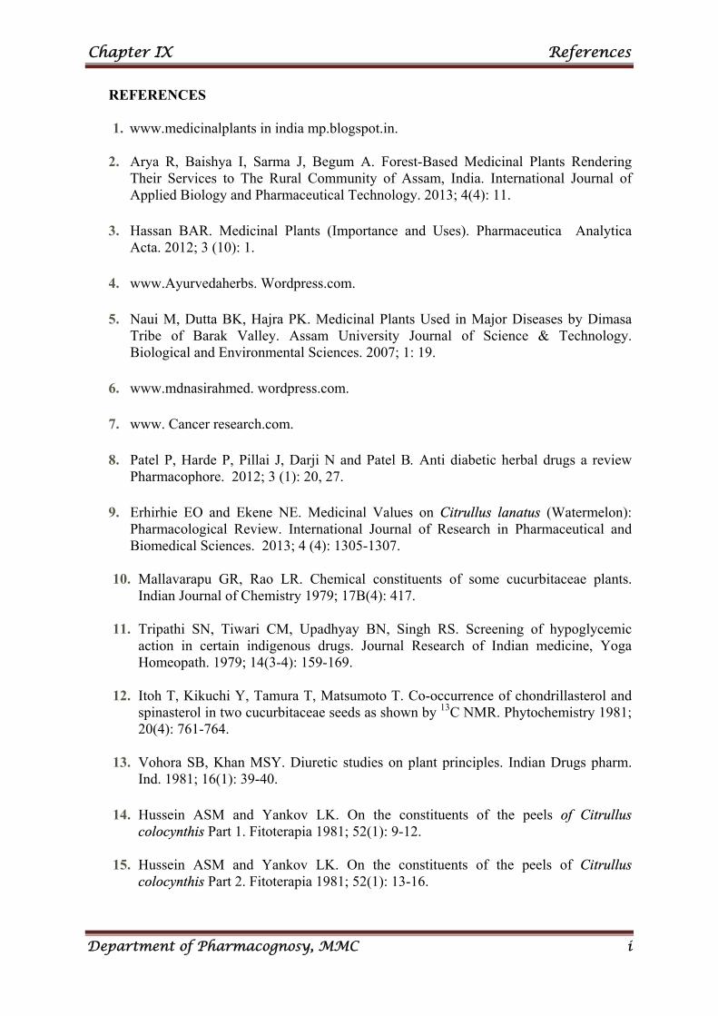
Chapter IX References
Department of Pharmacognosy, MMC i
REFERENCES
1. www.medicinalplants in india mp.blogspot.in.
2. Arya R, Baishya I, Sarma J, Begum A. Forest-Based Medicinal Plants Rendering Their Services to The Rural Community of Assam, India. International Journal of Applied Biology and Pharmaceutical Technology. 2013; 4(4): 11.
3. Hassan BAR. Medicinal Plants (Importance and Uses). Pharmaceutica Analytica Acta. 2012; 3 (10): 1.
4. www.Ayurvedaherbs. Wordpress.com.
5. Naui M, Dutta BK, Hajra PK. Medicinal Plants Used in Major Diseases by Dimasa Tribe of Barak Valley. Assam University Journal of Science & Technology. Biological and Environmental Sciences. 2007; 1: 19.
6. www.mdnasirahmed. wordpress.com.
7. www. Cancer research.com.
8. Patel P, Harde P, Pillai J, Darji N and Patel B. Anti diabetic herbal drugs a review Pharmacophore. 2012; 3 (1): 20, 27.
9. Erhirhie EO and Ekene NE. Medicinal Values on Citrullus lanatus (Watermelon): Pharmacological Review. International Journal of Research in Pharmaceutical and Biomedical Sciences. 2013; 4 (4): 1305-1307.
10. Mallavarapu GR, Rao LR. Chemical constituents of some cucurbitaceae plants. Indian Journal of Chemistry 1979; 17B(4): 417.
11. Tripathi SN, Tiwari CM, Upadhyay BN, Singh RS. Screening of hypoglycemic
action in certain indigenous drugs. Journal Research of Indian medicine, Yoga Homeopath. 1979; 14(3-4): 159-169.
12. Itoh T, Kikuchi Y, Tamura T, Matsumoto T. Co-occurrence of chondrillasterol and
spinasterol in two cucurbitaceae seeds as shown by 13C NMR. Phytochemistry 1981; 20(4): 761-764.
13. Vohora SB, Khan MSY. Diuretic studies on plant principles. Indian Drugs pharm.
Ind. 1981; 16(1): 39-40. 14. Hussein ASM and Yankov LK. On the constituents of the peels of Citrullus
colocynthis Part 1. Fitoterapia 1981; 52(1): 9-12. 15. Hussein ASM and Yankov LK. On the constituents of the peels of Citrullus
colocynthis Part 2. Fitoterapia 1981; 52(1): 13-16.
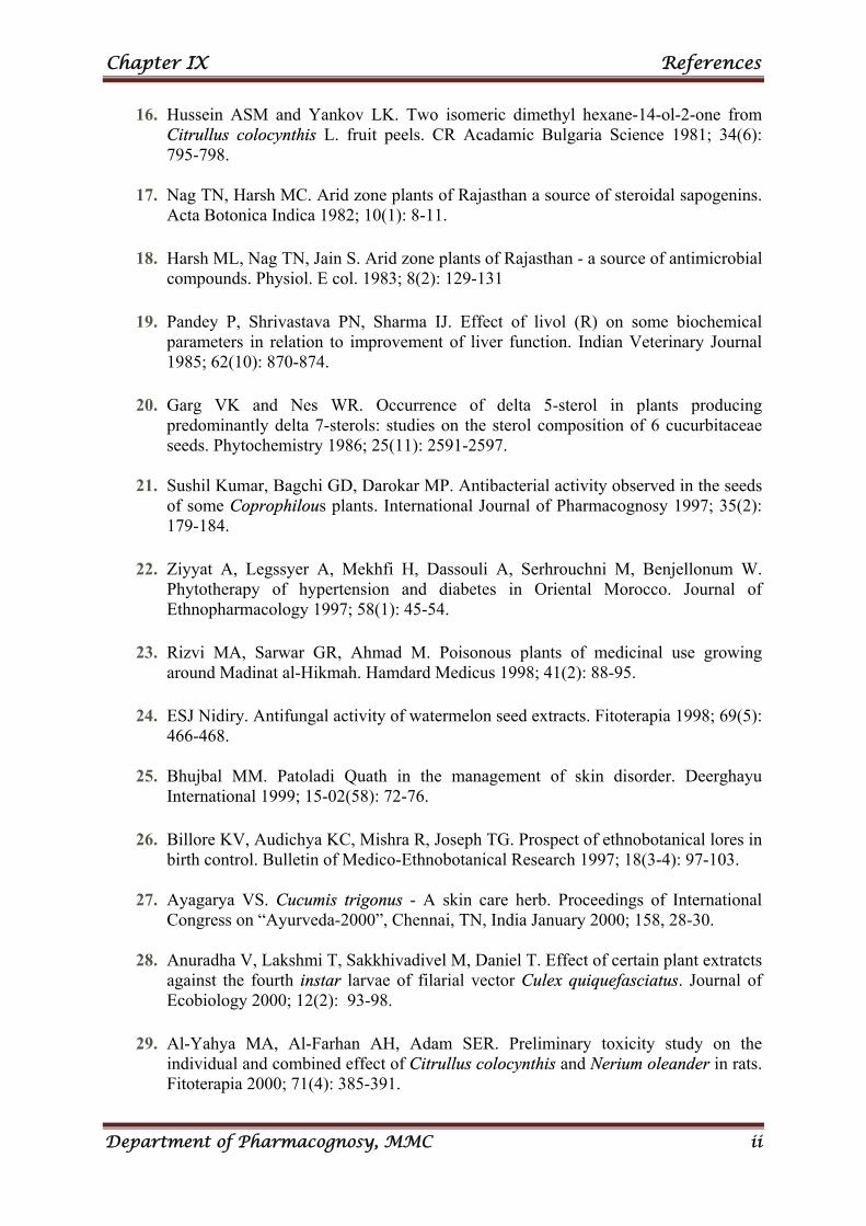
Chapter IX References
Department of Pharmacognosy, MMC ii
16. Hussein ASM and Yankov LK. Two isomeric dimethyl hexane-14-ol-2-one from Citrullus colocynthis L. fruit peels. CR Acadamic Bulgaria Science 1981; 34(6): 795-798.
17. Nag TN, Harsh MC. Arid zone plants of Rajasthan a source of steroidal sapogenins.
Acta Botonica Indica 1982; 10(1): 8-11. 18. Harsh ML, Nag TN, Jain S. Arid zone plants of Rajasthan - a source of antimicrobial
compounds. Physiol. E col. 1983; 8(2): 129-131 19. Pandey P, Shrivastava PN, Sharma IJ. Effect of livol (R) on some biochemical
parameters in relation to improvement of liver function. Indian Veterinary Journal 1985; 62(10): 870-874.
20. Garg VK and Nes WR. Occurrence of delta 5-sterol in plants producing
predominantly delta 7-sterols: studies on the sterol composition of 6 cucurbitaceae seeds. Phytochemistry 1986; 25(11): 2591-2597.
21. Sushil Kumar, Bagchi GD, Darokar MP. Antibacterial activity observed in the seeds
of some Coprophilous plants. International Journal of Pharmacognosy 1997; 35(2): 179-184.
22. Ziyyat A, Legssyer A, Mekhfi H, Dassouli A, Serhrouchni M, Benjellonum W.
Phytotherapy of hypertension and diabetes in Oriental Morocco. Journal of Ethnopharmacology 1997; 58(1): 45-54.
23. Rizvi MA, Sarwar GR, Ahmad M. Poisonous plants of medicinal use growing
around Madinat al-Hikmah. Hamdard Medicus 1998; 41(2): 88-95. 24. ESJ Nidiry. Antifungal activity of watermelon seed extracts. Fitoterapia 1998; 69(5):
466-468. 25. Bhujbal MM. Patoladi Quath in the management of skin disorder. Deerghayu
International 1999; 15-02(58): 72-76. 26. Billore KV, Audichya KC, Mishra R, Joseph TG. Prospect of ethnobotanical lores in
birth control. Bulletin of Medico-Ethnobotanical Research 1997; 18(3-4): 97-103. 27. Ayagarya VS. Cucumis trigonus - A skin care herb. Proceedings of International
Congress on “Ayurveda-2000”, Chennai, TN, India January 2000; 158, 28-30. 28. Anuradha V, Lakshmi T, Sakkhivadivel M, Daniel T. Effect of certain plant extratcts
against the fourth instar larvae of filarial vector Culex quiquefasciatus. Journal of Ecobiology 2000; 12(2): 93-98.
29. Al-Yahya MA, Al-Farhan AH, Adam SER. Preliminary toxicity study on the
individual and combined effect of Citrullus colocynthis and Nerium oleander in rats. Fitoterapia 2000; 71(4): 385-391.
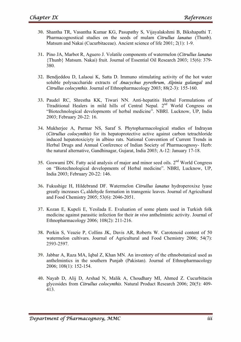
Chapter IX References
Department of Pharmacognosy, MMC iii
30. Shantha TR, Vasantha Kumar KG, Pasupathy S, Vijayalakshmi B, Bikshapathi T. Pharmacognostical studies on the seeds of mulam Citrullus lanatus (Thunb). Matsum and Nakai (Cucurbitaceae). Ancient science of life 2001; 2(1): 1-9.
31. Pino JA, Marbot R, Aguero J. Volatile components of watermelon (Citrullus lanatus
{Thunb} Matsum. Nakai) fruit. Journal of Essential Oil Research 2003; 15(6): 379-380.
32. Bendjeddou D, Lalaoui K, Satta D. Immuno stimulating activity of the hot water
soluble polysaccharide extracts of Anacychus pyrethrum, Alpinia galangal and Citrullus colocynthis. Journal of Ethnopharmacology 2003; 88(2-3): 155-160.
33. Paudel RC, Shrestha KK, Tiwari NN. Anti-hepatitis Herbal Formulations of
Ttraditional Healers in mild hills of Central Nepal. 2nd World Congress on “Biotechnological developments of herbal medicine”. NBRI. Lucknow, UP, India 2003; February 20-22: 16.
34. Mukherjee A, Parmar NS, Saraf S. Phytopharmacological studies of Indrayan
(Citrullus colocynthis) for its hepatoprotective active against carbon tetrachloride induced hepatotoxiciyty in albino rats. National Convention of Current Trends in Herbal Drugs and Annual Conference of Indian Society of Pharmacognosy- Herb: the natural alternative, Gandhinagar, Gujarat, India 2003; A-12: January 17-18.
35. Goswami DN. Fatty acid analysis of major and minor seed oils. 2nd World Congress
on “Biotechnological developments of Herbal medicine”. NBRI, Lucknow, UP, India 2003; February 20-22: 146.
36. Fukushige H, Hildebrand DF. Watermelon Citrullus lanatus hydroperoxixe lyase
greatly increases C6 aldehyde formation in transgenic leaves. Journal of Agricultural and Food Chemistry 2005; 53(6): 2046-2051.
37. Kozan E, Kupeli E, Yesilada E. Evaluation of some plants used in Turkish folk
medicine against parasitic infection for their in vivo anthelmintic activity. Journal of Ethnopharmacology 2006; 108(2): 211-216.
38. Perkin S, Veazie P, Collins JK, Davis AR, Roberts W. Carotenoid content of 50
watermelon cultivars. Journal of Agricultural and Food Chemistry 2006; 54(7): 2593-2597.
39. Jabbar A, Raza MA, Iqbal Z, Khan MN. An inventory of the ethnobotanical used as
anthelmintics in the southern Punjab (Pakistan). Journal of Ethnopharmacology 2006; 108(1): 152-154.
40. Nayab D, Alij D, Arshad N, Malik A, Choudhary MI, Ahmed Z. Cucurbitacin
glycosides from Citrullus colocynthis. Natural Product Research 2006; 20(5): 409-413.
Chapter IX References
Department of Pharmacognosy, MMC iv
41. Sharma M and Vats S. Ethnobotany of medicinal plants used by the tribal people of Rajasthan for digestive disorders. Journal Physiological Research 2007; 20(2): 337-338.
42. Qureshi R and Bhatti GR. Wild gourd; a green medicine. Hamdard Medicus 2007;
50(1): 156-162. 43. Khouri NA, El-Akawi Z, Daradka H. Effect of short term treatment with Citrullus
colocynthis L. on the lipid profile and other blood biochemical parameters in albino rats. Asian Journal of Chemistry 2007; 19(2): 1468-1472.
44. Joshva AJ, Vinod DR, Pravina K, Suja RS, Goudar KS, Pande CB, Amit A.
Lipoprotic activity of Natchol, a polyhedral formulation in albino wistar rats. Journal of Natural Remedies 2007; 6(2): 221-226.
45. Aburjai T, Tayyem R, Yousef M, Qishawi M. Ethnopharmacological survey of
medicinal herbs in Jordan, the Ajloun height region. Journal of Ethnopharmacology 2007; 110(2): 294-304.
46. Mukherjee A, Visen AKS, Saraf SA. Evaluation of hepatoprotective activity of
Citrullus colocynthis roots against carbon tetrachloride induced toxicity in albino rats. Natural Product Sciences 2007; 13(1): 23-26.
47. Yoshikava M, Morikawa T, Kobayashi H, Nakamura A, Matsuhira K, Nakamura S,
Matsuda H. Bioactive saponin and glycosides XXVII structures of new cucurbitane – type triterpene glycosides and antiallergic constituents from Citrullus colocynthis. Chemical and Pharmaceutical Bulletin 2007; 55(3): 428-434.
48. Meena MC and Patni V. Isolation and identification of flavonoid “quercetin” from
Citrullus colocynthis (Linn) Schrad. Asian Journal of Experimental sciences 2008; 22(1): 137-142.
49. Kumar S, Manjusha DK, Saroha K, Singh N, Vashishta B. Antioxidant and free
radical scavenging potential of Citrullus colocynthis (L) Schrad methanolic fruit extract. Acta Pharmaceutica 2008; 58(2): 215-220.
50. Allali H, Benmehdi H, Dib MA, Tabbi B, Ghalero S, Benabadji N. Phytotherapy of
diabetes in west Algeria. Asian Journal of Chemistry 2008; 20(4): 2701-2710. 51. Benmehdi H, Alladi H, Tabti B, Djabou N, Bendiadellah A, Lahfa F, Djazini R.
Hypo antihyperglycaemic effect of Citrullus colocynthis L. seed in normal and streptozotocin induced diabetic rats. Asian Journal of Chemistry 2008; 20(4): 2711-2788.
52. Sangameswaran B, Balakrishnan BR, Jayakar B. Oral hypoglycemic activity of
extract of leaves of Citrullus colocynthis (Schrad). Journal of Pharmaceutical Research 2008; 7(3): 138-140.
Chapter IX References
Department of Pharmacognosy, MMC v
53. Dinesh Kumar B, Mitra A, Manjunath M. A review of common Indian medicinal plants with anti-diabetic activities. Journal of Medicinal and Aromatic Plant Sciences 2009; 31(21): 152-158.
54. Dhanotia R, Chauhan NS, Saraf DK, Dixit VK. Effect of Citrullus colocynthis
Schrad fruits on testosterone induced alopecia. Natural Product Research 2011; 25(15): 1432-1443.
55. Upadhyay B, Singh KP, Kumar A. Ethno-veterinary use and informants consensus
factor of medicinal plants of Sariska region, Rajasthan, India. Journal of Ethnopharmacology 2011; 133(1): 14-25.
56. www.plants.usda.gov/core/ profile.
57. Mathew K.M. The Flora of the Tamil Nadu Carnatic Part I, The Rapinat herbarium, St. Joseph College, Thiruchirapalli, India. 1983; 636-637.
58. Burkill, H. M. The Useful Plants of West Tropical Africa, Families. M–R. 2nd
Edition. Royal Botanic Gardens, Kew, Richmond, UK. 1997; Volume 4. 59. Erhirhie and Ekene NE. Medicinal Values on Citrullus lanatus (Watermelon):
Pharmacological Review. International Journal of Research in Pharmaceutical and Biomedical Sciences. 2013; 4 (4): 1306-1307.
60. www.wikipedia.com 61. Mohammed Ali. The text book of Pharmacognosy. (Pharmacognosy &
Phytochemistry). 1; 1. 62. Edwin Jarald E, Sheeja Edwin Jarald. Text book of Pharmacognosy and
Phytochemistry. 96-97 & 100- 101.
63. Mathew K.M. The Flora of the Tamil Nadu Carnatic Part I, The Rapinat herbarium, St. Joseph College, Thiruchirapalli, India. 1983; 636-637.
64. Mathew KM. The Flora of Tamil Nadu Karnatic Polypetalae. Gamopetale &
Monochlamydae. The Ranipat Herbarium, St. John’s College, Tiruchirapalli, India. 1983; 1,3: 688, 689-1540.
65. Gamble J.S. Flora of the Presidency of Madras. Botanical survey of India, Calcutta,
India. 1935; Vol. I,II & III. 66. Henry AN, Kumari GR and Chitra V. Flora of Tamilnadu, India. Botanical survey of
India, Southern Circle, Coimbatore, India. 1987; 3: 258. 67. Sass JE. 1940. Elements of Botanical Microtechnique. McGraw Hill Book Co; New
York. 1940; 222. 68. Johansen DA. Plant Microtechnique. MC Graw Hill Book Co; New Yark. 1940; 523.
Chapter IX References
Department of Pharmacognosy, MMC vi
69. O’Brien TP, Feder N, Mc Cull ME. Polychromatic staining of plant Cell walls by
toluidine blue-O. Protoplasma. 1964; 59: 364-373. 70. Easu K. Plant Anatomy. John Wiley and Sons. New York. 1964; 767. 71. Easu K. Anatomy of seed plants. John Wiley and Sons. New York. 1979; 550. 72. Metcalfe CR and Chalk L. Anatomy of the Dicotyledons. Clarendon press, Oxford.
1950; I,II. 73. Metcalfe CR and Chalk L. Anatomy of the Dicotyledons. Clarendon press, Oxford.
1979; I: 276. 74. Yoganarasimhan SN. Medicinal plants of India. Tamilnadu. Regional Research
Institute (Ay), Tamilnadu, Banglore, India. 2000; 715. 75. Kay LA, Microscopical studies of drugs 1st edition, Bailliere, Tindal and Cox,
London. 1938; 17-18. 76. Wallis TE. The text book of pharmacognosy. CBS Publisher and Distributors,
Shahdara, Delhi, India. 1985. 77. Quality control methods for medicinal plant, WHO guidelines, Geneva. 1998; 10-
31,45. 78. Trease and Evans. Text Book of Pharmacognosy. 16th Edition. 2009; 124-125,568. 79. British Pharmacopoeia. 2012; Vol-5.
80. Indian Pharmacopoeia. Controller of publication, Government of India, Ministry of
health family welfare, Delhi. 1996; Volume-II : A53-A55.
81. Johansen DA. Plant Microtechnique. 1st edition, McGraw Hill Book Co. New York. 1940; 523.
82. Kay LA. Microscopical studies of Drugs. 1st edition. Bailliere, Tindal and Cox, London. 1938; 17-18.
83. Chase CR, Pratt R. Fluorescence powdered vegetable rugs with particular reference to development of system of identification, J Amer Pharm Assoc Sci Edition 1949; 28 : 324-331.
84. Onyeka EU and Nwambekwe IO. Phytochemical profile of some green leafy vegetables in South East, Nigeria. Nigerian Food Journal. 2007; 25(1): 67-76.
Chapter IX References
Department of Pharmacognosy, MMC vii
85. Igwenyi, I.O, Offor, C. E, Ajah,D.A, Nwankwo,O.C, Ukaomah,J.I, And Aja,P. M. Chemical compositions of Ipomea aquatica (green kangkong). International Journal of Pharma and Bio Sciences. 2011; 2(4): 594.
86. Harbone JB. Phytochemical Analysis – A guide to modern techniques of plant
analysis. Chapman & Hall, London, New York. 1980. 87. Bently & Drivers, Text Book Of Pharmaceutical Chemistry, 8th Edn Oxford
University Press, London. 1983; 13. 88. Finar IL, Organic Chemistry, Vol. 2, ELBS, London, 5th Edition. 1996; 771. 89. Chatwal GR, Organic Chemistry, 1st edn, Himalaya Publishing Home, Mumbai.
2000; 2,539. 90. Kokate CK, Purohit AP and Gokhale JB, Pharmacognosy, Nirali Prakasham, Pune,
36th edition; 593-597.
91. Arora A. Phytochemical analysis of methanolic extracts of leaves of some medicinal plants. Biological Forum – An International Journal. 2013; 5(2): 91-93.
92. Lai H and Singh NP. Oral artemisinin prevents and delays the development of 7, 12-
dimethylbenz[a]anthracene (DMBA)-induced breast cancer in the rat. Cancer Lett. 2006; 231(1):43-48.
93. Sun J, Chu, YF, Wu XZ, Liu RH. Antioxidant and anti-proliferative activities of
fruits. Journal of Agricultural and Food Chemistry. 2002; 50 (25):7449-7454. 94. Chang CC, Yang MH, Wen HM, Chern JC. Estimation of total flavonoid content in
Propolis by two complementary colorimetric methods. J Food Drug Analysis. 2002; 10(3): 178-82.
95. Mabry TJ, Markham KR, Thomas MB. The systematic identification of flavanoids.
Springer Verlay New York USA. 1970. 96. Chatwal GR and Anand SK .Instrumental Methods of Chemical Analysis. 2009; 2:
566-68. 97. Marinova D, Ribarova F, Atanassova M. Total phenolics and total flavonoids in
Bulgarian fruits and vegetables. Journal of the University of Chemical Technology and Metallurgy. 2005; 40(3): 255-260.
98. Patel A, Patel A and Patel NM. Estimation of Flavonoid, Polyphenolic Content and
In-vitro Antioxidant Capacity of leaves of Tephrosia purpurea Linn. (Leguminosae). International Journal of Pharma Sciences and Research. 2010; 1(1): 66-77.
99. Amola OJ, Saraste A, Pulkki K, Kallajoki M, Parvinen M andVoipo-Pulkki LM.
Acute doxorubicin cardiotoxicity involves cardiomyocyte apoptosis.,Cancer Res. 2000; 60: 1789-1792.
Chapter IX References
Department of Pharmacognosy, MMC viii
100. Powlowska JJ, Tarasiuk E, Borowski M, Wasowska I, Oszczapowicz and Wolf CR. The of new formamidine sugar-modified derivatives of dounorubicin to stimulate free radical formation in three enzymatic systems: NADH dehydrogenase,NADPH cytochrome P450 reductase and xanthine oxidase. Acta Biochim Pol. 2000; 47: 141-147.
101. Fard HM, Nasch G, Bodhankar SI, Dikshit M. Cardioprotective effect of Lagenaria
siceraria (Molina) Standley (Cucurbitaceae) fruit juice on Doxorubicin induced Cardio Toxicity in rats. American J Pharmacol & Toxicol. 2010; 5(2): 103-108.
102. Schanderl, SH. Method in food analysis. Academic Press, New York. 1970; 709.
103. Jain UK and Dixit VK. Spectrophotometrical estimation of tannins from the
Chyavanprash. Indian drugs. 2004; 41: 469-472.
104. Severi S, Bedogni G, Manzieri AM, Poli M, Battistini N. Effects of cooking and storage methods on the micronutrient content of foods. Euro J Cancer Prev. 1997; 6: S21-S24.
105. Vasundev V. Fundamentals of Biochemistry, Textbook of Biochemistry. 2006;
(2nd edn) 281-289.
106. Wagner KH, Kamal-Eldin A, Elmadfa I. Gamma-tocopherol-An Underestimated Vitamin? Ann Nutr Metab. 2004; 48: 169-188.
107. Eleazu CO, Eleazu KC. Determination of the Proximate Composition, Total
Carotenoid, Reducing Sugars and Residual Cyanide Levels of Flours of 6 New Yellow and White Cassava (Manihot esculenta Crantz) Varieties Am J Food Technol. 2012; 7: 642-649.
108. Onyeka EU and Nwambekwe IO. Phytochemical profile of some green leafy
vegetables in South East, Nigeria. Nigerian Food Journal. 2007; 25(1): 67-76.
109. Igwenyi, I.O, Offor, C. E, Ajah,D.A, Nwankwo,O.C, Ukaomah,J.I, And Aja,P. M. Chemical compositions of ipomea aquatica (green kangkong). International Journal of Pharma and Bio Sciences. 2011; 2(4): 594, 597.
110. Shaikh AA, Akter S, Rahman MS and Bakshi PK. Interaction of thiamine.hcl with
Cr2o72-. Journal of Bangladesh Academy of Sciences. 2011; 35(1): 51-59.
111. Okwu DE and Joshi C. Evaluation of the chemical composition of two Nigerian
medicinal plants. African Journal of Biotechnol. 2006; 5(4): 357-361.
112. USP29-NF24 page 2590.
113. Nath SC and Bordoloi DN. Major constituents of the essential oil of Clausena heptaphylla leaves. Indian Perfumer. 1991;35(2): 97-99.
Chapter IX References
Department of Pharmacognosy, MMC ix
114. Chakraborty A, Saha C, Podder G, Chowdhury BK, Bhattacharya P. Carbazole alkaloid with antimicrobial activity from Clausena heptaphylla. Phytochemisty 1995; 38(3): 787-789.
115. Applied Thin-Layer Chromatography: Best Practice and Avoidance of Mistakes, 2nd
Edition. Edited by Elke Hahn-Deinstrop. Copyright © 2007 WILEY-VCH Verlag GmbH & Co. KGaA,Weinheim.
116. Attimarad MKK, Ahmed M and Harsha S. High Performance Thin Layer
Chromatography. A powerful analytical technique in pharmaceutical drug discovery. Pharm methods. 2011; 2(2): 71-75.
117. Keuker H et al. in “Proceedings of the International Symposium on Instrumental
Thin Layer Chromatography”, Brighton, Sussex, UK. 1989; 105-114.
118. Srivastava (ed.) MM, High performance thin layer chromatography (HPTLC), Springer-Verlag Berlin Heidelberg 2011.
119. Satoskar R.S., Nirmala N.Rege, Bhandarkar S.D. text book of pharmacology and
pharmacotherapeutics. 23rd edition. 2013; 1-2. 120. Tripathi KD. Text book of essential of medicinal pharmacology. 6th edition. 3-4.
121. Ames BN, Shigenega MK, Hagen TM. Oxidants and the degenerative diseases of
ageing. Proc Nati Acad Sci. 1993; 90: 7915-22.
122. Shenoy R and Shirwaikar A. Anti-inflammatory and free radical scavenging studies of Hyptis suaveolens (Labiatae). Indian drugs 2002; 39: 574-577.
123. Evans P and Halliwall B. Free radicals and hearing Ann N Y Acad Sci. 1999; 884:
19.
124. Devasagayam TPA and Kesavan PC. Radio protective and antioxidant action of caffeine mechanistic considerations. Indian J Exptl Biol. 2003; 41: 267-269.
125. Peterhans E. Oxidants and antioxidants in viral diseases; disease mechanisms and
metabolic regulation. J. Nutr. 1997; 127: 962.
126. Sies H and Stahl W. Withania somnifera, vitamin E and C, β-carotene and other carotenoids as antioxidants. Am J Clin Nutr. 1995; 62: 1315-1321.
127. Parker R.S. Dietary and biochemical aspects of Vitamin E. Adv Food Nutr Res.
1980; 33: 157-232.
128. www.nccam.nih.gov/health/antioxidants.
129. Hackingthroughdistraction.blogspot.com.
Chapter IX References
Department of Pharmacognosy, MMC x
130. Brand Williams W, Cuvelier ME and Berset C. Use of Free Radical method to evaluate Antioxidant activity. LLebensm Wiss technol. 1995; 28: 25-30.
131. Blios MS. Antioxidant Determination by the use of stable free radicals. Nature
(1958); 26: 1199-1200.
132. Sahgal G, Ramanathan S, Sashidharan S, Mordi MN, Ismail S, Mansoor SM. In vitro antioxidant and Xanthine oxidase inhibitory activities of methanolic Swietenia mahogoni seedextracts. Molecules. 2009; 14: 4476-85.
133. Da Porto C, Calligaris S, Celotti E, Nicoli MC. Antiradical properties of commercial
cognags assessed by the DPPH test. J Agri Food Chem. 2000; 48: 4241-4245.
134. Jayanthi P and Lalitha P, Reducing Power of the Solvent of Eichhornia crassipes (Mart.) Solms, Indian J of Pharmacy and Pharm Sci. 2011; 3(3): 0975-1491.
135. Laloo D and Sahu AN. Antioxidant activities of three Indian commercially available
Nagakesar: An in-vitro study, J Chem Pharm Res. 2011; 3(1): 277-283.
136. Vector resistance to pesticides. Fifteenth report of the WHO expert committee on vector biology and control. WHO Tech Rep Ser. 1992; 818: 62.
137. Kumari R, Thapar BR, Dasgupta RK, Kaul SM, Shiv Lal. Susceptibility status of
malaria vectors to insecticides in India. J Com Dis. 1998; 30(3): 179–85.
138. Davidson G. Alternative measures to insecticides for mosquito control. Pesticide Sci. 1972; 3: 503–14.
139. Deshmukh PB, Chavan SR, Renapurkar DM. A study of insecticidal activity of
twenty indigenous plants. Pesticides 1982; (Dec); 7–10.
140. Mittal P and Subbarao SK. Prospects of using herbal products in the control of mosquito vectors. ICMR Bull. 2003; 33(1): 1–10.
141. Dharmagadda VSS, Naik SN, Mittal PK, Vasudevan P. Larvicidal activity of Tagetus
patula. Bioresource Technol. 2005; 96: 1235–40.
142. Singh RK, Mittal PK, Dhiman RC. Laboratory study on larvicidal properties of leaf extract of Calotropis procera (Family: Asclepiadaceae) against mosquito larvae. J Com Dis. 2005; 37: 109–13.
143. Thomas TG, Raghavendra K, Shiv Lal, Saxena VK. Larvicidal properties of latex
from unripe fruits of Carica papaya Linn (Caricaceae). J Com Dis. 2004; 36(4): 290–2.
144. Singh SP, Raghavendra K, Singh RK, Subbarao SK. Studies on the larvicidal
properties of leaf extract of Solanum nigrum Linn (Family: Solanaceae). Curr Sci 2001; 81(12): 1529-30.
Chapter IX References
Department of Pharmacognosy, MMC xi
145. Mehra BK and Haridhar PK. The effect of crude acetone extract of Annona squamosa
Linn (Family: Annonaceae) on possible control potential against larvae of Culex quinquefasciatus Say. J Entomol Res. 2001; 24(4): 141–6.
146. Thomas TG, Sharma SK, Jalees S, Rehman SJ. Larvicidal properties of an
indigenous plant Yucca aloifolia Linn against mosquito larvae. J Basic Appl Bio Med. 1994; 2:53–5.
147. Pathak N, Mittal PK, Singh OP, Sagar V, Vasudevan P. Larvicidal action of
essential oil from plants against the vector mosquitoes, Anopheles stephensi (Liston), Culex quinquefasciatus (Say) and Aedes aegypti (L). Intl Pest Contr. 2000; 46: 53–55.
148. Arivoli S, Samuvel, Tennyson. Larvicidal efficacy of Strychnus nuxvomica Linn.
(Family: Loganaceae). Leaf extract against the synarial vector Culex quinquefasciatus (Diptera: Culicidae); World J of zoology. 2012; 1: 6-11.
149. Hadjiakhoondi A, Aghel N, Zamanizadeh N, Vatandoost H. Chemical and
Biological study of Mentha spicata L. essential oil from Iran. Daru. 2000; 8(1-2): 19-21.
150. Hadjiakhoondi A, Sadeghipour-Roodsari HR, Vatandoost H, Khanavi M, Abai MR,
Vosoughi M, Kazemi M. Fatty acid composition and toxicity of Melia azedarach L. fruits against malaria vector Anopheles stephensi. Iran J Pharm Sci. 2006; 2(2): 97-102.
151. Schmid RD and Robert Verger, Lipases: Interfacial Enzymes with Attractive
Applications, Angew. Chem. Int. Ed. 1998; 37: 1608-633.
152. Choi SJ, Hwang JM Kim SA. Colorimetric microplate assay method for High Throughput Analysis of lipase activity, Journal of Biochemistry and Molecular Biology. 2003; 36(4): 417-420.
153. Prashith Kekuda TR, Raghavendra HL, Mallikarjun N, Venugopal TM and Anil
Kumar HS. Elemental composition, anticarcinogenic, pancreatic lipase inhibitory and cytotoxic activity of Atrocarpus lakoocha Roxb pericarp, Int. J Drug Dev. & Res., 2012; 4(1): 330-336.
154. NIH Medline plus. A publication of the National institutes of Health and the Friends
of the National library. 2010; 5(2): 17.
155. www.babymed.com/ breast cancer treatment.
156. www.healingwell.com / library/ breast cancer info1.asp.
157. www. Cancer research.com.
Chapter IX References
Department of Pharmacognosy, MMC xii
158. Mosmann, T. Rapid colorimetric assay for cellular growth and survival: application to proliferation and cytotoxicity assays. Journal of Immunological Methods. 1983; 65: 55-63.
159. Shoback, edited by David G. Gardner, Dolores. Greenspan's basic & clinical
endocrinology, 9th edn. New York: McGraw-Hill Medical. pp. Chapter 17. 2011.
160. Bhosale UP, Hallale BV. Asian Journal of Plant Science and Research. 2011; 1: 96.
161. Chakrabarti R and Rajagopalan R. Current Science. 2002; 83 (12): 1533-1538.
162. "Diabetes - Overview". NHS. Retrieved. 2013; 07-14.
163. Modak M, Dixit P, Londe J, Ghaskadbi S, Paul T, Devasagayam T. J Clin Biochem Nutr. 2007 May; 40(3): 163–173.
164. Patel P, Harde P, Pillai J, Darji B and Patel B. Antidiabetic herbal drugs a review
Pharmacophore. 2012; 3 (1): 20,27.
165. Bunn HF, Gabbay KH Gallop PM. The glycosylation of hemoglobin: relevance to Diabetes mellitus. Science. 1978; 200(4337): 21-27.
166. Stevens VJ, Vlassara H, Abati A and Cerami A. Nonenzymatic Glycosylation of
Hemoglobin. J. Biol. Chem. 1977; 252(9): 2998-3002.
167. Clifford J, Caroline Day B, et al. Review article. Traditional Plant Medicines as Treatments for Diabetes. Diabetes Care. 1989; 12(8).
168. Reddy NVLS, Anarthe SJ and Raghavendra NM, et al. In Vitro Antioxidant and
Antidiabetic activity of Asystasia gangetica (Chinese Violet) Linn. (Acanthaceae) International Journal of Research in Pharmaceutical and Biomedical Sciences. 2010; 1(2): 2229-3701.
169. Chaudhari MG, Joshi MB, Mistry KN, Dabhi B and Lal S, et al. In Vitro
Antimicrobial Activity of Stem Bark of Bauhinia Purpurea. International Science Press, (India). 2013; 4(1) : 29-35.
170. Gupta D, Chandrashekher KA, Pal G. In vitro antidiabetic activity of pentacyclic
triterpenoids and fatty acid ester from Bauhinia purpurea. International Journal of Pharmacology and Pharmaceutical Technology. 2013; 2(1): 3436.
171. Shreedhara CS, Vaidya VP, Vagdevi HM, Latha KP, Muralikrishna KS and
Krupanidhi AM. Screening of Bauhinia purpurea Linn. for analgesic and anti-inflammatory activities. Indian Journal of Pharmacolgy. 2009; 41: 75-79.
172. Kotowaroo MI, Mahomoodally, AGurib-Fakim, Subratty AH. Phytotherapy
Research. 2006; 20 (3): 228–231.
Chapter IX References
Department of Pharmacognosy, MMC xiii
173. Jumepaeng T, Prachakool S, Luthria DL and Chanthai S. Determination of antioxidant capacity and α-amylase inhibitory activity of the essential oils from citronella grass and lemongrass. International Food Research Journal. 2013; 20(1): 481-485.
174. Subramanian R, Asmawi AZ, Sadikun A. J Pol Biochem Soc. 2008; 55:391-398.
175. Mohammed A, Adelaiye AB, Bakari AG Mabrouk MA. International Journal of
Medicine and Medicinal Sciences. 2009; 1(12): 530-535.
176. Boivin M, Zinsmeister AR, Go VL. Mayo Clin Proc. 1987; 62:249-55.
177. Kumanan R, Manimaran S, Saleemulla K, Dhanabal SP, Nanjan MJ, Int J Pharmaceut Biomed Res. 2010; 1(2): 69-72.
178. Ashok Kumar BS, Khan S, Saran GS, Nandeesh R, Manjunath NK. In Vitro
Antidiabetic Activity of Nisamalaki Churna. Sains Malaysiana. 2013; 42(5): 625–628.
179. Hsieh PC, Huang GJ, Ho HL, Lin YH, Huang SS, Chiang C, Tseng MU, Chang YS.
Activities of antioxidants, α-Glucosidase inhibitors and aldose reductase inhibitors of the aqueous extracts of four Flemingia species in Taiwan. Botanical Studies (2010); 51: 293-302.
180. Kar DM, Maharana L, Parrnik S, Dash GK. Studies on hypoglycaemic activity of
Solanum xanthocarpum Schrad & Wendl fruit extract in rats. J.Ethno.Pharmacol. 2006; 108: 251-256.
181. Senthil Amudhan M and Hazeena Begum V. Alpha-Glucosidase inhibitory and
hypoglycaemic activities of Areca catechu extract. Pharmacognosy magazine. 2008; 15: 223-224.
182. Alberti KG, Zimmet PZ. Definition, diagnosis and classification of diabetes mellitus
provisional report of WHO consultations. Diabetic medicine.1998; 15: 539-553.
183. Prashanth D, Amit A, Samiulla DS, Asha MK, Padmaja R. α-glucosidase Inhibitory activity of Mangifera indica bark. Fitoterpia. 2001; 72: 686-8.
Related Documents


![Pharmacognostic and phytochemical evaluation of aerial parts of … s/january_2012/2.pdf · 2018. 10. 10. · Research Article [Pandya et al., 3(1): Jan., 2012] ISSN: 0976-7126 Int.](https://static.cupdf.com/doc/110x72/60b250ea224d8923396aaa86/pharmacognostic-and-phytochemical-evaluation-of-aerial-parts-of-sjanuary20122pdf.jpg)

