www.wjpps.com │ Vol 10, Issue 4, 2021. │ ISO 9001:2015 Certified Journal │ 1017 Rani et al. World Journal of Pharmacy and Pharmaceutical Sciences PHARMACOGNOSTIC AND PHYTOCHEMICAL ANALYSIS OF OXALIS LATIFOLIA KUNTH AND THEIR ANTIULCER ACTIVITY ON ALBINO RATS Hari Bhaskar 2 and Kothapalli Bannoth Chandra Sekhar 3 1 Research Scholar, Ratnam institute of Pharmacy, Pidathapoluru, Nellore, Andhra Pradesh, India. 2 Professor, Ratnam Institute of Pharmacy, Nellore, Andhra Pradesh, India. 3 Professor, Department of Chemistry, Krishna University, Machilipatnam, Andhra Pradesh, India, 521001. ABSTRACT The objective of the present study was to evaluate the pharmacognostic, phytochemical analysis and antiulcer potential of methanol extracts of Oxalis latifolia kunth in different experimental induced ulcer models in rats. In the present study different extracts, methanol (200 mg/kg and 400 mg/kg) extract of leaves of the plant were examined in Pylorus ligation and Indomethacin induced gastric ulcer in rats. Various parameters like volume of gastric acid secretion, pH, total acidity, ulcer index and antioxidant parameters were determined and compared between extract treated, standard and vehicle control group animals following ulcer induction. Among different dose of alcoholic extract, high dose showed significant antiulcer activity in Pylorus ligation and Indomethacin induced ulceration. The result of present study concluded that the alcoholic extract of leaves of the plant of Oxalis latifolia kunth has antiulcer activity in Pylorus ligation and Indomethacin induced gastric ulcer model in rats. The extract containing flavonoidal component latifolin A is as luteolin-6′′-(E-p-hydroxycinnamoyl) 4′-O-β-D- glucopyranoside is isolated, analysed by various spectral analysis and formulated as a capsule. This latifolin A capsule is main responsible for the anti ulcer activity. KEYWORDS: Pylorus ligation, Indomethacin, Total acidity, Latifolin A, Oxalis latifolia etc. WORLD JOURNAL OF PHARMACY AND PHARMACEUTICAL SCIENCES SJIF Impact Factor 7.632 Volume 10, Issue 4, 1017-1045 Research Article ISSN 2278 – 4357 *Corresponding Author M. Jhansi Rani Research Scholar, Ratnam institute of Pharmacy, Pidathapoluru, Nellore, Andhra Pradesh, India. Article Received on 25 Jan. 2021, Revised on 15 Feb. 2021, Accepted on 07 March 2021 DOI: 10.20959/wjpps20214-18565

Welcome message from author
This document is posted to help you gain knowledge. Please leave a comment to let me know what you think about it! Share it to your friends and learn new things together.
Transcript

www.wjpps.com │ Vol 10, Issue 4, 2021. │ ISO 9001:2015 Certified Journal │
1017
Rani et al. World Journal of Pharmacy and Pharmaceutical Sciences
PHARMACOGNOSTIC AND PHYTOCHEMICAL ANALYSIS OF
OXALIS LATIFOLIA KUNTH AND THEIR ANTIULCER ACTIVITY
ON ALBINO RATS
Hari Bhaskar2 and Kothapalli Bannoth
Chandra Sekhar
3
1Research Scholar, Ratnam institute of Pharmacy, Pidathapoluru, Nellore, Andhra Pradesh,
India.
2Professor, Ratnam Institute of Pharmacy, Nellore, Andhra Pradesh, India.
3Professor, Department of Chemistry, Krishna University, Machilipatnam, Andhra Pradesh,
India, 521001.
ABSTRACT
The objective of the present study was to evaluate the
pharmacognostic, phytochemical analysis and antiulcer potential of
methanol extracts of Oxalis latifolia kunth in different experimental
induced ulcer models in rats. In the present study different extracts,
methanol (200 mg/kg and 400 mg/kg) extract of leaves of the plant
were examined in Pylorus ligation and Indomethacin induced gastric
ulcer in rats. Various parameters like volume of gastric acid secretion,
pH, total acidity, ulcer index and antioxidant parameters were
determined and compared between extract treated, standard and
vehicle control group animals following ulcer induction. Among
different dose of alcoholic extract, high dose showed significant antiulcer activity in Pylorus
ligation and Indomethacin induced ulceration. The result of present study concluded that the
alcoholic extract of leaves of the plant of Oxalis latifolia kunth has antiulcer activity in
Pylorus ligation and Indomethacin induced gastric ulcer model in rats. The extract containing
flavonoidal component latifolin A is as luteolin-6′′-(E-p-hydroxycinnamoyl) 4′-O-β-D-
glucopyranoside is isolated, analysed by various spectral analysis and formulated as a
capsule. This latifolin A capsule is main responsible for the anti ulcer activity.
KEYWORDS: Pylorus ligation, Indomethacin, Total acidity, Latifolin A, Oxalis latifolia etc.
WORLD JOURNAL OF PHARMACY AND PHARMACEUTICAL SCIENCES
SJIF Impact Factor 7.632
Volume 10, Issue 4, 1017-1045 Research Article ISSN 2278 – 4357
*Corresponding Author
M. Jhansi Rani
Research Scholar, Ratnam
institute of Pharmacy,
Pidathapoluru, Nellore,
Andhra Pradesh, India.
Article Received on
25 Jan. 2021,
Revised on 15 Feb. 2021,
Accepted on 07 March 2021
DOI: 10.20959/wjpps20214-18565

www.wjpps.com │ Vol 10, Issue 4, 2021. │ ISO 9001:2015 Certified Journal │
1018
Rani et al. World Journal of Pharmacy and Pharmaceutical Sciences
INTRODUCTION
Peptic ulcer disease is a serious gastrointestinal disorder. The formation of peptic ulcers
depends on the presence of acid and peptic activity in gastric juice plus a breakdown in
mucosal defenses. There are two major factors that can disrupt the mucosal resistance to
injury: non-steroidal anti-inflammatory drugs NSAID like e.g. aspirin and Helicobacter pylori
(H. pylori) infection.[1]
Number of drugs including proton pump inhibitors, prostaglandins
analogs, histamine receptor antagonists and cytoprotective agents are available for the
treatment of peptic ulcer.[2]
But most of these drugs exhibit serious side effects like
arrhythmias, gynaecomastia, impotence, arthralgia, hypergastrinemia and haemopoeitic
changes.[3]
Hence, herbal medicines are generally used in such cases when drugs are to be
used for chronic periods. Several natural drugs have been reported to possess anti-ulcerogenic
activity by virtue of their predominant effect on mucosal defensive factors.[4]
The plant Oxalis latifolia kunth belonging to Oxalidaceae family is a stem less herb of
cosmopolitan distribution found abundantly in agricultural farms, gardens, lawns etc. Leaves
dark green, slight and charecteristic odour, sore and astringent taste. Leaves are digitately 3-
foliate, leaflets, obcordate, chartaceous, pilose base, cuneate, margin entire, apex,
emarginiate, Pseudoumbels, axiallary, 1-6flowered, bracts two, linear, bracteole, Sepals, five
lanceolate, petals oblanceolate apex, emarginated. The plant contains such as carbohydrates,
saponins, phenol, flavanoid, Flavonol glycosides[5]
cardiac glycosides, phytosterol, fixed oils
and fats, gums and mucilage in Oxalis latifolia kunth. The Oxalis species are reported to cure
various disorders such as paralysis, stomach disorder, antiulcer, anti-inflammatory,
antioxidant and it also acts as thirst reliever.[6]
MATERIALS AND METHODS
Plant material
Oxalis latifolia kunth plant was collected from in and around deciduous forest of Talakona
Forest in the state of Andhra Pradesh. India, during September 2019. Plant was authenticated
by Dr. K. Madhava Chetty, Plant Taxonomist, Department of Botany, Sri Venkateswara
University, Tirupati, Andhra Pradesh, India. The voucher specimen (2019/1214) of the plant
was deposited at the college for further reference.

www.wjpps.com │ Vol 10, Issue 4, 2021. │ ISO 9001:2015 Certified Journal │
1019
Rani et al. World Journal of Pharmacy and Pharmaceutical Sciences
Pharmacognostic study of oxalis latifolia kunth
Macroscopical study
The macroscopical description of plant include size, shape, nature of outer and inner surfaces,
types of fracture, and organoleptic characters like color, odour, taste etc. were studied.
Macroscopic characters of the plant Oxalis latifolia kunth (Oxalidaceae) was studied directly
in the field, and photographed under original environment.[7]
Microscopic study[8]
Transverse section of crude drug (Leaf)
Microscopical examination of the plant drugs is essential to study the adulterants also
indispensable in identification. Microscopical evaluation of the plant drugs helps to identify
the organized drugs by their known histological characters and used to confirm the structural
details of the drugs from plant origin.
Powder microscopy
The shade dried leaves of Oxalis latifolia kunth had been powdered and the powders that
passes through sieve number 60# individually and then afflicted by powder analysis.
Preparation of the extract
The plant leaves were dried on filter paper sheets under shade at room temperature until
changing of color of filter papers and milled into coarse powder. 200 g of powder material
placed was extracted with 70% methanol in a Soxhlet apparatus for 8-12 h. Solvent were
removed at temperature below 50°C in an oven. The residue (extract) of respective plant
material was stored at 4°C for further experimental studies. Aqueous extract were prepared
by taking 100g of the powdered plant material with 500 ml of distilled water in a Soxhlet
apparatus for 8-12 h. The filtrate was then concentrated and the extract was stored at 4°C for
further experimental studies.
Preliminary phytochemical screening
Methanolic extract of Oxalis latifolia kunth were subjected to preliminary phytochemical
screening for the detection of various plants constituents like Tannins, carbohydrates,
Saponins, Flavanoids, Glycosides, Proteins, Alkaloids and Phenols.[9]
www.wjpps.com │ Vol 10, Issue 4, 2021. │ ISO 9001:2015 Certified Journal │
1020
Rani et al. World Journal of Pharmacy and Pharmaceutical Sciences
Isolation of active constituent
5 gm of Methanolic extract Oxalis latifolia kunth (Oxalidaceae) was subjected to silica-gel
(100–200 mesh) column (length 100 cm and diameter 3 cm) chromatography and The elution
started with hexane followed by hexane- Ethyl acetate (EtOAc) mixtures (9 : 1, 8: 2; 7: 3, 6:
4, 5: 5, 4: 6; 3: 7, 2: 8, 1: 9), EtOAc (100%), EtOAc-methanol (MeOH) mixtures (9: 1, 8: 2
and 7: 3), and ended with MeOH (100%). A total of 55 fractions, 100 mL each, was collected
from the tubes; those with similar thin layer chromatography (TLC) profiles were combined
fractions as F1 l-6 (Pl); F2 7-12 (P2); F3 13-18 (P3), F4 19-32 (P4), F5 33-45 (P5); F34-43.
From the five pooled fractions (Pl to P5) eluted with hexane-EtOAc mixtures (9:1) eluted two
compounds, further this fraction subjected to column chromatography with hexane:
chloroform mixure (9:1, 5:5, 1:9). In this fraction, white yellow crystal powder (164 mg) was
eluted from hexane: chloroform (9:1) with melting point 65oC.
10, 11, 12
Ethyl acetate fraction
The column chromatography of the EtOAc soluble sub-fraction (90g) over silica gel was
performed using n-hexane/EtOAc (in increasing order of polarity) as solvent system, which
afforded six major sub-fractions A-F.
Sub-fraction D
It was subjected to further column chromatography over silica gel using n-hexane-EtOAc
(3.5:6.5) and n-hexane-EtOAc (4:6) solvent systems. Which afforded compound 2 (6 mg) and
compound 4 (21 mg) respectively.
Sub-fraction E
Further column chromatography of this fraction over silica gel eluting with
dichloromethane/methanol solvent system was carried out. It afforded two pure compounds.
Compound 3 (8 mg) was obtained from solvent system dichloromethane/methanol (9.5:0.5).
Similarly dichloromethane/methanol (9:1) solvent system gave compound 1 (25 mg).
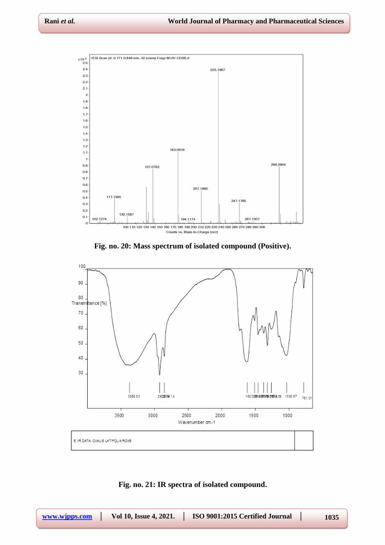
INSTRUMENTS AND MATERIALS
The UV and IR spectra were recorded on a Hitachi UV-3200 and JASCO 302-A
spectrometers. 1H NMR and 13C NMR and two-dimensional COSY, NOSEY, HMQC, and
HMBC, were recorded on the Bruker AV-400 spectrometer (400 MHz for 1H and 100 MHz
for 13C) in C5D5N with TMS as internal stander. Chemical shifts δ are shown in ppm

www.wjpps.com │ Vol 10, Issue 4, 2021. │ ISO 9001:2015 Certified Journal │
1021
Rani et al. World Journal of Pharmacy and Pharmaceutical Sciences
relative to TMS as internal standard and scalar coupling are reported in Hz. The HR-FAB-
MS were recorded on a JEOL JMS-HX-110 mass spectrometer. Analytical and preparative
TLC were carried out on pre- coated Silica gel 60 F254 plates (E. Merck, Darmstadt,
Germany), and visualized under UV radiation light and by spraying with the ceric sulfate
solution. Silica gel (230-400 mesh, E. Merck) was used for column chromatography.
Formulation development of isolated compound
Preparation of latifolin a granules[13]
(Jyothi D et al., 2017)
Latifolin A granules were prepared by wet granulation method.
Granules were also prepared containing sodium starch glycollate (SSG) as super disintegrant.
SSG is incorporated at different concentration (2%, 3%, 5%) separately and granulations
were carried out similar way as described above.
Evaluation of latifolin a granules[14]
(Assadpour E et al., 2017)
Prepared Latifolin A granules were subjected for determination of bulk density, tapped
density, Hausner ratio, Carr’s index, angle of repose in order to assess the flow property of
granules.
Formulation of latifolin a capsules
Prepared granules were packed into hard gelatin capsule (size 1) using hand operated capsule
filling machine such that each capsule contains 400 mg of granules. Latifolin A capsules
without SSG were labelled as F1 and capsules containing 2%, 3% and 5% of sodium starch
glycolate (SSG) were labelled as F2, F3, and F4 respectively.
Estimation of drug content (Latifolin A ) in capsules[15]
Granules from 10 capsules were mixed and weight of powder equivalent to 5 mg of Latifolin
A and extracted with the phosphate buffer of pH 6.8 for 30 min. These solutions were filtered,
suitably diluted and absorbance was measured at 208 nm against blank solution (phosphate
buffer pH 6.8) using a UV spectrophotometer.
Determination of uniformity of weight
Twenty capsules were selected. Each capsule was weighed on an analytical balance, carefully
emptied of its content, the shells reweighed and the weight of content determined. The
collective weight of content, average weight of content per capsule and the deviations (%) of
individual content weights from the mean were calculated.

www.wjpps.com │ Vol 10, Issue 4, 2021. │ ISO 9001:2015 Certified Journal │
1022
Rani et al. World Journal of Pharmacy and Pharmaceutical Sciences
Determination of disintegration time[16]
Disintegration times for capsules were determined by disintegration apparatus. Six capsules
were placed in six tubes of the basket and the apparatus was operated using water as release
medium maintained at 37 ± 2°C. The capsules were observed and the times taken for
complete disintegration of all capsules were determined.
In vitro dissolution study of capsules
In vitro dissolution study of all the prepared capsule formulations was done using USP Type
II paddle dissolution apparatus (Electrolab USP dissolution tester TDT-08L) using 900 ml
phosphate buffer pH 6.8 at 100 rpm and results were compared with drug release of Latifolin
A from capsule formulation F0. An aliquot amount of the sample was withdrawn at regular
time intervals and the same volume of pre-warmed (37±0.5ºC) fresh dissolution medium was
replaced. The samples were filtered, suitably diluted and Latifolin A in each sample was
analyzed by using Shimadzu UV-spectrophotometer at 208 nm.
Acute toxicity studies
The acute toxicity of MEOL was determined as per the OECD guideline no. 423 (Acute toxic
class method). It was observed that the rats were not mortal even at 2000 mg/kg dose of
MEOL. Hence, 1/5th (400 mg/kg) and 1/10th (200 mg/kg) of MEOL were selected as high
dose and low dose respectively for this study.[17]
Animals used
Experimental animals Albino wistar rats of both sexes weighing between 150-250 g were
used. The experimental protocol was approved from Institutional Animal Ethics Committee.
Animals were housed under standard conditions of temperature (24±2ºC) and relative
humidity (30-70%) with a 12:12 light: dark cycle
Model I: Pylorus ligation induced gastric ulceration in rats
Pyloric ligation of the stomach was done according to method of Shay et al. with slight
modification. Albino rats of either sex were divided into six groups of six animals each.
Animals were fasted for 24 h before the study, but had free access to water.
Animals in the control group received only 0.1% of Tween 80 (10 ml/kg orally). Methanolic
extracts of Oxalis latifolia kunth at 200 and 400 mg/kg, (p.o.) for each extract were given to
the animals in the treatment group. Omeprazole (10 mg/kg) was used as a standard. After 1h

www.wjpps.com │ Vol 10, Issue 4, 2021. │ ISO 9001:2015 Certified Journal │
1023
Rani et al. World Journal of Pharmacy and Pharmaceutical Sciences
of drugs treatment, they were anaesthetized with the help of anaesthetic ether; the abdomen
was opened by a small midline incision below the lipoid process. Pyloric portion of the
stomach was slightly lifted out and ligated according to method of Shay et al. avoiding
traction to the pylorus or damage to its blood supply. The stomach was replaced carefully and
the abdominal wall was closed by interrupted sutures. Rats were sacrificed by an over dose of
anaesthetic ether after four hours of pylorus ligation. The abdomen was opened, the stomach
was removed, and its content drained into a graduated centrifuge tube and centrifuged at 3000
rpm for 10 min. From the supernatant, aliquots (1 ml of each) were taken for the
determination of pH, and total acidity. Each stomach was examined for lesions in the fore
stomach portion and indexed according to severity.[18]
Determination of pH
An aliquot of 1 ml gastric juice was diluted with 1 ml of distilled water and pH of the
solution was measured using pH meter.
Determination of total acidity[19]
An aliquot of 1 ml gastric juice diluted with 1 ml of distilled water was taken into a 50 ml
conical flask and two drops of phenolphthalein indicator was added to it and titrated with
0.01 N NaOH until a permanent pink colour was observed. The volume of 0.01 N NaOH
consumed was noted. The total acidity is expressed as mEq/L by the following formula:
Acidity = (Vol. of NaOH×N×100)/0.1
Determination of free acidity[20]
Instead of phenolphthalein indicator, the Topfer's reagent was used. Aliquot of gastric juice
was titrated with 0.01 N NaOH until canary yellow colour was observed. The volume of 0.01
N NaOH consumed was noted. The free acidity was calculated by the same formula for the
determination of total acidity.
Macroscopic evaluation of stomach[21,22]
The stomachs were cut open along the greater curvature, rinsed with saline to remove gastric
contents and blood clots and examined by a 10X magnifier lens to assess the formation of
ulcers. Number of ulcers was counted and was given scores based on their intensity as
follows: 0= no ulcer, 0.5= red coloration, 1= superficial mucosal erosion, 1.5= hemorrhagic
streak, 2= deep ulcer or transmural necrosis, 3= perforated or penetrated ulcer.

www.wjpps.com │ Vol 10, Issue 4, 2021. │ ISO 9001:2015 Certified Journal │
1024
Rani et al. World Journal of Pharmacy and Pharmaceutical Sciences
Mean ulcer score for each animal will be expressed as ulcer index. The percentage of ulcer
protection was determined as follows: Ulcer index (UI) was measured by using following
formula:UI=UN+US+UP Χ 10−1
Where, UI= Ulcer Index; UN= Average number of ulcers per animal; US= Average number
of severity score; UP= Percentage of animals with ulcers.
Percentage inhibition of ulceration was calculated as below: % Inhibition of Ulceration =
(Ulcer indexControl–Ulcer indexTest) × 100/Ulcer index Control.
Model II
Indomethacin induced ulcer[23]
Albino rats of either sex were divided into six groups of six animals each. Animals were
fasted for 24 h before the study, but had free access to water. Animals in the control group
received only vehicle 10 ml/kg orally. Methanolic extracts of Oxalis latifolia kunth at 200
and 400 mg/kg, were administered orally for each extract were given to the animals in the
treatment group. Omeprazole (10 mg/kg) orally was used as a standard. Indomethacin (25
mg/kg body weight) was administrated orally to all animals 10 min prior to treatment. After 6
h of drugs treatment, rats were sacrificed by an over dose of anaesthetic ether and their
stomach was removed. The contents of the stomach were drained into a glass tube. The
volume of the gastric juice was measured and centrifuged at 2000 rpm for 10 min. From the
supernatant, aliquots (1 ml of each) were taken for the determination of pH, and total acidity.
10% v/v Formalin was injected into the totally ligated stomach for storage overnight. The
next day, the stomach were opened along the greater curvature, then washed in warm water,
and examined under a 3 fold magnifier. The ulcer index was determined as described above.
Statistical analysis
Values were expressed as mean ± SEM from 6 animals. Statistical differences were evaluated
using a One-way analysis of variance (ANOVA) followed by Dunnet's t-test. Results were
considered to be statistically significant at P<0.05.
RESULTS
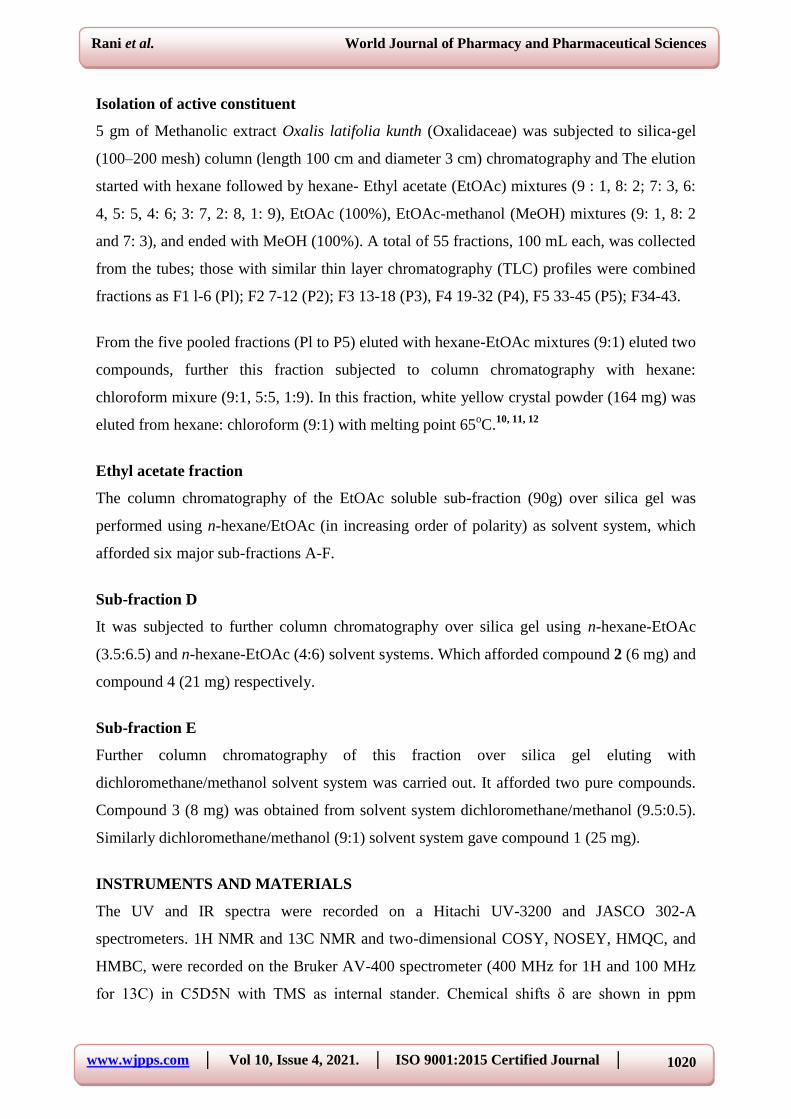
Macroscopic characters of the oxalis corniculata L
Oxalis latifolia kunth belonging to Oxalidaceae family is a stem less herb of cosmopolitan
distribution found abundantly in agricultural farms, gardens, lawns etc. Leaves dark green,
slight and charecteristic odour, sore and astringent taste. Leaves are digitately 3- foliate,
leaflets, obcordate, chartaceous, pilose base , cuneate, margin entire, apex, emarginiate,
www.wjpps.com │ Vol 10, Issue 4, 2021. │ ISO 9001:2015 Certified Journal │
1025
Rani et al. World Journal of Pharmacy and Pharmaceutical Sciences
Pseudoumbels, axiallary, 1-6flowered, bracts two, linear, bracteole, Sepals, five lanceolate,
petals oblanceolate apex, emarginated.
Fig no. 1: Macroscopic characters of the oxalis corniculata L.
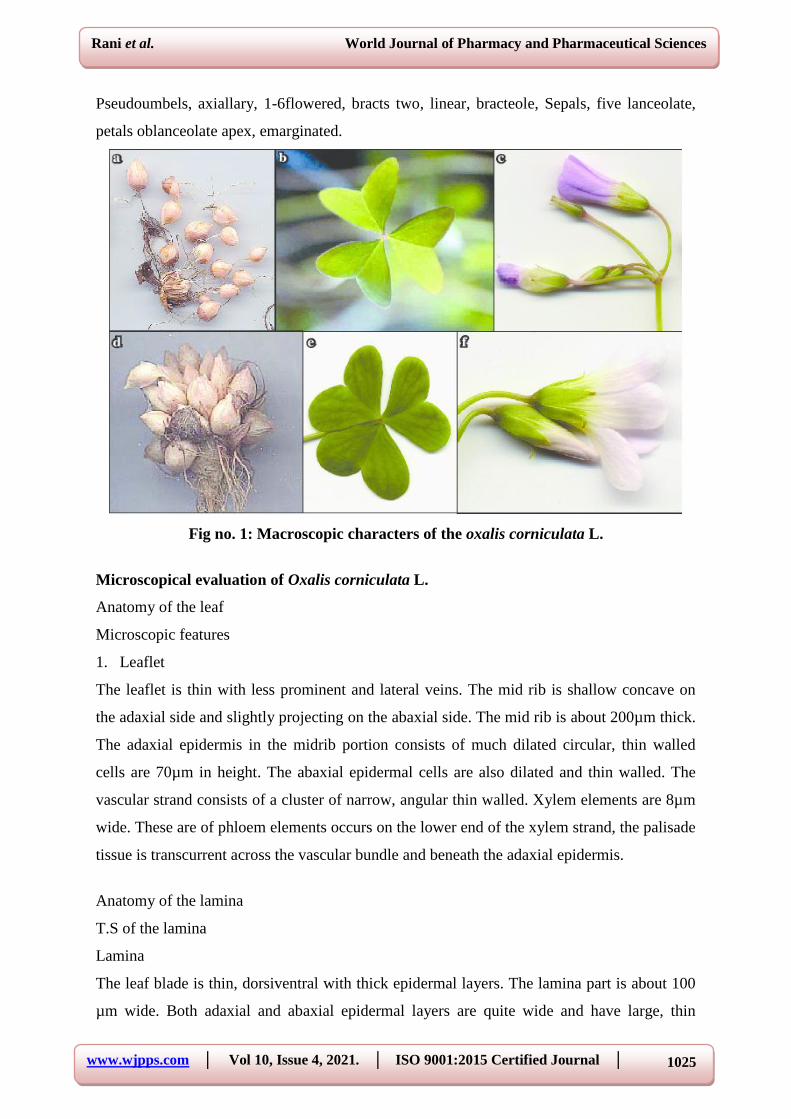
Microscopical evaluation of Oxalis corniculata L.
Anatomy of the leaf
Microscopic features
1. Leaflet
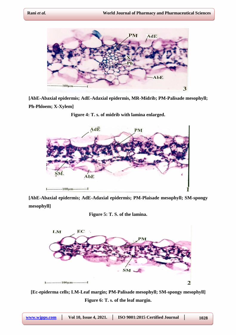
The leaflet is thin with less prominent and lateral veins. The mid rib is shallow concave on
the adaxial side and slightly projecting on the abaxial side. The mid rib is about 200µm thick.
The adaxial epidermis in the midrib portion consists of much dilated circular, thin walled
cells are 70µm in height. The abaxial epidermal cells are also dilated and thin walled. The
vascular strand consists of a cluster of narrow, angular thin walled. Xylem elements are 8µm
wide. These are of phloem elements occurs on the lower end of the xylem strand, the palisade
tissue is transcurrent across the vascular bundle and beneath the adaxial epidermis.
Anatomy of the lamina
T.S of the lamina
Lamina
The leaf blade is thin, dorsiventral with thick epidermal layers. The lamina part is about 100
µm wide. Both adaxial and abaxial epidermal layers are quite wide and have large, thin

www.wjpps.com │ Vol 10, Issue 4, 2021. │ ISO 9001:2015 Certified Journal │
1026
Rani et al. World Journal of Pharmacy and Pharmaceutical Sciences
walled circular cells, measuring 25µm in thickness. The mesophyll tissue consists of a narrow
adaxial zone of short, then cylindrical palisade cells and the four layers of small, lobed
spongy parenchyma cells.
T.S of the leaf margin
The leaf margin is slightly narrow leaflet and posses circular thin walled cells. They are 25
µm in diameter the mesophyll tissues are as in the middle portion of the lamina.
Epidermal morphology
Epidermal cells and stomata
The epidermal cells are thin walled; their anticlerical walls are highly wavy, so that the cells
appear amoeboid in outline. Stomata occur only the lower epidermis and they are absent on
the upper epidermis.
Abaxial epidermis with stomata
The stomata do not possess distinct subsidiary cells. The guard cells are elliptical with slit
like stomatal pores.
Adaxial epidermis
The guard cells are 15×20µm in size. The adaxial epidermal cells are similar to the abaxial
cells in shape and size; but it is apostomatic (without stomata).
Paradermal section showing venation pattern and crystal distribution
Venation pattern
The lateral veins and vein islets are uniformly thin comprising of one or two spiral xylem
elements, the veins are straight. They form wide, rectangular on many sided vein islets; the
vein islets have well defined vein terminations. Which are long, slender unbranched or
branched once or twice.
Crystals in the mesophyll tissue
Crystals
Calcium oxalate crystals are frequently seen in the mesophyll tissue. The crystals are mostly
druses or sphere crystals. They are diffuse in distribution and are located in ordinary
mesophyll cells. The crystals are up to 20 µm wide.

www.wjpps.com │ Vol 10, Issue 4, 2021. │ ISO 9001:2015 Certified Journal │
1027
Rani et al. World Journal of Pharmacy and Pharmaceutical Sciences
Powder microscopy of the whole plant
Leaf powder
Leaf powder are seen fragments lamina, with venation and trichomes, isolated trichomes and
epidermal peeling fragments of lamina show epidermal trichomes along the leaf margin, as
well as on the lamina surface.
Non-glandular covering trichome in the leaf powder
The trichomes are non-glandular type covering trichomes; they are unicellular, unbranched
and pointed at the tip. They are mostly curved and wavy. Their walls are fairly thick and
smooth. They are up to 300 µm long and 20 µm thick. Epidermal peeling in the powder
exhibit thin walled lobed cells. The stomata are anomocytic type.
[AdS – adaxial side; MR – Midrib; La – Lamina]
Figure 2: T. s. of through midrib with lamina.
[AdE-adaxial epidermis; Ph-Pholem; X-Xylem]
Figure 3: T. s. of midrib with lamina enlarged
www.wjpps.com │ Vol 10, Issue 4, 2021. │ ISO 9001:2015 Certified Journal │
1028
Rani et al. World Journal of Pharmacy and Pharmaceutical Sciences
[AbE-Abaxial epidermis; AdE-Adaxial epidermis, MR-Midrib; PM-Palisade mesophyll;
Ph-Phloem; X-Xylem]
Figure 4: T. s. of midrib with lamina enlarged.
[AbE-Abaxial epidermis; AdE-Adaxial epidermis; PM-Plaisade mesophyll; SM-spongy
mesophyll]
Figure 5: T. S. of the lamina.
[Ec-epiderma cells; LM-Leaf margin; PM-Palisade mesophyll; SM-spongy mesophyll]
Figure 6: T. s. of the leaf margin.

www.wjpps.com │ Vol 10, Issue 4, 2021. │ ISO 9001:2015 Certified Journal │
1029
Rani et al. World Journal of Pharmacy and Pharmaceutical Sciences
[Ec-Epidermal cells; St-Stomata]
Figure 7: Abaxial epidermis with stomata.
[Ec-Epidermal cells; St-Stomata]
Figure 8: Adaxial epidermis with stomata
[Ec-Epidermal cells]
Figure 9: Adaxial epidermis

www.wjpps.com │ Vol 10, Issue 4, 2021. │ ISO 9001:2015 Certified Journal │
1030
Rani et al. World Journal of Pharmacy and Pharmaceutical Sciences
[VI-vein islets; VT-Vein termination]
Figure 10: vein islets and vein termination.
[VI-vein islets]
Figure 11: Vein islets and vein termination.
[Cr-Crystal]
Figure 12: Crystals in the mesophyll tissue.

www.wjpps.com │ Vol 10, Issue 4, 2021. │ ISO 9001:2015 Certified Journal │
1031
Rani et al. World Journal of Pharmacy and Pharmaceutical Sciences
Figure 13: Cleared leaf showing vein islets and vein termination.
[VI-vein islets; VT-Vein termination]
Figure 14: Cleared leaf showing vein islets and vein termination enlarged.
[VI-vein islets; VT-Vein termination]
Figure 15: One vein islets and vein termination enlarged.

www.wjpps.com │ Vol 10, Issue 4, 2021. │ ISO 9001:2015 Certified Journal │
1032
Rani et al. World Journal of Pharmacy and Pharmaceutical Sciences
[LM-Leaf margin, Tr-Trichome]
Figure 16: Fragment of adaxial epidermis cells with covering trichome.
[Tr-Trichome]
Figure 17: A covering trichome enlarged.
[Tr-Trichome]
Figure 18: Non-glandular covering trichome in the leaf powder.

www.wjpps.com │ Vol 10, Issue 4, 2021. │ ISO 9001:2015 Certified Journal │
1033
Rani et al. World Journal of Pharmacy and Pharmaceutical Sciences
Table no 1: Preliminary Phyto-chemical screening.
Tests Pet. ether Ethanol Methanol Water
Alkaloids − − − −
Carbohydrates − + + +
Glycosides − − + +
Phytosterols − − − −
Fixed Oils & fats − + − +
Saponins − + + +
Phenolic compounds & Tannins − + + +
Proteins & Amino acids − − − −
Gums & mucilage + + + +
Flavonoids + + + −
"+" = Indicates Positive Result
"−" = Indicates Negative Results
Tab no. 2: Physicochemical parameters of Oxalis latifolia kunth.
Parameters Values (%w/w)
Moisture content (Loss on drying) 6.52±1.34
Total ash 6.80±1.52
Acid insoluble ash 3.52±0.72
Water soluble ash 2.02±0.55
Petroleum ether soluble extractive value 0.86±0.05
Chloroform soluble extractive value 2.56±0.06
Ethyl acetate soluble extractive value 3.65±0.82
Alcohol soluble extractive value 8.12±1.22
Water soluble extractive value 10.02±2.51
Tab no 3: Fluorescence analysis of Oxalis latifolia kunth.
Solvent used Visible light UV light
254nm
Water Buff Brown Water
NaOH Dark Brown Brown NaOH
HCl Reddish brown Black HCl
HNO3 Brown Green HNO3
Fecl3 Dark green Light green Fecl3

www.wjpps.com │ Vol 10, Issue 4, 2021. │ ISO 9001:2015 Certified Journal │
1034
Rani et al. World Journal of Pharmacy and Pharmaceutical Sciences
Fig no 19: TLC fingerprinting of Methanolic extract of Oxalis latifolia kunth with
compound on TLC Silica gel Kiesel gel 60 F254.
Tab no 4: Physical properties of isolated compound.
S. No Parameter Observation
1 Colour Yellowish
2 Shape Crystalline solid
Tab no 5: Chemical tests of isolated compound.
S. No Test Result
1 Shinoda test +
(Luteolin-6′′-(E-p-hydroxycinnamoyl) 4′-O-β-D-glucopyranoside)
Structure 1: Chemical structure of Latifolin A
www.wjpps.com │ Vol 10, Issue 4, 2021. │ ISO 9001:2015 Certified Journal │
1035
Rani et al. World Journal of Pharmacy and Pharmaceutical Sciences
Fig. no. 20: Mass spectrum of isolated compound (Positive).
Fig. no. 21: IR spectra of isolated compound.

www.wjpps.com │ Vol 10, Issue 4, 2021. │ ISO 9001:2015 Certified Journal │
1036
Rani et al. World Journal of Pharmacy and Pharmaceutical Sciences
Fig. no. 22: GCMC-Chromatogram of methanolic extract of Oxalis latifolia kunth.
Fig. no. 23: 1H NMR spectra of isolated constituent.

www.wjpps.com │ Vol 10, Issue 4, 2021. │ ISO 9001:2015 Certified Journal │
1037
Rani et al. World Journal of Pharmacy and Pharmaceutical Sciences
Fig. no. 24: 13
C NMR spectra of isolated constituent.
Table no. 14: Formulation of capsules.
Ingredients Quantity/Capsule (mg)
F1 F2 F3 F4
Latifolin A 5 5 5 5
Lactose Monohydrate 150 150 150 150
Starch Paste (5%) 50 50 50 50
Microcrystalline Cellulose 179 171 157 144
Sodium Starch Glycollate - 8 12 15
Talc (2%) 8 8 8 8
Magnesium Stearate (2%) 8 8 8 8
Tab. no. 13: Evaluation of latifolin a granules.
Formulation
Code
Evaluation Parameters
Bulk
Density
(g/ml)
Tapped
Density
(g/ml)
Hausner
Ratio
Carr’s
Index (%)
Angle of
Repose θ
F1 0.81±0.02 0.78±0.02 1.05±0.06 6.55±0.05 28.56±0.05
F2 0.78±0.05 0.76±0.02 1.08±0.02 8.02±0.02 23.21±0.06
F3 0.76±0.02 0.74±0.02 1.16±0.03 12.42±0.05 22.15±0.03
F4 0.75±0.03 0.72±0.02 1.14±0.02 14.21±0.04 23.52±0.05
www.wjpps.com │ Vol 10, Issue 4, 2021. │ ISO 9001:2015 Certified Journal │
1038
Rani et al. World Journal of Pharmacy and Pharmaceutical Sciences
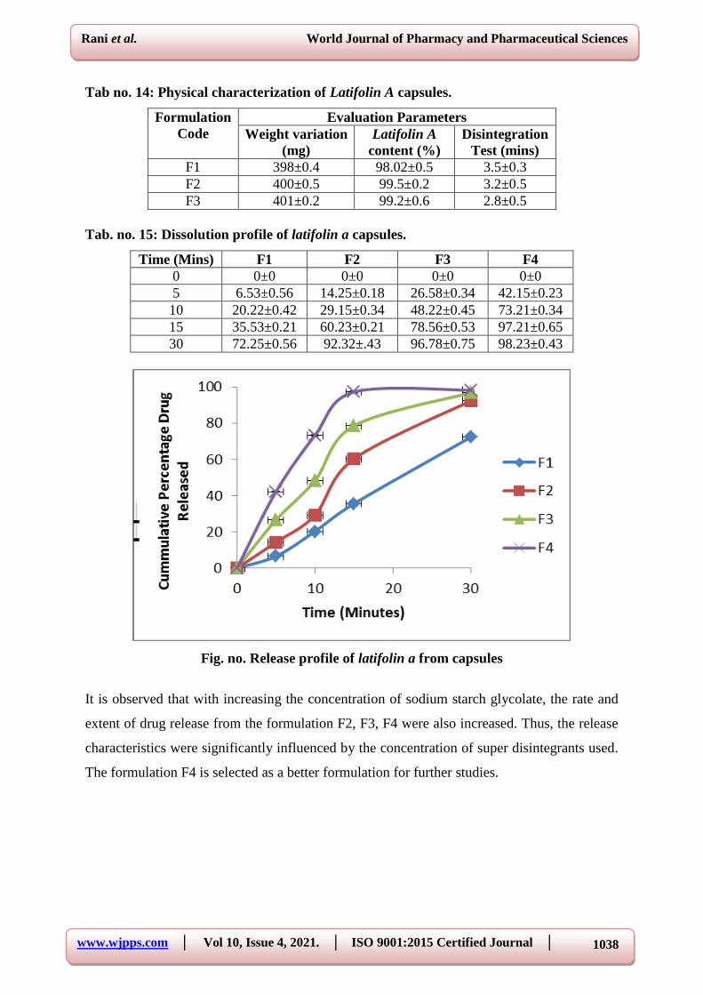
Tab no. 14: Physical characterization of Latifolin A capsules.
Formulation
Code
Evaluation Parameters
Weight variation
(mg)
Latifolin A
content (%)
Disintegration
Test (mins)
F1 398±0.4 98.02±0.5 3.5±0.3
F2 400±0.5 99.5±0.2 3.2±0.5
F3 401±0.2 99.2±0.6 2.8±0.5
Tab. no. 15: Dissolution profile of latifolin a capsules.
Time (Mins) F1 F2 F3 F4
0 0±0 0±0 0±0 0±0
5 6.53±0.56 14.25±0.18 26.58±0.34 42.15±0.23
10 20.22±0.42 29.15±0.34 48.22±0.45 73.21±0.34
15 35.53±0.21 60.23±0.21 78.56±0.53 97.21±0.65
30 72.25±0.56 92.32±.43 96.78±0.75 98.23±0.43
Fig. no. Release profile of latifolin a from capsules
It is observed that with increasing the concentration of sodium starch glycolate, the rate and
extent of drug release from the formulation F2, F3, F4 were also increased. Thus, the release
characteristics were significantly influenced by the concentration of super disintegrants used.
The formulation F4 is selected as a better formulation for further studies.

www.wjpps.com │ Vol 10, Issue 4, 2021. │ ISO 9001:2015 Certified Journal │
1039
Rani et al. World Journal of Pharmacy and Pharmaceutical Sciences
Fig. no. 1: Macroscopic evaluation of stomach ulcer in pyloric ligation induced
ulceration in rats.
Table 2: The Effect of methanolic extracts of oxalis latifolia kunth on gastric pH, Gastric
Volume, Free Acidity and Total Acidity in Pylorous Ligation Model.
Group Treatment Dose
(mg/kg
b.w.)
Gastric pH Gastric
Volume
(ml)
Free Acidity
(meq/ltr)
Mean Total
Acidity
(meq/ltr)
I Control 1 ml 2.9±0.10 8.6±0.28 6.56±0.07 70.77±0.25
II MEOL 200 3.8±0.05** 6.3±0.06** 5.65±0.19** 50.15±0.08**
III MEOL 400 4.9±0.17*** 5.2±0.18*** 4.24±0.29*** 44.8±0.05***
IV Omeprazole 10 5.7±0.09*** 4.5±0.29*** 3.43±0.08*** 34.4±0.14***
All the values are mean±SEM n=6. ***P<0.001, **
P<0.01, compare vs. control, data was
analysed using one way ANOVA followed by Tukey multiple comparison test.
Graph 1: Effect of Methanolic extracts of Oxalis latifolia kunth on total acidity and %
inhibition of ulcer in pyloric ligation induced ulceration in rats.

www.wjpps.com │ Vol 10, Issue 4, 2021. │ ISO 9001:2015 Certified Journal │
1040
Rani et al. World Journal of Pharmacy and Pharmaceutical Sciences
Fig. 2: Macroscopic evaluation of stomach ulcer in Indomethacin induced ulceration in
rats.
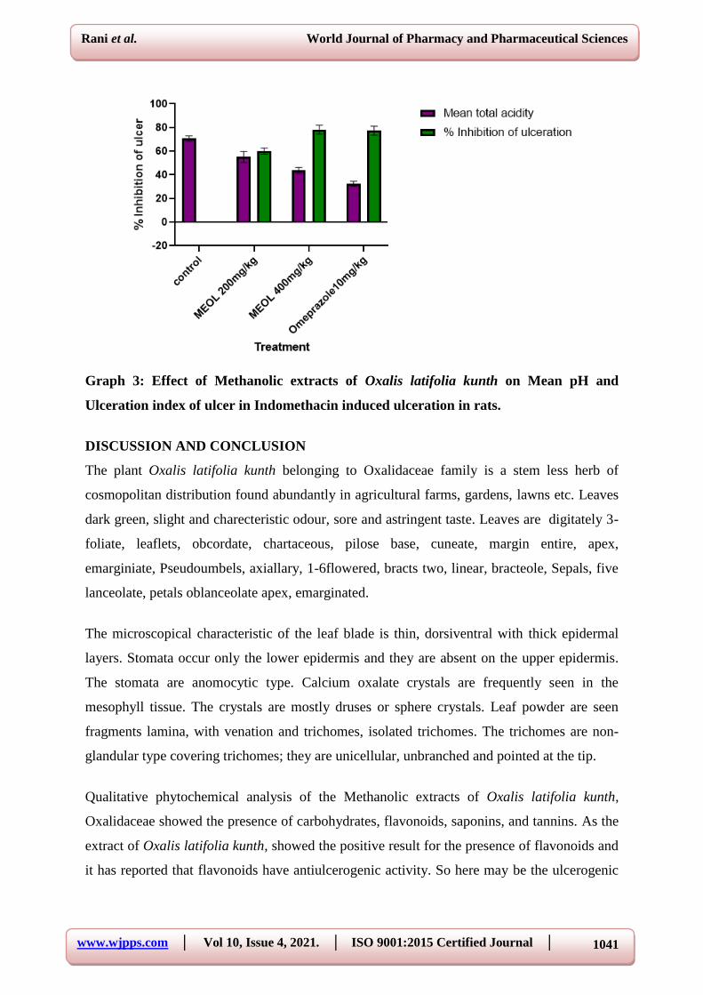
Table 3: Effect of methanolic extracts of Oxalis latifolia kunth on pH, total acidity, Ulcer
index and % inhibition of ulcer in Indomethacin induced ulceration in rats.
Group Treatment Gastric pH
Mean Total
Acidity
(meq/ltr)
Ulcer Index
%Inhibition
of
Ulceration
I Control(1 ml) 1.8±0.10 70.73±2.25 10.69±2.51 --
II MEOL (200mg/kg) 3.4±0.05** 55.15±4.68** 4.69±0.51 60.05%
III MEOL (400mg/kg) 4.9±0.17*** 43.68±2.55*** 2.32±0.51*** 78.29%
IV
Omeprazole
(10 mg/kg p.o.) 5.7±0.09*** 32.41±2.14*** 2.36±0.16** 77.29%
Significant values were compared with* P <0.05, ** P <0.01 and *** P <0.001 Indomethacin
control Vs treated groups using One way ANOVA followed by Dennett’s test.
www.wjpps.com │ Vol 10, Issue 4, 2021. │ ISO 9001:2015 Certified Journal │
1041
Rani et al. World Journal of Pharmacy and Pharmaceutical Sciences
Graph 3: Effect of Methanolic extracts of Oxalis latifolia kunth on Mean pH and
Ulceration index of ulcer in Indomethacin induced ulceration in rats.
DISCUSSION AND CONCLUSION
The plant Oxalis latifolia kunth belonging to Oxalidaceae family is a stem less herb of
cosmopolitan distribution found abundantly in agricultural farms, gardens, lawns etc. Leaves
dark green, slight and charecteristic odour, sore and astringent taste. Leaves are digitately 3-
foliate, leaflets, obcordate, chartaceous, pilose base, cuneate, margin entire, apex,
emarginiate, Pseudoumbels, axiallary, 1-6flowered, bracts two, linear, bracteole, Sepals, five
lanceolate, petals oblanceolate apex, emarginated.
The microscopical characteristic of the leaf blade is thin, dorsiventral with thick epidermal
layers. Stomata occur only the lower epidermis and they are absent on the upper epidermis.
The stomata are anomocytic type. Calcium oxalate crystals are frequently seen in the
mesophyll tissue. The crystals are mostly druses or sphere crystals. Leaf powder are seen
fragments lamina, with venation and trichomes, isolated trichomes. The trichomes are non-
glandular type covering trichomes; they are unicellular, unbranched and pointed at the tip.
Qualitative phytochemical analysis of the Methanolic extracts of Oxalis latifolia kunth,
Oxalidaceae showed the presence of carbohydrates, flavonoids, saponins, and tannins. As the
extract of Oxalis latifolia kunth, showed the positive result for the presence of flavonoids and
it has reported that flavonoids have antiulcerogenic activity. So here may be the ulcerogenic

www.wjpps.com │ Vol 10, Issue 4, 2021. │ ISO 9001:2015 Certified Journal │
1042
Rani et al. World Journal of Pharmacy and Pharmaceutical Sciences
activity reduced because of the flavonoids, which shows significant result as compared to
standard drug.
In this present study the Methanolic extracts of Oxalis latifolia kunth, Oxalidaceae were
investigated for antiulcer activity by using Pyloric ligation in rats, ethanol induced gastric
ulcer in rats and Indomethacin induced gastric ulcer in rats.
There are several risk factors that may contribute to formation of ulcer in human beings such
as stress, chronic use of anti-inflammatory drugs, continuous alcohol ingestion, H. pylori
infection, Zollinger Ellison syndrome, etc. Although in most cases the etiology of ulcer is
unknown. An effective anti-ulcer drug should act either by reducing the aggressive factors on
gastroduodenal mucosa or by increasing mucosal resistance against them. The critical factors
which maintain defense and integrity of gastric and intestinal mucosa include normal mucosal
blood flow, local prostaglandins, mucous and bicarbonate secretion, epithelial proliferation
and repair.
Non-steroidal anti-inflammatory drugs (NSAIDs) like Indomethacin are known to induce
gastric damage, particularly due to inhibition of the cyclooxygenase pathway of arachidonic
acid metabolism. It is currently believed that the ulcerogenic effects of the NSAIDs are due to
inhibition of cyclooxygenase 1 (COX-1) and that its isoforms, cyclooxygenase 2 (COX-2),
plays a pathological role in inflammation, pain and fever. Several studies shown that gastric
mucosal prostaglandins (PGs), produced mainly by COX-1, play an important role in
maintaining gastric mucosal integrity and Indomethacin markedly decrease mucosal PGE2
level.
On the other hand, recent reports show that Indomethacin is a dual inhibitor of COX-1 and
COX-2 because both tromboxanes and inflammatory PGE2 synthesis are suppressed, and that
inhibition of both isoform of these enzymes is required for the development of gastric
erosions after NSAID administration. Indeed, endogenous PG deficiency alone did not induce
visible gastric lesions and the pathogenesis of NSAID-induced gastric lesions also involves
luminal acid, neutrophils activation and gastric hyper motility.
The treatment of peptic ulcer is mainly aimed at reducing the hydrochloric acid secretion,
increasing gastric cytoprotection, eradication of H. pylori or curing Zollinger Ellison
syndrome. The discovery of potential antiulcer agent from plants is a developing area. So far,
www.wjpps.com │ Vol 10, Issue 4, 2021. │ ISO 9001:2015 Certified Journal │
1043
Rani et al. World Journal of Pharmacy and Pharmaceutical Sciences
several plants have been screened for antiulcer activity and many formulations have been
developed by combining extracts of these plants.
Pylorus ligation induced ulcer was used to study the effect of seed extracts on gastric acid
secretion and mucus secretion. The ligation of the pyloric end of the stomach causes
accumulation of gastric acid in the stomach. This increase in the gastric acid secretion causes
ulcers in the stomach. The original Shay rat model involves fasting of rats for 36 h followed
by ligation of pyloric end of the stomach. The ulcer index is determined 5 h after pylorus
ligation. The lesions produced by this method are located in the lumen region of the stomach.
Indomethacin is known to cause ulcer especially in an empty stomach and mostly on the
glandular (mucosal) part of the stomach by inhibiting prostaglandins synthetase through the
cyclooxygenase pathway. Prostaglandins function to protect the stomach from injury by
stimulating the secretion of bicarbonate and mucus, maintaining mucosal blood flow and
regulating mucosal turn over and repair. Suppression of prostaglandins by Indomethacin
results in increased susceptibility of the stomach to mucosal injury and gastro duodenal
ulceration. The extract was observed to significantly reduce mucosal damage in the
Indomethacin induced ulcer model, suggesting the possible extracts mobilization and
involvement of prostaglandin in the anti-ulcer effect of the extract.
It may act by multiple mechanisms. The activity might be due to increasing the gastric
mucosal resistance, local synthesis of cytoprotective prostaglandins and inhibiting the
leukotriene synthesis.
It has also been reported that the presence of phyto-constituents tannins, terpenoids, sterols
and flavonoids may be responsible for antiulcer activity. Recent reports and extensive
literature review indicated that flavonoids and tannins showed cytopro-tective action by
increasing mucosal content of prostaglandins and mucous in gastric mucosa.
The Methanolic extracts of Oxalis latifolia kunth, Oxalidaceae showed significant antiulcer
activity due to isolated the compound Latifolin A based on Analytical evidence.
Developed novel Latifolin A formulation showed significant anti-ulcer properties. Developed
formulation was evaluated on Pylorous ligated and Indomethacin induced ulcer in rats. The
extract significantly reduces mucosal damage on the raats. However further studies required

www.wjpps.com │ Vol 10, Issue 4, 2021. │ ISO 9001:2015 Certified Journal │
1044
Rani et al. World Journal of Pharmacy and Pharmaceutical Sciences
to elucidate the exact mechanism of action for develop its as potent antiulcer drug. These
herbal drugs will help to develop new drug molecules for antiulcer therapy.
BIBLIOGRAPHY
1. Jyoti Gupta, Dinesh Kumar, Ankit Gupta. Evaluation of gastric anti-ulcer activity of
methanolic extract of Cayratia trifolia in experimental animals, Asian Pacific Journal of
Tropical Disease, 2012; 99-102.
2. Ariyphisi I, Toshiharu A, Sugimura F, Abe M, Matsuo Y, Honda T. Recurrence during
maintenance therapy with histamine H2 receptors antagonist in cases of gastric ulcers,
Nikon University J Medical, 1986; 28: 69-74.
3. Akthar. M. S, A. H. Akthar and M.A. Khan, Int. J Pharmacog, 1992; 30: 97-104.
4. Sairam K, Rao CV, Goel RK. Effect of Centella asiatica linn on physical and chemical
factors induced gastric ulceration and secretion. Indian J Exp. Biol, 2001; 39: 137-142.
5. Muhammad Ibrahim, Iqbal Hussain, Muhammad Imran, Nusrat Hussain, Amjad Hussain,
Tooba Mahboob, Corniculatin A, a new flavonoidal glucoside from Oxalis corniculata,
Brazilian Journal of Pharmacognosy, 2013; 23(4): 630-634.
6. Madhavachetty K, Sivaj JK, Tulasi Rao K, Flowering plants of Chittoor district, Andhra
Pradesh, India, Student offset printers, Tirupati, 2008; 1: 75.
7. O’brien TP, Rder N and Mc Cull ME, Polychromatic staining of plant cell walls by
toludine blue-O’’, Protoplasma ,1964; 59: 364-3473.
8. Esau k, Plant Anatomy, John wiley and sons, Newyork, 1964; 767.
9. Khandelwal KR, Practical pharmacognosy, Nirali Prakashan, 2006; 1: 149-153.
10. Agrawal PK In: Agrawal PK, edi. Carbon-13 NMR of flavonoids. Amsterdam: Elsevier,
1989; 1: 297-321.
11. Della Greca M, Previtera L, Purcaro R, Zarrelli A Phytotoxic aromatic constituents of
Oxalis pes-caprae. Chem Biodivers, 2009; 6: 459-465.
12. Mizokami H, Yoshitama K Flavonoids in the leaves of Oxalis corniculata and
sequestration of the flavonoids in the wings scales of the pale grass blue butterfly,
Pseudozizeeria maha. J Plant Res, 2008; 121: 133-136.
13. Jyothi D, Koland M, Priya S, James JP. Formulation of herbal capsule containing
Trigonella foenum-graecum seed extract for the treatment of diabetes. Journal of Young
Pharmacists, 2017; 1, 9(3): 352.

www.wjpps.com │ Vol 10, Issue 4, 2021. │ ISO 9001:2015 Certified Journal │
1045
Rani et al. World Journal of Pharmacy and Pharmaceutical Sciences
14. Assadpour E, Jafari SM, Maghsoudlou Y. Evaluation of folic acid release from spray
dried powder particles of pectin-whey protein nano-capsules. International journal of
biological macromolecules, 2017; 1, 95: 238-47.
15. Singh A, Avupati VR. Development and validation of UV-spectrophotometric method for
the estimation of curcumin in standardised polyherbal formulations. Journal of Young
Pharmacists, 2017; 1, 9(4): 491.
16. Okunlola A, Adewoyin BA, Odeku OA. Evaluation of pharmaceutical and microbial
qualities of some herbal medicinal products in south western Nigeria. Tropical Journal of
Pharmaceutical Research, 2007; 31, 6(1): 661-70.
17. Ecobiochon DJ, Mekahel KM, Mjor P, Ogilvie KK, The acute toxicity of BIOLF-145 in
the rat, Fundamental appl Toxicol, 1988; 10(2): 313-20.
18. Shiva Shanker Rai, Atanu Banik, Ankit Singh, Meenu Singh, Evaluation of Anti-Ulcer
Activity of Aqueous and Ethanolic Extract of Whole Plant of Clitoria ternatea in Albino
Wistar Rats, International Journal of Pharmaceutical Sciences and Drug Research, 2015;
7(1): 33-39.
19. Manowar Hussain, Iswar Hazarika, Anju Das. Pylorus ligation induced gastric ulcer
protection by sesamum indicum ethanolic seed extract. Research & Reviews: A Journal of
Pharmaceutical Science, 2015; 6(3): 42–49.
20. Bae DK, Park D, Lee SH, Yang G, Yang YH, Kim TK et al. Different Antiulcer
Activities of Pantoprazole in Stress, Alcohol and Pylorus Ligation-Induced Ulcer Models.
Lab Anim Res, 2011; 27(1): 47-52.
21. Cantarella G, Martinez G, Cutuli VM, Loreto C, D'Alcamo M, Prato A, Amico-Roxas M,
Bernardini R, Clementi G. Adrenomedullin modulates COX-2 and HGF expression in
reserpine-injured gastric mucosa in the rat. Eur J Pharmacol, 2005; 518: 221-226.
22. Cantarella G, Martinez G, Di Benedetto G, Loreto C, Musumeci G, Prato A, Lempereur
L, Matera M, Amico-Roxas M, Bernardini R, Clementi G. Protective effects of amylin on
reserpine-injured gastric damage in the rat. Pharmacol Res, 2007; 56: 27-34.
23. Barathane Datchanamurty, Mythireyi D , Divyashanthi C. M, Evaluation of antiulcer
activity of ethanolic leaf extract of Coccinia grandis in indomethacin induced gastric ulcer
model, International Journal of Basic & Clinical Pharmacology, 2019: 8(4): 629-634.
Related Documents