Phage Therapy Is Effective against Infection by Mycobacterium ulcerans in a Murine Footpad Model Gabriela Trigo 1,2,3 , Teresa G. Martins 1,2 , Alexandra G. Fraga 1,2 , Adhemar Longatto-Filho 1,2,4,5 , Anto ´ nio G. Castro 1,2 , Joana Azeredo 3 , Jorge Pedrosa 1,2 * 1 Life and Health Sciences Research Institute (ICVS), School of Health Sciences, University of Minho, Braga, Portugal, 2 ICVS/3B’s - PT Government Associate Laboratory, Braga/Guimara ˜es, Portugal, 3 Institute for Biotechnology and Bioengineering (IBB), Centre of Biological Engineering, University of Minho, Campus de Gualtar, Braga, Portugal, 4 Laboratory of Medical Investigation (LIM), Faculty of Medicine, University of Sa ˜o Paulo, Sa ˜o Paulo, Brazil, 5 Molecular Oncology Research Center, Barretos, Sa ˜o Paulo, Brazil Abstract Background: Buruli Ulcer (BU) is a neglected, necrotizing skin disease caused by Mycobacterium ulcerans. Currently, there is no vaccine against M. ulcerans infection. Although the World Health Organization recommends a combination of rifampicin and streptomycin for the treatment of BU, clinical management of advanced stages is still based on the surgical resection of infected skin. The use of bacteriophages for the control of bacterial infections has been considered as an alternative or to be used in association with antibiotherapy. Additionally, the mycobacteriophage D29 has previously been shown to display lytic activity against M. ulcerans isolates. Methodology/Principal findings: We used the mouse footpad model of M. ulcerans infection to evaluate the therapeutic efficacy of treatment with mycobacteriophage D29. Analyses of macroscopic lesions, bacterial burdens, histology and cytokine production were performed in both M. ulcerans-infected footpads and draining lymph nodes (DLN). We have demonstrated that a single subcutaneous injection of the mycobacteriophage D29, administered 33 days after bacterial challenge, was sufficient to decrease pathology and to prevent ulceration. This protection resulted in a significant reduction of M. ulcerans numbers accompanied by an increase of cytokine levels (including IFN-c), both in footpads and DLN. Additionally, mycobacteriophage D29 treatment induced a cellular infiltrate of a lymphocytic/macrophagic profile. Conclusions/Significance: Our observations demonstrate the potential of phage therapy against M. ulcerans infection, paving the way for future studies aiming at the development of novel phage-related therapeutic approaches against BU. Citation: Trigo G, Martins TG, Fraga AG, Longatto-Filho A, Castro AG, et al. (2013) Phage Therapy Is Effective against Infection by Mycobacterium ulcerans in a Murine Footpad Model. PLoS Negl Trop Dis 7(4): e2183. doi:10.1371/journal.pntd.0002183 Editor: Christian Johnson, Fondation raoul Follereau, France Received December 19, 2012; Accepted March 18, 2013; Published April 25, 2013 Copyright: ß 2013 Trigo et al. This is an open-access article distributed under the terms of the Creative Commons Attribution License, which permits unrestricted use, distribution, and reproduction in any medium, provided the original author and source are credited. Funding: This work was supported by a grant from the Health Services of Fundac ¸a ˜o Calouste Gulbenkian, and the Portuguese Science and Technology Foundation (FCT) fellowships SFRH/BPD/64032/2009, SFRH/BD/41598/2007, and SFRH/BPD/68547/2010 to GT, TGM, and AGF, respectively. The funders had no role in study design, data collection and analysis, decision to publish, or preparation of the manuscript. Competing Interests: The authors have declared that no competing interests exist. * E-mail: [email protected] Introduction Buruli Ulcer (BU), caused by Mycobacterium ulcerans, is an emerging, devastating skin disease reported in more than 30 countries, mainly in West Africa [1,2]. BU is characterized by different clinical forms, including nonulcerative subcutaneous nodules, papules, edema, and plaques that can progress to necrotic ulcerative forms. The pathogenesis of BU is associated with mycolactone, a lipidic exotoxin presenting cytotoxic and immu- nosuppressive properties [3–7]. Prevention is difficult as little is known about disease transmission, although it has been shown that M. ulcerans is an environmental pathogen [8–10], and no vaccine is available. Since 2004, the World Health Organization (WHO) recom- mends the treatment of BU with a combination of rifampicin and streptomycin (RS) [11]. Nevertheless, this treatment presents several limitations: (i) it does not resolve extensive lesions (as a result, surgery is the only alternative) [12]; (ii) the long period of administration of streptomycin by muscular injection demands skilled personnel; (iii) it is associated with adverse side effects [13,14] leading to poor compliance; and (iv) importantly, it may lead to the occurrence of paradoxical reactions associated with the worsening of the lesion and/or the appearance of new lesions [14–18]. Bacteriophages (phages) have been proposed to treat human bacterial infections since their discovery in the early 20 th century [19]. Several well controlled studies in both animal models and human infections have successfully applied phage therapy to several types of bacterial infections, demonstrating its potential as an antibacterial therapy in vivo [20–30] Additionally, in the UK, the first phase II clinical trial performed under European regulations on phage treatment of chronic otitis has open the door for novel phage-based human applications [31]. Phage therapy presents several potential advantages for the treatment of BU patients, namely phages present lytic activity against extracellular bacteria which predominate in advanced lesions; phages may be used for the treatment of ulcerative lesions where the necrotic infection site would be accessible; and phages may be administered topically [28]. PLOS Neglected Tropical Diseases | www.plosntds.org 1 April 2013 | Volume 7 | Issue 4 | e2183

Welcome message from author
This document is posted to help you gain knowledge. Please leave a comment to let me know what you think about it! Share it to your friends and learn new things together.
Transcript
Phage Therapy Is Effective against Infection byMycobacterium ulcerans in a Murine Footpad ModelGabriela Trigo1,2,3, Teresa G. Martins1,2, Alexandra G. Fraga1,2, Adhemar Longatto-Filho1,2,4,5,
Antonio G. Castro1,2, Joana Azeredo3, Jorge Pedrosa1,2*
1 Life and Health Sciences Research Institute (ICVS), School of Health Sciences, University of Minho, Braga, Portugal, 2 ICVS/3B’s - PT Government Associate Laboratory,
Braga/Guimaraes, Portugal, 3 Institute for Biotechnology and Bioengineering (IBB), Centre of Biological Engineering, University of Minho, Campus de Gualtar, Braga,
Portugal, 4 Laboratory of Medical Investigation (LIM), Faculty of Medicine, University of Sao Paulo, Sao Paulo, Brazil, 5 Molecular Oncology Research Center, Barretos, Sao
Paulo, Brazil
Abstract
Background: Buruli Ulcer (BU) is a neglected, necrotizing skin disease caused by Mycobacterium ulcerans. Currently, there isno vaccine against M. ulcerans infection. Although the World Health Organization recommends a combination of rifampicinand streptomycin for the treatment of BU, clinical management of advanced stages is still based on the surgical resection ofinfected skin. The use of bacteriophages for the control of bacterial infections has been considered as an alternative or to beused in association with antibiotherapy. Additionally, the mycobacteriophage D29 has previously been shown to displaylytic activity against M. ulcerans isolates.
Methodology/Principal findings: We used the mouse footpad model of M. ulcerans infection to evaluate the therapeuticefficacy of treatment with mycobacteriophage D29. Analyses of macroscopic lesions, bacterial burdens, histology andcytokine production were performed in both M. ulcerans-infected footpads and draining lymph nodes (DLN). We havedemonstrated that a single subcutaneous injection of the mycobacteriophage D29, administered 33 days after bacterialchallenge, was sufficient to decrease pathology and to prevent ulceration. This protection resulted in a significant reductionof M. ulcerans numbers accompanied by an increase of cytokine levels (including IFN-c), both in footpads and DLN.Additionally, mycobacteriophage D29 treatment induced a cellular infiltrate of a lymphocytic/macrophagic profile.
Conclusions/Significance: Our observations demonstrate the potential of phage therapy against M. ulcerans infection,paving the way for future studies aiming at the development of novel phage-related therapeutic approaches against BU.
Citation: Trigo G, Martins TG, Fraga AG, Longatto-Filho A, Castro AG, et al. (2013) Phage Therapy Is Effective against Infection by Mycobacterium ulcerans in aMurine Footpad Model. PLoS Negl Trop Dis 7(4): e2183. doi:10.1371/journal.pntd.0002183
Editor: Christian Johnson, Fondation raoul Follereau, France
Received December 19, 2012; Accepted March 18, 2013; Published April 25, 2013
Copyright: � 2013 Trigo et al. This is an open-access article distributed under the terms of the Creative Commons Attribution License, which permitsunrestricted use, distribution, and reproduction in any medium, provided the original author and source are credited.
Funding: This work was supported by a grant from the Health Services of Fundacao Calouste Gulbenkian, and the Portuguese Science and TechnologyFoundation (FCT) fellowships SFRH/BPD/64032/2009, SFRH/BD/41598/2007, and SFRH/BPD/68547/2010 to GT, TGM, and AGF, respectively. The funders had norole in study design, data collection and analysis, decision to publish, or preparation of the manuscript.
Competing Interests: The authors have declared that no competing interests exist.
* E-mail: [email protected]
Introduction
Buruli Ulcer (BU), caused by Mycobacterium ulcerans, is an
emerging, devastating skin disease reported in more than 30
countries, mainly in West Africa [1,2]. BU is characterized by
different clinical forms, including nonulcerative subcutaneous
nodules, papules, edema, and plaques that can progress to necrotic
ulcerative forms. The pathogenesis of BU is associated with
mycolactone, a lipidic exotoxin presenting cytotoxic and immu-
nosuppressive properties [3–7]. Prevention is difficult as little is
known about disease transmission, although it has been shown that
M. ulcerans is an environmental pathogen [8–10], and no vaccine is
available.
Since 2004, the World Health Organization (WHO) recom-
mends the treatment of BU with a combination of rifampicin and
streptomycin (RS) [11]. Nevertheless, this treatment presents several
limitations: (i) it does not resolve extensive lesions (as a result,
surgery is the only alternative) [12]; (ii) the long period of
administration of streptomycin by muscular injection demands
skilled personnel; (iii) it is associated with adverse side effects [13,14]
leading to poor compliance; and (iv) importantly, it may lead to the
occurrence of paradoxical reactions associated with the worsening
of the lesion and/or the appearance of new lesions [14–18].
Bacteriophages (phages) have been proposed to treat human
bacterial infections since their discovery in the early 20th century
[19]. Several well controlled studies in both animal models and
human infections have successfully applied phage therapy to
several types of bacterial infections, demonstrating its potential as
an antibacterial therapy in vivo [20–30] Additionally, in the UK,
the first phase II clinical trial performed under European
regulations on phage treatment of chronic otitis has open the
door for novel phage-based human applications [31].
Phage therapy presents several potential advantages for the
treatment of BU patients, namely phages present lytic activity
against extracellular bacteria which predominate in advanced
lesions; phages may be used for the treatment of ulcerative lesions
where the necrotic infection site would be accessible; and phages
may be administered topically [28].
PLOS Neglected Tropical Diseases | www.plosntds.org 1 April 2013 | Volume 7 | Issue 4 | e2183

In the present study, following the screening of the lytic activity
of several mycobacteriophages, the therapeutic effect of the
selected mycobacteriophage D29 was evaluated against M. ulcerans
in the mouse footpad model of infection. The progression of
macroscopic/microscopic pathology and bacterial load, as well as
the cytokine profile, in both the footpad and the draining lymph
node (DLN), were evaluated after mycobacteriophage D29
administration.
Materials and Methods
In vitro mycobacteriophage activity against M. ulceransMycobacteriophages, kindly provided by Dr. Graham F. Hatfull
from the Pittsburgh Bacteriophage Institute and Department of
Biological Sciences, University of Pitsburgh, were screened against
M. ulcerans isolates. In order to select mycobacteriophages active
against M. ulcerans strains, we first selected representative isolates of
M. ulcerans from endemic BU areas, based on their genetic and
phenotypic characteristics, including the type of mycolactone
produced [3,32,33] and their virulence in mice [7,34] (see Table 1).
The strains were obtained from the collection of the Institute of
Tropical Medicine (ITM), Antwerp, Belgium.
This host-range determination was done by adapting a spot-test
technique described elsewhere [35,36]. Briefly, M. ulcerans was
grown to an OD600 of 1.0 and clumps were dispersed by passing
the bacterial suspension several times through a 25-gauge needle.
The suspension was plated on Middlebrook 7H9 agar medium
(Becton, Dickinson and Company). For each mycobacteriophage,
serial dilutions were prepared in phage buffer (MPB) (10 mM Tris,
pH 7.5, 1 mM MgSO4, 70 mM NaCl) and were plated onto the
M. ulcerans lawn and the spots were allowed to dry completely.
Plates were incubated at 32uC for approximately 6–8 weeks.
AnimalsA total of 120 (per experience) eight-week-old female BALB/c
mice were obtained from Charles River (Barcelona, Spain) and
were housed under specific-pathogen-free conditions with food
and water ad libitum.
Footpad mouse model of M. ulcerans infectionM. ulcerans 1615 is a mycolactone A/B producing strain isolated
in Malaysia from an ulcerative case [7]. The isolate was grown on
Middlebrook 7H9 agar medium at 32uC for approximately 6–8
weeks. For the preparation of inoculum, M. ulcerans was recovered,
diluted in phosphate-buffered saline (PBS) and vortexed using glass
beads. The number of acid-fast bacilli (AFB) in inocula were
determined as described previously using Ziehl-Neelsen (ZN)
staining [37]. Mice were infected in the left hind footpad with
0.03 ml of M. ulcerans suspension containing 5.5 log10 AFB.
Treatment of M. ulcerans-infected mice withmycobacteriophage D29
D29 particles were propagated in Mycobacterium smegmatis mc2155
(ATCC), as described elsewhere [36]. In brief, approximately 105
phage particles and 250 ml of M. smegmatis mc2155 (ATCC) (OD600
of 1.0) were plated on Middlebrook 7H9 overlays (0.6% agar) and
incubated at 37uC overnight. Phage particles were extracted with
3 ml of MPB and harvested filtering through a 0.2 mm pore-size
filter. Phages were concentrated through polyethylene glycol (PEG)
precipitation and purified using a CsCl equilibrium density gradient
centrifugation. Phage titers (PFU/ml) were determined by serial
dilution and plaque assays by the soft overlay technique with some
modifications [35]. Briefly, phage dilutions were spotted onto
Middlebrook 7H9 overlays (0.6% agar) with M. smegmatis mc2155
(ATCC) and incubated at 37uC overnight.
The treatment was initiated at day 33 post-infection, when the
footpad of mice were swollen to 3.0 mm, and was performed by
subcutaneous injection in the infected footpad with a single dose of
mycobacteriophage D29 containing 8 log10 PFU. MPB was given
to control (non-treated) mice.
Assessment of footpad swellingFootpad swelling was monitored throughout the experiment, as
an index of lesion development, by using a caliper to measure the
diameter of the frontal area of the footpad. For ethical reasons, the
non-treated mice were sacrificed after the emergence of ulceration
at day 68 post-infection, and no further parameters were evaluated
for this group.
Bacterial and phage growthM. ulcerans growth and phage proliferation were evaluated in
footpad tissues and in the DLN. Briefly, footpad tissue specimens
Table 1. Characteristics of M. ulcerans isolates used.
M. ulcerans OriginGeographicalorigin
Type ofMycolactone
98-912 Ulcer China D
97-1116 Plaque Benin A/B
1615 Ulcer Malaysia A/B
94-1331 nd Papua New Guinea A/B
94-1327 Ulcer Australia C
5114 Ulcer Mexico -
00-1441 Ulcer Benin A/B
94-1324 Aquatic insect Australia C
03-216 Ulcer Benin A/B
nd, not determined; -, Mycolactone negative.doi:10.1371/journal.pntd.0002183.t001
Author Summary
Buruli Ulcer (BU), caused by Mycobacterium ulcerans, is anecrotizing disease of the skin, subcutaneous tissue andbone. Standard treatment of BU patients consists of acombination of the antibiotics rifampicin and streptomycinfor 8 weeks. However, in advanced stages of the disease,surgical resection of the destroyed skin is still required. Theuse of bacterial viruses (bacteriophages) for the control ofbacterial infections has been considered as an alternativeor a supplement to antibiotic chemotherapy. By using amouse model of M. ulcerans footpad infection, we showthat mice treated with a single subcutaneous injection ofthe mycobacteriophage D29 present decreased footpadpathology associated with a reduction of the bacterialburden. In addition, D29 treatment induced increasedlevels of IFN-c and TNF in M. ulcerans-infected footpads,correlating with a predominance of a mononuclearinfiltrate. These findings suggest the potential use ofphage therapy in BU, as a novel therapeutic approachagainst this disease, particularly in advanced stages wherebacteria are found primarily in an extracellular location inthe subcutaneous tissue, and thus immediately accessibleby lytic phages.
Phage Therapy: M. ulcerans Experimental Infection
PLOS Neglected Tropical Diseases | www.plosntds.org 2 April 2013 | Volume 7 | Issue 4 | e2183

were minced, resuspended in PBS (Sigma) and vortexed with glass
beads to obtain homogenized suspensions. DLN were homoge-
nized, the cell numbers were counted and then suspensions were
lysed with saponin 0.1%. Serial dilutions of the footpad and DLN
homogenates were plated on Middlebrook 7H9 agar medium. M.
ulcerans numbers were counted after 6 to 8 weeks of incubation at
32uC and expressed as colony forming units (CFU/ml). Homog-
enized samples were also centrifuged for 10 min at 5000 rpm,
supernatant was used for phage determination by the soft overlay
technique [35] and expressed as plaque forming units (PFU/ml).
Phage dissemination was also investigated by detecting phages in
the spleen and sera of mice.
Detection of cytokinesThe levels of the cytokines tumor necrosis factor (TNF),
interleukin (IL)-6, gamma interferon (IFN-c) and IL-10 in the
supernatant of homogenized suspensions from DLN and footpad
tissues of control-infected and mycobacteriophage D29 treated
mice were quantified by using a Quantikine Murine ELISA kit
(eBioscience Inc), according to the manufacturer’s instructions.
Histological studiesMouse footpads and DLN were harvested, fixed in 10%
phosphate-buffered formalin and embedded in paraffin. Light
microscopy studies were performed on tissue sections stained with
hematoxylin and eosin (HE) or Ziehl-Neelsen (ZN). Images were
obtained with an Olympus BX61 microscope.
Statistical analysisDifferences between the means of experimental groups were
analyzed with the two-tailed Student t test. Differences with a P
value of #0.05 were considered significant.
Ethics statementThis study was approved by the Portuguese national authority
for animal experimentation Direcao Geral de Veterinaria (ID:
DGV 594 from 1st June 2010). Animals were kept and handled in
accordance with the guidelines for the care and handling of
laboratory animals in the Directive 2010/63/EU of the European
Parliament and of the Council.
Results
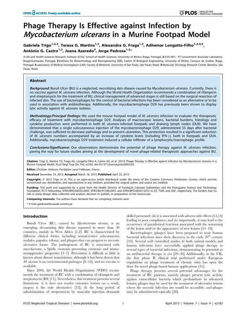
Mycobacteriophage D29 shows a broad lytic activityagainst M. ulcerans isolates in vitro
We first tested the lytic activity of different mycobacterio-
phages against several M. ulcerans isolates. The results for the
plaque formation on the tested M. ulcerans strains are given in
Table 2. We observed that some phages were more strain-
specific, such as the phages Adjutor, Kostya and Brujita, and
others presented a more narrow lytic host range spectrum (L5,
Chah and Phaedrus). A cluster of three phages, namely D29,
Bxz2 and Tweety, displayed the broadest lytic host range
spectrum and highest lytic activity against representative strains
of M. ulcerans. In line with a previous report [36], D29 phage
showed the broadest lytic host range spectrum amongst the
tested mycobacteriophages, affecting M. ulcerans isolates with
genetic heterogeneity, variable phenotypic characteristics and
from different geographic origins (Table 1). Based on these
results, we selected mycobacteriophage D29 for in vivo thera-
peutic studies against infection with M. ulcerans 1615, a well
characterized and stable strain that presents a mycolactone
profile identical to that of African strains [32].
Treatment with mycobacteriophage D29 preventsulceration caused by M. ulcerans and decreases thebacterial load in both the footpad and the DLN
To investigate the efficacy of mycobacteriophage D29 treatment
for the control of M. ulcerans, we used a footpad mouse model of
infection [34,38,39]. Mice were subcutaneously infected in
footpads with 5.5 log10 AFB of M. ulcerans strain 1615. At day
33 post-infection, when footpad swelling had reached 3.0 mm
(Figure 1A), mice were subcutaneously injected in the footpad with
a single dose of mycobacteriophage D29 (8 log10 PFU) or with the
vehicle MPB as a control.
In both control-infected and mycobacteriophage D29 treated
mice we observed an initial footpad swelling (Figure 1A). However,
at day 68 post-infection, footpads of non-treated mice started
showing signs of ulceration, while in mycobacteriophage D29
treated mice the progression of swelling halted after day 91 post-
infection (day 58 post-treatment) (Figure 1A). Furthermore, in
mycobacteriophage D29 treated mice, we observed a progressive
reduction of footpad swelling, until initial treatment values,
recorded by day 150 post-infection. Moreover, signs of ulceration
were continuously absent during the period of experimental
infection (Figure 1A). The administration of mycobacteriophage
D29 or vehicle MPB alone did not induce significant swelling of
the footpad (data not shown).
Regarding M. ulcerans growth in infected footpads of non-treated
mice, we observed a significant bacterial proliferation over the
course of experimental infection (P,0.01) (Figure 1B). On the
other hand, in footpads of mycobacteriophage D29 treated mice,
we observed a significant reduction in CFU counts (P,0.001) at
day 68 post-infection (day 35 post-treatment), following the
administration of a single dose of mycobacteriophage D29 on
day 33 post-infection (Figure 1B).
As previously described [38–40], we found that M. ulcerans
disseminates to the DLN after footpad infection (Figure 1C ),
probably due to continuous lymphatic dissemination of bacteria
either freely or shuttled within phagocytes. Here we show a
significant reduction in CFU counts (P,0.05) in the DLN of
mycobacteriophage D29 treated mice, as compared with non-
treated counterparts, at day 68 post-infection (day 35 post-
treatment) (Figure 1C), correlating with the reduction of M. ulcerans
numbers in the footpads.
Mycobacteriophage D29 disseminates from footpad tothe DLN
It is well known that bacteriophages can disseminate from the
administration site and reach several organs such as lymph nodes,
spleen and liver, which are the primary sites involved in phage
clearance [41,42]. In order to investigate the possible dissemina-
tion of mycobacteriophage D29, we determined phage titres in the
footpad, DLN, spleen and blood after its inoculation in M. ulcerans
infected footpads.
As shown in Figure 2, mycobacteriophage D29 numbers
significantly decreased (P,0.001) in infected footpads from 2 to
24 h post-treatment and no phages could be detected after this
time point (Figure 2). Phage numbers were also detected in the
DLN, as early as 2 h after the administration in infected footpads,
time point at which maximum phage counts were obtained
(Figure 2). After 24 h, we observed a significant decrease
(P,0.001) in phage titers in the DLN, but phages were still
present until day 15 post-treatment (Figure 2). No phages could be
detected in the DLN by the end of the experimental period of
infection (day 35 post-treatment) (Figure 2).
Phage Therapy: M. ulcerans Experimental Infection
PLOS Neglected Tropical Diseases | www.plosntds.org 3 April 2013 | Volume 7 | Issue 4 | e2183
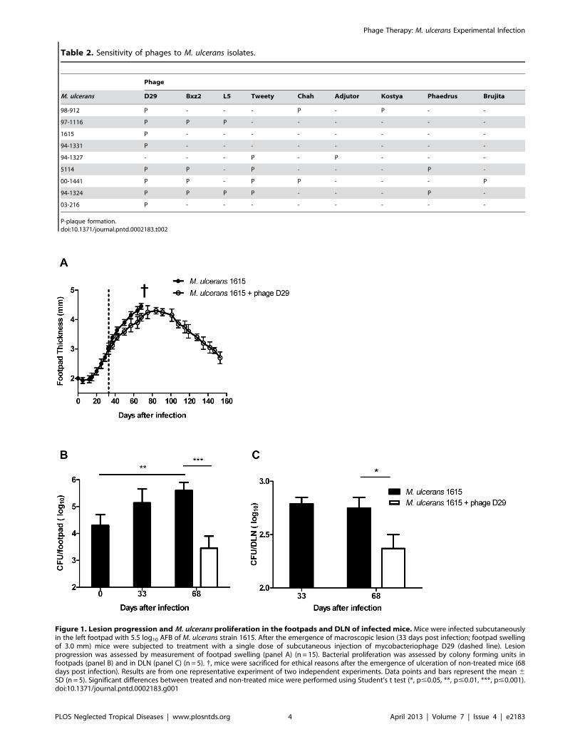
Table 2. Sensitivity of phages to M. ulcerans isolates.
Phage
M. ulcerans D29 Bxz2 L5 Tweety Chah Adjutor Kostya Phaedrus Brujita
98-912 P - - - P - P - -
97-1116 P P P - - - - - -
1615 P - - - - - - - -
94-1331 P - - - - - - - -
94-1327 - - - P - P - - -
5114 P P - P - - - P -
00-1441 P P - P P - - - P
94-1324 P P P P - - - P -
03-216 P - - - - - - - -
P-plaque formation.doi:10.1371/journal.pntd.0002183.t002
Figure 1. Lesion progression and M. ulcerans proliferation in the footpads and DLN of infected mice. Mice were infected subcutaneouslyin the left footpad with 5.5 log10 AFB of M. ulcerans strain 1615. After the emergence of macroscopic lesion (33 days post infection; footpad swellingof 3.0 mm) mice were subjected to treatment with a single dose of subcutaneous injection of mycobacteriophage D29 (dashed line). Lesionprogression was assessed by measurement of footpad swelling (panel A) (n = 15). Bacterial proliferation was assessed by colony forming units infootpads (panel B) and in DLN (panel C) (n = 5). {, mice were sacrificed for ethical reasons after the emergence of ulceration of non-treated mice (68days post infection). Results are from one representative experiment of two independent experiments. Data points and bars represent the mean 6SD (n = 5). Significant differences between treated and non-treated mice were performed using Student’s t test (*, p#0.05, **, p#0.01, ***, p#0.001).doi:10.1371/journal.pntd.0002183.g001
Phage Therapy: M. ulcerans Experimental Infection
PLOS Neglected Tropical Diseases | www.plosntds.org 4 April 2013 | Volume 7 | Issue 4 | e2183
D29 phages were also detected in the spleen (2.2 log1060.25)
and in the serum (2.3 log1060.17) of mycobacteriophage D29
treated mice as early as 2 h post-treatment but were no longer
detectable until the end of the experimental period.
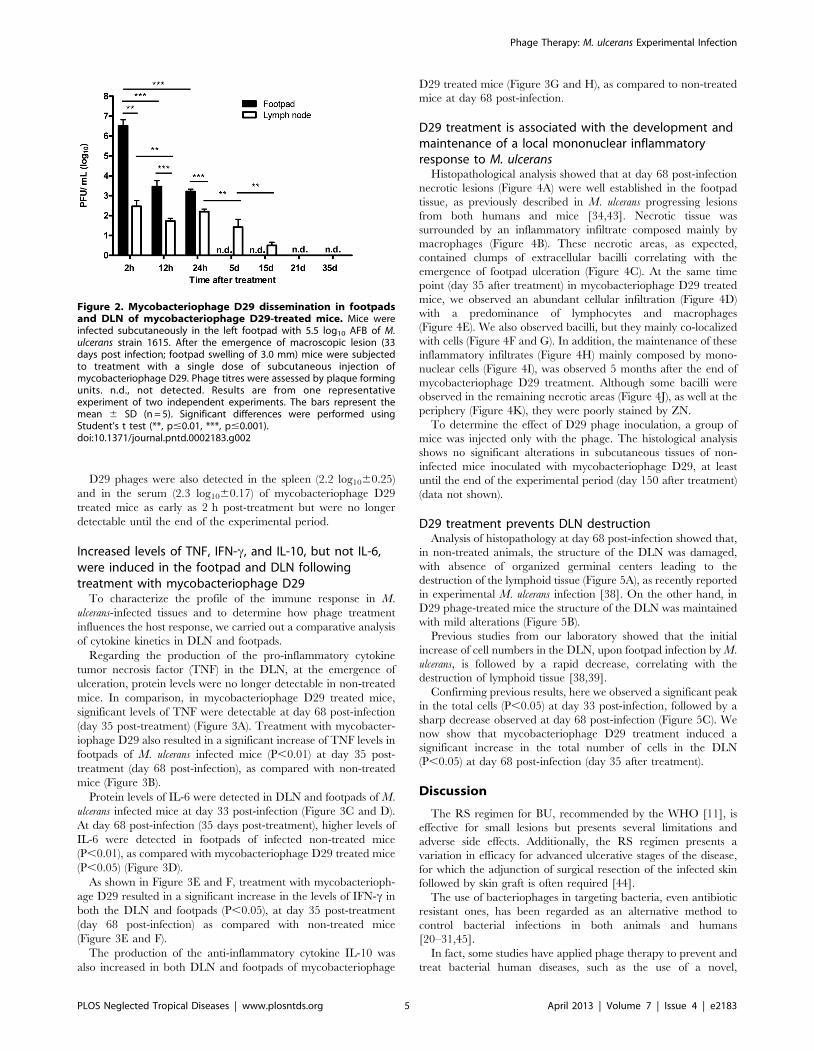
Increased levels of TNF, IFN-c, and IL-10, but not IL-6,were induced in the footpad and DLN followingtreatment with mycobacteriophage D29
To characterize the profile of the immune response in M.
ulcerans-infected tissues and to determine how phage treatment
influences the host response, we carried out a comparative analysis
of cytokine kinetics in DLN and footpads.
Regarding the production of the pro-inflammatory cytokine
tumor necrosis factor (TNF) in the DLN, at the emergence of
ulceration, protein levels were no longer detectable in non-treated
mice. In comparison, in mycobacteriophage D29 treated mice,
significant levels of TNF were detectable at day 68 post-infection
(day 35 post-treatment) (Figure 3A). Treatment with mycobacter-
iophage D29 also resulted in a significant increase of TNF levels in
footpads of M. ulcerans infected mice (P,0.01) at day 35 post-
treatment (day 68 post-infection), as compared with non-treated
mice (Figure 3B).
Protein levels of IL-6 were detected in DLN and footpads of M.
ulcerans infected mice at day 33 post-infection (Figure 3C and D).
At day 68 post-infection (35 days post-treatment), higher levels of
IL-6 were detected in footpads of infected non-treated mice
(P,0.01), as compared with mycobacteriophage D29 treated mice
(P,0.05) (Figure 3D).
As shown in Figure 3E and F, treatment with mycobacterioph-
age D29 resulted in a significant increase in the levels of IFN-c in
both the DLN and footpads (P,0.05), at day 35 post-treatment
(day 68 post-infection) as compared with non-treated mice
(Figure 3E and F).
The production of the anti-inflammatory cytokine IL-10 was
also increased in both DLN and footpads of mycobacteriophage
D29 treated mice (Figure 3G and H), as compared to non-treated
mice at day 68 post-infection.
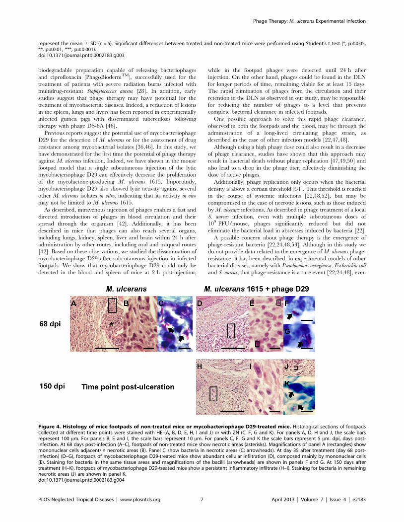
D29 treatment is associated with the development andmaintenance of a local mononuclear inflammatoryresponse to M. ulcerans
Histopathological analysis showed that at day 68 post-infection
necrotic lesions (Figure 4A) were well established in the footpad
tissue, as previously described in M. ulcerans progressing lesions
from both humans and mice [34,43]. Necrotic tissue was
surrounded by an inflammatory infiltrate composed mainly by
macrophages (Figure 4B). These necrotic areas, as expected,
contained clumps of extracellular bacilli correlating with the
emergence of footpad ulceration (Figure 4C). At the same time
point (day 35 after treatment) in mycobacteriophage D29 treated
mice, we observed an abundant cellular infiltration (Figure 4D)
with a predominance of lymphocytes and macrophages
(Figure 4E). We also observed bacilli, but they mainly co-localized
with cells (Figure 4F and G). In addition, the maintenance of these
inflammatory infiltrates (Figure 4H) mainly composed by mono-
nuclear cells (Figure 4I), was observed 5 months after the end of
mycobacteriophage D29 treatment. Although some bacilli were
observed in the remaining necrotic areas (Figure 4J), as well at the
periphery (Figure 4K), they were poorly stained by ZN.
To determine the effect of D29 phage inoculation, a group of
mice was injected only with the phage. The histological analysis
shows no significant alterations in subcutaneous tissues of non-
infected mice inoculated with mycobacteriophage D29, at least
until the end of the experimental period (day 150 after treatment)
(data not shown).
D29 treatment prevents DLN destructionAnalysis of histopathology at day 68 post-infection showed that,
in non-treated animals, the structure of the DLN was damaged,
with absence of organized germinal centers leading to the
destruction of the lymphoid tissue (Figure 5A), as recently reported
in experimental M. ulcerans infection [38]. On the other hand, in
D29 phage-treated mice the structure of the DLN was maintained
with mild alterations (Figure 5B).
Previous studies from our laboratory showed that the initial
increase of cell numbers in the DLN, upon footpad infection by M.
ulcerans, is followed by a rapid decrease, correlating with the
destruction of lymphoid tissue [38,39].
Confirming previous results, here we observed a significant peak
in the total cells (P,0.05) at day 33 post-infection, followed by a
sharp decrease observed at day 68 post-infection (Figure 5C). We
now show that mycobacteriophage D29 treatment induced a
significant increase in the total number of cells in the DLN
(P,0.05) at day 68 post-infection (day 35 after treatment).
Discussion
The RS regimen for BU, recommended by the WHO [11], is
effective for small lesions but presents several limitations and
adverse side effects. Additionally, the RS regimen presents a
variation in efficacy for advanced ulcerative stages of the disease,
for which the adjunction of surgical resection of the infected skin
followed by skin graft is often required [44].
The use of bacteriophages in targeting bacteria, even antibiotic
resistant ones, has been regarded as an alternative method to
control bacterial infections in both animals and humans
[20–31,45].
In fact, some studies have applied phage therapy to prevent and
treat bacterial human diseases, such as the use of a novel,
Figure 2. Mycobacteriophage D29 dissemination in footpadsand DLN of mycobacteriophage D29-treated mice. Mice wereinfected subcutaneously in the left footpad with 5.5 log10 AFB of M.ulcerans strain 1615. After the emergence of macroscopic lesion (33days post infection; footpad swelling of 3.0 mm) mice were subjectedto treatment with a single dose of subcutaneous injection ofmycobacteriophage D29. Phage titres were assessed by plaque formingunits. n.d., not detected. Results are from one representativeexperiment of two independent experiments. The bars represent themean 6 SD (n = 5). Significant differences were performed usingStudent’s t test (**, p#0.01, ***, p#0.001).doi:10.1371/journal.pntd.0002183.g002
Phage Therapy: M. ulcerans Experimental Infection
PLOS Neglected Tropical Diseases | www.plosntds.org 5 April 2013 | Volume 7 | Issue 4 | e2183

Figure 3. Cytokine profile in DLN and footpads of non-treated mice or mycobacteriophage D29-treated mice. Mice were infectedsubcutaneously in the left footpad with 5.5 log10 AFB of M. ulcerans strain 1615. After the emergence of macroscopic lesion (33 days post infection;footpad swelling of 3.0 mm) mice were subjected to treatment with a single dose of subcutaneous injection of mycobacteriophage D29. Levels of theTNF (panel A and B), IL- 6 (panel C and D), IFN-c (panel E and F) and IL-10 (panel G and H) in DLN (panel A, C, E and G) and footpads (panel B, D, F andH) of mice were quantified by ELISA assay. n.d., not detected. Results are from one representative experiment of two independent experiments. Bars
Phage Therapy: M. ulcerans Experimental Infection
PLOS Neglected Tropical Diseases | www.plosntds.org 6 April 2013 | Volume 7 | Issue 4 | e2183
biodegradable preparation capable of releasing bacteriophages
and ciprofloxacin (PhagoBiodermTM), successfully used for the
treatment of patients with severe radiation burns infected with
multidrug-resistant Staphylococcus aureus [28]. In addition, early
studies suggest that phage therapy may have potential for the
treatment of mycobacterial diseases. Indeed, a reduction of lesions
in the spleen, lungs and livers has been reported in experimentally
infected guinea pigs with disseminated tuberculosis following
therapy with phage DS-6A [46].
Previous reports suggest the potential use of mycobacteriophage
D29 for the detection of M. ulcerans or for the assessment of drug
resistance among mycobacterial isolates [36,46]. In this study, we
have demonstrated for the first time the potential of phage therapy
against M. ulcerans infection. Indeed, we have shown in the mouse
footpad model that a single subcutaneous injection of the lytic
mycobacteriophage D29 can effectively decrease the proliferation
of the mycolactone-producing M. ulcerans 1615. Importantly,
mycobacteriophage D29 also showed lytic activity against several
other M. ulcerans isolates in vitro, indicating that its activity in vivo
may not be limited to M. ulcerans 1615.
As described, intravenous injection of phages enables a fast and
directed introduction of phages in blood circulation and their
spread through the organism [42]. Additionally, it has been
described in mice that phages can also reach several organs,
including lungs, kidney, spleen, liver and brain within 24 h after
administration by other routes, including oral and traqueal routes
[42]. Based on these observations, we studied the dissemination of
mycobacteriophage D29 after subcutaneous injection in infected
footpads. We show that mycobacteriophage D29 could only be
detected in the blood and spleen of mice at 2 h post-injection,
while in the footpad phages were detected until 24 h after
injection. On the other hand, phages could be found in the DLN
for longer periods of time, remaining viable for at least 15 days.
The rapid elimination of phages from the circulation and their
retention in the DLN as observed in our study, may be responsible
for reducing the number of phages to a level that prevents
complete bacterial clearance in infected footpads.
One possible approach to solve this rapid phage clearance,
observed in both the footpads and the blood, may be through the
administration of a long-lived circulating phage strain, as
described in the case of other infection models [22,47,48].
Although using a high phage dose could also result in a decrease
of phage clearance, studies have shown that this approach may
result in bacterial death without phage replication [47,49,50] and
also lead to a drop in the phage titer, effectively diminishing the
dose of active phages.
Additionally, phage replication only occurs when the bacterial
density is above a certain threshold [51]. This threshold is reached
in the course of systemic infections [22,48,52], but may be
compromised in the case of necrotic lesions, such as those induced
by M. ulcerans infections. As described in phage treatment of a local
S. aureus infection, even with multiple subcutaneous doses of
109 PFU/mouse, phages significantly reduced but did not
eliminate the bacterial load in abscesses induced by bacteria [22].
A possible concern about phage therapy is the emergence of
phage-resistant bacteria [22,24,48,53]. Although in this study we
do not provide data related to the emergence of M. ulcerans phage-
resistance, it has been described, in experimental models of other
bacterial diseases, namely with Pseudomonas aeruginosa, Escherichia coli
and S. aureus, that phage resistance is a rare event [22,24,48], even
represent the mean 6 SD (n = 5). Significant differences between treated and non-treated mice were performed using Student’s t test (*, p#0.05,**, p#0.01, ***, p#0.001).doi:10.1371/journal.pntd.0002183.g003
Figure 4. Histology of mice footpads of non-treated mice or mycobacteriophage D29-treated mice. Histological sections of footpadscollected at different time points were stained with HE (A, B, D, E, H, I and J) or with ZN (C, F, G and K). For panels A, D, H and J, the scale barsrepresent 100 mm. For panels B, E and I, the scale bars represent 10 mm. For panels C, F, G and K the scale bars represent 5 mm. dpi, days post-infection. At 68 days post-infection (A–C), footpads of non-treated mice show necrotic areas (asterisks). Magnifications of panel A (rectangles) showmononuclear cells adjacent/in necrotic areas (B). Panel C show bacteria in necrotic areas (C; arrowheads). At day 35 after treatment (day 68 post-infection) (D–G), footpads of mycobacteriophage D29-treated mice show abundant cellular infiltration (D), composed mainly by mononuclear cells(E). Staining for bacteria in the same tissue areas and magnifications of the bacilli (arrowheads) are shown in panels F and G. At 150 days aftertreatment (H–K), footpads of mycobacteriophage D29-treated mice show a persistent inflammatory infiltrate (H–I). Staining for bacteria in remainingnecrotic areas (J) are shown in panel K.doi:10.1371/journal.pntd.0002183.g004
Phage Therapy: M. ulcerans Experimental Infection
PLOS Neglected Tropical Diseases | www.plosntds.org 7 April 2013 | Volume 7 | Issue 4 | e2183

more so than antibiotic resistance [22,54]. Even though we cannot
rule out that some phage resistance can occur, the use, in this study,
of a single phage treatment dose greatly reduces this hypothesis.
To characterize the type of immune response associated with
the administration of mycobacteriophage D29 and, particularly,
how phage treatment influences the host immune response against
M. ulcerans, we carried out a comparative analysis of cytokine
kinetics in footpads and DLN, where the initiation of the adaptive
immune response occurs [38]. It is known that the differentiation/
proliferation of mycobacteria-specific lymphocytes can occur in
the DLN, early after M. ulcerans infection, and that effector T cells
are recruited to the site of infection [38], where they mediate
partial protection by enhancing IFN-c-induced macrophage
antimicrobial mechanisms. In agreement, we detected IFN-c in
the DLN, however this host response is not sufficient to inhibit the
proliferation of virulent M. ulcerans, as increasing concentrations of
mycolactone impair the effector activity of macrophages [6].
Interestingly, we observed that mycobacteriophage D29 treatment
results in a significant increase in the total number of cells in the
DLN, as well as in an increase of IFN-c levels, correlating with a
decrease in the number of viable bacteria, both in footpads and
DLN, measured at day 68 post-infection (day 35 post-treatment).
Collectively, these results suggest that the dissemination and
prolonged permanence of phages in the DLN may prevent local
M. ulcerans proliferation and the associated accumulation of
mycolactone, therefore preventing DLN destruction.
As previously described [38–40] and confirmed in this study, the
tissue destruction of the DLN was associated with bacterial
colonization, which is consistent with the spreading of M. ulcerans
from the site of infection via afferent lymphatic drainage [55,56]
On the other hand, the increased immune activation induced in
the DLN of treated mice may explain an immune-mediated
Figure 5. Histology and leukocyte kinetics in DLN of non-treated mice or mycobacteriophage D29-treated mice. Histological sectionsof DLN collected at different time points were stained with HE. For panels A and B the scale bars represent 500 mm. At 68 days post-infection DLN ofnon-treated mice show severe damage of the lymphoid tissue (panel A). At day 35 after treatment (day 68 post-infection), DLN structure ofmycobacteriophage D29 treated animals was maintained (panel B). Total number of cells in the DLN was determined in DLN suspensions (panel C).Results are from one representative experiment of two independent experiments. n.d., not determined. In panel C data points represent the mean 6SD (n = 5). Significant differences between treated and non-treated mice were performed using Student’s t test (*, p#0.05).doi:10.1371/journal.pntd.0002183.g005
Phage Therapy: M. ulcerans Experimental Infection
PLOS Neglected Tropical Diseases | www.plosntds.org 8 April 2013 | Volume 7 | Issue 4 | e2183

control of bacterial proliferation in the footpad, despite the lack of
phages at the primary site of infection. In fact, as previously
described, IFN-c, and TNF play a protective role in immunity
against M. ulcerans experimental infections, contributing to control
bacterial proliferation [6,7,57]. Accordingly, we show here that
mycobacteriophage D29 treatment was associated with increased
levels of both IFN-c and TNF in M. ulcerans-infected footpads
[6,7], correlating with a predominance of a mononuclear infiltrate
and prevention of ulceration at 150 days post-infection. Addition-
ally, our histological data show that bacilli are still present in
footpad tissues, albeit with an altered morphology and poorly
stained with ZN. Although this observation may indicate that, as
described [39], M. ulcerans bacilli underwent degradation after
bacterial killing, a possible relapse of M. ulcerans infection after the
150 day period of experimental infection was not checked in this
study.
Here we show that IL-6 concentration was markedly lower in
footpads of mycobacteriophage D29 treated mice at day 68 post-
infection (day 35 after treatment) as compared to non-treated
mice, confirming that footpad tissue damage is less severe in D29
phage treated footpads.
Although production of IL-10 could be detected in skin lesions
of patients with BU [58,59] the exact role of this cytokine in the
progression of M. ulcerans infection has to be further analyzed. In
this experimental setting, it is possible that the increased levels of
IL-10 in mycobacteriophage D29 treated mice are modulating the
activity of the pro-inflammatory cytokines.
In summary, our results show that administration of the lytic
mycobacteriophage D29: (i) is an effective approach for reducing
M. ulcerans-induced pathology in the mouse model of infection; (ii)
reduced M. ulcerans numbers in the footpad and the DLN,
associated with increased IFN-c and TNF levels and; (iii) is not
associated with detectable side effects over a minimum delay of
150 day observation period.
To our knowledge, this is the first study on mycobacteriophage
therapy against M. ulcerans in vivo infection. It should be pointed out
that mice were treated at an advanced stage of M. ulcerans
infection, which is relevant for human infection since BU patients
often seek medical treatment in advanced stages of the disease.
More detailed studies examining the effects of phage dosage,
routes and timing of administration, as well as on pharmacoki-
netics, will be needed to determine if phage therapy will provide a
consistent alternative/supplement for the treatment of BU.
Although the development of a therapeutic regimen using phages
will involve a commitment to fulfill the scientific requirements of
current pharmaceutical agencies, our encouraging results justifies
further investigation on the potential of phages for the manage-
ment of this mycobacteriosis. Moreover, mycobacteriophage D29
represents an ideal agent from a regulatory standpoint in that it
has been fully characterized genetically [60] and is able to be used
on a stand-alone basis. Another approach could be based on the
therapeutic use of lysins bacteriophage proteins produced at the
end of a lytic life cycle, designed to attack peptidoglycan in order
to allow the release of the new synthesized phage particles [61].
Acknowledgments
We are grateful to Dr. Graham F. Hatfull for the kind donation of the
mycobacteriophages used in this study. The authors would like to thank
Luis Martins and Miguel Carneiro for laboratory assistance.
Author Contributions
Conceived and designed the experiments: GT AGC JA JP. Performed the
experiments: GT TGM AGF. Analyzed the data: GT ALF. Contributed
reagents/materials/analysis tools: JP AGC. Wrote the paper: GT AGF
AGC JA JP.
References
1. Portaels F, Silva MT, Meyers WM (2009) Buruli ulcer. Clin Dermatol 27: 291–
305.
2. Walsh DS, Portaels F Meyers WM (2011) Buruli ulcer: Advances inunderstanding Mycobacterium ulcerans infection. Dermatol Clin 29: 1–8.
3. Hong H, Spencer JB, Porter JL, Leadlay PF, Stinear T (2005) A novel
mycolactone from a clinical isolate of Mycobacterium ulcerans provides evidence foradditional toxin heterogeneity as a result of specific changes in the modular
polyketide synthase. Chem Bio Chem 6: 643–648.
4. George KM, Pascopella L, Welty DM, Small PL (2000) A Mycobacterium ulcerans
toxin, mycolactone, causes apoptosis in guinea pig ulcers and tissue culture cells.Infect Immun 68: 877–883.
5. George KM, Chatterjee D, Gunawardana G, Welty D, Hayman J, et al. (1999)
Mycolactone: a polyketide toxin from Mycobacterium ulcerans required forvirulence. Science 283: 854–857.
6. Torrado E, Fraga AG, Logarinho E, Martins G, Carmona JA, et al. (2010) IFN-
c-dependent activation of macrophages during experimental infections byMycobacterium ulcerans is impaired by the toxin mycolactone. J Immunol 184: 947–
955.
7. Torrado E, Adusumilli S, Fraga AG, Small PLC, Castro AG, et al. (2007)Mycolactone-mediated inhibition of tumor necrosis factor production by
macrophages infected with Mycobacterium ulcerans has implications for the control
of infection. Infect Immun 75: 3979–3988.
8. Marsollier L, Robert R, Aubry J, Saint Andre JP, Kouakou H, et al. (2002)Aquatic insects as a vector for Mycobacterium ulcerans. Appl Environ Microbiol 68:
4623–4628.
9. Portaels F, Meyers WM, Ablordey A, Castro AG, Chemlal K, et al. (2008) Firstcultivation and characterization of Mycobacterium ulcerans from the environment.
PLoS Negl Trop Dis 2: e178.
10. Silva MT, Portaels F, Pedrosa J (2009) Pathogenetic mechanisms of theintracellular parasite Mycobacterium ulcerans leading to Buruli ulcer. Lancet Infect
Dis 9: 699–710.
11. World Health Organization (2004) Provisional guidance on the role of specificantibiotics in the management of Mycobacterium ulcerans disease (Buruli ulcer).
WHO/CDS/CPE/GBUI/2004. 10.
12. Kibadi K, Boelaert M, Fraga AG, Kayinua M, Longatto-Filho A, et al. (2010)Response to treatment in a prospective cohort of patients with large ulcerated
lesions suspected to be Buruli Ulcer (Mycobacterium ulcerans disease). PLoS Negl
Trop Dis 4: e736.
13. Nienhuis WA, Stienstra Y, Thompson WA, Awuah PC, Abass KM, et al. (2010)Antimicrobial treatment for early, limited Mycobacterium ulcerans infection: a
randomised controlled trial. Lancet 375: 664–672.
14. Sarfo FS, Phillips R, Asiedu K, Ampadu E, Bobi N, et al. (2010) Clinical Efficacyof combination of rifampin and streptomycin for treatment of Mycobacterium
ulcerans disease. Antimicrob Agents Chemother 54: 3678–3685.
15. Ruf MT, Chauty A, Adeye A, Ardant MF, Koussemou H (2011) Secondary
Buruli ulcer skin lesions emerging several months after completion ofchemotherapy: paradoxical reaction or evidence for immune protection? PLoS
Negl Trop Dis 5: e1252.
16. Sopoh GE, Dossou AD, Brun LV, Barogui YT, Houezo JG, et al. (2010) Severemultifocal form of Buruli Ulcer after streptomycin and rifampin treatment:
comments on possible dissemination mechanisms. Am J Trop Med Hyg 83:307–313.
17. O’Brien DP, Robson ME, Callan PP, McDonald AH (2009) ‘‘‘Paradoxical’’’
immune-mediated reactions to Mycobacterium ulcerans during antibiotic treatment:a result of treatment success, not failure. Med J Aust 191: 564–566.
18. Gordon CL, Buntine JA, Hayman JA, Lavender CJ, Fyfe JAM, et al. (2010) All-
oral antibiotic treatment for Buruli Ulcer: a report of four patients. PLoS Negl
Trop Dis 4: e770.
19. Abedon ST, Kuhl SJ, Blasdel BG, Kutter EM (2011) Phage treatment of humaninfections. Bacteriophage 1: 66–85.
20. Ahmad SI (2002) Treatment of post-burns bacterial infections by bacteriophag-
es, specifically ubiquitous Pseudomonas spp. notoriously resistant to antibiotics.Med Hypoth 58: 327–331.
21. Biswas B, Adhya S, Washart P, Paul B, Trostel AN, et al. (2002) Bacteriophage
Therapy rescues mice bacteremic from a clinical isolate of vancomycin-resistantEnterococcus faecium. Infect immun 70(1): 204–210.
22. Capparelli R, Parlato M, Borriello G, Salvatore P, Iannelli D (2007)
Experimental phage therapy against Staphylococcus aureus in mice. AntimicrobAgents Chemother 51: 2765–2773.
23. Verma V, Harjai K, Chhibber S (2009) Characterization of a T7-like lytic
bacteriophage of Klebsiella pneumoniae B5055: a potential therapeutic agent. CurrMicrobiol 59: 274–281.
Phage Therapy: M. ulcerans Experimental Infection
PLOS Neglected Tropical Diseases | www.plosntds.org 9 April 2013 | Volume 7 | Issue 4 | e2183

24. McVay CS, Velasquez M, Fralick JA (2007) Phage therapy of Pseudomonas
aeruginosa infection in a mouse burn wound model. Antimicrob AgentsChemother 51: 1934–1938.
25. Hung CH, Kuo CF, Wang CH, Wu CM, Tsao N (2011) Experimental Phage
Therapy in treating Klebsiella pneumoniae-mediated liver abscesses and bacteremiain mice. Antimicrob Agents Chemother 55: 1358–1365.
26. Wills QF, Kerrigan C, Soothill JS (2005) Experimental bacteriophage protectionagainst Staphylococcus aureus abscesses in a rabbit model. Antimicrob Agents
Chemother 49: 1220–1221.
27. Weber-Dabrowska B, Mulczyk M, Gorski A (2000) Bacteriophage therapy ofbacterial infections: an update of our institute’s experience. Arch Immunol Ther
Ex 48: 547–551.28. Jikia D, Chkhaidze N, Imedashvili E, Mgaloblishvili I, Tsitlanadze G, et al.
(2005) The use of a novel biodegradable preparation capable of the sustainedrelease of bacteriophages and ciprofloxacin , in the complex treatment of
multidrug-resistant Staphylococcus aureus-infected local radiation injuries caused by
exposure to Sr90. Clin Exp Dermatol 30: 23–26.29. Barrow P, Lovell M, Jr Berchieri A (1998) Use of Lytic Bacteriophage for control
of experimental Escherichia coli septicemia and meningitis in chickens and calves.Clin Diagn Lab Immunol 5: 294–298.
30. Oliveira A, Sereno R, Azeredo J (2010) In vivo efficiency evaluation of a phage
cocktail in controlling severe colibacillosis in confined conditions andexperimental poultry houses. Vet Microbiol 146: 303–308.
31. Wright A, Hawkins CH, Anggard EE, Harper DR (2009) A controlled clinicaltrial of a therapeutic bacteriophage preparation in chronic otitis due to antibiotic
resistant Pseudomonas aeruginosa: a preliminary report of efficacy. Clin Otolaryngol34: 349–357.
32. Mve-Obiang A, Lee RE, Portaels F, Small PLC (2003) Heterogeneity of
mycolactones produced by clinical isolates of Mycobacterium ulcerans: implicationsfor virulence. Infect Immun 71: 774–783.
33. Stinear TP, Hong H, Frigui W, Pryor MJ, Brosch R, et al. (2005) Commonevolutionary origin for the unstable virulence plasmid pMUM found in
geographically diverse strains of Mycobacterium ulcerans. J Bacteriol 187: 1668–
1676.34. Oliveira MS, Fraga AG, Torrado E, Castro AG, Pereira JP, et al. (2005)
Infection with Mycobacterium ulcerans induces persistent inflammatory responses inmice. Infect Immun 73: 6299–6310.
35. Sambrook J, Russel DW (2001) Molecular Cloning: A Laboratory Manual. NewYork: Cold Spring Harbor Laboratory Press.
36. Rybniker J, Kramme S, Small PL (2006) Host range of 14 mycobacteriophages
in Mycobacterium ulcerans and seven other mycobacteria including Mycobacterium
tuberculosis–application for identification and susceptibility testing. J Med
Microbiol 55: 37–42.37. Shepard CC, McRae DH (1968) A method for counting acid-fast bacteria.
Int J Lepr Other Mycobact Dis 36: 78–82.
38. Fraga AG, Cruz A, Martins TG, Torrado E, Saraiva M, et al. (2011)Mycobacterium ulcerans triggers T-cell immunity followed by local and regional but
not systemic immunosuppression. Infec Immun 79: 421–430.39. Martins TG, Gama JB, Fraga AG, Saraiva M, Silva MT, et al. (2012) Local and
regional re-establishment of cellular immunity during curative antibiotherapy ofmurine Mycobacterium ulcerans infection. PLoS One 7: e32740.
40. Fraga AG, Martins TG, Torrado E, Huygen K, Portaels F, et al. (2012) Cellular
immunity confers transient protection in experimental Buruli Ulcer followingBCG or mycolactone-negative Mycobacterium ulcerans vaccination. PLoS 0ne 7:
e33406.41. Calendar RL, Abedon ST (2006) The Bacteriophages. New York: Oxford
University Press.
42. Dabrowska K, Switata-Jelen K, Opolski A, Weber-Dabrowska B, Gorski A(2005) Bacteriophage penetration in vertebrates. J Appl Microbiol 98: 7–13.
43. Torrado E, Fraga AG, Castro AG, Stragier P, Meyers WM, et al. (2007)
Evidence for an intramacrophage growth phase of Mycobacterium ulcerans. InfectImmun 75: 977–987.
44. Asiedu K, Scherpbier R, Raviglione M (2000) Buruli Ulcer, Mycobacterium ulcerans
infection. World Health Organization WHO/CDS/CPE/GBUI/2000 1: 118.
45. Bruttin A, Brussow H (2005) Human Volunteers Receiving Escherichia coli phage
T4 orally: a safety test of phage therapy. Antimicrob Agents Chemother 49:
2874–2878.
46. McNerney R, Traore H (2005) Mycobacteriophage and their application to
disease control. J Appl Microbiol 99: 223–233.
47. Merril CR, Biswas B, Carlton R, Jensen NC, Creed GJ, et al. (1996) Long-circulating bacteriophage as antibacterial agents. Proc Natl Acad Sci U S A 93:
3188–3192.
48. Capparelli R, Ventimiglia I, Roperto S, Fenizia D, Iannelli D (2006) Selection ofan Escherichia coli O157: H7 bacteriophage for persistence in the circulatory
system of mice infected experimentally. Clin Microbiol Infect 12: 248–253.
49. McNerney R, Kambashi BS, Kinkese J, Tembwe R, Godfrey-Faussett P (2004)Development of a bacteriophage phage replication assay for diagnosis of
pulmonary tuberculosis. J Clin Microbiol 42: 2115–2120.
50. Rabinovitch A, Aviram I, Zaritsky A (2003) Bacterial debris — an ecologicalmechanism for coexistence of bacteria and their viruses. J Theor Biol 224: 377–
383.
51. Payne RJH, Phil D, Jansen VAA, Kingdom U (2000) Phage therapy: The
peculiar kinetics of self-replicating pharmaceuticals. Clin Pharmacol Therap:
225–230.
52. Tiwari BR, Kim S, Rahman M, Kim J (2011) Antibacterial efficacy of lytic
Pseudomonas bacteriophage in normal and neutropenic mice models. J Microbiol
49: 994–999.
53. Loc Carrillo C, Atterbury RJ, El-Shibiny A, Connerton PL, Dillon E, et al.
(2005) Bacteriophage therapy to reduce Campylobacter jejuni colonization of broiler
chickens. Appl Environ Microbiol 71: 6554–6563.
54. Sulakvelidze A, Alavidze Z, JrMorris GJ (2001) Bacteriophage therapy.
Antimicrob Agents Chemother 45: 649–659.
55. Addo P, Owusu E, Adu-Addai B, Quartey M, Abbas M, et al. (2005) Findingsfrom a Buruli ulcer mouse model study. Ghana Med J 39: 86–93.
56. Coutanceau E, Marsollier L, Brosch R, Perret E, Goossens P, et al. (2005)
Modulation of the host immune response by a transient intracellular stage ofMycobacterium ulcerans: the contribution of endogenous mycolactone toxin. Cell
Microbiol 7: 1187–1196.
57. Phillips R, Kuijper S, Benjamin N, Wansbrough-Jones M, Wilks M, et al. (2004)In vitro killing of Mycobacterium ulcerans by acidified nitrite. Antimicrob Agents
Chemother 48: 3130–3132.
58. Kiszewski AE, Becerril E, Aguilar LD, Kader ITA, Myers W, et al. (2006) Thelocal immune response in ulcerative lesions of Buruli disease. Clin Exp Immunol
143: 445–451.
59. Prevot G, Bourreau E, Pascalis H, Pradinaud R, Tanghe A, et al. (2004)Differential production of systemic and intralesional Gamma Interferon and
Interleukin-10 in nodular and ulcerative forms of Buruli disease. Infect Immun
72: 958–965.
60. Ford ME, Sarkis GJ, Belanger AE, Hendrix RW, Hatfull GF (1998) Genome
structure of mycobacteriophage D29: implications for phage evolution. J Mol
Biol 279: 143–164.
61. Fischetti VA (2010) Bacteriophage endolysins: a novel anti-infective to control
Gram-positive pathogens. Int J Med Microbiol 300: 357–362.
Phage Therapy: M. ulcerans Experimental Infection
PLOS Neglected Tropical Diseases | www.plosntds.org 10 April 2013 | Volume 7 | Issue 4 | e2183
Related Documents


