Pftaire1 (Cyclin Dependent Kinase14): Role and Function in Axonal Outgrowth during the development of the CNS Fatemeh Kamkar Thesis Submitted to the Faculty of Graduate and Postdoctoral Studies in partial fulfillment of the requirements for the Doctorate in Philosophy degree in Cellular and Molecular Medicine Department of Cellular and Molecular Medicine Faculty of Medicine University of Ottawa © Fatemeh Kamkar, Ottawa, Canada, 2015

Welcome message from author
This document is posted to help you gain knowledge. Please leave a comment to let me know what you think about it! Share it to your friends and learn new things together.
Transcript
Pftaire1 (Cyclin Dependent Kinase14):
Role and Function in Axonal Outgrowth
during the development of the CNS
Fatemeh Kamkar
Thesis Submitted to the
Faculty of Graduate and Postdoctoral Studies
in partial fulfillment of the requirements
for the Doctorate in Philosophy degree
in Cellular and Molecular Medicine
Department of Cellular and Molecular Medicine
Faculty of Medicine
University of Ottawa
© Fatemeh Kamkar, Ottawa, Canada, 2015
II
ABSTRACT
Cyclin Dependent Kinase (Cdk) family members play a role in CNS development.
Cyclin Dependent Kinase 5 (Cdk5) is well known for its fundamental role in neuronal
development and axogenesis, as well as, cell death. Other Cdks include Pctaire and
Pftaire. Inhibition of Pctaire results in increased axon outgrowth, however, the role and
function of Pftaire is unknown. Pftaire1 is a novel member of the Cdk family that was
initially detected in a screen for cdc2-like kinases. Unpublished data from our lab reveals
that Pftaire1 (Eip63E) deficiency in Drosophila melanogaster results in defects in the axon
and neuronal structure of the ventral nerve cord (VNC). In mammals, Pftaire1 is highly,
expressed in the CNS. Here, we proposed that Pftaire1 might have a role in axon
outgrowth. To investigate the role of Pftaire1 in mammals, the first germline Pftaire1
knockout mice were generated. Considering the severe effects of Eip63E deficiency in
Drosophila and the homology between mammalian and fly Pftaire1, CNS defects in the
mouse were anticipated. However, to date, no gross abnormalities have been detected
in the overall morphology, fertility, life span, or anatomical brain structures of the Pftaire1
deficient mice. This may be due to the presence of other post-mitotic Cdk proteins that
are highly similar to Pftaire1. For instance, mammals possess Pftaire (1, and 2), as well as,
Pctaire (1, 2, and 3), while Drosophila only possess the Pftaire1 orthologue where the
Pftaire2 and Pctaire (1, 2, and 3) are absent. Furthermore, the mice were of mixed
background. In spite of this, we demonstrated that Pftaire1 deficient neurons showed
increased axon length, in the initial phases of culture. This was confirmed by expression
of dominant negative (DN) D228N-Pftaire1 in wild type neurons. Also classification of
III
axons into different ranges, reveals a higher percentage of hyperextended neurites in
D228N and Pftaire1 knockout mice. The mechanism by which Pftaire1 controls axon
outgrowth is unknown. In this study we show that, Pftaire1 interacts physically with the
small GTPase proteins Rac1, Cdc42, and RhoA. Importantly, we showed that Pftaire1
phosphorylates GDP-RhoA on a serine residue. We propose that this regulates RhoA
activity, which in turn controls axon outgrowth.

IV
ACKNOWLEDGEMENTS
The dissertation represented here is the result of contribution, guidance, and
efforts of a vast team during my journey as a PhD student at the University of Ottawa. I
have been privileged to have their great support and enjoyed working with each and
every person.
First, I would like to express my appreciation to my thesis supervisor, Dr. David
Park, for giving me the opportunity to discover the wonders of science and providing me
with guidance and constructive comments along the way. I have gained invaluable
knowledge and expertise from working under your supervision. Thank you for supporting
me through this challenging and rewarding experience.
I would like to express my deep appreciation to Dr. Antoine Hakim for his
invaluable support through this arduous phase of my life and for giving me hope and
courage to continue.
I would like to express my deep and sincere gratitude to Dr. Ruth Slack for her
invaluable support in the scientific field as our collaborator, as well as, her caring and
moral support. It would have been impossible for me to survive through the difficult
moments and be here today without your help.
I would like to express my appreciation to my advisory committee members, Dr.
Paul Albert, Dr. Antonio Colavita and Dr. Marc Ekker for their sound advice and insightful
comments during the development of my PhD research.
I would like to further extend my thanks to Dr. Paul Albert and Maribeth Lazzaro
for providing us with Pftaire1 AV viruses and plasmids. I would like to thank Dr. Gary

V
Bokoch for providing us with RhoA plasmids and Leticia Sanchez Alvarez for construction
of AV viruses.
My warm thanks to the members of the “Park” lab. To those who I am indebted
for all the invaluable help during this work, scientific discussions and for the friendly
moments at the times of joy and desperation. Thanks to Dr. Yasmilde Rodriguez Gonzalez
for her professional and moral support. For her insightful comments and invaluable
laboratory assistance. For her critical review of this dissertation. Many thanks to Steve
Callaghan and Carmen Estey for their valuable technical and scientific support during all
this years. I would like to especially thank Dr. Dianbo Qu, Yi-Hong Zhang, and Dr. Paymaan
Jafar-Nejad for their valuable insights and contributions to this work. I would also like to
express, my heartfelt thanks to my very good friend, Farzaneh Safarpour, for her great
technical, scientific and moral support at all times. My special thanks to Paul Marcogliese
and Elizabeth Abdel-Messih for their support and assistance in editing the primary draft
of my dissertation and their support at many occasions. I would like to extend my
gratitude to all my fellow lab members from the past and the present who made this a
memorable experience Dr. Mandana Amini, Dr. Mohammad Parsanejad, Dr. Grace
Iyirhiaro, Dr. Zohreh Galehdar, Dr. En Huang, Dr. Emdadul Haque, Wassamon Boonying,
Sarah Hewitt, and Katie Don-Carolis.
My special thanks to the Cellular and Molecular Medicine and Neuroscience
administrative and technical staff Karen Littlejohn, Bea Robertson, Charlotte McCusker,
Sylvie Deblois, Blanche Danielle, Marie La Fontant, Nancy MacDonald, Donna Hooper,

VI
Lynda Jui, Kim Wong, and the rest of my family at the “Neurosciences” and “CMM” for
their great assistance and moral support.
Also my special thanks to Devon Svoboda, Dr. Firouz Fallahi and Dr. Golnaz Sedigh,
for their expert help
And last but not least, my deep and heartfelt thanks to my family that supported
me with love and patience not only during my research life but also through every
moment of my life. My dad who was my true inspiration in life with his everlasting passion
for learning; my loving mum, my caring sisters Mary and Lily, my brothers Bahram and
Moayed for their constant support and encouragement, and my little niece and nephew
Parmida and Ryan that helped me survive the frustrations of science with their cheerful
spirit.
VII
TABLE OF CONTENTS
ABSTRACT ......................................................................................................................................... II
ACKNOWLEDGEMENTS ................................................................................................................... IV
TABLE OF CONTENTS...................................................................................................................... VII
LIST OF FIGURES ............................................................................................................................... X
LIST OF TABLES ................................................................................................................................ XI
LIST OF ABBREVIATIONS ................................................................................................................ XII
1. INTRODUCTION ........................................................................................................................ 2
1.1. Cyclin Dependent Kinases ............................................................................................ 3
1.1.1. Functions of Cdks ................................................................................................. 3
1.1.1.1. The Mitotic Cdks .............................................................................................. 4
1.1.1.2. Post-Mitotic Cdks ............................................................................................. 4
1.1.2. The Structure of Cdks ........................................................................................... 9
1.1.3. Modes of Regulation of Cdks ............................................................................. 16
1.1.3.1. Mechanism of Action of Cdks ........................................................................ 16
1.1.3.1.1. Activating Patterns .................................................................................. 16
- Cyclin Dependent Activation .......................................................................... 16
- Autophosphorylation ..................................................................................... 17
- Cyclin Independent Phosphorylation ............................................................. 18
1.1.3.1.2. Inhibitory Phosphorylation Patterns ....................................................... 18
- Glycine-Rich Loop Inhibitory Phosphorylation ............................................... 18
- Cdk Inhibitory Proteins (CDKIs/CKIs/CDIs) ..................................................... 19
1.1.4. Cdk5 ................................................................................................................... 20
1.1.4.1. Regulation of Cdk5 function .......................................................................... 22
1.1.4.2. Cdk5 Structure and Mechanism of Action ..................................................... 24
1.1.5. Other Post-Mitotic Cdks ..................................................................................... 26
1.1.5.1. PCTAIRE .......................................................................................................... 27
1.1.5.2. PFTAIRE: (Pro, Phe, Thr, Ala, Ile, Arg, Glu) ..................................................... 28
1.2. Neuronal Development and Migration ...................................................................... 36
1.3. Axon Outgrowth and Cellular Signaling ..................................................................... 39
1.4. Rho Family of GTPases ............................................................................................... 45
VIII
1.4.1. Structure of Rho GTPases .................................................................................. 46
1.4.2. Regulators of Rho GTPases ................................................................................ 55
1.5. Statement of Study .................................................................................................... 62
1.5.1. Rationale ............................................................................................................ 62
1.5.2. Hypothesis .......................................................................................................... 64
1.5.3. Objective ............................................................................................................ 64
2. METHODOLOGY ..................................................................................................................... 67
2.1. Transgenic Mice Systems ........................................................................................... 67
2.1.1. Generation of Pftaire 1 deficient Mice .............................................................. 67
2.2. Real Time PCR (Quantitative RT- PCR) ....................................................................... 69
2.3. Semi-Quantitative Reverse Transcriptase RT-PCR ..................................................... 70
2.4. Cell Culture ................................................................................................................. 71
2.4.1. Immortalized Cell Line ....................................................................................... 71
2.4.2. Primary Cortical Neuron Cultures ...................................................................... 71
2.5. Exogenous Gene Expression ...................................................................................... 72
2.5.1. Expression Vectors ............................................................................................. 72
2.5.2. Transient Transfection with Lipofectamine ....................................................... 73
2.5.3. Viral Infection ..................................................................................................... 74
2.6. Neuronal Death Assay ................................................................................................ 74
2.7. Total Protein Extraction ............................................................................................. 75
2.8. Immunoprecepitation (IP) .......................................................................................... 75
2.9. SDS-PAGE and Western Blot ...................................................................................... 77
2.10. Pftaire1 Kinase Assay ............................................................................................. 78
2.11. Assessment of Rho GTPase Activity ....................................................................... 79
2.12. Histology and Staining............................................................................................ 81
2.12.1. Brain Sectioning ................................................................................................. 81
2.12.2. Cresyl Violet Staining ......................................................................................... 82
2.12.3. Immunostaining ................................................................................................. 82
2.13. Microscopy and Imaging ........................................................................................ 83
2.14. Quantification of Signal Density on Images ........................................................... 84
2.15. Statistical Analysis .................................................................................................. 84
3. RESULTS ................................................................................................................................. 87
IX
3.1. Pftaire1 Transgenic Mice ........................................................................................... 87
3.1.1. Generation of Pftaire1 Transgenic Mice ............................................................ 87
3.1.2. No abnormal phenotype detected in Pftaire1 null mice ................................. 107
3.1.3. Microscopic Analysis of Brain Section.............................................................. 107
3.1.3.1. Brains of adult Knockout Pftaire1 mice did not show any gross anatomical
abnormality by cresyl violet staining ........................................................................... 107
3.1.4. Disruption in Pftaire1 does not lead to any difference in basal survival in
primary cortical cultures .................................................................................................. 112
3.2. Pftaire1 negatively regulates axon length in primary cortical cultures of mouse
embryos at E13.5-14.5 ......................................................................................................... 117
3.2.1. Dominant Negative D228N-Pftaire1 expression in vitro increases axonal length
in cortical cultures ............................................................................................................ 117
3.2.2. Primary neuronal cultures of Pftaire1 null mice produce longer axons .......... 123
3.3. Pftaire1 regulates axon outgrowth through RhoA GTPase ...................................... 130
3.3.1. Identification of RhoA as a Pftaire1 interacting protein .................................. 130
3.3.2. RhoA protein is upregulated in the cortex of Pftaire1 knockout mice ............ 135
3.3.3. RhoA interacts with Pftaire1 in mice brain ...................................................... 138
3.3.4. RhoA activity could not be detected in Pftaire1 deficient neurons by GTPase
assay in vitro .................................................................................................................... 141
3.3.5. Pftaire1 phosphorylates RhoA on a Serine residue in vitro ............................. 144
3.3.6. Pftaire1 mediated RhoA phosphorylation leads to RhoA activation ............... 144
4. DISCUSSION .......................................................................................................................... 149
4.1. Summary .................................................................................................................. 149
4.2. Overview .................................................................................................................. 150
4.2.1. Pftaire1: A Novel Protein and newly generated knockout mice ...................... 150
4.2.2. Axon outgrowth in vitro and Biological Relevance of Pftaire1 ........................ 154
4.2.3. Pftaire1: Possible Mechanism of Action .......................................................... 155
4.3. Conclusion ................................................................................................................ 161
REFERENCES ................................................................................................................................. 162
APPENDIX ..................................................................................................................................... 191
X
LIST OF FIGURES
FIGURE 1-1 STRUCTURE OF REPRESENTATIVE CDKS ..................................................................................... 15
FIGURE 1-2 STRUCTURE OF CDC42, RAC 1, RHO A ....................................................................................... 51
FIGURE 1-3 SCHEMATIC VIEW OF ROCK1 AND PAC1 DOMAINS ................................................................... 54
FIGURE 1-4 SCHEMATIC ILLUSTRATION OF MAIN RHO GTPASE EFFECTORS INVOLVED IN AXON
OUTGROWTH ........................................................................................................................................ 61
FIGURE 3-1 THE 14 CODING EXONS OF PFTAIRE1 IN MUS MUSCULUS ......................................................... 91
FIGURE 3-2 GENERATION OF PFTAIRE1 KNOCKOUT MICE ............................................................................ 94
FIGURE 3-3 PFTAIRE1 PROTEIN EXPRESSION IS SUCCESSFULLY DISRUPTED IN PFTAIRE1 HOMOZYGOTE
MUTANT MICE ...................................................................................................................................... 98
FIGURE 3-4 EXPRESSION LEVEL OF PFTAIRE1 HOMOLOGUES IS NOT AFFECTED BY PFTAIRE1 DEFICIENCY
............................................................................................................................................................ 100
FIGURE 3-5 SURVIVAL PERCENT OF PFTAIRE1 HOMOZYGOTE MUTANT MICE DOES NOT DIFFER
SIGNIFICANTLY FROM MENDELIAN RATIOS. ...................................................................................... 106
FIGURE 3-6 CRESYL VIOLET STAINED SECTIONS FROM PFTAIRE1 KNOCKOUT MICE REVEALS NO GROSS
ABNORMALITY IN COMPARISON TO WILD TYPE MICE ....................................................................... 111
FIGURE 3-7 PFTAIRE1 OVEREXPRESSION OR DISRUPTION DOES NOT AFFECT BASAL SURVIVAL RATES. ... 116
FIGURE 3-8 OVEREXPRESSION OF DOMINANT NEGATIVE PFTAIRE1, ENHANCES AXON OUTGROWTH IN
PRIMARY CORTICAL CULTURES ........................................................................................................... 122
FIGURE 3-9 KNOCKOUT OF PFTAIRE1 RESULTS IN ENHANCED AXONAL GROWTH, AT 16 AND 24 HOURS IN
VITRO .................................................................................................................................................. 129
FIGURE 3-10 PFTAIRE1 PHYSICALLY INTERACTS WITH RHO GTPASE PROTEINS IN VITRO .......................... 134
FIGURE 3-11 PFTAIRE1 KNOCKOUT MICE EXHIBIT HIGHER LEVELS OF BASAL RHOA PROTEIN .................. 137
FIGURE 3-12 PFTAIRE1 AND GTPASE PROTEIN RHO A INTERACT AT AN ENDOGENOUS LEVEL .................. 140
FIGURE 3-13 RHOA ACTIVITY WAS UNDETECTABLE IN THE ABSENCE OF PFTAIRE1 ................................... 143
FIGURE 3-14 PFTAIRE1 PHOSPHORYLATES RHOA AND ACTIVATES RHOA IN VITRO ................................... 147
FIGURE 4-1 A PROPOSED MODEL FOR THE MECHANISM OF ACTION OF PFTAIRE1 ................................... 160

XI
LIST OF TABLES
TABLE 1-1-SUMMARIZED BIOLOGICAL PROPERTIES AND FUNCTIONS OF CDKS ............................................. 7
TABLE 3-1 THE NUMBER OF PFTAIRE1 HOMOZYGOTE MUTANT MICE DOES NOT DIFFER SIGNIFICANTLY
FROM MENDELIAN RATIOS................................................................................................................. 103
TABLE 3-2 PFTAIRE1 OVEREXPRESSION OR DISRUPTION DOES NOT AFFECT BASAL SURVIVAL RATES OF
CORTICAL NEURONS IN VITRO. ........................................................................................................... 113
XII
LIST OF ABBREVIATIONS
° C Celsius
14-3-3 14th fraction of bovine brain homogenate, found on positions 3.3
3D Three dimensional
3’ 3rd Carbon in Sugar-Ring of Nucleic Acid, region upstream of gene
5’ 5th Carbon in Sugar-Ring of Nucleic Acid, region upstream of gene
A Alanine
AA Amino acid
Ab Antibody
Acetyl-H3 Acetylated histone 3
ALS Amyotrophic lateral sclerosis
ANOVA Analysis of variance
APC/C Anaphase-promoting complex/cyclosome
ApoE Apolipoprotein E
Arc 2/3 Actin Related Proteins 2 and 3
ATG Start of protein translation
ATP Adenosine-5’-triphosphate
AV Adenovirus
BDNF Brain-derived neurotrophic factor
β-geo Fusion of β-galactosidase and the Neomycin-resistance Gene
bHLH Basic helix-loop-helix
XIII
BMP Bone morphogenic protein
bp Base pair
BSA Bovine serum albumin
C Cysteine
C57Bl/6 C57 black 6
CAM Cell adhesion molecules
CAK Cdk activating kinase
CCND3 Cyclin D3
CCNY Cyclin Y
CCRK Cell cycle related kinases
Cdc-2 Cell division cycle homolog 2
Cdc25 Cell division cycle 25
Cdc28 Cell division cycle 28
Cdk Cyclin dependent kinase
CDKI Cyclin dependant kinase inhibitor
CDKL Cdk like kinase
cDNA Complementary DNA
Chk Checkpoint kinase
Cip/Kip Cdk interacting protein/ kinase inhibitory protein
CMV Cytomegalovirus
CNS Central nervous system
CRD Cysteine rich domain
XIV
CRIB Cdc42/rac1 interactive binding domain
CRMP2 Collapsin response mediator protein2
C-terminal Carboxy terminal
CTD Carboxy terminal domain
Cyc Cyclin
D Aspartic acid
D228N Dominant Negative Pftaire1 (mutation at aa 228 to asparagine)
DA Dopamine
Da Dalton
DCC Deleted in Colorectal Cancer
DIV Days in-vitro
Dlx Distaless related homologue
D. melanogaster Drosophila melanogaster
DN Dominant-negative
DNA Desoxyribonucleic acid
Dock Dedicator of Cytokinesis
Dreadlocks Dock/vertebrate nck
DTT 1,4-dithiothreitol
E Glutamic acid
E Embryonic day
E2F E2 promoter binding factor
EDTA Ethylene diamine tetra-acetic acid
XV
Eip63E Ecdysone-induced protein
ER Endoplasmic reticulum
F Phenyl-alanine
FITC Fluorescein isothiocyanate
FGF Fibroblast growth factor
G0 Gap 0 (quiescence)
G1 Gap 1 (interphase)
G2 Gap 2 (interphase)
GABA Γ-Aminobutyric Acid
GDP Guanosine diphosphate
GFP Green fluorescent protein
GnRH Gonadotropin-releasing hormone
GST Glutathione S-transferase
GTP Guanosine triphosphate
GTPyS Guanosine gamma thiophosphate
h Hour
HBSS Hank’s balanced salt solution
HCC Human hepatocellular carcinoma
HDL High density lipoprotein
HEK Human embryonic kidney
HEPES 4-(2-hydroxyethyl)-1-piperazineethanesulfonic acid
Hh Hedgehog
XVI
HIF1 Hypoxia-inducible factor 1
HPV Human papilloma virus
hr Hour
HRP Horseradish peroxidase
I Isoleucine
IgG Immunoglobulin G
Ink4 Inhibitors of cdk4
IP Immunoprecipitation
IRES Internal ribosomal entry site
ISH In situ hybridization
IZ Intermediate zone
JNK C-jun n-terminal kinase
K Lysine
kb Kilobase
kDa Kilodalton
KO Knockout gene
L Litre
LDL Low density lipoprotein
LGE Lateral ganglionic eminence
LOX Lipoxygenase
LRP Low-density lipoprotein receptor related protein
M Molar
XVII
M phase Mitosis
MAG Myelin associated glycoprotein
MAO Monoamine oxidase
MAP Microtubule associated protein
MAPK Mitogen-activated protein kinase
MCLK Myosin light chain kinases
mDia Diaphanous related formin
MEF Mouse embryonic fibroblast
MEG Medial ganglionic eminence
mg Milligram
min Minute
MKK Map Kinase
MLL Myeloid/lymphoid or mixed lineage leukemia
MOI Multiplicity of Infection
MPP+ 1-methyl-4-phenylpyridinium
MPTP 1-methyl-4-phenyl-1,2,3,6-tetrahydropyridine
mRNA Messenger RNA
Myc Myelocytomatosis viral oncogene
mut Mutant
MZ Marginal zone
N Asparagine
n Nano
XVIII
n Number
NeuN Neuronal nuclei
NF-B Nuclear factor kappa beta
NGF Nerve growth factor
NHBE Normal human bronchial cells
Nkx2.1 Nk2 homeobox 1
NLS Nuclear localization sequence
NMDA
NO-cGMP
N-methyl-d-aspartic acid
Nitric oxide-cyclic guanosine3’,5’-monophosphate signaling
pathway
NP-40 Nonidet P-40
NPC Niemann-Pick type C
N-terminal Amino-terminal
OB Olfactory bulb
O.C.T. Optimal cutting temperature compound
OMgp Oligodendrocyte myelin glycoprotein
P Proline
p Probability value
P19 Pluripotent embryonal carcinoma cell line
PAK P21-Activating Kinase
PARP Poly ADP-ribose polymerase
Pax6 Paired box gene 6
XIX
PBD P21 Binding Domain
PBS Phosphate buffered saline
PC12 Pheochromocytoma cell line
PCR Polymerase chain reaction
Pctaire Pctaire kinase
Pctaire (K194R) Kinase-dead Pctaire
PD Parkinson’s disease
PFA Paraformaldehyde
Pftaire Pftaire kinase
Pfu Plaque-forming unit
PGC1α Peroxisome proliferator-activated receptor- coactivator
pH Potential of Hydrogen
PH Pleckstrin homology
PI3K Phosphatidylinositol-3-kinase
PID Pak inhibition domain
PINK1 Pten induced putative kinase 1
PNS Peripheral nervous system
Poly A Polyadenylation site
PP1c Protein Phosphatase 1c
PPAR Peroxisome proliferator-activated receptor
pRB Protein retinoblastoma protein
PVDF Polyvinylidene fluoride
XX
PTEN Phosphatase and Tensin Homolog
qRT-PCR Quantitative real-time polymerase chain reaction
R Arginine
Ras Rat sarcoma
Rb Retinoblastoma protein
RB1 Retinoblastoma gene
Rbbp2 Retinoblastoma binding protein 2
RBD Rho binding domain
RCF Relative centrifugal force
RNA Ribonucleic acid
RNAi Interference RNA
Robo Roundabout protein
ROCK Rho-kinase
ROS Reactive oxygen species
RT Room temperature
RTK Receptor tyrosine kinases
S Serine
S phase DNA synthesis
SD Standard deviation
SEM Standard Error of The Mean
Sema 3 Class 3 semphorin
Sema 4 Class 3 semphorin
XXI
SGZ Subgranular zone
Shh Sonic hedgehog
shRNA Short hairpin RNA
siRNA Small interfering RNA
Stk9 Serine/threonine kinase 9
SV40 Simian virus 40
SVZ Subventricular zone
SWI/SNF Switching/sucrose non-fermenting
T Threonine
TAGLN2 Transgelin2
TIGM Texas A&M Institute for Genomic Medicine
TNF Tumor necrosis factor
TRH Thyrotropin-releasing hormone
Tris Tris(hydroxymethyl)aminomethane
UAS Upstream activated sequence
UNC5 Uncoordinated locomotion-5
VASP Vasodilator stimulated phosphoprotein
VEGF Vascular endothelial growth factor
VHL Von hipple lindau
VNC Ventral nerve cord
VZ Ventricular zone
WASP Wiskott -Aldrich -syndrome protein

XXII
Wnt Wingless, integration
Y Tyrosine
μ Micro

1
CHAPTER 1
INTRODUCTION

2
1. INTRODUCTION
The human brain is an intricate structure containing a complex network of billions
of neurons and even more non-neuronal support cells. The development of the brain and
the whole nervous system is an intriguing event that relies upon a balance between
proliferation and differentiation. The final morphology and function of the nervous
system is determined by multiple events including cell division and arrest, differentiation,
migration, axonal targeting, synaptogenesis and apoptosis. Tight regulation of these
events is important not only in the developing brain but also in the adult brain to maintain
plasticity. Dysregulation at any step would result in neurological disorders. Plasticity
includes all mechanisms that involve learning, memory and repair so that the proper
function of the central nervous system is restored after injury. For instance, after a stroke
lesion, profound remodeling processes including neurogenesis, axogenesis, and
angiogenesis occur in the penumbra area of an injured adult brain to maintain neuronal
plasticity and restore proper function of the nervous system (Dancause 2006, Font, Arboix
et al. 2010). Nonetheless, functional recovery after injury is limited in the adult brain.
This limited ability may be due to intrinsic factors, loss of the capacity to express growth
associated proteins in the neurons to regenerate rapidly, and lack of an environment that
is supportive of axon outgrowth (Fawcett 1992, Stuermer, Bastmeyer et al. 1992).
Having an advanced understanding of the processes involved in the development
of the nervous system is indispensable for developing new protocols in directing
differentiation of stem cells to the neural lineage and other therapeutic protocols. Cyclin
Dependent Kinases (Cdks), are a family of kinases with diverse functions in the CNS from
3
cell cycle regulation to differentiation (Malumbres, Harlow et al. 2009). This chapter will
focus on the role of Cdks and their mechanism of action in the central nervous system
(CNS).
1.1. Cyclin Dependent Kinases
Cyclin Dependent Kinases, are a family of serine/threonine(S/T) kinases. Currently,
twenty-six genes have been distinguished to encode for twenty-one Cdks and five Cdk like
(CDKL) kinases (Malumbres, Harlow et al. 2009) that have been associated to cell cycle
progress, cell death and differentiation during development of the nervous system, and
also in the adult brain (Tsai, Delalle et al. 1994, Park, Levine et al. 1997, Park, Morris et al.
1998, Park, Morris et al. 1998, Giovanni, Wirtz-Brugger et al. 1999, Giovanni, Keramaris
et al. 2000, Park, Obeidat et al. 2000, Morris, Keramaris et al. 2001, Wang, Corbett et al.
2002, Rideout, Wang et al. 2003, Smith, Crocker et al. 2003, Weishaupt, Neusch et al.
2003, Rashidian, Iyirhiaro et al. 2005).
1.1.1. Functions of Cdks
Cyclin Dependent Kinases (Cdks) were initially detected in the yeast and include a
growing family of cdc-2 related family of Serine/Threonine kinases (Hartwell 1974, Liu and
Kipreos 2000). The nomenclature cyclin dependent kinase was adopted since the first
identified Cdks, cdc28 and cdc2 (Cdk1), depended on binding to a cyclin regulatory
partner to become functional (Kaldis 1999, Liu and Kipreos 2000).
Cdks were initially defined specifically for their role in cell cycle regulation and
progression. For instance, Cdk1, Cdk2, Cdk3, Cdk4 and Cdk6 are Cdks with typical roles
during the cell cycle (John, Mews et al. 2001). However, later on other Cdks were
4
identified that had more diverse roles that were independent of their role in cell cycle
progress. For instance, Cdk7 (Kaldis 1999) and Cdk5 (Tang, Yeung et al. 1995) were
identified for their role in transcription (Kaldis 1999) and differentiation (Tang, Yeung et
al. 1995), respectively. Taking this into consideration, the Cdk family can be divided into
mitotic and postmitotic Cdks.
1.1.1.1. The Mitotic Cdks
The mitotic Cdks or cell cycle Cdks (Cdk1, 2,3, 4 and 6) have prominent roles in the
regulation of the cell cycle (Pines 1993, John, Mews et al. 2001), strictly depend upon a
cyclin partner for their regulation, and are responsible for transition between the gap
phases (G1 and G2) of cell replication by their kinase activity (John, Mews et al. 2001).
Other functions of the mitotic Cdks include, transcription, RNA splicing (Cdk7, 8, 9 and 11)
(Loyer, Trembley et al. 2005, Loyer, Trembley et al. 2008), and activation of other Cdks
(Cdk7) (Kaldis 1999). In the CNS, mitotic Cdks have an important role in regulating the
quantity and time of proliferation of the neural progenitors (Cunningham and Roussel
2001, Li and DiCicco-Bloom 2004, Dehay and Kennedy 2007). For instance, Cdk2-cyclin A2
is suggested to be required for renewal of neural stem cells. Its regulation by p27, an
endogenous inhibitor of the Cip/Kid family inhibits neural differentiation and delays
migration of the neuronal precursors (Richard-Parpaillon, Cosgrove et al. 2004, Itoh,
Masuyama et al. 2007, Jablonska, Aguirre et al. 2007). Cdk4-cyclin D increases the basal
progenitor cells and delays neurogenesis (Lange, Huttner et al. 2009).
1.1.1.2. Post-Mitotic Cdks
5
Post-mitotic Cdks are expressed abundantly in post-mitotic tissue. The role and
function of most post-mitotic Cdk family members remains to be fully elucidated. They
are distinct from mitotic Cdks since their activation does not specifically depend on
binding to a cyclin partner. This group of cdc-2 related kinases include PSSALRE(Cdk5),
PFTAIREs (Cdk14/15), PCTAIRE 1-3, KKIALRE, PITALRE, PISSLRE, and PITSLRE which
descend from the same evolutionary ancestor (Lazzaro, Albert et al. 1997, Liu and Kipreos
2000). Comparison of the amino acid sequences of Cdk family members reveals high
similarity between murine Pftaire-1 with Cdk5 and Pctaire, respectively, at 50-52% and
61% amino acid-identity (Besset, Rhee et al. 1998).
The known functions of Cdks could be summarized as follows, (1) Cdk1-4 and
Cdk6, are classified as classical Cdks and regulate cell cycle progression (John, Mews et al.
2001, Malumbres, Harlow et al. 2009); (2) Cdk5, is considered an atypical Cdk and is
involved in transcription, differentiation, cell death, migration, and plasticity (Tang,
Yeung et al. 1995, Dhavan and Tsai 2001, Malumbres, Harlow et al. 2009, Futatsugi,
Utreras et al. 2012) ; (3) Cdk7 is a Cdk activating kinase (CAK) that partners with cyclin H
and Mat1 and triggers downstream effectors by phosphorylating Cdks on their T-loop
(Kaldis 1999, Malumbres, Harlow et al. 2009); (4) Cdk 8-13 and Cdk 19 (or 11) are
transcriptional Cdks that control RNA polymerase II by phosphorylating its carboxy
terminal domain (CTD) (Loyer, Trembley et al. 2005, Loyer, Trembley et al. 2008,
Malumbres, Harlow et al. 2009) ; (5) Cdk 14-15 (PFTAIRE 1-2) (Lazzaro, Albert et al. 1997,
Besset, Rhee et al. 1998, Malumbres, Harlow et al. 2009) and Cdk 16-18 (PCTAIRE1-3)
(Okuda, Cleveland et al. 1992, Besset, Rhee et al. 1999, Malumbres, Harlow et al. 2009).
6
Although, typical Cdks contain a cyclin binding element in their primary structure; some
Cdks are activated by binding to non-cyclin partners where association with cyclins is not
essentially required for their activity (Meyerson, Enders et al. 1992, Okuda, Cleveland et
al. 1992, Brambilla and Draetta 1994, Grana, De Luca et al. 1994, Dhavan and Tsai 2001),
(6) Other Cdks have sequences similar to the original Cdks including cyclin activating
kinases-CAK (Cdk20-21), as well as, cdc2 like kinases including Cdc2L1 (KKIALRE) (Yen,
Kenessey et al. 1995), Cdc2L2 (KKIAMRE) (Sassa, Gomi et al. 2000), and Cdc2L6 and cell
cycle related kinases (CCRK)(Malumbres, Harlow et al. 2009, Malumbres 2014). (Table1-
1)(Lim and Kaldis 2013)
7
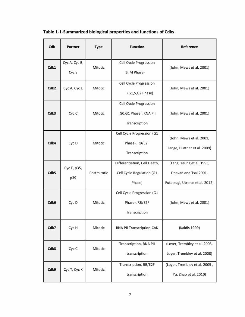
Table 1-1-Summarized biological properties and functions of Cdks
Cdk Partner Type Function Reference
Cdk1 Cyc A, Cyc B,
Cyc E Mitotic
Cell Cycle Progression
(S, M Phase) (John, Mews et al. 2001)
Cdk2 Cyc A, Cyc E Mitotic Cell Cycle Progression
(G1,S,G2 Phase) (John, Mews et al. 2001)
Cdk3 Cyc C Mitotic
Cell Cycle Progression
(G0,G1 Phase), RNA PII
Transcription
(John, Mews et al. 2001)
Cdk4 Cyc D Mitotic
Cell Cycle Progression (G1
Phase), RB/E2F
Transcription
(John, Mews et al. 2001,
Lange, Huttner et al. 2009)
Cdk5 Cyc E, p35,
p39 Postmitotic
Differentiation, Cell Death,
Cell Cycle Regulation (G1
Phase)
(Tang, Yeung et al. 1995,
Dhavan and Tsai 2001,
Futatsugi, Utreras et al. 2012)
Cdk6 Cyc D Mitotic
Cell Cycle Progression (G1
Phase), RB/E2F
Transcription
(John, Mews et al. 2001)
Cdk7 Cyc H Mitotic RNA PII Transcription-CAK (Kaldis 1999)
Cdk8 Cyc C Mitotic Transcription, RNA PII
transcription
(Loyer, Trembley et al. 2005,
Loyer, Trembley et al. 2008)
Cdk9 Cyc T, Cyc K Mitotic Transcription, RB/E2F
transcription
(Loyer, Trembley et al. 2005 ,
Yu, Zhao et al. 2010)
8
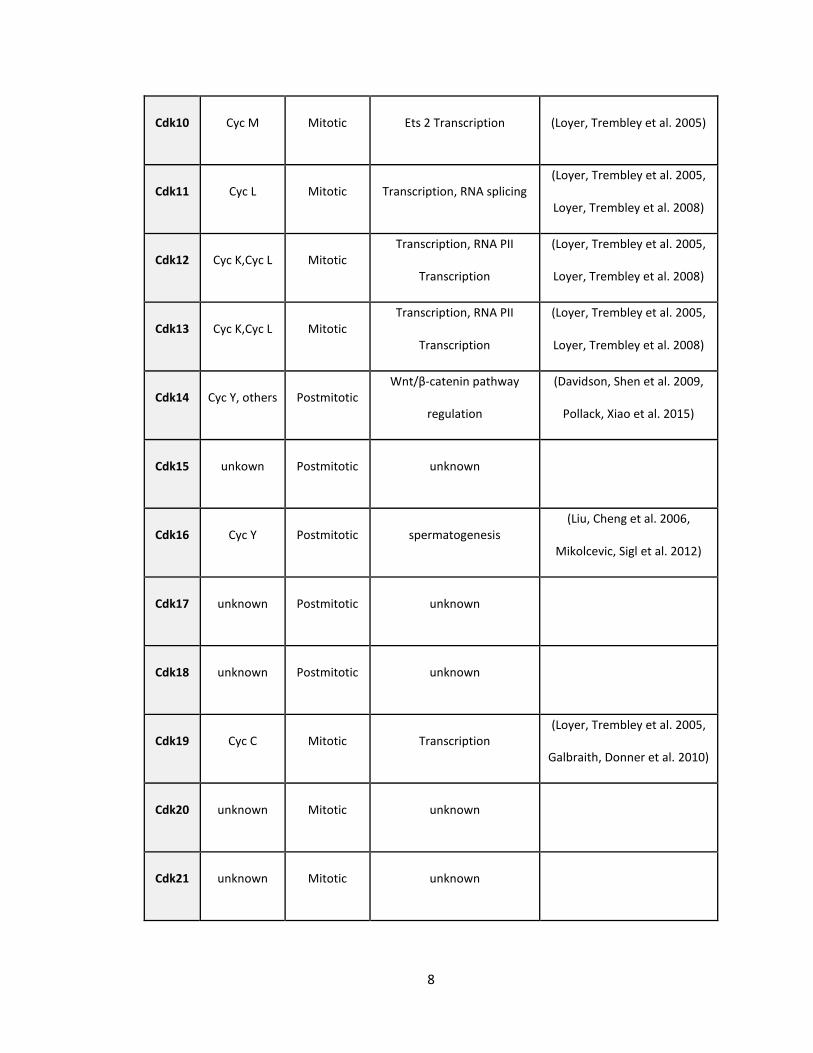
Cdk10 Cyc M Mitotic Ets 2 Transcription (Loyer, Trembley et al. 2005)
Cdk11 Cyc L Mitotic Transcription, RNA splicing (Loyer, Trembley et al. 2005,
Loyer, Trembley et al. 2008)
Cdk12 Cyc K,Cyc L Mitotic Transcription, RNA PII
Transcription
(Loyer, Trembley et al. 2005,
Loyer, Trembley et al. 2008)
Cdk13 Cyc K,Cyc L Mitotic Transcription, RNA PII
Transcription
(Loyer, Trembley et al. 2005,
Loyer, Trembley et al. 2008)
Cdk14 Cyc Y, others Postmitotic Wnt/β-catenin pathway
regulation
(Davidson, Shen et al. 2009,
Pollack, Xiao et al. 2015)
Cdk15 unkown Postmitotic unknown
Cdk16 Cyc Y Postmitotic spermatogenesis (Liu, Cheng et al. 2006,
Mikolcevic, Sigl et al. 2012)
Cdk17 unknown Postmitotic unknown
Cdk18 unknown Postmitotic unknown
Cdk19 Cyc C Mitotic Transcription (Loyer, Trembley et al. 2005,
Galbraith, Donner et al. 2010)
Cdk20 unknown Mitotic unknown
Cdk21 unknown Mitotic unknown
9
Although differentiation and cell cycle regulation would seem to be completely
independent processes there should be a cross-talk between differentiation and cell cycle
arrest, as the former normally follows the later (Hindley and Philpott 2012). Previous
studies signify the importance of Cdks in the tight regulation of proliferation in the
nervous system (Galderisi, Jori et al. 2003, Malumbres 2011). For instance, improper
activation of Cdks in a population of neurons that are terminally differentiated results in
apoptosis and neuronal death; both, in vitro (Freeman, Estus et al. 1994, Park, Farinelli et
al. 1996, Park, Levine et al. 1997, Park, Morris et al. 1997, Park, Morris et al. 1998, Park,
Morris et al. 1998, Giovanni, Wirtz-Brugger et al. 1999, Padmanabhan, Park et al. 1999,
Giovanni, Keramaris et al. 2000, O'Hare, Hou et al. 2000, Park, Obeidat et al. 2000, Morris,
Keramaris et al. 2001, Konishi, Lehtinen et al. 2002, Konishi and Bonni 2003, Rideout,
Wang et al. 2003, Sumrejkanchanakij, Tamamori-Adachi et al. 2003, Kruman, Wersto et
al. 2004, Liu, Biswas et al. 2004, Otsuka, Tanaka et al. 2004), and in vivo (Nagy, Esiri et al.
1997, Vincent, Jicha et al. 1997, Coelho and Leevers 2000, Osuga, Osuga et al. 2000, Wang,
Corbett et al. 2002, Nguyen, Boudreau et al. 2003, Rashidian, Iyirhiaro et al. 2005).
1.1.2. The Structure of Cdks
Cdks are all relatively small ranging from 34 to 60 kDa and rarely up to 110 kDa on
SDS-PAGE gel. Cdk proteins possess a similar structure, in which catalytic domains are
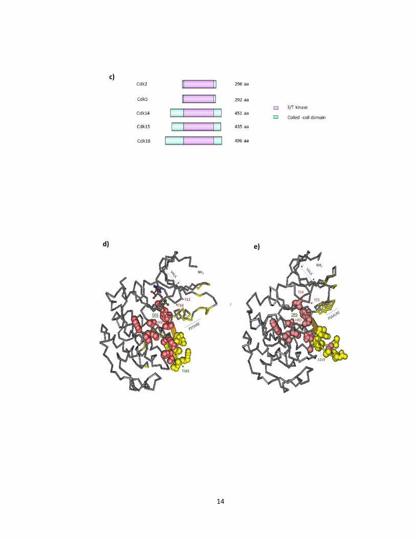
conserved. These domains include: (1) “VALK” motif in subdomain II of the kinase domain
that facilitates positioning of ATP in the active site, (2) “PCTAIRE/PSTAIRE” motif, a
characteristic of Cdk proteins, located in subdomain III of the kinase domain that is mainly
responsible for the Cdk/cyclin interaction, (3) ”HRD” motif in subdomain VIb that
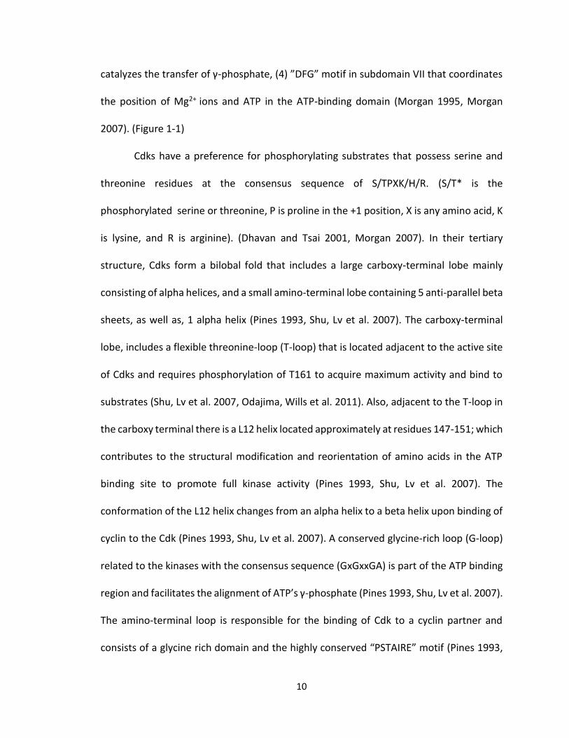
10
catalyzes the transfer of γ-phosphate, (4) ”DFG” motif in subdomain VII that coordinates
the position of Mg2+ ions and ATP in the ATP-binding domain (Morgan 1995, Morgan
2007). (Figure 1-1)
Cdks have a preference for phosphorylating substrates that possess serine and
threonine residues at the consensus sequence of S/TPXK/H/R. (S/T* is the
phosphorylated serine or threonine, P is proline in the +1 position, X is any amino acid, K
is lysine, and R is arginine). (Dhavan and Tsai 2001, Morgan 2007). In their tertiary
structure, Cdks form a bilobal fold that includes a large carboxy-terminal lobe mainly
consisting of alpha helices, and a small amino-terminal lobe containing 5 anti-parallel beta
sheets, as well as, 1 alpha helix (Pines 1993, Shu, Lv et al. 2007). The carboxy-terminal
lobe, includes a flexible threonine-loop (T-loop) that is located adjacent to the active site
of Cdks and requires phosphorylation of T161 to acquire maximum activity and bind to
substrates (Shu, Lv et al. 2007, Odajima, Wills et al. 2011). Also, adjacent to the T-loop in
the carboxy terminal there is a L12 helix located approximately at residues 147-151; which
contributes to the structural modification and reorientation of amino acids in the ATP
binding site to promote full kinase activity (Pines 1993, Shu, Lv et al. 2007). The
conformation of the L12 helix changes from an alpha helix to a beta helix upon binding of
cyclin to the Cdk (Pines 1993, Shu, Lv et al. 2007). A conserved glycine-rich loop (G-loop)
related to the kinases with the consensus sequence (GxGxxGA) is part of the ATP binding
region and facilitates the alignment of ATP’s γ-phosphate (Pines 1993, Shu, Lv et al. 2007).
The amino-terminal loop is responsible for the binding of Cdk to a cyclin partner and
consists of a glycine rich domain and the highly conserved “PSTAIRE” motif (Pines 1993,
11
Shu, Lv et al. 2007). The amino-terminal lobe also contains inhibitory threonine (T14) 14
and tyrosine 15 (Y15) phosphorylation sites (Morgan 1995). The amino and carboxy -
lobes are connected to each other through a domain that acts like a flexible hinge
(Morgan 1995). This region is termed the catalytic cleft or the ATP binding domain. The
G-loop forms the peak of the cleft that blocks the catalytic region when the Cdk is
inactivated (Morgan 1995). The catalytic domain is composed of aspartate and lysine
residues that bind to the substrate and ATP, as well. These residues include aspartate at
position (D127), lysine at position (K129) and asparagine at position (N132), as well as
lysine (K33) and aspartate (D145) (De Bondt, Rosenblatt et al. 1993, Morgan 1995) (Figure
1-1).
12
CDK2 ---------------------------------MENFQKVEKIGEGTYGV
CDK5 ---------------------------------MQKYEKLEKIGEGTYGT
CDK14 ------TSSTGKESPKVRRHSSPSSPTSPKFGKADSYEKLEKLGEGSYAT
CDK15 -------------EEDLR-QGFQW-RKSLPFGAASSYLNLEKLGEGSYAT
CDK16 YLEKLTLNSPIFDKPLSR-RLRRVSLSEIGFGKLETYIKLDKLGEGTYAT
CDK2 VYKARNKLTGEVVALKKIRXDTETEGVPSTAIREISLLKELNHPNIVKLL
CDK5 VFKAKNRETHEIVALKRVRLDDDDEGVPSSALREICLLKELKHKNIVRLH
CDK14 VYKGKSKVNGKLVALKVIRLQE-EEGTPFTAIREASLLKGLKHANIVLLH
CDK15 VYKGISRINGQLVALKVISMNA-EEGVPFTAIREASLLKGLKHANIVLLH
CDK16 VYKGKSKLTDNLVALKEIRLEH-EEGAPCTAIREVSLLKDLKHANIVTLH
CDK2 DVIHTENKLYLVFEFLHQDLKKFMDASALTGIPLPLIKSYLFQLLQGLAF
CDK5 DVLHSDKKLTLVFEFCDQDLKKYFDSCN-GDLDPEIVKSFLFQLLKGLGF
CDK14 DIIHTKETLTLVFEYVHTDLCQYMDKHP-GGLHPDNVKLFLFQLLRGLSY
CDK15 DIIHTKETLTFVFEYMHTDLAQYMSQHP-GGLHPHNVRLFMFQLLRGLAY
CDK16 DIIHTEKSLTLVFEYLDKDLKQYLDDCG-NIINMHNVKLFLFQLLRGLAY
CDK2 CHSHRVLHRDLKPQNLLINTEGAIKLADFGLARAFGVPVRTYTHEVVTLW
CDK5 CHSRNVLHRDLKPQNPLINRNGELKLADFGLARAFGIPVRCYSAEVVTLW
CDK14 IHQRYILHRDLKPQNLLISDTGELKLADFGLARAKSVPSHTYSNEVVTLW
CDK15 IHHQHVLHRDLKPQNLLISHLGELKLADFGLARAKSIPSQTYSSEVVTLW
CDK16 CHRQKVLHRDLKPQNLLINERGELKLADFGLARAKSIPTKTYSNEVVTLW
CDK2 YRAPEILLGCKYYSTAVDIWSLGCIFAEMVT-RRALFPGD-SEIDQLFRI
CDK5 YRPPDVLFGAKLYSTSIDMWSAGCIFAELANAGRPLFPGN-DVDDQLKRI
CDK14 YRPPDVLLGSTEYSTCLDMWGVGCIFVEMIQ-GVAAFPGMKDIQDQLERI
CDK15 YRPPDALLGATEYSSELDIWGAGCIFIEMFQ-GQPLFPGVSNILEQLEKI
CDK16 YRPPDILLGSTDYSTQIDMWGVGCIFYEMAT-GRPLFPGS-TVEEQLHFI
CDK2 FRTLGTPDEVVWPGVTSMPDYKPS-FPKW-ARQDFSKVVPPLD--EDGRS
CDK5 FRLLGTPTEEQWPSMTKLPDYKPY--PMYPATTSLVNVVPKLN--ATGRD
CDK14 FLVLGTPNEDTWPGVHSLPHFKPERFTLY-SSKNLRQAWNKLSYVNHAED
CDK15 WEVLGVPTEDTWPGVSKLPNYNPEWFPLP-TPRSLHVVWNRLGRVPEAED
CDK16 FRILGTPTEETWPGILSNEEFKTYNYPKY-RAEALLSHAPRLD--SDGAD
CDK2 LLSQMLHYDPNKRISAKAALAHPFFQDVTKPVPH----------------
CDK5 LLQNLLKCNPVQRISAEEALQHPYFSDFCP--------------------
CDK14 LASKLLQCSPKNRLSAQAALSHEYFSDLPPRLWELTDMSSIFTVPNVRLQ
CDK15 LASQMLKGFPRDRVSAQEALVHDYFSALPSQLYQLPDEESLFTVSGVRLK
CDK16 LLTKLLQFEGRNRISAEDAMKHPFFLSLGERIHKLPDTTSIFALKEIQLQ
a)
13
b)
b)
14
c)
d) e)
15
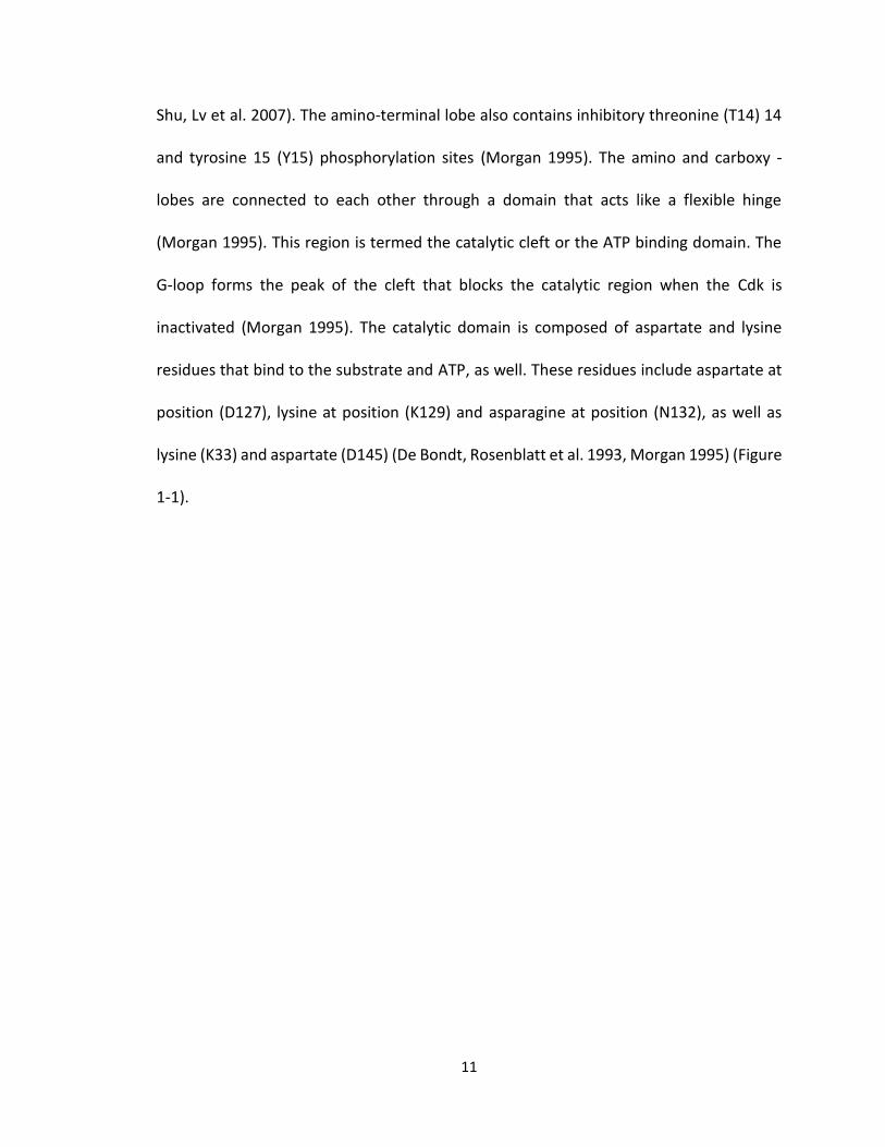
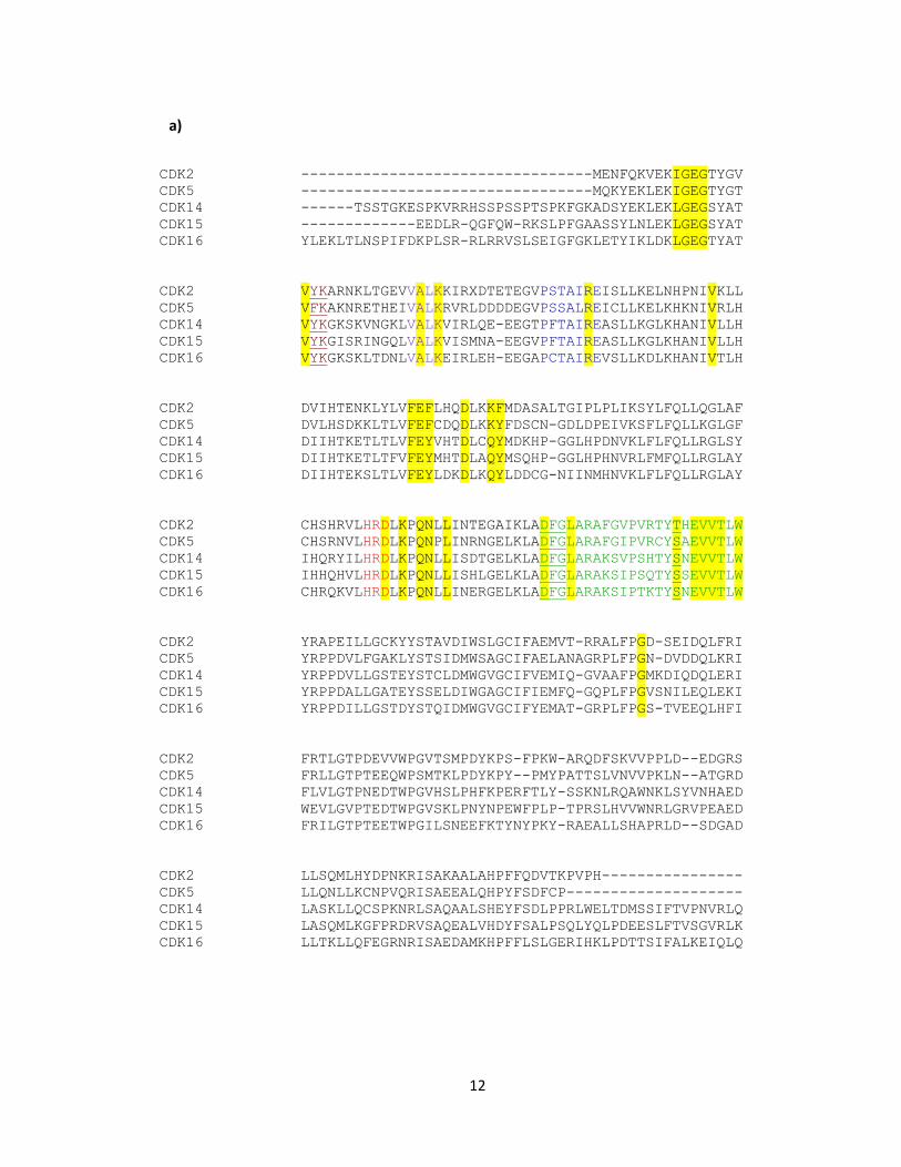
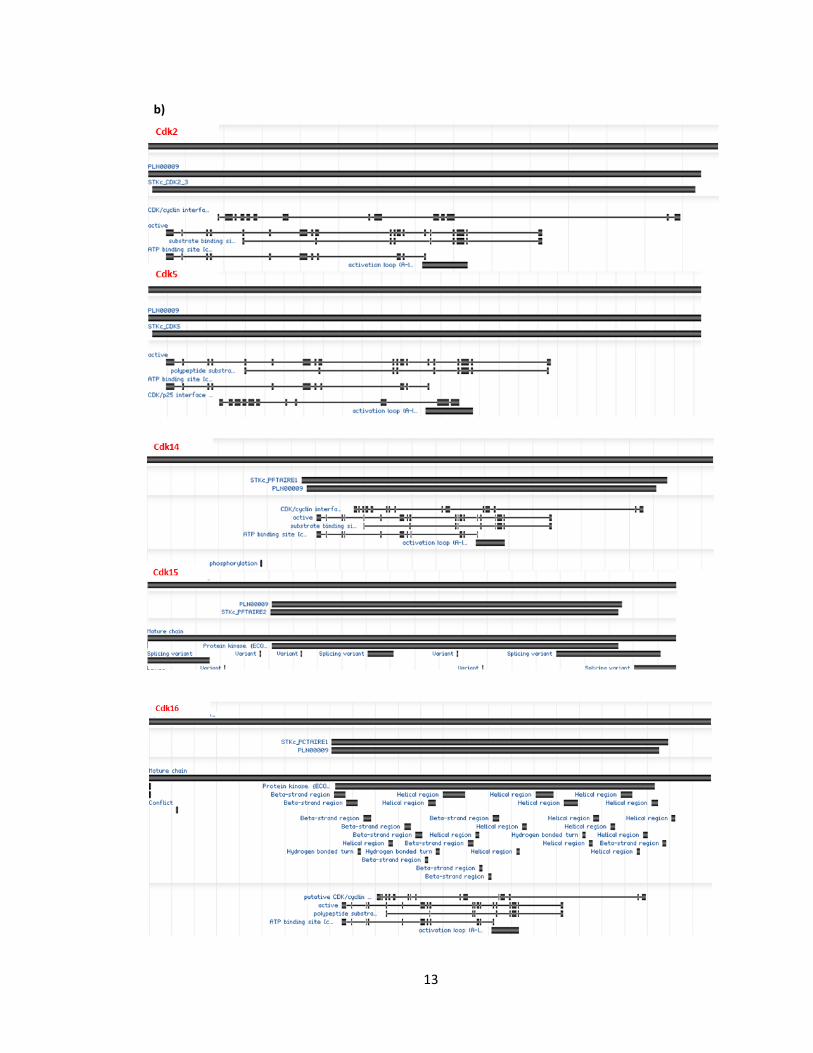
a) Alignment of sequences for human CDK2 (Accession: CAA43985.1), CDK5 (Accession:
CAG33322.1), CDK14 (Accession: NP001274064.1), CDK15 (Accession: Q96Q40.2), and
CDK16 (Accession: Q00536.1). Activation loop (yellow highlight), T14 and Y15 - inhibitory
sites ( red), S/T- activation site (green), conserved Cdk PSTAIRE motif (blue), VLAK, HRD,
DFG domains (respectively purple, red, green) (see text for further description). b)
Graphical summary of conserved domains of CDK2, CDK5, CDK14, CDK15, and CDK16
respectively with alignment footprint indicating region similarity. c) S/T kinases
highlighted in red in CDK2, CDK5, CDK14, CDK15, CDK16 (Modified from Table 1
(Malumbres and Barbacid 2005). 3D structure and relative position of conserved domains
d) CDK2 and e) CDK5 using PubMed, Cn3D tool (Wang, Addess et al. 2007, Marchler-
Bauer, Zheng et al. 2013).
Figure 1-1 Structure of Representative Cdks
16
1.1.3. Modes of Regulation of Cdks
Cdks are commonly regulated through different mechanisms which include: (1)
binding to their cyclin partners, (2) phosphorylation of Cdk and/or cylin-Cdk complex at
their conserved serine/threonine residue by CAK (Cdk activating kinase), (3) inhibitory
phosphorylation of Cdks, (4) CDK inhibitory subunits also known as cyclin-dependent
kinase inhibitor protein (CDKIs, CKIs or CDIs) that result in cell cycle arrest during the G1
phase of cell cycle progression (Morgan 1995, Morgan 2007).
1.1.3.1. Mechanism of Action of Cdks
1.1.3.1.1. Activating Patterns
- Cyclin Dependent Activation
Cdks are generally inactive in their monomer form. In their inactive form the T-
loop (activation loop) located at the carboxy-terminal blocks the cleft thus the amino
acids in the side chain of the Cdk are not accessible for binding of ATP (Morgan 1995,
Morgan 2007). Upon binding of cyclin with Cdk at the conserved (PSTAIRE) motif,
conformational changes occur in the tertiary structure of the Cdk. The L12 alpha helix at
the carboxy-terminal changes to a beta sheet. The T-loop that is adjacent to it has a
flexible conformation thus the T-loop rearranges in a manner that it moves outwards from
the cleft entrance. Later, the PSTAIRE helix at the amino-terminal moves inwards. This
conformational change flattens of the carboxy-terminal lobe; which in turn results in an
optimal position for ATP to bind. ATP binds within the cleft in a manner that the
phosphates are positioned outwards and accessible for the transfer of phosphate. The
17
Cdk substrate containing the S/TPXK/H/R sequence binds to the entrance of the cleft and
mainly positions towards the carboxy-terminal. (Russo, Jeffrey et al. 1996, Russo, Jeffrey
et al. 1996, Kim and Zhao 2005, Morgan 2007). Nevertheless, not all of the classic Cdks
become fully active upon binding to their cyclin partner and require binding to other non-
cyclin partners. For instance, the Cdk4/cyclin D complex is not fully active and it requires
binding to other non-cyclin partners including p34SEI1 or Sertad1 for full activity (Sugimoto,
Nakamura et al. 1999, Li and DiCicco-Bloom 2004).
Cyclin dependent kinases are regulated by phosphorylation of different residues,
at different phases. The phosphorylation of Cdks may occur at residues that enhance the
activity of Cdks. For instance, the Cdk-activating kinase (CAK) phosphorylates some Cdks
(Morgan 1995) at threonine (T160) and increases the activity of Cdk7/cyclin H complex
in combination with MAT1 or Cdk2/cyclin A complex (Gu, Rosenblatt et al. 1992) by
stabilizing their binding (Kaldis 1999). Phosphorylation of the conserved tyrosine and
threonine residues in the ATP binding region of Cdk results in electrostatic repulsion
between the phosphate linked to the kinase and those of ATP (Russo, Jeffrey et al. 1996,
Russo, Jeffrey et al. 1996, Kim and Zhao 2005, Morgan 2007).
- Autophosphorylation
Despite the similarities in the conformation of the Cdks, Cdk9 has a different
phosphorylation pattern in comparison to classical Cdks. The interface area of Cdk9 with its
partner cyclin T is 40% less than classical Cdks, such as Cdk2 with its partner cyclin A. Due to this
reduced interface area, the threonine (T186) in the T-loop is only able to interact with two arginine
residues R148 and R172 on the side chains of Cdk9 and this results in autophosphorylation of Cdk9
18
(Baumli, Lolli et al. 2008, Echalier, Endicott et al. 2010). Cdk14 has also been reported to have
autophosphorylation activity (Lazzaro, Albert et al. 1997) which suggests their independence from
Cdk7 kinase activity for becoming activated.
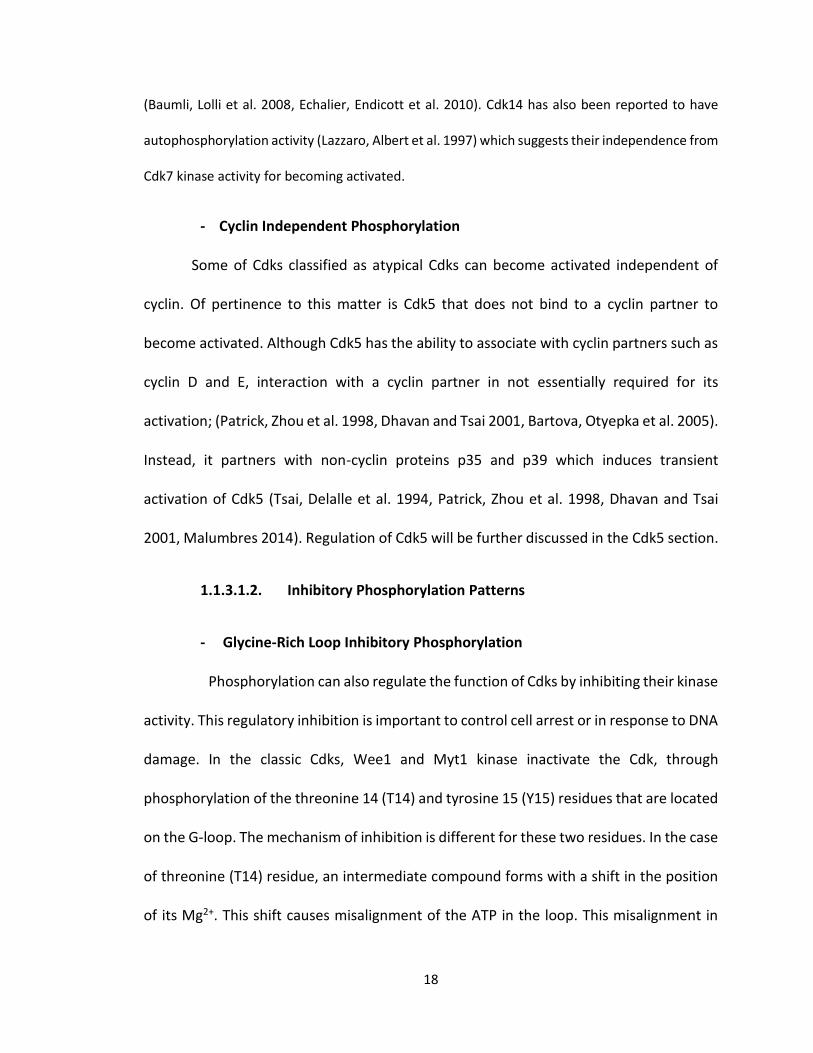
- Cyclin Independent Phosphorylation
Some of Cdks classified as atypical Cdks can become activated independent of
cyclin. Of pertinence to this matter is Cdk5 that does not bind to a cyclin partner to
become activated. Although Cdk5 has the ability to associate with cyclin partners such as
cyclin D and E, interaction with a cyclin partner in not essentially required for its
activation; (Patrick, Zhou et al. 1998, Dhavan and Tsai 2001, Bartova, Otyepka et al. 2005).
Instead, it partners with non-cyclin proteins p35 and p39 which induces transient
activation of Cdk5 (Tsai, Delalle et al. 1994, Patrick, Zhou et al. 1998, Dhavan and Tsai
2001, Malumbres 2014). Regulation of Cdk5 will be further discussed in the Cdk5 section.
1.1.3.1.2. Inhibitory Phosphorylation Patterns
- Glycine-Rich Loop Inhibitory Phosphorylation
Phosphorylation can also regulate the function of Cdks by inhibiting their kinase
activity. This regulatory inhibition is important to control cell arrest or in response to DNA
damage. In the classic Cdks, Wee1 and Myt1 kinase inactivate the Cdk, through
phosphorylation of the threonine 14 (T14) and tyrosine 15 (Y15) residues that are located
on the G-loop. The mechanism of inhibition is different for these two residues. In the case
of threonine (T14) residue, an intermediate compound forms with a shift in the position
of its Mg2+. This shift causes misalignment of the ATP in the loop. This misalignment in
19
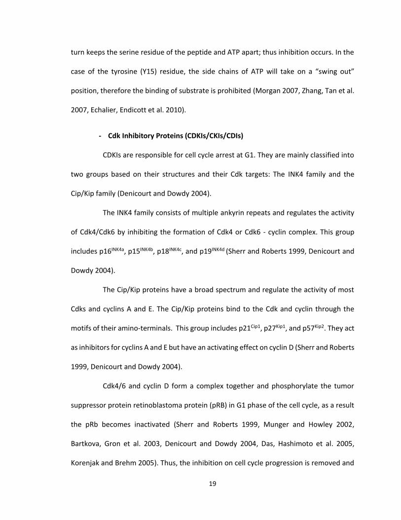
turn keeps the serine residue of the peptide and ATP apart; thus inhibition occurs. In the
case of the tyrosine (Y15) residue, the side chains of ATP will take on a “swing out”
position, therefore the binding of substrate is prohibited (Morgan 2007, Zhang, Tan et al.
2007, Echalier, Endicott et al. 2010).
- Cdk Inhibitory Proteins (CDKIs/CKIs/CDIs)
CDKIs are responsible for cell cycle arrest at G1. They are mainly classified into
two groups based on their structures and their Cdk targets: The INK4 family and the
Cip/Kip family (Denicourt and Dowdy 2004).
The INK4 family consists of multiple ankyrin repeats and regulates the activity
of Cdk4/Cdk6 by inhibiting the formation of Cdk4 or Cdk6 - cyclin complex. This group
includes p16INK4a, p15INK4b, p18INK4c, and p19INK4d (Sherr and Roberts 1999, Denicourt and
Dowdy 2004).
The Cip/Kip proteins have a broad spectrum and regulate the activity of most
Cdks and cyclins A and E. The Cip/Kip proteins bind to the Cdk and cyclin through the
motifs of their amino-terminals. This group includes p21Cip1, p27Kip1, and p57Kip2. They act
as inhibitors for cyclins A and E but have an activating effect on cyclin D (Sherr and Roberts
1999, Denicourt and Dowdy 2004).
Cdk4/6 and cyclin D form a complex together and phosphorylate the tumor
suppressor protein retinoblastoma protein (pRB) in G1 phase of the cell cycle, as a result
the pRb becomes inactivated (Sherr and Roberts 1999, Munger and Howley 2002,
Bartkova, Gron et al. 2003, Denicourt and Dowdy 2004, Das, Hashimoto et al. 2005,
Korenjak and Brehm 2005). Thus, the inhibition on cell cycle progression is removed and
20
the cell transits from G1 to S phase. On the other hand, cyclin D sequesters the Cip/Kip
proteins. Subsequently, Cdk2 becomes active and forms a complex with cyclin E. Cdk2-
cyclin E, then, phosphorylates Rb and p27. Phosphorylation of p27 triggers its degradation
through the proteasomal pathway. Phosphorylation of Rb in late G1 disrupts the
association of Rb with E2F and disrupts its repression; since the repression is removed the
transition throughout S-G2 and M phases is facilitated (Sherr and Roberts 1999, Munger
and Howley 2002, Stevaux and Dyson 2002, Denicourt and Dowdy 2004).
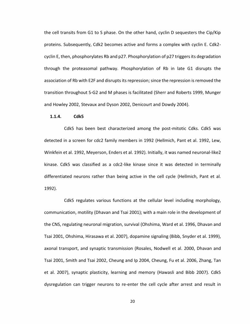
1.1.4. Cdk5
Cdk5 has been best characterized among the post-mitotic Cdks. Cdk5 was
detected in a screen for cdc2 family members in 1992 (Hellmich, Pant et al. 1992, Lew,
Winkfein et al. 1992, Meyerson, Enders et al. 1992). Initially, it was named neuronal-like2
kinase. Cdk5 was classified as a cdc2-like kinase since it was detected in terminally
differentiated neurons rather than being active in the cell cycle (Hellmich, Pant et al.
1992).
Cdk5 regulates various functions at the cellular level including morphology,
communication, motility (Dhavan and Tsai 2001); with a main role in the development of
the CNS, regulating neuronal migration, survival (Ohshima, Ward et al. 1996, Dhavan and
Tsai 2001, Ohshima, Hirasawa et al. 2007), dopamine signaling (Bibb, Snyder et al. 1999),
axonal transport, and synaptic transmission (Rosales, Nodwell et al. 2000, Dhavan and
Tsai 2001, Smith and Tsai 2002, Cheung and Ip 2004, Cheung, Fu et al. 2006, Zhang, Tan
et al. 2007), synaptic plasticity, learning and memory (Hawasli and Bibb 2007). Cdk5
dysregulation can trigger neurons to re-enter the cell cycle after arrest and result in
21
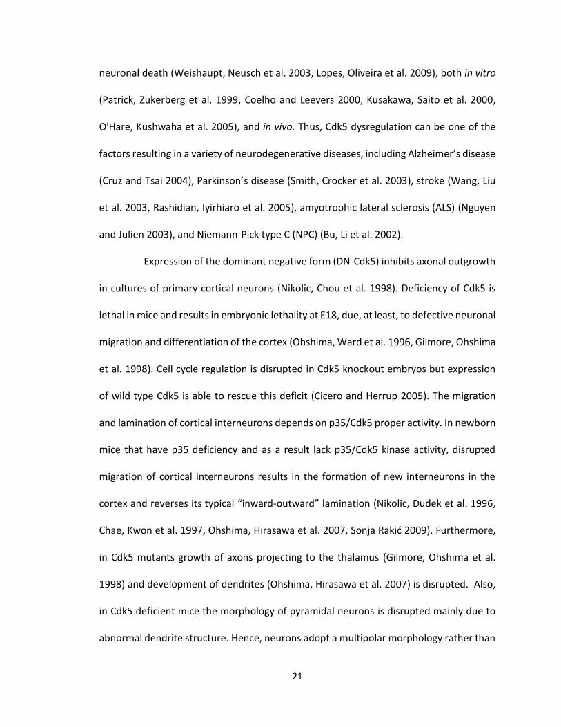
neuronal death (Weishaupt, Neusch et al. 2003, Lopes, Oliveira et al. 2009), both in vitro
(Patrick, Zukerberg et al. 1999, Coelho and Leevers 2000, Kusakawa, Saito et al. 2000,
O'Hare, Kushwaha et al. 2005), and in vivo. Thus, Cdk5 dysregulation can be one of the
factors resulting in a variety of neurodegenerative diseases, including Alzheimer’s disease
(Cruz and Tsai 2004), Parkinson’s disease (Smith, Crocker et al. 2003), stroke (Wang, Liu
et al. 2003, Rashidian, Iyirhiaro et al. 2005), amyotrophic lateral sclerosis (ALS) (Nguyen
and Julien 2003), and Niemann-Pick type C (NPC) (Bu, Li et al. 2002).
Expression of the dominant negative form (DN-Cdk5) inhibits axonal outgrowth
in cultures of primary cortical neurons (Nikolic, Chou et al. 1998). Deficiency of Cdk5 is
lethal in mice and results in embryonic lethality at E18, due, at least, to defective neuronal
migration and differentiation of the cortex (Ohshima, Ward et al. 1996, Gilmore, Ohshima
et al. 1998). Cell cycle regulation is disrupted in Cdk5 knockout embryos but expression
of wild type Cdk5 is able to rescue this deficit (Cicero and Herrup 2005). The migration
and lamination of cortical interneurons depends on p35/Cdk5 proper activity. In newborn
mice that have p35 deficiency and as a result lack p35/Cdk5 kinase activity, disrupted
migration of cortical interneurons results in the formation of new interneurons in the
cortex and reverses its typical “inward-outward” lamination (Nikolic, Dudek et al. 1996,
Chae, Kwon et al. 1997, Ohshima, Hirasawa et al. 2007, Sonja Rakić 2009). Furthermore,
in Cdk5 mutants growth of axons projecting to the thalamus (Gilmore, Ohshima et al.
1998) and development of dendrites (Ohshima, Hirasawa et al. 2007) is disrupted. Also,
in Cdk5 deficient mice the morphology of pyramidal neurons is disrupted mainly due to
abnormal dendrite structure. Hence, neurons adopt a multipolar morphology rather than
22
the normal bipolar structure (Ohshima, Hirasawa et al. 2007). Despite the severe effect
of Cdk5 deficiency during embryonic development and its lethality, the deficiency of Cdk5
does not completely prevent axons from projecting; (Gilmore, Ohshima et al. 1998,
Connell-Crowley, Vo et al. 2007); suggesting that other post-mitotic Cdks may play a role
in axogenesis.
1.1.4.1. Regulation of Cdk5 function
Like other Cdks, Cdk5 does not show catalytic activity in its monomeric form
and it has been known to bind to non-cyclin partners such as p35 (Tsai, Delalle et al. 1994)
or its isoform p39 (Tang, Yeung et al. 1995, Kwon and Tsai 2000, Ko, Humbert et al. 2001)
and their cleavage products p25 and p29, respectively (Patrick, Zhou et al. 1998, Dhavan
and Tsai 2001). Despite their differences with cyclins in their sequence identity, p35 and
p39 resemble cyclins in their ternary structure (Lew, Huang et al. 1994, Tsai, Delalle et al.
1994, Tang, Yeung et al. 1995, Tarricone, Dhavan et al. 2001, Lin, Wang et al. 2008, Jiang,
Gao et al. 2009). In spite of the ubiquitous expression of Cdk5, binding to its non-cyclin
neural-specific partner, p35 (Tsai, Delalle et al. 1994) or its isoform p39 (Tang, Yeung et
al. 1995, Kwon and Tsai 2000, Ko, Humbert et al. 2001) activates Cdk5, predominantly, in
the nervous system (Johansson, Lilja et al. 2005, Hawasli and Bibb 2007, Ohshima,
Hirasawa et al. 2007).
Cdk5 normally associates with the membrane due to the myristoylation signal
on p35 or p39 (Tsai, Delalle et al. 1994, Tang, Yeung et al. 1995, Patrick, Zhou et al. 1998,
Tarricone, Dhavan et al. 2001, Asada, Yamamoto et al. 2008, Lin, Wang et al. 2008). p35
directed activation of Cdk5 results in transient activation due to proteasomal
23
degradation. The deficiency of either Cdk5, or p35 and p39 in mice results in phenotypes
that are similar, which confirms the dependence of Cdk5 activity on p35 and p39 (Patrick,
Zhou et al. 1998, Patrick, Zukerberg et al. 1999). Furthermore, under pathological
conditions, calpain-mediated cleavage of p35 can result in formation and binding of p25
to Cdk5. Since, p25 lacks the myristoylation signal, it results in prolonged activation of
Cdk5 in the cytosol (Patrick, Zhou et al. 1998, Dhavan and Tsai 2001) therefore it interacts
with a different subset of substrates that induce neuronal death (Dhavan and Tsai 2001)
through phosphorylation of MEF2 (Smith, Mount et al. 2006) and Prx2 (Qu, Rashidian et
al. 2007) in Parkinson’s disease and Stroke (Rashidian, Rousseaux et al. 2009), as well. At
early stages of Alzheimer’s disease Cdk5 kinase activity is increased. Cdk5/p25 complex
hyperphosphorylates Tau and enhances the formation of neurofibrillary tangles (NFTs)
which is a hallmark of Alzheimer’s and Parkinson’s disease (Ishiguro, Takamatsu et al.
1992, Pei, Grundke-Iqbal et al. 1998) . Inhibition of Cdk5 by an inhibitor such as roscovitine
inhibits cell cycle progress to death (Lopes, Oliveira et al. 2009). Also, Cdk5 inhibition in
models of stroke and MPTP a model of Parkinson’s disease, in-vivo, results in
neuroprotection (Smith, Mount et al. 2006, Qu, Rashidian et al. 2007, Rashidian,
Rousseaux et al. 2009).
Despite not always depending on a cyclin partner for activation, association of
Cdk5 with cyclins D and cyclin E has been observed (Miyajima, Nornes et al. 1995, Patrick,
Zhou et al. 1998, Dhavan and Tsai 2001). Cyclin E has been attributed a role in cell cycle
regulation in the CNS and has been detected in conjunction with Cdk5 in adult brains;
however, in embryonic brain only Cdk1 and Cdk2 are associated with it (Odajima, Wills et
24
al. 2011, Lim and Kaldis 2013, Kawauchi 2014). In its inactive state Cdk5 forms a ternary
complex with p27kip1 in association with cyclin E that is also enriched in adult brain tissue.
It is possible that contrary to most cyclins that activate their Cdk partner (John, Mews et
al. 2001, Loyer, Trembley et al. 2005, Lange, Huttner et al. 2009), cyclin E inactivates Cdk5
by inhibiting its association with its activating p35/p39 partner (Dhavan and Tsai 2001,
John, Mews et al. 2001, Lim and Kaldis 2013, Kawauchi 2014). One possibility is that Cyclin
E decreases synapse number and functionality by inactivating Cdk5; thus, contributing to
decreased synaptic plasticity in neurological conditions such as Alzheimer’s disease
(Dhavan and Tsai 2001, Cruz and Tsai 2004). Park2, a Parkinson’s disease associated gene
with neuroprotective effect exerts its function through targeting cyclin E function and its
ubiquitination (Lucking, Durr et al. 2000, Staropoli, McDermott et al. 2003).
1.1.4.2. Cdk5 Structure and Mechanism of Action
Cdk5 contains the conserved PSTAIRE motif of Cdks with alanine (A) and leucine
(L) replacing threonine (T) and isoleucine (I); respectively, forming the PSAALRE motif
(Zhang, Tan et al. 2007). Similar to other Cdks, Cdk5 has a preference for the consensus
sequence (S/T) PX (K/H/R), the proline in +1 position, as well as, basic residues at +3
position (Zhang, Tan et al. 2007). As a result of this change, phosphorylation of the T-loop
is not essentially required for Cdk5 activation.
In studies on the mechanism of function of Cdk5, a variety of substrates are
involved including cytoskeletal elements (Xie, Samuels et al. 2006), signaling molecules
and regulatory proteins. In this section the signaling cues that have a role in Cdk5
regulation will be described briefly. A more detailed explanation will be included later on
25
in this chapter. Cdk5 has an important role in neurite formation. It inhibits the stimulatory
effect of nerve growth factor (NGF) on neurite outgrowth in PC12 cells (Harada, Morooka
et al. 2001). Cdk5 regulates both dendrite outgrowth and axonal outgrowth and guidance.
In hippocampal neurons, dendrite outgrowth is regulated by phosphorylating Trk B at
serine (S478) which in turn activates brain-derived neurotrophic factor (BDNF) (Cheung,
Chin et al. 2007). Cdk5 dependent axon outgrowth is regulated via phosphorylation of
MAP1b (Del Rio, Gonzalez-Billault et al. 2004) which in turn enhances axon outgrowth and
regulates its guidance via netrin1 and ephrinA5, respectively. The kinase activity of Cdk5
is suppressed by s-nitrosylation at cysteine (C83) (Zhang, Yu et al. 2010, Ye, Fu et al. 2012).
The loss of kinase activity, inhibits ephrinA5 induced growth cone collapse in retinal
ganglion neurons (Cheng, Sasaki et al. 2003). On the other hand, increase in Cdk5 kinase
activity through its phosphorylation on tyrosine (Y15) induces epherin4 dendritic spine
retraction (Fu, Chen et al. 2007). Contrary to classical cell cycle Cdks, phosphorylation of
the tyrosine 15 (Y15) residue in Cdk5 has an activating effect on Cdk5. In classical Cdks,
phosphorylation of Tyrosine 15 (Y15) occurs through Wee1 and this causes an inhibitory
effect. However, in Cdk5 this phosphorylation occurs through c-abl and Cables (an
adaptor protein for Cables), this causes a different conformational rearrangement upon
phosphorylation. It is suggested that in this case, the phosphorylated side chains fail to
take on a “swing out” position thus binding of the substrate is not inhibited (Zhang, Tan
et al. 2007). Rho GTPases are essential factors for the growth cone machinery and are
activated as Cdk5 targets. Cdk5 interacts with Rac1 via p35 in a GTP dependent manner
(Nikolic, Chou et al. 1998, Rashid, Banerjee et al. 2001). Cdk5/p35, Rac and Pak1 kinase
26
co-localize in the periphery of the neurites and regulate its outgrowth. Cdk5/p35 kinase
complex induces Pak1 hyper-phosphorylation. This downregulates the kinase activity of
Pak1, which results in actin re-organization and increases neurite outgrowth (Nikolic,
Chou et al. 1998). Cdk5 phosphorylates p27Kip1 in neurons, and this stabilization is critical
to keep the proper level of F-actins in the leading processes during migration (Nikolic,
Chou et al. 1998, Dhavan and Tsai 2001, Kawauchi, Chihama et al. 2006). Cdk5 increases
the activity of Rac and RhoA through phosphorylation of downstream proteins such as
kalirin7 (Xin, Wang et al. 2008) and ephexin1 (Fu, Chen et al. 2007?). Cdk5 also activates
calpain dependent protein degradation through phosphorylation of RasGRF1
(Kesavapany, Amin et al. 2004, Kesavapany, Pareek et al. 2006).
1.1.5. Other Post-Mitotic Cdks
The amino acid sequence of post-mitotic Cdk’s is highly identical. Pftaire-1,
Pctaire, show 50-52% and 61% similarity to Cdk5, respectively (Besset, Rhee et al. 1998).
Cdk5 has a central role in the in the development of the nervous system and cell death.
Similar to Cdk5, Pctaire and Pftaire1 are all highly expressed in the brain (Liu and Kipreos
2000). However, Pctaire1 (Liu, Cheng et al. 2006, Mikolcevic, Sigl et al. 2012) and Pftaire1
(Besset, Rhee et al. 1998), as well as, mitotic Cdk4 has also been detected in non-dividing
sertoli cells (Rhee and Wolgemuth 1995). Pctaire is absent in Drosophila. Initially, Sauer
et al. (Sauer, Weigmann et al. 1996) reported detection of Drosophila Pctaire-1 but
further investigation by Besset et al. (Besset, Rhee et al. 1998) showed that the detected
gene had higher homology with murine Pftaire-1 rather than Pctaire, 75% versus 58%,
respectively. Furthermore, comparison of the conserved kinase domain of murine Pftaire-
27
1 with Drosophila reveals higher similarity in their amino acid sequences as compared to
other Cdk family members (Besset, Rhee et al. 1998).
1.1.5.1. PCTAIRE
PCTAIRE is another post mitotic Cdk protein, which is highly conserved through
evolution (Cole 2009). It is conserved among most of clade holozoa but absent in
Drosophila. PCTAIRE includes three homologues, Pctaire 1-3 (Cdk 16-18) that are distinct
in their c-terminal but more conserved in the N-terminal and central domain (Cole 2009).
Pctaire1-3 proteins are expressed at high levels in the testis (Hirose, Kawabuchi et al.
2000, Mikolcevic, Sigl et al. 2012) and the brain (Okuda, Cleveland et al. 1992, Hirose,
Kawabuchi et al. 2000, Fu, Cheng et al. 2011, Mikolcevic, Sigl et al. 2012). Pctaire1 has
been detected at high levels in growth cones of newly developing neurons and also in
dendrites (Fu, Cheng et al. 2011). However, in post-mitotic tissue Pctaire1 is not detected
in the axons. It is only present in cell bodies and in the proximal region of neurites which
indicate it might not be directly involved in axon outgrowth and only have a role in the
transfer of cytoskeletal elements to the neurites (Besset, Rhee et al. 1999). Similarly,
Pctaire2 has been detected in neurons and neurites, as well (Hirose, Kawabuchi et al.
2000). The activity of Pctaire1-3 has a negative effect on neurite outgrowth. When the
activity of Pctaire 1-3 is abolished, there is an increase in neurite outgrowth; for instance,
overexpression of the kinase-dead form of Pctaire (K194R) results in increased neurite
outgrowth (Cole 2009). Phosphorylation of Pctaire at serine 153 by PKA decreases the
kinase activity of Pctaire1 which increases neurite outgrowth. On the contrary,
phosphorylation of Pctaire1 at serine (S95) by Cdk5, in neurons, increases it kinase activity
28
(Cole 2009). Mutations in the position serine (S95) of Pctaire1 interfere with regular
dendrite development (Cole 2009). Cdk5 could be a potential regulator of Pctaire1 activity
since the activity of Pctaire1 changes are in line with Cdk5 activity (Fu, Cheng et al. 2011).
1.1.5.2. PFTAIRE: (Pro, Phe, Thr, Ala, Ile, Arg, Glu)
Pftaire1 was, initially, identified in a screen for neuronal cdc2-like kinases by
Lazzaro et al in 1997 (Lazzaro, Albert et al. 1997). Later in 1998, Besset et al (Besset, Rhee
et al. 1998), reported the detection of the same protein and named it Pftaire1.
Nevertheless, the latter group reported a small difference in the cDNA sequence; in
which, three single amino acids are varied, as well as, 46 amino acids in the amino-
terminal. They reported transcripts that are different from the ones reported by Lazzaro
et al (Lazzaro, Albert et al. 1997). The first group had reported one single transcript with
the size of 5 kb, expressed almost exclusively in the nervous system; whereas, Besset et
al (Besset, Rhee et al. 1998) reported two transcripts at the sizes of 4.9 and 5.5 kb which
showed significant expression in the nervous system, testis, and embryo but a ubiquitous
and lower expression in the other tissues (Lazzaro, Albert et al. 1997, Besset, Rhee et al.
1998).
At present, the consensus is that PFTAIRE is highly conserved among different
species from yeast to mammals (Liu and Kipreos 2000). Nonetheless, the fly and worm
genome code for only one complex gene known as Eip63-Ecdysone-induced protein 63E
that is 70% identical to the mouse homolog Pftaire1 (Liu and Kipreos 2000, Stowers, Garza
et al. 2000) and Pftaire-interacting factor 2-pa (PubMed Gene ID: 9953769). In mammals,
PFTAIRE includes 2 separate genes: (1) Pftaire1/ PFTK1/CDK14 (PubMed Gene ID: 5218).
29
The human PFTAIRE1 and mouse Pftaire1 are located at (7q21-q22) and (5 A1; 2.61 cM)
(Lazzaro and Julien 1997), respectively, and show considerably high homology of
95%(Lazzaro, Albert et al. 1997, Yang and Chen 2001). (2) Pftaire2/PFTK2/Als2cr7/CDK15
(PubMed Gene ID: 65061) is recognized in GenBank database; although, to date no
research is reported on Pftaire2. The human PFTAIRE2 and mouse Pftaire2 are located at
(2q33.2) and (1 C1.3; 1), respectively. There are 14 known transcripts for the mouse
Pftaire1 gene (ENSMUSG00000028926). Only six of the isoforms result in a coded protein.
Two of the transcripts are putative isoforms and another two are incomplete in the 3’
coding region. Thus, only two of the sequences are bona fide (1) Cdk14-002
(ENSMUST00000030763 ) results in a transcript with a total of 15 exons of which the last
exon only contains 3’UTR and no coding sequence. It produces a transcript of 4,851
bps that translates into a protein product with 469 residues; and (2) Cdk14-09
(ENSMUST00000115452) a transcript with a total of 14 exons, of which 13 are coding and
results in a 4,866 bps mRNA and a protein product with 451 residues.
Similar to Cdk5, Pftaire1 transcript is concentrated in the brain (Lazzaro, Albert
et al. 1997, Besset, Rhee et al. 1998). The localization of Pftaire1 in the brain is exclusive
to the marginal layer that consists of differentiated cells. No Pftaire1 was detected in deep
layers or layer1, which is mainly composed of axons and dendrites (Lazzaro, Albert et al.
1997, Besset, Rhee et al. 1998). This is different from the expression pattern of cdc2 that
is detected in proliferative tissue and only inner layers of the brain (Lazzaro, Albert et al.
1997, Besset, Rhee et al. 1998) and Cdk5 that concentrated in the axons (Tsai, Takahashi
et al. 1993). In comparison, Pftaire1 is expressed in neuronal cell bodies and close
30
proximities (Lazzaro, Albert et al. 1997). The expression of Pftaire1 in comparison to the
more ubiquitous expression of cdc-2 (Lazzaro, Albert et al. 1997, Besset, Rhee et al. 1998)
suggests a more eminent role for Pftaire1 in the development of the nervous system.
Interestingly, Cdk14 is expressed highly in testicular tissue as well (Shu, Lv et al. 2007),
(Besset, Rhee et al. 1998). However, there is no evidence with regards to Pftaire1’s role
in the nervous system.
In the adult mouse, Pftaire1 mRNA is located both in neuroglia and neurons
(Lazzaro, Albert et al. 1997, Besset, Rhee et al. 1998) in different regions of the brain such
as cortex, hippocampus, thalamus, cerebellum, and also in the spine. Pftaire1 is localized
to neuronal cell bodies but not neurite extensions. Pftaire1 mRNA is also strongly
expressed in the testis and lung; yet, it does not translate to a protein (Lazzaro, Albert et
al. 1997). There is also low expression of Pftaire1 in the heart but no expression is
detected in spleen and thymus which supports the notion that Pftaire1 is primarily and
mainly expressed in the nervous system in adult mice (Lazzaro, Albert et al. 1997). During
mouse development, Pftaire1 mRNA is expressed at very low levels at embryonic days
12.5, 15.5, and even 18.5 but there is a rise right after birth, at P 10.5, it reaches a
maximum that is similar to its level in adult mice. This is different from the expression
pattern of Cdk5 that has a continuous increase from embryonic day 12, followed by a
significant increase at birth (Lazzaro, Albert et al. 1997). The expression pattern of human
PFTAIRE1 differs from mouse Pftaire1 to some extent. Similar to mouse Pftaire1, the
expression level of human PFTAIRE1 is relatively high in the brain and testis; however, in
human it is also highly expressed in the pancreas, kidney, heart and ovary. It is also
31
detected at lower levels in other tissues such as placenta, lung, liver, skeletal muscle,
prostate, small intestine, colon, and peripheral blood leukocyte, but is minimal in spleen
and thymus (Yang and Chen 2001, Shu, Lv et al. 2007). The expression pattern of human
PFTAIRE2 resembles that of PFTAIRE1 and is expressed highly in the brain and testis and
also in prostate, kidney, and skin (Yang and Chen 2001), PubMed Cdk15 Gene ID: 65061).
To determine potential interacting partners of Pftaire1, different methods have
been applied. In a 2D-PAGE mass spectrometric analysis ẞ-actin and Transgelin2
(TAGLN2), a tumor suppressor protein, heat shock protein 70, aldehyde dehydrogenase,
and 14-3-3 proteins were identified as Pftaire1 substrates (Leung, Ching et al. 2011). In a
yeast two-hybrid screen for proteins interacting with human PFTAIRE1, seven proteins
were detected including (1) two non-cyclin proteins such as KIAA0202 (septin8) (Yang,
Gao et al. 2002) and 14-3-3 isoforms β,ε,η,τ (Gao, Jiang et al. 2006), (2) cyclin proteins
such as cyclin Y (CCNY) (Jiang, Gao et al. 2009) and cyclin D3 (CCND3) (Shu, Lv et al. 2007),
(3) p21Cip1 (Shu, Lv et al. 2007), (4) PLZF protein (Gao, Jiang et al. 2006).
ẞ-actin and TAGLN2 (Transgelin2), an actin binding protein, reveal the most
intensified signal in a 2D-PAGE mass spectrophotometric analysis. The biological
relevance of this interaction was examined in vitro. In HEP3B and Human Hepatocellular
Carcinoma (HCC) cells upon PFTAIRE1 suppression, the total level of ẞ-actin remains
unchanged; however, the levels of TAGLN2 increases (Leung, Ching et al. 2011). TAGLN2
exhibits actin binding ability in its unphosphorylated state. Increase in PFTAIRE1 levels
increases phosphorylation of serine on TAGLN2, upon phosphorylation by PFTAIRE1, at
serine 83 and serine 163 residues, the interaction of TAGLN2 and actin is disrupted (Fu,
32
Subramanian et al. 2000, Leung, Ching et al. 2011) and the inhibitory effect of TAGLN2 on
actin dynamics is removed, actin gets depolymerized and HCC become motile (Pang, Bai
et al. 2007, Leung, Ching et al. 2011).
Caldesmone, 82 kDa, is another substrate for PFTAIRE1 detected in human
hepatocellular carcinoma (HCC) cells. Caldesmone and human PFTAIRE1, physically
interact with each other in vitro, in Hep3B cells and the level of caldesmone
phosphorylation is affected by the kinase activity of hPFTAIRE1. Inhibition of PFTAIRE1
decreases the levels of threonine and tyrosine phosphorylation and disrupts the
caldesmone-actin interaction. Phosphorylation of caldesmone enhances the actin binding
ability of caldesmone and determines its localization to membrane ruffles and stress
fibers (Leung, Ching et al. 2011).
At the subcellular level, ectopically expressed human PFTAIRE1 was localized to
the cytosol of Hela cells (Yang and Chen 2001), cytosol and nucleus of immature sertoli
cells, primary NHBE (normal human bronchial cells) (Pollack, Xiao et al. 2015), and also in
the plasma membrane of HEK293 cells through its interaction with cyclin Y (Jiang, Gao et
al. 2009) in vitro. Also, in cytosol, nucleus of the cell body of the motor neurons of the
spinal cord of adult C3H mice, (Lazzaro, Albert et al. 1997), and mouse testis (Jiang, Gao
et al. 2009) in vivo. Although, Pftaire1 has two nuclear localization sequences (NLS) at
residues 66 to 72 (PEDKKVR) and 113 to 119 (PKVRRHS) (Yang and Chen 2001) it is
predominantly localized in the cytosol (Lazzaro, Albert et al. 1997, Yang and Chen 2001)
and in the plasma membrane (Jiang, Gao et al. 2009). Generally, proteins that contain a
NLS sequence shuttle between the nucleus and the cytoplasm through binding to an
33
importin / complex. It is suggested that KIAA0202 or human septin8 which is a
cytosolic protein, interacts with PFTAIRE1 and localizes PFTAIRE1 to the cytoplasm (Yang,
Gao et al. 2002). We speculate that the presence of the highly phosphorylated 58-60 kDa
protein that was detected among the substrates of Pftaire1 in adult mouse brains lysates
(Lazzaro, Albert et al. 1997) and is similar to the size of Septin 8 (Yang, Gao et al. 2002)
could be a possible explanation to this. In an assessment for the kinase activity of Pftaire1
in a mouse brain lysate sample, a subset of three endogenous phosphoproteins were
suggested to interact with Pftaire1. Of the three substrates, the 58-60 kDa protein was
highly phosphorylated and considered specific to Pftaire1; whereas, the phosphoproteins
in 200-205 kDa range are believed to be unspecific and detected in association with Cdk5
(Lazzaro, Albert et al. 1997). Similar to all Cdk proteins, Pftaire1 contains three conserved
regulatory phosphorylation residues that include serine (S99), tyrosine (Y100), and serine
(S243). The equivalent of these residues in other Cdk family members are threonine (T14),
tyrosine (Y15), and threonine (T161) (Lazzaro, Albert et al. 1997).
As mentioned above, the 14-3-3 proteins also interact with PFTAIRE1. Four of
the 14-3-3 ( , ,, ,, ) isoforms are detected in association with PFTAIRE1 (Gao, Jiang et
al. 2006). These isoforms are mainly cytoplasmic and abundant in the CNS. It is suggested
that similar to KIAA0202, 14-3-3 proteins have the ability to localize PFTAIRE1 to the
cytoplasm (Muslin and Xing 2000, Voigt, Liebich et al. 2000, Gao, Jiang et al. 2006). Two
consensus binding motifs (1) RSxpSxP, and (2) Rx1-2Sx2-3S (Fu, Subramanian et al. 2000)
are detected in 14-3-3 proteins. It is suggested that the consensus motif RHSSPSS (117 to
120) of 14-3-3 overlaps with the second NLS of the PFTAIRE1 protein and interacts with it
34
through phosphorylation of 14-3-3 on the serine (S199) residue (Muslin and Xing 2000,
Voigt, Liebich et al. 2000).
Retinoblastoma protein (Rb) is a substrate for hPFTAIRE1 in vitro in many
different cell lines including human embryonic kidney cells (HEK-293) (Shu, Lv et al. 2007,
Jiang, Gao et al. 2009), SH-SY5Y, and SK-N-SH human neuroblastoma cells (Shu, Lv et al.
2007). The phosphorylation of retinoblastoma protein (Rb) is activated by the association
of human PFTAIRE1 with CCND3 (cyclin D3) and inhibited by interaction with p21Cip1 (Shu,
Lv et al. 2007). This might suggest that the activity of PFTAIRE1 is regulated by (CCND3)
cyclin D3 (Shu, Lv et al. 2007) and CCNY (cyclin Y) (Jiang, Gao et al. 2009) and inhibited by
the p21 (Shu, Lv et al. 2007). Since, phosphorylation of Rb results in inhibition of its activity
(Shu, Lv et al. 2007); this indicates a role in cell cycle regulation for Pftaire1. Furthermore,
overexpression of human PFTAIRE enhanced cell cycle progression in human
osteosarcoma U2OS cell line through transition from G1 to S (Shu, Lv et al. 2007).
Although phosphorylation of Rb occurs only in the cytoplasm, interaction of PFTAIRE1
with the membrane-associated cyclin Y in vitro, in HEK cells, recruits PFTAIRE1 to the
plasma membrane through its N-terminal myristoylation (Jiang, Gao et al. 2009). This
interaction is facilitated through phosphorylation of CCNY on the serine residues by
PFTAIRE1 kinase activity (Jiang, Gao et al. 2009). The interaction of PFTAIRE1 with Cyclin
Y phosphorylates low density lipoprotein receptor related protein 6 (LRP6) in vivo, which
also requires Cdc25 activity (Davidson, Shen et al. 2009). LRP6, on the other hand,
modulates the Wnt signaling pathway that is involved in neurogenesis (Ille, Atanasoski et
al. 2007, Davidson, Shen et al. 2009). Wnt signaling in combination with CCND regulate
35
spermatogenesis during mouse development (Golestaneh, Beauchamp et al. 2009, Kerr,
Young et al. 2014) and in postmitotic cells, as well (Pollack, Xiao et al. 2015).
Downregulation of Pftaire1 disturbs Wnt signaling and β-catenin levels (Pollack, Xiao et
al. 2015) and result in cell-cycle arrest (Shu, Lv et al. 2007).
PLZF protein interacts with other proteins including hPFTAIRE1 and PCTAIRE1,
through (1) a proline-rich domain (Pro domain) in the linker region, (2) the 9 Krüppel zinc
finger domains in its carboxy terminal, and (3) the conserved POZ/BTB domain at its
amino terminal (Li, English et al. 1997). PLZF proteins are highly conserved (Cook, Gould
et al. 1995) and contain a consensus sequence for cdc2 phosphorylation (Cook, Gould et
al. 1995) that contains serine and threonine residues (Ball, Melnick et al. 1999). PLZF
negatively regulates cell cycle progress by inhibiting cyclin A (Yeyati, Shaknovich et al.
1999). Ectopic expression of PLZF inhibits growth and results in apoptosis by arrest of cell
cycle progression at G1 (Shaknovich, Yeyati et al. 1998). Taking this into consideration, we
hypothesize that Pftaire1 might also regulate cell cycle through PLZF and cyclin A.
Nevertheless, no biological function for this interaction has been examined yet.
Thyrotropin-Releasing Hormone (TRH) is a neuropeptide expressed in
hypothalamus and other regions of the brain. TRH regulates the expression of Pftaire1 in
mice cerebellum through NO-cGMP pathway. TRH acts on anterior pituitary cells that
secrete thyroid stimulating Hormone (TSH). This suggests an active role for Pftaire1 in
postnatal tissue, as a downstream target of TRH, perhaps playing a role in differentiation
rather than cell proliferation (Hashida, Yamada et al. 2002).
36
In Drosophila, the Pftaire homologue Eip63E is crucial for development and its
absence is embryonically lethal (Stowers, Garza et al. 2000). Expression of its conserved
kinase domain can reverse this lethality (Stowers, Garza et al. 2000). Also, data from our
lab described later in this chapter, suggests the importance of Eip63E in axogenesis in
Drosophila (Rodríguez González and S. 2011). Our study on Pftaire1 is novel, as it focuses
on the neuronal function and targets of Pftaire1. To date, no reports exist on the role and
mechanism of action of Pftaire1 in the nervous system.
1.2. Neuronal Development and Migration
Cdks regulate different aspects of neurogenesis from proliferation and
differentiation to apoptosis and cell death. One of the pathways that are renown for
regulating progenitors and cell lineage fate is Wnt signaling which is important in insects
and vertebrates , (Zechner, Fujita et al. 2003, Ille, Atanasoski et al. 2007, Grigoryan, Wend
et al. 2008). As mentioned earlier Pftaire1 mediates Wnt signaling (Davidson, Shen et al.
2009). Neurogenesis is defined as the process by which neurons are developed
from neural stem cells and progenitor cells. In addition to neurons, glial cells originate
from the stem cells. Neurogenesis is highly active both perinatally and later during
adulthood. During development, initially proliferative divisions occur in stem cells, they
increase in number and produce progenitor cells. Upon neurogenesis, the progenitor cells
undergo an asymmetrical division and different types of progenitor cells are produced
(Kawauchi, Shikanai et al. 2013). This division is followed by symmetric, differentiating
divisions of progenitor cells that produce immature neurons (neuroblasts), which still do
not possess processes. Commonly, the new neurons are formed in locations, different

37
from the position that they finally acquire. These immature neurons have the ability to
migrate to new positions and reside at their final destination in the mature brain (Rakic
1990). The migration of these neuroblasts is controlled by “leading processes” that are
structures at the tip of the migrating neurons. The leading processes sense the
environmental cues and direct the neuroblasts to their new locations (Tabata and
Nakajima 2003). Migration occurs in two stages, locomotion and somal translocation
(Nadarajah, Brunstrom et al. 2001). During locomotion (1) neuroblasts extend a leading
process, (2) nucleokinesis occurs and the prenucleous compartment (centrosome and
Golgi apparatus) moves into the leading process along with the nucleus. Remodeling of
the leading process facilitates its movement. Once the migrating neuron reaches its
destination, it undergoes somal translocation and once again remodeling and
nucleokinesis occur in the leading process of the neuroblast, then the extended leading
tip shrinks (Rakic 1990, Miyata, Kawaguchi et al. 2001, Nadarajah and Parnavelas 2002,
Tabata and Nakajima 2003, Miyata and Ogawa 2007, Marin, Valiente et al. 2010).
Migration mainly occurs through two main modes: radial migration and
tangential migration (Umeshima and Kengaku 2013). Radial migration occurs mainly
during early development of the brain, as opposed to tangential migration that occurs
during final stages of development. The ventricular zone contains the soma of radial glial
cells and gives rise to the neurons that form the cortical plate. The neuroblasts migrate
perpendicular to the ventricular zone and all the way through the developing cortex to
reach their final destination. A single extended process named the leading process aids
the neuroblasts in locating their path during migration as they follow the long processes

38
of the radial glial cells (Rakic 1990, Campbell and Gotz 2002, Marin, Valiente et al. 2010).
Membrane bound cell adhesion molecules such as astrotactin, neuregulin, and integrins
aid the interaction of the neurons with the radial glial cells during migration (Anton,
Marchionni et al. 1997, Anton, Kreidberg et al. 1999, Adams, Tomoda et al. 2002, Marin,
Valiente et al. 2010). Other regulators of the migration process include environmental
cues such as slits, netrins, semaphorins, and reelin (Marin, Valiente et al. 2010). Inhibition
of Cdk5 activity results in lengthening of radial fibers into the white matter of cerebellum
and disrupted radial migration of neurons (Umeshima and Kengaku 2013).
In tangential migration of neuroblasts, the leading process forms two branches
with a steady angle. They possess a dynamic structure and depending on the direction of
migration, only one branch remains active at each migratory cycle and the other branch
retracts (Ward, Jiang et al. 2005, Martini, Valiente et al. 2009). The migration path of
tangential cells is parallel to the ventricular surface and orthogonal to the radial glia
palisade (Polleux, Whitford et al. 2002, Yokota, Gashghaei et al. 2007, Martini, Valiente et
al. 2009). Rather than interacting with radial glial cells (Corbin, Nery et al. 2001, Marin
and Rubenstein 2001) , the tangentially migrating neurons interact with a variety of cells
either homotypic or heterotypic in nature as they pass through different environments
(Marin, Valiente et al. 2010). Homotypic interactions usually occur when neurons migrate
through unpermissive environments. In this case, a cluster of neuroblasts migrate
together and utilize neighbouring cells as their substrate (Marin, Valiente et al. 2010). An
example of homotypic interaction is the formation of the interneurons of the olfactory
bulb which migrate through the lateral ventricles of the telencephalon (Wichterle, Garcia-
39
Verdugo et al. 1997, Marin, Valiente et al. 2010). Contrary to the movement of
neuroblasts in clusters, inhibitory homotypic interactions may occur. This assists the
neuroblasts to move to low density areas. An example of this movement is the dispersion
of Cajal-Retzius cells (which originate from the cortical hem) over the surface of the
cerebral cortex, during early corticogenesis (Borrell and Marin 2006). Tangential
migration may also be heterotypic in which case the direction of migration is determined
by the environmental cues that are present in the surface of other cells including axons.
For instance, the neurons that produce Gonadotropin-releasing hormone (GnRH) pass
through the forebrain following vomeronasal axons (Wray 2002, Marin, Valiente et al.
2010). The guidance cues that regulate tangential migration of neuroblasts are mainly the
same cues that regulate axon guidance, which include slits, netrins, semaphorins, as well
as, growth factors including BDNF, NT4, and HGF or morphogenetic proteins (Polleux,
Whitford et al. 2002, Pozas and Ibanez 2005, Marin, Valiente et al. 2010). Once the
neuroblasts reach their final destination, the new neurons become polarized and take up
various morphological changes dendrites and axons form and extend toward long-
distance targets (Kawauchi and Hoshino 2008).
1.3. Axon Outgrowth and Cellular Signaling
In addition to the aforementioned functions the timing of Pftaire1 expression
could be an important factor in regulating axon outgrowth and fasciculation (Sanchez-
Soriano and Prokop 2005). Precise connectivity within the nervous system is important
for proper development and plasticity in the adult nervous system. Our knowledge about
the patterns of axogenesis in the nervous system began over a century ago, when Golgi
40
and Cajal stained brain sections using the Golgi method (Cajal 1906, Mazzarello 1999).
This resulted in Cajal’s remarkable discovery of the axonal structure and the growth cone
and our cellular and molecular understanding of the biology of neurons and their
outgrowth has continued to develop especially during the recent decades. The polarized
structure of neurons plays an important role in relaying information in the nervous
system. The growth cone is the motile terminal of the neurite that leads the axon through
the complex environment of the tissues. The structure of a growth cone includes actin
microfilaments and cytoskeletal microtubules in the peripheral and central region,
respectively (Nikolic 2002, Huber, Kolodkin et al. 2003). The peripheral actin
microfilaments are connected to the central microtubular region by the actin arc. Upon
axonal outgrowth cohered rearrangements occur in the actin superstructure of a growth
cone (Condeelis 1993, Rodriguez, Schaefer et al. 2003).
This guidance of an axon is modulated through multiple events at the growth
cone and axogenesis occurs in a few stages, as defined in neuronal cultures in vitro. At
the time of plating, unpolarized sphere-shaped neurons are cultured. The neuron then
becomes polarized and axogenesis initiates: (1) During initiation, the actin structure of
the immature postmitotic neurons goes through rearrangement and cellular protrusions
appear in the form of sheet like structures and lamellipodia and filopodia microspikes;
respectively (Nikolic 2002); (2) Lamellipodia and filopodia give rise to immature neurites;
(3) In the next step elongation occurs, the neuron becomes asymmetrical, one particular
neurite grows rapidly and forms the axon while the remaining become retracted and form
dendrites. The attribute to become an axon or dendrite is inherited from the progenitor
41
cells that give rise to these neurites in vivo (Polleux and Snider 2010). One possible
mechanism in determining the fate of an axon is that the axon sends out inhibitory signals
to dendrites and excitatory signals to itself; (4) While the axon continues to grow rapidly,
arborization of the dendrites occurs; (5) Neurons become differentiated, axons and
dendrites become specialized, the dendritic spines and synapses form (Dotti, Sullivan et
al. 1988, da Silva and Dotti 2002).
Polarity in the neuron and axon outgrowth is modulated by signaling cues.
During axon outgrowth, extracellular guidance cues bind to the receptors at the periphery
of the growth cone and link to downstream signaling cascades (Nikolic 2002, Jaffe and
Hall 2005). The signaling pathways may become activated or deactivated (Huber, Kolodkin
et al. 2003) depending upon repulsive and attractive cues. While excitatory cues enhance
axon outgrowth, the Inhibitory cues result in growth cone collapse and arrest of axon
growth (Nikolic 2002, Lowery and Van Vactor 2009). The direction and rate of axon
growth also depends upon these signaling cues (Dickson 2002, Huber, Kolodkin et al.
2003) which are present in a gradient. The growth cone integrates different guidance
cues, modifies the actin superstructure and alters the morphology of the growth cone.
Thus, axons are either directed to their targets or retracted from them (Nikolic 2002).
Signaling cues including intrinsic factors, Rho GTPase regulatory molecules,
developmental morphogens, mature neurotrophins, receptors, neurotransmitters,
extracellular matrix and adhesion molecules and cell adhesion molecules (CAMs),
regulate the formation of neurites (Nikolic 2002, Jaffe and Hall 2005).
42
Intrinsic factors that appear to be important and regulate cytoskeletal dynamics
and axon growth include proteins such as transcription factors, GAP-43, cell adhesion
molecules, cytoskeletal proteins, the Rho-family of GTPases (Hall and Lalli 2010), and Cdk5
(Ye, Fu et al. 2012). We hypothesize that Pftaire1 plays a role in the growth cone
machinery, as well.
Developmental morphogenes are signaling cues that are present in a gradient
and exert different responses with regards to axon outgrowth depending on their
absolute concentration. They alter the transcription of their target cells. (Gurdon and
Bourillot 2001, Schnorrer and Dickson 2004). Among the morphogenes are sonic
hedgehog (Shh) (Charron, Stein et al. 2003), Wnt (Yoshikawa, McKinnon et al. 2003) and
bone morphogenic protein (BMP)s (Butler and Dodd 2003).
Neurotrophins play an important role in neurite outgrowth and differentiation.
Neurotrophin precursors split and form mature neurotrophins (Sun, Lim et al. 2012) and
usually enhance axon outgrowth through Trk receptors. The Ras linked tyrosine kinase
receptor Trk is a regulator for the Rho family of GTPases (Nusser, Gosmanova et al. 2002,
Huang and Reichardt 2003). Nerve Growth Factor NGF regulates neurite formation by
activating Rac1 and Cdc42 (Aoki, Nakamura et al. 2004) and deactivating RhoA (Nusser,
Gosmanova et al. 2002, Huang and Reichardt 2003). The Brain derived neurotrophic
factor (BDNF) enhances neurite outgrowth in an inhibitory environment (Huang and
Reichardt 2003, Williams, Williams et al. 2005). There are also various downstream
molecules that trigger intercellular signaling. For instance, Pak interacts with LIM kinases
that are serine/threonine kinases that phosphorylate and deactivate the destabilizing
43
protein cofilin (Maekawa, Ishizaki et al. 1999). This induces the formation of stress fibers
and enhances the outgrowth of axon (Yang, Higuchi et al. 1998, Amano, Tanabe et al.
2001, Bernard 2007). Pak can inactivate myosin light chain kinases (MCLK) and as a result
control retrograde formation of F-actin (Arber, Barbayannis et al. 1998, Yang, Higuchi et
al. 1998). Also, the Src-family of tyrosine kinases including Src, Fyn, Lck regulate axon and
dendrite outgrowth through their effector the small GTPase TC10, a Cdc42-GAP, and the
CAP-associated complex (Liu, Nakazawa et al. 2006). A number of actin-binding proteins
like cofilin and profilin inhibit neurite formation. Profilin II is mainly expressed in the brain
(Witke, Podtelejnikov et al. 1998, Da Silva, Medina et al. 2003). In addition, profilin-
binding proteins of the vasodilator stimulated phosphoprotein (Ena/VASP) or EVH family
regulate actin polymerization (Bear, Svitkina et al. 2002, Huber, Kolodkin et al. 2003).
Other effector proteins include Wiskott -Aldrich -syndrome protein (WASP) and neuronal
N-WASP that are scaffolding proteins that are regulated by Cdc42 and regulate branching
(Rohatgi, Ma et al. 1999). SCAR/WAVE proteins that are regulated by Rac and
subsequently regulate the actin related proteins 2 and 3 (Arp2/3 complex) which increase
actin nucleation (Machesky, Mullins et al. 1999, Rohatgi, Ma et al. 1999, Higgs and Pollard
2001). Filamin (Revenu, Athman et al. 2004) is another effector that regulates
cytoskeleton remodeling and neurite outgrowth (Stuermer, Bastmeyer et al. 1992, Hall
1998, Burridge and Wennerberg 2004, Langhorst, Jaeger et al. 2008, Hall and Lalli 2010,
Stuermer 2010). Inhibitory molecules such as those with the inhibitory effect of glia
(Schwab and Thoenen 1985, Carbonetto, Evans et al. 1987, Savio and Schwab 1989),
oligodendrocyte myelin glycoprotein (OMgp) (Wang, Corbett et al. 2002), myelin
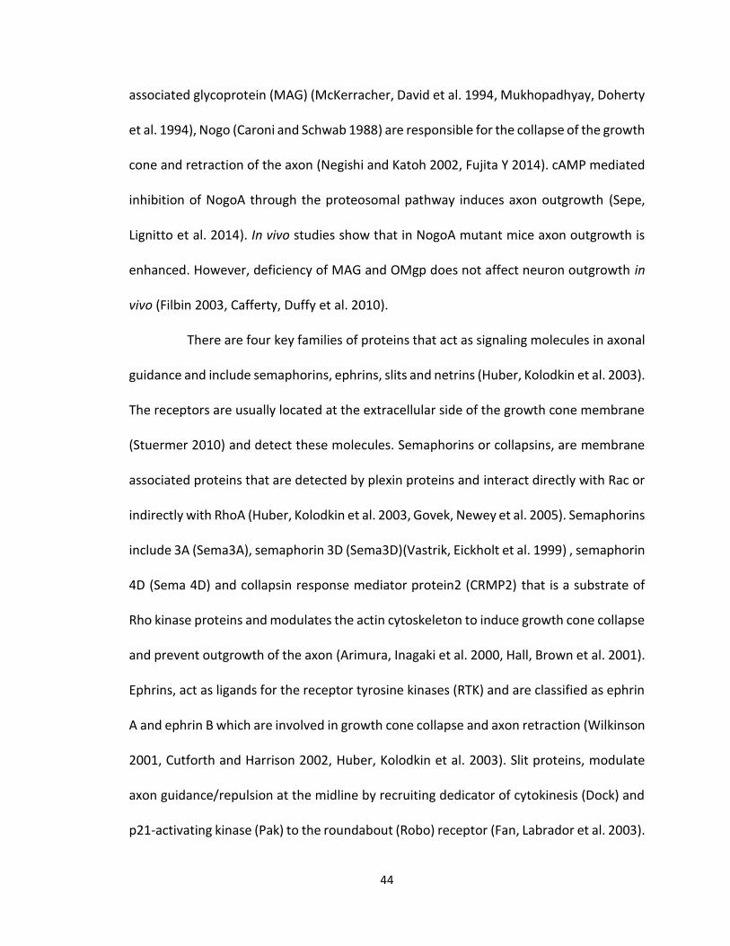
44
associated glycoprotein (MAG) (McKerracher, David et al. 1994, Mukhopadhyay, Doherty
et al. 1994), Nogo (Caroni and Schwab 1988) are responsible for the collapse of the growth
cone and retraction of the axon (Negishi and Katoh 2002, Fujita Y 2014). cAMP mediated
inhibition of NogoA through the proteosomal pathway induces axon outgrowth (Sepe,
Lignitto et al. 2014). In vivo studies show that in NogoA mutant mice axon outgrowth is
enhanced. However, deficiency of MAG and OMgp does not affect neuron outgrowth in
vivo (Filbin 2003, Cafferty, Duffy et al. 2010).
There are four key families of proteins that act as signaling molecules in axonal
guidance and include semaphorins, ephrins, slits and netrins (Huber, Kolodkin et al. 2003).
The receptors are usually located at the extracellular side of the growth cone membrane
(Stuermer 2010) and detect these molecules. Semaphorins or collapsins, are membrane
associated proteins that are detected by plexin proteins and interact directly with Rac or
indirectly with RhoA (Huber, Kolodkin et al. 2003, Govek, Newey et al. 2005). Semaphorins
include 3A (Sema3A), semaphorin 3D (Sema3D)(Vastrik, Eickholt et al. 1999) , semaphorin
4D (Sema 4D) and collapsin response mediator protein2 (CRMP2) that is a substrate of
Rho kinase proteins and modulates the actin cytoskeleton to induce growth cone collapse
and prevent outgrowth of the axon (Arimura, Inagaki et al. 2000, Hall, Brown et al. 2001).
Ephrins, act as ligands for the receptor tyrosine kinases (RTK) and are classified as ephrin
A and ephrin B which are involved in growth cone collapse and axon retraction (Wilkinson
2001, Cutforth and Harrison 2002, Huber, Kolodkin et al. 2003). Slit proteins, modulate
axon guidance/repulsion at the midline by recruiting dedicator of cytokinesis (Dock) and
p21-activating kinase (Pak) to the roundabout (Robo) receptor (Fan, Labrador et al. 2003).
45
Netrins, are a family of proteins that modulate the guidance of commissural and
peripheral axons by acting as chemoattractants (Sun KLW 2011). Their function is
controversial. Different netrin receptors include uncoordinated locomotion-5 (UNC5) and
is absent in colorectal cancer (DCC) family members. If netrin binds to DCC homodimers
it acts as a chemoattractant but if it binds to UNC5/DCC heterodimer it acts as a
chemorepellent (Huber, Kolodkin et al. 2003).
NMDA and GABA neurotransmitters regulate axon outgrowth and fasciculation.
They exert their action on axonal guidance through glutamate and Ca2+ influx (Georgiev,
Taniura et al. 2008, Sernagor, Chabrol et al. 2010). Ca2+ has an important role in regulating
neuronal development. Changes in the frequency of Ca2+ release regulate
neurotransmitter expression and neurite outgrowth. This regulation is achieved through
fast Ca2+ spikes and slow Ca2+ waves, respectively (Gu, Olson et al. 1994, Gu and Spitzer
1995, Gomez and Spitzer 1999, Ciccolini, Collins et al. 2003).
CAMs include cadherins and integrins, which are important regulators of
plasticity in the nervous system, as mentioned earlier in this chapter. They modulate a
variety of processes from migration to differentiation, axon outgrowth, synapse (Walsh,
Meiri et al. 1997, Hansen, Berezin et al. 2008) formation by regulating intracellular Ca 2+
levels and are usually located at the extracellular side of the membrane (Stuermer 2010,
Sheng, Leshchyns'ka et al. 2013).
1.4. Rho Family of GTPases
Cell signaling at the growth cone is essential for proper axonal outgrowth and
guidance. Rho GTPase family members are critical regulators in the process of axon
46
specification and elongation. Rho GTPases were initially identified about 30 years ago
(Madaule and Axel 1985) but their Rho GTPase activity was not discovered until 1992
(Ridley and Hall 1992). The Rho family of small GTPases is a subfamily of the Ras
superfamily of GTPases (Negishi and Katoh 2002). The Rho family includes 7 subgroups of
small monomeric GTPases with 23 representatives in mammals (Wherlock and Mellor
2002); including Rac (1,2,3), Cdc42, Rho(A,B,C,D), and Rho(G and H) (Negishi and Katoh
2002, Burridge and Wennerberg 2004). Rac1, Cdc42, and RhoA, respectively, stimulate
formation of veil-shaped lamellipodia, mirospiked filopodia, stress fibers, and focal
adhesions (Nobes and Hall 1995, Hall and Lalli 2010). They have an essential role in neurite
outgrowth and guidance by regulating the dynamics of the actin and microtubule
cytoskeleton via their associated kinases and these roles may overlap at times (Jalink, van
Corven et al. 1994, Luo, Hensch et al. 1996, Zhao and Manser 2005).
1.4.1. Structure of Rho GTPases
The structure of Rho GTPases governs its spatiotemporal regulation. They
contain: (1) a conserved G domain that includes 5 motifs (G1-G5) involved in the
positioning of Mg2+ and nucleotide binding for the base guanine (Vetter and Wittinghofer
2001, Wennerberg, Rossman et al. 2005, Wittinghofer and Vetter 2011); (2) 2
homologous switch domains which are responsible for GEF binding (V43 in RhoA) and GTP
hydrolysis (Z63). Formation of hydrogen bonds between the ϒ-phosphate of GTP and T37
and G62 residues in Rho-GTPases results in conformational changes that regulate the
interaction of effector proteins (Vetter and Wittinghofer 2001, Wennerberg, Rossman et
al. 2005, Wittinghofer and Vetter 2011); (3) an insert region, involved in the binding of

47
GEF and effector proteins which in RhoA includes residues 123-137; (4) a hypervariable
C-terminal and a CAAX motif that mediate association with the membrane (Wennerberg,
Rossman et al. 2005, ten Klooster and Hordijk 2007) and binding of effector proteins
(Schaefer, Reinhard et al. 2014). (Figure 1-2)
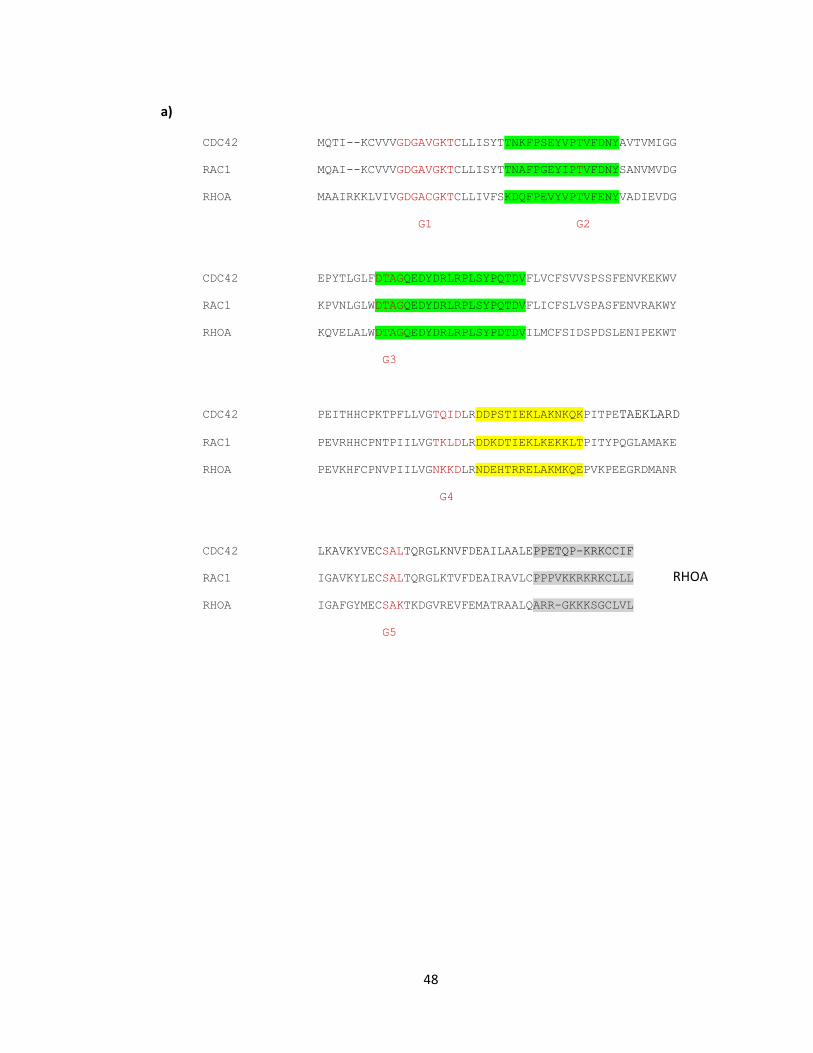
48
CDC42 MQTI--KCVVVGDGAVGKTCLLISYTTNKFPSEYVPTVFDNYAVTVMIGG
RAC1 MQAI--KCVVVGDGAVGKTCLLISYTTNAFPGEYIPTVFDNYSANVMVDG
RHOA MAAIRKKLVIVGDGACGKTCLLIVFSKDQFPEVYVPTVFENYVADIEVDG
G1 G2
CDC42 EPYTLGLFDTAGQEDYDRLRPLSYPQTDVFLVCFSVVSPSSFENVKEKWV
RAC1 KPVNLGLWDTAGQEDYDRLRPLSYPQTDVFLICFSLVSPASFENVRAKWY
RHOA KQVELALWDTAGQEDYDRLRPLSYPDTDVILMCFSIDSPDSLENIPEKWT
G3
CDC42 PEITHHCPKTPFLLVGTQIDLRDDPSTIEKLAKNKQKPITPETAEKLARD
RAC1 PEVRHHCPNTPIILVGTKLDLRDDKDTIEKLKEKKLTPITYPQGLAMAKE
RHOA PEVKHFCPNVPIILVGNKKDLRNDEHTRRELAKMKQEPVKPEEGRDMANR
G4
CDC42 LKAVKYVECSALTQRGLKNVFDEAILAALEPPETQP-KRKCCIF
RAC1 IGAVKYLECSALTQRGLKTVFDEAIRAVLCPPPVKKRKRKCLLL
RHOA IGAFGYMECSAKTKDGVREVFEMATRAALQARR-GKKKSGCLVL
G5
a)
RHOA
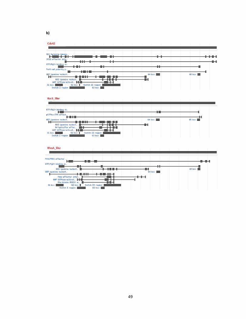
49
b)
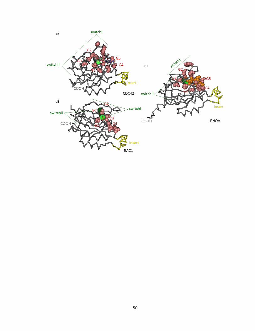
50
51
a) Alignment of the CDC42 (AAM21109.1), RAC1 (AAM21111.1) and RHOA (AAM21117.1)
amino acid sequences. G domain sequences (red), Switch I, switch II, respectively (green
highlight), the insert region (yellow highlight) and the hypervariable C-terminus (grey
highlight). b) Graphical summary of conserved domains of GTPase proteins, CDC42, RAC1
and RHOA, respectively with alignment footprint indicating region similarity. 3D structure
of and relative position of conserved domains c) CDC42, d) RAC1 and e) RHOA using
PubMed, Cn3D tool (Wang, Addess et al. 2007, Marchler-Bauer, Zheng et al. 2013).
Figure 1-2 Structure of CDC42, RAC 1, RHO A
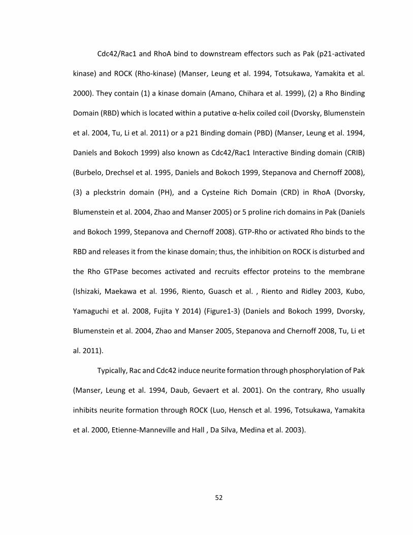
52
Cdc42/Rac1 and RhoA bind to downstream effectors such as Pak (p21-activated
kinase) and ROCK (Rho-kinase) (Manser, Leung et al. 1994, Totsukawa, Yamakita et al.
2000). They contain (1) a kinase domain (Amano, Chihara et al. 1999), (2) a Rho Binding
Domain (RBD) which is located within a putative α-helix coiled coil (Dvorsky, Blumenstein
et al. 2004, Tu, Li et al. 2011) or a p21 Binding domain (PBD) (Manser, Leung et al. 1994,
Daniels and Bokoch 1999) also known as Cdc42/Rac1 Interactive Binding domain (CRIB)
(Burbelo, Drechsel et al. 1995, Daniels and Bokoch 1999, Stepanova and Chernoff 2008),
(3) a pleckstrin domain (PH), and a Cysteine Rich Domain (CRD) in RhoA (Dvorsky,
Blumenstein et al. 2004, Zhao and Manser 2005) or 5 proline rich domains in Pak (Daniels
and Bokoch 1999, Stepanova and Chernoff 2008). GTP-Rho or activated Rho binds to the
RBD and releases it from the kinase domain; thus, the inhibition on ROCK is disturbed and
the Rho GTPase becomes activated and recruits effector proteins to the membrane
(Ishizaki, Maekawa et al. 1996, Riento, Guasch et al. , Riento and Ridley 2003, Kubo,
Yamaguchi et al. 2008, Fujita Y 2014) (Figure1-3) (Daniels and Bokoch 1999, Dvorsky,
Blumenstein et al. 2004, Zhao and Manser 2005, Stepanova and Chernoff 2008, Tu, Li et
al. 2011).
Typically, Rac and Cdc42 induce neurite formation through phosphorylation of Pak
(Manser, Leung et al. 1994, Daub, Gevaert et al. 2001). On the contrary, Rho usually
inhibits neurite formation through ROCK (Luo, Hensch et al. 1996, Totsukawa, Yamakita
et al. 2000, Etienne-Manneville and Hall , Da Silva, Medina et al. 2003).
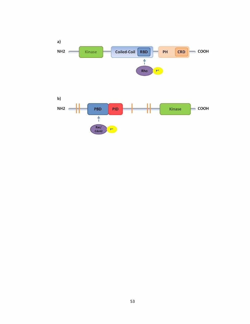
53
54
a) ROCK I consists of an N-terminal serine/threonine kinase domain (green), a Rho binding
domain (RBD) located within the central putative coiled-coil domain (blue), and a C-
terminal cysteine-rich domain (CRD) that is located within a pleckstrin homology (PH)
domain (orange). Upon activation of Rho, GTP-Rho it binds to the RBD and disturbs the
inhibition on ROCK (modified from Figure1A (Dvorsky, Blumenstein et al. 2004)). b) p21-
activated kinase (Pak) consists of an N-terminal p21-binding domain (PBD) (blue) followed
by a Pak inhibition domain (PID) (red), 5 proline rich domains that are the binding site for
-SH3 units (orange), and a C-terminal kinase domain (green). Upon activation of
(Rac/Cdc42) GTP-Rac/Cdc42 binds to the PBD, unfolds the molecule and activates Pak
(Modified from Figure (Stepanova and Chernoff 2008)).
Figure 1-3 Schematic view of ROCK1 and Pac1 domains
55
1.4.2. Regulators of Rho GTPases
Rho GTPase molecules transit between a GTP-bound and a GDP-bound state in
the cell. Generally, the GTP-bound state of the GTPase is deemed active; however,
occasionally the GDP state acts as regulatory; for instance, GDP-Cdc42 regulates MAPK
(Arozarena, Matallanas et al. 2001). The higher concentration of GTP in the cell acts in
favor of GTP-bound state (Schaefer, Reinhard et al. 2014). Guanine nucleotide Exchange
Factors (GEFs), GTPase activating proteins (GAPs), and guanine nucleotide dissociation
Inhibitors (GDIs) regulate this transition and Rho GTPase signaling (Jaffe and Hall 2005,
Hall and Lalli 2010).
GEFs are multidomain proteins, most of them consisting a Dbl homology (DH)
domain and a Pleckstrin homology (PH) domain.GEFs enhance the GTP bound state by
catalyzing GTP (Schmidt and Hall 2002). The invasion inducing-lymphoma and metastasis1
protein (Tiam1) is a Rac-GEF that is expressed in the developing nervous system (Ehler,
van Leeuwen et al. 1997) and is enriched in the neurite that is destined to become an
axon and also in the growth cone of the developing axon (Kunda, Paglini et al. 2001).
Other GEFs include Kalirin that contains GEF domains for both Rac1 and RhoG and
modulates axon protrusion (Ma, Johnson et al. 2001, Penzes, Johnson et al. , May, Schiller
et al. 2002) . The Rac-GEF, SIF and Tiam like exchange factor (STEF) that is involved in
neurite formation (Hoshino, Sone et al. 1999), Ost (Horii, Beeler et al. 1994), Dock 180,
Sos (Ng and Luo 2004), and Rho-GEF KIAA0380 that is usually present in the tip of neurites
(Togashi, Nagata et al. 2000).
56
On the contrary are GAPs that enhance the hydrolytic activity of GTPase and act
in favor of the GDP bound state by dissociating GTP. GAP proteins include CrGAP/Vilse,
p190RhoGAP (Brouns, Matheson et al. 2001), α-chimaerin (Hall, Michael et al. 2001),and
oligophrenin-1 (Billuart, Bienvenu et al. 1998, Bernards and Settleman). GAPs usually
interact with the P-loop and switch I and II regions of GTPases and their activity normally
results in neurite retraction (Dvorsky and Ahmadian 2004, Cherfils and Zeghouf 2013,
Schaefer, Reinhard et al. 2014).
GDIs are Chaperon proteins, which lack enzymatic activity and consist of an N-
terminal domain that interacts with switch domains I and II within the GTPase and a C-
terminal that binds to the switch II region and acts as an membrane anchor (Longenecker,
Read et al. 1999, Hoffman, Nassar et al. 2000, Scheffzek, Stephan et al. 2000, Schaefer,
Reinhard et al. 2014). GDIs inhibit dissociation of GDP and transfer of phosphate. RhoGDIγ
is an example of a GDI that is mainly located in the nervous system (Adra, Manor et al.
1997).
Nonetheless, the regulation of axon outgrowth is not always a straightforward
process and depending on the receptors different results could be observed. This
variation depends on the developmental stage and type of neuron and also the
subcellular localization of the GTPases, as well as, the upstream and/or downstream
signaling that becomes activated. In their active state, Rho GTPases recruit effector
proteins to the membrane. Rac, as well as Rho, have dual functions. For instance, if Rac is
modulated through Rac GEFs (such as Tiam, STEF and Dock 180) and then activates
IQGAP3 as its downstream target, axon outgrowth is stimulated (Aoki, Nakamura et al.
57
2004). On the contrary, Rac suppress axon outgrowth if it is activated by Trio to
phosphorylate cofilin through Pak and LIMK (Aoki, Nakamura et al. 2004). Also, Slit
dependent activation of Rac which is regulated by GEF Sos and GAP CrGAP/Vilse via
Drosophila Dock/vertebrate Nck (Dreadlocks), Pak and Robo complex results in axon
retraction (Hall and Lalli 2010). Interaction of Rac with plexinA1 causes a change in the
cytoplasmic tail of plexin and results in receptor endocytosis (Hall and Lalli 2010). This
enhances the activity of Rho/Rock signaling cascade and the inhibition of axonal
outgrowth. Furthermore, Rac inhibits axon outgrowth when it is activated as a
downstream target of plexinB1 (Hall and Lalli 2010). p190RhoGAP which is a RhoA-specific
GAP transiently downregulates the activity of Rho but activates Rho activity through Rho
GEFs (PDZ-RhoGEF and LARG) in a Sema dependent manner (Hall and Lalli 2010). In
contrast, if Rho is activated through a GEF such as Kailirin9 or if it functions through
Diaphanous related formin (mDia), axon outgrowth is stimulated (Hall and Lalli 2010).
Inactivation of Rho through Rap/RA-Rho GAP or p190RhoGAP stimulates axon growth, as
well; whereas, phosphorylation of cofilin through ROCK and Pak activated LIMK inhibits
axon outgrowth (Govek, Newey et al. 2005). IRSp53 modulates neurite outgrowth by
recruiting actin cytoskeleton effectors (Kast, Yang et al. 2014). Cdk5/p35 complex
hyperphosphorylates Pak and disrupts its kinase activity (Govek, Newey et al. 2005).
(Figure1-4) (Modified from (Govek, Newey et al. 2005, Hall and Lalli 2010)).
The activation and inactivation of the Rho GTPases controls downstream signaling
cascades; which regulate actin disassembly and growth cone regulation including
outgrowth and axonal guidance (Nikolic 2002, Jaffe and Hall 2005). Cdc42 may play an
58
important role in polarity of neuron as it is a downstream effector of small GTPase (Ras
proximate1). Accumulation of active Rap1 and the selective degradation of its inactive
form is one possible mechanism of regulation while the neuron acquires polarity
(Schwamborn and Puschel 2004, Schwamborn, Muller et al. 2007). Cdc42 may also
interact with atypical protein kinase C (aPKC) through mPar6 and modulate apical/basal
polarity of Drosophila neuroblasts and rat astrocytes (Kemphues 2000, Ohno 2001,
Etienne-Manneville and Hall 2002). In Drosophila, mutant forms of Rac1 inhibit axon
outgrowth whereas dendrites are not affected (Hakeda-Suzuki, Ng et al. 2002, Ng,
Nardine et al. 2002). In comparison, Cdc42 mutants affect both axon and dendrite
outgrowth and migration and RhoA mutants have no effect on axon outgrowth but extend
the dendrite and reduce its branching (Lee, Winter et al. 2000, Aoki, Nakamura et al.
2004). Loss- of- function mutations in the Rac genes of Drosophila result in defects in
axonal outgrowth and guidance (Hakeda-Suzuki, Ng et al. 2002). Deficiency of Ena/Vasp
family members in flies, worms, and mice causes defects in axonal outgrowth and
guidance (Lanier, Gates et al. 1999, Wills, Bateman et al. 1999, Yu, Hao et al. 2002).
The Rac signaling pathway is modulated by Cdk5. In Xenopus, neuronal
morphology of retinal ganglion cells (RGCs) is regulated by Rac via Cdk 5/p35 (Ruchhoeft,
Ohnuma et al. 1999). Cdk5 interacts with Rac1 via p35 in a GTP dependent manner
(Nikolic, Chou et al. 1998, Rashid, Banerjee et al. 2001). P35/Cdk5, Rac and Pak1 kinase
co-localize in the periphery of the neurites and regulate its outgrowth. P35/Cdk5 kinase
complex induces Pak1 hyper-phosphorylation. This downregulates the kinase activity of

59
Pak1 and actin re-organization which results in increased neurite outgrowth (Nikolic,
Chou et al. 1998).
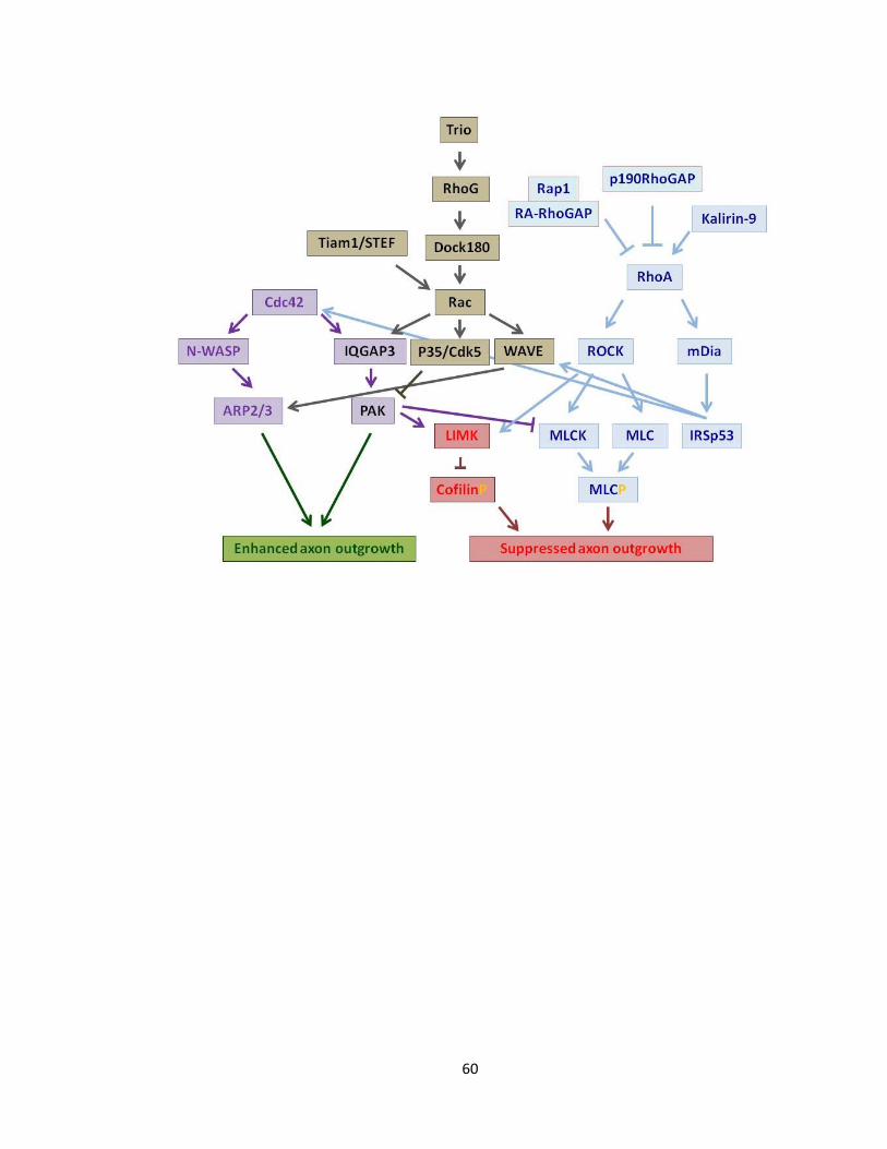
60
61
Rho and Rac can inhibit or activate axon outgrowth depending on their upstream and/or
downstream effectors. To simplify the pathway, different colors are utilized for
illustrating each GTPase protein and its upstream and downstream effectors, i.e. Rac,
Cdc42, RhoA and their effectors are illustrated in black, purple and blue, respectively.
Also, the final enhancement (green) and suppression (red) of axon outgrowth is illustrated
(See text for further description- Modified from Figure1 (Govek, Newey et al. 2005) and
Figure3 (Hall and Lalli 2010))
Figure 1-4 Schematic illustration of main Rho GTPase effectors involved in axon
outgrowth
62
1.5. Statement of Study
1.5.1. Rationale
Pctaire, Pftaire and Cdk5 are post-mitotic Cdk proteins that share the same most
recent common ancestor. Previous research indicates that Cdk5 and Pctaire have
important roles in the nervous system. For instance, Cdk5 inactivation leads to inhibition
of axonal growth (Nikolic, Dudek et al. 1996, Nikolic, Chou et al. 1998). Furthermore,
deletion of Cdk5 results in abnormal brain structure and embryonic lethality at embryonic
day 18 (Ohshima, Ward et al. 1996). Pctaire has been detected at high levels, in growth
cones of newly developing neurons and also in dendrites (Hirose, Kawabuchi et al. 2000,
Fu, Cheng et al. 2011). The activity of Pctaire1-3 negatively regulates neurite outgrowth
(Cole 2009). In addition, Pctaire seems to be a target of Cdk5 since the activity of Pctaire1
changes is in line with Cdk5 activity (Fu, Cheng et al. 2011).
Pftaire1 is highly expressed in the nervous system (Lazzaro, Albert et al. 1997, Yang
and Chen 2001). Our lab has previously, examined the role of Pftaire (Eip63E, ecdysone-
induced protein) in Drosophila. Initially, Jafar-nejad P (unpublished data), showed that
commissural and longitudinal axons of the VNC were defasciculated and disrupted in two
Eip63E mutant Drosophila lines (1) Df (L63) E1 and (2) L6381. Homozygosity for both of
these mutant alleles are embryonic lethal. Df(L63)E1 consists of a large deletion including
the transcription initiation point and almost the entire conserved kinase domain resulting
in no L63 (fly PFTAIRE) expression; whereas L6381 represents a smaller 48bp (226-241) in-
frame deletion within the conserved kinase domain resulting in a allele that is possibly
null (Stowers, Garza et al. 2000) due to the loss of a highly conserved lysine residue K234
63
that is a part of the ATP binding domain in the Cdk2 protein (Jeffrey, Russo et al. 1995).
Deficiency of the Pftaire1 homologue in Drosophila results in severe defects in axon
patterning. This includes axon misguidance and defasciculation accompanied by
disorganization of neuronal and cell bodies, as well as, abnormal arrangements of both
commissural and longitudinal axons of the Ventral Nerve Cord (VNC) (Rodríguez González
and S. 2011). Furthermore, our lab showed disorganization of cell bodies and diffused
expression of elav, (neuronal marker for soma), in Eip63E mutants. Additionally, staining
with HRP (sensitive axonal marker), reveals premature axonal growth along with
defasciculation and misguidance. Eip63E deficiency results in concerted defects in axons
and neurons of the Drosophila VNC. The defects emerge at stage 11 of embryonic
development. The penetrance of the defects increases during development (Rodríguez
González and S. 2011) and the flies dye at early larval stages (Stowers, Garza et al. 2000).
The majority of the flies die during larval development; a few of them survive to form
pupae that are smaller in size and rarely a fly survives to adult life. Taking into
consideration the smaller size of the fly, one could infer that Eip63E may play a direct or
indirect role in the size of the organism. For instance Eip63E may indirectly affect the size
of the flies through disrupting the proper development of the nervous system (Stowers,
Garza et al. 2000).
By analogy to Cdk5 and Pctaire, we speculated that Pftaire1 may play a role in
neuronal development that could be more significant than that of Pctaire, as Pctaire is
absent in Drosophila whereas Pftaire1 is highly conserved through evolution (Liu and
64
Kipreos 2000). This hypothesis is further supported through the study our lab is
conducting on flies in parallel as mentioned above (Rodríguez González and S. 2011).
We also speculate that Pftaire1 may interact with Rho GTPases since they are
involved in the regulation of neurite outgrowth. Nikolic et al. had shown that Cdk5 kinase
interacts with Rac and regulates the activity of Pak1 (Nikolic, Chou et al. 1998). Since, Cdk5
interacts with the Rac family members, we propose that Pftaire1 may interact with Rho
GTPases, as well. Our lab’s study on flies supports this notion as functional interaction of
Eip63E and RhoA is observed and the co-expression of Eip63E and RhoA modifies the
axonal defects.
1.5.2. Hypothesis
We hypothesized that Pftaire1 is critical for neuronal development and
differentiation, particularly in axon outgrowth and guidance, during the CNS
development. However, we did not expect the lack of Pftaire1 to be lethal in mice due to
the presence of the other homologues including Pftaire2 and Pctaire 1, 2, and 3; thus,
only some degree of CNS abnormalities was anticipated.
We next proposed that Pftaire1 may functionally interact with known pathways
relevant to axon guidance / outgrowth. Rho family GTPases (Rac1/RhoA/cdc42) may
interact with Pftaire1 and modulate Pftaire1’s function in the neurons. Based on previous
reports by Nikolic et al. on Cdk5 (Nikolic, Dudek et al. 1996, Nikolic, Chou et al. 1998), we
wonder if Pftaire1 negatively regulates axogenesis, similar to the function of Pctaire and
opposite to Cdk5 (Nikolic, Dudek et al. 1996).
1.5.3. Objective
65
The purpose of this study was to examine the effect of Pftaire1 on axonal features.
To execute this goal, the length of axons of Pftaire1 mutants in primary cortical cultures
was compared with wild type controls. We also examined the interaction between
Pftaire1 and the well-known Rho GTPases, which are confirmed to regulate neurite
outgrowth.
To have a better understanding of the function of Pftaire1 in vivo, TIGM (Texas
A&M Institute for Genomic Medicine) commercially designed Pftaire1 standard knockout
mice specifically for our use1. Morphological, functional, and anatomical factors were
examined in this transgenic mouse model, which lacked the Pftaire1 allele. To this end,
we compared the differences between this group and their wild type littermates to
pinpoint possible abnormalities that were caused by the absence of the gene and which
would aid in determining the cellular role of Pftaire1.
We are cognizant that due to the presence of two highly similar Pftaire genes in
the mammalian system, Pftaire2 could compensate for any defects related to the
disruption of Pftaire1. To have a clearer understanding, this study needs to be continued
with analyses of the Pftaire2 gene individually and in conjunction with Pftaire1.
1 Pftaire1 was commercially obtained from TIGM (Texas A&M Institute for Genomic Medicine)

66
CHAPTER 2
METHODOLOGY
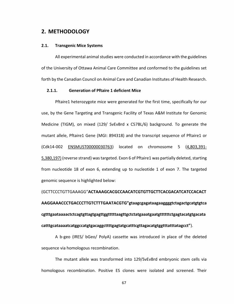
67
2. METHODOLOGY
2.1. Transgenic Mice Systems
All experimental animal studies were conducted in accordance with the guidelines
of the University of Ottawa Animal Care Committee and conformed to the guidelines set
forth by the Canadian Council on Animal Care and Canadian Institutes of Health Research.
2.1.1. Generation of Pftaire 1 deficient Mice
Pftaire1 heterozygote mice were generated for the first time, specifically for our
use, by the Gene Targeting and Transgenic Facility of Texas A&M Institute for Genomic
Medicine (TIGM), on mixed (129/ SvEvBrd x C57BL/6) background. To generate the
mutant allele, Pftaire1 Gene (MGI: 894318) and the transcript sequence of Pftaire1 or
(Cdk14-002 ENSMUST00000030763) located on chromosome 5 (4,803,391-
5,380,197) (reverse strand) was targeted. Exon 6 of Pftaire1 was partially deleted, starting
from nucleotide 18 of exon 6, extending up to nucleotide 1 of exon 7. The targeted
genomic sequence is highlighted below:
(GCTTCCCTGTTGAAAGG“ACTAAAGCACGCCAACATCGTGTTGCTTCACGACATCATCCACACT
AAGGAAACCCTGACCCTTGTCTTTGAATACGTG”gtaagcgagataagaaggggtctagactgcatgtgtca
cgtttgaataaaactctcagtgttagtgagttggtttttaagttgctctatgaaatgaatgttttttctgagtacatgtgacata
catttgcataaaatcatggccatgtgacaggcttttgagtatgcatttcgttagacatgtggtttatttatagcct”).
A b-geo (IRES/ bGeo/ PolyA) cassette was introduced in place of the deleted
sequence via homologous recombination.
The mutant allele was transformed into 129/SvEvBrd embryonic stem cells via
homologous recombination. Positive ES clones were isolated and screened. Their
68
sequences were confirmed by Polymerase Chain Reaction (PCR) and Southern blot
hybridization analysis. Positive clones were delivered into C57BL/6 blastocysts by
microinjection and implanted into pseudo-pregnant foster mothers to produce chimeras.
Chimeric mice were bred with wild type mice to produce the Pftaire1 heterozygote
deficient mice. PFTAIRE F1 generation homozygote/null mice were obtained by breeding
heterozygous parents and their genotypes were confirmed by Polymerase Chain Reaction
(PCR). To establish Pftaire1 knockout line on a pure C57BL6 background we backcrossed
mixed (129/SvEvBrd x C57BL/6) heterozygote mice from the Pftaire1 deficient line with
C57BL6 for 10 generations (Figure 3-2 b)
Germ line chimera was confirmed by Southern blot followed by PCR analysis using
primers recommended by the Jackson Laboratory as described in their protocol. To
confirm genotypes, primers for Southern blot screening were designed as follows:
5’probes, A: (5’–CATACATCAGGCATCCAGGGTA) and B: (5’– GGCTGGACTCCTGACTTCACCA);
to generate a 644 bp product. 3’probes, C: (5’– CTTAGGAGAATTTGACATCCTCA) and D: (5’
– CAGTGAGTTCCGGAGCTAGTCT); to generate a 684 bp product. Furthermore, to confirm
successful disruption of Pftaire1 gene in Pftaire1 final mouse strain, PCR primers were
designed as follows: (fwd. 5’-TGACCCTTGTCTTTGAATACGTG-3’ and 5’-
GGAAGCCTAAATTGTAAGGATCAGG-3’) and (fwd. 5’–GCAGCGCATCGCCTTCTATC-3’ and
5’-GGAAGCCTAAATTGTAAGGATCAGG-3’); to generate a 418 bp product and reverse to
generate a 341 bp product for wild type and knockout, respectively.
PCR mix was run with GoTaq Green Master Mix 2x (Promega, M712B). The PCR
amplification process was as follows: initial amplification designed as touchdown PCR
69
including: 6 cycles of denaturation 98 °C for 17 sec, annealing 69 °C (touchdown, -1°C per
cycle) for 15 sec, amplification 72°C for 15 sec, and further amplification of the target
genes was as follows: 35 cycles of 98 °C for 17 sec, 60 °C for 15 sec, and 72 °C for 15 sec.
PCR products were resolved on a 1.5 % agarose gel- Ultra Pure Agarose (Invitrogen,
15510-027) and visualized by ethidium bromide (Ethyl hexadecyl dimethyl bromide
C20H44BrN) (Sigma Ultra, C5335-1000).
2.2. Real Time PCR (Quantitative RT- PCR)
All steps were performed in RNAase-free environment. To ensure RNA-free
surface, RNase away (MBP Molecular Bioproducts, 7003) was applied. Total RNA was
extracted from mouse embryo whole brains/cortices at embryonic day E 13.5-14.5 using
Trizol Reagent (Ambion, 15596026) based on Invitrogen protocols. 25 ng of extracted RNA
was used per reaction. To determine disruption of Pftaire1 transcription the SuperScript
III Platinum SYBR Green One-Step RT-PCR kit (Invitrogen, 204174) was used. Negative
controls, no RNA and no RT were run in parallel with both the GAPDH housekeeping gene
and Pftaire1. Each sample was run in quadruplets and n=3. Results were normalized
against GAPDH. Primers used for amplification of the target genes were as follows:
(GAPDH-Fwd.: 5’-GGTGAAGGTCGGTGTGAACG-3’, GAPDH-Rev: 5’-
CTCGCTCCTGGAAGATGGTG-3′) to generate a 233 bp product and (Pftaire1-Fwd: 5′-
CAGCGATCTGCCTCCACGGC-3’) designed within exon13. (Pftaire1-Rev: 5’-
GGCCCTCATGCTCTCTCCAGC-3’) designed within exon 14, to generate a 341 bp product.
The primers would also be able to produce a short heptamer within mutated region (exon
8); thus, it could not interfere with mRNA levels for Pftaire1-KO. The program for RT-PCR
70
amplification was as follows: 48 °C for 30 min, 95 °C for 10 min, and 40 cycles of 95 °C for
15 sec, 60 °C for 30 sec and 72°C for 30 sec. The RT-PCR products were resolved on a 1.5
% agarose-ethidium bromide gel as well.
2.3. Semi-Quantitative Reverse Transcriptase RT-PCR
All steps were performed in RNAase-free environment. To ensure RNA-free
surface, RNase away (MBP Molecular Bioproducts, 7003) was applied. Total RNA was
extracted from whole brains/cortices of WT-Pftaire1 and KO-Pftaire1 mouse embryo at
embryonic day E 13.5-14.5 using Trizol Reagent (Ambion, 15596026) based on Invitrogen
protocols. 50 ng of extracted total RNA was used per reaction for cDNA synthesis and
gene amplification using SuperScript III Platinum SYBR Green One-Step RT-PCR kit
(Invitrogen, 204174). Targeting primers designed for RhoA were as follows: (RhoA-fwd:
5’-GACCTTCGGAATGACGAGCA-3’ and RhoA-Rev: 5’-TTCCCACGTCTAGCTTGCAG-3’).
Results were normalized against S12 and primers were as follows (5′-
GGAAGGCATAGCTGCTGG-3′ and 5′-CCTCGATGACATCCTTGG-3′) (Gonzalez, Zhang et al.
2008, Biswas, Zhang et al. 2010). cDNA synthesis was performed at 45°C for 45 min. Then,
amplification was performed by a 2 min initial denaturation step at 94°C, followed by 24
cycles of 94°C for 30 sec, 60°C melting temperature (Tm) for 30 sec, and 72°C for 1 min,
and 72°C for 10 min . RT-PCR products were resolved on a 2% agarose (Invitrogen)–
ethidium bromide gel, and correct band for RhoA, 197 bp was processed by densitometry
using Image J. Transcript levels were normalized against S12 signals and results were
reported as relative mRNA level (normalized value). Data is represented as (Mean ±SEM)
of at least three independent experiments.
71
2.4. Cell Culture
2.4.1. Immortalized Cell Line
Human Embryonic Kidney cells (HEK-293T) were cultured in Dulbecco's Modified
Eagle Medium (DMEM) (High Glucose 4500 mg/L with 4 mM L-Glutamine without Sodium
Pyruvate 0.1 µm sterile filtered) (ThermoScientific, SH30022-01) supplemented with 10 %
Fetal Bovine Serum (FBS) (Hyclone, SH30397.03) and 1 % Antibiotic/ Antimicotic
Solution100x (10,000 units/ml of penicillin, 10,000 µg/ml of streptomycin, and 25 µg/ml
of Amphotericin B , 0.2 µm filtered) (ThermoScientific, SV30079.01) at 37°C in a 5% CO2
atmosphere.
2.4.2. Primary Cortical Neuron Cultures
Cortical neuron cultures were prepared either from wild-type CD1 mouse embryos
purchased from Charles River or from heterozygous crosses between Pftaire1 mice, at
gestational day /embryonic day E 13.5-14.5. Embryos were considered E 0.5 d, at the time
vaginal plug was detected. Embryos were removed from the uterine horns and
transferred to Hanks' Balanced Salt Solution (HBSS) (modified without Calcium and
Magnesium) (Hyclone Gibco, SH3003102). Brains were excised by cutting the skull open
at the midline, from base of the head towards the front. Two lateral incisions were made
under the cerebral cortices and skull was peeled off, meninges were removed and
individual cortices were transferred to HBSS containing 8 µl Trypsin (25g/L in 0.9 % NaCl)
(Sigma, T4549), incubated for 20 min at 37 ⁰C with agitation. 10.7 µl of Trypsin inhibitor
in neurobasal medium was added to stop trypsinization, and 13.34 µl DNase1 (10 mg /
ml) (Boehringer Mannheim Roche Diagnostics, 10137100) to degrade extracellular DNA
72
medium were added to the solution. Cells were pelleted at 1000 x, for 5 min at 4 °C and
10.7 µl of trypsin inhibitor and 13.34 µl of DNase were added. Cells were triturated; viable
cells were counted using sterile filtered trypan blue solution (0.4 %) (Sigma, T8154).
(Number of cells per ml was calculated by: cell count in the central square x dilution factor
x 104). Cells were diluted to desired concentration for culture, in Complete Neurobasal
Medium containing: Neurobasal medium (Gibco LifeTechnologies, 21103-049), B27
supplement (Invitrogen, 17504-044), N2 supplement (Invitrogen, 17502-048), L-
Glutamine 200 mM (Gibco, 25030-081) and Penicillin-Streptomycin. Primary cortical
neuron cells were plated into 24 or 6 well plates. Plates were coated with Poly-D-Lysine-
Hydrobromide (1 mg/ml, sterile) (Sigma, P0899) prior to use. Poly-D-Lysine incubation
was performed for at least 30 min and then plates were washed. For immunostaining,
sterilized coverslips were placed into 24 well culture dishes and then coated with poly-D-
lysine. Cultures were incubated at 37 °C in a 5% CO2 atmosphere and fixed or lysed at
varied times.
2.5. Exogenous Gene Expression
2.5.1. Expression Vectors
Plasmids for transfection: GFP subcloned into pCig2 vector (generously gifted by
Carola Sherman), FLAG-Tagged: WT-Pftaire1 and D228N-Pftaire1 cDNA (mutation of
conserved aspartic acid residue to asparagine, by a point mutation) (1284 kb) subcloned
into pCMV vector (generously gifted by Dr. Paul Albert and Maribeth Lazzaro) (Lazzaro,
Albert et al. 1997); GST-Tagged: WT-Rac1, G12V-Rac1, S17N-Rac1 subcloned in pEBG
vector; GST-tagged: WT-Cdc42, G12V-Cdc42, S17N-Cdc42 subcloned in pEBG vector and
73
MYC-tagged: WT-RhoA, CA-RhoA Q63L, DN-RhoA L19N in pRK5 vector. Rho GTPases
(generously gifted by Dr. Gary Bokoch and Dr. Margaret Chou, University of Pennsylvania).
Constructs were sequenced at Ottawa Genomic Center, using following primers: (1) For
Rac1 and Cdc42 constructs in the pEBG backbone: (pGEX 5' Sequencing Primer: 5'-
[GGGCTGGCAAGCCACGTTTGGTG]-3' and pGEX 3' Sequencing Primer 5'-
[CCGGGAGCTGCATGTGTCAGAGG]-3') (2) To sequence Pftaire1, CMV virus backbone:
(pCMV 5' Sequencing Primer: 5'-CGCAAATGGGCGGTAGGCGTG-3’ Pftaire1 GC rich region
at 3’ end: 5’-GGGGGAGTTGAAGCTGGCAG-3’). Sequencing was performed 100 bp
upstream of start codon and 100 bp downstream of stop codon. Plasmids were
transformed with Top10 competent cells. Transformed cells were cultured on Ampicillin
resistant agar plates (LB Agar Granulated, Molecular Genetics BP-1423-500). A single
colony was picked and grown in Luria-Bertani (LB) Broth (Miller Amresco, J-106-500G).
DNA was purified using the Pure Yield Plasmid Midi Prep System (Promega, A2495).
2.5.2. Transient Transfection with Lipofectamine
HEK 293T Cells were cultured to 70% confluency, then co-transfected with Flag
tagged (WT-Pftaire1 or D228N-Pftaire1) and GST tagged [WT-Rac1, G12V-Rac1
(constitutively active), S17N-Rac1 (dominant negative inactive)] or GST tagged [WT-
Cdc42, G12V-Cdc42 (constitutively active), S17N-Cdc-42 (dominant negative inactive)] or
GST tagged [WT-RhoA, Q63L- RhoA (constitutively active), L19N-RhoA (dominant negative
inactive)], as well as, the reporter vector (GST::PEGB, MYC::PRK5, Flag::CMV using
lipofectamine 2000 reagent (1mg/ml) (Invitrogen, 11668-019 ). Opti-MEM (Minimal
Essential Medium), 1x Reduced Serum (Gibco, 31985-070) was used to dilute the plasmid
74
and lipofectamine according to the manufacturer’s instructions. Diluted plasmid and
lipofectamine were mixed and incubated for 20 minutes at room temperature. Cell
culture medium was changed to media without FBS at the time of transfection. The
plasmid-lipofectamine complex was added to culture media in droplets. 24 hours post
transfection, cells were triturated off culture plates by scraping, cells were collected into
ice cold PBS and spun down at 1.4 x g for 1 min for pull down assay.
2.5.3. Viral Infection
Gene delivery via Adenoviral (AV) infection was performed at the time of plating.
Adenovirus expressing WT-Pftaire1 (Ad ET-CMV-3x Flag-WT-Pftaire1, Pfu: 1.00 x 10¹² CsCl
Pure, Plaque pure), D228-Pftaire1 (Ad ET-CMV-3x Flag-D228N-Pftaire1, Pfu: 9.20 x 10¹¹
CsCl Pure, Plaque pure), and GFP (Ad ET-CMV-EGFP, Pfu: 4.89 x 10¹¹ CsCl Pure, 3x Plaque
pure), was mixed with neuronal cortical culture cells at 50 -100 MOI (multiplicity of
infection). Cells were fixed or lysed, at different time points, post-culture, for further
analysis, depending on the nature of the experiment.
2.6. Neuronal Death Assay
Cortical culture cells from mouse embryos at E13.5-14.5 were seeded at (1 x 10⁵)
in 96 well plates and infected/co-infected with AV viruses (GFP alone or GFP + WT-
Pftaire1, D228N-Pftaire1) at MOI=100 at the time of plating, for survival assay. Cells were
washed with 1X PBS and then fixed with 4% PFA 12, 16 and 24 hours post-infection and
then stained with Hoechst 33258 (1:10000) (Sigma-Aldrich B2883) in 1x PBS and
incubated for the minimum of 30 min for nuclei staining. Intact nuclei were counted
against condensed and fragmented nuclei to determine live neurons versus dead ones.
75
To determine survival the number of live GFP positive neurons were assessed in each well,
each treatment was performed in triplets, and the average for each treatment was
calculated. Survival was assessed for WT-Pftaire1 or D228N-Pftaire1 as the percent of live
GFP positive neurons in comparison to GFP. Survival percentage represents the ratio of
GFP expressing neurons with morphologically intact nuclei at (12, 16, and 24 h) in WT-
Pftaire1 or KO-Pftaire1 to the total number of GFP expressing neurons. The values were
normalized to GFP alone as control. Data are presented as (Mean± SEM) of at least three
independent experiments.
2.7. Total Protein Extraction
To compare protein expression levels, whole brain lysates were prepared from
mouse brains at E13.5-14-5 or from mouse pups at P21. Basically, mouse brains were
dissected and washed with ice-cold PBS. Brains were homogenized in lysis buffer: [50mM
Tris-HCl (pH=7.5), 150mM NaCl,1 mM MgCl2, 1mM EDTA, 1% triton X-100 (Boehringer
Mannheim, 789704), 1mM L-Dithiothreitol (DTT , C4H10O2S2) (Sigma Aldrich, D0632-5G),
protease inhibitor (Halt Protease inhibitor Cocktail EDTA-free 100x (Roche, 87785)] for 20
min, at 4 °C, with agitation. Lysates were clarified by centrifugation at 19500 x g, 4 °C, for
20 min. Supernatant was collected (approximately 2mg of protein was collected per
brain), mixed with 0.1% w/v bromophenol blue/ DTT sample buffer and heated at 100°C,
for 10 min.
2.8. Immunoprecepitation (IP)
All steps were performed on ice. Cell lysate was prepared as described, previously.
Lysate was prepared using lysis buffer 50 mM Tris-HCl (pH=7.5), 150 mM NaCl, 1 mM
76
MgCl2, 1mM EDTA, 1% Triton X-100 (Boehringer Mannheim, 789704), 1mM DTT (L-
Dithiothreitol, C4H10O2S2) (Sigma Aldrich, D0632-5G), protease inhibitor, for 20 min, at 4°C
with agitation. Lysates were clarified by centrifugation at 19500 x g, 4°C, for 20 min.
Supernatant was collected and incubated with 20 µl of washed bead per reaction. After
immunoprecipitation, beads were pelleted at 1000 x g , washed five times with wash
buffer (Tris-HCl (pH≈7.3-7.5), NaCl, EDTA, Triton 10 % , DTT), then mixed with 0.1 % w/v
bromophenol blue / DTT sample buffer, and heated at 100°C, for 10 min to release the
protein complex. Proteins were resolved on 12 % SDS-polyacrylamide gels.
For immunoprecipitation of ectopically expressed GST tagged proteins, GST beads
- (Glutathione Sepharose 4b beads, particle size 45 µm - 165 µm, GE Healthcare 17-0756-
05) were incubated with the lysate overnight, at 4 °C, on a rotor. For immunoprecipitation
of endogenous protein, Trueblot Anti Rabbit IgIP beads - (eBioScience, 00-8800-25) or
Anti mouse IgIP beads - (eBioScience, 00-8811-25) were used, depending on the origin of
the bait antibody. Lysate was collected and precleared by incubating 20 µl of the washed
bead that was to be used for IP, for 30 min, at 4 °C, on a rotor. Supernatant was cleared
by centrifugation at 1000 x g, 1 min, at 4°C. 4 - 8 µg of antibody per reaction, normal
mouse IgG (SantaCruz, sc-2025) or normal rabbit IgG (SantaCruz, sc-2027) and Pftaire-1
(H-140) (1:500) rabbit polyclonal IgG (SantaCruz, sc-50475), RhoA (1:500) mouse
monoclonal IgG (SantaCruz, sc-418), Rac1 (C-11) (1:500) rabbit polyclonal IgG (SantaCruz,
sc-95), Cdc42 (1:500) rabbit polyclonal IgG (SantaCruz, sc-87) was incubated with the
cleared supernatant, overnight, at 4°C, on a rotor. Pftaire1 was used to IP GTPase and vice
77
versa. 20 µl of the washed beads were incubated with the antibody-lysate mix for 3 hours,
at 4 °C, on a rotor to precipitate the antibody.
2.9. SDS-PAGE and Western Blot
Protein extracts were resolved on a 10 or 12 % SDS - polyacrylamide gel, along
with Molecular Marker, Blueye Prestained Protein Ladder (GeneDirex, P14007-0500).
Transfers were done on 0.45 µm PolyVinylidene DiFluoride (PVDF) Transfer membrane
(Immobilon-P, IPVH00010). Membrane was blocked in 10 % non-fat dry milk in 1 x PBST
[Phosphate Buffered Saline (pH=8.0) and 0.1 % Tween] at room temperature, for 1 hour.
Proteins were detected using antibodies. Primary antibodies were diluted in [ 3 % Bovine
Serum Albumin (BSA) fraction V (Fisher, BP1600-100) in PBST and 0.1 % Sodium Azide
NaN3 (Sigma Ultra, s-8032)] as follows: Pftaire1 (H-140) (1:500) rabbit polyclonal IgG,
(SantaCruz, sc-50475), Pftaire2 (CDK15) (1:500) rabbit polyclonal (Life Span Biosciences,
LS-C119219/34326), RhoA (1:500) mouse monoclonal IgG (SantaCruz, sc-418), Rac1 (C-
11) (1:500) rabbit polyclonal IgG (SantaCruz, sc-95), Cdc42 (1:500) rabbit polyclonal IgG
(SantaCruz, sc-87), Cdk5 (C-8) (1:500) rabbit polyclonal IgG (SantaCruz, sc-173), Pctaire1
(CDK16) (1:500) rabbit polyclonal, (Life Span Biosciences, LS-C112292/34325), Anti-Flag
(1:500) rabbit monoclonal IgG (Sigma, F7425), Anti-GST (1:500) mouse monoclonal IgG
(SantaCruz, sc-138) and incubated with the membranes overnight, at 4 °C, with agitation.
Membranes were washed and then incubated with secondary antibodies diluted in 5 %
non-fat milk, for 1 hour, at room temperature [goat anti-mouse 170-6516 or goat anti-
rabbit 170-6515 IgG (H+L) HRP conjugate (Bio-Rad)] were all used at 1:2000 in 3 % BSA in
0.1 % Tween 20 (PBS/T). Immunoreactivity was detected using Immobilin Western
78
Chemiluminescence HRP substrate (Millipore, WBKLS0500) or Pierce ECL Western
Blotting Substrate (Thermo Scientific, 32106) and visualization HyBlot CL Autoradiography
film, Denville Scientific. Protein levels were normalized against β-actin signals, and results
were reported in reference to control values (untreated control for each individual
experiment).
2.10. Pftaire1 Kinase Assay
Kinase assay was performed using immunoprecipitated WT-Pftaire1 protein or IgG
as a control. Briefly, Neuronal cortical cells prepared from WT-Pftaire1 mouse embryos
E13.5-14.5 were cultured in Neurobasal media (Gibco, 21103-049) containing B27
(Invitrogen, 17504-044) and N2 (Invitrogen, 17502-048), in 12 well plates, at the ratio of
1 embryo per well, for 18-20 hour, at 37°C, 5 % CO2. After washing with ice-cold 1x PBS,
lysates were collected directly from plates by scraping into ice-cold immunoprecipitation
(IP) buffer (Tris-HCL 25mM (pH 7.4) , Na4P2O7 10 mM, Na3VO4 25 mM, NaF 50 mM, EDTA
1 mM , Digitonin 2 mg/ml) and sonicated twice, for 3 cycles, with a 20 minute incubation
in between the 2 set of cycles, and then supernatant was cleared by centrifugation at
14000x g, 4°C, for 10 min. 1 mg of protein( adjusted to the volume of 300 µl was incubated
with 4 µg antibody either normal rabbit IgG (200 µg/0.5 ml) or Pftaire1 (200 µg/ml)
antibodies, overnight, at 4°C, on the shaker. IP-ed, WT-Pftaire1 and IgG were mixed with
40 µl of pre-equilibrated (performed washes with IP wash buffer) Trueblot anti-rabbit IgIP
beads per reaction and incubated for 2 hours, at 4°C, on the shaker. Beads were then
washed and pelleted at 2000rpm, for 1 min, at 4°C, with IP wash buffer and kinase assay
buffer [HEPES 50 mM (pH 7.5) and 1mM DTT], respectively. Next, kinase reaction buffer
79
(HEPES 50 mM (pH 7.5), 1 mM DTT, 10 mM MgCl, and 20 µM ATP)) was added to a final
volume of 25 µl. 10 µg of RhoA substrate His-Recombinant human RhoA (Prospec, pro-
057), was added to reactions with or without (5 µl, 100 x) GDP. Reactions were incubated
overnight, at 30°C. Samples were then centrifuged at 2000 rpm, for 1 min, at 4°C, then
supernatant was collected and subjected to either western blot for rabbit polyclonal IgG
phosphoserine (Invitrogen, 61-8100) signal; or G-LISA assay to assess RhoA activity as
described below.
2.11. Assessment of Rho GTPase Activity
Activation of RhoA, as a function of Pftaire1 kinase activity was determined using
commercially available “RhoA Activation Assay Kit (CellBioLabs, STA-403-A)” according to
the manufacturer's instructions. Primary Cortical Culture Cells were prepared from Het x
Het crosses at embryonic day 13.5-14.5, at the density of 1 embryo per well, in 1.5 ml,
cultured in complete Neurobasal media supplemented with B27 and N2 , in 12 well plates,
and incubated at 37 °C and 5 % CO2. 18-20 hours post-plating cells were washed with ice-
cold PBS and GTPase assay was performed according to the manufacturer’s kit. To
decrease rapid hydrolysis of active GTP bound to inactive GDP bound protein all following
procedures were performed on ice. Briefly, cells were incubated on ice, for 20 minutes,
with 1x Assay / lysis buffer provided with the kit (Part No. 240102) (containing 125 mM
HEPES, pH 7.5, 750 mM NaCl, 5% NP-40, 50 mM MgCl2, 5 mM EDTA, 10% Glycerol) and
then collected by scraping. Due to very low expression of RhoA same-genotype extracts
were pooled to achieve large amounts of Protein. Total protein was quantified by
Bradford protein assay to achieve at least 800ug of protein for each sample/assay. Cell
80
lysates were clarified by centrifugation at 14,000 g x, at 4 °C, for 10 min. (at least 800 µg
protein per reaction). 100x GTPS (Part No. 240103) and GDP (Part No. 240104) were
added to the cells for positive and negative control, respectively. Control samples were
incubated for 30 minutes, at 30 ⁰C, with agitation, and the reaction was terminated by
adding 1M MgCl2 to the samples. Volume of sample per reaction was adjusted to be
identical in all reactions and 40 µl of bead added per reaction. Active-proteins were
immunopercipitated with respective rhotekin RBD agarose beads for RhoA. Beads were
pelleted by centrifugation at 14000 x g, for 10 seconds, and washed three times with 1x
Assay buffer supplied in the kit. Afterwards, beads were resuspended in SDS-PAGE sample
buffer and boiled for 5 minutes for detection by western blotting. Separated proteins
were visualized using RhoA, Mouse Monoclonal antibody (Part No. 240302) provided with
the kit.
To quantify Rho GTPase activity after the pftaire1 Kinase assay, in a more
accurate/sensitive manner, G-LISA RhoA activation assay biochem kit (Absorbance based)
(Cytoskeleton, BK124) was used according to the manufacturer’s protocol. Samples from
the kinase reactions, (IgG-IP: RhoA, IgG-IP: RhoA+GDP, Pftaire1-IP: RhoA and Pftaire1-IP:
RhoA+GDP) were diluted and pre-equalized with ice cold lysis buffer /binding buffer (Part
# GL37). To pre-equalize samples, protein concentration was determined at 600nm, using
Precision RedTM Advanced Protein Assay Reagent (Part # GL50) as suggested by the
manufacturer. Same buffer mix (no protein) and RhoA (provided by manufacturer), were
respectively, used as blank and positive control for the assay. Next, samples were
transferred to 96 well Rho-GTP binding plates (Part # GL25), equipped with pre-washed
81
GLISA assay strips, incubated at 4°C, on an orbital microplate shaker at 400 rpm, for
30min. Samples were then washed with wash buffer (Part # GL38) and incubated with
Antigen Presenting Buffer (Part # GL45) for 2 min, washed again with wash buffer, then
incubated at room temperature for 45 min, on orbital microplate shaker at 400rpm, with
anti-RhoA primary antibody (Part # GL01) (1:250) in Antigen dilution buffer (Part # GL40)
and washed subsequently. Samples were next incubated with (1:62.5) secondary
horseradish peroxidase (HRP) conjugate antibody (Part # GL02) at room temperature, for
45 min, on an orbital microplate shaker at 400 rpm and washed afterwards. Then samples
were incubated with HRP detection reagents A (Part # GL43) and B (Part # GL44) (1:1 ratio
) for 15 min, at 37 °C, and reaction was stopped by adding HRP Stop Buffer (Part # GL80),
and absorbance measured at 490 nm using a microplate spectrophotometer. To measure
RhoA activity, values were determined as follows, (GLISA value(ng)
ELISA value(ng)× 100 =
normalized % of active RhoA). The detected signal was normalized against total RhoA
using G-LISA and E-LISA (BK # 150) kits side by side. To determine if any significant
difference existed in the activity of RhoA among samples, two way ANOVA was performed
in the grouped data followed by Bonferroni’s post-hoc test. Results were presented as
average of at least three independent experiments.
2.12. Histology and Staining
2.12.1. Brain Sectioning
Brains were collected from the Wt-Pftaire1 and KO-Pftaire1 mice at 8 weeks of
age. Animals were humanely anesthetized with euthasol and transcardially perfused with
0.9 % saline, followed by fixative solution 4 % Paraformaldehyde (PFA) (EMD, PX0055-3)
82
in 1x PBS at pH=7.4. Brains were then removed from the skull and fixed further with 4 %
PFA, at 4 °C, for 24 hrs. Fixed brain tissue was cryopreserved in 20 % sucrose in 0.1 M
phosphate buffer and stored at 4°C, over a 3 day course, and the solution was changed 3
times per day. Brains were then snap-frozen with Nitrogen balanced with CO2 and then
embedded with O.C.T. (Tissue - Tek /O.C.T compound, 4583) for sectioning. 14 μm coronal
sections were collected from the brains; starting at the frontal cortex at Bregma: ~ 2.710
mm up to the 4th ventricle at Bregma: ~ - 4.20 mm. For collecting, 3 sections were skipped
in between each selection. Sections were directly mounted onto SuperFrost Plus
microscopic slides 25 x 75 x 1.0 mm (Fisher brand, 12-550-15) and subjected to2 % cresyl
violet staining as described below.
2.12.2. Cresyl Violet Staining
Sections were thawed on slide warmer for 15-20 min, at 35 °C, and then placed in
distilled water for 5 min. Sections were then stained with 2 % filtered cresyl violet
(pH=3.5) cresyl violet acetate (C18H15N3O3, Sigma C5042-10G), for 10-30 minutes
depending on the stain concentration. Sections were then dehydrated in graded ethanol
solutions from 50 % to 100 %, followed by defatting with xylene. Sections were mounted
with coverslips using toluene.
2.12.3. Immunostaining
Cells plated on glass coverslips were fixed in 4 % PFA in 1 x PBS, for 15 min. Fixed
cells were permeabilized and blocked simultaneously in 0.1 % Triton X-100, and 5 % BSA
in 1x PBS, respectively, for 20 min, at room temperature. Coverslips were incubated with
primary antibodies diluted in 5 % BSA in 1x PBS: Tau1 clone 46 (1:500) mouse monoclonal
83
IgG (Sigma-Aldrich, T-9450), GFP (H-140) (1:500) mouse monoclonal IgG (Abcam, ab1218),
Pftaire1 (H-140) (1:500) rabbit polyclonal IgG (SantaCruz, sc-50475), MAP2 (H-300)
(1:500) rabbit polyclonal IgG (SantaCruz, sc-20172) for 1hr, at room temperature.
Coverslips were washed; incubated with FITC-secondary antibodies: Alexafluor 488
(1:1000) Goat-Anti-mouse (Molecular Probes, A11001), Streptavidin Alexafluor 488
(1:1000) Goat-Anti-mouse (Molecular Probes, S11223), Streptavidin Alexafluor 546
(1:1000) Goat-Anti-mouse (Molecular Probes, S11225), Alexafluor 594 (1:1000) Goat-
Anti-mouse (Molecular Probes, A11005), Alexafluor 594 (1:1000) Goat-Anti-rabbit
(Molecular Probes, A11037), Alexafluor 594 (1:1000) Donkey-Anti-Goat (Molecular
Probes, A11058) were diluted in 5% BSA in 1x PBS, and then incubated with coverslips for
30 min, at room temperature. To detect intact nuclei, cells were stained with Hoechst
33258 (1:10000) (Sigma-Aldrich B2883) in 1x PBS, incubated for the minimum of 30 min,
at room temperature. Slides were immersed in distilled water to remove excess salt and
then mounted on slides applying approximately 25 µl of Vectashield Mounting Medium
(Vector laboratories, H-1200).
2.13. Microscopy and Imaging
Images were taken using upright Zeiss Axioskop 2 Mot or inverted Zeiss
AxioObserver-Z1 fluorescent microscope with Northern Eclipse Software or Zeiss and
Axiovision Rel. 4.8 software, captured in XY mosaic format, in rectangle mode. Images
were taken at 20x, 32x, and 40 x magnification lenses. To determine the total diameter of
the field of view for each objective, Ocular Field of View (F.O.V.) number (32) was divided
by objective magnification and following measurements were obtained: for 20x (1500
84
µm), for 32x (719 µm), and for 40x (575 µm). Images were processed for length
measurements using Axiovision software Rel. 4.8, spline mode to trace axon length or
Stereo Investigator microscopy imaging system (version 6; MicroBrightField, Williston,
VT) software using contour mapping mode and tracing the length of axons. The distance
from the cell body of the GFP expressing neuron or Tau1 to the distal region of the longest
neurite was measured as axon/lengthiest neurite.
2.14. Quantification of Signal Density on Images
Immunoblots were further analyzed by Image J software to compare the
density/intensity of the bands of an agar gel or a western blot membrane. Following
imaging of the blots, representative bands were selected and their intensity was
quantified. Then quantified measurements were transferred to excel and results were
normalized against relative controls (β-actin for western blot and S12 for RT-PCR).
2.15. Statistical Analysis
Experiments were performed for a minimum of 3 times and each treatment was
done in replicates of 3. Data and graphs were represented in the format of (Mean ± SEM)
or percentage. Graphs were plotted as column charts for probability of getting a specific
range of values, to compare the mean of the experimental groups. To compare axon
lengths among different litters and to avoid divergence and variability between different
litters, data was either transformed to log10 format or classified into length intervals and
then data was plotted onto a linear or bar graph, respectively. To classify measurements,
the average and median axon lengths were calculated for each time-point. The maximum
and minimum numbers related to average and median were considered as average range

85
for that time-point and neurites diverting from this range were considered as either short
or long, relatively. Comparison against different litters was performed, as well. Statistical
analysis was performed using excel, Eviews 6 and SPSS software and processed using chi
square, Levene's Test for Equality of Variances, student’s two tailed t-Test considering the
variance between the two experimental groups equal, or one way and two way ANOVAs
followed by Student-Newman-Keuls post-hoc test or Bonferroni’s test.

86
CHAPTER 3
RESULTS
87
3. RESULTS
3.1. Pftaire1 Transgenic Mice
3.1.1. Generation of Pftaire1 Transgenic Mice
To examine the role of PFTAIRE1 in the CNS, Pftaire1 deficient mice were
generated*2. These mice are the first of their type since no other Pftaire1 deficient mice
exist, to date. To generate the Pftaire1 deficient mouse, Pftaire1 heterozygotes were
designed by replacing the majority of exon 6, starting from nucleotide 18, of the Mus
Musculus Pftaire1 transcript (ENSMUST00000030763) (Figure 3-1, 3-2 a) with the
neomycin (Neo) resistance cassette via homologous recombination (Figure3-2 b). The
transgenic mutation produced in the Pftaire1 gene causes a frame shift which alters the
open reading frame and leads to a premature stop codon and produces a truncated
protein that does not have the full kinase domain (Figure3-3); thus, is expected to degrade
or lack kinase activity. The DNA sequence of the 129/SvEvBrd embryonic cells that were
transformed with Pftaire1 mutant allele was confirmed by Polymerase Chain Reaction
(PCR) and Southern blot hybridization analysis. The transformed Pftaire1 allele was easily
detectable in the transformed mice through the agouti coat color of the chimeras. The
produced chimeric mice were viable and fertile; after being crossed with wild type mice
they also produce the Pftaire1 heterozygote deficient mice that were viable and fertile.
Pftaire1 F1 generation, homozygote (null) mice, which were obtained by breeding
2 Pftaire1 deficient mouse was designed and developed for our use by the TIGM (Texas A&M Institute for Genomic Medicine) company.

88
heterozygous parents were also viable and fertile. To establish Pftaire1 knockout line on
a pure C57BL6 background we backcrossed mixed (129/SvEvBrd x C57BL/6) heterozygote
mice from the Pftaire1 deficient line with C57BL6 for 10 generations, which continue to
be viable (Figure 3-2 b). Nevertheless, due to the length of time the complete cleaning of
the genomic background entitles; in most cases the results included in this thesis were
obtained using mice of 6th backcross generation or lower.
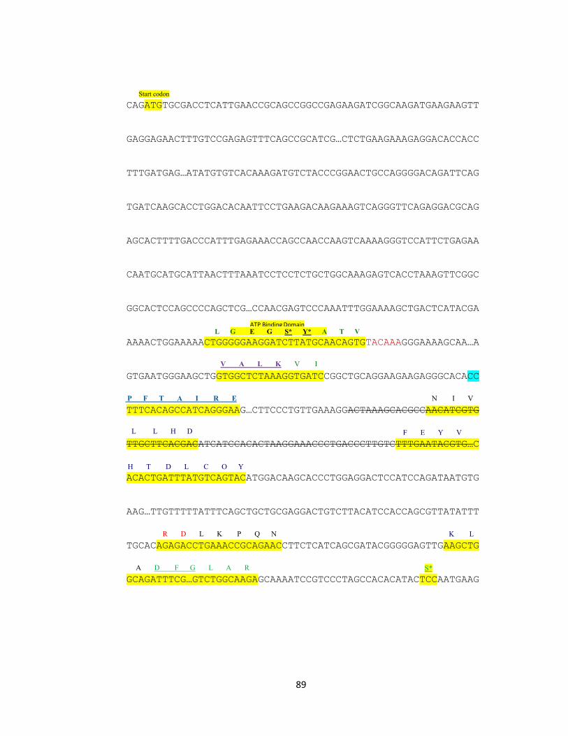
89
CAGATGTGCGACCTCATTGAACCGCAGCCGGCCGAGAAGATCGGCAAGATGAAGAAGTT
GAGGAGAACTTTGTCCGAGAGTTTCAGCCGCATCG…CTCTGAAGAAAGAGGACACCACC
TTTGATGAG…ATATGTGTCACAAAGATGTCTACCCGGAACTGCCAGGGGACAGATTCAG
TGATCAAGCACCTGGACACAATTCCTGAAGACAAGAAAGTCAGGGTTCAGAGGACGCAG
AGCACTTTTGACCCATTTGAGAAACCAGCCAACCAAGTCAAAAGGGTCCATTCTGAGAA
CAATGCATGCATTAACTTTAAATCCTCCTCTGCTGGCAAAGAGTCACCTAAAGTTCGGC
GGCACTCCAGCCCCAGCTCG…CCAACGAGTCCCAAATTTGGAAAAGCTGACTCATACGA
AAAACTGGAAAAACTGGGGGAAGGATCTTATGCAACAGTGTACAAAGGGAAAAGCAA…A
GTGAATGGGAAGCTGGTGGCTCTAAAGGTGATCCGGCTGCAGGAAGAAGAGGGCACACC
TTTCACAGCCATCAGGGAAG…CTTCCCTGTTGAAAGGACTAAAGCACGCCAACATCGTG
TTGCTTCACGACATCATCCACACTAAGGAAACCCTGACCCTTGTCTTTGAATACGTG…C
ACACTGATTTATGTCAGTACATGGACAAGCACCCTGGAGGACTCCATCCAGATAATGTG
AAG…TTGTTTTTATTTCAGCTGCTGCGAGGACTGTCTTACATCCACCAGCGTTATATTT
TGCACAGAGACCTGAAACCGCAGAACCTTCTCATCAGCGATACGGGGGAGTTGAAGCTG
GCAGATTTCG…GTCTGGCAAGAGCAAAATCCGTCCCTAGCCACACATACTCCAATGAAG
Start codon
S* A D F G L A R
K L R D L K P Q N
H T D L C O Y
F E Y V
L L H D
N I V P F T A I R E
V A L K V I
ATP Binding Domain L G E G S* Y* A T V
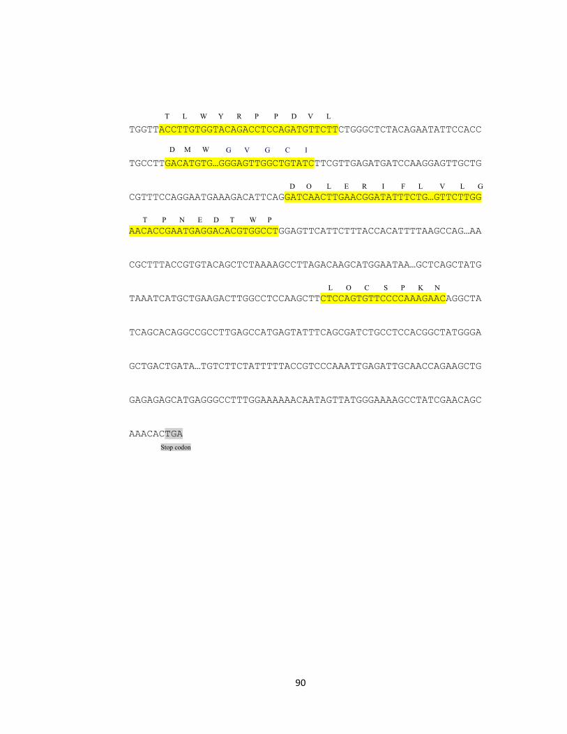
90
TGGTTACCTTGTGGTACAGACCTCCAGATGTTCTTCTGGGCTCTACAGAATATTCCACC
TGCCTTGACATGTG…GGGAGTTGGCTGTATCTTCGTTGAGATGATCCAAGGAGTTGCTG
CGTTTCCAGGAATGAAAGACATTCAGGATCAACTTGAACGGATATTTCTG…GTTCTTGG
AACACCGAATGAGGACACGTGGCCTGGAGTTCATTCTTTACCACATTTTAAGCCAG…AA
CGCTTTACCGTGTACAGCTCTAAAAGCCTTAGACAAGCATGGAATAA…GCTCAGCTATG
TAAATCATGCTGAAGACTTGGCCTCCAAGCTTCTCCAGTGTTCCCCAAAGAACAGGCTA
TCAGCACAGGCCGCCTTGAGCCATGAGTATTTCAGCGATCTGCCTCCACGGCTATGGGA
GCTGACTGATA…TGTCTTCTATTTTTACCGTCCCAAATTGAGATTGCAACCAGAAGCTG
GAGAGAGCATGAGGGCCTTTGGAAAAAACAATAGTTATGGGAAAAGCCTATCGAACAGC
AAACACTGA
T L W Y R P P D V L
Stop codon
L O C
S P K N
T P N E D T W P
D O L E R I F L V L G
G V G C I
D M W
T L W Y R P P D V L
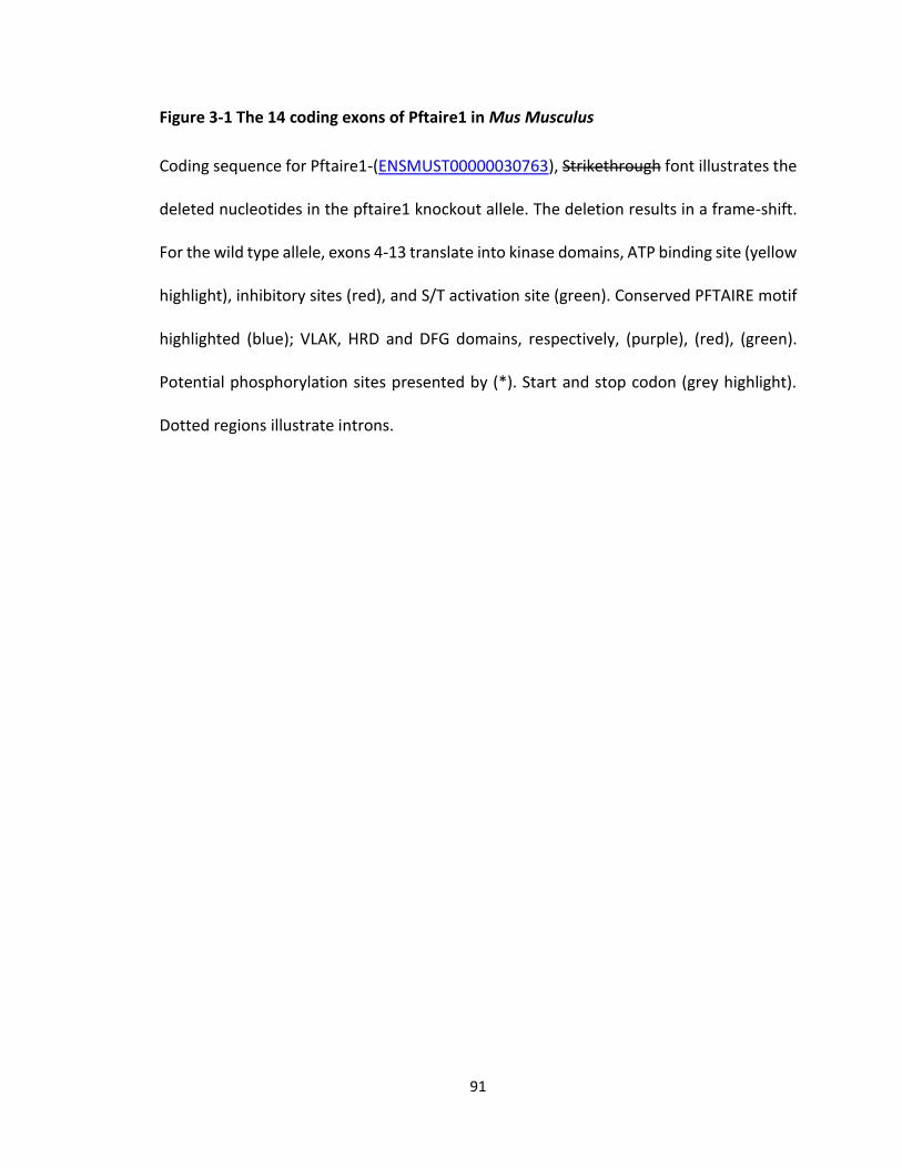
91
Coding sequence for Pftaire1-(ENSMUST00000030763), Strikethrough font illustrates the
deleted nucleotides in the pftaire1 knockout allele. The deletion results in a frame-shift.
For the wild type allele, exons 4-13 translate into kinase domains, ATP binding site (yellow
highlight), inhibitory sites (red), and S/T activation site (green). Conserved PFTAIRE motif
highlighted (blue); VLAK, HRD and DFG domains, respectively, (purple), (red), (green).
Potential phosphorylation sites presented by (*). Start and stop codon (grey highlight).
Dotted regions illustrate introns.
Figure 3-1 The 14 coding exons of Pftaire1 in Mus Musculus
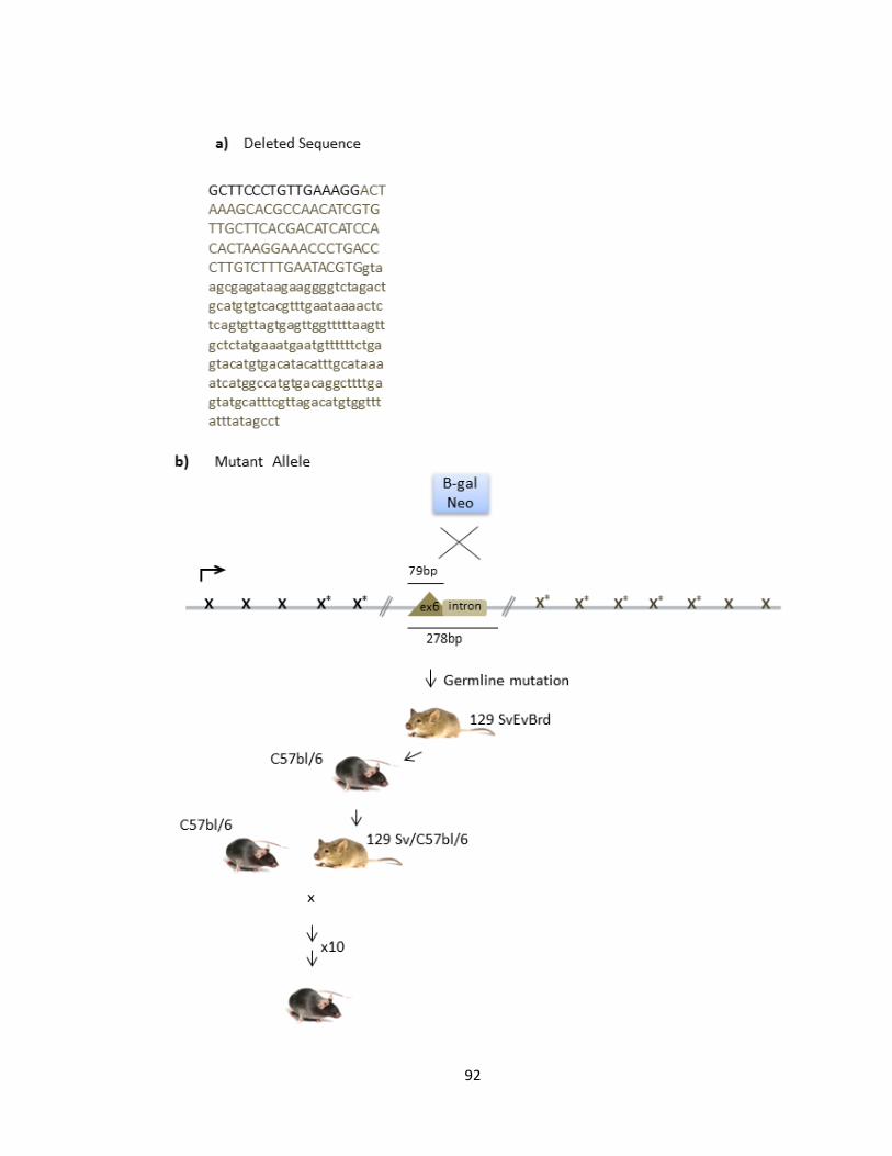
92
93
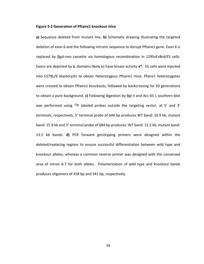
94
a) Sequence deleted from mutant line, b) Schematic drawing illustrating the targeted
deletion of exon 6 and the following intronic sequence to disrupt Pftaire1 gene. Exon 6 is
replaced by βgal-neo cassette via homologous recombination in 129SvEvBrd/ES cells.
Exons are depicted by x, domains likely to have kinase activity x*. ES cells were injected
into C57BL/6 blastocysts to obtain Heterozygous Pftaire1 mice. Pfaire1 heterozygotes
were crossed to obtain Pftaire1 knockouts, followed by backcrossing for 10 generations
to obtain a pure background. c) Following digestion by Bgl II and Acc 65 I, southern blot
was performed using 32P labeled probes outside the targeting vector, at 5’ and 3’
terminals, respectively. 5’ terminal probe of 644 bp produces WT band: 10.9 kb, mutant
band: 15.9 kb and 3’ terminal probe of 684 bp produces: WT band: 11.2 kb, mutant band:
13.5 kb bands. d) PCR forward genotyping primers were designed within the
deleted/replacing regions to ensure successful differentiation between wild type and
knockout alleles; whereas a common reverse primer was designed with the conserved
area of intron 6-7 for both alleles. Polymerization of wild type and knockout bands
produces oligomers of 418 bp and 341 bp, respectively.
Figure 3-2 Generation of Pftaire1 knockout mice
95
Successful removal of exon 6 and its replacement with the neo cassette in the
Pftaire1 gene of Pftaire1 knockouts was confirmed by PCR genotyping on mouse tail
genomic DNA at the time of weaning (P21). To ensure primer specificity, forward primers
were designed specifically within the deletion cassette of exon 6 (the last 23 nucleotides
of exon 6) and within the Neo cassette, for wild type and knockout, respectively. Also,
Reverse primer was designed within a conserved area of intron 6-7 (Figure 3-2). Pftaire1
wild type and knockout mice, respectively, produced bands of 418 bp or 341 bp.
Heterozygote mice were identified by the presence of PCR bands from both wild type and
knockout alleles (Figure 3-2 d).
Subsequently, to determine if the translation of full length Pftaire1 is abolished in
the Pftaire1 knockout mice, we examined for the presence of the full Pftaire1 product by
western blot analysis of whole brain lysates from mouse embryos at E13.5-14.5 and adult
mice, as well. Protein was extracted from brains of individual mice from each genotype
and resolved on 12 % SDS-PAGE gels and then probed for Pftaire1 rabbit polyclonal
antibody. Wild type, heterozygote mutants or homozygote mutants of Pftaire1 were
compared for Pftaire1 protein expression. When processing embryonic samples, a band
was detected for wild type Pftaire1 at about 48 kDa; the intensity of the signal was lower
in heterozygote mice. No band was detected when blotting for Pftaire1 in the knockout
(Figure 3-3 d). Previous reports by Lazzaro et al. (Lazzaro, Albert et al. 1997) and Besset
et al. (Besset, Rhee et al. 1998), confirm that Pftaire1 protein migrates at 47.5 (Lazzaro,
Albert et al. 1997) to 53 kDa (Besset, Rhee et al. 1998). However, Besset et al. detected
an extra band at 39 kDa in brain extracts (Besset, Rhee et al. 1998). In our case, when we
96
processed adult and pups samples, we observed a double band for Pftaire1 in wild type
and the heterozygote mutant of Pftaire1 but none in Pftaire1 homozygote mutants
(Figure 3-3 d). This observation is consistent with the splice forms reported for Pftaire1,
Cdk14-002 (ENSMUST00000030763) and Cdk14-09 (ENSMUST00000115452), which
translate into a protein product with 469 and 451 residues, respectively. As discussed
previously, the transgenic mutation produced in Pftaire1 gene results in a frame shift
mutation and a premature stop codon. Next, we examined whether disruption of Pftaire1
affects the protein levels of any of its close family members. We did not observe any
difference in the levels of Pftaire2, Pctaire1, and Cdk5 among wild type and knockout
Pftaire1 mice pups at P21 (Figure 3-4). The equal level of these Pftaire1 family members
in the knockout mice suggests that they may not be compensating for any Pftaire1 deficits
at the expression level. However, their kinase activity needs to be determined.
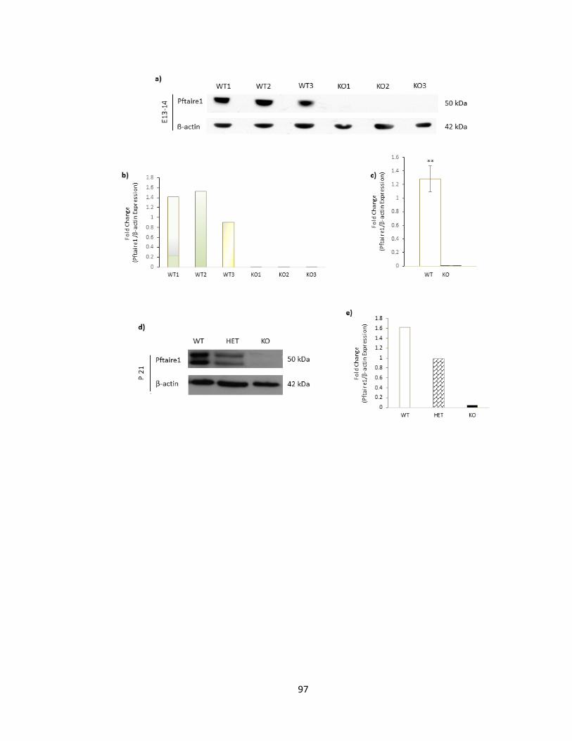
97
98
Disruption of Pftaire1 in knockout mice in comparison to wild type mice observed. Equal
loading was confirmed by β-actin probing. a) Western blot of Pftaire1 extracted from
whole brain lysates from the progeny of a Het x Het intercross. Whole brain lysates were
prepared from mouse embryos E13.5-14.5. Pftaire1 was probed with Pftaire1 rabbit
polyclonal antibody (SantaCruz). b) Column graph illustrating quantification of Western
blot by densitometry analysis for representative blot shown above in section a, c) Column
graph representing quantification of signal for western blot b, columns represent (Mean
± SEM), n=3 for wild type and knockout quantification s p=0.003 ** denotes significant
at p<0.01. d) Western blot of Pftaire1 extracted from whole brain lysates from the
progeny of a Het x Het intercross. Whole brain lysates were prepared from mouse pups
at P21, respectively. Pftaire1 was probed with Pftaire1 rabbit polyclonal antibody
(SantaCruz). e) Quantification of Western blot by densitometry analysis for representative
blot shown beside in section d. Columns represent fold changes of Pftaire1 expression in
wild type, heterozygote and homozygote mutants of Pftaire1 normalized to β-actin.
Figure 3-3 Pftaire1 protein expression is successfully disrupted in Pftaire1 homozygote
mutant mice
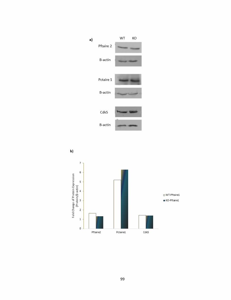
99
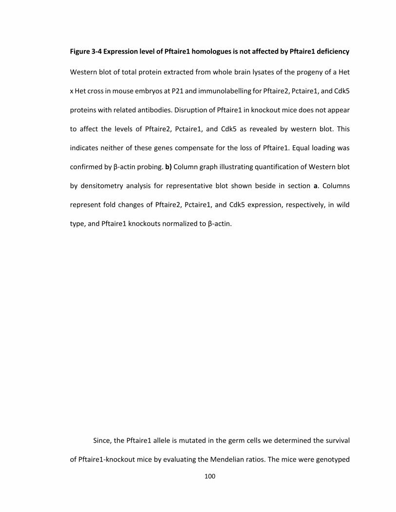
100
Western blot of total protein extracted from whole brain lysates of the progeny of a Het
x Het cross in mouse embryos at P21 and immunolabelling for Pftaire2, Pctaire1, and Cdk5
proteins with related antibodies. Disruption of Pftaire1 in knockout mice does not appear
to affect the levels of Pftaire2, Pctaire1, and Cdk5 as revealed by western blot. This
indicates neither of these genes compensate for the loss of Pftaire1. Equal loading was
confirmed by β-actin probing. b) Column graph illustrating quantification of Western blot
by densitometry analysis for representative blot shown beside in section a. Columns
represent fold changes of Pftaire2, Pctaire1, and Cdk5 expression, respectively, in wild
type, and Pftaire1 knockouts normalized to β-actin.
Since, the Pftaire1 allele is mutated in the germ cells we determined the survival
of Pftaire1-knockout mice by evaluating the Mendelian ratios. The mice were genotyped
Figure 3-4 Expression level of Pftaire1 homologues is not affected by Pftaire1 deficiency
101
by PCR as described earlier and Mendelian ratios were obtained using the chi2-test
(p<0.05). We evaluated a total of F1: n=21 (Het x Het) breeding crosses that were
backcrossed with C57Bl6, at least for a minimum of 9 times (Table 3-1). Heterozygote
intercrossing of Pftaire1 mice F1: (Het x Het) produces F2 offspring of wild type,
heterozygote, and homozygote mutant that do not differ significantly from the expected
1:2:1 Mendelian ratios.
At the approximate age of E13.5-14.5, from a total of F1: (Het x Het) of 15 breeding
pairs, F2 produced a total of 132 mice, of which 28% (n=37) were knockout mice, which
is not significantly different from expected Mendelian ratios (χ2 = 5.8, p = 0.055) (Table 3-
1 a). At the approximate age of P21 (time of weaning), from the total of F1: (Het x Het) of
6 breeding pairs, F2 produced a total of 37 mice of which 24.3% (n=9) were knockout
mice, which is not significantly different from expected Mendelian ratios (χ2 = 0.46, p =
0.79) (Table 3-1 b). Also among another 18 breeding pairs that were not fully backcrossed
were compared, F1: (Het x Het) of 18 breeding pairs, F2 produced a total of 128 mice of
which 18.8% (n=24) were knockout mice, which is not significantly different from
expected Mendelian ratios (χ2 = 3.3, p = 0.2) (Table 3-1 c) (Figure 3-5). Moreover, within
our observations the Pftaire1-knockout mice seem to survive to adulthood and be
normally fertile. Four Pftaire1-knockout animals (n=4) were maintained for over eighteen
months. These mice were mated as two individual breeding pairs (n=2), which remained
fertile up to 13 months and 18 months of age, respectively. The aged mice were humanely
culled at the age of 18 months. To conclude a normal lifespan within Pftaire1- knockout

102
mice, investigation is required upon completion of backcrossing into C57BL6 background,
with higher number of mice.
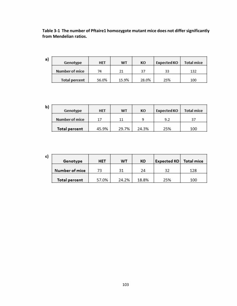
103
Table 3-1 The number of Pftaire1 homozygote mutant mice does not differ significantly from Mendelian ratios.

104
Table 3-1 summarizes the PCR results of the F2 offspring. F2 offspring from F1
intercrossed (HET x HET) mice were genotyped for Pftaire1 gene by PCR. A total of 39
breeding crosses were evaluated which were subsequently divided into 3 categories.
Percentage of knockout mice does not differ from Mendelian ratios, significantly (p >
0.05). a) Mouse embryos at E13.5-14.5, backcrossed with C57Bl6 for a minimum of 9
times, b) Mouse pups at P21, backcrossed with C57Bl6 for a minimum of 9 times, and c)
Mouse pups at P21, not fully backcrossed to C57Bl6.
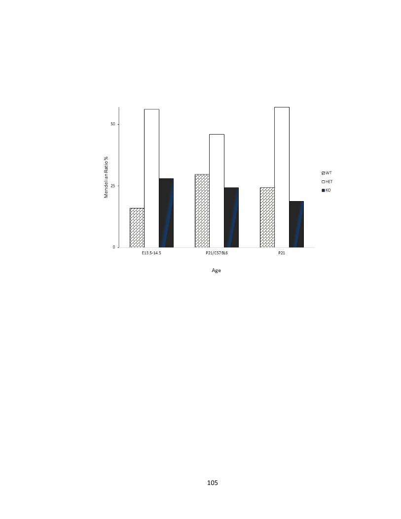
105
106
Column graph representing Mendelian ratios in F2 progeny. Effect of genotype on percent
of survival of progeny resulted from heterozygous intercrossing of Pftaire1 mice plotted
on a chart. Significance determined by χ2 test (p> 0.05).
Figure 3-5 Survival percent of Pftaire1 homozygote mutant mice does not differ
significantly from Mendelian ratios.
107
3.1.2. No abnormal phenotype detected in Pftaire1 null mice
Since PFTAIRE deficiency leads to embryonic lethality in fruit flies (Stowers, Garza
et al. 2000); we questioned whether loss of Pftaire1 could have a significant impact on
mouse development. Macroscopic and morphological examination of Pftaire1 knockout
mice, in comparison to their wild type littermates revealed no abnormal phenotype
within our observations to date: the pups are viable, age normally and do not show any
obvious morphological defects during their life span. We did not detect any obvious
alterations in brain morphology within our observations.
3.1.3. Microscopic Analysis of Brain Section
3.1.3.1. Brains of adult Knockout Pftaire1 mice did not show any gross anatomical
abnormality by cresyl violet staining
Microscopic analysis of coronal sections of the adult mice brain on a mixed C57Bl6
background revealed no major abnormalities upon cresyl violet staining. Brain sections of
two month old, adult wild-type (n=3) and Pftaire1 null mice (n=3), were collected starting
at the frontal cortex at bregma: ~ 2.710 mm up through the 4th ventricle at bregma: ~-
4.20 mm. The structure of the cortex, septostriatal region, the organization of the
hippocampus, shape of the dentate gyrus, diencephalon, mesencephalon, all follow the
normal gross anatomy of the brain. Sections representing bregma 1.98, 1.70, 0.74, 1.00,
1.64, and 2.75 mm are presented, respectively, in section “a-f” and “g-l” (Figure 3-6).
These results suggest that Pftaire1 loss, alone, in mice, might not have the same effects
observed with L63E deficiency in the fly (Rodríguez González and S. 2011). However, the
effect of Pftaire1 on development of the brains needs to be examined during younger

108
ages including the embryonic period using different methods. For instance, silver staining
could give us a better idea with regards to the structure of the brain.
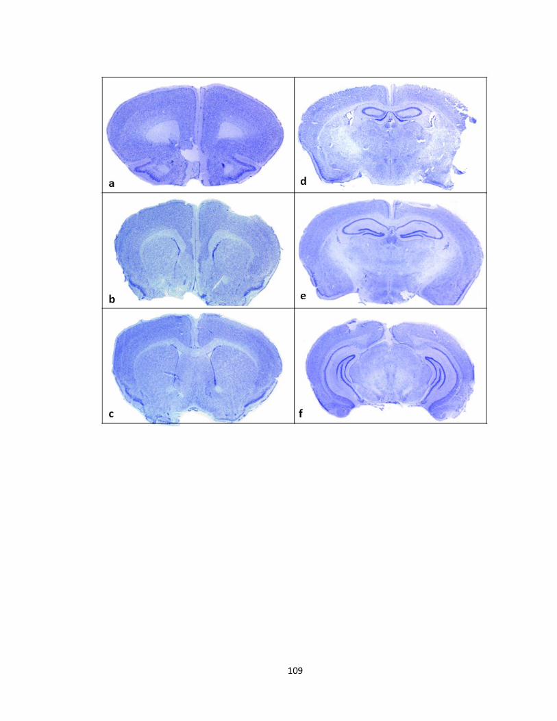
109
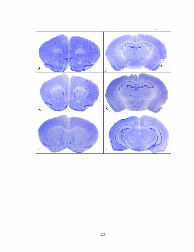
110
111
Sections representing bregmas: 1.98, 1.70, 0.74, 1.00, 1.64, and 2.75 mm are presented,
respectively, in section “a-f” for wild type (n=3) and “g-l” for knockout (n=3). Brains were
collected from three month old adult mice, perfused and fixed for sectioning. 14µm
sections were collected from cryopreserved samples, sections were collected starting
from the cortex at bregma: ~ 2.710 mm up through the 4th ventricle at bregma: ~-4.20
mm. Sections were then stained with cresyl violet to examine the gross anatomy of the
brain. Histological examination following cresyl violet staining of Pftaire1 knockout mice
brain, reveals no abnormality in the structure of the cortex, septostriatal region,
hippocampal organization, shape of the dentate gyrus, diencephalon, and
mesencephalon.
Figure 3-6 Cresyl violet stained sections from Pftaire1 knockout mice reveals no gross
abnormality in comparison to wild type mice
112
3.1.4. Disruption in Pftaire1 does not lead to any difference in basal survival in
primary cortical cultures
We investigated whether manipulating Pftaire1 activity has any effect on its basal
cell survival or it acts in a manner similar to Cdk5 (O'Hare, Kushwaha et al. 2005). To
determine cell viability upon disruption of Pftaire1 activity, we co-infected primary
cortical neurons (MOI=100) with GFP along with WT Pftaire1 or the dominant negative
form of Pftaire1, (D228N), in which the highly conserved aspartic acid residue of Pftaire1
at residue 228 was mutated to asparagine, by a point mutation (Lazzaro, Albert et al.
1997) as a kinase inactive control for transient transfections. This residue is highly
conserved among all protein kinases, and inhibits the endogenous kinase activity of cdc2
kinases in its mutated form upon ectopic expression (Taylor, Knighton et al. 1993, van den
Heuvel and Harlow 1993). Neurons were cultured for 12, 16, and 24 hours post-infection,
then fixed and stained with Hoechst for detection of morphologically intact healthy
nuclei. GFP expressing neurons with viable nuclei were counted. We did not observe a
difference in basal survival rate among neurons expressing GFP with Pftaire1 or D228N-
Pftaire1 and GFP alone (Table 3-2), (Figure 3-7). From a total of n=1602, (77.1 ± 1.8%), of
n=2293, (77.8 ± 1.0%), of n=4533, ( 92.9 ± 0.6% )of D228N expressing neurons survived at
12, 16, and 24 hours, respectively, which does not significantly differ (p>0.05 ) from GFP
control and WT (Table 3-2), (Figure 3-7). Our result, suggests that Pftaire1 and its
dominant negative form D228N do not affect cell viability under normal circumstances.
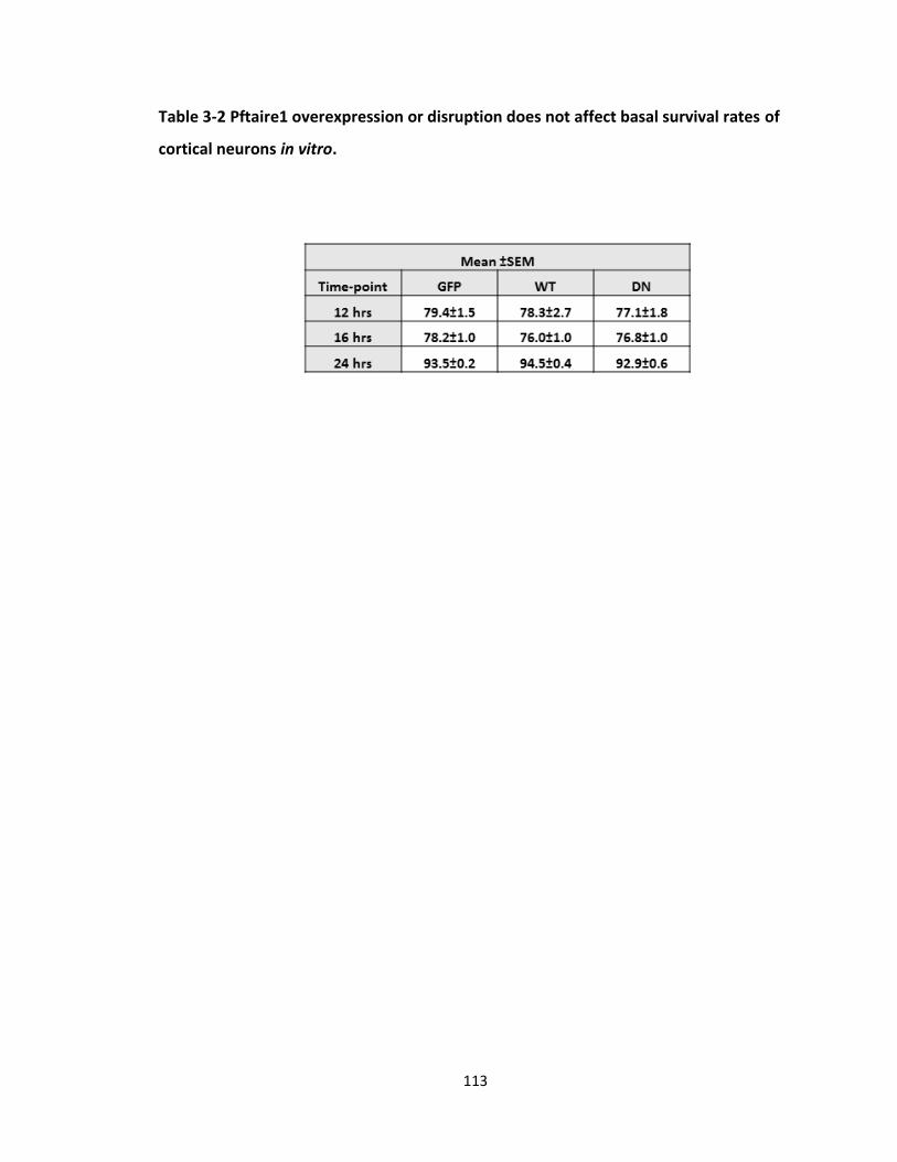
113
Table 3-2 Pftaire1 overexpression or disruption does not affect basal survival rates of
cortical neurons in vitro.

114
Table 3-2 represents survival of cortical neurons as (Mean ± SEM), n=3 and 3 replicas per
treatment. Basal survival rates of Pftaire1 wild type and D228N vs control is not significant
as per ANOVA and Newman-Keuls for means p>0.05.
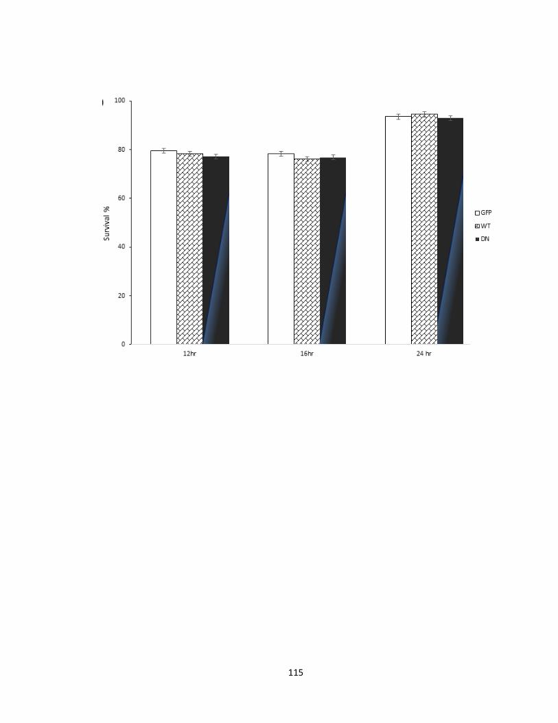
115
116
Column Graph representing percent of survival for each treatment with relation to its
incubation period. Primary cortical neurons derived from CD1 mice were infected with
GFP alone, or co-infected with GFP and WT-Pftaire1 or D228N-Pftaire1 (MOI=100),
incubated for 12, 16, and 24 hours, followed by fixation, and Hoechst 33258 staining.
Percentage of survival was calculated as percentage of morphologically intact live nuclei
over total GFP-expressing neurons. Data/columns represent the (Mean ± SEM), n=3 and
3 replicas per treatment. p>0.05 ANOVA, t-Test: paired two sample for means is N.S., no
significant difference is observed in basal survival rates of Pftaire1 wild type and D228N
vs control.
Figure 3-7 Pftaire1 overexpression or disruption does not affect basal survival rates.
117
3.2. Pftaire1 negatively regulates axon length in primary cortical cultures of mouse
embryos at E13.5-14.5
3.2.1. Dominant Negative D228N-Pftaire1 expression in vitro increases axonal
length in cortical cultures
Previous research indicates that Cdk5 inactivation leads to inhibition of axonal
growth (Nikolic, Dudek et al. 1996, Nikolic, Chou et al. 1998). In addition, Pctaire1-3
negatively regulates neurite outgrowth (Cole 2009). We speculated whether
manipulation of Pftaire1 activity would also have an impact on axon outgrowth. To test
this notion, primary cortical cultures were prepared from CD1 mouse embryos at E 13.5-
14.5 days and subjected to adenoviral infection with GFP tagged-D228N Pftaire1, GFP
tagged-wild type Pftaire1 or GFP control alone, at the time of plating. After allowing
cultured neurons to grow and differentiate, they were fixed at subsequent time points:
12, 24, and 48 hours post-infection (Figure 3-8). Neurites were then stained with GFP for
an enhanced infection signal and reliable tracing. Also, Hoechst 33258 staining was
performed to assess viability of neurons (Figure 3-8 a). Digital images were acquired with
Zeiss Axioskop 2 Mot microscope. The total length of neurites was measured on digitally
acquired images utilizing Stereo-investigator (version 6; MicroBrightField, Williston, VT)
software using the contour mapping mode and tracing the length of axons. Lengths were
determined measuring at the 40X objective magnification and diameter of field of view
was 575 µm. Measurements were commenced from the edge of the GFP expression in
the cell body to the distal terminal of the neurite using the longest neurite for
comparative purposes. Each experiment was repeated for a minimum of 3 times and each
118
treatment was replicated in triplets. The change in axon length is represented and the
significance is determined by one way-ANOVA followed by Student-Newman-Keuls post–
hoc test and Bonferroni. At 12 hours, a total of 372 neurites were measured. For D228N-
Pftaire1 infected neurons, n=124 and (Mean ± SEM) was (14.5 ± 0.6) was statistically
significant, as confirmed by one way ANOVA (p<0.001) followed by Student-Newman-
Keuls method, from GFP control (10.9 ± 0.7 µm) (p=8.2E-06) and WT-Pftaire1 (10.16 ± 0.5
µm) (p=2.9E-07) but not WT relative to GFP (p=0.3) (Figure 3-8 b). At 16 hours, a total of
1408 neurites were measured and for D228N-Pftaire1 infected neurons, n=509, (Mean ±
SEM) was (33.0 ± 0.8 µm) that was statistically significant by one way ANOVA (p<0.001)
with Student-Newman-Keuls method from GFP control (24.5±0.7 µm) (p=7E-13) and WT-
Pftaire1 (24.6±0.8 µm) (p=3E-11) but WT relative to GFP (p=0.9) (Figure 3-8 b). At 24
hours, a total of 891 neurites were measured. For D228N-Pftaire1 infected neurons,
n=241 (Mean ± SEM) was (129.6 ± 3.6 µm) that was statistically significant (p<0.001,
ANOVA) with Student-Newman-Keuls method from GFP control (97.9 ± 3.9 µm) (p=6.4E-
09) and WT-Pftaire1 (89.5 ± 2.6 µm) (p=3.4E-18) but not WT relative to GFP (p=0.07)
(Figure 3-8 b). At 48 hours, a total of 724 neurites were measured. For D228N-Pftaire1
infected neurons, n=283, (Mean ± SEM) was (253.6 ± 9.1 µm) that was not statistically
significant (p > 0.05, ANOVA) with Student-Newman-Keuls method from GFP control
(275.0 ± 10 µm) (p=0.1) and WT-Pftaire1 (241.3 ± 12.4 µm) (p=0.4); also, GFP and WT
(p=0.03) were not significant (Figure 3-8 b). According to our data the D228N-Pftaire1
expressing neurons, showed an enhanced ability to elongate when they were compared
to wild type and GFP control expressing neurons, at early time-points (12-24 hours);
119
however, later at 48 hours, they do not show any difference with GFP control or wild type
mice (Figure 3-8 b, c, and d). Also, we show that the percentage of difference to GFP
control, in mean neurite length, is higher in D228N expressing neurons. At 12, 16, and 24
hours, we observe a 32.4%, 34.7%, and 32.3% increase in mean neurite length compared
to GFP which significantly differs from GFP but not WT in comparison to GFP (Figure 3-8
c). Also, average of log10 of axon measurements of D228N expressing neurons, revealed
a linear distribution with higher measures as compared to wild type, at 12, 16, and 24
hours (Figure3-8 d). Furthermore, classifying axons into groups based on their length,
reveals a significant difference (p < 0.0001) between the GFP, wild-type and the D228N-
Pftaire1 group, as determined by chi-square test. We classified neurons into three groups
based on the length of their neurites. To classify, average and median were calculated for
each time-point. The maximum and minimum numbers related to average and median
were considered as average range for that time-point and neurites diverting were from
this range were considered as either short or long, relatively. In neurons expressing
D228N-Pftaire1, a significantly higher percent of neurons, had longer axons at early time-
points 12 (p= 7E-09), 16 (p= 1.6E-12), and 24 hours (p= 1.9E-14) but this difference
became insignificant at 48 hours (p= 0.07) (Figure 3-8 d, e). Our data suggests that there
is an increase in neurite length in vitro upon ectopic expression of D228N-Pftaire1.
Previous studies report that loss of Cdk5 results in a decrease in axon length (Nikolic,
Dudek et al. 1996). Our observations, suggest that D228N might have an effect that is
opposite to that of Cdk5 on axon growth in vitro. Thus, we postulate that Pftaire1 might
have a role opposite to Cdk5 in regulating axon growth.
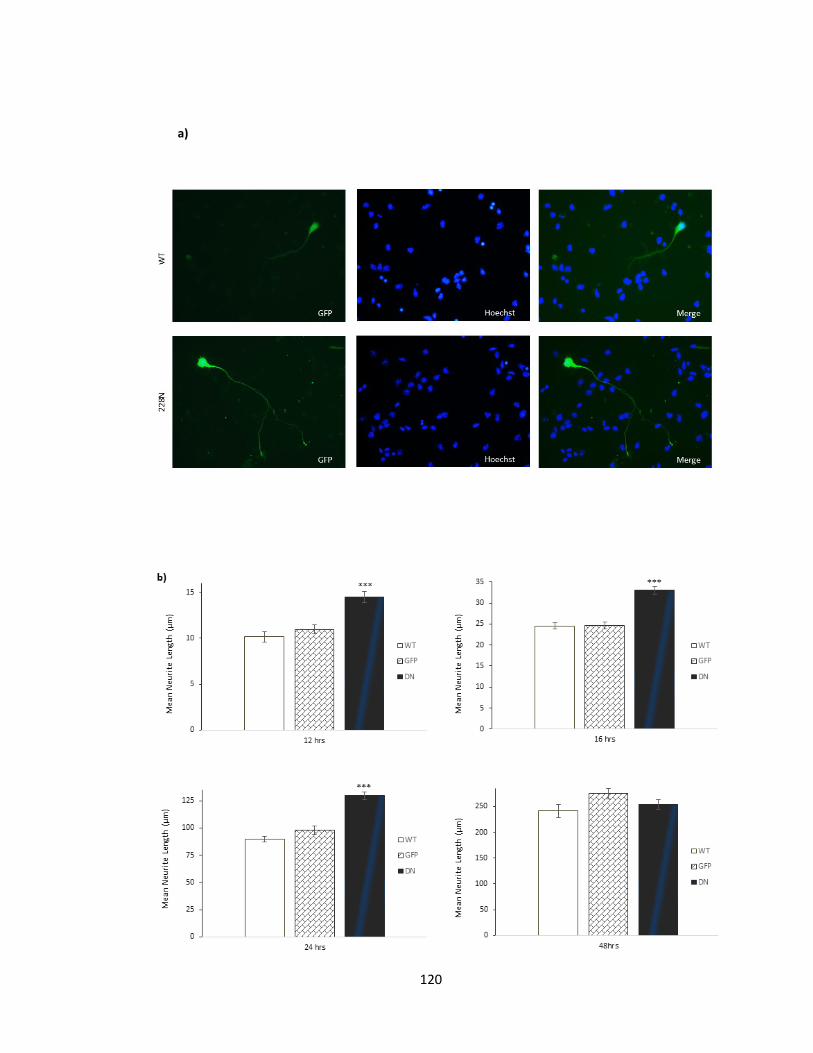
120
a)
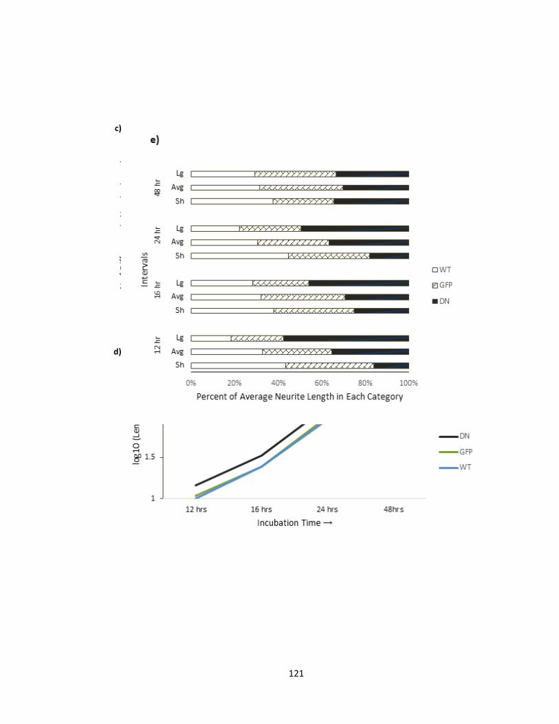
121
c)
d)
122
Figure 3-8 Overexpression of dominant negative Pftaire1, enhances axon outgrowth in
primary cortical cultures
Image, representing immunostaining of infected neurons with GFP, Hoechst, and merged.
Staining was performed to enhance GFP signal for reliable neurite tracing and to
determine the viability of neurons assessed. b) Column graphs, representing (Mean ±
SEM) at different time points (12, 16, 24, and 48 hours post infection. c) Column graph,
representing percent of difference in mean neurite length of WT and D228N neurites in
relation with GFP control. d) Same data, represented as log10 of neurites at different time
points to homogenize the different distribution pattern over time for easier comparison.
e) Also, classification of axons into different group based on their lengths (Sh: short, Avg:
average, and L: long. Cut off values for each category are 9-14.4; 23.3-33; 85.4-129.5; and
201-275 µm at 12, 16, 24, 48 hrs respectively) reveals more neurons with longer axons
especially at earlier time points in the D228N-Pftaire1 cultures, as opposed to, GFP control
and WT. Primary cortical cultures were prepared from mouse embryos at E13.5-14.5,
infected with AV-virus expressing Pftaire1, D228N-Pftaire1 and GFP (MOI=100), and fixed
123
at different time points post infection, stained with GFP and Hoechst to enhance signal
strength to determine cell viability, respectively. Each experiment was performed for a
minimum of three times (n=3) and each treatment was replicated three times.
Fluorescent pictures were taken and measurements were done using stereoinvestigator
software, as described earlier. The measurements were statistically tested with one way
ANOVA (p < 0.05) followed by student Newman-Keuls test to determine significance.
3.2.2. Primary neuronal cultures of Pftaire1 null mice produce longer axons
Ectopic expression of dominant negative Pftaire1 in vitro, resulted in enhanced
axon length in cortical cultures (Figure 3-9). To investigate, whether the effects of D228N
overexpression on axon outgrowth could be replicated by Pftaire1 deficiency, we studied
populations of axons in primary cortical cultures from Pftaire1 deficient mice. We crossed
heterozygote Pftaire1 mice to obtain littermates that were either Pftaire1 wild types or
homozygote mutants. Cultures were prepared from mouse embryos at gestation days
13.5-14.5 from generation 6 backcrossed animals. To detect the genotype of the
littermates, samples were taken from each embryo at the time of dissection but all further
processing of axon assessment was done in a blind manner. The cells were plated and
fixed and stained at 16 and 24 hours after seeding (Figure 3-9 a). At both time points, the
axon length of Pftaire1-knockout neurons was found to be significantly longer than WT
cultures (p < 0.0001, upon student’s paired two tailed t-test) (Figure 3-9). At 16 hours,
axons from primary cortical cultures prepared from 3 different Het x Het crosses (litters)
were compared, in 3 replicates, and a total n=15 (wild type n=6, knockout n=9) mice were
124
examined. Axons were detected using Tau-1 microtubule marker and viability of neurons
was assessed using Hoechst 33258 (Figure 3-9 a). Length of axons was measured, on
digitally acquired fluorescent images at the 40X objective magnification with a diameter
of field of view of 575 µm using Northern Eclipse Zeiss on Zeiss Axioskop 2 Mot fluorescent
microscope, utilizing Stereo-investigator (version 6; MicroBrightField, Williston, VT)
software using the contour mapping mode. Measurements were commenced from the
edge of the Tau-1 expression in the cell body to the distal terminal of the axon. We
observed a significant increase in the length of axons of KO-Pftaire1 neurons when
compared to WT-Pftaire1. (Mean ± SEM) was (131.2 ± 1.9) µm (n= 2510) for KO axons and
(90.0 ± 1.6) µm (n=1738) for WT axons (p=2E-37) (Figure 3-9 b). The same trend was
observed at 24 hours while fluorescent Digital images were acquired with Zeiss Axioskop
2 Mot fluorescent microscope at objective 20 x magnification with a field of view diameter
of 1150 µm. For measuring, axons were traced on digitally acquired images using Stereo-
investigator (version 6; MicroBrightField, Williston, VT) software utilizing the contour
mapping mode. At 24 hours, among two different litters n=6 (wild type n=3, knockout
n=3), (Mean ± SEM) was (204.0 ± 3.3) µm (n= 1499) for KO axons and (158.68 ± 3.0) µm
(n=1272) for WT axons (p = 2E-37) (Figure 3-9 b). In other words, KO axons revealed a
45.6% and 28.5% increase in relative axonal length in comparison to WT axons, prepared
from the progeny of Het x Het crosses primary cortical neurons, at 16 and 24 hours,
respectively (Figure 3-9 c). Also, average of log10 of axon measurements of knockout
Pftaire1 axon lengths revealed a linear distribution with higher measures, as compared to
wild type, both at 16 and 24 hours (Figure 3-9 d). Next, axons were binned according to
125
their lengths into short, average, and long ranges, as explained previously. The maximum
and minimum numbers related to average and median were considered as average range
for that time-point and neurites diverting were from this range were considered as either
short or long, relatively and statistical significance was determined by chi-square (p <
0.001). At 16 hours, KO and WT neurons consisted of 40.8% and 25% long axons (p =
2.12E-64), respectively. At 24 hours, the KO and WT neurons consisted of 41.8 % and 28.3
% longer axons (p= 9.58E-15), respectively (Figure 3-9 e).
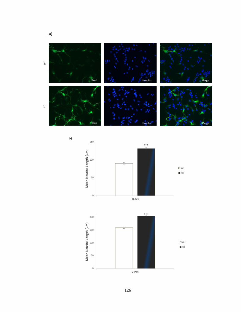
126
a)
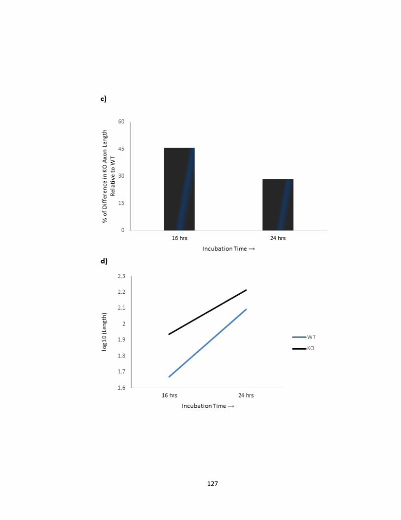
127
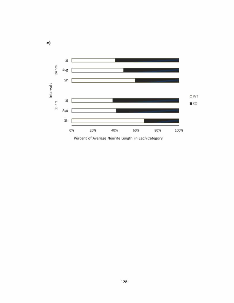
128
129
a) Image, representing immunostaining of neurons with Tau-1, Hoechst, and merged.
Staining was performed to immunolabel axon for tracing and to determine viability of
neurons measured. b) Column graphs, representing (Mean ± SEM) for different time
points. At 16 and 24 hours, the knockout neurons have significantly longer axons in
comparison to the WT (p<0.001) groups. c) Column graphs representing percent of
difference in axonal length in knockout relative to wild type d) Linear graph, represents
log10 of axons to simplify comparison at different time points due to changes in length.
e) Also, classification of axons into different groups based on their lengths (Sh: short, Avg:
average, and L: long) reveals more neurons with longer axons KO-Pftaire1 cultures as
opposed to WT as determined by chi square (p < 0.001). Primary cortical cultures were
prepared from mouse embryos at E13.5-14.5 from Pftaire1 Het x Het crosses and fixed at
different time points. Fluorescent pictures were taken and measurements were done
using stereo-investigator software as described earlier. Statistical significance of the
measurements were determined with student’s paired two tail t-test (p < 0.001).
Figure 3-9 Knockout of Pftaire1 results in enhanced axonal growth, at 16 and 24 hours
in vitro
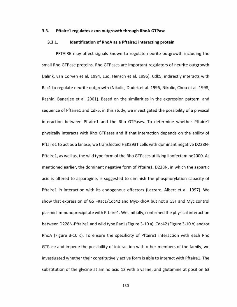
130
3.3. Pftaire1 regulates axon outgrowth through RhoA GTPase
3.3.1. Identification of RhoA as a Pftaire1 interacting protein
PFTAIRE may affect signals known to regulate neurite outgrowth including the
small Rho GTPase proteins. Rho GTPases are important regulators of neurite outgrowth
(Jalink, van Corven et al. 1994, Luo, Hensch et al. 1996). Cdk5, indirectly interacts with
Rac1 to regulate neurite outgrowth (Nikolic, Dudek et al. 1996, Nikolic, Chou et al. 1998,
Rashid, Banerjee et al. 2001). Based on the similarities in the expression pattern, and
sequence of Pftaire1 and Cdk5, in this study, we investigated the possibility of a physical
interaction between Pftaire1 and the Rho GTPases. To determine whether Pftaire1
physically interacts with Rho GTPases and if that interaction depends on the ability of
Pftaire1 to act as a kinase; we transfected HEK293T cells with dominant negative D228N-
Pftaire1, as well as, the wild type form of the Rho GTPases utilizing lipofectamine2000. As
mentioned earlier, the dominant negative form of Pftaire1, D228N, in which the aspartic
acid is altered to asparagine, is suggested to diminish the phosphorylation capacity of
Pftaire1 in interaction with its endogenous effectors (Lazzaro, Albert et al. 1997). We
show that expression of GST-Rac1/Cdc42 and Myc-RhoA but not a GST and Myc control
plasmid immunoprecipitate with Pftaire1. We, initially, confirmed the physical interaction
between D228N-Pftaire1 and wild type Rac1 (Figure 3-10 a), Cdc42 (Figure 3-10 b) and/or
RhoA (Figure 3-10 c). To ensure the specificity of Pftaire1 interaction with each Rho
GTPase and impede the possibility of interaction with other members of the family, we
investigated whether their constitutively active form is able to interact with Pftaire1. The
substitution of the glycine at amino acid 12 with a valine, and glutamine at position 63
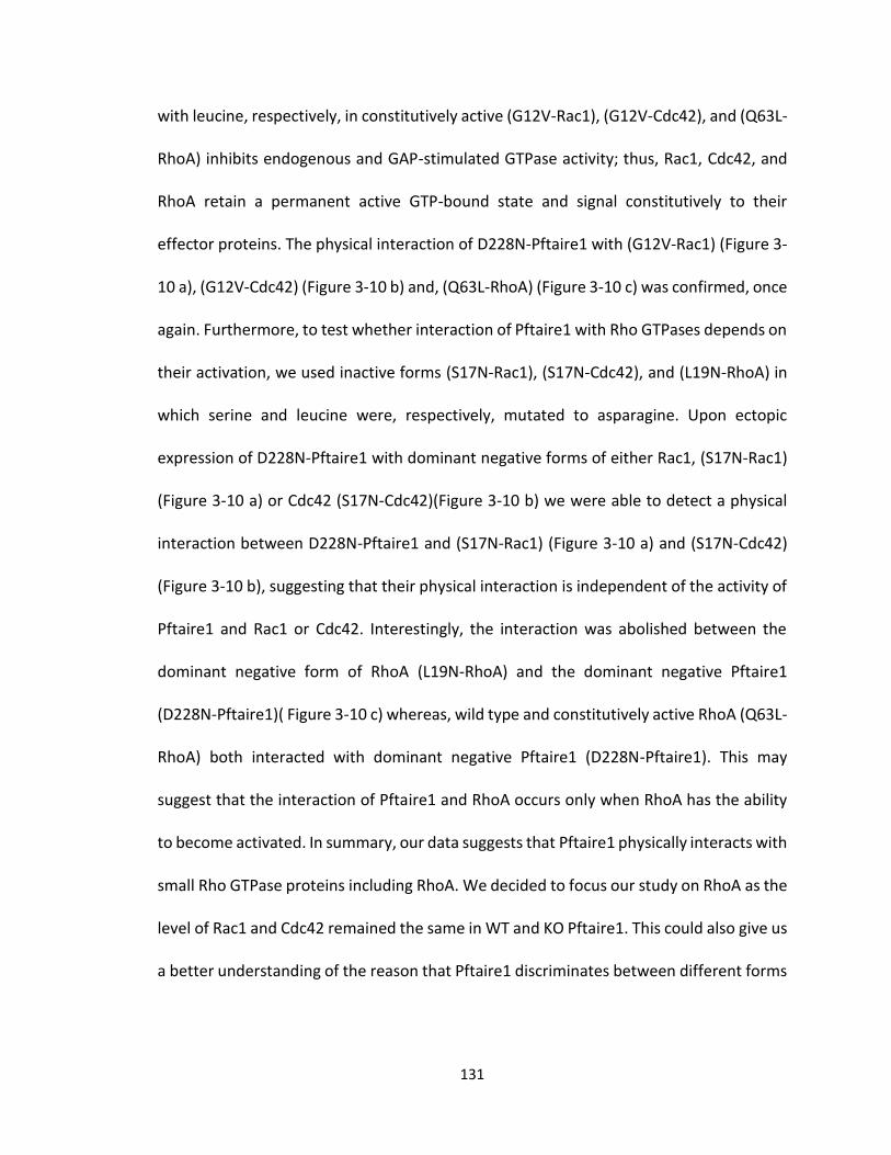
131
with leucine, respectively, in constitutively active (G12V-Rac1), (G12V-Cdc42), and (Q63L-
RhoA) inhibits endogenous and GAP-stimulated GTPase activity; thus, Rac1, Cdc42, and
RhoA retain a permanent active GTP-bound state and signal constitutively to their
effector proteins. The physical interaction of D228N-Pftaire1 with (G12V-Rac1) (Figure 3-
10 a), (G12V-Cdc42) (Figure 3-10 b) and, (Q63L-RhoA) (Figure 3-10 c) was confirmed, once
again. Furthermore, to test whether interaction of Pftaire1 with Rho GTPases depends on
their activation, we used inactive forms (S17N-Rac1), (S17N-Cdc42), and (L19N-RhoA) in
which serine and leucine were, respectively, mutated to asparagine. Upon ectopic
expression of D228N-Pftaire1 with dominant negative forms of either Rac1, (S17N-Rac1)
(Figure 3-10 a) or Cdc42 (S17N-Cdc42)(Figure 3-10 b) we were able to detect a physical
interaction between D228N-Pftaire1 and (S17N-Rac1) (Figure 3-10 a) and (S17N-Cdc42)
(Figure 3-10 b), suggesting that their physical interaction is independent of the activity of
Pftaire1 and Rac1 or Cdc42. Interestingly, the interaction was abolished between the
dominant negative form of RhoA (L19N-RhoA) and the dominant negative Pftaire1
(D228N-Pftaire1)( Figure 3-10 c) whereas, wild type and constitutively active RhoA (Q63L-
RhoA) both interacted with dominant negative Pftaire1 (D228N-Pftaire1). This may
suggest that the interaction of Pftaire1 and RhoA occurs only when RhoA has the ability
to become activated. In summary, our data suggests that Pftaire1 physically interacts with
small Rho GTPase proteins including RhoA. We decided to focus our study on RhoA as the
level of Rac1 and Cdc42 remained the same in WT and KO Pftaire1. This could also give us
a better understanding of the reason that Pftaire1 discriminates between different forms

132
of RhoA. We next decided to examine the level of RhoA in Pftaire1 deficient mice and
investigate the possibility of an endogenous interaction between Pftaire1 and RhoA.
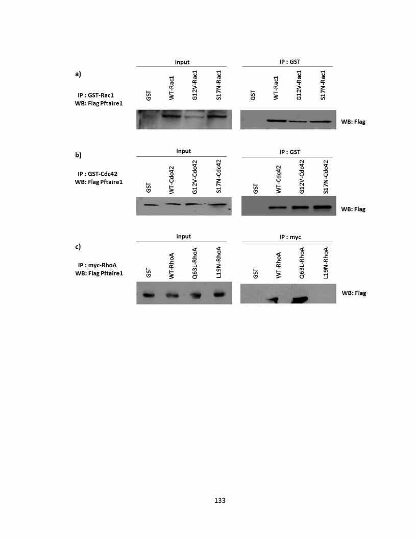
133
134
Western blots representing a) physical interaction of D228N-Pftaire1 with Rac1, wild type
(WT-Rac1), constitutively active (G12V-Rac1) and dominant negative (S17N-Rac1). b)
D228N-Pftaire1 also interacts with wild type (WT-Cdc42), constitutively active (G12V-
Cdc42) and dominant negative (S17N-Cdc42), c) D228N-Pftaire1 interacts with both wild
type RhoA and constitutively active (Q63L-RhoA) but not with dominant negative (L19N-
RhoA). Ectopic expression of Flag-D228N-Pftaire and GST-Rac1, GST-Cdc42 or Myc-RhoA
in HEK293T cell line via lipofectamine transfection. 24 hours post transfection cells were
collected and lysed for pull down assay. Pftaire1, input samples were collected and GST
tagged Rac1 and Cdc42 were precipitated using GST glutathione sepharose beads. In case
of RhoA samples were immunoprecipitated using myc antibody and mouse –IgG True blot
beads. Protein complexes were then resolved on a 12% SDS-gel followed by western
blotting. Membranes were probed for Flag protein.
Figure 3-10 Pftaire1 physically interacts with Rho GTPase proteins in vitro
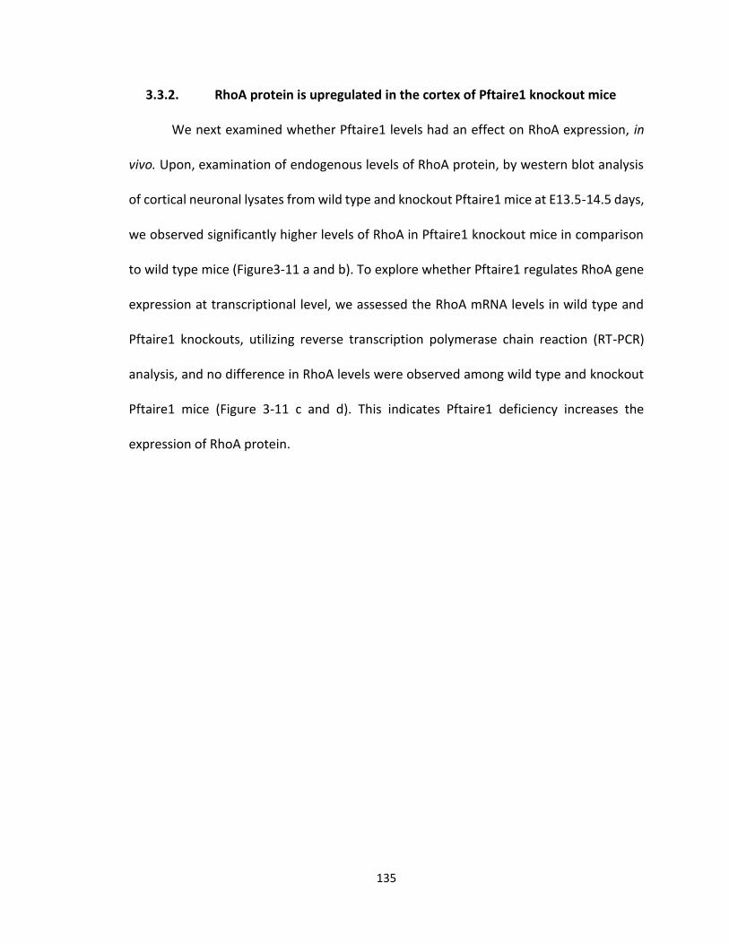
135
3.3.2. RhoA protein is upregulated in the cortex of Pftaire1 knockout mice
We next examined whether Pftaire1 levels had an effect on RhoA expression, in
vivo. Upon, examination of endogenous levels of RhoA protein, by western blot analysis
of cortical neuronal lysates from wild type and knockout Pftaire1 mice at E13.5-14.5 days,
we observed significantly higher levels of RhoA in Pftaire1 knockout mice in comparison
to wild type mice (Figure3-11 a and b). To explore whether Pftaire1 regulates RhoA gene
expression at transcriptional level, we assessed the RhoA mRNA levels in wild type and
Pftaire1 knockouts, utilizing reverse transcription polymerase chain reaction (RT-PCR)
analysis, and no difference in RhoA levels were observed among wild type and knockout
Pftaire1 mice (Figure 3-11 c and d). This indicates Pftaire1 deficiency increases the
expression of RhoA protein.
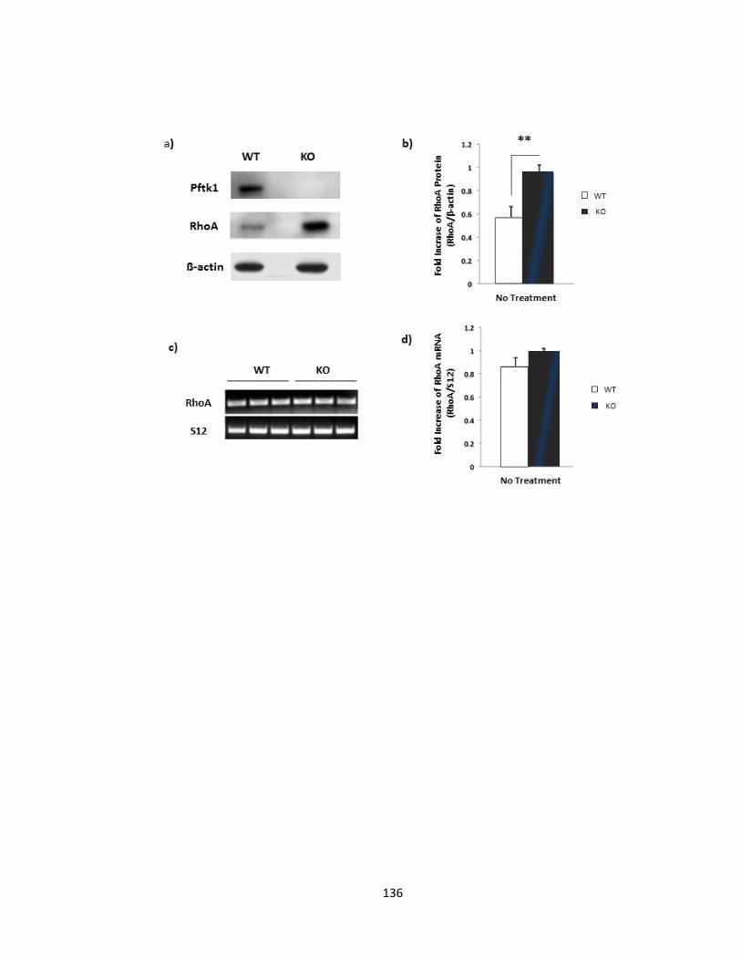
136
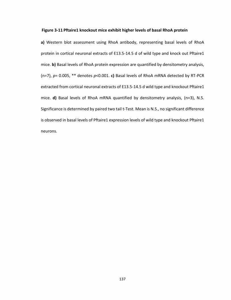
137
a) Western blot assessment using RhoA antibody, representing basal levels of RhoA
protein in cortical neuronal extracts of E13.5-14.5 d of wild type and knock out Pftaire1
mice. b) Basal levels of RhoA protein expression are quantified by densitometry analysis,
(n=7), p= 0.005, ** denotes p<0.001. c) Basal levels of RhoA mRNA detected by RT-PCR
extracted from cortical neuronal extracts of E13.5-14.5 d wild type and knockout Pftaire1
mice. d) Basal levels of RhoA mRNA quantified by densitometry analysis, (n=3), N.S.
Significance is determined by paired two tail t-Test. Mean is N.S., no significant difference
is observed in basal levels of Pftaire1 expression levels of wild type and knockout Pftaire1
neurons.
Figure 3-11 Pftaire1 knockout mice exhibit higher levels of basal RhoA protein
138
3.3.3. RhoA interacts with Pftaire1 in mice brain
Next, we examined the possibility of an endogenous interaction between Pftaire1
and Rho GTPase. The endogenous expression level of RhoA GTPase is low and it is rapidly
subject to hydrolysis. Thus, for immunoprecipitation purposes high amounts of protein
(approximately 2000 µg of total protein) was required per reaction. Moreover, due to
high sensitivity of Rho GTPases to temperature, all steps were performed at 4˚C using ice
cold solutions to ensure the stability of reactions. Protein lysates were extracted from
embryonic mouse brains at E13.5-14.5. We immunoprecipitated endogenous Pftaire1
utilizing rabbit polyclonal Pftaire1 (H-140) antibody from SantaCruz and used rabbit
polyclonal IgG as control antibody. Our result indicates physical interaction of Pftaire1
and RhoA at an endogenous level (Figure 3-12).
To determine whether the activity of RhoA has any effect on this interaction we
added GTPγS to stabilize the active form. Both the endogenous form of RhoA and GTP
bound RhoA interacted with Pftaire1 (Figure 3-12).
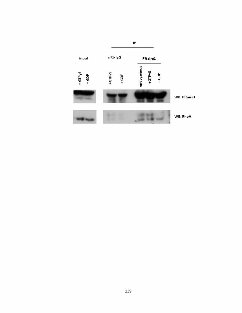
139
140
Western blot representing physical interaction of Pftaire1 and RhoA in vivo, on whole
brain extracts from wild type CD1 mouse embryo at E13.5-14.5 d. Inputs and
immunoprecipitated proteins were separated on 12% SDS gel, followed by western blot.
Immune complexes were detected using RhoA antibody. Pftaire1 antibody and RhoA
antibody were used for western blotting. To immunoprecipitate, lysates were collected
and incubated with Pftaire1 antibody and true blot rabbit IgG beads. GTPγS was loaded
with the interactions to determine the effect of GTP bound RhoA on interaction with
Pftaire1.
Figure 3-12 Pftaire1 and GTPase protein Rho A interact at an endogenous level
141
3.3.4. RhoA activity could not be detected in Pftaire1 deficient neurons by
GTPase assay in vitro
Our data revealed an increase in levels of RhoA protein in primary cortical cultures
of Pftaire1 knockout mice, p=0.005 (Figure 3-11 a). To further investigate the relationship
between the activity of RhoA and Pftaire1; the GTPase activity of RhoA was examined
under conditions where Pftaire1 was absent. The activation level of RhoA was determined
by Rho GTPase activity assay utilizing GTPase activation kit from Cell Biolabs, on cell
lysates as described earlier in methodology. Briefly, cell extracts were prepared from
cortical cultures of Pftaire1 wild type and knockout littermates at E13.5-14.5 and then
incubated with Rho-binding domain (RBD) of Rhotekin PBD beads provided with the kit
which exclusively pull down the active form of RhoA from the endogenous cell lysate. We
immunoprecipitated rhotekin /active RhoA complex with the provided PBD beads. Active
RhoA levels were revealed by western blotting probing with RhoA specific antibody. The
detected signal would correlate with the activity level of RhoA. Interestingly, in spite of
RhoA protein upregulation in Pftaire1 knockout mice, we did not detect RhoA activity in
the KO extracts (Figure 3-13).
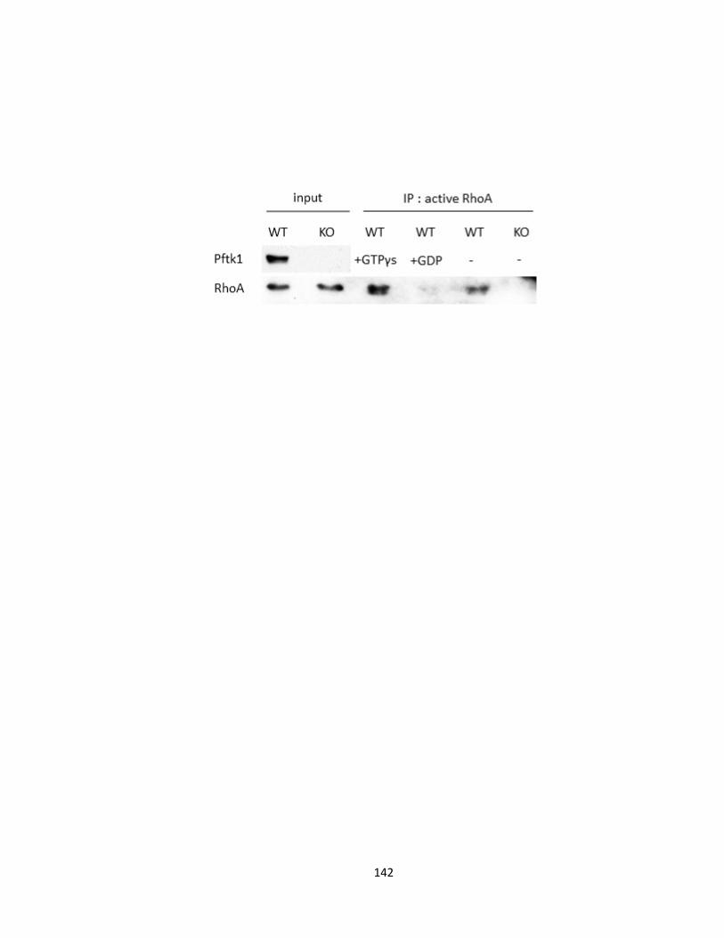
142
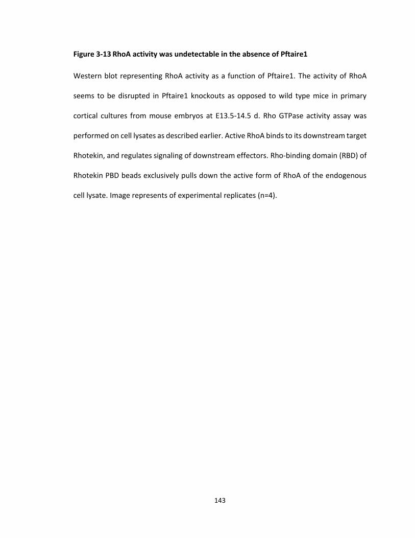
143
Western blot representing RhoA activity as a function of Pftaire1. The activity of RhoA
seems to be disrupted in Pftaire1 knockouts as opposed to wild type mice in primary
cortical cultures from mouse embryos at E13.5-14.5 d. Rho GTPase activity assay was
performed on cell lysates as described earlier. Active RhoA binds to its downstream target
Rhotekin, and regulates signaling of downstream effectors. Rho-binding domain (RBD) of
Rhotekin PBD beads exclusively pulls down the active form of RhoA of the endogenous
cell lysate. Image represents of experimental replicates (n=4).
Figure 3-13 RhoA activity was undetectable in the absence of Pftaire1
144
3.3.5. Pftaire1 phosphorylates RhoA on a Serine residue in vitro
Since according to our results, RhoA activity was undetectable in Pftaire1
knockout cortical neurons in spite of its upregulation (Figure 3-11 a, 3-13), we decided to
investigate whether Pftaire1 has direct kinase activity over RhoA in vitro. We performed
an in vitro kinase assay using immunoprecipitated Pftaire1. Briefly, we
immunoprecipitated Pftaire1 from mouse embryonic cortical neurons at E13.5-14.5, as
described in methods section. Immunoprecipitated Pftaire1 was incubated overnight with
human recombinant RhoA substrate for kinase assay. The reaction was then resolved on
a 12% SDS gel and subjected to western blotting. Phosphorylated RhoA was detected
using phosphoserine antibody. Our results revealed Pftaire1 mediated the
phosphorylation of RhoA but IgG control had no effect (Figure 3-13a). We next examined
the ability of Pftaire1 to phosphorylate the GDP bound form of RhoA. Upon adding GDP
to our reactions we observed increased phosphorylation of RhoA-GDP as opposed to
RhoA alone. Our results suggest RhoA as a direct in vitro substrate of Pftaire1 (Figure 3-
14 a).
3.3.6. Pftaire1 mediated RhoA phosphorylation leads to RhoA activation
To investigate the effects of pftaire1 mediated phosphorylation over RhoA activity
more accurately, we analyzed RhoA activity following kinase activity utilizing an
absorbance based G-LISA assay biochem kit from Cytoskeleton as described by the
manufacturer. We performed the in vitro Kinase Assay as described earlier, with or
without GDP in the reaction mix followed by the G-LISA reaction. A significant increase in
RhoA activity was detected as a function of Pftaire1 versus IgG control, as determined by
145
two-way ANOVA (p= 0.046). Post-hoc Bonfferoni’s test, revealed that the highest
difference was associated with the presence of GDP, suggesting that RhoA-GDP is the
most efficient target for Pftaire1 phosphorylation (p= 0.033). In summary, in the presence
of GDP, Pftaire1-RhoA reveals significantly higher activity when compared to IgG-RhoA
(Figure 3-14 b).
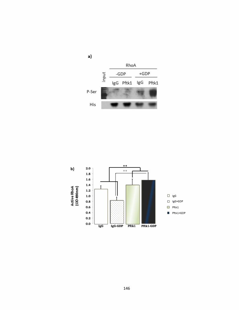
146
147
a) Western blot analysis, representing in vitro kinase assay using IP-Pftaire1 or IP-IgG from
wild type mouse embryonic cortical neurons at E13.5-14.5. Human recombinant RhoA
was used as substrate. Beads were incubated with RhoA O/N at 4 ˚C and then reactions
were separated on a 12% SDS-PAGE gel followed by western blotting. Blots were probed
with polyclonal rabbit phosphoserine antibody to detect RhoA phosphorylation. Pftaire1
but not control IgG phosphorylates RhoA. GDP bound RhoA reveals a significantly stronger
phosphorylation pattern as compared to RhoA alone as detected by phosphoserine
antibody. b) RhoA activity assay following in vitro kinase assay. Reactions were prepared
in the presence and absence of GDP and analyzed by G-LISA as described earlier. A
significant difference was observed following GDP treatment of the Pftaire1 reaction with
IgG control p= 0.046 between Pftaire1 and control groups, as determined by two-way
ANOVA. Post-hoc Bonfferoni’s test suggests significant difference between GDP-RhoA is
the most significant target for Pftaire1 (p=0.033).
Figure 3-14 Pftaire1 phosphorylates RhoA and activates RhoA in vitro

148
CHAPTER 4
DISCUSSION
149
4. DISCUSSION
4.1. Summary
Neurodegenerative diseases result from disturbed function and structure of
neurons that lead to progressive damage in the CNS and PNS. Presently, much focus has
been directed on developing therapies and strategies to prevent or reverse these
conditions. Success in developing therapeutic protocols, depends on a profound
understanding of the function of the nervous system. Cdks have a crucial role in cell cycle
regulation and neurogenesis. Numerous studies, including work done in our laboratory,
support the indispensable role of Cdks especially postmitotic Cdks, including Cdk5 in the
regulation of the nervous system. In the work presented here, we focus on a novel
postmitotic Cdk family member, Pftaire1 (Cdk14), and we explore its effect on axon
outgrowth during the development of CNS.
Pftaire1, is a novel serine/threonine protein kinase that was identified when
cloning for cdc2 related kinases (Lazzaro, Albert et al. 1997). Our lab, described the effect
of Pftaire (Eip63E) deficiency on CNS development in Drosophila, for the first time
(Rodríguez González and S. 2011). Eip63E deficiency results in concerted defects in axons
and neurons of the Drosophila ventral nerve cord (VNC). Premature axonal outgrowth, as
well as, defasciculation and misguidance of the commissural and longitudinal axon tracks
at the ventral nerve cord (VNC) were among the phenotypic features detected in Eip63
deficient embryos. This suggested a role for Eip63E in the regulation of axon development
in Drosophila (Rodríguez González and S. 2011). Furthermore, Stowers et al. had
previously shown that Eip63E deficient flies die mainly during larval development and
150
only a few that survive to pupae are smaller in size (Stowers, Garza et al. 2000). We
speculate that the smaller size of Eip63E mutants could be due to loss of functionality in
the nervous system. Considering the severe effects of Eip63E deficiency in Drosophila,
and the high degree of similarity (about 75 %) with the mammalian homologue, one could
infer that Pftaire1 has an important role in the development of axons in the mammalian
system, as well. Taking this into consideration, in addition to the high expression of
Pftaire1 in the central nervous system (Lazzaro, Albert et al. 1997, Besset, Rhee et al.
1998, Yang and Chen 2001), we anticipated defects in the mouse system with Pftaire1
deficiency. While this is complicated by the presence of two Pftaire genes (Pftaire 1 and
2) in the mammalian system; we nonetheless, focused on evaluating the effects of
Pftaire1, in of Pftaire1 deficient mice. In our study, presented in “Chapter Three” of this
thesis, we developed3 Pftaire1 homozygous deficient mice, for the first time, to explore
the role and function of Pftaire1 in the CNS. We initially focused on obtaining and
characterizing Pftaire1 homozygous mice. Then, we examined the effects of Pftaire1
deficiency on axon length, in vitro; and finally we investigated the possible mechanism of
action of Pftaire1. Our study, provides evidence that Pftaire1 deficiency results in
premature axon outgrowth, in vitro; and interacts physically with RhoA by
phosphorylating it, in vitro.
4.2. Overview
4.2.1. Pftaire1: A Novel Protein and newly generated knockout mice
3 Pftaire1 was commercially obtained from TIGM (Texas A&M Institute for Genomic Medicine)
151
In the first part of this thesis, we report, for the first time, the design of a standard
germline Pftaire1 knockout mouse model, specifically for our study. We hypothesized that
Pftaire1 is critical for neuronal development. In this knockout line, the 6th exon (out of a
total 15) was replaced by the IRES / bGeo / PolyA cassette via homologous recombination
and successful replacement was confirmed by PCR genotyping of the DNA samples (Figure
3-1, and 3-2). This replacement interferes with the coding sequence and results in a
frameshift that translates into a truncated protein. Loss of full length Pftaire1 protein in
the 48 kDa was confirmed by western blot (Figure 3-3). It is unlikely that the produced
truncated protein (about 18 kDa) could have any kinase activity. In their tertiary structure,
Cdks form a bilobal fold which includes a large carboxy-terminal lobe, a small amino-
terminal lobe and a flexible hinge that connects the two lobes to each other and acts as
the ATP binding domain or the catalytic cleft. This mutated protein lacks the carboxy
terminal and the majority of the subkinase domains except subdomains I-III. It also does
not have the flexible threonine-loop (T-loop) that binds to the substrate. Taking into
consideration, the structural and functional characteristics of Cdk family members; we
speculate that the truncated protein lacks the L12 helix, which contributes to the
structural modification and reorientation of amino acids site in the ATP binding site
though it likely contains the conserved glycine-rich loop (G-loop), which facilitates the
binding and alignment of γ-phosphate of ATP for transfer(Dhavan and Tsai 2001, Bartova,
Otyepka et al. 2005, Morgan 2007).
The Pftaire1 deficient mice that were designed for the purpose of this study,
followed normal Mendelian ratios (Table 3-1, Figure 3-5) and no gross abnormalities were
152
detected in their morphology, fertility, or life span at early mix background generations.
Also, upon cresyl violet staining of brain sections collected from Bregma: ~ 2.710 to 4.20
no gross abnormality was detected in the anatomical structure of the brain within our
observations (Figure 3-6). However, the possibility of abnormal structure cannot be ruled
out until more specific methods, for instance Golgi staining, are utilized to examine the
structure of the brain further. Additionally, axonal guidance during development of the
nervous system is an important feature to further study considering that Pftaire (Eip63E)
deficiency results in concerted defects in axons and neurons of the Drosophila VNC
(Rodríguez González and S. 2011). Although, the lack of gross developmental defects is
not inconsistent with effects on axon length since the latter might not be readily
observable by gross analyses due to the presence of other post-mitotic Cdk genes. For
instance, Pftaire2, Pctaire1-3, and Cdk5 thus more careful observation of Pftaire1 and
detection of the kinase activity of other genes is required.
The question that arises is why does there appear to be a lack of apparent gross
CNS phenotype with Pftaire1 loss in comparison to the fly? One possible explanation
could be the methods and the age of the mice in our study. In this study, we examined
the structure of the brain in 2 month old adult mice and E17.5-18.5 embryos using cresyl
violet (Figure 3-6). The defects in axons of Eip63E flies appeared as early as stage 11 of
embryonic development (Rodríguez González and S. 2011). Although, this might not be a
major problem due to survival of the mouse embryos to adulthood. It would be
interesting to have a closer investigation. Furthermore, the adult Pftaire1 null mice were
still on a mixed background as opposed to a pure C57BL6 background. To rule out any
153
possible differences, the gross structure of the brains need to be examined at younger
ages, during embryonic period, on fully backcrossed mice. Another, obvious explanation
is that Drosophila merely possess the Pftaire1 homologue Eip63L but not Pftaire2 and
PCTAIRE kinases. In comparison, mammals contain two highly similar Pftaire kinase
proteins, Pftaire1 and 2 that are about 68 % similar, as well as, three Pctaire kinases,
Pctaire 1-3, which are about 61 % similar to Pftaire1 and are all highly expressed in the
mouse brain (Okuda, Cleveland et al. 1992, Hirose, Kawabuchi et al. 2000, Cole 2009,
Mikolcevic, Sigl et al. 2012). The presence of these highly similar genes in the mammalian
system could compensate for the lack of Pftaire1 and result in a lower level of
abnormalities in the knockout mice. This concern was taken into consideration by
examining levels of Pftaire2, Pctaire1 and Cdk5 proteins in Pftaire1 knockouts. No
increase was observed in the level of these proteins in wild type mice versus Pftaire1
knockouts (Figure 3-4 a, b), yet, this does not rule out the possibility of functional
redundancy between any of them and pftaire1. Nevertheless, we have already generated
a Pftarie2 deficient mouse line and it would be critical to determine the phenotype of this
mouse, as well as, a double Pftaire1/2 knock out mouse. Finally, although Pftaire1
deficiency did not affect basal cell survival levels of cortical cell cultures at E13.5-14.5
(Figure 3-7), there is the possibility that loss of Pftaire1 may result in abnormal features
under stress. There is precedence for this. Cdk5, another neuronal Cdk member has been
shown to be associated with the degenerative process in animal models of CNS disease
(Patrick, Zhou et al. 1998, Dhavan and Tsai 2001, Cruz and Tsai 2004, Rashidian, Iyirhiaro
154
et al. 2005, Smith, Mount et al. 2006). It would be interesting to determine whether this
is true for Pftaire1.
4.2.2. Axon outgrowth in vitro and Biological Relevance of Pftaire1
Our studies indicate that Pftaire-1 plays a role in regulating axon length. Cdk5 and
Pctaire, two close family members of Pftaire1 have been implicated in axon outgrowth.
For instance, Cdk5 inactivation leads to inhibition of axonal growth (Nikolic, Dudek et al.
1996, Nikolic, Chou et al. 1998), while Pctaire1 negatively regulates neurite outgrowth
(Graeser, Gannon et al. 2002, Cole 2009). Ectopic expression of dominant negative
Pctaire1 (K194R) in Neuro-2A neuroblastoma cells resulted in a 5 % increase in neurite
outgrowth of Pctaire1 mutants, as opposed to their wild type counterparts (Graeser,
Gannon et al. 2002). Likewise, our data suggests a role for Pftaire1 in regulation of axonal
length. A significant increase, about 32.4%, 34.7%, and 32.3%, change in axonal length
relative to GFP control was observed upon ectopic expression of dominant negative
D228N-Pftaire1 in cortical neurons at 12, 16, and 24 hours, respectively (Figure 3-8 c).
Also, a 45.6% and 28.5% increase in relative axonal length is observed in cultures prepared
from primary cortical neurons of Pftaire1 knockout mice, at 16 and 24 hours, respectively
(Figure 3-9 c). The higher percent of change at earlier time-points might indicate that
Pftaire1 is more important in regulating the initiation of axon outgrowth rather than being
involved in the growth of axon afterwards. Interestingly, categorizing the axons to
different ranges, based on their lengths, reveals that in Pftaire1 deficient mice, the
percent of neurons with longer axons are higher at 16 hours 40.8% and 48.3%; in
comparison to at 24 hours 43.1% and 43.3%, in D228N infected and knockout cortical
155
cultures, respectively (Figure 3-8 e, 3-9 e). Furthermore, at 48 hours D228N infected
neurons do not differ significantly from wild type and GFP (Figure 3-8 e). This indicates
that the rate of axonal outgrowth as a function of time decreases in Pftaire1 deficient
cortical cultures and supports the notion that Pftaire1 negatively regulates the initiation
of axon growth and later on this effect is diminished. The rate of axon outgrowth as an
effect of Pftaire1 could be easily assessed using special gridded glass bottom culture
dishes that sustain CO2 and facilitate tracing individual cells at different time points.
Another interesting observation is that not all cultures obtained from Pftaire1 deficient
embryos showed increased axonal length when compared to wild type cultures (although
collectively the data shows a difference). This could indicate a partial penetrance
phenotype, which requires further investigation. In this regard, epigenetic control of gene
expression results in diversity of phenotypes within a population independent of their
genotype. Epigenetic inheritance occurs in multiple kingdoms of life including: plants,
fungi (Henderson and Jacobsen 2007, Jablonka and Raz 2009), flies, mice and humans
(Roemer, Reik et al. 1997, Cavalli and Paro 1998, Morgan, Sutherland et al. 1999, Hitchins,
Wong et al. 2007, Cropley, Dang et al. 2012) and it may be an explanation for observing
different phenotypes among the knockouts of a same litter; specifically, given the fact
that the assessments were done in generation 6th embryos.
4.2.3. Pftaire1: Possible Mechanism of Action
How Pftaire1 regulates axon outgrowth is still unknown. In line with our data, we
suggest that it does so through regulation of RhoA. In this thesis, we revealed a physical
interaction between Pftaire1 and RhoA both upon ectopic expression and at endogenous
156
state. To test if pftaire1, like its family members, would interact with molecules known to
regulate axon development, we performed a biased investigation focusing on Rho
GTPases. We hypothesized that Pftaire1 interacts with known regulators of growth cone
machinery, Rho GTPases specifically RhoA. Rho GTPases are implicated in neurite
outgrowth, the activation and inactivation of the Rho GTPases controls downstream
signaling cascades which in return regulate outgrowth and guidance of an axon (Nikolic
2002, Jaffe and Hall 2005). Furthermore, the Rac signaling pathway is shown to be
modulated by Cdk5. Cdk5 inactivation leads to inhibition of axonal growth (Nikolic, Dudek
et al. 1996, Nikolic, Chou et al. 1998). In Xenopus, neuronal morphology of retinal ganglion
cells (RGCs) is regulated by Rac via Cdk5/p35 (Ruchhoeft, Ohnuma et al. 1999). Cdk5
interacts with Rac1 via p35 in a GTP dependent manner (Nikolic, Chou et al. 1998, Rashid,
Banerjee et al. 2001). Our investigations indicate that RhoA interacts with Pftaire1 both
under expressed (Figure 3-10 c) and endogenous (Figure 3-12) conditions. In HEK293T
cells, L19N-RhoA inactivates the endogenous RhoA and disrupts the interaction (Figure 3-
10 c). We initially, interpreted that this interaction depends upon the ability of RhoA to
become activated but not on the kinase activity of Pftaire1. Interestingly, RhoA activity
was undetectable in the absence of Pftaire1 (Figure 3-13), whereas, RhoA basal levels
were higher in Pftaire1 knockouts (Figure 3-11).
These results in conjunction with the observed effects of Pftaire1 deficiency on
axon length is suggestive of a model by which Pftaire1 regulates outgrowth through its
effects on RhoA. This infers that GTP-RhoA impedes excessive axon outgrowth; thus,
regulates axon phenotype. We speculate that the inactive or not activated RhoA becomes
157
upregulated or does not get eliminated in the absence of Pftaire1. In spite of the
interaction of active RhoA with Pftaire1, our in vitro kinase assay results suggest that GDP
increases phosphorylation of RhoA by Pftaire1 (Figure 3-14 a). Furthermore, our GLISA
assay results suggest the kinase activity of Pftaire1, results in a significant increase in the
activity of RhoA bound GDP (Figure 3-14 b). How might this occur? One possibility is that
under basal circumstances Pftaire1 interacts with active RhoA and regulates its function.
We show that Pftaire1 phosphorylates GDP-RhoA on a serine residue. We speculate that
Pftaire1 may directly phosphorylate RhoA, in vivo, in cortical neurons, as well. We propose
that this leads to an increase in RhoA activity (Figure 3-13), which in turn controls axon
outgrowth. Alternatively, we speculate in Pftaire1 deficient cortical neurons, upregulation
of basal RhoA levels might be in an attempt to compensate for the low basal activity of
RhoA. We speculate that hyperextention of axons in D228N and Pftaire1 knockouts in
vitro (Figure 3-8 and 3-9) is due to impediment of the negative regulation of RhoA on axon
outgrowth (Sebok, Nusser et al. 1999). However, we need to further investigate this by
determining whether RhoA function can be modulated in the absence of Pftaire1 to
reverse the axon effects of Pftaire1 loss. Also, it is necessary to determine if RhoA is able
to rescue the phenotype of knockout-Pftaire1 cortical neurons.
We have shown that in vitro, Pftaire1 phosphorylation of RhoA leads to an
increase in RhoA activity. Nevertheless, this neither does rule out the possibility of
Pftaire1 regulating the pathway of axon outgrowth at different levels nor suggest RhoA
as the universal target for Pftaire1. At the moment, we cannot explain how
phosphorylation of RhoA leads to increased activity. Could it be due to a conformational
158
change? This is hard to test at this point. One possibility is that in vivo Pftaire1 may
phosphorylate other targets in addition to RhoA, which in turn could affect RhoA activity.
For instance, we have not tested whether Rho regulating proteins, like GDI or GEFs, are
Pftaire1 targets. Moreover, we have shown that other Rho GTPase proteins like Cdc42
and Rac1 physically interact with Pftaire1 in vitro (Figure 3-10 a and b). However, we do
not know, yet, if any of the Rho GTPase proteins are Pftaire1 targets. That is important
because any changes in their activity levels could affect RhoA activity due to the fact that
they share most of their regulators.
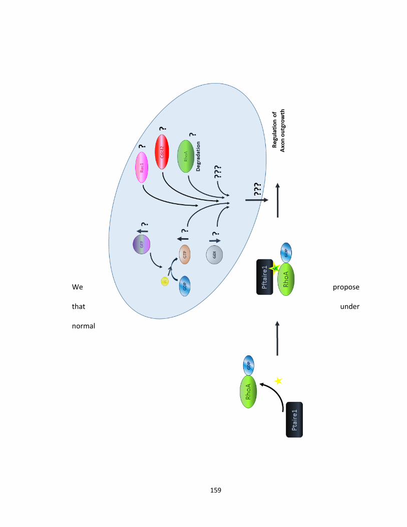
159
We propose
that under
normal
160
circumstances, Pftaire1 (black) and RhoA (green) physically interact. Pftaire1
phosphorylates (yellow star), GDP-RhoA on a serine residue (yellow-green star), activates
it and regulates axon outgrowth in a negative manner. Potential factors that may be
involved include but are not limited to, are exemplified and demonstrated by question
marks (?) in the blue background. pi (yellow), GDIs (gray), GEFs (purple), other Rho GTPase
proteins, , Cdc42 (red), Rac1 (pink), GDP to GTP (brown) exchange, also other unknown
factors (???).
Figure 4-1 A proposed model for the mechanism of action of Pftaire1
161
4.3. Conclusion
The role of Pftaire1 in the development of the CNS is virtually unknown. We are
the first to develop / obtain Pftaire1 deficient mice and study its effect on axon
outgrowth. Our in vitro data, suggests a role for mammalian Pftaire1 in axogenesis from
a phenotypic aspect, revealing a hyperextension in axonal length upon inhibition or
absence of Pftaire1. Furthermore, our study, confirms a physical interaction between
Pftaire1 and the small GTPase proteins Rac1, Cdc42, and RhoA. RhoA, is known to
negatively regulate axon outgrowth (Sebok, Nusser et al. 1999). Importantly, we showed
that Pftaire1 phosphorylates GDP-RhoA on a serine residue. We propose that this leads
to an increase in RhoA activity, which in turn controls axon outgrowth.
162
REFERENCES
Adams, N. C., T. Tomoda, M. Cooper, G. Dietz and M. E. Hatten (2002). "Mice that lack astrotactin
have slowed neuronal migration." Development 129(4): 965-972.
Adra, C. N., D. Manor, J. L. Ko, S. Zhu, T. Horiuchi, L. Van Aelst, R. A. Cerione and B. Lim (1997).
"RhoGDIgamma: a GDP-dissociation inhibitor for Rho proteins with preferential expression in
brain and pancreas." Proc Natl Acad Sci U S A 94(9): 4279-4284.
Amano, M., K. Chihara, N. Nakamura, T. Kaneko, Y. Matsuura and K. Kaibuchi (1999). "The COOH
terminus of Rho-kinase negatively regulates rho-kinase activity." J Biol Chem 274(45): 32418-
32424.
Amano, T., K. Tanabe, T. Eto, S. Narumiya and K. Mizuno (2001). "LIM-kinase 2 induces formation
of stress fibres, focal adhesions and membrane blebs, dependent on its activation by Rho-
associated kinase-catalysed phosphorylation at threonine-505." Biochem J 354(Pt 1): 149-
159.
Anton, E. S., J. A. Kreidberg and P. Rakic (1999). "Distinct functions of alpha3 and alpha(v) integrin
receptors in neuronal migration and laminar organization of the cerebral cortex." Neuron
22(2): 277-289.
Anton, E. S., M. A. Marchionni, K. F. Lee and P. Rakic (1997). "Role of GGF/neuregulin signaling in
interactions between migrating neurons and radial glia in the developing cerebral cortex."
Development 124(18): 3501-3510.
Aoki, K., T. Nakamura and M. Matsuda (2004). "Spatio-temporal regulation of Rac1 and Cdc42
activity during nerve growth factor-induced neurite outgrowth in PC12 cells." J Biol Chem
279(1): 713-719.
Arber, S., F. A. Barbayannis, H. Hanser, C. Schneider, C. A. Stanyon, O. Bernard and P. Caroni
(1998). "Regulation of actin dynamics through phosphorylation of cofilin by LIM-kinase."
Nature 393(6687): 805-809.
Arimura, N., N. Inagaki, K. Chihara, C. Menager, N. Nakamura, M. Amano, A. Iwamatsu, Y. Goshima
and K. Kaibuchi (2000). "Phosphorylation of collapsin response mediator protein-2 by Rho-
kinase. Evidence for two separate signaling pathways for growth cone collapse." J Biol Chem
275(31): 23973-23980.
163
Arozarena, I., D. Matallanas and P. Crespo (2001). "Maintenance of CDC42 GDP-bound state by
Rho-GDI inhibits MAP kinase activation by the exchange factor Ras-GRF. evidence for Ras-GRF
function being inhibited by Cdc42-GDP but unaffected by CDC42-GTP." J Biol Chem 276(24):
21878-21884.
Asada, A., N. Yamamoto, M. Gohda, T. Saito, N. Hayashi and S. Hisanaga (2008). "Myristoylation
of p39 and p35 is a determinant of cytoplasmic or nuclear localization of active cyclin-
dependent kinase 5 complexes." J Neurochem 106(3): 1325-1336.
Ball, H. J., A. Melnick, R. Shaknovich, R. A. Kohanski and J. D. Licht (1999). "The promyelocytic
leukemia zinc finger (PLZF) protein binds DNA in a high molecular weight complex associated
with cdc2 kinase." Nucleic Acids Res 27(20): 4106-4113.
Bartkova, J., B. Gron, E. Dabelsteen and J. Bartek (2003). "Cell-cycle regulatory proteins in human
wound healing." Arch Oral Biol 48(2): 125-132.
Bartova, I., M. Otyepka, Z. Kriz and J. Koca (2005). "The mechanism of inhibition of the cyclin-
dependent kinase-2 as revealed by the molecular dynamics study on the complex CDK2 with
the peptide substrate HHASPRK." Protein Sci 14(2): 445-451.
Baumli, S., G. Lolli, E. D. Lowe, S. Troiani, L. Rusconi, A. N. Bullock, J. E. Debreczeni, S. Knapp and
L. N. Johnson (2008). "The structure of P-TEFb (CDK9/cyclin T1), its complex with flavopiridol
and regulation by phosphorylation." EMBO J 27(13): 1907-1918.
Bear, J. E., T. M. Svitkina, M. Krause, D. A. Schafer, J. J. Loureiro, G. A. Strasser, I. V. Maly, O. Y.
Chaga, J. A. Cooper, G. G. Borisy and F. B. Gertler (2002). "Antagonism between Ena/VASP
proteins and actin filament capping regulates fibroblast motility." Cell 109(4): 509-521.
Bernard, O. (2007). "Lim kinases, regulators of actin dynamics." Int J Biochem Cell Biol 39(6): 1071-
1076.
Bernards, A. and J. Settleman (2004). "GAP control: regulating the regulators of small GTPases."
Trends Cell Biol 14(7): 377-385.
Besset, V., K. Rhee and D. J. Wolgemuth (1998). "The identification and characterization of
expression of Pftaire-1, a novel Cdk family member, suggest its function in the mouse testis
and nervous system." Mol Reprod Dev 50(1): 18-29.
Besset, V., K. Rhee and D. J. Wolgemuth (1999). "The cellular distribution and kinase activity of
the Cdk family member Pctaire1 in the adult mouse brain and testis suggest functions in
differentiation." Cell Growth Differ 10(3): 173-181.
164
Bibb, J. A., G. L. Snyder, A. Nishi, Z. Yan, L. Meijer, A. A. Fienberg, L. H. Tsai, Y. T. Kwon, J. A. Girault,
A. J. Czernik, R. L. Huganir, H. C. Hemmings, Jr., A. C. Nairn and P. Greengard (1999).
"Phosphorylation of DARPP-32 by Cdk5 modulates dopamine signalling in neurons." Nature
402(6762): 669-671.
Billuart, P., T. Bienvenu, N. Ronce, V. des Portes, M. C. Vinet, R. Zemni, H. Roest Crollius, A. Carrie,
F. Fauchereau, M. Cherry, S. Briault, B. Hamel, J. P. Fryns, C. Beldjord, A. Kahn, C. Moraine and
J. Chelly (1998). "Oligophrenin-1 encodes a rhoGAP protein involved in X-linked mental
retardation." Nature 392(6679): 923-926.
Biswas, S. C., Y. Zhang, G. Iyirhiaro, R. T. Willett, Y. Rodriguez Gonzalez, S. P. Cregan, R. S. Slack, D.
S. Park and L. A. Greene (2010). "Sertad1 plays an essential role in developmental and
pathological neuron death." J Neurosci 30(11): 3973-3982.
Borrell, V. and O. Marin (2006). "Meninges control tangential migration of hem-derived Cajal-
Retzius cells via CXCL12/CXCR4 signaling." Nat Neurosci 9(10): 1284-1293.
Brambilla, R. and G. Draetta (1994). "Molecular cloning of PISSLRE, a novel putative member of
the cdk family of protein serine/threonine kinases." Oncogene 9(10): 3037-3041.
Brouns, M. R., S. F. Matheson and J. Settleman (2001). "p190 RhoGAP is the principal Src substrate
in brain and regulates axon outgrowth, guidance and fasciculation." Nat Cell Biol 3(4): 361-
367.
Bu, B., J. Li, P. Davies and I. Vincent (2002). "Deregulation of cdk5, hyperphosphorylation, and
cytoskeletal pathology in the Niemann-Pick type C murine model." J Neurosci 22(15): 6515-
6525.
Burbelo, P. D., D. Drechsel and A. Hall (1995). "A conserved binding motif defines numerous
candidate target proteins for both Cdc42 and Rac GTPases." J Biol Chem 270(49): 29071-
29074.
Burridge, K. and K. Wennerberg (2004). "Rho and Rac take center stage." Cell 116(2): 167-179.
Butler, S. J. and J. Dodd (2003). "A role for BMP heterodimers in roof plate-mediated repulsion of
commissural axons." Neuron 38(3): 389-401.
Cafferty, W. B., P. Duffy, E. Huebner and S. M. Strittmatter (2010). "MAG and OMgp synergize
with Nogo-A to restrict axonal growth and neurological recovery after spinal cord trauma." J
Neurosci 30(20): 6825-6837.
Cajal, S. R. y. (1906). "The structure and connexions of neurons." Nobel Lecture.
165
Campbell, K. and M. Gotz (2002). "Radial glia: multi-purpose cells for vertebrate brain
development." Trends Neurosci 25(5): 235-238.
Carbonetto, S., D. Evans and P. Cochard (1987). "Nerve fiber growth in culture on tissue substrata
from central and peripheral nervous systems." J Neurosci 7(2): 610-620.
Caroni, P. and M. E. Schwab (1988). "Two membrane protein fractions from rat central myelin
with inhibitory properties for neurite growth and fibroblast spreading." J Cell Biol 106(4):
1281-1288.
Cavalli, G. and R. Paro (1998). "The Drosophila Fab-7 chromosomal element conveys epigenetic
inheritance during mitosis and meiosis." Cell 93(4): 505-518.
Chae, T., Y. T. Kwon, R. Bronson, P. Dikkes, E. Li and L. H. Tsai (1997). "Mice lacking p35, a neuronal
specific activator of Cdk5, display cortical lamination defects, seizures, and adult lethality."
Neuron 18(1): 29-42.
Charron, F., E. Stein, J. Jeong, A. P. McMahon and M. Tessier-Lavigne (2003). "The morphogen
sonic hedgehog is an axonal chemoattractant that collaborates with netrin-1 in midline axon
guidance." Cell 113(1): 11-23.
Cheng, Q., Y. Sasaki, M. Shoji, Y. Sugiyama, H. Tanaka, T. Nakayama, N. Mizuki, F. Nakamura, K.
Takei and Y. Goshima (2003). "Cdk5/p35 and Rho-kinase mediate ephrin-A5-induced signaling
in retinal ganglion cells." Mol Cell Neurosci 24(3): 632-645.
Cherfils, J. and M. Zeghouf (2013). "Regulation of small GTPases by GEFs, GAPs, and GDIs." Physiol
Rev 93(1): 269-309.
Cheung, Z. H., W. H. Chin, Y. Chen, Y. P. Ng and N. Y. Ip (2007). "Cdk5 is involved in BDNF-
stimulated dendritic growth in hippocampal neurons." PLoS Biol 5(4): e63.
Cheung, Z. H., A. K. Fu and N. Y. Ip (2006). "Synaptic roles of Cdk5: implications in higher cognitive
functions and neurodegenerative diseases." Neuron 50(1): 13-18.
Cheung, Z. H. and N. Y. Ip (2004). "Cdk5: mediator of neuronal death and survival." Neurosci Lett
361(1-3): 47-51.
Ciccolini, F., T. J. Collins, J. Sudhoelter, P. Lipp, M. J. Berridge and M. D. Bootman (2003). "Local
and global spontaneous calcium events regulate neurite outgrowth and onset of GABAergic
phenotype during neural precursor differentiation." J Neurosci 23(1): 103-111.
Cicero, S. and K. Herrup (2005). "Cyclin-dependent kinase 5 is essential for neuronal cell cycle
arrest and differentiation." J Neurosci 25(42): 9658-9668.
166
Coelho, C. M. and S. J. Leevers (2000). "Do growth and cell division rates determine cell size in
multicellular organisms?" J Cell Sci 113 ( Pt 17): 2927-2934.
Cole, A. R. (2009). "PCTK proteins: the forgotten brain kinases?" Neurosignals 17(4): 288-297.
Condeelis, J. (1993). "Life at the leading edge: the formation of cell protrusions." Annu Rev Cell
Biol 9: 411-444.
Connell-Crowley, L., D. Vo, L. Luke and E. Giniger (2007). "Drosophila lacking the Cdk5 activator,
p35, display defective axon guidance, age-dependent behavioral deficits and reduced
lifespan." Mech Dev 124(5): 341-349.
Cook, M., A. Gould, N. Brand, J. Davies, P. Strutt, R. Shaknovich, J. Licht, S. Waxman, Z. Chen, S.
Gluecksohn-Waelsch and et al. (1995). "Expression of the zinc-finger gene PLZF at
rhombomere boundaries in the vertebrate hindbrain." Proc Natl Acad Sci U S A 92(6): 2249-
2253.
Corbin, J. G., S. Nery and G. Fishell (2001). "Telencephalic cells take a tangent: non-radial migration
in the mammalian forebrain." Nat Neurosci 4 Suppl: 1177-1182.
Cropley, J. E., T. H. Dang, D. I. Martin and C. M. Suter (2012). "The penetrance of an epigenetic
trait in mice is progressively yet reversibly increased by selection and environment." Proc Biol
Sci 279(1737): 2347-2353.
Cruz, J. C. and L. H. Tsai (2004). "Cdk5 deregulation in the pathogenesis of Alzheimer's disease."
Trends Mol Med 10(9): 452-458.
Cunningham, J. J. and M. F. Roussel (2001). "Cyclin-dependent kinase inhibitors in the
development of the central nervous system." Cell Growth Differ 12(8): 387-396.
Cutforth, T. and C. J. Harrison (2002). "Ephs and ephrins close ranks." Trends Neurosci 25(7): 332-
334.
da Silva, J. S. and C. G. Dotti (2002). "Breaking the neuronal sphere: regulation of the actin
cytoskeleton in neuritogenesis." Nat Rev Neurosci 3(9): 694-704.
Da Silva, J. S., M. Medina, C. Zuliani, A. Di Nardo, W. Witke and C. G. Dotti (2003). "RhoA/ROCK
regulation of neuritogenesis via profilin IIa-mediated control of actin stability." J Cell Biol
162(7): 1267-1279.
Dancause, N. (2006). "Vicarious function of remote cortex following stroke: recent evidence from
human and animal studies." Neuroscientist 12(6): 489-499.
Daniels, R. H. and G. M. Bokoch (1999). "p21-activated protein kinase: a crucial component of
morphological signaling?" Trends Biochem Sci 24(9): 350-355.
167
Das, S. K., T. Hashimoto, K. Shimizu, T. Yoshida, T. Sakai, Y. Sowa, A. Komoto and K. Kanazawa
(2005). "Fucoxanthin induces cell cycle arrest at G0/G1 phase in human colon carcinoma cells
through up-regulation of p21WAF1/Cip1." Biochim Biophys Acta 1726(3): 328-335.
Daub, H., K. Gevaert, J. Vandekerckhove, A. Sobel and A. Hall (2001). "Rac/Cdc42 and p65PAK
regulate the microtubule-destabilizing protein stathmin through phosphorylation at serine
16." J Biol Chem 276(3): 1677-1680.
Davidson, G., J. Shen, Y. L. Huang, Y. Su, E. Karaulanov, K. Bartscherer, C. Hassler, P. Stannek, M.
Boutros and C. Niehrs (2009). "Cell cycle control of wnt receptor activation." Dev Cell 17(6):
788-799.
De Bondt, H. L., J. Rosenblatt, J. Jancarik, H. D. Jones, D. O. Morgan and S. H. Kim (1993). "Crystal
structure of cyclin-dependent kinase 2." Nature 363(6430): 595-602.
Dehay, C. and H. Kennedy (2007). "Cell-cycle control and cortical development." Nat Rev Neurosci
8(6): 438-450.
Del Rio, J. A., C. Gonzalez-Billault, J. M. Urena, E. M. Jimenez, M. J. Barallobre, M. Pascual, L.
Pujadas, S. Simo, A. La Torre, F. Wandosell, J. Avila and E. Soriano (2004). "MAP1B is required
for Netrin 1 signaling in neuronal migration and axonal guidance." Curr Biol 14(10): 840-850.
Denicourt, C. and S. F. Dowdy (2004). "Cip/Kip proteins: more than just CDKs inhibitors." Genes
Dev 18(8): 851-855.
Dhavan, R. and L. H. Tsai (2001). "A decade of CDK5." Nat Rev Mol Cell Biol 2(10): 749-759.
Dickson, B. J. (2002). "Molecular mechanisms of axon guidance." Science 298(5600): 1959-1964.
Dotti, C. G., C. A. Sullivan and G. A. Banker (1988). "The establishment of polarity by hippocampal
neurons in culture." J Neurosci 8(4): 1454-1468.
Dvorsky, R. and M. R. Ahmadian (2004). "Always look on the bright site of Rho: structural
implications for a conserved intermolecular interface." EMBO Rep 5(12): 1130-1136.
Dvorsky, R., L. Blumenstein, I. R. Vetter and M. R. Ahmadian (2004). "Structural insights into the
interaction of ROCKI with the switch regions of RhoA." J Biol Chem 279(8): 7098-7104.
Echalier, A., J. A. Endicott and M. E. Noble (2010). "Recent developments in cyclin-dependent
kinase biochemical and structural studies." Biochim Biophys Acta 1804(3): 511-519.
Ehler, E., F. van Leeuwen, J. G. Collard and P. C. Salinas (1997). "Expression of Tiam-1 in the
developing brain suggests a role for the Tiam-1-Rac signaling pathway in cell migration and
neurite outgrowth." Mol Cell Neurosci 9(1): 1-12.
168
Etienne-Manneville, S. and A. Hall (2002). "Rho GTPases in cell biology." Nature 420(6916): 629-
635.
Fan, X., J. P. Labrador, H. Hing and G. J. Bashaw (2003). "Slit stimulation recruits Dock and Pak to
the roundabout receptor and increases Rac activity to regulate axon repulsion at the CNS
midline." Neuron 40(1): 113-127.
Fawcett, J. W. (1992). "Intrinsic neuronal determinants of regeneration." Trends Neurosci 15(1):
5-8.
Filbin, M. T. (2003). "Myelin-associated inhibitors of axonal regeneration in the adult mammalian
CNS." Nat Rev Neurosci 4(9): 703-713.
Font, M. A., A. Arboix and J. Krupinski (2010). "Angiogenesis, neurogenesis and neuroplasticity in
ischemic stroke." Curr Cardiol Rev 6(3): 238-244.
Freeman, R. S., S. Estus and E. M. Johnson, Jr. (1994). "Analysis of cell cycle-related gene
expression in postmitotic neurons: selective induction of Cyclin D1 during programmed cell
death." Neuron 12(2): 343-355.
Fu, H., R. R. Subramanian and S. C. Masters (2000). "14-3-3 proteins: structure, function, and
regulation." Annu Rev Pharmacol Toxicol 40: 617-647.
Fu, W. Y., Y. Chen, M. Sahin, X. S. Zhao, L. Shi, J. B. Bikoff, K. O. Lai, W. H. Yung, A. K. Fu, M. E.
Greenberg and N. Y. Ip (2007). "Cdk5 regulates EphA4-mediated dendritic spine retraction
through an ephexin1-dependent mechanism." Nat Neurosci 10(1): 67-76.
Fu, W. Y., K. Cheng, A. K. Fu and N. Y. Ip (2011). "Cyclin-dependent kinase 5-dependent
phosphorylation of Pctaire1 regulates dendrite development." Neuroscience 180: 353-359.
Fujita Y, Y. T. (2014). "Axon growth inhibition by RhoA/ROCK in the central nervous system." Front
Neurosci. 8: 338.
Futatsugi, A., E. Utreras, P. Rudrabhatla, H. Jaffe, H. C. Pant and A. B. Kulkarni (2012). "Cyclin-
dependent kinase 5 regulates E2F transcription factor through phosphorylation of Rb protein
in neurons." Cell Cycle 11(8): 1603-1610.
Galbraith, M. D., A. J. Donner and J. M. Espinosa (2010). "CDK8: a positive regulator of
transcription." Transcription 1(1): 4-12.
Galderisi, U., F. P. Jori and A. Giordano (2003). "Cell cycle regulation and neural differentiation."
Oncogene 22(33): 5208-5219.
Gao, Y., M. Jiang, T. Yang, J. Ni and J. Chen (2006). "A Cdc2-related protein kinase hPFTAIRE1 from
human brain interacting with 14-3-3 proteins." Cell Res 16(6): 539-547.
169
Gao, Y. K., M. Jiang, T. Yang and J. Y. Chen (2006). "Analysis of the interaction between hPFTAIRE1
and PLZF in a yeast two-hybrid system." Acta Biochim Biophys Sin (Shanghai) 38(3): 164-170.
Georgiev, D., H. Taniura, Y. Kambe, T. Takarada and Y. Yoneda (2008). "A critical importance of
polyamine site in NMDA receptors for neurite outgrowth and fasciculation at early stages of
P19 neuronal differentiation." Exp Cell Res 314(14): 2603-2617.
Gilmore, E. C., T. Ohshima, A. M. Goffinet, A. B. Kulkarni and K. Herrup (1998). "Cyclin-dependent
kinase 5-deficient mice demonstrate novel developmental arrest in cerebral cortex." J
Neurosci 18(16): 6370-6377.
Giovanni, A., E. Keramaris, E. J. Morris, S. T. Hou, M. O'Hare, N. Dyson, G. S. Robertson, R. S. Slack
and D. S. Park (2000). "E2F1 mediates death of B-amyloid-treated cortical neurons in a
manner independent of p53 and dependent on Bax and caspase 3." J Biol Chem 275(16):
11553-11560.
Giovanni, A., F. Wirtz-Brugger, E. Keramaris, R. Slack and D. S. Park (1999). "Involvement of cell
cycle elements, cyclin-dependent kinases, pRb, and E2F x DP, in B-amyloid-induced neuronal
death." J Biol Chem 274(27): 19011-19016.
Golestaneh, N., E. Beauchamp, S. Fallen, M. Kokkinaki, A. Uren and M. Dym (2009). "Wnt signaling
promotes proliferation and stemness regulation of spermatogonial stem/progenitor cells."
Reproduction 138(1): 151-162.
Gomez, T. M. and N. C. Spitzer (1999). "In vivo regulation of axon extension and pathfinding by
growth-cone calcium transients." Nature 397(6717): 350-355.
Gonzalez, Y. R., Y. Zhang, D. Behzadpoor, S. Cregan, S. Bamforth, R. S. Slack and D. S. Park (2008).
"CITED2 signals through peroxisome proliferator-activated receptor-gamma to regulate death
of cortical neurons after DNA damage." J Neurosci 28(21): 5559-5569.
Govek, E. E., S. E. Newey and L. Van Aelst (2005). "The role of the Rho GTPases in neuronal
development." Genes Dev 19(1): 1-49.
Graeser, R., J. Gannon, R. Y. Poon, T. Dubois, A. Aitken and T. Hunt (2002). "Regulation of the CDK-
related protein kinase PCTAIRE-1 and its possible role in neurite outgrowth in Neuro-2A cells."
J Cell Sci 115(Pt 17): 3479-3490.
Grana, X., A. De Luca, N. Sang, Y. Fu, P. P. Claudio, J. Rosenblatt, D. O. Morgan and A. Giordano
(1994). "PITALRE, a nuclear CDC2-related protein kinase that phosphorylates the
retinoblastoma protein in vitro." Proc Natl Acad Sci U S A 91(9): 3834-3838.
170
Grigoryan, T., P. Wend, A. Klaus and W. Birchmeier (2008). "Deciphering the function of canonical
Wnt signals in development and disease: conditional loss- and gain-of-function mutations of
beta-catenin in mice." Genes Dev 22(17): 2308-2341.
Gu, X., E. C. Olson and N. C. Spitzer (1994). "Spontaneous neuronal calcium spikes and waves
during early differentiation." J Neurosci 14(11 Pt 1): 6325-6335.
Gu, X. and N. C. Spitzer (1995). "Distinct aspects of neuronal differentiation encoded by frequency
of spontaneous Ca2+ transients." Nature 375(6534): 784-787.
Gu, Y., J. Rosenblatt and D. O. Morgan (1992). "Cell cycle regulation of CDK2 activity by
phosphorylation of Thr160 and Tyr15." EMBO J 11(11): 3995-4005.
Gurdon, J. B. and P. Y. Bourillot (2001). "Morphogen gradient interpretation." Nature 413(6858):
797-803.
Hakeda-Suzuki, S., J. Ng, J. Tzu, G. Dietzl, Y. Sun, M. Harms, T. Nardine, L. Luo and B. J. Dickson
(2002). "Rac function and regulation during Drosophila development." Nature 416(6879):
438-442.
Hall, A. (1998). "Rho GTPases and the actin cytoskeleton." Science 279(5350): 509-514.
Hall, A. and G. Lalli (2010). "Rho and Ras GTPases in axon growth, guidance, and branching." Cold
Spring Harb Perspect Biol 2(2): a001818.
Hall, C., M. Brown, T. Jacobs, G. Ferrari, N. Cann, M. Teo, C. Monfries and L. Lim (2001). "Collapsin
response mediator protein switches RhoA and Rac1 morphology in N1E-115 neuroblastoma
cells and is regulated by Rho kinase." J Biol Chem 276(46): 43482-43486.
Hall, C., G. J. Michael, N. Cann, G. Ferrari, M. Teo, T. Jacobs, C. Monfries and L. Lim (2001). "alpha2-
chimaerin, a Cdc42/Rac1 regulator, is selectively expressed in the rat embryonic nervous
system and is involved in neuritogenesis in N1E-115 neuroblastoma cells." J Neurosci 21(14):
5191-5202.
Hansen, S. M., V. Berezin and E. Bock (2008). "Signaling mechanisms of neurite outgrowth induced
by the cell adhesion molecules NCAM and N-cadherin." Cell Mol Life Sci 65(23): 3809-3821.
Harada, T., T. Morooka, S. Ogawa and E. Nishida (2001). "ERK induces p35, a neuron-specific
activator of Cdk5, through induction of Egr1." Nat Cell Biol 3(5): 453-459.
Hartwell, L. H. (1974). "Saccharomyces cerevisiae cell cycle." Bacteriol Rev 38(2): 164-198.
Hashida, T., M. Yamada, K. Hashimoto, N. Shibusawa, T. Monden, T. Satoh and M. Mori (2002). "A
novel TRH-PFTAIRE protein kinase 1 pathway in the cerebellum: subtractive hybridization
analysis of TRH-deficient mice." Endocrinology 143(7): 2808-2811.
171
Hawasli, A. H. and J. A. Bibb (2007). "Alternative roles for Cdk5 in learning and synaptic plasticity."
Biotechnol J 2(8): 941-948.
Hellmich, M. R., H. C. Pant, E. Wada and J. F. Battey (1992). "Neuronal cdc2-like kinase: a cdc2-
related protein kinase with predominantly neuronal expression." Proc Natl Acad Sci U S A
89(22): 10867-10871.
Henderson, I. R. and S. E. Jacobsen (2007). "Epigenetic inheritance in plants." Nature 447(7143):
418-424.
Higgs, H. N. and T. D. Pollard (2001). "Regulation of actin filament network formation through
ARP2/3 complex: activation by a diverse array of proteins." Annu Rev Biochem 70: 649-676.
Hindley, C. and A. Philpott (2012). "Co-ordination of cell cycle and differentiation in the developing
nervous system." Biochem J 444(3): 375-382.
Hirose, T., M. Kawabuchi, T. Tamaru, N. Okumura, K. Nagai and M. Okada (2000). "Identification
of tudor repeat associator with PCTAIRE 2 (Trap). A novel protein that interacts with the N-
terminal domain of PCTAIRE 2 in rat brain." Eur J Biochem 267(7): 2113-2121.
Hitchins, M. P., J. J. Wong, G. Suthers, C. M. Suter, D. I. Martin, N. J. Hawkins and R. L. Ward (2007).
"Inheritance of a cancer-associated MLH1 germ-line epimutation." N Engl J Med 356(7): 697-
705.
Hoffman, G. R., N. Nassar and R. A. Cerione (2000). "Structure of the Rho family GTP-binding
protein Cdc42 in complex with the multifunctional regulator RhoGDI." Cell 100(3): 345-356.
Horii, Y., J. F. Beeler, K. Sakaguchi, M. Tachibana and T. Miki (1994). "A novel oncogene, ost,
encodes a guanine nucleotide exchange factor that potentially links Rho and Rac signaling
pathways." EMBO J 13(20): 4776-4786.
Hoshino, M., M. Sone, M. Fukata, S. Kuroda, K. Kaibuchi, Y. Nabeshima and C. Hama (1999).
"Identification of the stef gene that encodes a novel guanine nucleotide exchange factor
specific for Rac1." J Biol Chem 274(25): 17837-17844.
Huang, E. J. and L. F. Reichardt (2003). "Trk receptors: roles in neuronal signal transduction." Annu
Rev Biochem 72: 609-642.
Huber, A. B., A. L. Kolodkin, D. D. Ginty and J. F. Cloutier (2003). "Signaling at the growth cone:
ligand-receptor complexes and the control of axon growth and guidance." Annu Rev Neurosci
26: 509-563.
Ille, F., S. Atanasoski, S. Falk, L. M. Ittner, D. Marki, S. Buchmann-Moller, H. Wurdak, U. Suter, M.
M. Taketo and L. Sommer (2007). "Wnt/BMP signal integration regulates the balance between
172
proliferation and differentiation of neuroepithelial cells in the dorsal spinal cord." Dev Biol
304(1): 394-408.
Ishiguro, K., M. Takamatsu, K. Tomizawa, A. Omori, M. Takahashi, M. Arioka, T. Uchida and K.
Imahori (1992). "Tau protein kinase I converts normal tau protein into A68-like component of
paired helical filaments." J Biol Chem 267(15): 10897-10901.
Ishizaki, T., M. Maekawa, K. Fujisawa, K. Okawa, A. Iwamatsu, A. Fujita, N. Watanabe, Y. Saito, A.
Kakizuka, N. Morii and S. Narumiya (1996). "The small GTP-binding protein Rho binds to and
activates a 160 kDa Ser/Thr protein kinase homologous to myotonic dystrophy kinase." EMBO
J 15(8): 1885-1893.
Itoh, Y., N. Masuyama, K. Nakayama, K. I. Nakayama and Y. Gotoh (2007). "The cyclin-dependent
kinase inhibitors p57 and p27 regulate neuronal migration in the developing mouse
neocortex." J Biol Chem 282(1): 390-396.
Jablonka, E. and G. Raz (2009). "Transgenerational epigenetic inheritance: prevalence,
mechanisms, and implications for the study of heredity and evolution." Q Rev Biol 84(2): 131-
176.
Jablonska, B., A. Aguirre, R. Vandenbosch, S. Belachew, C. Berthet, P. Kaldis and V. Gallo (2007).
"Cdk2 is critical for proliferation and self-renewal of neural progenitor cells in the adult
subventricular zone." J Cell Biol 179(6): 1231-1245.
Jaffe, A. B. and A. Hall (2005). "Rho GTPases: biochemistry and biology." Annu Rev Cell Dev Biol
21: 247-269.
Jalink, K., E. J. van Corven, T. Hengeveld, N. Morii, S. Narumiya and W. H. Moolenaar (1994).
"Inhibition of lysophosphatidate- and thrombin-induced neurite retraction and neuronal cell
rounding by ADP ribosylation of the small GTP-binding protein Rho." J Cell Biol 126(3): 801-
810.
Jeffrey, P. D., A. A. Russo, K. Polyak, E. Gibbs, J. Hurwitz, J. Massague and N. P. Pavletich (1995).
"Mechanism of CDK activation revealed by the structure of a cyclinA-CDK2 complex." Nature
376(6538): 313-320.
Jiang, M., Y. Gao, T. Yang, X. Zhu and J. Chen (2009). "Cyclin Y, a novel membrane-associated
cyclin, interacts with PFTK1." FEBS Lett 583(13): 2171-2178.
Johansson, J. U., L. Lilja, X. L. Chen, H. Higashida, B. Meister, M. Noda, Z. G. Zhong, S. Yokoyama,
P. O. Berggren and C. Bark (2005). "Cyclin-dependent kinase 5 activators p35 and p39 facilitate
formation of functional synapses." Brain Res Mol Brain Res 138(2): 215-227.
173
John, P. C., M. Mews and R. Moore (2001). "Cyclin/Cdk complexes: their involvement in cell cycle
progression and mitotic division." Protoplasma 216(3-4): 119-142.
Kaldis, P. (1999). "The cdk-activating kinase (CAK): from yeast to mammals." Cell Mol Life Sci 55(2):
284-296.
Kast, D. J., C. Yang, A. Disanza, M. Boczkowska, Y. Madasu, G. Scita, T. Svitkina and R. Dominguez
(2014). "Mechanism of IRSp53 inhibition and combinatorial activation by Cdc42 and
downstream effectors." Nat Struct Mol Biol 21(4): 413-422.
Kawauchi, T. (2014). "Cdk5 regulates multiple cellular events in neural development, function and
disease." Dev Growth Differ 56(5): 335-348.
Kawauchi, T., K. Chihama, Y. Nabeshima and M. Hoshino (2006). "Cdk5 phosphorylates and
stabilizes p27kip1 contributing to actin organization and cortical neuronal migration." Nat Cell
Biol 8(1): 17-26.
Kawauchi, T. and M. Hoshino (2008). "Molecular pathways regulating cytoskeletal organization
and morphological changes in migrating neurons." Dev Neurosci 30(1-3): 36-46.
Kawauchi, T., M. Shikanai and Y. Kosodo (2013). "Extra-cell cycle regulatory functions of cyclin-
dependent kinases (CDK) and CDK inhibitor proteins contribute to brain development and
neurological disorders." Genes Cells 18(3): 176-194.
Kemphues, K. (2000). "PARsing embryonic polarity." Cell 101(4): 345-348.
Kerr, G. E., J. C. Young, K. Horvay, H. E. Abud and K. L. Loveland (2014). "Regulated Wnt/beta-
catenin signaling sustains adult spermatogenesis in mice." Biol Reprod 90(1): 3.
Kesavapany, S., N. Amin, Y. L. Zheng, R. Nijhara, H. Jaffe, R. Sihag, J. S. Gutkind, S. Takahashi, A.
Kulkarni, P. Grant and H. C. Pant (2004). "p35/cyclin-dependent kinase 5 phosphorylation of
ras guanine nucleotide releasing factor 2 (RasGRF2) mediates Rac-dependent Extracellular
Signal-regulated kinase 1/2 activity, altering RasGRF2 and microtubule-associated protein 1b
distribution in neurons." J Neurosci 24(18): 4421-4431.
Kesavapany, S., T. K. Pareek, Y. L. Zheng, N. Amin, J. S. Gutkind, W. Ma, A. B. Kulkarni, P. Grant and
H. C. Pant (2006). "Neuronal nuclear organization is controlled by cyclin-dependent kinase 5
phosphorylation of Ras Guanine nucleotide releasing factor-1." Neurosignals 15(4): 157-173.
Kim, Y. T. and M. Zhao (2005). "Aberrant cell cycle regulation in cervical carcinoma." Yonsei Med
J 46(5): 597-613.
174
Ko, J., S. Humbert, R. T. Bronson, S. Takahashi, A. B. Kulkarni, E. Li and L. H. Tsai (2001). "p35 and
p39 are essential for cyclin-dependent kinase 5 function during neurodevelopment." J
Neurosci 21(17): 6758-6771.
Konishi, Y. and A. Bonni (2003). "The E2F-Cdc2 cell-cycle pathway specifically mediates activity
deprivation-induced apoptosis of postmitotic neurons." J Neurosci 23(5): 1649-1658.
Konishi, Y., M. Lehtinen, N. Donovan and A. Bonni (2002). "Cdc2 phosphorylation of BAD links the
cell cycle to the cell death machinery." Mol Cell 9(5): 1005-1016.
Korenjak, M. and A. Brehm (2005). "E2F-Rb complexes regulating transcription of genes important
for differentiation and development." Curr Opin Genet Dev 15(5): 520-527.
Kruman, II, R. P. Wersto, F. Cardozo-Pelaez, L. Smilenov, S. L. Chan, F. J. Chrest, R. Emokpae, Jr.,
M. Gorospe and M. P. Mattson (2004). "Cell cycle activation linked to neuronal cell death
initiated by DNA damage." Neuron 41(4): 549-561.
Kubo, T., A. Yamaguchi, N. Iwata and T. Yamashita (2008). "The therapeutic effects of Rho-ROCK
inhibitors on CNS disorders." Ther Clin Risk Manag 4(3): 605-615.
Kunda, P., G. Paglini, S. Quiroga, K. Kosik and A. Caceres (2001). "Evidence for the involvement of
Tiam1 in axon formation." J Neurosci 21(7): 2361-2372.
Kusakawa, G., T. Saito, R. Onuki, K. Ishiguro, T. Kishimoto and S. Hisanaga (2000). "Calpain-
dependent proteolytic cleavage of the p35 cyclin-dependent kinase 5 activator to p25." J Biol
Chem 275(22): 17166-17172.
Kwon, Y. T. and L. H. Tsai (2000). "The role of the p35/cdk5 kinase in cortical development."
Results Probl Cell Differ 30: 241-253.
Lange, C., W. B. Huttner and F. Calegari (2009). "Cdk4/cyclinD1 overexpression in neural stem cells
shortens G1, delays neurogenesis, and promotes the generation and expansion of basal
progenitors." Cell Stem Cell 5(3): 320-331.
Langhorst, M. F., F. A. Jaeger, S. Mueller, L. Sven Hartmann, G. Luxenhofer and C. A. Stuermer
(2008). "Reggies/flotillins regulate cytoskeletal remodeling during neuronal differentiation via
CAP/ponsin and Rho GTPases." Eur J Cell Biol 87(12): 921-931.
Lanier, L. M., M. A. Gates, W. Witke, A. S. Menzies, A. M. Wehman, J. D. Macklis, D. Kwiatkowski,
P. Soriano and F. B. Gertler (1999). "Mena is required for neurulation and commissure
formation." Neuron 22(2): 313-325.
Lazzaro, M. A., P. R. Albert and J. P. Julien (1997). "A novel cdc2-related protein kinase expressed
in the nervous system." J Neurochem 69(1): 348-364.
175
Lazzaro, M. A. and J. P. Julien (1997). "Chromosomal mapping of the PFTAIRE gene, Pftk1, a cdc2-
related kinase expressed predominantly in the mouse nervous system." Genomics 42(3): 536-
537.
Lee, T., C. Winter, S. S. Marticke, A. Lee and L. Luo (2000). "Essential roles of Drosophila RhoA in
the regulation of neuroblast proliferation and dendritic but not axonal morphogenesis."
Neuron 25(2): 307-316.
Leung, W. K., A. K. Ching, A. W. Chan, T. C. Poon, H. Mian, A. S. Wong, K. F. To and N. Wong (2011).
"A novel interplay between oncogenic PFTK1 protein kinase and tumor suppressor TAGLN2 in
the control of liver cancer cell motility." Oncogene 30(44): 4464-4475.
Leung, W. K., A. K. Ching and N. Wong (2011). "Phosphorylation of Caldesmon by PFTAIRE1 kinase
promotes actin binding and formation of stress fibers." Mol Cell Biochem 350(1-2): 201-206.
Lew, J., Q. Q. Huang, Z. Qi, R. J. Winkfein, R. Aebersold, T. Hunt and J. H. Wang (1994). "A brain-
specific activator of cyclin-dependent kinase 5." Nature 371(6496): 423-426.
Lew, J., R. J. Winkfein, H. K. Paudel and J. H. Wang (1992). "Brain proline-directed protein kinase
is a neurofilament kinase which displays high sequence homology to p34cdc2." J Biol Chem
267(36): 25922-25926.
Li, B. and E. DiCicco-Bloom (2004). "Basic fibroblast growth factor exhibits dual and rapid
regulation of cyclin D1 and p27 to stimulate proliferation of rat cerebral cortical precursors."
Dev Neurosci 26(2-4): 197-207.
Li, J. Y., M. A. English, H. J. Ball, P. L. Yeyati, S. Waxman and J. D. Licht (1997). "Sequence-specific
DNA binding and transcriptional regulation by the promyelocytic leukemia zinc finger
protein." J Biol Chem 272(36): 22447-22455.
Lim, S. and P. Kaldis (2013). "Cdks, cyclins and CKIs: roles beyond cell cycle regulation."
Development 140(15): 3079-3093.
Lin, S., J. Wang, Z. Ye, N. Y. Ip and S. C. Lin (2008). "CDK5 activator p35 downregulates E-cadherin
precursor independently of CDK5." FEBS Lett 582(8): 1197-1202.
Liu, D. X., S. C. Biswas and L. A. Greene (2004). "B-myb and C-myb play required roles in neuronal
apoptosis evoked by nerve growth factor deprivation and DNA damage." J Neurosci 24(40):
8720-8725.
Liu, H., T. Nakazawa, T. Tezuka and T. Yamamoto (2006). "Physical and functional interaction of
Fyn tyrosine kinase with a brain-enriched Rho GTPase-activating protein TCGAP." J Biol Chem
281(33): 23611-23619.
176
Liu, J. and E. T. Kipreos (2000). "Evolution of cyclin-dependent kinases (CDKs) and CDK-activating
kinases (CAKs): differential conservation of CAKs in yeast and metazoa." Mol Biol Evol 17(7):
1061-1074.
Liu, Y., K. Cheng, K. Gong, A. K. Fu and N. Y. Ip (2006). "Pctaire1 phosphorylates N-ethylmaleimide-
sensitive fusion protein: implications in the regulation of its hexamerization and exocytosis."
J Biol Chem 281(15): 9852-9858.
Longenecker, K., P. Read, U. Derewenda, Z. Dauter, X. Liu, S. Garrard, L. Walker, A. V. Somlyo, R.
K. Nakamoto, A. P. Somlyo and Z. S. Derewenda (1999). "How RhoGDI binds Rho." Acta
Crystallogr D Biol Crystallogr 55(Pt 9): 1503-1515.
Lopes, J. P., C. R. Oliveira and P. Agostinho (2009). "Cdk5 acts as a mediator of neuronal cell cycle
re-entry triggered by amyloid-beta and prion peptides." Cell Cycle 8(1): 97-104.
Lowery, L. A. and D. Van Vactor (2009). "The trip of the tip: understanding the growth cone
machinery." Nat Rev Mol Cell Biol 10(5): 332-343.
Loyer, P., J. H. Trembley, J. A. Grenet, A. Busson, A. Corlu, W. Zhao, M. Kocak, V. J. Kidd and J. M.
Lahti (2008). "Characterization of cyclin L1 and L2 interactions with CDK11 and splicing factors:
influence of cyclin L isoforms on splice site selection." J Biol Chem 283(12): 7721-7732.
Loyer, P., J. H. Trembley, R. Katona, V. J. Kidd and J. M. Lahti (2005). "Role of CDK/cyclin complexes
in transcription and RNA splicing." Cell Signal 17(9): 1033-1051.
Lucking, C. B., A. Durr, V. Bonifati, J. Vaughan, G. De Michele, T. Gasser, B. S. Harhangi, G. Meco,
P. Denefle, N. W. Wood, Y. Agid and A. Brice (2000). "Association between early-onset
Parkinson's disease and mutations in the parkin gene." N Engl J Med 342(21): 1560-1567.
Luo, L., T. K. Hensch, L. Ackerman, S. Barbel, L. Y. Jan and Y. N. Jan (1996). "Differential effects of
the Rac GTPase on Purkinje cell axons and dendritic trunks and spines." Nature 379(6568):
837-840.
Ma, X. M., R. C. Johnson, R. E. Mains and B. A. Eipper (2001). "Expression of kalirin, a neuronal
GDP/GTP exchange factor of the trio family, in the central nervous system of the adult rat." J
Comp Neurol 429(3): 388-402.
Machesky, L. M., R. D. Mullins, H. N. Higgs, D. A. Kaiser, L. Blanchoin, R. C. May, M. E. Hall and T.
D. Pollard (1999). "Scar, a WASp-related protein, activates nucleation of actin filaments by the
Arp2/3 complex." Proc Natl Acad Sci U S A 96(7): 3739-3744.
Madaule, P. and R. Axel (1985). "A novel ras-related gene family." Cell 41(1): 31-40.
177
Maekawa, M., T. Ishizaki, S. Boku, N. Watanabe, A. Fujita, A. Iwamatsu, T. Obinata, K. Ohashi, K.
Mizuno and S. Narumiya (1999). "Signaling from Rho to the actin cytoskeleton through protein
kinases ROCK and LIM-kinase." Science 285(5429): 895-898.
Malumbres, M. (2011). "Physiological relevance of cell cycle kinases." Physiol Rev 91(3): 973-1007.
Malumbres, M. (2014). "Cyclin-dependent kinases." Genome Biol 15(6): 122.
Malumbres, M. and M. Barbacid (2005). "Mammalian cyclin-dependent kinases." Trends Biochem
Sci 30(11): 630-641.
Malumbres, M., E. Harlow, T. Hunt, T. Hunter, J. M. Lahti, G. Manning, D. O. Morgan, L. H. Tsai and
D. J. Wolgemuth (2009). "Cyclin-dependent kinases: a family portrait." Nat Cell Biol 11(11):
1275-1276.
Manser, E., T. Leung, H. Salihuddin, Z. S. Zhao and L. Lim (1994). "A brain serine/threonine protein
kinase activated by Cdc42 and Rac1." Nature 367(6458): 40-46.
Marchler-Bauer, A., C. Zheng, F. Chitsaz, M. K. Derbyshire, L. Y. Geer, R. C. Geer, N. R. Gonzales,
M. Gwadz, D. I. Hurwitz, C. J. Lanczycki, F. Lu, S. Lu, G. H. Marchler, J. S. Song, N. Thanki, R. A.
Yamashita, D. Zhang and S. H. Bryant (2013). "CDD: conserved domains and protein three-
dimensional structure." Nucleic Acids Res 41(Database issue): D348-352.
Marin, O. and J. L. Rubenstein (2001). "A long, remarkable journey: tangential migration in the
telencephalon." Nat Rev Neurosci 2(11): 780-790.
Marin, O., M. Valiente, X. Ge and L. H. Tsai (2010). "Guiding neuronal cell migrations." Cold Spring
Harb Perspect Biol 2(2): a001834.
Martini, F. J., M. Valiente, G. Lopez Bendito, G. Szabo, F. Moya, M. Valdeolmillos and O. Marin
(2009). "Biased selection of leading process branches mediates chemotaxis during tangential
neuronal migration." Development 136(1): 41-50.
May, V., M. R. Schiller, B. A. Eipper and R. E. Mains (2002). "Kalirin Dbl-homology guanine
nucleotide exchange factor 1 domain initiates new axon outgrowths via RhoG-mediated
mechanisms." J Neurosci 22(16): 6980-6990.
Mazzarello, P. (1999). "The Hidden Structure. Translated from Cajal’s Histologie du Systeme
Nerveux, describing a first view of the results of the Golgi-staining method.".
McKerracher, L., S. David, D. L. Jackson, V. Kottis, R. J. Dunn and P. E. Braun (1994). "Identification
of myelin-associated glycoprotein as a major myelin-derived inhibitor of neurite growth."
Neuron 13(4): 805-811.
178
Meyerson, M., G. H. Enders, C. L. Wu, L. K. Su, C. Gorka, C. Nelson, E. Harlow and L. H. Tsai (1992).
"A family of human cdc2-related protein kinases." EMBO J 11(8): 2909-2917.
Mikolcevic, P., R. Sigl, V. Rauch, M. W. Hess, K. Pfaller, M. Barisic, L. J. Pelliniemi, M. Boesl and S.
Geley (2012). "Cyclin-dependent kinase 16/PCTAIRE kinase 1 is activated by cyclin Y and is
essential for spermatogenesis." Mol Cell Biol 32(4): 868-879.
Miyajima, M., H. O. Nornes and T. Neuman (1995). "Cyclin E is expressed in neurons and forms
complexes with cdk5." Neuroreport 6(8): 1130-1132.
Miyata, T., A. Kawaguchi, H. Okano and M. Ogawa (2001). "Asymmetric inheritance of radial glial
fibers by cortical neurons." Neuron 31(5): 727-741.
Miyata, T. and M. Ogawa (2007). "Twisting of neocortical progenitor cells underlies a spring-like
mechanism for daughter-cell migration." Curr Biol 17(2): 146-151.
Morgan, D. O. (1995). "Principles of CDK regulation." Nature 374(6518): 131-134.
Morgan, D. O. (2007). "The Cell Cycle: Principles of Control. ." London: New Science Press, 1st ed.
Morgan, H. D., H. G. Sutherland, D. I. Martin and E. Whitelaw (1999). "Epigenetic inheritance at
the agouti locus in the mouse." Nat Genet 23(3): 314-318.
Morris, E. J., E. Keramaris, H. J. Rideout, R. S. Slack, N. J. Dyson, L. Stefanis and D. S. Park (2001).
"Cyclin-dependent kinases and P53 pathways are activated independently and mediate Bax
activation in neurons after DNA damage." J Neurosci 21(14): 5017-5026.
Mukhopadhyay, G., P. Doherty, F. S. Walsh, P. R. Crocker and M. T. Filbin (1994). "A novel role for
myelin-associated glycoprotein as an inhibitor of axonal regeneration." Neuron 13(3): 757-
767.
Munger, K. and P. M. Howley (2002). "Human papillomavirus immortalization and transformation
functions." Virus Res 89(2): 213-228.
Muslin, A. J. and H. Xing (2000). "14-3-3 proteins: regulation of subcellular localization by
molecular interference." Cell Signal 12(11-12): 703-709.
Nadarajah, B., J. E. Brunstrom, J. Grutzendler, R. O. Wong and A. L. Pearlman (2001). "Two modes
of radial migration in early development of the cerebral cortex." Nat Neurosci 4(2): 143-150.
Nadarajah, B. and J. G. Parnavelas (2002). "Modes of neuronal migration in the developing
cerebral cortex." Nat Rev Neurosci 3(6): 423-432.
Nagy, Z., M. M. Esiri, A. M. Cato and A. D. Smith (1997). "Cell cycle markers in the hippocampus in
Alzheimer's disease." Acta Neuropathol 94(1): 6-15.
179
Negishi, M. and H. Katoh (2002). "Rho family GTPases as key regulators for neuronal network
formation." J Biochem 132(2): 157-166.
Ng, J. and L. Luo (2004). "Rho GTPases regulate axon growth through convergent and divergent
signaling pathways." Neuron 44(5): 779-793.
Ng, J., T. Nardine, M. Harms, J. Tzu, A. Goldstein, Y. Sun, G. Dietzl, B. J. Dickson and L. Luo (2002).
"Rac GTPases control axon growth, guidance and branching." Nature 416(6879): 442-447.
Nguyen, M. D., M. Boudreau, J. Kriz, S. Couillard-Despres, D. R. Kaplan and J. P. Julien (2003). "Cell
cycle regulators in the neuronal death pathway of amyotrophic lateral sclerosis caused by
mutant superoxide dismutase 1." J Neurosci 23(6): 2131-2140.
Nguyen, M. D. and J. P. Julien (2003). "Cyclin-dependent kinase 5 in amyotrophic lateral sclerosis."
Neurosignals 12(4-5): 215-220.
Nikolic, M. (2002). "The role of Rho GTPases and associated kinases in regulating neurite
outgrowth." Int J Biochem Cell Biol 34(7): 731-745.
Nikolic, M., M. M. Chou, W. Lu, B. J. Mayer and L. H. Tsai (1998). "The p35/Cdk5 kinase is a neuron-
specific Rac effector that inhibits Pak1 activity." Nature 395(6698): 194-198.
Nikolic, M., H. Dudek, Y. T. Kwon, Y. F. Ramos and L. H. Tsai (1996). "The cdk5/p35 kinase is
essential for neurite outgrowth during neuronal differentiation." Genes Dev 10(7): 816-825.
Nobes, C. D. and A. Hall (1995). "Rho, rac, and cdc42 GTPases regulate the assembly of
multimolecular focal complexes associated with actin stress fibers, lamellipodia, and
filopodia." Cell 81(1): 53-62.
Nusser, N., E. Gosmanova, Y. Zheng and G. Tigyi (2002). "Nerve growth factor signals through TrkA,
phosphatidylinositol 3-kinase, and Rac1 to inactivate RhoA during the initiation of neuronal
differentiation of PC12 cells." J Biol Chem 277(39): 35840-35846.
O'Hare, M. J., S. T. Hou, E. J. Morris, S. P. Cregan, Q. Xu, R. S. Slack and D. S. Park (2000). "Induction
and modulation of cerebellar granule neuron death by E2F-1." J Biol Chem 275(33): 25358-
25364.
O'Hare, M. J., N. Kushwaha, Y. Zhang, H. Aleyasin, S. M. Callaghan, R. S. Slack, P. R. Albert, I. Vincent
and D. S. Park (2005). "Differential roles of nuclear and cytoplasmic cyclin-dependent kinase
5 in apoptotic and excitotoxic neuronal death." J Neurosci 25(39): 8954-8966.
Odajima, J., Z. P. Wills, Y. M. Ndassa, M. Terunuma, K. Kretschmannova, T. Z. Deeb, Y. Geng, S.
Gawrzak, I. M. Quadros, J. Newman, M. Das, M. E. Jecrois, Q. Yu, N. Li, F. Bienvenu, S. J. Moss,
180
M. E. Greenberg, J. A. Marto and P. Sicinski (2011). "Cyclin E constrains Cdk5 activity to
regulate synaptic plasticity and memory formation." Dev Cell 21(4): 655-668.
Ohno, S. (2001). "Intercellular junctions and cellular polarity: the PAR-aPKC complex, a conserved
core cassette playing fundamental roles in cell polarity." Curr Opin Cell Biol 13(5): 641-648.
Ohshima, T., M. Hirasawa, H. Tabata, T. Mutoh, T. Adachi, H. Suzuki, K. Saruta, T. Iwasato, S.
Itohara, M. Hashimoto, K. Nakajima, M. Ogawa, A. B. Kulkarni and K. Mikoshiba (2007). "Cdk5
is required for multipolar-to-bipolar transition during radial neuronal migration and proper
dendrite development of pyramidal neurons in the cerebral cortex." Development 134(12):
2273-2282.
Ohshima, T., J. M. Ward, C. G. Huh, G. Longenecker, Veeranna, H. C. Pant, R. O. Brady, L. J. Martin
and A. B. Kulkarni (1996). "Targeted disruption of the cyclin-dependent kinase 5 gene results
in abnormal corticogenesis, neuronal pathology and perinatal death." Proc Natl Acad Sci U S
A 93(20): 11173-11178.
Okuda, T., J. L. Cleveland and J. R. Downing (1992). "PCTAIRE-1 and PCTAIRE-3, two members of a
novel cdc2/CDC28-related protein kinase gene family." Oncogene 7(11): 2249-2258.
Osuga, H., S. Osuga, F. Wang, R. Fetni, M. J. Hogan, R. S. Slack, A. M. Hakim, J. E. Ikeda and D. S.
Park (2000). "Cyclin-dependent kinases as a therapeutic target for stroke." Proc Natl Acad Sci
U S A 97(18): 10254-10259.
Otsuka, Y., T. Tanaka, D. Uchida, Y. Noguchi, N. Saeki, Y. Saito and I. Tatsuno (2004). "Roles of
cyclin-dependent kinase 4 and p53 in neuronal cell death induced by doxorubicin on
cerebellar granule neurons in mouse." Neurosci Lett 365(3): 180-185.
Padmanabhan, J., D. S. Park, L. A. Greene and M. L. Shelanski (1999). "Role of cell cycle regulatory
proteins in cerebellar granule neuron apoptosis." J Neurosci 19(20): 8747-8756.
Pang, E. Y., A. H. Bai, K. F. To, S. M. Sy, N. L. Wong, P. B. Lai, J. A. Squire and N. Wong (2007).
"Identification of PFTAIRE protein kinase 1, a novel cell division cycle-2 related gene, in the
motile phenotype of hepatocellular carcinoma cells." Hepatology 46(2): 436-445.
Park, D. S., S. E. Farinelli and L. A. Greene (1996). "Inhibitors of cyclin-dependent kinases promote
survival of post-mitotic neuronally differentiated PC12 cells and sympathetic neurons." J Biol
Chem 271(14): 8161-8169.
Park, D. S., B. Levine, G. Ferrari and L. A. Greene (1997). "Cyclin dependent kinase inhibitors and
dominant negative cyclin dependent kinase 4 and 6 promote survival of NGF-deprived
sympathetic neurons." J Neurosci 17(23): 8975-8983.
181
Park, D. S., E. J. Morris, L. A. Greene and H. M. Geller (1997). "G1/S cell cycle blockers and
inhibitors of cyclin-dependent kinases suppress camptothecin-induced neuronal apoptosis."
J Neurosci 17(4): 1256-1270.
Park, D. S., E. J. Morris, J. Padmanabhan, M. L. Shelanski, H. M. Geller and L. A. Greene (1998).
"Cyclin-dependent kinases participate in death of neurons evoked by DNA-damaging agents."
J Cell Biol 143(2): 457-467.
Park, D. S., E. J. Morris, L. Stefanis, C. M. Troy, M. L. Shelanski, H. M. Geller and L. A. Greene (1998).
"Multiple pathways of neuronal death induced by DNA-damaging agents, NGF deprivation,
and oxidative stress." J Neurosci 18(3): 830-840.
Park, D. S., A. Obeidat, A. Giovanni and L. A. Greene (2000). "Cell cycle regulators in neuronal
death evoked by excitotoxic stress: implications for neurodegeneration and its treatment."
Neurobiol Aging 21(6): 771-781.
Patrick, G. N., P. Zhou, Y. T. Kwon, P. M. Howley and L. H. Tsai (1998). "p35, the neuronal-specific
activator of cyclin-dependent kinase 5 (Cdk5) is degraded by the ubiquitin-proteasome
pathway." J Biol Chem 273(37): 24057-24064.
Patrick, G. N., L. Zukerberg, M. Nikolic, S. de la Monte, P. Dikkes and L. H. Tsai (1999). "Conversion
of p35 to p25 deregulates Cdk5 activity and promotes neurodegeneration." Nature
402(6762): 615-622.
Pei, J. J., I. Grundke-Iqbal, K. Iqbal, N. Bogdanovic, B. Winblad and R. F. Cowburn (1998).
"Accumulation of cyclin-dependent kinase 5 (cdk5) in neurons with early stages of Alzheimer's
disease neurofibrillary degeneration." Brain Res 797(2): 267-277.
Penzes, P., R. C. Johnson, R. Sattler, X. Zhang, R. L. Huganir, V. Kambampati, R. E. Mains and B. A.
Eipper (2001). "The neuronal Rho-GEF Kalirin-7 interacts with PDZ domain-containing proteins
and regulates dendritic morphogenesis." Neuron 29(1): 229-242.
Pines, J. (1993). "Cyclins and cyclin-dependent kinases: take your partners." Trends Biochem Sci
18(6): 195-197.
Pollack, D., Y. Xiao, V. Shrivasatava, A. Levy, M. Andrusier, J. D'Armiento, M. K. Holz and M.
Vigodner (2015). "CDK14 expression is down-regulated by cigarette smoke in vivo and in
vitro." Toxicol Lett 234(2): 120-130.
Polleux, F. and W. Snider (2010). "Initiating and growing an axon." Cold Spring Harb Perspect Biol
2(4): a001925.
182
Polleux, F., K. L. Whitford, P. A. Dijkhuizen, T. Vitalis and A. Ghosh (2002). "Control of cortical
interneuron migration by neurotrophins and PI3-kinase signaling." Development 129(13):
3147-3160.
Pozas, E. and C. F. Ibanez (2005). "GDNF and GFRalpha1 promote differentiation and tangential
migration of cortical GABAergic neurons." Neuron 45(5): 701-713.
Qu, D., J. Rashidian, M. P. Mount, H. Aleyasin, M. Parsanejad, A. Lira, E. Haque, Y. Zhang, S.
Callaghan, M. Daigle, M. W. Rousseaux, R. S. Slack, P. R. Albert, I. Vincent, J. M. Woulfe and D.
S. Park (2007). "Role of Cdk5-mediated phosphorylation of Prx2 in MPTP toxicity and
Parkinson's disease." Neuron 55(1): 37-52.
Rakic, P. (1990). "Principles of neural cell migration." Experientia 46(9): 882-891.
Rashid, T., M. Banerjee and M. Nikolic (2001). "Phosphorylation of Pak1 by the p35/Cdk5 kinase
affects neuronal morphology." J Biol Chem 276(52): 49043-49052.
Rashidian, J., G. Iyirhiaro, H. Aleyasin, M. Rios, I. Vincent, S. Callaghan, R. J. Bland, R. S. Slack, M. J.
During and D. S. Park (2005). "Multiple cyclin-dependent kinases signals are critical mediators
of ischemia/hypoxic neuronal death in vitro and in vivo." Proc Natl Acad Sci U S A 102(39):
14080-14085.
Rashidian, J., M. W. Rousseaux, K. Venderova, D. Qu, S. M. Callaghan, M. Phillips, R. J. Bland, M. J.
During, Z. Mao, R. S. Slack and D. S. Park (2009). "Essential role of cytoplasmic cdk5 and Prx2
in multiple ischemic injury models, in vivo." J Neurosci 29(40): 12497-12505.
Revenu, C., R. Athman, S. Robine and D. Louvard (2004). "The co-workers of actin filaments: from
cell structures to signals." Nat Rev Mol Cell Biol 5(8): 635-646.
Rhee, K. and D. J. Wolgemuth (1995). "Cdk family genes are expressed not only in dividing but also
in terminally differentiated mouse germ cells, suggesting their possible function during both
cell division and differentiation." Dev Dyn 204(4): 406-420.
Richard-Parpaillon, L., R. A. Cosgrove, C. Devine, A. E. Vernon and A. Philpott (2004). "G1/S phase
cyclin-dependent kinase overexpression perturbs early development and delays tissue-
specific differentiation in Xenopus." Development 131(11): 2577-2586.
Rideout, H. J., Q. Wang, D. S. Park and L. Stefanis (2003). "Cyclin-dependent kinase activity is
required for apoptotic death but not inclusion formation in cortical neurons after proteasomal
inhibition." J Neurosci 23(4): 1237-1245.
Ridley, A. J. and A. Hall (1992). "The small GTP-binding protein rho regulates the assembly of focal
adhesions and actin stress fibers in response to growth factors." Cell 70(3): 389-399.
183
Riento, K., R. M. Guasch, R. Garg, B. Jin and A. J. Ridley (2003). "RhoE binds to ROCK I and inhibits
downstream signaling." Mol Cell Biol 23(12): 4219-4229.
Riento, K. and A. J. Ridley (2003). "Rocks: multifunctional kinases in cell behaviour." Nat Rev Mol
Cell Biol 4(6): 446-456.
Rodríguez González, Y. and P. D. S. (2011). "Cited2 and PFTAIRE, two Ways in Which Cyclin
Dependent Kinases Impact on Development and Degeneration in the Central Nervous
System." Thesis submitted to the Faculty of Graduate and Postdoctoral Studies of University
of Ottawa.
Rodriguez, O. C., A. W. Schaefer, C. A. Mandato, P. Forscher, W. M. Bement and C. M. Waterman-
Storer (2003). "Conserved microtubule-actin interactions in cell movement and
morphogenesis." Nat Cell Biol 5(7): 599-609.
Roemer, I., W. Reik, W. Dean and J. Klose (1997). "Epigenetic inheritance in the mouse." Curr Biol
7(4): 277-280.
Rohatgi, R., L. Ma, H. Miki, M. Lopez, T. Kirchhausen, T. Takenawa and M. W. Kirschner (1999).
"The interaction between N-WASP and the Arp2/3 complex links Cdc42-dependent signals to
actin assembly." Cell 97(2): 221-231.
Rosales, J. L., M. J. Nodwell, R. N. Johnston and K. Y. Lee (2000). "Cdk5/p25(nck5a) interaction
with synaptic proteins in bovine brain." J Cell Biochem 78(1): 151-159.
Ruchhoeft, M. L., S. Ohnuma, L. McNeill, C. E. Holt and W. A. Harris (1999). "The neuronal
architecture of Xenopus retinal ganglion cells is sculpted by rho-family GTPases in vivo." J
Neurosci 19(19): 8454-8463.
Russo, A. A., P. D. Jeffrey, A. K. Patten, J. Massague and N. P. Pavletich (1996). "Crystal structure
of the p27Kip1 cyclin-dependent-kinase inhibitor bound to the cyclin A-Cdk2 complex."
Nature 382(6589): 325-331.
Russo, A. A., P. D. Jeffrey and N. P. Pavletich (1996). "Structural basis of cyclin-dependent kinase
activation by phosphorylation." Nat Struct Biol 3(8): 696-700.
Sanchez-Soriano, N. and A. Prokop (2005). "The influence of pioneer neurons on a growing motor
nerve in Drosophila requires the neural cell adhesion molecule homolog FasciclinII." J
Neurosci 25(1): 78-87.
Sassa, T., H. Gomi, W. Sun, T. Ikeda, R. F. Thompson and S. Itohara (2000). "Identification of
variants and dual promoters of murine serine/threonine kinase KKIAMRE." J Neurochem
74(5): 1809-1819.
184
Sauer, K., K. Weigmann, S. Sigrist and C. F. Lehner (1996). "Novel members of the cdc2-related
kinase family in Drosophila: cdk4/6, cdk5, PFTAIRE, and PITSLRE kinase." Mol Biol Cell 7(11):
1759-1769.
Savio, T. and M. E. Schwab (1989). "Rat CNS white matter, but not gray matter, is nonpermissive
for neuronal cell adhesion and fiber outgrowth." J Neurosci 9(4): 1126-1133.
Schaefer, A., N. R. Reinhard and P. L. Hordijk (2014). "Toward understanding RhoGTPase
specificity: structure, function and local activation." Small GTPases 5(2): 6.
Scheffzek, K., I. Stephan, O. N. Jensen, D. Illenberger and P. Gierschik (2000). "The Rac-RhoGDI
complex and the structural basis for the regulation of Rho proteins by RhoGDI." Nat Struct
Biol 7(2): 122-126.
Schmidt, A. and A. Hall (2002). "Guanine nucleotide exchange factors for Rho GTPases: turning on
the switch." Genes Dev 16(13): 1587-1609.
Schnorrer, F. and B. J. Dickson (2004). "Axon guidance: morphogens show the way." Curr Biol
14(1): R19-21.
Schwab, M. E. and H. Thoenen (1985). "Dissociated neurons regenerate into sciatic but not optic
nerve explants in culture irrespective of neurotrophic factors." J Neurosci 5(9): 2415-2423.
Schwamborn, J. C., M. Muller, A. H. Becker and A. W. Puschel (2007). "Ubiquitination of the
GTPase Rap1B by the ubiquitin ligase Smurf2 is required for the establishment of neuronal
polarity." EMBO J 26(5): 1410-1422.
Schwamborn, J. C. and A. W. Puschel (2004). "The sequential activity of the GTPases Rap1B and
Cdc42 determines neuronal polarity." Nat Neurosci 7(9): 923-929.
Sebok, A., N. Nusser, B. Debreceni, Z. Guo, M. F. Santos, J. Szeberenyi and G. Tigyi (1999).
"Different roles for RhoA during neurite initiation, elongation, and regeneration in PC12 cells."
J Neurochem 73(3): 949-960.
Sepe, M., L. Lignitto, M. Porpora, R. Delle Donne, L. Rinaldi, G. Belgianni, G. Colucci, O. Cuomo, D.
Viggiano, A. Scorziello, C. Garbi, L. Annunziato and A. Feliciello (2014). "Proteolytic control of
neurite outgrowth inhibitor NOGO-A by the cAMP/PKA pathway." Proc Natl Acad Sci U S A
111(44): 15729-15734.
Sernagor, E., F. Chabrol, G. Bony and L. Cancedda (2010). "GABAergic control of neurite outgrowth
and remodeling during development and adult neurogenesis: general rules and differences in
diverse systems." Front Cell Neurosci 4: 11.
185
Shaknovich, R., P. L. Yeyati, S. Ivins, A. Melnick, C. Lempert, S. Waxman, A. Zelent and J. D. Licht
(1998). "The promyelocytic leukemia zinc finger protein affects myeloid cell growth,
differentiation, and apoptosis." Mol Cell Biol 18(9): 5533-5545.
Sheng, L., I. Leshchyns'ka and V. Sytnyk (2013). "Cell adhesion and intracellular calcium signaling
in neurons." Cell Commun Signal 11: 94.
Sherr, C. J. and J. M. Roberts (1999). "CDK inhibitors: positive and negative regulators of G1-phase
progression." Genes Dev 13(12): 1501-1512.
Shu, F., S. Lv, Y. Qin, X. Ma, X. Wang, X. Peng, Y. Luo, B. E. Xu, X. Sun and J. Wu (2007). "Functional
characterization of human PFTK1 as a cyclin-dependent kinase." Proc Natl Acad Sci U S A
104(22): 9248-9253.
Smith, D. S. and L. H. Tsai (2002). "Cdk5 behind the wheel: a role in trafficking and transport?"
Trends Cell Biol 12(1): 28-36.
Smith, P. D., S. J. Crocker, V. Jackson-Lewis, K. L. Jordan-Sciutto, S. Hayley, M. P. Mount, M. J.
O'Hare, S. Callaghan, R. S. Slack, S. Przedborski, H. Anisman and D. S. Park (2003). "Cyclin-
dependent kinase 5 is a mediator of dopaminergic neuron loss in a mouse model of
Parkinson's disease." Proc Natl Acad Sci U S A 100(23): 13650-13655.
Smith, P. D., M. P. Mount, R. Shree, S. Callaghan, R. S. Slack, H. Anisman, I. Vincent, X. Wang, Z.
Mao and D. S. Park (2006). "Calpain-regulated p35/cdk5 plays a central role in dopaminergic
neuron death through modulation of the transcription factor myocyte enhancer factor 2." J
Neurosci 26(2): 440-447.
Sonja Rakić, Y. Y., Kunihiko Obata, Clare Faux,John G. Parnavelas,and Margareta Nikolić (2009).
"Cortical Interneurons Require p35/Cdk5 for their Migration and Laminar Organization."
Cereb Cortex. 19(8): 1857-1869.
Staropoli, J. F., C. McDermott, C. Martinat, B. Schulman, E. Demireva and A. Abeliovich (2003).
"Parkin is a component of an SCF-like ubiquitin ligase complex and protects postmitotic
neurons from kainate excitotoxicity." Neuron 37(5): 735-749.
Stepanova, D. and J. Chernoff (2008). "PAK1 (p21/Cdc42/Rac1-activated kinase 1 (STE20 homolog,
yeast))." Atlas Genet Cytogenet Oncol Haematol. 12(4): 318-319.
Stevaux, O. and N. J. Dyson (2002). "A revised picture of the E2F transcriptional network and RB
function." Curr Opin Cell Biol 14(6): 684-691.
186
Stowers, R. S., D. Garza, A. Rascle and D. S. Hogness (2000). "The L63 gene is necessary for the
ecdysone-induced 63E late puff and encodes CDK proteins required for Drosophila
development." Dev Biol 221(1): 23-40.
Stuermer, C. A. (2010). "The reggie/flotillin connection to growth." Trends Cell Biol 20(1): 6-13.
Stuermer, C. A., M. Bastmeyer, M. Bahr, G. Strobel and K. Paschke (1992). "Trying to understand
axonal regeneration in the CNS of fish." J Neurobiol 23(5): 537-550.
Sugimoto, M., T. Nakamura, N. Ohtani, L. Hampson, I. N. Hampson, A. Shimamoto, Y. Furuichi, K.
Okumura, S. Niwa, Y. Taya and E. Hara (1999). "Regulation of CDK4 activity by a novel CDK4-
binding protein, p34(SEI-1)." Genes Dev 13(22): 3027-3033.
Sumrejkanchanakij, P., M. Tamamori-Adachi, Y. Matsunaga, K. Eto and M. A. Ikeda (2003). "Role
of cyclin D1 cytoplasmic sequestration in the survival of postmitotic neurons." Oncogene
22(54): 8723-8730.
Sun KLW, C. J., Kennedy, TE (2011). "Netrins: versatile extracellular cues with diverse functions."
Development 138(11): 2153-2169.
Sun, Y., Y. Lim, F. Li, S. Liu, J. J. Lu, R. Haberberger, J. H. Zhong and X. F. Zhou (2012). "ProBDNF
collapses neurite outgrowth of primary neurons by activating RhoA." PLoS One 7(4): e35883.
Tabata, H. and K. Nakajima (2003). "Multipolar migration: the third mode of radial neuronal
migration in the developing cerebral cortex." J Neurosci 23(31): 9996-10001.
Tang, D., J. Yeung, K. Y. Lee, M. Matsushita, H. Matsui, K. Tomizawa, O. Hatase and J. H. Wang
(1995). "An isoform of the neuronal cyclin-dependent kinase 5 (Cdk5) activator." J Biol Chem
270(45): 26897-26903.
Tarricone, C., R. Dhavan, J. Peng, L. B. Areces, L. H. Tsai and A. Musacchio (2001). "Structure and
regulation of the CDK5-p25(nck5a) complex." Mol Cell 8(3): 657-669.
Taylor, S. S., D. R. Knighton, J. Zheng, J. M. Sowadski, C. S. Gibbs and M. J. Zoller (1993). "A
template for the protein kinase family." Trends Biochem Sci 18(3): 84-89.
ten Klooster, J. P. and P. L. Hordijk (2007). "Targeting and localized signalling by small GTPases."
Biol Cell 99(1): 1-12.
Togashi, H., K. Nagata, M. Takagishi, N. Saitoh and M. Inagaki (2000). "Functions of a rho-specific
guanine nucleotide exchange factor in neurite retraction. Possible role of a proline-rich motif
of KIAA0380 in localization." J Biol Chem 275(38): 29570-29578.
Totsukawa, G., Y. Yamakita, S. Yamashiro, D. J. Hartshorne, Y. Sasaki and F. Matsumura (2000).
"Distinct roles of ROCK (Rho-kinase) and MLCK in spatial regulation of MLC phosphorylation
187
for assembly of stress fibers and focal adhesions in 3T3 fibroblasts." J Cell Biol 150(4): 797-
806.
Tsai, L. H., I. Delalle, V. S. Caviness, Jr., T. Chae and E. Harlow (1994). "p35 is a neural-specific
regulatory subunit of cyclin-dependent kinase 5." Nature 371(6496): 419-423.
Tsai, L. H., T. Takahashi, V. S. Caviness, Jr. and E. Harlow (1993). "Activity and expression pattern
of cyclin-dependent kinase 5 in the embryonic mouse nervous system." Development 119(4):
1029-1040.
Tu, D., Y. Li, H. K. Song, A. V. Toms, C. J. Gould, S. B. Ficarro, J. A. Marto, B. L. Goode and M. J. Eck
(2011). "Crystal structure of a coiled-coil domain from human ROCK I." PLoS One 6(3): e18080.
Umeshima, H. and M. Kengaku (2013). "Differential roles of cyclin-dependent kinase 5 in
tangential and radial migration of cerebellar granule cells." Mol Cell Neurosci 52: 62-72.
van den Heuvel, S. and E. Harlow (1993). "Distinct roles for cyclin-dependent kinases in cell cycle
control." Science 262(5142): 2050-2054.
Vastrik, I., B. J. Eickholt, F. S. Walsh, A. Ridley and P. Doherty (1999). "Sema3A-induced growth-
cone collapse is mediated by Rac1 amino acids 17-32." Curr Biol 9(18): 991-998.
Vetter, I. R. and A. Wittinghofer (2001). "The guanine nucleotide-binding switch in three
dimensions." Science 294(5545): 1299-1304.
Vincent, I., G. Jicha, M. Rosado and D. W. Dickson (1997). "Aberrant expression of mitotic
cdc2/cyclin B1 kinase in degenerating neurons of Alzheimer's disease brain." J Neurosci
17(10): 3588-3598.
Voigt, J., I. Liebich, J. Wostemeyer, K. H. Adam and O. Marquardt (2000). "Nucleotide sequence,
genomic organization and cell-cycle-dependent expression of a Chlamydomonas 14-3-3
gene." Biochim Biophys Acta 1492(2-3): 395-405.
Walsh, F. S., K. Meiri and P. Doherty (1997). "Cell signalling and CAM-mediated neurite
outgrowth." Soc Gen Physiol Ser 52: 221-226.
Wang, F., D. Corbett, H. Osuga, S. Osuga, J. E. Ikeda, R. S. Slack, M. J. Hogan, A. M. Hakim and D.
S. Park (2002). "Inhibition of cyclin-dependent kinases improves CA1 neuronal survival and
behavioral performance after global ischemia in the rat." J Cereb Blood Flow Metab 22(2):
171-182.
Wang, J., S. Liu, Y. Fu, J. H. Wang and Y. Lu (2003). "Cdk5 activation induces hippocampal CA1 cell
death by directly phosphorylating NMDA receptors." Nat Neurosci 6(10): 1039-1047.
188
Wang, Y., K. J. Addess, J. Chen, L. Y. Geer, J. He, S. He, S. Lu, T. Madej, A. Marchler-Bauer, P. A.
Thiessen, N. Zhang and S. H. Bryant (2007). "MMDB: annotating protein sequences with
Entrez's 3D-structure database." Nucleic Acids Res 35(Database issue): D298-300.
Ward, M. E., H. Jiang and Y. Rao (2005). "Regulated formation and selection of neuronal processes
underlie directional guidance of neuronal migration." Mol Cell Neurosci 30(3): 378-387.
Weishaupt, J. H., C. Neusch and M. Bahr (2003). "Cyclin-dependent kinase 5 (CDK5) and neuronal
cell death." Cell Tissue Res 312(1): 1-8.
Wennerberg, K., K. L. Rossman and C. J. Der (2005). "The Ras superfamily at a glance." J Cell Sci
118(Pt 5): 843-846.
Wherlock, M. and H. Mellor (2002). "The Rho GTPase family: a Racs to Wrchs story." J Cell Sci
115(Pt 2): 239-240.
Wichterle, H., J. M. Garcia-Verdugo and A. Alvarez-Buylla (1997). "Direct evidence for homotypic,
glia-independent neuronal migration." Neuron 18(5): 779-791.
Wilkinson, D. G. (2001). "Multiple roles of EPH receptors and ephrins in neural development." Nat
Rev Neurosci 2(3): 155-164.
Williams, G., E. J. Williams, P. Maison, M. N. Pangalos, F. S. Walsh and P. Doherty (2005).
"Overcoming the inhibitors of myelin with a novel neurotrophin strategy." J Biol Chem 280(7):
5862-5869.
Wills, Z., J. Bateman, C. A. Korey, A. Comer and D. Van Vactor (1999). "The tyrosine kinase Abl and
its substrate enabled collaborate with the receptor phosphatase Dlar to control motor axon
guidance." Neuron 22(2): 301-312.
Witke, W., A. V. Podtelejnikov, A. Di Nardo, J. D. Sutherland, C. B. Gurniak, C. Dotti and M. Mann
(1998). "In mouse brain profilin I and profilin II associate with regulators of the endocytic
pathway and actin assembly." EMBO J 17(4): 967-976.
Wittinghofer, A. and I. R. Vetter (2011). "Structure-function relationships of the G domain, a
canonical switch motif." Annu Rev Biochem 80: 943-971.
Wray, S. (2002). "Molecular mechanisms for migration of placodally derived GnRH neurons."
Chem Senses 27(6): 569-572.
Xie, Z., B. A. Samuels and L. H. Tsai (2006). "Cyclin-dependent kinase 5 permits efficient
cytoskeletal remodeling--a hypothesis on neuronal migration." Cereb Cortex 16 Suppl 1: i64-
68.
189
Xin, X., Y. Wang, X. M. Ma, P. Rompolas, H. T. Keutmann, R. E. Mains and B. A. Eipper (2008).
"Regulation of Kalirin by Cdk5." J Cell Sci 121(Pt 15): 2601-2611.
Yang, N., O. Higuchi, K. Ohashi, K. Nagata, A. Wada, K. Kangawa, E. Nishida and K. Mizuno (1998).
"Cofilin phosphorylation by LIM-kinase 1 and its role in Rac-mediated actin reorganization."
Nature 393(6687): 809-812.
Yang, T. and J. Y. Chen (2001). "Identification and cellular localization of human PFTAIRE1." Gene
267(2): 165-172.
Yang, T., Y. K. Gao and J. Y. Chen (2002). "KIAA0202, a human septin family member, interacting
with hPFTAIRE1." Sheng Wu Hua Xue Yu Sheng Wu Wu Li Xue Bao (Shanghai) 34(4): 520-525.
Ye, T., A. K. Fu and N. Y. Ip (2012). "Cyclin-dependent kinase 5 in axon growth and regeneration."
Int Rev Neurobiol 105: 91-115.
Yen, S. H., A. Kenessey, S. C. Lee and D. W. Dickson (1995). "The distribution and biochemical
properties of a Cdc2-related kinase, KKIALRE, in normal and Alzheimer brains." J Neurochem
65(6): 2577-2584.
Yeyati, P. L., R. Shaknovich, S. Boterashvili, J. Li, H. J. Ball, S. Waxman, K. Nason-Burchenal, E.
Dmitrovsky, A. Zelent and J. D. Licht (1999). "Leukemia translocation protein PLZF inhibits cell
growth and expression of cyclin A." Oncogene 18(4): 925-934.
Yokota, Y., H. T. Gashghaei, C. Han, H. Watson, K. J. Campbell and E. S. Anton (2007). "Radial glial
dependent and independent dynamics of interneuronal migration in the developing cerebral
cortex." PLoS One 2(8): e794.
Yoshikawa, S., R. D. McKinnon, M. Kokel and J. B. Thomas (2003). "Wnt-mediated axon guidance
via the Drosophila Derailed receptor." Nature 422(6932): 583-588.
Yu, D. S., R. Zhao, E. L. Hsu, J. Cayer, F. Ye, Y. Guo, Y. Shyr and D. Cortez (2010). "Cyclin-dependent
kinase 9-cyclin K functions in the replication stress response." EMBO Rep 11(11): 876-882.
Yu, T. W., J. C. Hao, W. Lim, M. Tessier-Lavigne and C. I. Bargmann (2002). "Shared receptors in
axon guidance: SAX-3/Robo signals via UNC-34/Enabled and a Netrin-independent UNC-
40/DCC function." Nat Neurosci 5(11): 1147-1154.
Zechner, D., Y. Fujita, J. Hulsken, T. Muller, I. Walther, M. M. Taketo, E. B. Crenshaw, 3rd, W.
Birchmeier and C. Birchmeier (2003). "beta-Catenin signals regulate cell growth and the
balance between progenitor cell expansion and differentiation in the nervous system." Dev
Biol 258(2): 406-418.
190
Zhang, B., V. B. Tan, K. M. Lim and T. E. Tay (2007). "The activation and inhibition of cyclin-
dependent kinase-5 by phosphorylation." Biochemistry 46(38): 10841-10851.
Zhang, P., P. C. Yu, A. H. Tsang, Y. Chen, A. K. Fu, W. Y. Fu, K. K. Chung and N. Y. Ip (2010). "S-
nitrosylation of cyclin-dependent kinase 5 (cdk5) regulates its kinase activity and dendrite
growth during neuronal development." J Neurosci 30(43): 14366-14370.
Zhao, Z. S. and E. Manser (2005). "PAK and other Rho-associated kinases--effectors with
surprisingly diverse mechanisms of regulation." Biochem J 386(Pt 2): 201-214.
191
APPENDIX
Additional Publications
__________________________________
Regulation of the VHL/HIF-1 Pathway by DJ-1 Mohammad Parsanejad, Yi Zhang, Dianbo Qu, Isabella Irrcher, Maxime W.C. Rousseaux, Hossein
Aleyasin, Fatemeh Kamkar, Steve Callaghan, Ruth S. Slack, Tak W. Mak, Stephen Lee, Daniel
Figeys, and David S. Park
The Journal of Neuroscience, 4 June 2014, 34(23):8043-8050; doi:10.1523/JNEUROSCI.1244-13.2014
192
From: jn permissions <[email protected]> To: Fatemeh Kamkar Sent: Thursday, July 30, 2015 5:03 PM Subject: RE: Request for permission to reprint in Dissertation/Thesis
Dear Dr. Kamkar, Thank you for your email. Permission is granted to reproduce the requested material listed below with NO fee in print and electronic format for use in your doctoral thesis/dissertation. Please contact me if you have any questions or if you need another form of permission. Regards, Michael SfN Central Office
Regulation of the VHL/HIF-1 Pathway by DJ-1 Mohammad Parsanejad, Yi Zhang, Dianbo Qu, Isabella Irrcher, Maxime W.C. Rousseaux, Hossein Aleyasin,
Fatemeh Kamkar, Steve Callaghan, Ruth S. Slack, Tak W. Mak, Stephen Lee, Daniel Figeys, and David S. Park The Journal of Neuroscience, 4 June 2014, 34(23):8043-8050; doi:10.1523/JNEUROSCI.1244-13.2014
From: Fatemeh Kamkar Sent: Tuesday, July 28, 2015 6:49 PM
To: jn permissions Subject: Request for permission to reprint in Dissertation/Thesis
Dear Sir/Madam, I am a co-author of the research article published in J Neuroscience. I would like to request permission for reprint of this article to include it in my PhD dissertation, as part of the manuscript collection.
Parsanejad M, Zhang Y, Qu D, Irrcher I, Rousseaux MW, Aleyasin H, Kamkar F,
Callaghan S, Slack RS, Mak TW, Lee S, Figeys D, Park DS. Regulation of the
VHL/HIF-1 pathway by DJ-1. J Neurosci. 2014 Jun 4;34(23):8043-50. doi:
10.1523/JNEUROSCI.1244-13.2014. PubMed PMID: 24899725.
Thank you for your time and consideration. Regards, Fatemeh Kamkar
193
Related Documents


![Elevated Cyclins and Cyclin-dependent Kinase Activity in ...[CANCER RESEARCH 58, 2042-2049, May I, 1998] Elevated Cyclins and Cyclin-dependent Kinase Activity in the Rhabdomyosarcoma](https://static.cupdf.com/doc/110x72/5e4e63ca3358114ff2317f00/elevated-cyclins-and-cyclin-dependent-kinase-activity-in-cancer-research-58.jpg)
