Pex3p-Dependent Peroxisomal Biogenesis Initiates in the Endoplasmic Reticulum of Human Fibroblasts Andre ´s A. Toro, 1 Claudia A. Araya, 1 Gonzalo J. Co ´rdova, 1 Cristian A. Arredondo, 1 Hugo G. Ca ´rdenas, 2 Regina E. Moreno, 3 Alejandro Venegas, 4 Cecilia S. Koenig, 1 Jorge Cancino, 5 Alfonso Gonzalez, 5 and Manuel J. Santos 1 * 1 Departamento de Biologı ´a Celular y Molecular, Centro de Regulacio ´n Celular y Patologı ´a, Facultad de Ciencias Biolo ´gicas, Pontificia Universidad Cato ´lica de Chile, and MIFAB, Chile 2 Departamento de Biologı ´a, Facultad de Ciencias, Universidad de Santiago de Chile, Santiago, Chile 3 Facultad de Medicina, Universidad de la Frontera, Temuco, Chile 4 Departamento de Microbiologı ´a y Gene ´tica Molecular, Facultad de Ciencias Biolo ´gicas, Pontificia Universidad Cato ´lica de Chile, Chile 5 Departamento de Inmunologı ´a Clı ´nica y Reumatologı ´a, Facultad de Medicina, and Centro de Regulacio ´n Celular y Patologı ´a, Centro de Envejecimiento y Regeneracio ´n, Facultad de Ciencias Biolo ´gicas, Pontificia Universidad Cato ´lica de Chile, and MIFAB, Chile ABSTRACT The mechanisms of peroxisomal biogenesis remain incompletely understood, specially regarding the role of the endoplasmic reticulum (ER) in human cells, where genetic disorders of peroxisome biogenesis lead to Zellweger syndrome (ZS). The Pex3p peroxisomal membrane protein (PMP) required for early steps of peroxisome biogenesis has been detected in the ER in yeast but not in mammalian cells. Here, we show that Pex3p-GFP expressed in a new ZS cell line (MR), which lacks peroxisomes due to a mutation in the PEX3 gene, localizes first in the ER and subsequently in newly formed peroxisomes. Pex3p bearing an artificial N-glycosylation site shows an electrophoretic shift indicative of ER targeting while en route to preformed peroxisomes in normal fibroblast. A signal peptide that forces its entry into the ER does not eliminate its capability to drive peroxisome biogenesis in ZS cells. Thus, Pex3p is able to drive peroxisome biogenesis from the ER and its ER pathway is not privative of ZS cells. Cross-expression experiments of Pex3p in GM623 cells lacking Pex16p or Pex16p in MR cells lacking Pex3p, showed evidence that Pex3p requires Pex16p for ER location but is dispensable for the ER location of Pex16p. These results indicate that Pex3p follows the ER-to-peroxisomal route in mammalian cells and provides new clues to understand its function. J. Cell. Biochem. 107: 1083– 1096, 2009. ß 2009 Wiley-Liss, Inc. KEY WORDS: PEROXISOME; BIOGENESIS; ENDOPLASMIC RETICULUM O ur understanding of peroxisome biogenesis has been evolving dramatically during recent years. The previously controversial notion of an intermediary step in the endoplasmic reticulum (ER) is now relatively well accepted [Schrader and Fahimi, 2008], but still certain aspects need to be defined in mammalian cells. Most of the basic elements involved in peroxisome biogenesis mechanisms have been identified and their function elucidate mainly as the result of studies on a group of human genetic disorders derived from defects in peroxisome biogenesis, including the prototypic Zellweger syndrome (ZS) [Brosius and Gartner, 2002]. Peroxisomes, like mitochondria and chloroplasts, have been considered for a long-time autonomous organelles, which multiply exclusively by growth and division of their pre-existing parental organelles [Lazarow and Fujiki, 1985]. This early paradigm is supported by evidence showing that both peroxisomal matrix and PMPs are synthesized on free ribosomes and imported posttransla- tionally into preexisting peroxisomes [Heiland and Erdmann, 2005]. Kinetic studies in vivo also support this model [Lazarow and Fujiki, Journal of Cellular Biochemistry ARTICLE Journal of Cellular Biochemistry 107:1083–1096 (2009) 1083 Grant sponsor: Fondo Nacional de Ciencia y Tecnologı ´a (FONDECYT); Grant number: 1040792; Grant sponsor: Fondo Nacional de Areas Prioritarias (FONDAP); Grant number: 13980001; Grant sponsor: VRAID; Grant number: Puente 07/ 2007. *Correspondence to: Dr. Manuel J. Santos, MD, PhD, Departamente de Biologı ´a Celular y Molecular, Facultad de Ciencias Biologicas, Pontificia Universidad Cato ´lica de Chile Alameda 340, Santiago, Chile. E-mail: [email protected] Received 19 January 2009; Accepted 14 April 2009 DOI 10.1002/jcb.22210 ß 2009 Wiley-Liss, Inc. Published online 28 May 2009 in Wiley InterScience (www.interscience.wiley.com).

Welcome message from author
This document is posted to help you gain knowledge. Please leave a comment to let me know what you think about it! Share it to your friends and learn new things together.
Transcript
Journal of CellularBiochemistry
ARTICLEJournal of Cellular Biochemistry 107:1083–1096 (2009)
Pex3p-Dependent Peroxisomal Biogenesis Initiates in theEndoplasmic Reticulum of Human Fibroblasts
GN2
*CE
R
P
Andres A. Toro,1 Claudia A. Araya,1 Gonzalo J. Cordova,1 Cristian A. Arredondo,1
Hugo G. Cardenas,2 Regina E. Moreno,3 Alejandro Venegas,4 Cecilia S. Koenig,1
Jorge Cancino,5 Alfonso Gonzalez,5 and Manuel J. Santos1*1Departamento de Biologıa Celular y Molecular, Centro de Regulacion Celular y Patologıa, Facultad de CienciasBiologicas, Pontificia Universidad Catolica de Chile, and MIFAB, Chile
2Departamento de Biologıa, Facultad de Ciencias, Universidad de Santiago de Chile, Santiago, Chile3Facultad de Medicina, Universidad de la Frontera, Temuco, Chile4Departamento de Microbiologıa y Genetica Molecular, Facultad de Ciencias Biologicas, Pontificia UniversidadCatolica de Chile, Chile
5Departamento de Inmunologıa Clınica y Reumatologıa, Facultad de Medicina, and Centro de Regulacion Celular yPatologıa, Centro de Envejecimiento y Regeneracion, Facultad de Ciencias Biologicas, Pontificia Universidad Catolicade Chile, and MIFAB, Chile
ABSTRACTThe mechanisms of peroxisomal biogenesis remain incompletely understood, specially regarding the role of the endoplasmic reticulum (ER) in
human cells, where genetic disorders of peroxisome biogenesis lead to Zellweger syndrome (ZS). The Pex3p peroxisomal membrane protein
(PMP) required for early steps of peroxisome biogenesis has been detected in the ER in yeast but not in mammalian cells. Here, we show that
Pex3p-GFP expressed in a new ZS cell line (MR), which lacks peroxisomes due to a mutation in the PEX3 gene, localizes first in the ER and
subsequently in newly formed peroxisomes. Pex3p bearing an artificial N-glycosylation site shows an electrophoretic shift indicative of ER
targeting while en route to preformed peroxisomes in normal fibroblast. A signal peptide that forces its entry into the ER does not eliminate its
capability to drive peroxisome biogenesis in ZS cells. Thus, Pex3p is able to drive peroxisome biogenesis from the ER and its ER pathway is not
privative of ZS cells. Cross-expression experiments of Pex3p in GM623 cells lacking Pex16p or Pex16p in MR cells lacking Pex3p,
showed evidence that Pex3p requires Pex16p for ER location but is dispensable for the ER location of Pex16p. These results indicate that Pex3p
follows the ER-to-peroxisomal route in mammalian cells and provides new clues to understand its function. J. Cell. Biochem. 107: 1083–
1096, 2009. � 2009 Wiley-Liss, Inc.
KEY WORDS: PEROXISOME; BIOGENESIS; ENDOPLASMIC RETICULUM
O ur understanding of peroxisome biogenesis has been
evolving dramatically during recent years. The previously
controversial notion of an intermediary step in the endoplasmic
reticulum (ER) is now relatively well accepted [Schrader and Fahimi,
2008], but still certain aspects need to be defined in mammalian
cells. Most of the basic elements involved in peroxisome biogenesis
mechanisms have been identified and their function elucidate
mainly as the result of studies on a group of human genetic disorders
derived from defects in peroxisome biogenesis, including the
rant sponsor: Fondo Nacional de Ciencia y Tecnologıa (FONDECYT); Grantacional de Areas Prioritarias (FONDAP); Grant number: 13980001; Grant sp007.
Correspondence to: Dr. Manuel J. Santos, MD, PhD, Departamente de Biencias Biologicas, Pontificia Universidad Catolica de Chile Alameda 340-mail: [email protected]
eceived 19 January 2009; Accepted 14 April 2009 � DOI 10.1002/jcb.22
ublished online 28 May 2009 in Wiley InterScience (www.interscience.w
prototypic Zellweger syndrome (ZS) [Brosius and Gartner, 2002].
Peroxisomes, like mitochondria and chloroplasts, have been
considered for a long-time autonomous organelles, which multiply
exclusively by growth and division of their pre-existing parental
organelles [Lazarow and Fujiki, 1985]. This early paradigm is
supported by evidence showing that both peroxisomal matrix and
PMPs are synthesized on free ribosomes and imported posttransla-
tionally into preexisting peroxisomes [Heiland and Erdmann, 2005].
Kinetic studies in vivo also support this model [Lazarow and Fujiki,
number: 1040792; Grant sponsor: Fondoonsor: VRAID; Grant number: Puente 07/
iologıa Celular y Molecular, Facultad de, Santiago, Chile.
210 � � 2009 Wiley-Liss, Inc.
iley.com).
1083
1985]. However, later studies revealed a more complex scenario,
demonstrating a de novo pathway for peroxisome generation
involving insertion of at least certain PMPs in the ER [Hoepfner
et al., 2005; Kragt et al., 2005; Tam et al., 2005]. Since then, the role
of ER as platform for an initial peroxisomal biosynthetic event has
been under intense scrutiny. Accumulated evidence from yeast,
plants and mammalian cells strongly supports the ER hypothesis
[Titorenko and Mullen, 2006; Tabak et al., 2008]. In mammalian
cells, however, the kind of peroxisomal proteins that are
incorporated first into the ER while en route to peroxisomes
remains only partially defined.
Although former observations in liver biopsies suggested that
patients with ZS lack peroxisomes [Goldfischer et al., 1973], later
studies in Zellweger fibroblasts discovered membranes containing
peroxisomal membrane proteins (PMPs), but lacking most of the
matrix proteins, thus called ‘‘peroxisomal membrane ghosts’’
[Santos et al., 1988a,b, 2000]. This finding suggested that ZS
entails defects in the peroxisomal import machinery for matrix
proteins [Santos et al., 1988b]. Up to now, at least 14 different
complementation groups (CG) have been described among ZS
patients, most of them displaying peroxisomal ghosts [Brosius and
Gartner, 2002]. These studies together with genetic studies in yeast
have identified at least 32 proteins, called peroxins, and their
corresponding PEX genes, as required for peroxisome biogenesis
[Heiland and Erdmann, 2005; Platta and Erdmann, 2007].
Remarkably, only cells that carry a mutation in any of three PEX
genes, namely PEX3 (CG12), PEX16 (GC9), and PEX19 (CG 14), lack
peroxisomes, peroxisome ghosts and any peroxisomal membrane
[Shimozawa et al., 2004]. Each of these three genes, which
respectively encode the peroxines Pex3p, Pex16p, and Pex19p,
has the ability to restore the biogenesis of peroxisomes when
reintroduced to mutant cells lacking peroxisomes [South and Gould,
1999; South et al., 2000; Honsho et al., 2002; Heiland and Erdmann,
2005; Kragt et al., 2005; Tam et al., 2005]. This seminal observation
suggests the capability for de novo synthesis of peroxisomes, and
posit the challenge of elucidating the mechanism of action of Pex3p,
Pex16p, and Pex19p.
We know now that Pex3p, Pex16p, and Pex19p act in early stages
of peroxisome biogenesis [Heiland and Erdmann, 2005], more
precisely in peroxisomal membrane formation and initial protein
import. Pex19p is found distributed in both cytosol and peroxi-
somes, reflecting its mechanism of function by binding nascent
PMPs in the cytosol and targeting them into the peroxisomal
membrane [Heiland and Erdmann, 2005]. Thus, import of most
PMPs, including Pex16 [Sacksteder et al., 2000; Jones et al., 2001;
Jones et al., 2004], depends on Pex19p [Jones et al., 2004]. Pex16p is
an integral membrane protein crucial for peroxisomal biogenesis at
least in mammalian cells. The lack of Pex16p expression completely
abrogates peroxisome biogenesis and determines development of a
ZS disease [Honsho et al., 1998; South and Gould, 1999]. The role of
Pex16p is not completely clear. Some evidence suggests that it
might act as a membrane translocator component upstream of
Pex3p [Honsho et al., 2002; Kim et al., 2006]. However, its role is
dispensable at least in certain yeasts. Pex3p is the only PMP known
to be imported independently of Pex19p and, therefore, currently
defines a mechanistically distinct PMP import pathway [Jones et al.,
1084 HUMAN Pex3p TARGETING TO ENDOPLASMIC RETICULUM
2004]. Pex3p is a peroxisomal integral membrane protein that
functions as a docking factor for Pex19p, recruiting complexes of
Pex19p and newly synthesized PMPs [Heiland and Erdmann, 2005].
Its role is then essential for Pex19p-dependent import of PMPs [Fang
et al., 2004]. Elucidating the biogenetic pathways and functional
mechanisms of Pex3p constitute a crucial requirement to under-
stand the peroxisome biogenesis problem.
The role of ER as a platform for the peroxisomal biosynthetic
machinery is mainly sustained by the detection of crucial
peroxisomal proteins in the ER and the demonstration that they
can drive peroxisome biogenesis from this location [Hoepfner et al.,
2005; Kragt et al., 2005; Tam et al., 2005; Mullen and Trelease,
2006]. In plants, Pex16p has been consistently reported in the ER, as
well as in peroxisomes [Mullen and Trelease, 2006]. In Arabidopsis
thaliana, Pex16p has been detected as more concentrated in discrete
regions of the ER, suggesting that specific domains of the ER might
be involved in peroxisomal biogenesis [Mullen and Trelease, 2006].
In certain yeast strains, N-glycosylation has been detected on
Pex16p and Pex2p, indirectly indicating trafficking through the ER
[Titorenko and Rachubinski, 1998a]. In mammalian cells, the most
clear evidence involving the ER in peroxisome biogenesis has been
obtained by live imaging studies in COS cells, which showed Pex16p
targeting first the ER and then budding off in vesicles that evolve
into peroxisomes [Kim et al., 2006]. Also, PEX13 and PMP70 have
been reported in discrete regions of the ER in mouse dendritic cells
[Geuze et al., 2003]. Regarding Pex3p, complementation experi-
ments in yeast lacking Pex3p, and thus lacking peroxisomes,
demonstrated that certain structures growing out from the ER, and
containing Pex3p-GFP, constitute peroxisomal precursors [Hoepf-
ner et al., 2005; Tam et al., 2005]. Also in yeast, experiments with a
recombinant Pex3p bearing an attached signal peptide and,
therefore, forced to enter the ER, restores peroxisome biogenesis
and becomes integrated into peroxisomes [Kragt et al., 2005],
demonstrating that Pex3p is capable of promoting peroxisomal
biogenesis from the ER. Observations in yeast might not be entirely
extensible to mammalian cells. For instance, Pex16p seems to
be dispensable in certain yeasts, such as Saccharomyces cerevisiae
[Eitzen et al., 1997], but is absolutely required for peroxisome
biogenesis in mammalian cells. Despite the evidence in yeast,
the experiments in plants have been incongruent with the possibility
that Pex3p uses the ER-to-peroxisome pathway [Mullen and
Trelease, 2006] and previous studies in mammalian cells failed to
detect Pex3p in the ER [South et al., 2000; Voorn-Brouwer et al.,
2001], even in cells lacking peroxisomes [Fang et al., 2004], unless
Pex16p is overexpressed [Kim et al., 2006]. These incongruent
observations prompted us to re-evaluate the sorting behavior of
Pex3p in mammalian cells.
We took advantage of a new fibroblast cell line obtained from a
Chilean patient with ZS in which we found a nonsense mutation in
the PEX3 gene and a complete lack of peroxisome remnants. We first
found that these Pex3p-lacking cells as well as the well known ZS
fibroblast cell line GM6231 lacking Pex16p [South and Gould, 1999]
both have detectable levels of endogenous PMP70 in the ER, not
previously reported in PBD. Thus, this feature represents a new
cellular phenotype in ZS. Our ZS fibroblast cell line not only restored
peroxisomal biogenesis upon exogenous expression of Pex3p (GFP
JOURNAL OF CELLULAR BIOCHEMISTRY
tagged), but also showed that Pex3p-GFP reached first the ER and
afterwards the newly formed peroxisomes. Furthermore, Pex3p
bearing a signal peptide to force its entry into the ER also led to
peroxisomal appearance, as previously shown in yeast [Kragt et al.,
2005]. We also provide evidence indicating that Pex3p requires
Pex16p for its ER location. These results provide definitive evidence
that Pex3p has an ER-mediated pathway to peroxisomes and can
promote peroxisomal biogenesis from the ER in mammalian cells.
MATERIALS AND METHODS
CLINICAL CASE
MR was the first child of a young consanguineous couple without
family disease history. In the neonatal period he was hypotonic and
dysmorphic (flat face, flattened supraciliary arches, wide sutures,
broad forehead). He also had hepatomegaly, congenital cardiopathy
(small auricular communication and persistent Ductus), hypospa-
dias, central neurological damage, and diffuse hepatic damage,
compatible with atresia of the biliary tract. Aminoacidemia, amino-
aciduria, lactic acid and ammonium were normal; karyotype
normal: 46,XY; electromyography: normal. The nuclear magnetic
resonance and brain scanner showed non-specific alterations. The
diagnosis of ZS was based on the following findings: (1) Increase of
the very long chain fatty acid in fibroblasts: C22 g/mg protein:
1.259 (control: 0.9� 0.4); C26:0 g/mg protein: 0.706 (control:
0.07� 0.04); C26:1 g/mg protein: 0.622 (control: 0.09� 0.07); C26/
C22: 0.585 (control: 0.08� 0.03). (2) Severe deficiency in the
synthesis of plasmalogens in culture fibroblasts: 3H/14C: 19.54
(control: 0.67� 0.19); 14C/3H: 0.05 (1.65� 0.66). (3) Marked
deficiency in the oxidation of phytanic acid: 0.730 pmol/h/mg
protein (control: 31.6� 6.9 pmol/h/mg protein). (4) Matrix proteins
TABLE I. Primer Sequences Used in this Study
Plasmid/gene
pPEX3-GFP
cDNA hPEX16
pPEX16-GFP
Calreticulin
SP-pex3
sp-PEX3
pSP-PEX3-GFP
SP-pex16
sp-PEX16
pSP-PEX16-GFP
pcmyc-PEX3
pcmyc-GLY-PEX3
JOURNAL OF CELLULAR BIOCHEMISTRY
mislocalized to the cell cytosol. The patient died of respiratory
insufficiency at the age of 3 months.
CELL CULTURE
Skin fibroblasts were from control subjects, from our Chilean patient
(MR) and from two other ZS patients previously characterized,
GM4340 and GM6231, belonging to complementation groups 4 and
9, respectively, obtained from the Mutant Cell Repository (New
Jersey). These cells were cultured in DMEM supplemented with
antibiotics and 10% fetal calf serum in 5% CO2 in air at 378C.
TRANSFORMATION OF CONTROL AND ZS FIBROBLASTS
Transformation was carried out with a recombinant SV40
adenovirus as described by Van Doren and Gluzman [1984].
FRACTIONATION OF FIBROBLASTS
Cells grown in 10 cm culture dish were washed with phosphate-
buffer saline (PBS), trypsinized, homogenized and fractionated to
obtain a nuclear pellet and a post-nuclear (PNS), according to Santos
et al. [1988a].
ENZYME ASSAYS
Established procedures were employed for the determination of
catalase [Leighton et al., 1968], cytochrome c oxidase [Cooperstein
and Lazarow, 1951]; N-acetylglucosaminidase [Sellinger et al.,
1960]. Proteins were measured by the Bio-Rad protein assay method
(Bio-Rad Laboratories).
PLASMIDS
pPEX3-GFP, pcmyc-PEX3 and pcmyc-GLY-PEX3: These plasmids
were created by PCR using specific primers (Table I) and the
Primers
Forward: 50-TCAGCTAGCATGCTGAGGTCTGTA-30
Reverse: 50-GCAGGTACCTCTTTCTCCAGTTGCTG-30
Forward: 50-ATGGAGAAGCTGCGGCTCCTG-30
Reverse: 50-TCAGCCCCAACTGTAGAAGTA-30
Forward: 50-ATAAAGCTTCATGGAGAAGCTGCGG-30
Reverse: 50-GAGGAATTCGCCCCAACTGTAGAA-30
Forward: 50-TATTGCTAGCATGCTGCTATCCGTGCCGCTGCTG-30
Reverse: 30-GGTCCGGTTCCTGCTCGACATCCTTAAGATTAT-50
Forward: 50-TATTGCTAGCATGCTGCTATCCGTGCCGCTGCTG-30
Reverse: 50-ATTCCATACAGACCTCAGGGCGGGCTCGGCGACGGC-30
Forward: 50-GCCGTCGCCGAGCCCGCCCTGAGGTCTGTATGGAAT-30
Reverse: 50-GCAGGTACCTCTTTCTCCAGTTGCTG-30
Amplicon SP-pex3Amplicon sp-PEX3Forward: 50-TATTGCTAGCATGCTGCTATCCGTGCCGCTGCTG-30
Reverse: 50-GCAGGTACCTCTTTCTCCAGTTGCTG-30
Forward: 50-TATTGCTAGCATGCTGCTATCCGTGCCGCTGCTG-30
Reverse: 50-CAGGAGCCGCAGCTTCTCGGCGGGCTCGGCGACGGC-30
Forward: 50-GCCGTCGCCGAGCCCGCCGAGAAGCTGCGGCTCCTG-30
Reverse: 50-GAGGAATTCGCCCCAACTGTAGAA-30
Amplicon SP-pex16Amplicon sp-PEX16Forward: 50-TATTGCTAGCATGCTGCTATCCGTGCCGCTGCTG-30
Reverse: 50-GAGGAATTCGCCCCAACTGTAGAA-30
Forward: 50-TATACCATGGAGATGCTGAGGTCTGTATGGAATTTTC-30
Reverse: 50-TATAGCGGCCGCTCATTTCTCCAGTTGCTGAGGG-30
Forward: 50-TATACCATGGAGATGAACAGGTCTGTATGGAATTTTC-30
Reverse: 50-TATAGCGGCCGCTCATTTCTCCAGTTGCTGAGGG-30
HUMAN Pex3p TARGETING TO ENDOPLASMIC RETICULUM 1085
pcDNA3-PEX3 plasmid. For pcmyc-GLY-PEX3, a specific change in
the 2L to N was produced, in order to generate an N-glycosylation
site NRS. The amplification product PEX3-GFP was digested with
Nhe I and Kpn I enzymes, and ligated to the pEGFP-N1 plasmid
(Clontech), and PEX3 and GLY-PEX3 were digested with Nco I and
Not I enzymes, and ligated into the vector pCS2þMT. This vector
has the myc sequence immediately after the promoter, and allow
fuse it to the 50 of our genes.
pPEX16-GFP: The human PEX16 gene was obtained by RT-PCR
using mRNA from human control fibroblasts and specific primers
(Table I). For the generation of pPEX16-GFP, the cDNA of human
PEX16 was used as template in a new PCR, using specific primers
(Table I). The amplification product was digested with Hind III and
EcoR I enzymes, and ligated to the pEGFP-N2 plasmid (Clontech).
pSP-PEX3-GFP: This plasmid was created using the overlapping
PCR technique to fuse the signal peptide (SP) of the human protein
calreticulin, to the entire sequence of human Pex3 gene. The
calreticulin gene was obtained from human fibroblast by RT-PCR,
using specific primers (Table I). The SP of the calreticulin gene was
obtained by PCR, and a tail corresponding of the first six codons
(without the ATG) of the human PEX3 gene was added to it. The
primers used to obtain this amplicon (SP-pex3) are indicated in
Table I. On the other hand, the last six codons of the SP of
calreticulin gene was added to the 50- of the PEX3 gene by PCR. The
primers used to obtain this new amplicon (sp-PEX3) are indicated in
Table I. Next, in order to obtain the complete construct NheI-PS-
PEX3-Kpn-I, an overlapping PCR was performed, using the primers
and sequences shown in Table I. Finally, this construct was digested
with Nhe I and Kpn I enzymes, and ligated to the pEGFP-N1 plasmid
(Clontech).
pSP-PEX16-GFP: the same protocol used to generate pSP-PEX3-
GFP, was employed to produce this construction. The sets of primers
used to generate the different amplicons are listed in Table I. Finally,
the gene NheI-PS-PEX16-EcoR-I was digested with Nhe I and Kpn I
enzymes, and ligated to the pEGFP-N1 plasmid (Clontech).
INDIRECT IMMUNOFLUORESCENCE, ANTIBODIES, TRANSFECTION,
AND MITOCHONDRION-SELECTIVE DYE
Cultured control and ZS fibroblasts were subjected to immuno-
fluorescence according to Santos et al. [1988b]. The following
primary antibodies were used: monospecific affinity-puriffied
antibodies against PMP70 and PMP22 [Santos et al., 1988b]; rabbit
polyclonal antibody against GFP, human autoantibody against
the Golgi protein p230 [Mardones and Gonzalez, 2003]; mouse
monoclonal antibody against the ER protein disulfide isomerase
(PDI) (StressGen Biotechnologies Corp., Canada); rabbit antisera
against catalase or human PMPs [Santos et al., 1988b]; polyclonal
antibody against Thiolase. Secondary antibodies used were: FITC-
conjugated goat anti-rabbit IgG (Sigma Chemical Co., St. Louis),
Texas Red-conjugated goat anti-mouse IgG (KPL Laboratories),
Rhodamine-conjugated donkey anti-human IgG (Sigma Chemical
Co.).
Cells growing in coverslips were transfected with Lipofectamin
2000 (Invitrogen), according to manufacturer’s instructions. After
48 h, the cells were fixed with 2% parafolmaldehyde and mounted.
1086 HUMAN Pex3p TARGETING TO ENDOPLASMIC RETICULUM
MitoTracker (Molecular Probes, Oregon), a mitochondrion-selective
dye, were used following manufacturer’s instructions.
MICROINJECTIONS
The plasmid pPEX3-GFP (25mg/ml) alone or with Texas Red-
conjugated antibodies (40mg/ml), were microinjected into the
fibroblasts nucleus using a workstation for microinjection into
adherent cells (InjectMan NI 2 with Transjector 5246 (Eppendorf,
USA), coupled to a Zeiss microscope Axiovert S100 (Carl Zeiss,
Germany) as described [Cancino et al., 2007]. After different time
periods, the cells were processed for immunofluorescence with FICT-
conjugated antibodies against GFP to enhance the signal. Digital
images of cells with Texas Red and FITC signal, were then analyzed
with MetaMorph 6.1 imaging software (Universal Imagining, West
Chester, PA) as described in Soza et al. [2004].
WESTERN BLOTTING
Membranes were isolated by the alkaline carbonate method [Fujiki
et al., 1982], and the proteins were separated by SDS–PAGE (0.1%
SDS and 11% polyacrilamide), transferred to nitocellulose mem-
branes, incubated with polyclonal antibodies and then with
secondary antibody conjugated to horseradish peroxidase (HRP)
(Calbiochem-Novabiochem Intl., La Jolla, CA) and the chemilumi-
nescene reagent plus kit (NENTM Life Science Products Inc., USA).
Membranes were exposed to HyperfilmTM ECLTM chemilumines-
cence film (Amershan Life Science) and the film was developed in a
Kodak X-OMAT 1000 processor.
Transfected fibroblasts with pcmyc-PEX3 and pcmyc-GLY-PEX3
were cultured for 48 h, and then trypsinized and lyzed with 80ml of
lysis buffer (20 mM Tris–HCl pH 7.5, 137 mM NaCl, 1% Triton X-
100, 10% Glycerol, 2 mM EDTA, and 10 mg/ml PMSF). Total proteins
were separated in SDS–PAGE (0.1% SDS and 15% polyacrilamide),
transferred to nitocellulose membranes, incubated with monoclonal
antibodies against cmyc (sc-40, Santa Cruz, USA), and processed as
mentioned above.
ELECTRON MICROSCOPY, CATALASE CYTOCHEMISTRY, AND
IMMUNOCYTOCHEMISTRY
Confluent cultures of fibroblasts were fixed in situ with 2%
glutaraldehyde in 0.1 M cacodylate buffer, pH 7.4 for 1 h at room
temperature. The fixed cells were removed with a rubber scraper,
washed with 0.1 M cacodylate buffer, postfixed in 1% osmium
tetroxide, dehydrated and embedded in Epon for conventional EM.
Ultrathin sections were further stained with uranyl acetate and lead
citrate and examined in a Phillips electron microscope. Cytochem-
istry for catalase was carried out using the alkaline 3,3-
diamonibenzidine (Sigma) procedure as described [Santos et al.,
1994].
For immunocytochemical localization of PMPs, fibroblasts were
fixed for 1 h at 48C in 2% paraformaldehyde, 0.01 M periodate,
0.075 M lysine and 0.037 M phosphate buffer pH7.4. Cell sections
were incubated in a solution of anti-PMP antibody diluted in TCT
(0.25% Triton X-100, 0.7% carrageine and 40 mM Tris phosphate
buffer pH 7.6) for 18 h at 48C. After washings, cells were incubated in
anti-mouse IgG (Sigma), for 1 h, and then were incubated for 1 h in
the complex peroxidase-antiperoxidase (Sigma). After washings, the
JOURNAL OF CELLULAR BIOCHEMISTRY
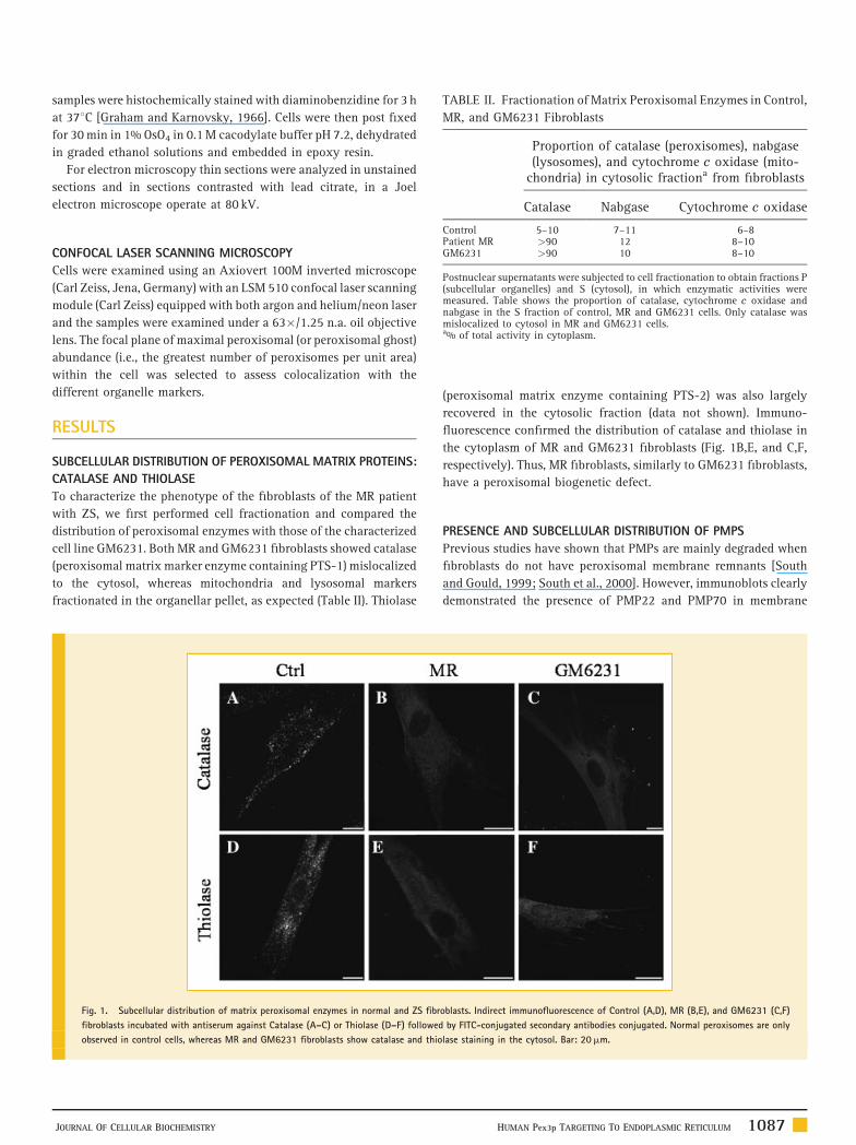
TABLE II. Fractionation of Matrix Peroxisomal Enzymes in Control,
MR, and GM6231 Fibroblasts
Proportion of catalase (peroxisomes), nabgase(lysosomes), and cytochrome c oxidase (mito-
chondria) in cytosolic fractiona from fibroblasts
Catalase Nabgase Cytochrome c oxidase
Control 5–10 7–11 6–8Patient MR >90 12 8–10GM6231 >90 10 8–10
Postnuclear supernatants were subjected to cell fractionation to obtain fractions P(subcellular organelles) and S (cytosol), in which enzymatic activities weremeasured. Table shows the proportion of catalase, cytochrome c oxidase andnabgase in the S fraction of control, MR and GM6231 cells. Only catalase wasmislocalized to cytosol in MR and GM6231 cells.a% of total activity in cytoplasm.
samples were histochemically stained with diaminobenzidine for 3 h
at 378C [Graham and Karnovsky, 1966]. Cells were then post fixed
for 30 min in 1% OsO4 in 0.1 M cacodylate buffer pH 7.2, dehydrated
in graded ethanol solutions and embedded in epoxy resin.
For electron microscopy thin sections were analyzed in unstained
sections and in sections contrasted with lead citrate, in a Joel
electron microscope operate at 80 kV.
CONFOCAL LASER SCANNING MICROSCOPY
Cells were examined using an Axiovert 100M inverted microscope
(Carl Zeiss, Jena, Germany) with an LSM 510 confocal laser scanning
module (Carl Zeiss) equipped with both argon and helium/neon laser
and the samples were examined under a 63�/1.25 n.a. oil objective
lens. The focal plane of maximal peroxisomal (or peroxisomal ghost)
abundance (i.e., the greatest number of peroxisomes per unit area)
within the cell was selected to assess colocalization with the
different organelle markers.
RESULTS
SUBCELLULAR DISTRIBUTION OF PEROXISOMAL MATRIX PROTEINS:
CATALASE AND THIOLASE
To characterize the phenotype of the fibroblasts of the MR patient
with ZS, we first performed cell fractionation and compared the
distribution of peroxisomal enzymes with those of the characterized
cell line GM6231. Both MR and GM6231 fibroblasts showed catalase
(peroxisomal matrix marker enzyme containing PTS-1) mislocalized
to the cytosol, whereas mitochondria and lysosomal markers
fractionated in the organellar pellet, as expected (Table II). Thiolase
Fig. 1. Subcellular distribution of matrix peroxisomal enzymes in normal and ZS fibr
fibroblasts incubated with antiserum against Catalase (A–C) or Thiolase (D–F) followed
observed in control cells, whereas MR and GM6231 fibroblasts show catalase and thio
JOURNAL OF CELLULAR BIOCHEMISTRY
(peroxisomal matrix enzyme containing PTS-2) was also largely
recovered in the cytosolic fraction (data not shown). Immuno-
fluorescence confirmed the distribution of catalase and thiolase in
the cytoplasm of MR and GM6231 fibroblasts (Fig. 1B,E, and C,F,
respectively). Thus, MR fibroblasts, similarly to GM6231 fibroblasts,
have a peroxisomal biogenetic defect.
PRESENCE AND SUBCELLULAR DISTRIBUTION OF PMPS
Previous studies have shown that PMPs are mainly degraded when
fibroblasts do not have peroxisomal membrane remnants [South
and Gould, 1999; South et al., 2000]. However, immunoblots clearly
demonstrated the presence of PMP22 and PMP70 in membrane
oblasts. Indirect immunofluorescence of Control (A,D), MR (B,E), and GM6231 (C,F)
by FITC-conjugated secondary antibodies conjugated. Normal peroxisomes are only
lase staining in the cytosol. Bar: 20mm.
HUMAN Pex3p TARGETING TO ENDOPLASMIC RETICULUM 1087
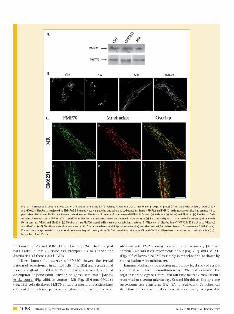
Fig. 2. Presence and subcellular localization of PMPs in normal and ZS fibroblasts. A: Western blot of membranes (100mg of protein) from organellar pellets of control, MR
and GM6231 fibroblasts subjected to SDS–PAGE. Immunoblots were carried out using antibodies against human PMP22 and PMP70, and secondary antibodies conjugated to
peroxidase. PMP22 and PMP70 are detected in both mutant fibroblasts. B: Immunofluorescence of PMP70 in Control (a), GM4340 (b), MR (c) and GM6231 (d) fibroblasts. Cells
were incubated with anti-PMP70 affinity purified antibodies. Normal peroxisomes are observed in control cells (a). Peroxisomal ghosts are shown in Zellweger syndrome cells
(b). In contrast, MR (c) and GM6231 (d) fibroblasts have PMP70 assembled in membranous tubular structures. C: Mitocondrial distribution of PMP70 in ZS fibroblasts. MR (a–c)
and GM6231 (d–f) fibroblasts were first incubated at 378C with the mitochondrial dye Mitotracker (b,e) and then treated for indirect immunofluorescence of PMP70 (a,d).
Fluorescence images obtained by confocal laser scanning microscopy show PMP70 containing tubules in MR and GM6231 fibroblasts colocalizing with mitochondria (c,f).
N: nucleus. Bar: 20mm.
fractions from MR and GM6231 fibroblasts (Fig. 2A). The finding of
both PMPs in our ZS fibroblasts prompted us to analyze the
distribution of these class I PMPs.
Indirect immunofluorescence of PMP70 showed the typical
pattern of peroxisomes in control cells (Fig. 2Ba) and peroxisomal
membrane ghosts in GM 4340 ZS fibroblasts, in which the original
description of peroxisomal membrane ghosts was made [Santos
et al., 1988b] (Fig. 2Bb). In contrast, MR (Fig. 2Bc) and GM6231
(Fig. 2Bd) cells displayed PMP70 in tubular membranous structures
different from classic peroxisomal ghosts. Similar results were
1088 HUMAN Pex3p TARGETING TO ENDOPLASMIC RETICULUM
obtained with PMP53 using laser confocal microscopy (data not
shown). Colocalization experiments of MR (Fig. 2Cc) and GM6231
(Fig. 2Cf) cells revealed PMP70 mainly in mitochondria, as shown by
colocalization with mitotracker.
Immunolabeling at the electron microscopy level showed results
congruent with the immunofluorescence. We first examined the
regular morphology of control and MR fibroblasts by conventional
transmission electron microscopy. Control fibroblasts display some
peroxisome-like structures (Fig. 3A, arrowheads). Cytochemical
detection of catalase makes peroxisomes easily recognizable
JOURNAL OF CELLULAR BIOCHEMISTRY
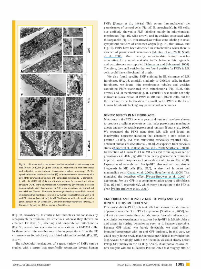
Fig. 3. Ultrastructural, cytochemical and immunoelectron microscopy stu-
dies. Control (A–E), MR (F–J), and GM6231(K–M) fibroblasts were fixed in situ
and subjected to conventional transmission electron microscopy (A,F,K),
cytochemistry for catalase detection (B) or immunoelectron microscopy with
anti-PMPs serum and peroxidase-anti-peroxidase detection (C–E, control; G–
J, MR; L,M GM6231). Only the ultrathin sections for conventional ultra-
structure (A,F,K) were counterstained. Cytochemistry (arrowheads in B) and
immunocytochemistry (arrowheads in C–E) show peroxisomes in control but
not in MR and GM6231 fibroblasts. Immunocytochemistry shows PMPs located
in mitochondrial membranes (arrows in G,H), small vesicles (thin arrows in G–I)
and ER cisternae (asterisk in J) in MR fibroblasts, as well as in small vesicles
(thin arrows in M), ER (asterisk in L) and thin membranous tubules in GM6231
fibroblasts (arrows in L,M). n: nucleus. Bar: 0.5mm.
(Fig. 3B, arrowheads). In contrast, MR fibroblasts did not show any
recognizable peroxisome-like structures, whereas they showed an
enlarged ER (Fig. 3F, asterisk) and long-tubular mitochondria
(Fig. 3F, arrow). We made similar observations in GM6231 cells.
In these cells, thin membranous tubular projections from the ER
cisternae were found closely associated with mitochondria (Fig. 3K,
arrow).
The subcellular localization of a great variety of PMPs can be
studied with a serum that specifically recognizes several human
JOURNAL OF CELLULAR BIOCHEMISTRY
PMPs [Santos et al., 1988a]. This serum immunolabeled the
peroxisomes of control cells (Fig. 3C–E, arrowheads). In MR cells,
our antibody showed a PMP-labeling mainly in mitochondrial
membranes (Fig. 3G, wide arrow), and in vesicles associated with
this organelle (Fig. 3H, thin arrows), as well as some labeling in small
cytoplasmic vesicles of unknown origin (Fig. 3G, thin arrow, and
Fig. 3I). PMPs have been described in mitochondria when there is
absence of peroxisomal membranes [Muntau et al., 2000; South
et al., 2000]. More recently, mitochondria derived vesicles
accounting for a novel vesicular traffic between this organelle
and peroxisomes was reported [Schumann and Subramani, 2008].
Therefore, the small vesicles that we found positive for PMPs in MR
cells could have mitochondrial origin.
We also found specific PMP staining in ER cisternae of MR
fibroblasts, (Fig. 3J, asterisk), similarly to GM6231 cells. In these
fibroblasts, we found thin membranous tubules and vesicles
containing PMPs associated with mitochondria (Fig. 3L,M, thin
arrows) and ER membranes (Fig. 3L, asterisk). These results not only
indicate mislocalization of PMPs in MR and GM6231 cells, but for
the first time reveal localization of a small pool of PMPs in the ER of
human fibroblasts lacking any peroxisomal membranes.
GENETIC DEFECTS IN MR FIBROBLASTS
Mutations in the PEX3 gene in yeast and humans have been shown
to produce a cellular phenotype that lacks peroxisome membrane
ghosts and any detectable peroxisomal remnant [South et al., 2000].
We sequenced the PEX3 gene from MR cells and found an
inactivating nonsense mutation that generates a stop codon at
position 53 (Fig. 4A), thus mimicking previously reported PEX3
deficient human cells [South et al., 2000]. As expected from previous
studies [Ghaedi et al., 2000a; Muntau et al., 2000; South et al., 2000],
transfection of human PEX3 in MR cells led to the appearance of
peroxisomes in 48 h (Fig. 4B). These newly generated peroxisomes
imported matrix enzymes such as catalase and thiolase (Fig. 4C,D).
Expression of recombinant Pex3p-GFP also restored peroxisome
biogenesis in MR cells (Fig. 4E,F), as described in yeast and
mammalian cells [Ghaedi et al., 2000b; Hoepfner et al., 2005]. This
mimicked the described effect [Voorn-Brouwer et al., 2001] of
expressing Pex16p-GFP in a complementation group 9 fibroblasts
(Fig. 4G and H, respectively), which carry a mutation in the PEX16
gene [Voorn-Brouwer et al., 2001].
TIME COURSE AND ER INVOLVEMENT OF Pex3p AND Pex16p
DRIVEN PEROXISOME BIOGENESIS
Previous studies in PEX3 deficient cells have shown reestablishment
of peroxisomes after 3 h of PEX3 expression [South et al., 2000], but
did not analyze shorter time periods. We performed similar nuclear
microinjection experiments to express Pex3p-GFP in MR fibroblasts
and assess its sorting behavior as soon as it became detectable.
Because GFP signal was barely detectable, we used indirect
immunofluorescence with an anti-GFP antibody. In this way, we
could easily detect newly made peroxisomes 4 h post-microinjection
(Fig. 5Ac,d). Strikingly, within the first hour, we could clearly detect
Pex3p-GFP mainly in the ER (Fig. 5Aa,b). Quantitative colocaliza-
tion analysis with the ER marker PDI indicated that roughly 70% of
HUMAN Pex3p TARGETING TO ENDOPLASMIC RETICULUM 1089
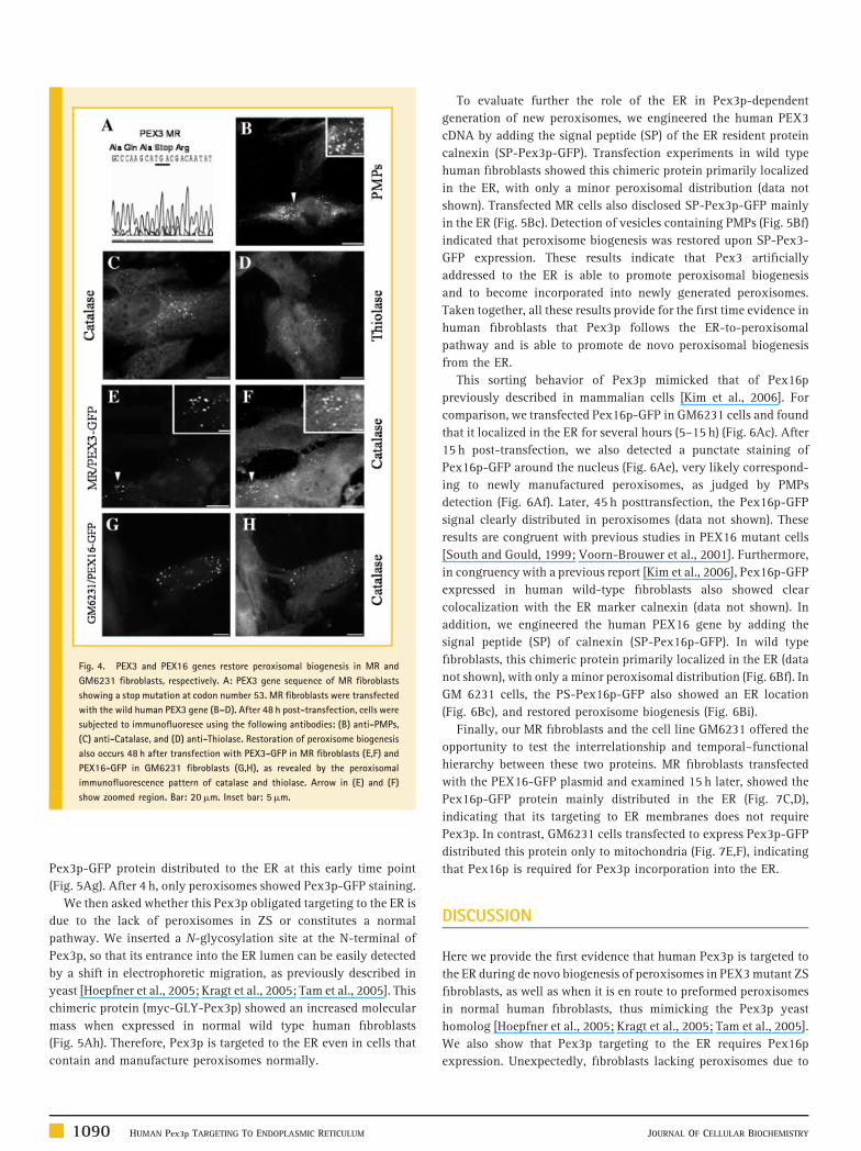
Fig. 4. PEX3 and PEX16 genes restore peroxisomal biogenesis in MR and
GM6231 fibroblasts, respectively. A: PEX3 gene sequence of MR fibroblasts
showing a stop mutation at codon number 53. MR fibroblasts were transfected
with the wild human PEX3 gene (B–D). After 48 h post-transfection, cells were
subjected to immunofluoresce using the following antibodies: (B) anti-PMPs,
(C) anti-Catalase, and (D) anti-Thiolase. Restoration of peroxisome biogenesis
also occurs 48 h after transfection with PEX3-GFP in MR fibroblasts (E,F) and
PEX16-GFP in GM6231 fibroblasts (G,H), as revealed by the peroxisomal
immunofluorescence pattern of catalase and thiolase. Arrow in (E) and (F)
show zoomed region. Bar: 20mm. Inset bar: 5mm.
Pex3p-GFP protein distributed to the ER at this early time point
(Fig. 5Ag). After 4 h, only peroxisomes showed Pex3p-GFP staining.
We then asked whether this Pex3p obligated targeting to the ER is
due to the lack of peroxisomes in ZS or constitutes a normal
pathway. We inserted a N-glycosylation site at the N-terminal of
Pex3p, so that its entrance into the ER lumen can be easily detected
by a shift in electrophoretic migration, as previously described in
yeast [Hoepfner et al., 2005; Kragt et al., 2005; Tam et al., 2005]. This
chimeric protein (myc-GLY-Pex3p) showed an increased molecular
mass when expressed in normal wild type human fibroblasts
(Fig. 5Ah). Therefore, Pex3p is targeted to the ER even in cells that
contain and manufacture peroxisomes normally.
1090 HUMAN Pex3p TARGETING TO ENDOPLASMIC RETICULUM
To evaluate further the role of the ER in Pex3p-dependent
generation of new peroxisomes, we engineered the human PEX3
cDNA by adding the signal peptide (SP) of the ER resident protein
calnexin (SP-Pex3p-GFP). Transfection experiments in wild type
human fibroblasts showed this chimeric protein primarily localized
in the ER, with only a minor peroxisomal distribution (data not
shown). Transfected MR cells also disclosed SP-Pex3p-GFP mainly
in the ER (Fig. 5Bc). Detection of vesicles containing PMPs (Fig. 5Bf)
indicated that peroxisome biogenesis was restored upon SP-Pex3-
GFP expression. These results indicate that Pex3 artificially
addressed to the ER is able to promote peroxisomal biogenesis
and to become incorporated into newly generated peroxisomes.
Taken together, all these results provide for the first time evidence in
human fibroblasts that Pex3p follows the ER-to-peroxisomal
pathway and is able to promote de novo peroxisomal biogenesis
from the ER.
This sorting behavior of Pex3p mimicked that of Pex16p
previously described in mammalian cells [Kim et al., 2006]. For
comparison, we transfected Pex16p-GFP in GM6231 cells and found
that it localized in the ER for several hours (5–15 h) (Fig. 6Ac). After
15 h post-transfection, we also detected a punctate staining of
Pex16p-GFP around the nucleus (Fig. 6Ae), very likely correspond-
ing to newly manufactured peroxisomes, as judged by PMPs
detection (Fig. 6Af). Later, 45 h posttransfection, the Pex16p-GFP
signal clearly distributed in peroxisomes (data not shown). These
results are congruent with previous studies in PEX16 mutant cells
[South and Gould, 1999; Voorn-Brouwer et al., 2001]. Furthermore,
in congruency with a previous report [Kim et al., 2006], Pex16p-GFP
expressed in human wild-type fibroblasts also showed clear
colocalization with the ER marker calnexin (data not shown). In
addition, we engineered the human PEX16 gene by adding the
signal peptide (SP) of calnexin (SP-Pex16p-GFP). In wild type
fibroblasts, this chimeric protein primarily localized in the ER (data
not shown), with only a minor peroxisomal distribution (Fig. 6Bf). In
GM 6231 cells, the PS-Pex16p-GFP also showed an ER location
(Fig. 6Bc), and restored peroxisome biogenesis (Fig. 6Bi).
Finally, our MR fibroblasts and the cell line GM6231 offered the
opportunity to test the interrelationship and temporal–functional
hierarchy between these two proteins. MR fibroblasts transfected
with the PEX16-GFP plasmid and examined 15 h later, showed the
Pex16p-GFP protein mainly distributed in the ER (Fig. 7C,D),
indicating that its targeting to ER membranes does not require
Pex3p. In contrast, GM6231 cells transfected to express Pex3p-GFP
distributed this protein only to mitochondria (Fig. 7E,F), indicating
that Pex16p is required for Pex3p incorporation into the ER.
DISCUSSION
Here we provide the first evidence that human Pex3p is targeted to
the ER during de novo biogenesis of peroxisomes in PEX3 mutant ZS
fibroblasts, as well as when it is en route to preformed peroxisomes
in normal human fibroblasts, thus mimicking the Pex3p yeast
homolog [Hoepfner et al., 2005; Kragt et al., 2005; Tam et al., 2005].
We also show that Pex3p targeting to the ER requires Pex16p
expression. Unexpectedly, fibroblasts lacking peroxisomes due to
JOURNAL OF CELLULAR BIOCHEMISTRY
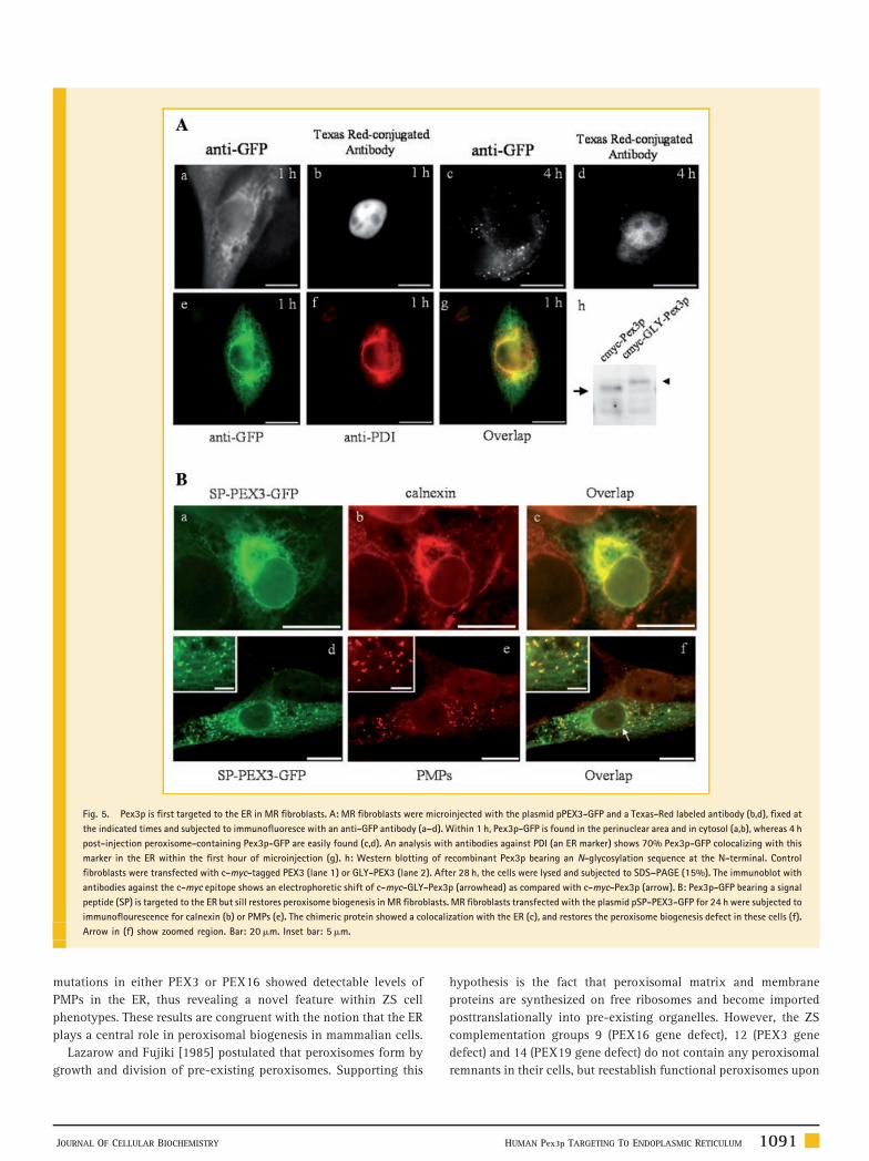
Fig. 5. Pex3p is first targeted to the ER in MR fibroblasts. A: MR fibroblasts were microinjected with the plasmid pPEX3-GFP and a Texas-Red labeled antibody (b,d), fixed at
the indicated times and subjected to immunofluoresce with an anti-GFP antibody (a–d). Within 1 h, Pex3p-GFP is found in the perinuclear area and in cytosol (a,b), whereas 4 h
post-injection peroxisome-containing Pex3p-GFP are easily found (c,d). An analysis with antibodies against PDI (an ER marker) shows 70% Pex3p-GFP colocalizing with this
marker in the ER within the first hour of microinjection (g). h: Western blotting of recombinant Pex3p bearing an N-glycosylation sequence at the N-terminal. Control
fibroblasts were transfected with c-myc-tagged PEX3 (lane 1) or GLY-PEX3 (lane 2). After 28 h, the cells were lysed and subjected to SDS–PAGE (15%). The immunoblot with
antibodies against the c-myc epitope shows an electrophoretic shift of c-myc-GLY-Pex3p (arrowhead) as compared with c-myc-Pex3p (arrow). B: Pex3p-GFP bearing a signal
peptide (SP) is targeted to the ER but sill restores peroxisome biogenesis in MR fibroblasts. MR fibroblasts transfected with the plasmid pSP-PEX3-GFP for 24 h were subjected to
immunoflourescence for calnexin (b) or PMPs (e). The chimeric protein showed a colocalization with the ER (c), and restores the peroxisome biogenesis defect in these cells (f).
Arrow in (f) show zoomed region. Bar: 20mm. Inset bar: 5mm.
mutations in either PEX3 or PEX16 showed detectable levels of
PMPs in the ER, thus revealing a novel feature within ZS cell
phenotypes. These results are congruent with the notion that the ER
plays a central role in peroxisomal biogenesis in mammalian cells.
Lazarow and Fujiki [1985] postulated that peroxisomes form by
growth and division of pre-existing peroxisomes. Supporting this
JOURNAL OF CELLULAR BIOCHEMISTRY
hypothesis is the fact that peroxisomal matrix and membrane
proteins are synthesized on free ribosomes and become imported
posttranslationally into pre-existing organelles. However, the ZS
complementation groups 9 (PEX16 gene defect), 12 (PEX3 gene
defect) and 14 (PEX19 gene defect) do not contain any peroxisomal
remnants in their cells, but reestablish functional peroxisomes upon
HUMAN Pex3p TARGETING TO ENDOPLASMIC RETICULUM 1091
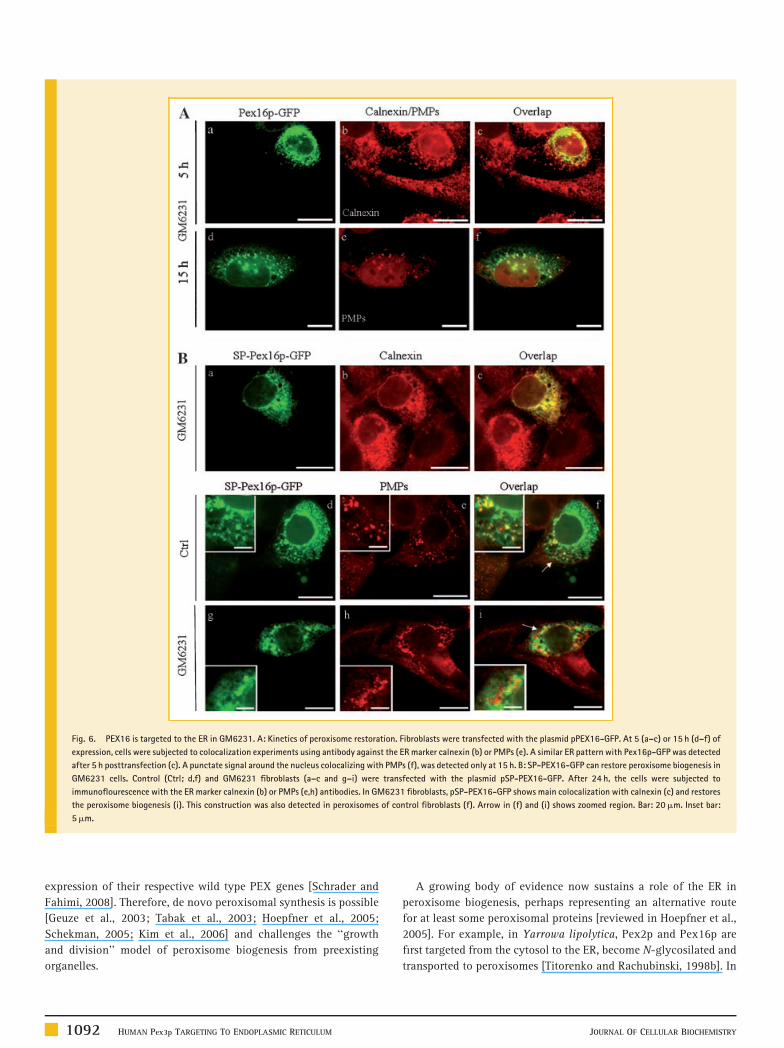
Fig. 6. PEX16 is targeted to the ER in GM6231. A: Kinetics of peroxisome restoration. Fibroblasts were transfected with the plasmid pPEX16-GFP. At 5 (a–c) or 15 h (d–f) of
expression, cells were subjected to colocalization experiments using antibody against the ER marker calnexin (b) or PMPs (e). A similar ER pattern with Pex16p-GFP was detected
after 5 h posttransfection (c). A punctate signal around the nucleus colocalizing with PMPs (f), was detected only at 15 h. B: SP-PEX16-GFP can restore peroxisome biogenesis in
GM6231 cells. Control (Ctrl; d,f) and GM6231 fibroblasts (a–c and g–i) were transfected with the plasmid pSP-PEX16-GFP. After 24 h, the cells were subjected to
immunoflourescence with the ER marker calnexin (b) or PMPs (e,h) antibodies. In GM6231 fibroblasts, pSP-PEX16-GFP shows main colocalization with calnexin (c) and restores
the peroxisome biogenesis (i). This construction was also detected in peroxisomes of control fibroblasts (f). Arrow in (f) and (i) shows zoomed region. Bar: 20mm. Inset bar:
5mm.
expression of their respective wild type PEX genes [Schrader and
Fahimi, 2008]. Therefore, de novo peroxisomal synthesis is possible
[Geuze et al., 2003; Tabak et al., 2003; Hoepfner et al., 2005;
Schekman, 2005; Kim et al., 2006] and challenges the ‘‘growth
and division’’ model of peroxisome biogenesis from preexisting
organelles.
1092 HUMAN Pex3p TARGETING TO ENDOPLASMIC RETICULUM
A growing body of evidence now sustains a role of the ER in
peroxisome biogenesis, perhaps representing an alternative route
for at least some peroxisomal proteins [reviewed in Hoepfner et al.,
2005]. For example, in Yarrowa lipolytica, Pex2p and Pex16p are
first targeted from the cytosol to the ER, become N-glycosilated and
transported to peroxisomes [Titorenko and Rachubinski, 1998b]. In
JOURNAL OF CELLULAR BIOCHEMISTRY
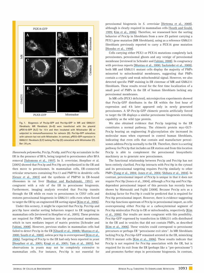
Fig. 7. Biogenesis of Pex3p-GFP and Pex16p-GFP in MR and GM6231
fibroblasts. MR fibroblasts (A–D) were transfected with the plasmid
pPEX16-GFP (A,C) for 15 h and then incubated with Mitotracker (B) or
subjected to immunofluorescence for calnexin (D). Pex16p-GFP colocalizes
with calnexin but not with Mitotracker. In contrast, pPEX3-GFP expression in
GM6231 fibroblasts (E,F) lacking Pex16p (E) colocalized with Mitotracker (F).
Bar: 20mm.
Hansenula polymorha, Pex3p, Pex8p, and Pex14p accumulate in the
ER in the presence of BFA, being targeted to peroxisomes after BFA
removal [Salomons et al., 1997]. In S. cerevisiae, Hoepfner et al.
[2005] showed that Pex3p and Pex19p are synthesized in the ER and
then move to peroxisomes. In mammalian cells, ER-connected
reticular structures containing Pex13 and PMP70 in dendritic cells
[Geuze et al., 2003] and the synthesis of PMP50 in ER-bound
ribosomes in rat liver [Bodnar and Rachubinski, 1991], are
congruent with a role of the ER in peroxisome biogenesis.
Furthermore, imaging analysis revealed that Pex16p transits
through the ER while en route to peroxisomes in COS7 cells and
restores peroxisomal biogenesis in GM6231 cells even when forced
to target the ER by an engineered ER sorting signal [Kim et al., 2006].
Under this scenery, it might be expected that Pex3p, Pex16p and
pex19p have similar sorting behaviors respect to ER targeting in
mammalian cells [reviewed in Hoepfner et al., 2005]. These proteins
are required for PMPs insertion into the peroxisomal membrane,
which in turn mediates import of matrix proteins [Schrader and
Fahimi, 2008]. However, previous studies in mammalian cells had
failed to detect Pex3p in the ER [Ghaedi et al., 2000b; Muntau et al.,
2000; South et al., 2000], unless pex16p is overexpressed [Kim et al.,
2006]. Targeting of Pex3p to the ER had only been reported in yeast
[Hoepfner et al., 2005; Kragt et al., 2005; Tam et al., 2005], but
observations in yeasts may not be completely extensive to
mammalian cells. For instance, Pex16p is not essential for
JOURNAL OF CELLULAR BIOCHEMISTRY
peroxisomal biogenesis in S. cerevisiae [Hettema et al., 2000],
although is clearly required in mammalian cells [South and Gould,
1999; Kim et al., 2006]. Therefore, we reassessed here the sorting
behavior of Pex3p in fibroblasts from a new ZS patient carrying a
PEX3 gene mutation (MR fibroblasts), using as a reference GM6231
fibroblasts previously reported to carry a PEX16 gene mutation
[Honsho et al., 1998].
Cells carrying either PEX3 or PEX16 mutations completely lack
peroxisomes, peroxisomal ghosts and any vestige of peroxisomal
membrane [reviewed in Schrader and Fahimi, 2008]. In congruency
with previous reports [Muntau et al., 2000; Sacksteder et al., 2000],
both MR and GM6231 mutant cells display the majority of PMPs
missorted to mitochondrial membranes, suggesting that PMPs
contain a cryptic and weak mitochondrial signal. However, we also
detected specific PMP staining in ER cisternae of MR and GM6231
fibroblasts. These results reveal for the first time localization of a
small pool of PMPs in the ER of human fibroblasts lacking any
peroxisomal membranes.
In MR cells (PEX3 deficient), microinjection experiments showed
that Pex3p-GFP distributes in the ER within the first hour of
expression and 4 h later appeared only in newly generated
peroxisomes. A SP-Pex3p-GFP chimeric protein artificially forced
to target the ER displays a similar peroxisome biogenesis restoring
capability as the wild type protein.
We also obtained evidence that Pex3p targeting to the ER
constitutes a normal pathway. The chimeric protein myc-GLY-
Pex3p bearing an engineering N-glycosylation site increased its
molecular mass when expressed in control human fibroblasts,
indicating that even cells that contain and manufacture peroxi-
somes address Pex3p normally to the ER. Therefore, there is a sorting
pathway for Pex3p that includes an ER station and from this location
Pex3p is able to complement the peroxisomal biosynthetic
machinery as to generate new peroxisomes.
The functional relationship between Pex3p and Pex16p has not
been entirely clarified. Pex16p interacts with Pex19p in the cytosol
and its peroxisomal targeting requires Pex19p similarly to other
PMPs [Fang et al., 2004; Jones et al., 2004; Shibata et al., 2004]. In
contrast, peroxisomal import of Pex3p is unique in that it does not
require Pex19p [Jones et al., 2004], although a Pex16p and Pex19p-
dependent peroxisomal import of this peroxin has recently been
shown by Matsuzaki and Fujiki [2008]. Because Pex3p acts as a
docking factor for Pex19p it could be predicted that is required for
Pex16p peroxisomal import. However, there is evidence suggesting
Pex16p functions upstream of Pex3p in peroxisomal import, as cells
overexpressing either Pex16p or a carboxyterminal segment of
Pex16p mislocalize Pex3p to ER or mitochondria, respectively [Kim
et al., 2006]. Our results are more congruent with this possibility.
Pex16p-GFP expressed by transfection in GM6231 cells distributed
in the ER and in vesicles that did not contain PMPs, as described
[Kim et al., 2006]. These vesicles could correspond to peroxisome
precursors or perhaps ER ‘‘peroxisome exit sites’’. In MR fibroblasts
lacking Pex3p, Pex16p-GFP remained arrested in the ER, mimicking
PEX19 mutant cells [Kim et al., 2006]. These results indicate that
Pex3p is not required for Pex16p association with the ER, but is
required for its exit from the ER (perhaps like a ‘‘pre-peroxisome’’)
and promotes further steps in peroxisome biogenesis. In contrast,
HUMAN Pex3p TARGETING TO ENDOPLASMIC RETICULUM 1093
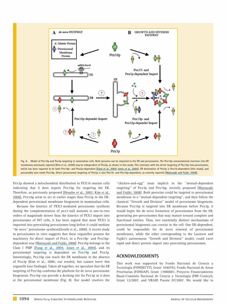
Fig. 8. Model of Pex16p and Pex3p targeting in mammalian cells. Both peroxins can be imported to the ER and peroxisomes. The Pex16p cotranslational insertion into ER
membranes previously reported [Kim et al., 2006] may be independent of Pex3p, as shown in this study. This contrasts with the direct targeting of Pex16p into peroxisomes,
which has been reported to be both Pex19p- and Pex3p-dependent [Fang et al., 2004; Jones et al., 2004]. ER destination of Pex3p is Pex16-dependent (this study), and
presumably also needs Pex19p. Direct peroxisomal targeting of Pex3p is also Pex19- and Pex16p-dependent, as recently reported [Matsuzaki and Fujiki, 2008].
Pex3p showed a mitochondrial distribution in PEX16 mutant cells
indicating that it does require Pex16p for targeting the ER.
Therefore, as previously proposed [Honsho et al., 2002; Kim et al.,
2006], Pex16p seem to act at earlier stages than Pex3p in the ER-
dependent peroxisomal membrane biogenesis in mammalian cells.
Because the kinetics of PEX3-mediated peroxisome synthesis
during the complementation of pex3-null mutants is one-to-two
orders of magnitude slower than the kinetics of PEX3 import into
peroxisomes of WT cells, it has been argued that most PEX3 is
imported into preexisting peroxisomes long before it could mediate
‘‘de novo’’ peroxisome synthesis[South et al., 2000]. A recent study
in peroxisomes in vitro suggests that these organelles possess the
machinery for direct import of Pex3, in a Pex19p- and Pex16p-
dependent way [Matsuzaki and Fujiki, 2008]. Pex16p belongs to the
Class I PMP [Fang et al., 2004; Jones et al., 2004], and its
peroxisomal targeting is dependent on Pex19p and Pex3p.
Interestingly, Pex16p can reach the ER membrane in the absence
of Pex3p [Kim et al., 2006; our results], but cannot leave this
organelle (our findings). Taken all together, we speculate that the ER
targeting of Pex16p conforms the platform for de novo peroxisome
biogenesis. Pex16p can provide a docking site for Pex3p as it does
at the peroxisomal membrane (Fig. 8). Our model resolves the
1094 HUMAN Pex3p TARGETING TO ENDOPLASMIC RETICULUM
‘‘chicken-and-egg’’ issue implicit in the ‘‘mutual-dependent
targeting’’ of Pex3p and Pex16p, recently proposed [Matsuzaki
and Fujiki, 2008]. Both peroxins could be targeted to peroxisomal
membrane in a ‘‘mutual-dependent targeting’’, and then follow the
classical ‘‘Growth and Division’’ model of peroxisome biogenesis.
Because Pex16p is targeted into ER membrane before Pex3p, it
would begin the de novo formation of peroxisomes from the ER,
generating pre-peroxisomes that may mature toward complete and
functional entities. Thus, two essentially distinct mechanisms of
peroxisomal biogenesis can coexist in the cell. One ER-dependent,
could be responsible for de novo renewal of peroxisomal
membranes, while the other corresponding to the Lazarow and
Fujiki’s autonomous ‘‘Growth and Division’’ model, could exert
rapid and direct protein import into preexisting peroxisomes.
ACKNOWLEDGMENTS
This work was supported by Fondo Nacional de Ciencia yTecnologıa (FONDECYT), Grant 1040792, Fondo Nacional de AreasPrioritarias (FONDAP), Grant 13980001, Proyecto FinanciamientoBasal-Comision Nacional de Ciencia y Tecnologıa (PBF-Conicyt),Grant 12/2007, and VRAID Puente 07/2007. We would like to
JOURNAL OF CELLULAR BIOCHEMISTRY
thank Ms. A.B. Moser and Dr. H.W. Moser, Kennedy KriegerInstitute, Baltimore, USA, for their helpful discussions and thecomplementation studies; Dr. Y. Gluzman, from the Department ofMicrobiology and Kaplan Comprehensive Cancer Center, New YorkUniversity School of Medicine, New York, USA, who kindlyprovided the recombinant SV40 adenovirus; Dr. S.J. Gould, fromthe Department of Biological Chemistry, The Johns HopkinsUniversity School of Medicine, USA, for his generous gift of thepcDNA3-PEX3 plasmid; Dr. T. Imanaka, from the Department ofBiological Chemistry, Graduate School of Medicine and Pharma-ceutical Sciences, University of Toyama, Japan, for his generousgift of the polyclonal antibody against Thiolase, Dr. Estela Andres(Department of Cellular and Molecular Biology, Faculty ofBiological Sciences, Catholic University of Chile), for his generousgift of the pCS2 R MT plasmid and Dr. Gillian Small, CityUniversity of New York for her helpful discussions.
REFERENCES
Bodnar AG, Rachubinski RA. 1991. Characterization of the integral mem-brane polypeptides of rat liver peroxisomes isolated from untreated andclofibrate-treated rats. Biochem Cell Biol 69:499–508.
Brosius U, Gartner J. 2002. Cellular and molecular aspects of Zellwegersyndrome and other peroxisome biogenesis disorders. Cell Mol Life Sci59:1058–1069.
Cancino J, Torrealba C, Soza A, Yuseff MI, Gravotta D, Henklein P, Rodri-guez-Boulan E, Gonzalez A. 2007. Antibody to AP1B adaptor blocks bio-synthetic and recycling routes of basolateral proteins at recyclingendosomes. Mol Biol Cell 18:4872–4884.
Cooperstein SJ, Lazarow A. 1951. A microspectrophotometric method for thedetermination of cytochrome oxidase. J Biol Chem 189:665–670.
Eitzen GA, Szilard RK, Rachubinski RA. 1997. Enlarged peroxisomes arepresent in oleic acid-grown Yarrowia lipolytica overexpressing the PEX16gene encoding an intraperoxisomal peripheral membrane peroxin. J Cell Biol137:1265–1278.
Fang Y, Morrell JC, Jones JM, Gould SJ. 2004. PEX3 functions as a PEX19docking factor in the import of class I peroxisomal membrane proteins. J CellBiol 164:863–875.
Fujiki Y, Hubbard AL, Fowler S, Lazarow PB. 1982. Isolation of intracellularmembranes by means of sodium carbonate treatment: Application to endo-plasmic reticulum. J Cell Biol 93:97–102.
Geuze HJ, Murk JL, Stroobants AK, Griffith JM, Kleijmeer MJ, Koster AJ,Verkleij AJ, Distel B, Tabak HF. 2003. Involvement of the endoplasmicreticulum in peroxisome formation. Mol Biol Cell 14:2900–2907.
Ghaedi K, Honsho M, Shimozawa N, Suzuki Y, Kondo N, Fujiki Y. 2000a.PEX3 is the causal gene responsible for peroxisome membrane assembly-defective Zellweger syndrome of complementation group G. Am J HumGenet 67:976–981.
Ghaedi K, Tamura S, Okumoto K, Matsuzono Y, Fujiki Y. 2000b. The peroxinpex3p initiates membrane assembly in peroxisome biogenesis. Mol Biol Cell11:2085–2102.
Goldfischer S, Moore CL, Johnson AB, Spiro AJ, Valsamis MP, WisniewskiHK, Ritch RH, Norton WT, Rapin I, Gartner LM. 1973. Peroxisomal andmitochondrial defects in the cerebro-hepato-renal syndrome. Science182:62–64.
Graham RC, Jr., Karnovsky MJ. 1966. The early stages of absorption ofinjected horseradish peroxidase in the proximal tubules of mouse kidney:Ultrastructural cytochemistry by a new technique. J Histochem Cytochem14:291–302.
Heiland I, Erdmann R. 2005. Biogenesis of peroxisomes. Topogenesis of theperoxisomal membrane and matrix proteins. FEBS J 272:2362–2372.
JOURNAL OF CELLULAR BIOCHEMISTRY
Hettema EH, Girzalsky W, van Den Berg M, Erdmann R, Distel B. 2000.Saccharomyces cerevisiae pex3p and pex19p are required for proper localiza-tion and stability of peroxisomal membrane proteins. EMBO J 19:223–233.
Hoepfner D, Schildknegt D, Braakman I, Philippsen P, Tabak HF. 2005.Contribution of the endoplasmic reticulum to peroxisome formation. Cell122:85–95.
Honsho M, Tamura S, Shimozawa N, Suzuki Y, Kondo N, Fujiki Y. 1998.Mutation in PEX16 is causal in the peroxisome-deficient Zellweger syn-drome of complementation group D. Am J Hum Genet 63:1622–1630.
Honsho M, Hiroshige T, Fujiki Y. 2002. The membrane biogenesis peroxinPex16p. Topogenesis and functional roles in peroxisomal membrane assem-bly. J Biol Chem 277:44513–44524.
Jones JM, Morrell JC, Gould SJ. 2001. Multiple distinct targeting signals inintegral peroxisomal membrane proteins. J Cell Biol 153:1141–1150.
Jones JM, Morrell JC, Gould SJ. 2004. PEX19 is a predominantly cytosolicchaperone and import receptor for class 1 peroxisomal membrane proteins.J Cell Biol 164:57–67.
Kim PK, Mullen RT, Schumann U, Lippincott-Schwartz J. 2006. The originand maintenance of mammalian peroxisomes involves a de novo PEX16-dependent pathway from the ER. J Cell Biol 173:521–532.
Kragt A, Voorn-Brouwer T, van den Berg M, Distel B. 2005. Endoplasmicreticulum-directed Pex3p routes to peroxisomes and restores peroxisomeformation in a Saccharomyces cerevisiae pex3Delta strain. J Biol Chem280:34350–34357.
Lazarow PB, Fujiki Y. 1985. Biogenesis of peroxisomes. Annu Rev Cell Biol1:489–530.
Leighton F, Poole B, Beaufay H, Baudhuin P, Coffey JW, Fowler S, De Duve C.1968. The large-scale separation of peroxisomes, mitochondria, and lyso-somes from the livers of rats injected with triton WR-1339. Improvedisolation procedures, automated analysis, biochemical and morphologicalproperties of fractions. J Cell Biol 37:482–513.
Mardones G, Gonzalez A. 2003. Selective plasma membrane permeabiliza-tion by freeze-thawing and immunofluorescence epitope access to determinethe topology of intracellular membrane proteins. J Immunol Methods275:169–177.
Matsuzaki T, Fujiki Y. 2008. The peroxisomal membrane protein importreceptor Pex3p is directly transported to peroxisomes by a novel Pex19p- andPex16p-dependent pathway. J Cell Biol 183:1275–1286.
Mullen RT, Trelease RN. 2006. The ER-peroxisome connection in plants:Development of the ‘‘ER semi-autonomous peroxisome maturation andreplication’’ model for plant peroxisome biogenesis. Biochim Biophys Acta1763:1655–1668.
Muntau AC, Mayerhofer PU, Paton BC, Kammerer S, Roscher AA. 2000.Defective peroxisome membrane synthesis due to mutations in human PEX3causes Zellweger syndrome, complementation group G. Am J Hum Genet67:967–975.
Platta HW, Erdmann R. 2007. The peroxisomal protein import machinery.FEBS Lett 581:2811–2819.
Sacksteder KA, Jones JM, South ST, Li X, Liu Y, Gould SJ. 2000. PEX19 bindsmultiple peroxisomal membrane proteins, is predominantly cytoplasmic, andis required for peroxisome membrane synthesis. J Cell Biol 148:931–944.
Salomons FA, van der Klei IJ, Kram AM, Harder W, Veenhuis M. 1997.Brefeldin A interferes with peroxisomal protein sorting in the yeast Hanse-nula polymorpha. FEBS Lett 411:133–139.
Santos MJ, Imanaka T, Shio H, Lazarow PB. 1988a. Peroxisomal integralmembrane proteins in control and Zellweger fibroblasts. J Biol Chem263:10502–10509.
Santos MJ, Imanaka T, Shio H, Small GM, Lazarow PB. 1988b. Peroxisomalmembrane ghosts in Zellweger syndrome-aberrant organelle assembly.Science 239:1536–1538.
HUMAN Pex3p TARGETING TO ENDOPLASMIC RETICULUM 1095
Santos MJ, Kawada ME, Espeel M, Figueroa C, Alvarez A, Hidalgo U, Metz C.1994. Characterization of human peroxisomal membrane proteins. J BiolChem 269:24890–24896.
Santos MJ, Henderson SC, Moser AB, Moser HW, Lazarow PB. 2000.Peroxisomal ghosts are intracellular structures distinct from lysosomalcompartments in Zellweger syndrome: A confocal laser scanning microscopystudy. Biol Cell 92:85–94.
Schekman R. 2005. Peroxisomes: Another branch of the secretory pathway?Cell 122:1–2.
Schrader M, Fahimi HD. 2008. The peroxisome: Still a mysterious organelle.Histochem Cell Biol 129:421–440.
Schumann U, Subramani S. 2008. Special delivery from mitochondria toperoxisomes. Trends Cell Biol 18:253–256.
Sellinger OZ, Beaufay H, Jacques P, Doyen A, De Duve C. 1960. Tissuefractionation studies. 15. Intracellular distribution and properties of beta-N-acetylglucosaminidase and beta-galactosidase in rat liver. Biochem J 74:450–456.
Shibata H, Kashiwayama Y, Imanaka T, Kato H. 2004. Domain architectureand activity of human Pex19p, a chaperone-like protein for intracellulartrafficking of peroxisomal membrane proteins. J Biol Chem 279:38486–38494.
Shimozawa N, Tsukamoto T, Nagase T, Takemoto Y, Koyama N, Suzuki Y,Komori M, Osumi T, Jeannette G, Wanders RJ, Kondo N. 2004.Identification of a new complementation group of the peroxisomebiogenesis disorders and PEX14 as the mutated gene. Hum Mutat 23:552–558.
South ST, Gould SJ. 1999. Peroxisome synthesis in the absence of preexistingperoxisomes. J Cell Biol 144:255–266.
1096 HUMAN Pex3p TARGETING TO ENDOPLASMIC RETICULUM
South ST, Sacksteder KA, Li X, Liu Y, Gould SJ. 2000. Inhibitors of COPI andCOPII do not block PEX3-mediated peroxisome synthesis. J Cell Biol149:1345–1360.
Soza A, Norambuena A, Cancino J, de la Fuente E, Henklein P, Gonzalez A.2004. Sorting competition with membrane-permeable peptides in intactepithelial cells revealed discrimination of transmembrane proteins not onlyat the trans-Golgi network but also at pre-Golgi stages. J Biol Chem279:17376–17383.
Tabak HF, Murk JL, Braakman I, Geuze HJ. 2003. Peroxisomes start their lifein the endoplasmic reticulum. Traffic 4:512–518.
Tabak HF, van der Zand A, Braakman I. 2008. Peroxisomes: Minted by the ER.Curr Opin Cell Biol 20:393–400.
Tam YY, Fagarasanu A, Fagarasanu M, Rachubinski RA. 2005. Pex3pinitiates the formation of a preperoxisomal compartment from a subdomainof the endoplasmic reticulum in Saccharomyces cerevisiae. J Biol Chem280:34933–34939.
Titorenko VI, Mullen RT. 2006. Peroxisome biogenesis: The peroxisomalendomembrane system and the role of the ER. J Cell Biol 174:11–17.
Titorenko VI, Rachubinski RA. 1998a. The endoplasmic reticulum plays anessential role in peroxisome biogenesis. Trends Biochem Sci 23:231–233.
Titorenko VI, Rachubinski RA. 1998b. Mutants of the yeast Yarrowialipolytica defective in protein exit from the endoplasmic reticulum are alsodefective in peroxisome biogenesis. Mol Cell Biol 18:2789–2803.
Van Doren K, Gluzman Y. 1984. Efficient transformation of human fibroblastsby adenovirus-simian virus 40 recombinants. Mol Cell Biol 4:1653–1656.
Voorn-Brouwer T, Kragt A, Tabak HF, Distel B. 2001. Peroxisomal membraneproteins are properly targeted to peroxisomes in the absence of COPI- andCOPII-mediated vesicular transport. J Cell Sci 114:2199–2204.
JOURNAL OF CELLULAR BIOCHEMISTRY
Related Documents