Original article Persistent cognitive and motor deficits after successful antimalarial treatment in murine cerebral malaria Minxian Dai a , Sandra E. Reznik a , David C. Spray b,c , Louis M. Weiss c,d , Herbert B. Tanowitz c,d , Maria Gulinello b,e , Mahalia S. Desruisseaux c,d, * a Department of Pharmaceutical Sciences, College of Pharmacy and Allied Health Professions, St Johns University, Queens, NY 11439, USA b The Dominick P. Purpura Department of Neuroscience, Albert Einstein College of Medicine, Bronx, NY 10461, USA c Department of Medicine, Albert Einstein College of Medicine, Bronx, NY 10461, USA d Department of Pathology, Albert Einstein College of Medicine, Yeshiva University, 1300 Morris Park Avenue, Forchheimer 504, Bronx, NY 10461, USA e Behavioral Core Facility, Albert Einstein College of Medicine, Bronx, NY 10461, USA Received 13 May 2010; accepted 13 August 2010 Available online 26 August 2010 Abstract Human cerebral malaria causes neurological and behavioral deficits which persist long after resolution of infection and clearance of parasites with antimalarial drugs. Previously, we demonstrated that during active infection, mice with cerebral malaria demonstrated negative behavioral outcomes. Here we used a chloroquine treatment model of cerebral malaria to determine whether these abnormal outcomes would be persistent in the mouse model. C57BL/6 mice were infected with Plasmodium berghei ANKA, and treated for ten days. After cessation of chloroquine, a comprehensive assessment of cognitive and motor function demonstrated persistence of abnormal behavioral outcomes, 10 days after successful eradication of parasites. Furthermore, these deficits were still evident forty days after cessation of chloroquine, indicating persistence long after successful treatment, a hallmark feature of human cerebral malaria. Thus, cognitive tests similar to those used in these mouse studies could facilitate the development of adjunctive therapies that can ameliorate adverse neurological outcomes in human cerebral malaria. Published by Elsevier Masson SAS on behalf of the Institut Pasteur. Keywords: Plasmodium berghei ANKA; Object recognition; Spatial memory; Motor coordination; Brain pathology; Chloroquine treatment 1. Introduction Cerebral malaria (CM) in humans is a severe neurological manifestation of Plasmodium falciparum infection [1,2], which results in neurological and behavioral deficits that span a wide range of behavioral domains and persist long after the infection has been successfully eradicated with antimalarial drugs [3e12]. It is estimated that 20e25% of children with successfully treated CM demonstrate residual neurological or cognitive deficits, including memory disturbances, speech and language difficul- ties, disorders of concentration and attention, seizures, visuo- spatial and motor deficits [3e12]. These deficits have been determined to persist after cessation of therapy, with follow-up periods extending to 9 years after infection [3e12]. Rodent models have been critical in elucidating the underlying mechanisms of CM and in the development of therapeutic agents that might salvage CNS function in patients with CM [13]. Susceptible mouse strains such as C57BL/6 infected with Plas- modium berghei ANKA (PbA) have been developed to investigate the pathogenesis of CM because they exhibit similar characteris- tics to human CM [13e23], including hemorrhage, axonal and neuronal damage [16,24e30], as well as behavioral and neuro- logical impairments during active infection [12,19,22,31]. To date, however, there have been no studies of negative cognitive and Abbreviations: PbA, Plasmodium berghei ANKA; CQ, chloroquine; CM, Cerebral malaria; RBC, red blood cell; cm, centimeter; dpi, days post- infection. * Corresponding author. Department of Pathology, Albert Einstein College of Medicine, Yeshiva University, 1300 Morris Park Avenue, Forchheimer 504, Bronx, NY 10461, USA. Tel.: þ1 718 430 4070; fax: þ1 718 430 8543. E-mail address: [email protected] (M.S. Desruisseaux). 1286-4579/$ - see front matter Published by Elsevier Masson SAS on behalf of the Institut Pasteur. doi:10.1016/j.micinf.2010.08.006 Microbes and Infection 12 (2010) 1198e1207 www.elsevier.com/locate/micinf

Welcome message from author
This document is posted to help you gain knowledge. Please leave a comment to let me know what you think about it! Share it to your friends and learn new things together.
Transcript
Microbes and Infection 12 (2010) 1198e1207www.elsevier.com/locate/micinf
Original article
Persistent cognitive and motor deficits after successful antimalarialtreatment in murine cerebral malaria
Minxian Dai a, Sandra E. Reznik a, David C. Spray b,c, Louis M. Weiss c,d, Herbert B. Tanowitz c,d,Maria Gulinello b,e, Mahalia S. Desruisseaux c,d,*
aDepartment of Pharmaceutical Sciences, College of Pharmacy and Allied Health Professions, St Johns University, Queens, NY 11439, USAb The Dominick P. Purpura Department of Neuroscience, Albert Einstein College of Medicine, Bronx, NY 10461, USA
cDepartment of Medicine, Albert Einstein College of Medicine, Bronx, NY 10461, USAdDepartment of Pathology, Albert Einstein College of Medicine, Yeshiva University, 1300 Morris Park Avenue, Forchheimer 504, Bronx, NY 10461, USA
eBehavioral Core Facility, Albert Einstein College of Medicine, Bronx, NY 10461, USA
Received 13 May 2010; accepted 13 August 2010
Available online 26 August 2010
Abstract
Human cerebral malaria causes neurological and behavioral deficits which persist long after resolution of infection and clearance of parasiteswith antimalarial drugs. Previously, we demonstrated that during active infection, mice with cerebral malaria demonstrated negative behavioraloutcomes. Here we used a chloroquine treatment model of cerebral malaria to determine whether these abnormal outcomes would be persistentin the mouse model. C57BL/6 mice were infected with Plasmodium berghei ANKA, and treated for ten days. After cessation of chloroquine,a comprehensive assessment of cognitive and motor function demonstrated persistence of abnormal behavioral outcomes, 10 days aftersuccessful eradication of parasites. Furthermore, these deficits were still evident forty days after cessation of chloroquine, indicating persistencelong after successful treatment, a hallmark feature of human cerebral malaria. Thus, cognitive tests similar to those used in these mouse studiescould facilitate the development of adjunctive therapies that can ameliorate adverse neurological outcomes in human cerebral malaria.Published by Elsevier Masson SAS on behalf of the Institut Pasteur.
Keywords: Plasmodium berghei ANKA; Object recognition; Spatial memory; Motor coordination; Brain pathology; Chloroquine treatment
1. Introduction
Cerebral malaria (CM) in humans is a severe neurologicalmanifestation of Plasmodium falciparum infection [1,2], whichresults in neurological and behavioral deficits that span a widerange of behavioral domains and persist long after the infectionhas been successfully eradicated with antimalarial drugs [3e12].It is estimated that 20e25% of children with successfully treated
Abbreviations: PbA, Plasmodium berghei ANKA; CQ, chloroquine; CM,
Cerebral malaria; RBC, red blood cell; cm, centimeter; dpi, days post-
infection.
* Corresponding author. Department of Pathology, Albert Einstein College
of Medicine, Yeshiva University, 1300 Morris Park Avenue, Forchheimer 504,
Bronx, NY 10461, USA. Tel.: þ1 718 430 4070; fax: þ1 718 430 8543.
E-mail address: [email protected] (M.S. Desruisseaux).
1286-4579/$ - see front matter Published by Elsevier Masson SAS on behalf of th
doi:10.1016/j.micinf.2010.08.006
CM demonstrate residual neurological or cognitive deficits,including memory disturbances, speech and language difficul-ties, disorders of concentration and attention, seizures, visuo-spatial and motor deficits [3e12]. These deficits have beendetermined to persist after cessation of therapy, with follow-upperiods extending to 9 years after infection [3e12].
Rodentmodels have been critical in elucidating the underlyingmechanisms of CM and in the development of therapeutic agentsthat might salvage CNS function in patients with CM [13].Susceptible mouse strains such as C57BL/6 infected with Plas-modiumbergheiANKA (PbA) have been developed to investigatethe pathogenesis of CM because they exhibit similar characteris-tics to human CM [13e23], including hemorrhage, axonal andneuronal damage [16,24e30], as well as behavioral and neuro-logical impairments duringactive infection [12,19,22,31].Todate,however, there have been no studies of negative cognitive and
e Institut Pasteur.
1199M. Dai et al. / Microbes and Infection 12 (2010) 1198e1207
behavioral outcomes after successful antimalarial therapy inexperimental CM, despite the fact that persistent deficits occur inhumans after resolution of malaria [3e12].
Using a chloroquine (CQ) treated CM model in C57BL/6mice, we have demonstrated, for the first time, usinga comprehensive test battery to determine whether the previ-ously evident cognitive deficits during active infection are alsoobserved after successful anti-parasitic treatment, the persis-tence of such cognitive defects in this rodent CM model. Thismodel will be useful in the exploration of the mechanismsinvolved in long-term neuro-cognitive sequelae in CM, therebyfacilitating the development of adjunctive therapy directed atthe prevention of neuronal damage during treatment of CM.
2. Materials and methods
2.1. Animal infection and study design
Female C57BL/6 mice 4e6 weeks old were randomlyassigned into 2 groups and either infected with P. bergheiANKA (PbA) or left uninfected for a comparative controlgroup. Mice were injected intraperitoneally with either 1 � 105
red blood cells parasitized with PbA diluted in 200 mL of PBS(infected mice) or with uninfected blood diluted in PBS (controlmice). This age group was chosen as older mice with PbAinfection do not readily develop CM, indicating that age isimportant in this model [32]. Parasitemia, or the percentage ofparasitized red blood cells, was assessed by microscopicexamination of Giemsa-stained tail blood smears. Weight andactivity levels were monitored. The infected mice were allowedto manifest the signs of CM, as described below, then treatedwith chloroquine (CQ) at a dose of 20 mg/kg for 10 days inorder to prevent recrudescence of parasitism. Treatment wasstarted when the infected mice reached 3 pre-determinedcriteria: (1) parasitemia of 7% or greater, (2) lower activitylevels (as measured by the number of grid crosses and of rearsdefined below e in 1 min), and (3) a weight loss of 2% baselinebody weight or greater. Previous unpublished observationsindicate that neither parasite levels nor weight loss alone pre-dicted the development of CM or negative behavioral sequelae,whereas the combination of all the above criteria resulted instable and reproducible numbers of animals with evidence ofCM, brain pathology and behavioral deficits. CQ therapy wasinitiated at 7 dpi in all control mice, the earliest day wheninfected mice first reached treatment criteria.
The experiments were replicated two times with the secondcohort serving as its own internal replication. Both cohortsunderwent behavioral testing 10� 2 days after cessation off CQ(henceforth referred to as the 10 day time point). The first cohortwas tested at the 10-day time point, after which time the brainswere harvested for pathology assessment. The second cohort, inwhich two groups of mice were infected on separate days,underwent behavioral testing at 10 days and at an additional timepoint, 40 days after the cessation of CQ, or one month after theadministration of the first tests, in order to determine thepersistence of any deficits demonstrated at the early time point.
Experiments were performed with the approval of TheInstitutional Animal Care and Use Committee of the AlbertEinstein College of Medicine.
2.2. Open field
An open field test was performed whereby mice were placedin an opaque plastic arena (106 cm� 106 cm square) for 9 min,and activity was assessed with automated tracking software(Viewer: Biobserve, Bonn, Germany). General locomotoractivity was assessed as well as total track length and explora-tion. Exploration, scored manually, was assessed as the numberof rears, defined as lifting of the upper body and forepaws off theground, whisking and sniffing. The open field was also used forhabituation for the cognitive tests as visual cues were presentduring the tests.
2.3. Cognitive tests
The object placement and object recognition tests utilizethe inherent tendency of rodents to preferentially explorenovel objects and allow us to assess spatial and recognitionmemory, respectively. These assays are analogous to cognitivetests conducted in humans [33].
In both the object placement and object recognition tests,animals received a sample trial (Trial 1) and a test trial(Trial 2). In the object recognition test, mice were placed in anopen field and allowed to freely explore two identical objectsfor 3 min and then returned to the home cage (Trial 1). Objectexploration was recorded manually. After a retention intervalof either 45 or 120 min, mice were returned to the open fieldfor trial 2 where one of the familiar objects was replaced bya novel object. The mice were again allowed to freely explorefor 3 min and exploration of objects was recorded. The objectplacement test was conducted similarly except that trial 1 wasextended to 5 min, and in trial 2, the original identical objectsremained the same; however one of the objects was displacedto a conspicuously different position in the open field.Retention intervals in the object placement test were 30 minand 120 min. Both the objects and the arena were cleanedbetween each subject. In addition, the objects were counter-balanced so that the new and old objects were not always thesame. For the 10-day assays, only the 45-min and 30-minretention intervals were used for the object recognition and theobject placement tests respectively.
In both assays, normal mice are expected to preferentiallyexplore the novel (or displaced) object. This is analyzed andillustrated as a preference score, defined as the percentage oftime spent exploring the novel object (exploration of novelobject/exploration of both objects � 100). A preference scoreof 50% indicates random exploration. Mice are also evaluatedby their “success rate” or the proportion of animals witha novel object preference of 55% or better in each experi-mental group. The time spent exploring both objects is rep-resented as total novel object exploration. The arena andobjects were cleaned with 70% ethanol after each trial andafter completion of the day’s testing to avoid confounding

1200 M. Dai et al. / Microbes and Infection 12 (2010) 1198e1207
effects of familiar odor. In addition, the objects were coun-terbalanced so that old and new objects were not always thesame to remove the confounds of preferential exploration ofparticular objects. Examiners were blind to the conditions ofthe mice.
2.4. Motor function
The balance beam test provides a sensitive assessment ofmotor coordination [34]. The start side is highly illuminatedand the goal side contains a darkened shelter and palatablefood to encourage reliable crossing. Mice first received 3e4pre-training trials on a plank 4 cm wide. They were then testedfor motor coordination on 2 balance beams (65 cm long) ofvarying difficulty e a more difficult beam measuring 1.2 cmbeam in diameter, and an easier 1.8 cm diameter beam. Motorcoordination was assessed as both the number of slips and thelatency to cross the beam. Grip strength was assessed bylatency to fall when suspended by the forepaws from a wire.
2.5. Statistics
Preference scores, number of slips, latencies and othernormally distributed data were statistically analyzed whencomparing controls to infected mice at a single time point bya t-test (JMP: SAS, Cary. NC). Success rate in the cognitivetests was analyzed by Chi Square tests.
Comparisons over time were analyzed in a mixed (fixed andrandom) design as some subjects were not tested at both timepoints. This is similar in principle to a repeated measures anal-ysis, as it compares within (random) and between (fixed) subjectsvariability but is not compromised by missing data points.
2.6. Histopathology and immunohistochemistry
The first cohort of mice was sacrificed 5 days after thecompletion of the 10-day behavioral assays. In addition,parallel experiments with untreated PbA infected mice anduntreated control mice were performed for histologicalexamination. These mice were sacrificed when the infectedmice developed the above-described CQ treatment criteria forcomparison. Mice were euthanized using CO2 gas, and theirbrains were harvested and fixed in 10% buffered formalin.Brain specimens were sectioned into 5-mm sagittal slices,embedded in paraffin, and stained with hematoxylineeosin. Atleast two separate midline sagittal sections were examined foreach animal (N ¼ 5 PbA and 5 Control in the CQ treatmentgroup and 6 PbA and 4 Control in the untreated group). Areasof the brain were examined in each section using a NikonMicrophot-FXA microscope system and a Nikon digital sightDS-5M camera (Nikon Corporation, Japan). A semi-quantitative method was used for analysis of hemorrhagebased on a scale of 0e4: 0 ¼ red blood cells (RBCs) areconfined within the blood vessels; 1 ¼ one vessel withminimal extravasation of RBCs in one field at 10� magnifi-cation; 2 ¼ greater than 1 vessel with minimal extravasation inone field; 3 ¼ 1 vessel with extensive extravasation in one
field; 4 ¼ greater than 1 vessel with extensive extravasation inone field. Areas of hemorrhage were then evaluated at 40�magnification to confirm vascular damage (Fig. 1). T-testswere used to compare the scores of the control to the infectedmice. Two examiners blinded to the conditions of the speci-mens reviewed the slides.
Examination of a single midline section was also per-formed in each of the chloroquine-treated mice for evidenceof microglial activation by immunostaining with a rabbitantibody to ionized calcium-binding adaptor molecule 1(Iba1) (Wako Chemicals USA) [21]. Photographs wereobtained of the cortex, cerebellum and hypothalamus (N ¼ 5controls, 5 PbA for cortex and hypothalamus, and 5 controls,4 PbA for cerebellum) and Iba1 intensity was quantified asa percentage of the region of interest using ImageJ 1.42v(National Institutes of Health). T-tests were used for statis-tical analysis.
3. Results
3.1. Parasitemia, weight loss and locomotor activity
The percent parasitemia in the infected mice was10.1 � 1.8% infected red blood cells at the start of chloroquine(CQ) treatment and returned to 0% by 3 days after CQ treatment(Fig. 2A). CQ was started when individual animals reached thepreviously determined treatment criteria. Infected mice hada significant decrease in locomotor activity at 9 days post-infection (dpi) as demonstrated by the number of grids crossedin 1min (PbA 29� 3.5 vs. control 55.4� 2.4, p< 0.001) and thenumber of rears (PbA 10.9 � 1.7 vs. control 24.8 � 1.8,p < 0.001). Infected mice also showed fluctuations in weight(illustrated as the percent change from original weight) duringthe course of active infection, with significant decreases (2% orgreater) of original body weight occurring between 8 and 10 dpi(Fig. 2B). A mortality of 67.9% was observed which occurredbetween 7 and 10 dpi (Fig. 2C). This mortality increased to71.4% at 29 days post-CQ treatment, prior to retesting at 40days.
All surviving infected mice recovered their weight by thetime of testing (Fig. 2B), though there was a transientdecrease in weight at day 20, and none of the mice demon-strated differences in activity or exploration after the reso-lution of infection as assessed in the open field. Generallocomotor activity, measured by track length (PbA3224 � 128.1 cm vs. control 3431 � 155.0 cm), and explo-ration, measured by the number of rears (PbA 72.7 � 6.0 vs.control 85.6 � 6.0) were not significantly different betweencontrol and infected mice after the resolution of infection(Fig. S1AeC).
3.2. Mice with CM have cognitive deficits afterresolution of infection
Infected mice demonstrated evidence of cognitive deficitsin both spatial memory and recognition memory when tested

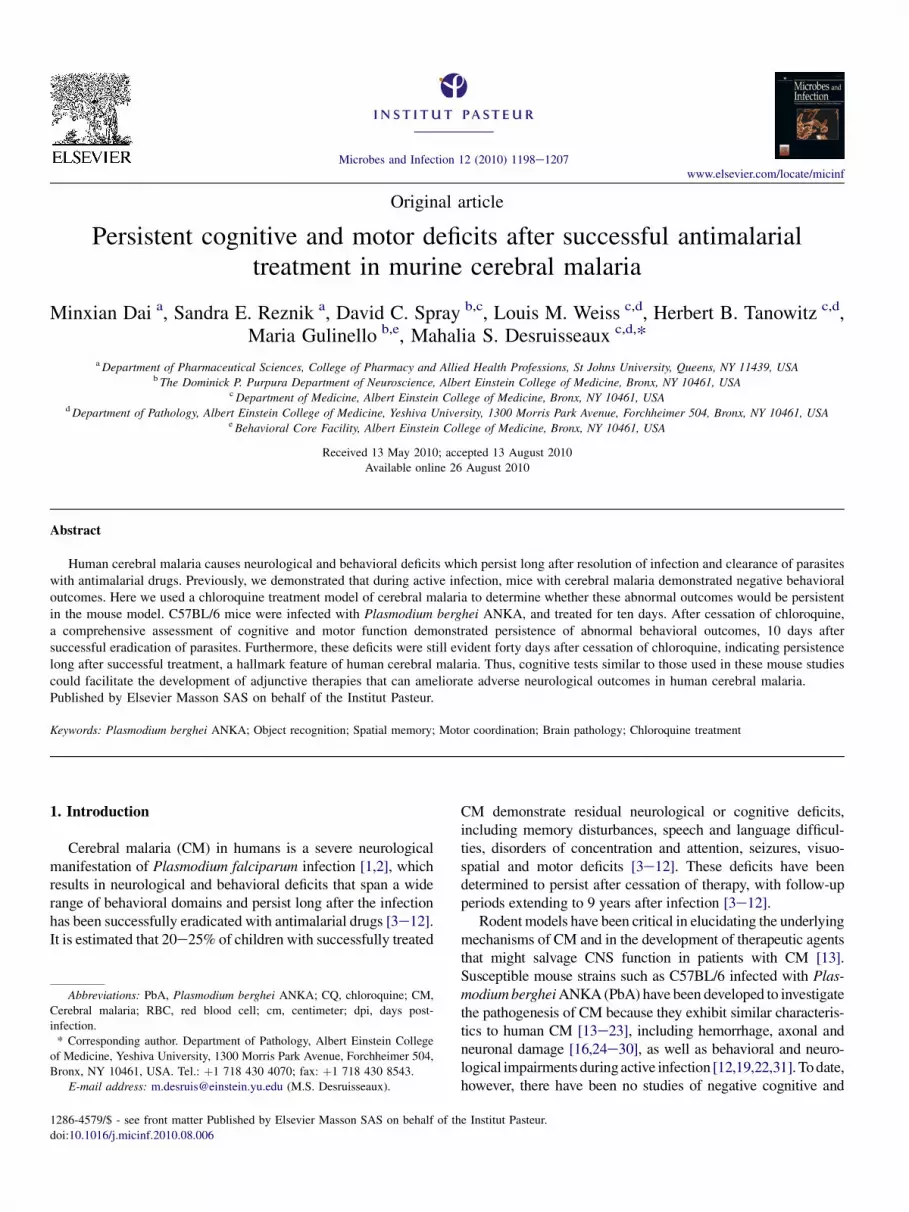
Fig. 1. Pathology observed during CM. (A) Uninfected untreated control mice demonstrate no hemorrhage on histological examination of the brain; score ¼ 0.
(BeE) Untreated PbA infected mice demonstrated various degrees of hemorrhage (B) Arrow-one visible vessel in one field with extravasation of red blood cells
(RBCs) in the hippocampus; score ¼ 1. (C) Arrows-multiple vessels in the cortex with extravasation of RBCs; score ¼ 2. (D) Arrow-single vessel in one field with
more extensive vascular damage in the pons; score ¼ 3. (E) Arrows-multiple vessels in cerebellum with extensive damage; score ¼ 4. (F) Arrow and insert arrow-
perivascular inflammation was also observed in different regions of the brain with CM. Hemorrhage largely resolved after anti-parasitic treatment (G) Chloroquine-
treated control. (H, I) Arrows-RBC extravasation was virtually confined to scores of 1 or 2 in CQ treated PbA infected mice. Brains harvested 17 days after
cessation of CQ therapy for mice in panels GeI.
1201M. Dai et al. / Microbes and Infection 12 (2010) 1198e1207
10 days after the cessation of CQ treatment despite the totalresolution of malarial infection.
Infected mice demonstrated significantly lower preferencescores in the object placement test of spatial memory aftera 30 min retention interval when tested 10 days after thecessation of CQ (Fig. 3A; p < 0.05). Furthermore, a signifi-cantly higher proportion of the infected mice showed spatialmemory deficits (Fig. 3C) based on the criteria for randomexploration ( p < 0.05).
Infected mice also had evidence of cognitive deficits ina test of recognition memory after a 45 min retention intervalwhen tested 10 days after the cessation of CQ, as indicated bythe lower preference scores in the object recognition test(Fig. 4A; p < 0.05). Furthermore, a significantly higherproportion of the infected mice showed recognition memorydeficits (Fig. 4C; p < 0.05).
3.3. Cognitive deficits are persistent
The cognitive deficits evident early after CQ treatment per-sisted when the mice were tested one month later. Spatialmemory deficits were evident using a 30 min retention interval,when infected mice were tested 40 days after the cessation ofCQ (Fig. 3A; p < 0.05). A significantly higher proportion ofmice still exhibited random exploration (Fig. 3C; p < 0.05) inthe object placement test. Testing after a 120 min retentioninterval also revealed significant deficits in CM mice, withlower preference scores (Fig. 3B; p< 0.05) and a higher rate ofrandom exploration (Fig. 3D; p < 0.05).
Though CM mice did not exhibit significant deficits aftera 45 min retention interval in the object recognition 40 daysafter the cessation of CQ (Fig. 4A, C), further testing usinga 120 min retention revealed significantly lower preference

Fig. 2. Parasitemia, weight change and survival of PbA infectedmice in this study.
(A) By 8 days post-infection (dpi), parasitemiawas greater than 7% in all infected
mice. (B) Infected animals demonstrated a gradual decrease in weight after
infection with significant decrease occurring between 8 and 10 days. Weights of
infected animals increased to original levels after CQ treatment, though there was
a transient loss of weight in both groups at day 20, which resolved by the time of
testing. (C) Survival curve of PbA infectedmicewithmortality occurring between
7 and 10 days.*p< 0.05,**p< 0.001. Arrow-ten-day course of CQ treatment in
accordance with the previously selected treatment criteria was initiated at
8.5 � 1.5 dpi in PbA mice. CQ was started at 7 dpi in control mice.
1202 M. Dai et al. / Microbes and Infection 12 (2010) 1198e1207
scores in CM mice (Fig. 4B; p < 0.01) and a higher proportionof the infected mice performed at random exploration levels(Fig. 4D; p < 0.05) compared to controls.
Infected and control mice had similar durations of totalnovel object exploration (Fig. S1DeF) when tested either 10days (PbA 13.6 � 2.5 s vs. control 14 � 3.5 s) or 40 days (PbA23.6 � 2.0 s vs. control 27.2 � 5.0 s) after the cessation of CQ,using a 45-min retention interval. Similar results were alsoobtained after 120 min retention (PbA 29.7 � 4.9 s vs. control36.5 � 6.4 s). Furthermore, infected mice were able to performthe object placement task when tested after a short (10 min)
retention interval, indicating that the cognitive defects werenot purely due to non-specific behavioral impairment.
3.4. Mice with CM have motor coordination deficits afterresolution of infection
Impairment of motor coordination on the balance beam waspresent in infected mice 10 days after the cessation of CQ,evidenced by the number of slips (Fig. 5A; p < 0.01) andlatency to cross the 1.2 cm diameter beam (Fig. 5B; p < 0.01).Moreover, the infected mice took a significantly longer timethan controls to cross the 1.8 cm diameter beam (Fig. 5D;p < 0.05).
The early motor coordination deficits were still evident 40days after the cessation of CQ on the 1.2 cm beam as infectedmice had significantly more slips (Fig. 5A; p < 0.05) anda longer latency to cross (Fig. 5B; p < 0.05) the beam.However, no significant difference in slips or latency existedon 1.8 cm beam between infected and control animals 40 daysafter the cessation of CQ (Fig. 5C,D).
Though over time, there was a trend toward improvement onthe 1.2 cm beam, there were no significant effects of time onbalance beam performance (1.2 cm: F (1, 36) ¼ 3.71;p ¼ 0.062. 1.8 cm: F (1, 8) ¼ 1.1; p ¼ 0.3). However, there wasa significant effect of infection condition on performance on the1.2 cm beam as CM mice continued to perform worse thancontrols (F (1, 38)¼ 8.72; p< 0.01). This effect was lost on the1.8 cm beam (p ¼ NS). Interestingly, there were no interactionsbetween time and infection condition with respect to perfor-mance on either beam, suggesting that learning capacity ininfected mice may be intact (1.2 cm: F (1, 38) ¼ 0.58; 1.8 cm:F (1, 5) ¼ 0.69; p ¼ NS).
3.5. Brain inflammation and parenchymal hemorrhagelargely resolve in CM mice with CQ treatment
We previously demonstrated that during acute malarialinfection, mice with CM sustain substantial hemorrhage inseveral regions of the brain [21]. Hematoxylin and eosin (H&E)staining of the sagittal section of acutely infected mice (N ¼ 6)corroborated our previous demonstration of significant hemor-rhage compared to untreated controls (N ¼ 4) (Fig. 1AeE),with the more striking scores occurring in the cerebellum(PbA 1.94 � 0.54 vs. Con 0.08 � 0.08; p < 0.05), the midbrain(PbA 1.17 � 0.33 vs. Con 0; p < 0.05), and the medulla (PbA1.08 � 0.38 vs. Con 0; p < 0.05). Interestingly, the centralrespiratory center, the brainstem, including the pons, demon-strated the most significant changes (PbA 1.14 � 0.23 vs. Con0.025 � 0.025; p < 0.001). In addition, though individually thehemorrhage scores in the cortex, hippocampus, and fornix werenot different from controls, when combined there was a ten-foldincrease in the hemorrhage score in some of the regions mostcommonly associated with cognitive function (PbA 0.58 � 0.2vs. Con 0.05 � 0.03; p < 0.05). There was also an increase inperivascular and periventricular inflammation adjacent to thoseregions (Fig. 1F). These lesions were largely resolved after CQtreatment, as there were no significant differences in the
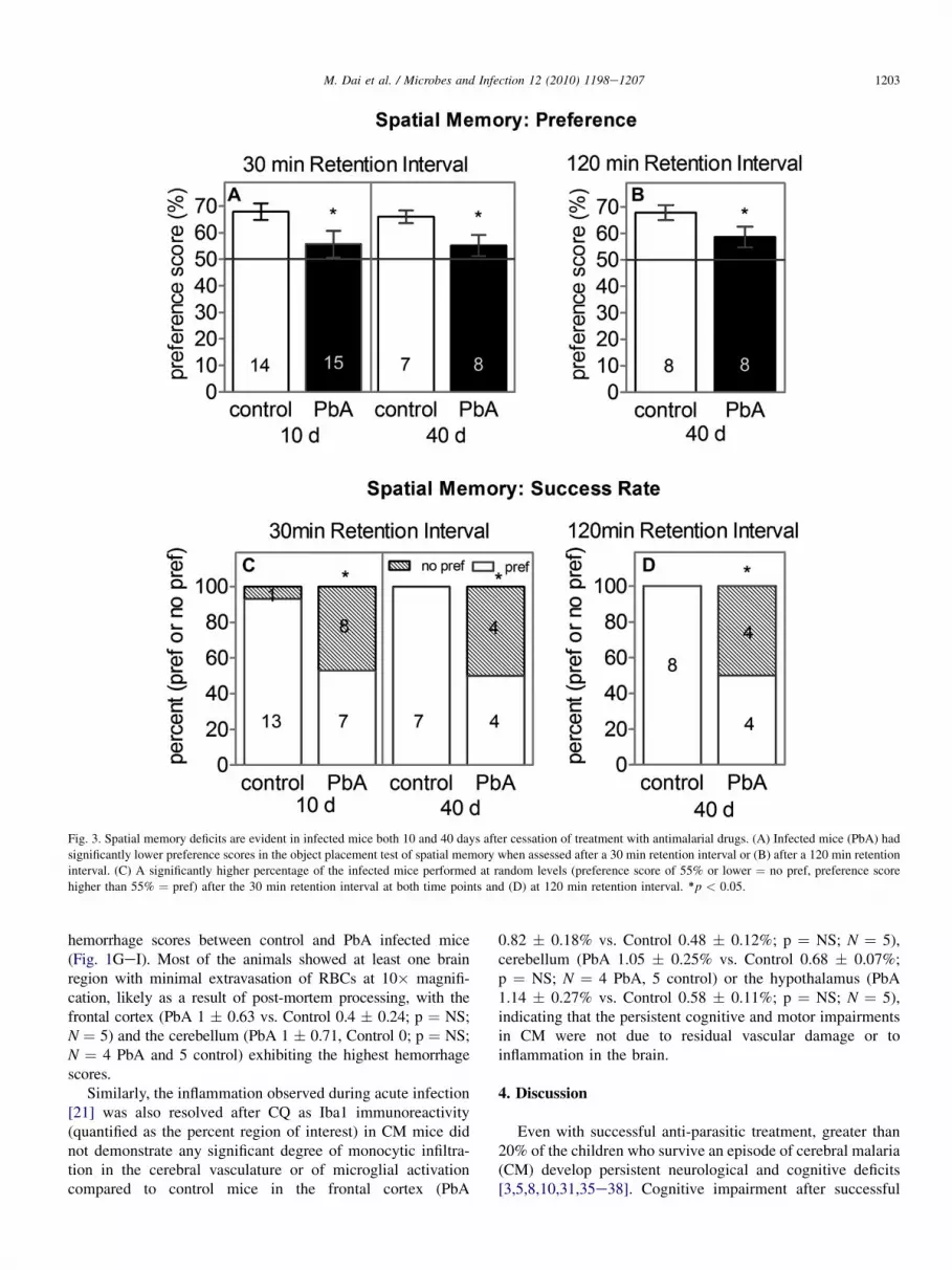
Fig. 3. Spatial memory deficits are evident in infected mice both 10 and 40 days after cessation of treatment with antimalarial drugs. (A) Infected mice (PbA) had
significantly lower preference scores in the object placement test of spatial memory when assessed after a 30 min retention interval or (B) after a 120 min retention
interval. (C) A significantly higher percentage of the infected mice performed at random levels (preference score of 55% or lower ¼ no pref, preference score
higher than 55% ¼ pref) after the 30 min retention interval at both time points and (D) at 120 min retention interval. *p < 0.05.
1203M. Dai et al. / Microbes and Infection 12 (2010) 1198e1207
hemorrhage scores between control and PbA infected mice(Fig. 1GeI). Most of the animals showed at least one brainregion with minimal extravasation of RBCs at 10� magnifi-cation, likely as a result of post-mortem processing, with thefrontal cortex (PbA 1 � 0.63 vs. Control 0.4 � 0.24; p ¼ NS;N ¼ 5) and the cerebellum (PbA 1 � 0.71, Control 0; p ¼ NS;N ¼ 4 PbA and 5 control) exhibiting the highest hemorrhagescores.
Similarly, the inflammation observed during acute infection[21] was also resolved after CQ as Iba1 immunoreactivity(quantified as the percent region of interest) in CM mice didnot demonstrate any significant degree of monocytic infiltra-tion in the cerebral vasculature or of microglial activationcompared to control mice in the frontal cortex (PbA
0.82 � 0.18% vs. Control 0.48 � 0.12%; p ¼ NS; N ¼ 5),cerebellum (PbA 1.05 � 0.25% vs. Control 0.68 � 0.07%;p ¼ NS; N ¼ 4 PbA, 5 control) or the hypothalamus (PbA1.14 � 0.27% vs. Control 0.58 � 0.11%; p ¼ NS; N ¼ 5),indicating that the persistent cognitive and motor impairmentsin CM were not due to residual vascular damage or toinflammation in the brain.
4. Discussion
Even with successful anti-parasitic treatment, greater than20% of the children who survive an episode of cerebral malaria(CM) develop persistent neurological and cognitive deficits[3,5,8,10,31,35e38]. Cognitive impairment after successful

Fig. 4. Recognition memory deficits are evident in infected mice. (A) Infected mice had significantly lower preference scores in the object recognition test after
a 45 min retention interval, 10 days after the cessation of CQ and (B) after a 120 min retention interval 40 days after cessation of CQ. (C) A significantly higher
percentage of the infected mice also performed at random levels after a 30 min retention interval 10 days post-CQ treatment and (D) after a 120 min retention
interval 40 days post-CQ. There were no deficits after a 45 min retention 40 days post-CQ treatment (A and C). *p < 0.05, **p < 0.01. One control mouse was
excluded from testing at 40 days since there was no exploration of either object after a 30 min retention.
1204 M. Dai et al. / Microbes and Infection 12 (2010) 1198e1207
therapy for malaria is becoming increasingly evident, and theglobal economic and scholastic health consequences of long-term residual deficits in CM patients cannot be underestimated,considering the vast number of people affected in endemicregions. Despite extensive studies on the pathogenesis of CM,significant gaps exist in our understanding of the preciseetiology of the cerebral dysfunction seen in CM because thepathological processes are difficult to document in humans. Themurine model of C57BL/6, infection with P. berghei ANKA(PbA) used in this study enables us to assess a disease process inwhich several aspects are analogous to human infection.
Previously, our laboratory group has demonstrated thatsusceptiblemice sustain significantmemorydeficits during acuteinfection with PbA [21], and demonstrate many of the histo-pathologic features of human CM such as hemorrhage, neuronaland axonal damage [21,24,30]. With this study we presenta treatment model of PbA infection that mimics the problem ofpersistency in neurological sequelae after parasitological cure
that is observed in human CMwhen infected with P. falciparum.In our model, both cognitive and motor deficits were evident ininfected mice early after the eradication of parasites and per-sisted on retesting one month after cure of infection and afterresolution of cerebral pathology. Significant deficits wereobserved in the object recognition and object placement tests ofspatial and recognition memory 10 days after the completion ofsuccessful antimalarial therapywith CQ. These deficits persistedwhen the animals were evaluated more than a month after theresolution of their infection.
In addition, CM mice also demonstrated significant motorcoordination deficits when evaluated on the balance beam bothearly and after a prolonged period post-resolution of malaria,consistent with the pronounced pathology in the cerebellumobserved during acute disease. Motor deficits on the balancebeam were not accompanied by deficits in grip strength orgeneral locomotor activity suggesting some specificity formotorcoordination rather than peripheral muscle dysfunction or

Fig. 5. Infected mice performed poorly in balance beam test of motor coordination when compared to controls. (A) The infected mice had a greater number of slips
and longer latency (B) on the 1.2 cm beam at both 10 and 40 days time point. On the 1.8 cm beam, infected mice had no significant difference in slips (C), but
exhibited significantly longer latency (D) 10 days post-CQ treatment. There were no significant deficits on the larger beam 40 days post-CQ treatment (C and D).
*p < 0.05, **p < 0.01. A control mouse was excluded from testing at 40 days as there was no exploration of either objects using 45 min or a 120 min retention
intervals.
1205M. Dai et al. / Microbes and Infection 12 (2010) 1198e1207
general sickness behavior. Although there seemed to bea tendency toward improvement in motor coordination on the1.2 cm balance beam with time, this was not significant. Inaddition, there were no interactions between time and infectionstatus with regard to motor coordination suggesting that motorlearning capacity in infectedmicemay be at least partially intact.
Although cognitive deficits were persistent 40 days aftercessation of CQ, infected mice approached the performancelevels of controls and demonstrated no significant deficits in theobject recognition test after a 45 min retention. This may bea reflection of experimental variables such as the loss of sickerand presumably more impaired animals, or possible variabilityin improvement of symptoms and the rate of such improve-ments. The nature of the improved performance also suggeststhat these deficits over time might be more subtle as brain
pathology resolves, or that the mice might have learned tocompensate for their deficits, as more sensitive tests, forexample the extension of the retention interval to 120 min, werestill able to detect significant impairment in infected mice.
It is important to separate the specific cognitive deficits fromnon-specific behavioral deficits induced by previous illness,general sickness due to malarial infection, CQ therapy orperipheral pathology. A recent article by Reis et al. [39] foundthat CM-resistant mice infectedwith PbA did not incur cognitivedeficits after chloroquine treatment, indicating that the neuro-cognitive impairment observed during CM are specific, and notjust due to general severe malarial disease. In this regard, in thepresent study, the lack of impairment in total novel exploration,locomotor activity and exploration, and normal cognitiveperformance using very short retention intervals (3 min) in the

1206 M. Dai et al. / Microbes and Infection 12 (2010) 1198e1207
object placement test in CMmice (data not shown) indicate thatthe cognitive deficits observed were not likely to be due toconfounding factors such as perceptual deficits, development ofneophobia, peripheral organ or muscle dysfunction due togeneral systemic illness, or effects of CQ treatment.
Several theories exist about the etiology of long-term cogni-tive impairment with CM. It is postulated that these cognitivedeficits occur as a result of a complicated host inflammatoryresponse in conjunction with vascular impairment [40], causingabnormalities in neuronal cells, which adversely affect cognitivefunction [41e43]. This was consistent with our observations inthe acute CM murine model [26,44]. However, more recentanalysis suggests that the development of long-term deficits withCM is more complex [45] and may involve a host of metabolicand physiological processes in several brain regions that remainlong after malaria parasites have been cleared. This idea is morein line with our demonstration that after the resolution ofinflammation and hemorrhage with parasitological cure, micestill remained with persistent cognitive and motor deficits.
Both experimental and human models of CM have alreadydemonstrated evidence for neuronal damage during activeinfection [21e28,30]. In this regard neuronal apoptosis has beendemonstrated in mice with CM [46,47], and irreversible glialcell injury and dysfunction in the brainstem has been describedin cases of severe human CM [25]. These data not only lendsupport to our observed increase in hemorrhagic damage in thebrainstem during infection which may be implicated in down-stream neuronal damage, but also point toward damage to thecentral respiratory center, both vascular and neuronal, asa possible explanation for the increased mortality associatedwith CM in this model. Our data demonstrate that geographicaldistribution of vascular damage during CM is clearly importantas persistent cognitive and motor deficits as well as mortalitycorrelate with the areas of damage in acute infection. However,as demonstrated in this study, vascular damage or inflammationalone cannot result in the long-term neuro-cognitive effects ofcerebral malaria. There is a dearth of information on neuralfactors thatmay affect cognitive and sensorimotor function aftermalarial treatment. The persistence of negative functionaloutcomes despite the resolution of hemorrhage and inflamma-tion underscores the need for further evaluation of preventableneuronal damage as a possible etiologic factor of long-termneuro-cognitive sequelae in CM.
Though this study does not expressly resolve the etiology oflong-term negative neurological outcomes with CM, it doesrecapitulate the process observed in human CM. This report, toour knowledge, is the first of this kind. This important modelcan not only serve as a rationale for existing analogous tests inhuman CM, but it can aid in the exploration of the mechanisminvolved in neuro-cognitive sequelae in CM; thereby allowingus to devise adjunctive therapy directed at the prevention ofneuronal damage during treatment of CM.
Conflict of interest statement
The authors report no commercial or other association thatmight pose a conflict of interest.
Acknowledgments
The authors acknowledge Mr. Dazhi Zhao for his consid-erable assistance with the maintenance, infection and treat-ment of the mice.
This work was supported by the NIH Training Grant inMechanisms of Cardiovascular Diseases (T32 HL-07675) toMSD; Dominick P. Purpura Department of Neuroscience andDepartment of Psychiatry and Behavioral Sciences (neurosci-ence fellowship support to MSD), Albert Einstein College ofMedicine; Burroughs-Wellcome Fund’s Career Awards forMedical Scientists to MSD; Einstein-Montefiore Institute forClinical and Translational Research Career Development Awardto MSD; AI076248 to HBT; AI39454 to LMW; NS041282 toDCS.
Appendix. Supplementary material
Supplementary data related to this article can be foundonline at doi:10.1016/j.micinf.2010.08.006.
References
[1] R.K. Garg, Cerebral malaria, J. Assoc. Physicians India 48 (2000)
1004e1013.
[2] C.L. Mackintosh, J.G. Beeson, K. Marsh, Clinical features and
pathogenesis of severe malaria, Trends Parasitol. 20 (2004)
597e603.[3] M.J. Boivin, P. Bangirana, J. Byarugaba, R.O. Opoka, R. Idro, A.M.
Jurek, C.C. John, Cognitive impairment after cerebral malaria in chil-
dren: a prospective study, Pediatrics 119 (2007) E360eE366.
[4] R. Idro, M. Ndiritu, B. Ogutu, S. Mithwani, K. Maitland, J. Berkley, J.
Crawley, G. Fegan, E. Bauni, N. Peshu, K. Marsh, B. Neville, C. Newton,
Burden, features, and outcome of neurological involvement in acute
falciparum malaria in Kenyan children, J. Am. Med. Assoc. 297 (2007)
2232e2240.
[5] J.A. Carter, A.J. Ross, B.G. Neville, E. Obiero, K. Katana, V. Mung’ala-
Odera, J.A. Lees, C.R. Newton, Developmental impairments following
severe falciparum malaria in children, Trop. Med. Int. Health 10 (2005)
3e10.
[6] J. Crawley, S. Smith, P. Muthinji, K. Marsh, F. Kirkham, Electroen-
cephalographic and clinical features of cerebral malaria, Arch. Dis.
Child. 84 (2001) 247e253.[7] G.S. Falchook, C.M. Malone, S. Upton, W.X. Shandera, Postmalaria
neurological syndrome after treatment of Plasmodium falciparum
malaria in the United States, Clin. Infect. Dis. 37 (2003) e22e24.[8] P.A. Holding, J. Stevenson, N. Peshu, K. Marsh, Cognitive sequelae of
severe malaria with impaired consciousness, Trans. R. Soc. Trop. Med.
Hyg. 93 (1999) 529e534.
[9] C.C. John, P. Bangirana, J. Byarugaba, R.O. Opoka, R. Idro, A.M.
Jurek, B.L. Wu, M.J. Boivin, Cerebral malaria in children is associ-
ated with long-term cognitive impairment, Pediatrics 122 (2008)
E92eE99.
[10] M. Kihara, J.A. Carter, C.R. Newton, The effect of Plasmodium falci-
parum on cognition: a systematic review, Trop. Med. Int. Health 11
(2006) 386e397.
[11] E.D. Richardson, N.R. Varney, R.J. Roberts, J.A. Springer, P.S. Wood,
Long-term cognitive sequelae of cerebral malaria in Vietnam veterans,
Appl. Neuropsychol. 4 (1997) 238e243.
[12] N. Senanayake, H.J. de Silva, Delayed cerebellar ataxia complicating
falciparum malaria: a clinical study of 74 patients, J. Neurol. 241 (1994)
456e459.

1207M. Dai et al. / Microbes and Infection 12 (2010) 1198e1207
[13] N.H. Hunt, G.E. Grau, Cytokines: accelerators and brakes in the patho-
genesis of cerebral malaria, Trends Immunol. 24 (2003) 491e499.
[14] T. Schetters, J. Curfs, A. van Zon, C. Hermsen, W. Eling, Cerebral
lesions in mice infected with Plasmodium berghei are the result of an
immunopathological reaction, Trans. R. Soc. Trop. Med. Hyg. 83 (1989)
103e104.
[15] J.R. Rest, Cerebral malaria in inbred mice. I. A new model and its
pathology, Trans. R. Soc. Trop. Med. Hyg. 76 (1982) 410e415.[16] I.M. Medana, N.H. Hunt, T. Chan-Ling, Early activation of microglia in
the pathogenesis of fatal murine cerebral malaria, Glia 19 (1997)
91e103.
[17] Y.C. Martins, M.J. Smith, M. Pelajo-Machado, G.L. Werneck, H.L.
Lenzi, C.T. Daniel-Ribeiro, L.J. Carvalho, Characterization of cerebral
malaria in the outbred Swiss Webster mouse infected by Plasmodium
berghei ANKA, Int. J. Exp. Pathol. 90 (2009) 119e130.
[18] S. Bagot, M. Idrissa Boubou, S. Campino, C. Behrschmidt, O. Gorgette,
J.L. Guenet, C. Penha-Goncalves, D. Mazier, S. Pied, P.A. Cazenave,
Susceptibility to experimental cerebral malaria induced by Plasmodium
berghei ANKA in inbred mouse strains recently derived from wild stock,
Infect. Immun. 70 (2002) 2049e2056.
[19] J. Lou, R. Lucas, G.E. Grau, Pathogenesis of cerebral malaria: recent
experimental data and possible applications for humans, Clin. Microbiol.
Rev. 14 (2001) 810e820.
[20] J.B. de Souza, E.M. Riley, Cerebral malaria: the contribution of studies in
animal models to our understanding of immunopathogenesis, Microbes
Infect. 4 (2002) 291e300.
[21] M.S. Desruisseaux, M. Gulinello, D.N. Smith, S.C. Lee, M. Tsuji, L.M.
Weiss, D.C. Spray, H.B. Tanowitz, Cognitive dysfunction in mice
infected with Plasmodium berghei strain ANKA, J. Infect. Dis. 197
(2008) 1621e1627.[22] P. Lackner, R. Beer, V. Heussler, G. Goebel, D. Rudzki, R. Helbok,
E. Tannich, E. Schmutzhard, Behavioural and histopathological alter-
ations in mice with cerebral malaria, Neuropathol. Appl. Neurobiol. 32
(2006) 177e188.[23] G. Stoltenburgdidinger, S. Neifer, U. Bienzle, W.M.C. Eling, P.G.
Kremsner, Selective damage of hippocampal neurons in murine cerebral
malaria prevented by pentoxifylline, J. Neurol. Sci. 114 (1993) 20e24.
[24] I.M. Medana, N.P. Day, T.T. Hien, N.T.H. Mai, D. Bethell, N.H. Phu, J.
Farrar, M.M. Esiri, N.J. White, G.D. Turner, Axonal injury in cerebral
malaria, Am. J. Pathol. 160 (2002) 655e666.
[25] I.M. Medana, N.T.H. Mai, N.P.J. Day, T.T. Hien, D. Bethell, N.H. Phu,
J. Farrar, N.J. White, G.D.H. Turner, Cellular stress and injury responses
in the brains of adult Vietnamese patients with fatal Plasmodium falci-
parum malaria, Neuropathol. Appl. Neurobiol. 27 (2001) 421e433.
[26] R.P. Kennan, F.S. Machado, S.C. Lee, M.S. Desruisseaux, M. Wittner,
M. Tsuji, H.B. Tanowitz, Reduced cerebral blood flow and N-acetyl
aspartate in a murine model of cerebral malaria, Parasitol. Res. 96 (2005)
302e307.
[27] N.L. Ma, M.C. Madigan, T. ChanLing, N.H. Hunt, Compromised blood-
nerve barrier, astrogliosis, and myelin disruption in optic nerves during
fatal murine cerebral malaria, Glia 19 (1997) 135e151.
[28] S. Lewallen, R.N. Bronzan, N.A. Beare, S.P. Harding, M.E. Molyneux,
T.E. Taylor, Using malarial retinopathy to improve the classification of
children with cerebral malaria, Trans. R. Soc. Trop. Med. Hyg. 102
(2008) 1089e1094.
[29] N.A.V. Beare, C.E. Riva, T.E. Taylor, M.E. Molyneux, K. Kayira, V.A.
White, S. Lewallen, S.P. Harding, Changes in optic nerve head blood
flow in children with cerebral malaria and acute papilloedema, J. Neurol.
Neurosurg. Psychiatry 77 (2006) 1288e1290.
[30] V.A. White, S. Lewallen, N.A.V. Beare, M.E. Molyneux, T.E. Taylor,
Retinal pathology of pediatric cerebral malaria in Malawi, Plos One 4
(2009).
[31] M.J. Boivin, Effects of early cerebral malaria on cognitive ability in
Senegalese children, J. Dev. Behav. Pediatr. 23 (2002) 353e364.
[32] J. Hearn, N. Rayment, D.N. Landon, D.R. Katz, J.B. de Souza, Immu-
nopathology of cerebral malaria: morphological evidence of parasite
sequestration in murine brain microvasculature, Infect. Immun. 68 (2000)
5364e5376.
[33] F. Caterini, S. Della Sala, H. Spinnler, C. Stangalino, O.H. Turnbull,
Object recognition and object orientation in Alzheimer’s disease,
Neuropsychology 16 (2002) 146e155.
[34] J.L. Stanley, R.J. Lincoln, T.A. Brown, L.M. McDonald, G.R. Dawson,
D.S. Reynolds, The mouse beam walking assay offers improved sensi-
tivity over the mouse rotarod in determining motor coordination deficits
induced by benzodiazepines, J. Psychopharmacol. 19 (2005) 221e227.
[35] J.A. Carter, V. Mung’ala-Odera, B.G. Neville, G. Murira, N. Mturi,
C. Musumba, C.R. Newton, Persistent neurocognitive impairments
associated with severe falciparum malaria in Kenyan children, J. Neurol.
Neurosurg. Psychiatry 76 (2005) 476e481.
[36] J.A. Carter, J.A. Lees, J.K. Gona, G. Murira, K. Rimba, B.G. Neville,
C.R. Newton, Severe falciparum malaria and acquired childhood
language disorder, Dev. Med. Child. Neurol. 48 (2006) 51e57.
[37] D. Fernando, D. De Silva, R. Wickremasinghe, Short-term impact of an
acute attack of malaria on the cognitive performance of schoolchildren
living in a malaria-endemic area of Sri Lanka, Trans. R. Soc. Trop. Med.
Hyg. 97 (2003) 633e639.
[38] M.M. Meremikwu, A.A. Asindi, E. Ezedinachi, The pattern of neuro-
logical sequelae of childhood cerebral malaria among survivors in
Calabar, Nigeria, Cent. Afr. J. Med. 43 (1997) 231e234.
[39] P.A. Reis, C.M. Comim, F. Hernani, B. Silva, T. Barichello, A.C. Por-
tella, F.C. Gomes, I.M. Sab, V.S. Frutuoso, M.F. Oliveira, P.T. Bozza, F.
A. Bozza, F. Dal-Pizzol, G.A. Zimmerman, J. Quevedo, H.C. Castro-
Faria-Neto, Cognitive dysfunction is sustained after rescue therapy in
experimental cerebral malaria, and is reduced by additive antioxidant
therapy, PLoS Pathog. 6 (2010) e1000963.
[40] C.L. Grote, S.J.C. PierreLouis, W.F. Durward, Deficits in delayed
memory following cerebral malaria: a case study, Cortex 33 (1997)
385e388.
[41] N.L. Sparkman, J.B. Buchanan, J.R.R. Heyen, J. Chen, J.L. Beverly,
R.W. Johnson, Interleukin-6 facilitates lipopolysaccharide-induced
disruption in working memory and expression of other proinflammatory
cytokines in hippocampal neuronal cell layers, J. Neurosci. 26 (2006)
10709e10716.
[42] A. Bessis, C. Bechade, D. Bernard, A. Roumier, Microglial control of
neuronal death and synaptic properties, Glia 55 (2007) 233e238.
[43] R. Griffin, R. Nally, Y. Nolan, Y. McCartney, J. Linden, M.A. Lynch, The
age-related attenuation in long-term potentiation is associated with
microglial activation, J. Neurochem. 99 (2006) 1263e1272.
[44] F.S. Machado, M.S. Desruisseaux, Nagajyothi, R.P. Kennan, H.P.
Hetherington, M. Wittner, L.M. Weiss, S.C. Lee, P.E. Scherer, M. Tsuji,
H.B. Tanowitz, Endothelin in a murine model of cerebral malaria, Exp.
Biol. Med. 231 (2006) 1176e1181.
[45] M. Kihara, J.A. Carter, P.A. Holding, F. Vargha-Khadem, R.C. Scott,
R. Idro, G.W. Fegan, M. de Haan, B.G.R. Neville, C. Newton, Impaired
everyday memory associated with encephalopathy of severe malaria: the
role of seizures and hippocampal damage, Malar. J. 8 (2009).
[46] P. Lackner, C. Burger, K. Pfaller, V. Heussler, R. Helbok, M. Morandell,
G. Broessner, E. Tannich, E. Schmutzhard, R. Beer, Apoptosis in
experimental cerebral malaria: spatial profile of cleaved caspase-3 and
ultrastructural alterations in different disease stages, Neuropathol. Appl.
Neurobiol. 33 (2007) 560e571.
[47] L. Wiese, J.A.L. Kurtzhals, M. Penkowa, Neuronal apoptosis, metal-
lothionein expression and proinflammatory responses during cerebral
malaria in mice, Exp. Neurol. 200 (2006) 216e226.
Related Documents


