Peripheral Nerve Repair David Moss, MD

Welcome message from author
This document is posted to help you gain knowledge. Please leave a comment to let me know what you think about it! Share it to your friends and learn new things together.
Transcript


Indications
• Preserve or restore function– Innervation of skin, muscle, other target
organs
• After transection, direct repair or graft offers only chance of recovery
• Irreparable proximal stump → nerve transfers
• Irreparable distal stump → muscular neurotization (implant into muscle)
• Quicker reconnection of nerve → better result– > 18 months → permanent end organ failure
(muscle cell death)
• Isolated nerve laceration repair timing:– Within 1-2 weeks to avoid retraction and
scarring

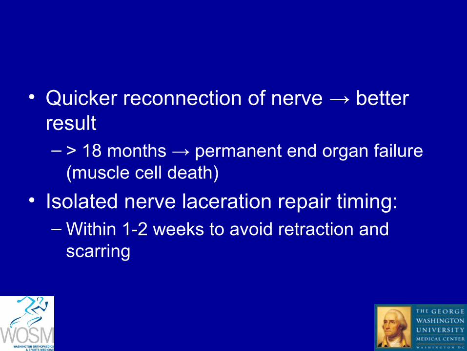
• Epineurium – Nourish and protect
fascicles
• Perineurium – Major contributor to
nerve tensile strength
• Endoneurium– Nourish axons

Fascicular arrangement
• Median nerve motor branch – Volar-radial
• Ulnar nerve motor branch– Dorsal-ulnar


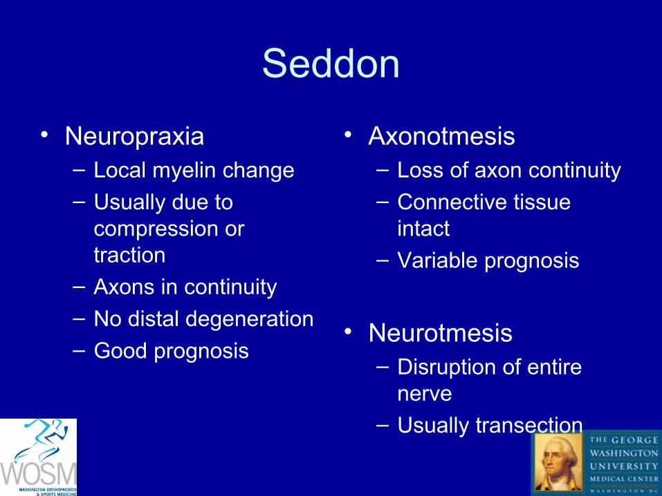
Seddon
• Neuropraxia– Local myelin change– Usually due to
compression or traction
– Axons in continuity
– No distal degeneration
– Good prognosis
• Axonotmesis– Loss of axon continuity– Connective tissue
intact– Variable prognosis
• Neurotmesis – Disruption of entire
nerve– Usually transection


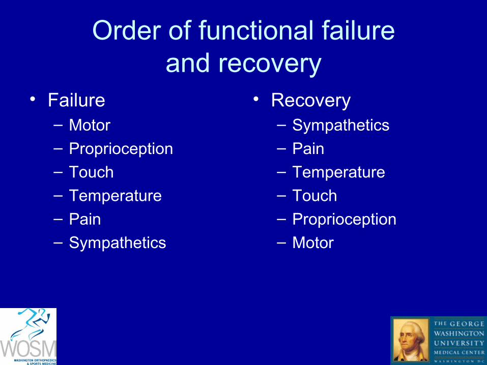
Order of functional failureand recovery
• Failure – Motor– Proprioception– Touch
– Temperature
– Pain
– Sympathetics
• Recovery– Sympathetics– Pain– Temperature
– Touch
– Proprioception
– Motor

Physiology of nerve degeneration
• Laceration– Cell body swells and becomes eosinophilic– Cell nucleus displaced peripherally– Proximal stump degeneration to proximal
node of ranvier

Wallerian degeneration
• Nerve breakdown distal to site of injury
• Begins 48-96 hours after transection
• Myelin deteriorates
• Schwann cells proliferate– Phagocytose myelin and debris
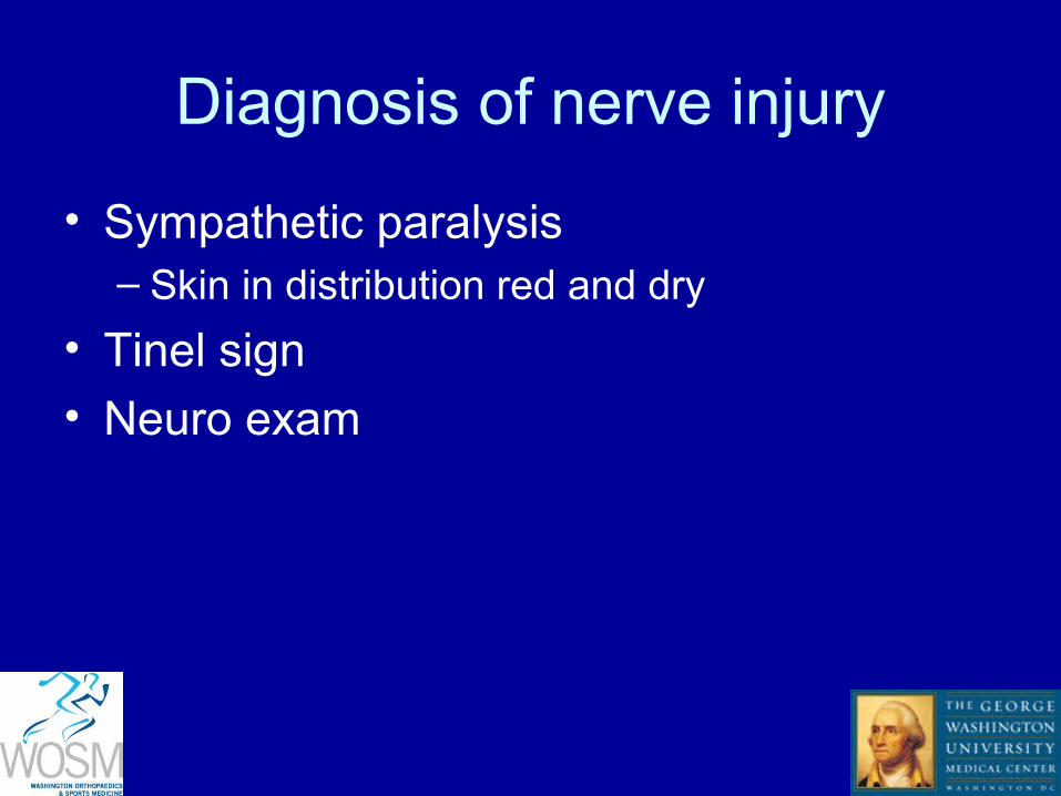
Diagnosis of nerve injury
• Sympathetic paralysis– Skin in distribution red and dry
• Tinel sign
• Neuro exam

• Martin-Gruber anastomosis– Median to ulnar nerve crossover in forearm– “overly median innervated hand”– AIN to ulnar nerve– Motor only
• Riche-Cannieu anastomosis – Median to ulnar nerve crossover in hand– May be median to ulnar or reverse

EMG
• Fibrillations – Appear as muscles are denervated– Onset 10-14 days after injury


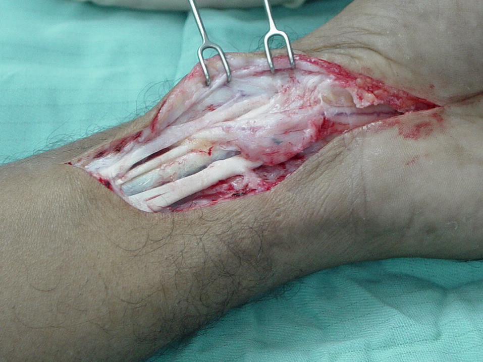
• Freshen ends to healthy nerve
• Suture epineurium only– No advantage to intra-fascicular repair
• Use as few sutures as possible to re-approximate nerve ends
• Tension-free
• 8-0 suture for the digital nerves
• Microscope often helpful
Fibrin glue
• No definitive evidence yet
• Gaining popularity
• Faster than suture
• Useful for multiple repairs– Cable graft
• Can be used to augment suture repair to prevent gapping

Nerve graft
• If direct, tension-free repair not possible
• Sural nerve
• MABCN
Nerve tubes
• Up to 3 cm gap – Tubes equivalent to nerve graft
• PLA and collagen superior to PGA
Related Documents

![Review Article Past, Present, and Future of Nerve Conduits ...downloads.hindawi.com/journals/bmri/2015/237507.pdf · ] performed peripheral nerve conduit repair in patients with peripheral](https://static.cupdf.com/doc/110x72/5f08867b7e708231d422712f/review-article-past-present-and-future-of-nerve-conduits-performed-peripheral.jpg)