PERIPHERAL BLOOD SMEAR DR. SNEHAL KOSALE GUIDES- DR. VANDANA SANKLECHA DR. BHAVNA BHARAMBE

Welcome message from author
This document is posted to help you gain knowledge. Please leave a comment to let me know what you think about it! Share it to your friends and learn new things together.
Transcript
PERIPHERAL BLOOD SMEAR
DR. SNEHAL KOSALE
GUIDES- DR. VANDANA SANKLECHA DR. BHAVNA BHARAMBE

Role of peripheral blood examination• Evaluation of anemia
• Evaluation of thrombocytopenia/ thrombocytosis
• Identification of abnormal cells
• Inclusions like basophilic stippling, Howell-Jolly bodies, Cabot ring
• Infections like malaria, microfilaria etc

Collection of blood

EDTA• Collected in lavender (purple)–topped tubes
• Contain disodium or tripotassium ethylenediaminetetraacetic acid (EDTA) anticoagulates the blood by chelating the calcium that is essential for coagulation.
• High-quality blood films can be made within 2 to 3 hours of drawing the specimen.
• Blood films from EDTA tubes that remain at room temperature for more than 5 hours often have unacceptable blood cell artifacts• Echinocytic red blood cells, • Spherocytes, • Degenerated leukocytes, and • Vacuolated neutrophils
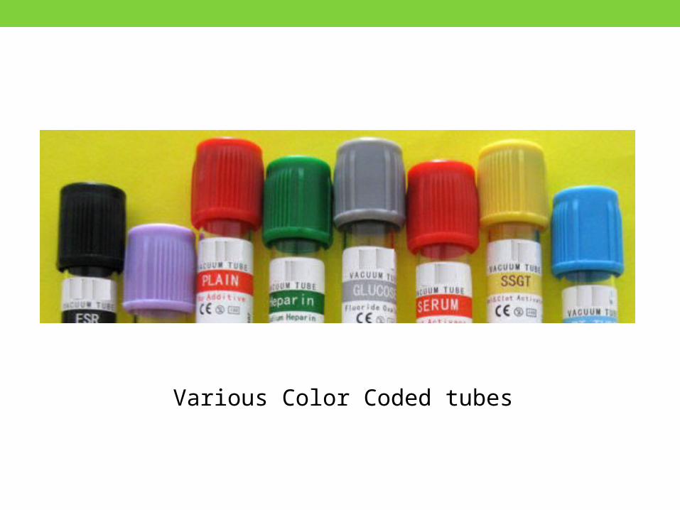
Various Color Coded tubes

Collection of bloodAdvantages
Many smears can be done in just a single draw Immediate preparation of the smear is not necessary
Prevents platelet clumping on the glass slide
Disadvantages: Platelet satellitosis: causes pseudothrombocytopenia and pseudoleukocytosis
Cause: Platelet specific auto antibodies that react best at room temperature

Platelet satellitism

PREPARATION OF SMEARWEDGECOVER SLIPAUTOMATED

Preparation of smear There are three types of blood smears:
The wedge smear The cover glass smear The spun smear
The are two additional types of blood smear used for specific purposes Buffy coat smear Thick blood smears for blood parasites

Wedge techniqueEasiest to master
Most convenient and most commonly used technique
EquipmentSpreadersClean slidesBlood capillary tube or micropipette 10 µL

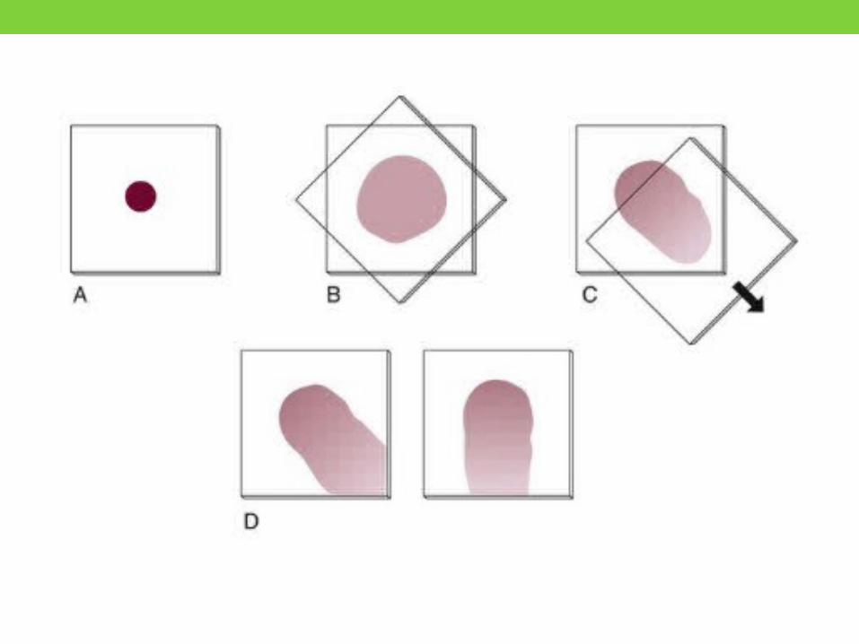
Place a drop of blood, about 2-3 mm in diameter approximately 1 cm from one
end of slide.
Precaution: Too large drop = too thick smearToo small drop = too thin smear

a. Place the slide on a flat surface, and hold the other end between your left thumb and forefinger.
b. With your right hand, place the smooth clean edge of a second (spreader) slide on the specimen slide, just in front of the blood drop.
c. Hold the spreader slide at a 30°- 45° angle, and draw it back against the drop of blood

Allow the blood to spread almost to the edges of the slide
Precautions: Ensure that the whole drop of blood is picked up and spread Angle correction:
High Hct: Angle should be loweredLow Hct: Angle should be raised

Large angle
Low HCT
(thinner)
Small angle
High HCT (thicker)

Push the spread forward with one light, smooth moderate speed.
Make a thin film of blood in the shape of tongue.
Precautions:• Too slow a slide push will accentuate poor leukocyte distribution,
larger cells are pushed at the end of the slide• Maintain an even gentle pressure on the slide• Keep the same angle all the way to the end of the smear.

Label one edge with patient name, lab id and date.
The slides should be rapidly air dried by waving the slides or using an electrical fan

Cover Slip Technique Rarely used Bone Marrow Aspirate smears Advantage: excellent leukocyte distribution Disadvantage: labeling, transport, staining and storage is a problem
Technique: A drop of marrow aspirate is placed on top of 1 coverslip
Another coverslip is placed over the other allowing the aspirate to spread.
One is pulled over the other to create 1 thin smears
Mounted on a 3x1 inch glass slide
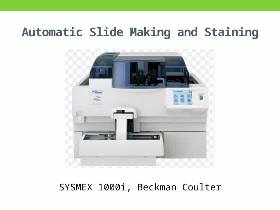
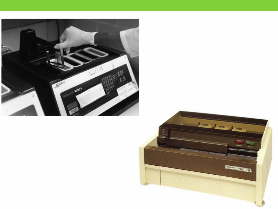
Automatic Slide Making and Staining
SYSMEX 1000i, Beckman Coulter

Automatic Slide Making and Staining• Performs a CBC for a specimen
•Dependent on the hematocrit reading, the system adjusts • Size of the drop of blood used and • Angle and speed of the spreader slide in making a wedge preparation.
• After each blood film is prepared, the spreader slide is automatically cleaned.
Automatic Slide Making and Staining• Films can be produced approximately every 30 seconds.
• Name, number, and date for the specimen is printed on the slide.
• The slide is dried, loaded into a cassette, and moved to the staining position, where stain and then buffer and rinse are added at designated times.
• When staining is complete, the slide is moved to a dry position, then to a collection area where it can be picked up for microscopic evaluation.
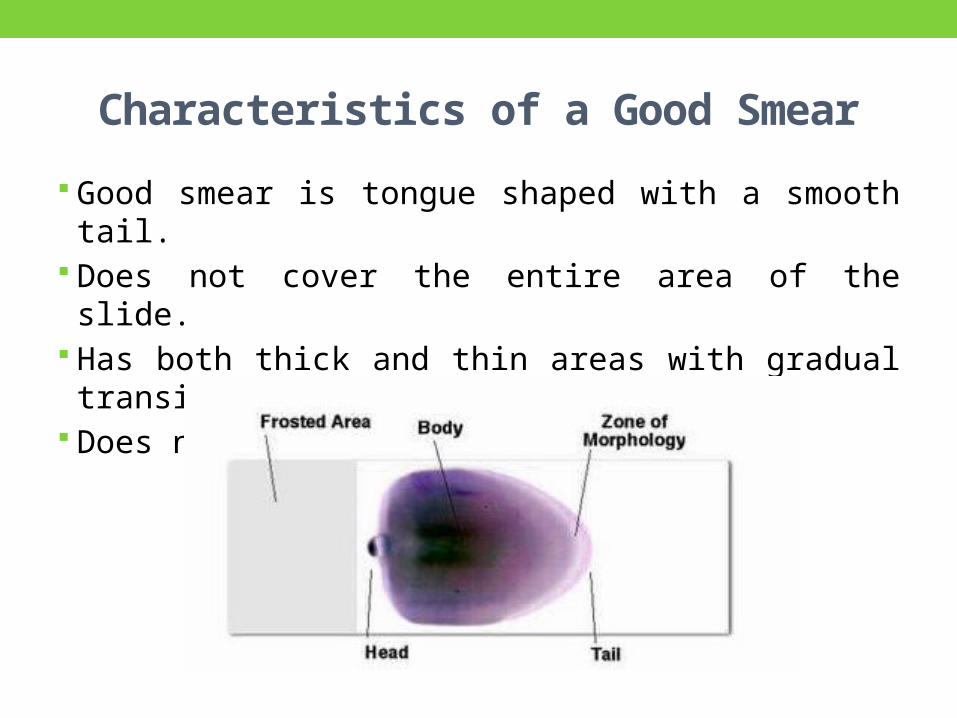
Characteristics of a Good Smear Good smear is tongue shaped with a smooth tail. Does not cover the entire area of the slide. Has both thick and thin areas with gradual transition.
Does not contain any lines or holes.


SLIDE FIXATION & STAININGLEISHMAN'S STAINFIELD’S STAIN- RAPID METHODAUTOMATED SLIDE STAINERS
Principle Leishman's stain is a polychromatic stain
Components: Methanol: fixes cells to slide Methylene blue stains RNA,DNA: blue-grey color Eosin stains hemoglobin, eosin granules orange-red
color Eosin + Methylene Blue = thiazine eosinate complex
The complex will not stain any color unless a buffer is added: 0.05M sodium phosphate (pH 6.4) and aged distilled water (pH 6.4-6.8)
Staining Procedure • Thin smear are air dried.• Flood the smear with stain. • Stain for 1-5 min. • Experience will indicate the optimum time.
• Add an equal amount of buffer solution and mix the stain by blowing an eddy in the fluid.• Leave the mixture on the slide for 10-15 min. •Wash off by running water directly to the centre of the slide to prevent a residue of precipitated stain.• Stand slide on end, and let dry in air.


Automated Slide StainersIt takes about 5-10 minutes to stain a batch of smears
Slides are just automatically dipped in the stain in the buffer and a series of rinses
DisadvantagesStaining process has begun, no STAT slides can be added in the batch
Aqueous solutions of stains are stable only for 3-6 hours
Rapid staining method- Field’s stain
AdvantageFast, convenient and takes about 1 minute
Cost effective
ComponentsMethanol Solution B- contains eosinSolution A- contains methylene blue

Dip in methanol to fix the smear for 1 minute
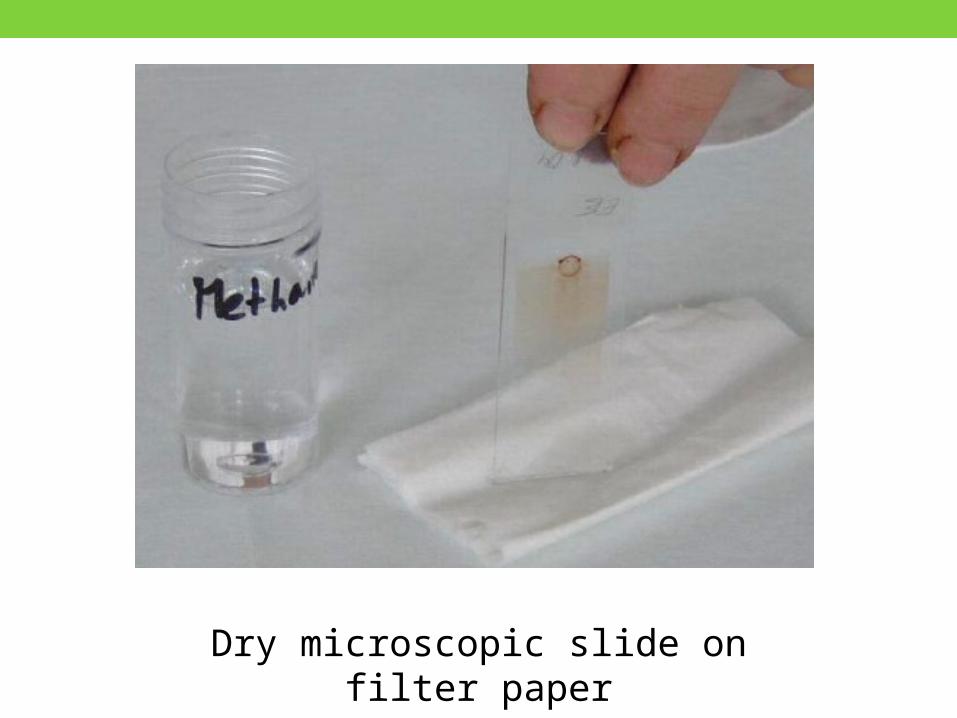
Dry microscopic slide on filter paper

Immerse slide in Field’s stain B (Eosin) for 5 seconds
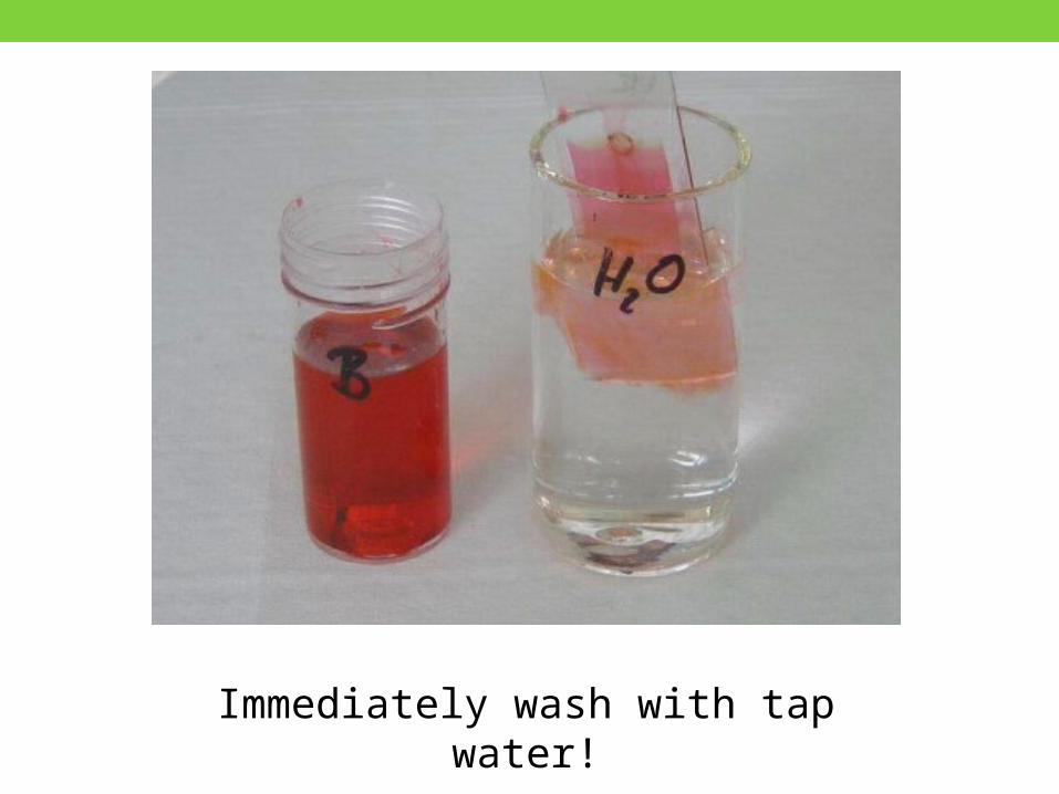
Immediately wash with tap water!

Immerse slide in Field‘s stain A (Methylene blue) for 10 seconds

Immediately wash with tap water
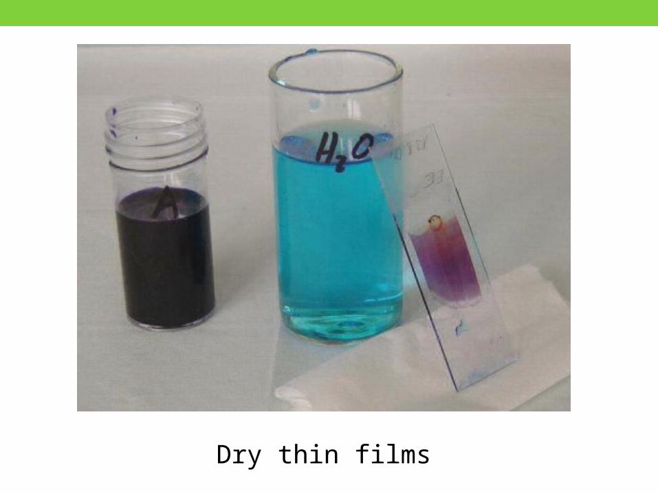
Dry thin films

MICROSCOPIC OVERVIEW

TOO ACIDIC SUITABLE TOO BASIC

Causes and correctionsToo acid stain:
Insufficient staining timeProlonged buffering or washingOld stain
Correction:Lengthen staining timeCheck stain and buffer pHShorten buffering or wash time
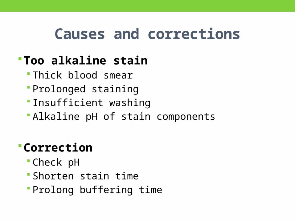
Causes and correctionsToo alkaline stain
Thick blood smear Prolonged staining Insufficient washing Alkaline pH of stain components
Correction Check pH Shorten stain time Prolong buffering time
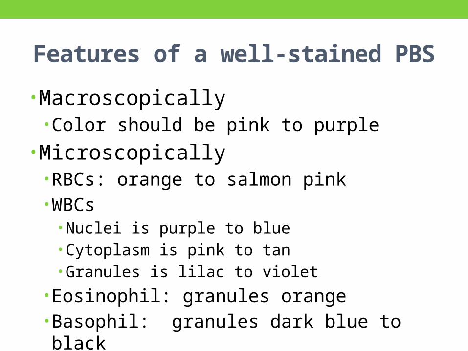
Features of a well-stained PBS•Macroscopically• Color should be pink to purple
•Microscopically• RBCs: orange to salmon pink•WBCs• Nuclei is purple to blue• Cytoplasm is pink to tan • Granules is lilac to violet
• Eosinophil: granules orange• Basophil: granules dark blue to black

tail body headZones of a slide
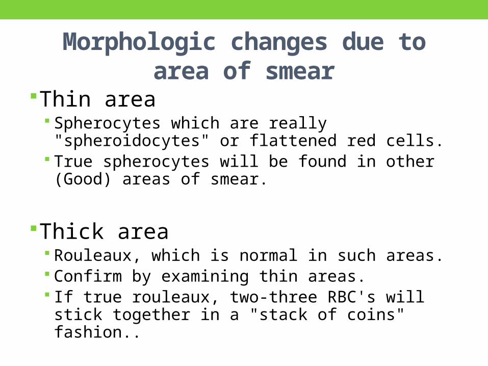
Morphologic changes due to area of smear
Thin area Spherocytes which are really "spheroidocytes" or flattened red cells.
True spherocytes will be found in other (Good) areas of smear.
Thick area Rouleaux, which is normal in such areas. Confirm by examining thin areas. If true rouleaux, two-three RBC's will stick together in a "stack of coins" fashion..
10x ObjectiveAssess overall quality of the smear i.e feathery edge, quality of the color, distributin of the cells and the lateral edges can be checked for WBC distribution
Snow-plow effect: more than 4x/cells per field on the feathery edge: Reject
Fibrin strands: Reject
Total Leucocyte Count 40x Objective
Use dry without oil
Choose a portion of the peripheral smear where there is only slight overlapping of the RBCs.
Count 10 fields, take the total number of white cells and divide by 10.
To do a WBC estimate by taking the average number of white cells and multiplying by 2000.
Normal leucocyte count ranges from 4000 to 11000/µl

Observe one field and record the number of WBC according to the different type then turn to another field in the snake-like direction
To avoid repeat or miss some cells

Manual Differential Counts These counts are done in the same area as WBC and platelet estimates with the red cells barely touching.
This takes place under × 100 (oil) using the zigzag method.
Count 100 WBCs including all cell lines from immature to mature.
Expressed as percentage.
Absolute number of cells/µl = % of cell type in differential x white cell count
Nucleated Red Blood Cells (nRBCs)• If 10 or more nucleated RBC's (nRBC) are seen, correct the TLC
• Corrected WBC Count = WBC x 100/( nRBC + 100)
Example: If WBC = 5000 and 10 nRBCs have been countedThen 5,000× 100/110 = 4545.50The corrected white count is 4545.50.

Do Not Count• Disintegrating cells
• Eosinophil with no cytoplasmic membrane and with scattered granules
• Smudge cells
• Pyknotic cell• Nucleus condensed and
degenerated, • Lobes in small, round clumps• No interconnecting filaments

RECORDING WBC MORPHOLOGYNORMAL COMPONENTSABNORMAL FORMS
WBCs in PBS•GRANULOCYES
Neutrophils ( polymorphonuclear leucocytes)Eosinophils Basophils
• AGRANULOCYTES Lymphocytes Monocytes
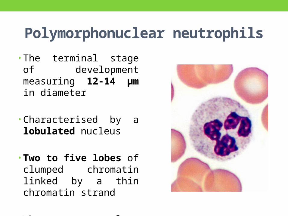
Polymorphonuclear neutrophils• The terminal stage of development measuring 12-14 μm in diameter
• Characterised by a lobulated nucleus
• Two to five lobes of clumped chromatin linked by a thin chromatin strand
• The cytoplasm contains fine azurophilic granules
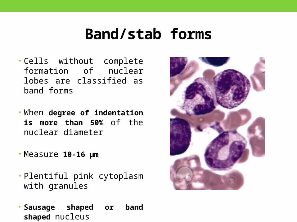
Band/stab forms• Cells without complete
formation of nuclear lobes are classified as band forms
• When degree of indentation is more than 50% of the nuclear diameter
• Measure 10-16 μm
• Plentiful pink cytoplasm with granules
• Sausage shaped or band shaped nucleus

Hypersegmented neutrophils
Presence of even a single neutrophils with six or more lobes or the presence of more than 5% of neutrophils with five lobes.
Seen in Megaloblastic anemia Uraemia Drugs-cytotoxic treatment
with methotrexate Hydroxycarbamide Myelokathexis
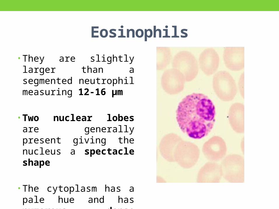
Eosinophils• They are slightly larger than a segmented neutrophil measuring 12-16 μm
• Two nuclear lobes are generally present giving the nucleus a spectacle shape
• The cytoplasm has a pale hue and has numerous dense orange red granules

Abnormalities in Eosinophils Increase in the absolute eosinophil count in the peripheral blood
Mild eosinophilia- Allergic conditions hay fever, Asthma, eczema
Severe eosinophilia- Parasitic infection Reactive eosinophilia Eosinophilic leukaemia Idiopathic hypereosinophilic syndrome

Basophils• Basophils have a diameter of 10-14 μm• Lobulated nucleus• Distinguished by their large, coarse, purplish-black granules • Fill the cytoplasm• Obscure the nucleus
• Granules are rich in histamine, serotonin and heparin

Basophila Myeloproliferative disorders (e.g., chronic myelogenous leukemia)
IgE mediated Hypersensitivity reactions
Mastocytosis
Ulcerative colitis
Hypothyroidism
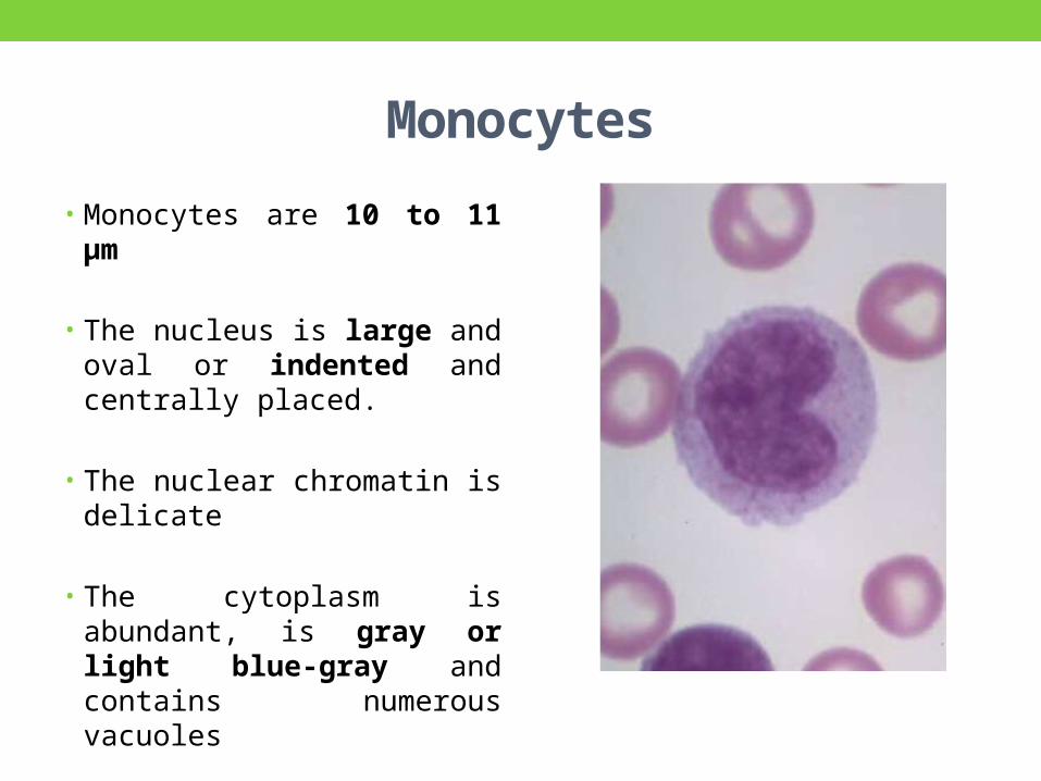
Monocytes• Monocytes are 10 to 11 μm
• The nucleus is large and oval or indented and centrally placed.
• The nuclear chromatin is delicate
• The cytoplasm is abundant, is gray or light blue-gray and contains numerous vacuoles
• The granules resemble fine dust and give the bluish cytoplasm a ground-glass appearance
MonocytosisChronic infections and inflammatory conditions such as Malaria, Typhoid, Bacterial endocarditis, Kala- azar Tuberculosis Crohn’s disease Infectious Mononucleosis
Hematolymphoid malignancies Acute myelomonocytic leukaemia (AML M4),
Acute monocytic leukaemia (AML M5),
Myeloproliferative neoplasms,
Chronic myelomonocytic leukaemia,
Myelodysplastic syndrome
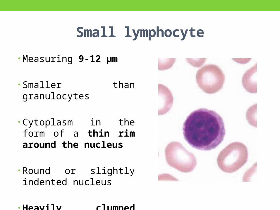
Small lymphocyte• Measuring 9-12 μm
• Smaller than granulocytes
• Cytoplasm in the form of a thin rim around the nucleus
• Round or slightly indented nucleus
• Heavily clumped deeply staining chromatin

Large lymphocyte• Measuring 12-15 μm
• Round in outline
• Nucleus is round to slightly indented with clumped chromatin
• Cytoplasm is more abundant than lymphocyte and is pale blue in color

Reactive lymphocytes (Downey cells)
•Have slightly larger nuclei with more open chromatin
• Abundant cytoplasm that may be irregular. (scalloping/skirting RBCs)
• Seen in • Infectious mononucleosis• Viral infections
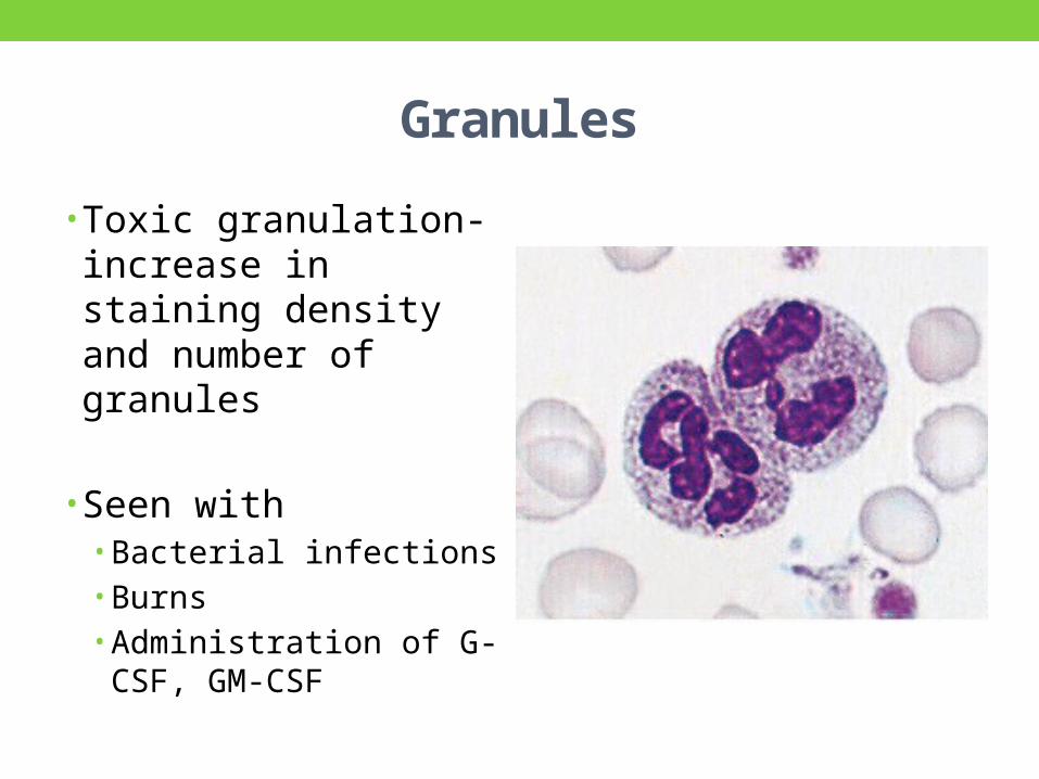
Granules• Toxic granulation- increase in staining density and number of granules
• Seen with • Bacterial infections• Burns• Administration of G-CSF, GM-CSF
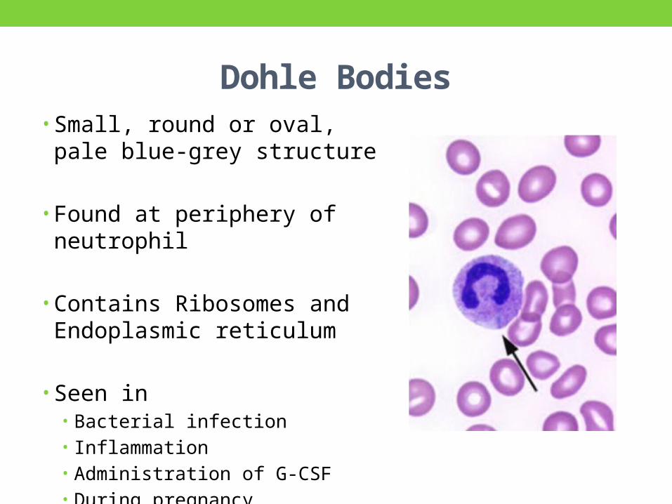
Dohle Bodies• Small, round or oval, pale blue-grey structure
• Found at periphery of neutrophil
• Contains Ribosomes and Endoplasmic reticulum
• Seen in • Bacterial infection• Inflammation • Administration of G-CSF• During pregnancy
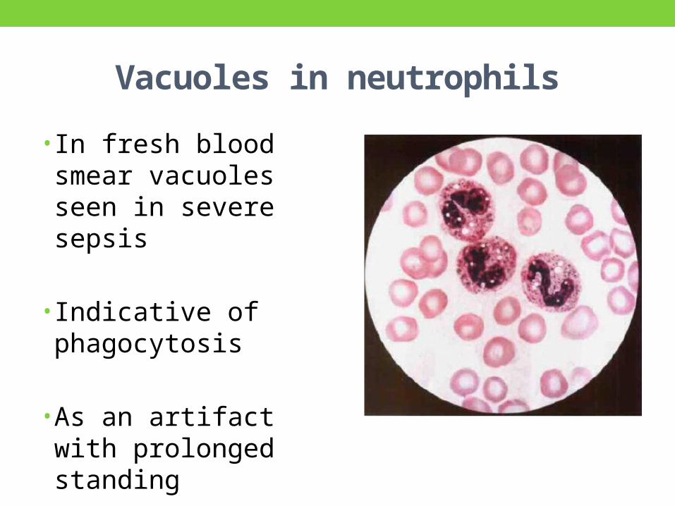
Vacuoles in neutrophils• In fresh blood smear vacuoles seen in severe sepsis
• Indicative of phagocytosis
• As an artifact with prolonged standing
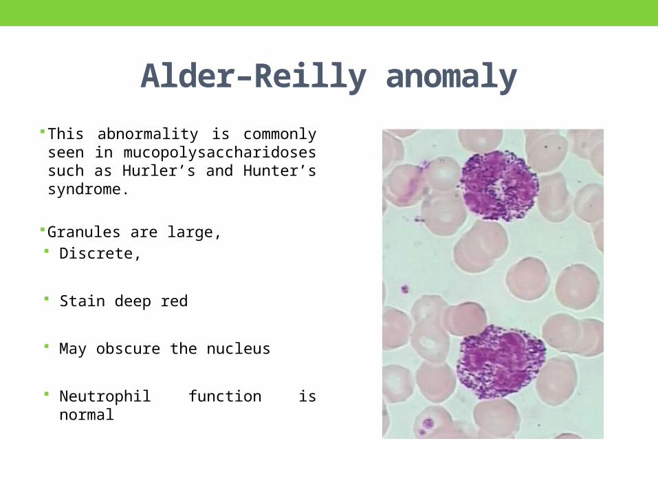
Alder–Reilly anomaly This abnormality is commonly
seen in mucopolysaccharidoses such as Hurler’s and Hunter’s syndrome.
Granules are large, Discrete,
Stain deep red
May obscure the nucleus
Neutrophil function is normal
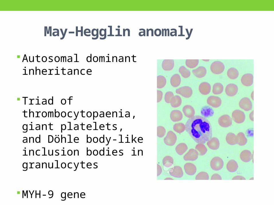
May–Hegglin anomalyAutosomal dominant inheritance
Triad of thrombocytopaenia, giant platelets, and Döhle body-like inclusion bodies in granulocytes
MYH-9 gene

Chédiak-Higashi Syndrome Rare autosomal recessive disease Immune deficiency, Poor resistance to bacterial
infections, Oculocutaneous albinism, Bleeding tendency, Multiple neurologic
abnormalities
Giant peroxidase-positive lysosomal granules in granulocytes
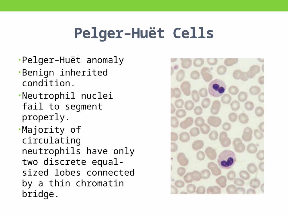
Pelger–Huët Cells• Pelger–Huët anomaly• Benign inherited condition.•Neutrophil nuclei fail to segment properly.•Majority of circulating neutrophils have only two discrete equal-sized lobes connected by a thin chromatin bridge.

Pseudo-Pelger cells• Pseudo-Pelger cells or the acquired Pelger–Huët anomaly• Acquired condition• Morphologically similar to Pelger–Huët anomaly• Seen in • Myelodysplastic syndromes,• Acute myeloid
leukaemia with dysplastic maturation,
• Occasionally inchronic myelogenous leukaemia

RECORDING RBC MORPHOLOGY
RBC morphology Scan area using ×100 (oil immersion).
Observe 10 fields.
Red cells are observed for Size, Shape, Hemoglobin content, Inclusions
Abnormal morphology Note that red cell morphology must be scanned in a good
counting area.

RBC• In the blood from healthy person RBCs are • Circular , Homogenous disc nearly of uniform size (7–8 µm)• Deep pink cytoplasm with Central pallor <1/3rd

Hypochromia•Decrease in hemoglobin content of RBC
• Increase in central pallor(>1/3rd)
•Decrease in MCH and MCHC
• Seen in various anaemias

Dimorphic anaemiaPresence of anisocytosis and anisochromia in the same film.
Seen in Coexistence of iron deficiency and megaloblastic anaemia
Sideroblastic anemia Some weeks after iron therapy for iron deficiency anemia
Hypochromic anemia after transfusion with normal cells

Dimorphic blood picture

Polychromatophilia• Blue grey tint of red cells
•Due to presence of residual RNA in young cells.
• Larger than normal and may lack central pallor
• Implies Reticulocytosis and therefore marrow response
Variation In Size • Anisocytosis- Variation in size of the red blood cells
•Normal MCV is -80-100 fl
•Microcytes ( MCV <80 fl)
•Macrocytes (MCV >100fl)
• Anisocytosis is a feature of most anemias.

Microcytes• Size of RBC is reduced (<80fl)
• Seen when hemoglobin synthesis is defective• Iron deficiency
anemia• Thalassemia• Anemia of chronic
disease• Sideroblastic anemia
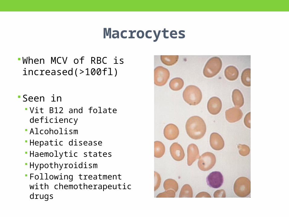
Macrocytes When MCV of RBC is increased(>100fl)
Seen in Vit B12 and folate
deficiency Alcoholism Hepatic disease Haemolytic states Hypothyroidism Following treatment with
chemotherapeutic drugs

Shape•Variation in shape is called Poikilocytosis.• It is of following types-•Elliptocytes•Spherocytes•Target cells•Schistocytes•Acanthocytes•Keratocytes•Echinocytes

Elliptocytes
• Elipitical in shapes
•Most abundant in hereditary elliptocytosis
• Seen in –• Iron deficiency anemia• Myelofibrosis with myeloid metaplasia• Megaloblastic anemia• Sickle cell anemia

Spherocytes• Nearly spherical • Diameter is smaller than normal• Lack central pale area or have a smaller , eccentric, pale area• Seen in • Hereditary spherocytosis• Some cases of autoimmune hemolytic anemia• Direct physical or chemical injury
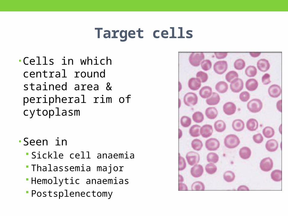
Target cells• Cells in which central round stained area & peripheral rim of cytoplasm
• Seen in Sickle cell anaemia Thalassemia major Hemolytic anaemias Postsplenectomy
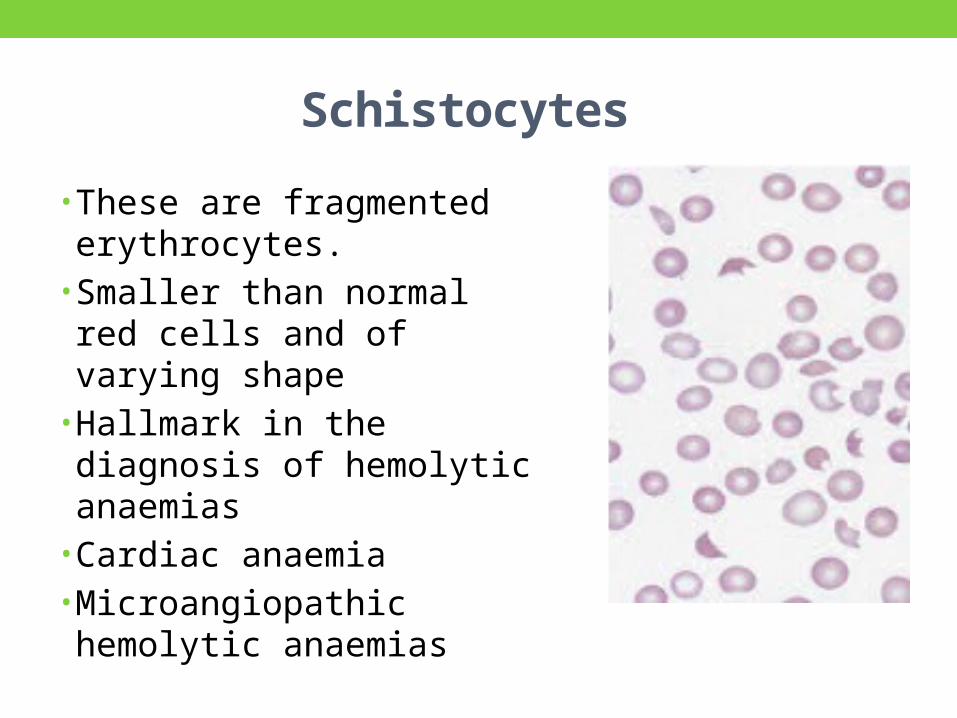
Schistocytes • These are fragmented erythrocytes.• Smaller than normal red cells and of varying shape•Hallmark in the diagnosis of hemolytic anaemias• Cardiac anaemia•Microangiopathic hemolytic anaemias
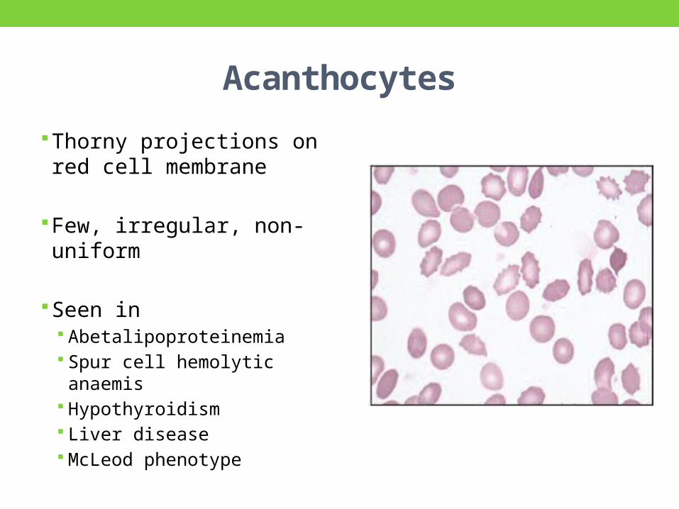
Acanthocytes Thorny projections on red cell membrane
Few, irregular, non-uniform
Seen in Abetalipoproteinemia Spur cell hemolytic anaemis Hypothyroidism Liver disease McLeod phenotype
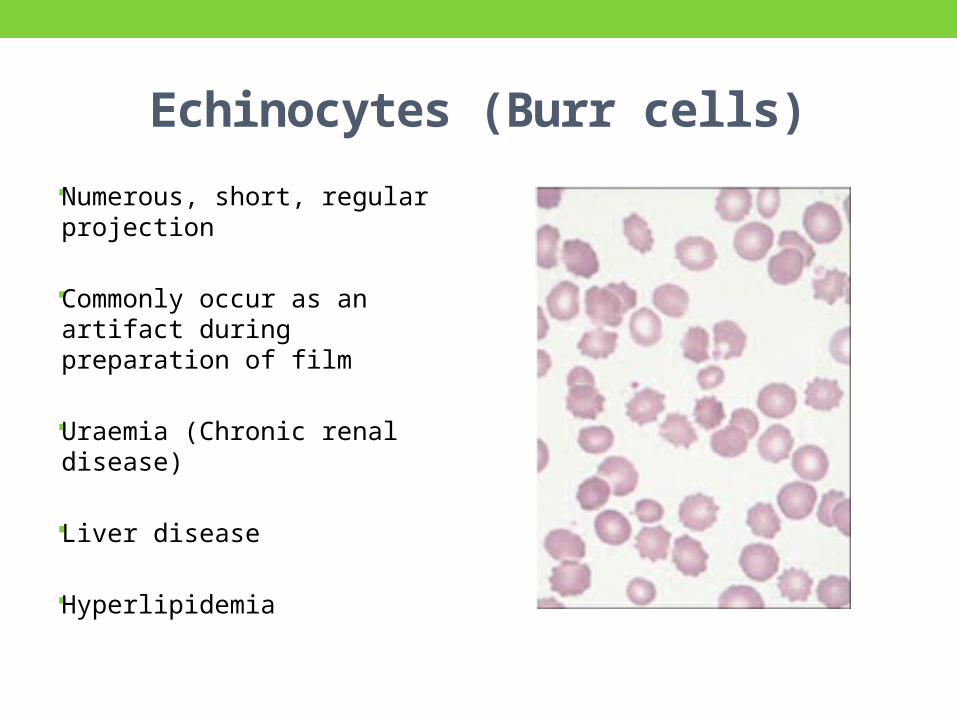
Echinocytes (Burr cells) Numerous, short, regular projection
Commonly occur as an artifact during preparation of film
Uraemia (Chronic renal disease)
Liver disease
Hyperlipidemia
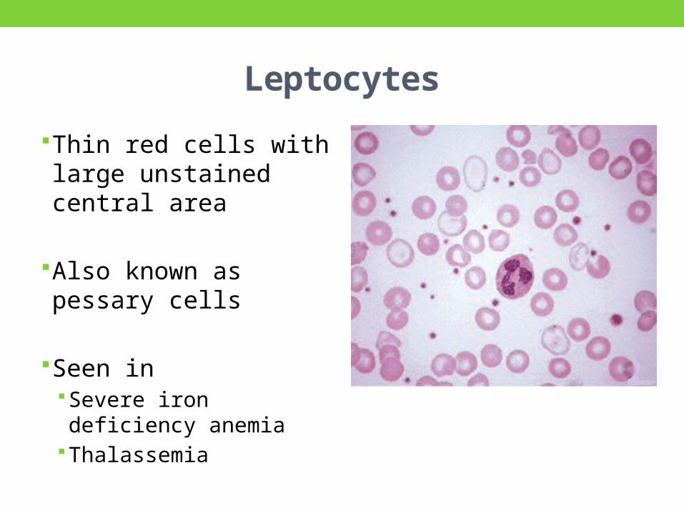
LeptocytesThin red cells with large unstained central area
Also known as pessary cells
Seen in Severe iron deficiency anemia
Thalassemia
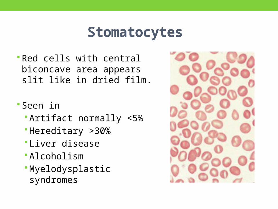
Stomatocytes Red cells with central biconcave area appears slit like in dried film.
Seen in Artifact normally <5% Hereditary >30% Liver disease Alcoholism Myelodysplastic syndromes

Sickle cellCells are sickle (boat shape) or crescent shape
Present in film of patient with homozygosity for HbS.
Usually absent in neonates and rare in patients with high Hb F percentage
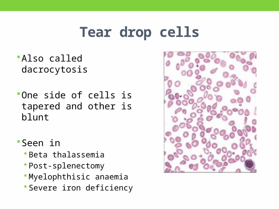
Tear drop cells Also called dacrocytosis
One side of cells is tapered and other is blunt
Seen in Beta thalassemia Post-splenectomy Myelophthisic anaemia Severe iron deficiency

RBC InclusionsName of Inclusion Content
Howell-Jolly body DNA
Basophilic stippling RNA
Pappenheimer body Iron
Heinz body (supravital only) Denatured hemoglobin
Crystals Hemoglobin-C
Cabot rings Mitotic spindle remnants
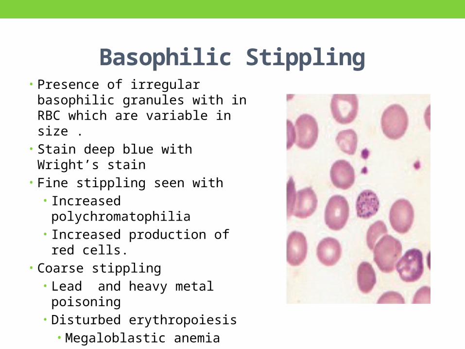
Basophilic Stippling• Presence of irregular basophilic
granules with in RBC which are variable in size .
• Stain deep blue with Wright’s stain
• Fine stippling seen with • Increased polychromatophilia• Increased production of red
cells. • Coarse stippling• Lead and heavy metal
poisoning• Disturbed erythropoiesis• Megaloblastic anemia • Thalassaemia• infection• liver disease
• Unstable Hb• Pyrimidine-5’-nucleotidase def.
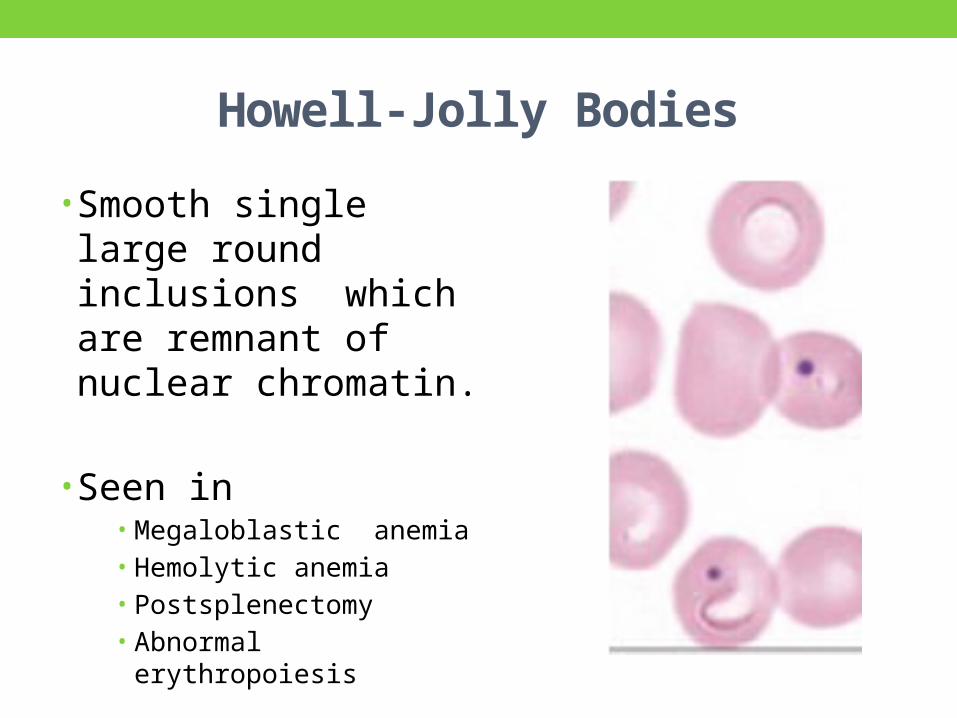
Howell-Jolly Bodies• Smooth single large round inclusions which are remnant of nuclear chromatin.
• Seen in • Megaloblastic anemia• Hemolytic anemia• Postsplenectomy• Abnormal erythropoiesis
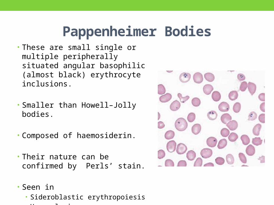
Pappenheimer Bodies• These are small single or multiple
peripherally situated angular basophilic (almost black) erythrocyte inclusions.
• Smaller than Howell–Jolly bodies.
• Composed of haemosiderin.
• Their nature can be confirmed by Perls’ stain.
• Seen in• Sideroblastic erythropoiesis• Hyposplenism• Myelodysplastic syndrome• Hemolytic anemia

Heinz bodies• Seen on supravital stains• Purple, blue, large, single or multiple
inclusions attached to the inner surface of the red blood cell.
• Represent precipitated normal or unstable hemoglobins.
• Seen in• Postsplenectomy• Oxidative stress• Glucose-6-phosphate
dehydrogenase deficiency,• Glutathione synthetase deficiency• Drugs• Toxins• Unstable hemoglobins

Cabot Rings• These are Ring shaped, figure of eight or loop shaped
• Red or Reddish purple with Wright’s stain and have no internal structure
• Observed in• Megaloblastic anaemia• Pernicious anemia• Lead poisoning
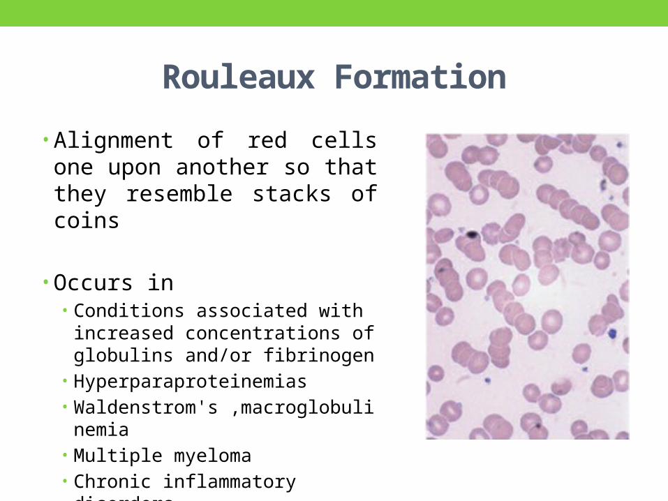
Rouleaux Formation• Alignment of red cells one upon another so that they resemble stacks of coins
• Occurs in• Conditions associated with
increased concentrations of globulins and/or fibrinogen
• Hyperparaproteinemias• Waldenstrom's ,macroglobuline
mia• Multiple myeloma• Chronic inflammatory disorders

Agglutination• It is more irregular and round clumping than linear rouleaux
• Cannot distinguish the outlines of individual RBCs
• Seen with cold agglutinin• Anti RBC antibody• Autoimmune hemolytic anemia• Macroglobulinemia

PLATELETS
Platelets• Use the oil immersion lens estimate the number of platelets per field.
• Look at 5-6 fields and take an average.
• Multiply the average by 15,000.
• Platelets per oil immersion field (OIF)• <7 platelets/OIF = decreased• 7 to 15 platelets/OIF = adequate• >15 platelets/OIF = increased

Platelets• Size -1-3µm
• Normal count – 1.5 to 4.5 lac/cmm
• Non nucleated derived from cytoplasmic fragments of Megakaryocytes
• Have an irregular outline and fine purple red granules

Thrombocytopenia Artifactual thrombocytopenia
Platelet clumping caused by anticoagulant-dependent immunoglobulin Platelet satellitismGiant platelets
Decreased platelet productionHypoplasia of megakaryocytesIneffective thrombopoiesisDisorders of thrombopoietic controlHereditary thrombocytopenias
Abnormal platelet distribution or pooling Disorders of the spleen (neoplastic, congestive, infiltrative, infectious, of unknown cause)HypothermiaDilution of platelets with massive transfusions
• Increased platelet destructionCaused by immunologic processes Autoimmune Idiopathic Secondary: Infections, pregnancy, collagen vascular disorders, lymphoproliferative disorders, drugs, miscellaneous Alloimmune Neonatal thrombocytopenia Posttransfusion purpura Thrombotic microangiopathies Disseminated intravascular coagulation Thrombotic thrombocytopenic purpura Hemolytic-uremic syndrome Platelet damage by abnormal vascular surfaces Miscellaneous Infection Massive blood transfusions
ThrombocytosisEssential thrombocythemia
CML
Reactive thrombocytosisPost infection Iron deficiency InflammationCollagen vascular disease
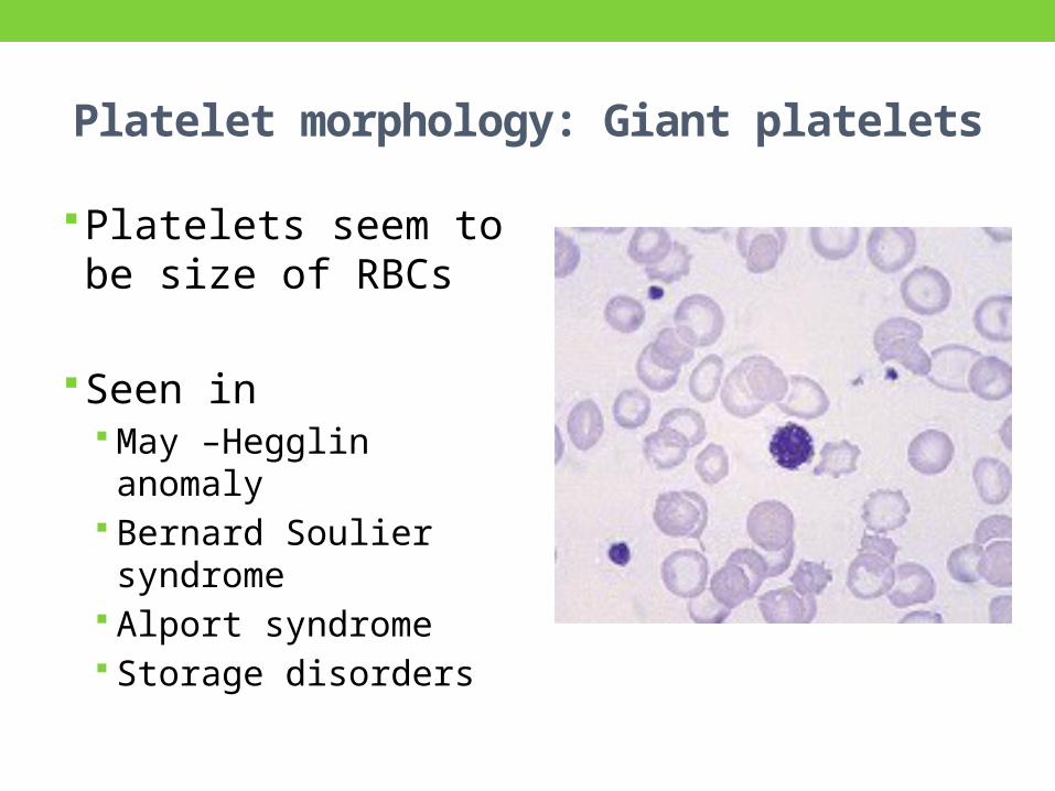
Platelet morphology: Giant platelets
Platelets seem to be size of RBCs
Seen in May –Hegglin anomaly
Bernard Soulier syndrome
Alport syndrome Storage disorders

HEMOPARASITES
Malaria Remains the gold standard for diagnosis
Giemsa stain distinguishes between species and life cycle stages
parasitemia is quantifiable
Threshold of detection thin film: 100 parasites/µl thick film: 5 -20 parasites/µl
Requirements: equipment, training, reagents, supervision
Simple, inexpensive yet labor-intensive

Plasmodium falciparum Infected rbcs are of normal size Multiple infections
Gametocytes: Mature Immature
Immature forms rarely Seen in peripheral blood
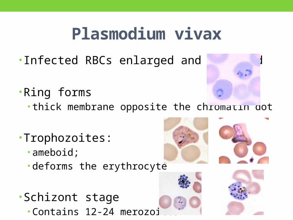
Plasmodium vivax• Infected RBCs enlarged and deformed
• Ring forms • thick membrane opposite the chromatin dot
• Trophozoites: • ameboid; • deforms the erythrocyte
• Schizont stage• Contains 12-24 merozoites
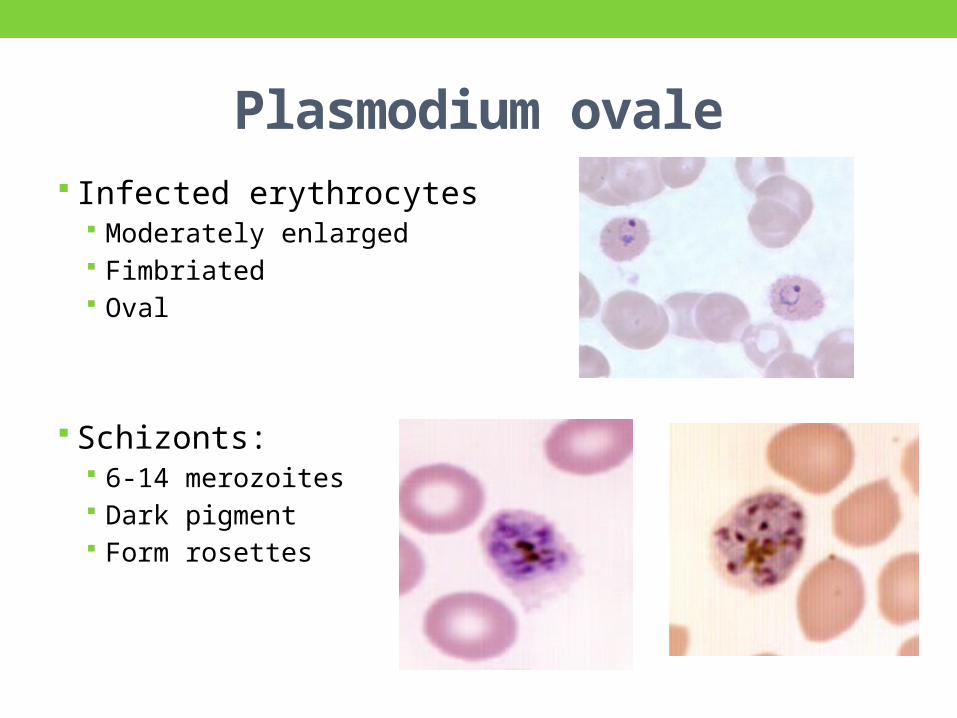
Plasmodium ovale Infected erythrocytes
Moderately enlarged Fimbriated Oval
Schizonts: 6-14 merozoites Dark pigment Form rosettes
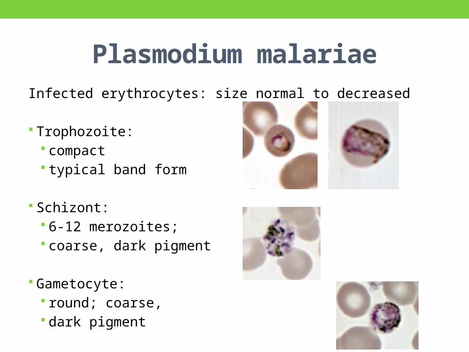
Plasmodium malariaeInfected erythrocytes: size normal to decreased
Trophozoite: compact typical band form
Schizont: 6-12 merozoites; coarse, dark pigment
Gametocyte: round; coarse, dark pigment
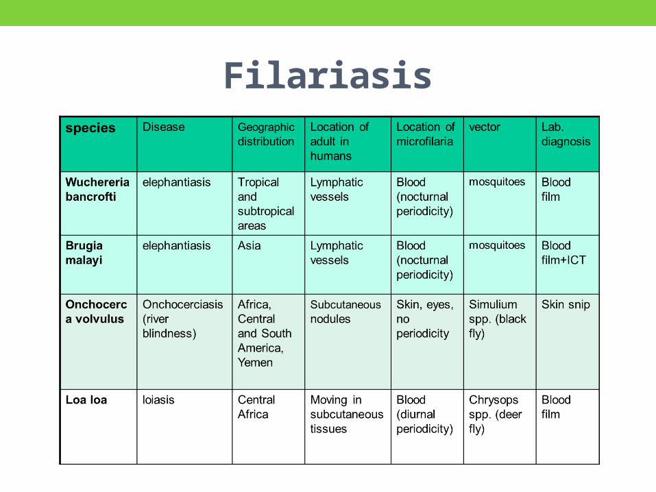
Filariasis Causes elephantiasis
Mainly caused by Wuchereria bancrofti and Brugia malayi
Pathology: Due to adult worm obstructing lymphatics.
Acute: lymphadenitis lymphatic varices
Chronic: lymphedema, hydrocele, chyluria.
Filariasis

Filariasis Detection of microfilariae in blood in early stages of the disease:
Blood film, Knott’s method (concentration of 1 ml of blood)
Best 10 pm to 2 am (nocturnal periodicity)

Trypanosomiasis• Causes African Sleeping sickness• Transmitted by tse tse fly

Babesia• Caused by species of the intraerythrocytic protozoan Babesia• B. microti• B. divergens
• Vector is tick
• Causes a malaria-like sickness
• Maltese cross appearance in erythrocytes

Thank You

Summarizing WBC parameters• STEP 1
WBC increased : leukocytosisWBC decreases: leukopenia
• STEP 2Relative differential count
• STEP 3Absolute Cell Counts
• STEP 4Examination for immature cellsYoung cells should not be seen in the peripheral blood smearImmature cells: possess a nucleusdo not lyse during testingcan be counted as WBC and falsely elevate WBC results
Summarizing RBC Parameters• Step1
Examne Hb an Hct for anemia or polycythemiaIf the RBC morphology is normal: Use rule of three to estimate the Hct
• Step 2MCV: to check and correlate to the morpholic apperance of the cells
• Step 3Examine MCHCDescribes how well the cells are filled with HbHypochromic, normochromic2 conditions when MCHC should be evaluated:1. spherocytosis: slight elevation2. lipemia/icterus: markedly increase

• Step 4Examine MCHCDescribes how well the cells are filled with HbHypochromic, normochromic2 conditions when MCHC should be evaluated:1. spherocytosis: slight elevation2. lipemia/icterus: markedly increase
• Step 5Morphology
1. Size2. Shape3. Inclusions4. Young rbcs5. Color6. Arrangement
Summarizing Platelet Parameters• Platelet count (x 109/L)• Mean Platelet Volume MPV, fl• Morphology

• White Blood Cells.1. Check for even distribution and estimate the
number present (also, look for any gross abnormalities present on the smear).
2. Perform the differential count. 3. Examine for morphologic abnormalities.

• Red Blood Cells, Examine for: 1.Size and shape. 2.Relative hemoglobin content. 3.Polychromatophilia. 4.Inclusions. 5. Rouleaux formation or agglutination
• Platelets. 1.Estimate number present. 2. Examine for morphologic abnormalities.
Related Documents