Percutaneous transthoracic localization of pulmonary nodules under C-arm cone-beam CT virtual navigation guidance Tae Ho Kim Chang Min Park Sang Min Lee H. Page McAdams Young Tae Kim Jin Mo Goo W ith the widespread utilization of low-dose computed tomography (CT) screening, detection of early lung cancers, which manifest as small solid nodules or ground- glass nodules (GGNs), has been possible. According to one large observational cohort study (1, 2), solitary noncalcified pulmonary nodules were detected at the rate of 15%–20% among the high-risk population screened on baseline screening CT and 15% of these nodules proved to be malignant (1, 2). Therefore, it is crucial to safely and accurately diagnose these suspicious nodules that have a high probability of malignancy. Bronchoscopic biopsy or percutaneous transthoracic needle biopsy (PTNB) using CT have been commonly used for tissue sampling and pathologic confirmation. However, these meth- ods sometimes show limitations for tissue sampling of small or faint lesions. Furthermore, their ability to distinguish between pulmonary adenocarcinomas and their preinvasive lesions has been questioned (3, 4). In this context, video-assisted thoracoscopic surgery (VATS) excision has been utilized in clinical practice. Although VATS excision can be more invasive than PTNB or bronchoscopic biopsy, it can certainly offer a definitive diagnosis for suspicious pulmonary nod- ules even if they are small or faint (5, 6). VATS excision also provides simultaneous treatment and diagnosis. However, resection of these targets through VATS can be very challenging, and com- plete resection of the targets sometimes proves impossible, especially when the target nodules are too small or faint for surgeons to detect (7). Therefore, prior to VATS excision for small or faint lung nodules, percutaneous transthoracic localization (PTL) has been commonly performed un- der imaging guidance using several marking devices or materials (8–10). Recently C-arm cone-beam CT (CBCT) and state-of-the-art CBCT virtual navigation systems have been introduced in the field of radiologic intervention (11–15). In particular, CBCT virtual navigation systems provide a virtual needle pathway as the needle approaches the target point 224 From the Department of Radiology (T.H.K., C.M.P. [email protected] S.M.L., J.M.G.), Seoul National University College of Medicine, and Institute of Radiation Medicine, Seoul National University Medical Research Center, Seoul, Korea; the Cancer Research Institute (C.M.P., J.M.G.), Seoul National University, Seoul, Korea; the Department of Radiology (C.M.P., H.P.M.), Duke University Medical Center, Durham, NC, USA; the Department of Thoracic Surgery (Y.T.K.), Seoul National University College of Medicine, Seoul, Korea. Received 14 July 2015; revision requested 30 July 2015; last revision received 21 September 2015; accepted 22 September 2015. Published online 22 March 2016. DOI 10.5152/dir.2015.15297 Diagn Interv Radiol 2016; 22:224–230 © Turkish Society of Radiology 2016 CHEST IMAGING ORIGINAL ARTICLE PURPOSE We aimed to describe our initial experience with percutaneous transthoracic localization (PTL) of pulmonary nodules using a C-arm cone-beam CT (CBCT) virtual navigation guidance system. METHODS From February 2013 to March 2014, 79 consecutive patients (mean age, 61±10 years) with 81 sol- id or ground-glass nodules (mean size, 12.36±7.21 mm; range, 4.8–25 mm) underwent PTLs prior to video-assisted thoracoscopic surgery (VATS) excision under CBCT virtual navigation guidance using lipiodol (mean volume, 0.18±0.04 mL). Their procedural details, radiation dose, and com- plication rates were described. RESULTS All 81 target nodules were successfully localized within 10 mm (mean distance, 2.54±3.24 mm) from the lipiodol markings. Mean number of CT acquisitions was 3.2±0.7, total procedure time was 14.6±5.14 min, and estimated radiation exposure during the localization was 5.21±2.51 mSv. Postprocedural complications occurred in 14 cases (17.3%); complications were minimal pneu- mothorax (n=10, 12.3%), parenchymal hemorrhage (n=3, 3.7%), and a small amount of hemop- tysis (n=1, 1.2%). All target nodules were completely resected; pathologic diagnosis included invasive adenocarcinoma (n=53), adenocarcinoma-in-situ (n=10), atypical adenomatous hyper- plasia (n=4), metastasis (n=7), and benign lesions (n=7). CONCLUSION PTL procedures can be performed safely and accurately under the guidance of a CBCT virtual navigation system.

Welcome message from author
This document is posted to help you gain knowledge. Please leave a comment to let me know what you think about it! Share it to your friends and learn new things together.
Transcript

Percutaneous transthoracic localization of pulmonary nodules under C-arm cone-beam CT virtual navigation guidance
Tae Ho KimChang Min Park Sang Min LeeH. Page McAdams Young Tae KimJin Mo Goo
With the widespread utilization of low-dose computed tomography (CT) screening, detection of early lung cancers, which manifest as small solid nodules or ground-glass nodules (GGNs), has been possible. According to one large observational
cohort study (1, 2), solitary noncalcified pulmonary nodules were detected at the rate of 15%–20% among the high-risk population screened on baseline screening CT and 15% of these nodules proved to be malignant (1, 2). Therefore, it is crucial to safely and accurately diagnose these suspicious nodules that have a high probability of malignancy.
Bronchoscopic biopsy or percutaneous transthoracic needle biopsy (PTNB) using CT have been commonly used for tissue sampling and pathologic confirmation. However, these meth-ods sometimes show limitations for tissue sampling of small or faint lesions. Furthermore, their ability to distinguish between pulmonary adenocarcinomas and their preinvasive lesions has been questioned (3, 4). In this context, video-assisted thoracoscopic surgery (VATS) excision has been utilized in clinical practice. Although VATS excision can be more invasive than PTNB or bronchoscopic biopsy, it can certainly offer a definitive diagnosis for suspicious pulmonary nod-ules even if they are small or faint (5, 6). VATS excision also provides simultaneous treatment and diagnosis. However, resection of these targets through VATS can be very challenging, and com-plete resection of the targets sometimes proves impossible, especially when the target nodules are too small or faint for surgeons to detect (7). Therefore, prior to VATS excision for small or faint lung nodules, percutaneous transthoracic localization (PTL) has been commonly performed un-der imaging guidance using several marking devices or materials (8–10).
Recently C-arm cone-beam CT (CBCT) and state-of-the-art CBCT virtual navigation systems have been introduced in the field of radiologic intervention (11–15). In particular, CBCT virtual navigation systems provide a virtual needle pathway as the needle approaches the target point
224
From the Department of Radiology (T.H.K., C.M.P. [email protected] S.M.L., J.M.G.), Seoul National University College of Medicine, and Institute of Radiation Medicine, Seoul National University Medical Research Center, Seoul, Korea; the Cancer Research Institute (C.M.P., J.M.G.), Seoul National University, Seoul, Korea; the Department of Radiology (C.M.P., H.P.M.), Duke University Medical Center, Durham, NC, USA; the Department of Thoracic Surgery (Y.T.K.), Seoul National University College of Medicine, Seoul, Korea.
Received 14 July 2015; revision requested 30 July 2015; last revision received 21 September 2015; accepted 22 September 2015.
Published online 22 March 2016.DOI 10.5152/dir.2015.15297
Diagn Interv Radiol 2016; 22:224–230
© Turkish Society of Radiology 2016
CHEST IMAGINGORIGINAL AR TICLE
PURPOSE We aimed to describe our initial experience with percutaneous transthoracic localization (PTL) of pulmonary nodules using a C-arm cone-beam CT (CBCT) virtual navigation guidance system.
METHODSFrom February 2013 to March 2014, 79 consecutive patients (mean age, 61±10 years) with 81 sol-id or ground-glass nodules (mean size, 12.36±7.21 mm; range, 4.8–25 mm) underwent PTLs prior to video-assisted thoracoscopic surgery (VATS) excision under CBCT virtual navigation guidance using lipiodol (mean volume, 0.18±0.04 mL). Their procedural details, radiation dose, and com-plication rates were described.
RESULTSAll 81 target nodules were successfully localized within 10 mm (mean distance, 2.54±3.24 mm) from the lipiodol markings. Mean number of CT acquisitions was 3.2±0.7, total procedure time was 14.6±5.14 min, and estimated radiation exposure during the localization was 5.21±2.51 mSv. Postprocedural complications occurred in 14 cases (17.3%); complications were minimal pneu-mothorax (n=10, 12.3%), parenchymal hemorrhage (n=3, 3.7%), and a small amount of hemop-tysis (n=1, 1.2%). All target nodules were completely resected; pathologic diagnosis included invasive adenocarcinoma (n=53), adenocarcinoma-in-situ (n=10), atypical adenomatous hyper-plasia (n=4), metastasis (n=7), and benign lesions (n=7).
CONCLUSIONPTL procedures can be performed safely and accurately under the guidance of a CBCT virtual navigation system.
Transthoracic localization of pulmonary nodules under CBCT guidance • 225
and additional navigation after the skin en-try site and the target point within the body are located using preprocedural CBCT data (11). This brand-new guiding system has the technical advantage of additional safety fea-tures, including accurate and flexible needle route selection based on three-dimensional (3D) volume CT data, real-time fluoroscopic navigation, and comfortable operating con-ditions through an open gantry system (11). Due to its inherent advantages and virtual navigation systems, CBCT has great potential to provide very accurate and safe localiza-tion of small or faint pulmonary nodules. To the best of our knowledge, there have been no studies dealing with CBCT virtual navi-gation-guided PTL of pulmonary nodules to date. Thus, the purpose of our study was to describe our initial experiences with PTL of pulmonary nodules using a CBCT virtual nav-igation guidance system.
MethodsThe Institutional Review Board of Seoul
National University Hospital approved this retrospective study with a waiver of the re-quirement for patients’ informed consent for inclusion into this study. Written in-formed consent for PTL under the guidance of a CBCT virtual navigation system was ob-tained from each patient after explanation of all potential risks and benefits.
Patients and nodules characteristicsFrom February 2013 to March 2014, 81
consecutive PTLs for 81 pulmonary nodules (mean size, 12.36±7.21 mm; range, 4.8–25mm) in 79 consecutive patients (37 males and 42 females; mean age, 61±10 years; range, 39–80 years) were included in this study. There were seven solid nodules, 48 pure GGNs, and 26 part-solid GGNs. Most of these pulmonary nodules (91.4%) present-ed ground glass opacity and because these characteristics have high risk of malignancy,
the surgeons requested lipiodol localization before VATS excision. The remaining seven solid nodules were requested to have lipi-odol localization, because they showed rel-atively small size (mean size, 7.7±1.86 mm) with suggestive findings of malignancy. The mean distance from nodule to the pleu-ra was 24.9±13 mm (range, 0.4–66 mm). Nodules were detected on screening CTs in 53 patients, during diagnostic work-ups for underlying malignancies in 23 patients, and were detected for symptom evaluation of blood-tingled sputum in three. There were 52 never-smokers, 22 ex-smokers, and five current smokers. The average smoking amount was 21.5 pack-years (range, 4–75 pack-years). Characteristics of the patients and nodules are summarized in Table 1.
Procedural details of CBCT virtual navigation-guided PTLs
All PTL procedures were performed un-der the guidance of CBCT virtual navigation system in combination with CBCT system
(AlluraXperFD20; Phillips Healthcare) and virtual guiding software (XperCT and Xper-Guide software; Phillips Healthcare) by, or under the supervision of, a chest radiologist (C.M.P. with 10 years of experience in im-age-guided thoracic intervention). All PTL procedures were performed on the same day or one day prior to VATS in collabora-tion with the thoracic surgery team. The procedural details are as follows:
1. Prior to the PTL procedure, the target nodule was carefully reviewed on the diag-nostic CT previously taken and the optimal and safest trajectory was decided by the operators.
2. For PTL procedures, each patient was positioned on a CBCT table in either a su-pine or prone position depending on the location of lung nodule (Fig. 1a). After prop-erly positioning the patient, preprocedural CBCT was performed in the region of inter-est during a single breath-hold in inspira-tion or expiration, as appropriate. Simulta-neously, C-arm rotated 240˚ in 5.2 seconds
Main points
• C-arm cone-beam CT (CBCT) virtual navigation systems based on three-dimensional CBCT data could guide percutaneous transthoracic localization for the small lung nodule very accurately.
• Virtual navigation guidance enables safe localization procedures for small lung nodules prior to video-assisted thoracoscopic sugery (VATS).
• Nodule localization with lipiodol can be performed safely and precisely using CBCT.
Figure 1. a, b. Patients were placed in supine or prone position according to the proper needle trajectory established based on the prior diagnostic CT (a). After local anesthesia, a 21-gauge needle was introduced to the target nodule under the guidance of fluoroscopy (b).
a b
Table 1. Nodule characteristics
Characteristics Values
Nodule size (mm), mean±SD 12.36±7.21 (range; 4.8–25)
Distance from nodule to pleura (mm), mean±SD 24.90±13 (range; 0.4–66)
Nodule feature (n)
Solid nodule 7
Pure GGN 48
Part-solid GGN 26
Location (n)
Right upper lobe 23
Right middle lobe 3
Right lower lobe 26
Left upper lobe 16
Left lower lobe 13
SD, standard deviation; GGN, ground-glass nodule; n, number of patients.

generating 312 images in a 512×512 matrix (Fig. 2a).
3. After the preprocedural CBCT was per-formed, the projectional images acquired were automatically transmitted to a com-mercially-available dedicated workstation (Xtravision; Philips Healthcare) and 3D CT images were reconstructed using 1 mm thick multiplanar reformations in axial, cor-onal, and sagittal planes with a total recon-struction time of 25 seconds. Subsequently, operators determined the proper skin entry site, target points of localization-marker placement, and trajectories with distance to target points using the virtual navigation software (XperGuide) (Fig. 2b). Target points of localization were determined within a 10 mm range around target lung nodules.
4. According to the needle trajectories determined using the virtual guidance soft-ware, C-arm gantry of the CBCT system au-tomatically rotated until the skin (red circle) and target points (green circle) were super-imposed making a “bull’s eye view” and the virtual navigation system virtually marked the skin entry site on fluoroscopy (Fig. 2c). After local anesthesia at the point of entry
with 2% lidocaine, a 21-gauge needle was carefully introduced to the intended local-ization site through the planned skin-entry site under fluoroscopic guidance, keeping the needle angle in the “bull’s eye view” (Fig. 1b). After the needle was placed in the planned target point, intraprocedural CBCT was performed to check the exact loca-tion of the needle tip around the targeted pulmonary nodule. If the needle tip was located exactly at the target point, the in-ner stylet of the needle was withdrawn and lipiodol (Guerbet Laboratories) was slowly and carefully injected through the needle under surveillance via fluoroscopy. Maxi-mum capacity of lipiodol was predefined as 0.2 mL (mean amount, 0.18±0.04 mL; range, 0.15–0.20 mL) considering the size of radiopaque markers of 1 cm and potential risk of lipiodol embolism (16). Also during lipiodol injection, we carefully monitored the movement of lipiodol droplets using fluoroscopy.
5. Afterwards, postprocedural CBCT was performed to identify the exact location of the lipiodol marker with respect to the tar-get pulmonary nodules (Fig. 2d). Postpro-
cedural complications were also carefully investigated with CBCT images. Blood pres-sure, heart rate, breathing rate, and the ar-terial oxygen saturation of all patients were closely monitored throughout the entire procedure.
Data analysisOne radiologist identified the procedur-
al records that included technical success or failure, diameter of lipiodol marker, dis-tance from nodule to lipiodol marker, num-ber of CT exams, total procedure time, radi-ation exposure through the PTL procedure, procedure-related complications, and the time interval between the PTL procedure and the VATS. Total procedural time was defined as the time period from the ad-ministration of local anesthesia to the end of postprocedure CBCT. Technical success was defined as the location of the lipiodol markers within a 10 mm range around tar-get nodules on postprocedural CBCT imag-es. Radiation exposure was also recorded during the entire PTL procedures (both the fluoroscopy dose and the CBCT dose) and total dose area product (DAP) during the
226 • May–June 2016 • Diagnostic and Interventional Radiology Kim et al.
Figures 2. a–e. A 63-year-old female who underwent a lower anterior resection due to colon cancer, followed by cone-beam CT (CBCT)-guided percutaneous transthoracic localization (PTL) for a 6 mm pure ground-glass nodule (GGN) in the right lower lobe of the lung. This nodule, undetectable by fluoroscopy, can be easily visualized through preprocedural CBCT (a). Proper skin entry site and target point of lipiodol marker placement were carefully planned using 3D CBCT data with virtual navigation guidance software (b). The CBCT virtual navigation system enables the automatic alignment of X-ray tube, target point and detector, providing “bull’s eye view,” then the needle was carefully introduced to the intended localization site under fluoroscopic guidance (c). Afterwards, lipiodol was slowly and carefully injected, with no procedure-related complications (d). Localized marker was easily detectable with intraoperative fluoroscopy before the excision of the target nodule and the lipiodol marker (e). This nodule was identified as adenocarcinoma-in-situ.
d
a
e
b c

Transthoracic localization of pulmonary nodules under CBCT guidance • 227
fluoroscopy and the CBCT acquisition was recorded for the absorbed radiation doses (mGy). Subsequently, DAPs were converted into effective doses using the conversion factor of 0.45 mSv/Gy∙cm2 obtained from our previous study (11).
Surgical findings, such as the visibility of the radiopaque lipiodol marker under intra-operative C-arm fluoroscopy and success in surgical resection of both the marker and the target nodule, were recorded (Fig. 2e). In addition, all pathologic reports of VATS specimens were reviewed to evaluate the final diagnosis of targeted lung nodules. All lesions were evaluated in terms of the pathologic diagnosis of the target nodule.
ResultsThe technical success rate of PTLs under
CBCT virtual navigation guidance was 100% (81/81). The mean diameter of lipiodol markers was 9.75±9.09 mm (range, 5–19 mm) and the distance from target nod-ules to lipiodol markers was 2.54±3.24 mm (range, 0–10 mm). Mean overall DAP during the total procedure was 11730±5463.6 mGy cm2. Thus, the mean effective dose during CBCT virtual navigation-guided PTLs was 5.21±2.51 mSv in the present study. The procedural records and radiation doses of the 81 cases are summarized in Table 2. Postprocedural complications occurred in 14 cases (17.3%). Small pneumothorax occurred in 10 cases (12.3%), parenchymal hemorrhage in three cases (3.7%), and a small amount of hemoptysis occurred in one case (1.2%). However, there were no serious complications that required any further procedures such as chest drainage catheter insertion or embolism events.
VATS excision was performed on the same day in 68 patients (84%) and the next day in 13 patients (16%) after PTLs. Lipiodol markers were identifiable in all cases under intraoperative fluoroscopy (n=81) and all radiopaque lipiodol and targeted nodules were successfully resected with a negative surgical margin through VATS. Pathological-ly, all nodules were definitively diagnosed. Diagnoses of the 81 nodules included in-vasive adenocarcinoma (n=53), adenocar-cinoma-in-situ (n=10), metastasis (n=7), atypical adenomatous hyperplasia (n=4), and nonspecific benign pathologies (n=7) (Table 3).
DiscussionIn this study, we found that all 81 PTL
procedures under CBCT virtual naviga-
tion guidance were successful, delivering lipiodol markers within 10 mm distance from the targeted pulmonary nodules be-fore VATS excision. Postprocedural com-plications were relatively low and total procedure time was short. These targeted nodules were successfully resected with a negative surgical margin through VATS.
VATS excision has several benefits, includ-ing shorter hospitalizations, less pain for patients, lower postoperative wound prob-lems, and the preservation of normal lung parenchyma compared with conventional
thoracic surgery. In general, the sensitivi-ty and specificity of malignancy through VATS excision is known to be 100% (5, 6). However, VATS excision can sometimes be very challenging, especially when the pul-monary nodules are too small, too faint, or too deeply located for surgeons to detect through palpation (7). In particular, pure or part-solid GGNs on CT, which represent ad-enocarcinoma containing lepidic compo-nents (or its preinvasive lesions), could be typical examples of this kind of challenge, even though these nodules are located just
Table 2. Procedural details of CBCT virtual navigation-guided PTLs
Characteristics Values
Technical success rate, n/N (%) 81/81 (100)
Amount of lipiodol (mL), mean±SD 0.18±0.04 (range; 0.15–0.2)
Diameter of lipiodol (mm), mean±SD 9.75±9.09 (range; 5–19)
Distance from nodule to marking (mm), mean±SD 2.54±3.24 (range; 0–10)
Patient’s position, n
Supine 22
Prone 59
Number of CBCT data acquisitions, mean±SD 3.2±0.7 (3–7)
Total procedure time (min), mean±SD 14.6±5.14 (range; 9–31)
Effective radiation dose (mSv), mean±SD 5.21±2.51 (range; 0.67–13.09)
Postprocedural complications, n (%)
Small pneumothorax 10 (12.3)
Parenchymal hemorrhage 3 (3.7)
Small hemoptysis 1 (1.2)
Time interval between PTL and operation, n (%)
Same day 68 (84)
Next day 13 (16)
CBCT, C-arm cone-beam computed tomography; PTL, percutaneous transthoracic localization; n, number of patients; SD, standard deviation.
Table 3. Pathologic diagnosis of nodules
Pathologic diagnosis Number of nodules
Invasive adenocarcinoma 53
Adenocarcinoma-in-situ 10
Metastatic nodule 7
Atypical adenomatous hyperplasia 4
Benign nodules 7
Smooth muscular proliferation 1
Nonspecific inflammation 2
Reactive hyperplasia 1
Fibrotic nodule 2
Anthracofibrotic lesion 1
beneath the pleura (17). Therefore, it is es-sential to perform PTL before VATS excision to guarantee the successful and effective resection of these faint or small, but clini-cally relevant lesions. Furthermore, operat-ing time has been reported to significantly decrease with PTL compared with patients not undergoing PTL (18).
Several previous studies reported the effectiveness of CT- or CT fluoroscopy (CTF)-guided PTL on small pulmonary nod-ules (10, 19). However, CT- or CTF-guided PTLs have several inherent disadvantages, such as small and deeply located closed gantry systems, limited real-time guidance, or limited imaging plane orientation of the needle trajectory, which has the potential to deteriorate the operators’ performance or confidence (20–23).
Recently, CBCT and CBCT virtual naviga-tion systems have been increasingly used in various fields of radiologic interventional procedures (24–26). CBCT-guided, or CBCT virtual navigation-guided PTNB have been reported to provide very excellent and, at least, equivalent diagnostic accuracy to state-of-the-art CTF-guided PTNB (11, 12, 27). In fact, given the flexible tilting and
open gantry system of CBCT, as well as im-proved guidance of needle trajectory based on 3D volume CT data, operators can per-form interventional procedures more com-fortably and confidently (Fig. 3). Also we believe that these kinds of technical advan-tages could be implemented even in local-ization of small lung nodules, which could lead to lower postprocedural complication rates, providing improved awareness and avoidance of dangerous organs or vessels (Fig. 4).
In our study, technical success rate of PTL procedures was 100%. Technical success of PTL meant that the lipiodol markers were placed within 10 mm from target nod-ules. The needle did not have to be exactly placed in the target nodule. So, the results of technical success of PTL may not be di-rectly applied to other CBCT-guided pro-cedures such as biopsy, where the needle should hit the nodule precisely.
Postprocedural complications occurred in 14 cases (17.3%) in our study. Small pneu-mothorax occurred in 10 cases (12.3%), parenchymal hemorrhage in three cases (3.7%), and a small amount of hemoptysis in one case (1.2%). Moreover, there were no
serious complications requiring any further procedures such as surgical intervention or needle aspiration. Considering that in CT- or CTF-guided biopsy, postprocedural pneu-mothorax has been reported to occur up to 17.7%–39% and hemoptysis in 5%–10% of all biopsy procedures (20, 28, 29), our post-procedural complication rates of 17.3% are relatively low. One previous study showed that the use of virtual navigation guidance was able to significantly reduce the occur-rence of biopsy-related pneumothorax and hemoptysis (12). We think that our relatively low complication rates could be attributed to the avoidance of emphysema and im-portant vascular structures along the nee-dle trajectory using accurate planning soft-ware and navigation systems.
In our study, the total procedure time was 14.6±5.14 min, which was substantial-ly shorter than the reported time of a pre-vious CT-guided localization study (25.6 min) (12). Considering that additional time was spent in our study to determine the tra-jectories using virtual navigation software, this result is noteworthy. We believe that CBCT and CBCT virtual navigation systems based on 3D CBCT data can enable easier and more precise needle guidance through PTL procedures in a more comfortable and confident manner, which can then lead to shorter total procedure times.
In our study, the mean effective dose of CBCT virtual navigation-guided PTL was 5.21 mSv. This result is similar to that of the previous CBCT-guided PTNB study (5.72 mSv) (11). As with the previous study (11), we performed CBCT approximately three times for each nodule (mean, 3.2±0.7): the initial CT for preprocedural planning, the second for the exact location of the coax-ial needle tip around target points, and the third for evaluation of postprocedural complication. These results of radiation exposure are similar, or somewhat lower, compared with those of CTF-guided PTNB (6.53 mSv) (20). We believe that the cur-rent radiation dose during CBCT virtual navigation-guided PTL is not a substantial hindrance. However, increased awareness and efforts such as the application of a rea-sonably small field-of-view or collimation should be used to reduce radiation expo-sure during PTL.
Although lipiodol-related acute pulmo-nary embolism has been reported (16), small amounts of lipiodol usage have been reported to be safe for the localization of small pulmonary nodules (30, 31). Kawana-
228 • May–June 2016 • Diagnostic and Interventional Radiology Kim et al.
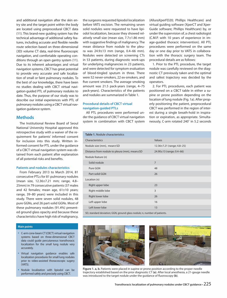
Figures 3. a–d. A 70-year-old male who had CBCT-guided PTL for two different GGNs. Chest CT for lung cancer screening shows an 8 mm pure GGN in the right upper lobe of the lung (a) and a 6 mm pure GGN in the right lower lobe of the lung (b). Panels (c) and (d) show the localization of GGNs under CBCT with virtual navigation guidance. Patient was placed in supine position for localization of the GGN in the right upper lobe (c). Then, patient’s position was changed to prone for localization of the GGN in the right lower lobe (d). GGN in the right upper lobe was identified as adenocarcinoma-in-situ, while the one in the right lower lobe was identified as atypical adenomatous hyperplasia.
c
a
d
b

Transthoracic localization of pulmonary nodules under CBCT guidance • 229
ka et al. (8) reported 0.3–0.5 mL of lipiodol marking was safe in their 107 cases without any lipiodol-related complication. In our study, 0.2 mL or less of lipiodol was used and there were no lipiodol-related compli-cations with this minimal usage. However, it should be kept in mind that embolization can be a possible complication of lipiodol use; the placement of the needle tip within the pulmonary vessel and any movement of lipiodol droplets during the lipiodol in-jection must be monitored carefully.
There are several limitations to our study. First, our study had a retrospective design with a relatively small study population. Sec-ond, we did not review the resected surgical specimen containing lipiodol markers, thus we did not evaluate the histopathologic effects of lipiodol on the nodule patholo-gy. However, according to several previous studies, lipiodol is a safe and inert material without adverse effects on the histologic diagnosis due to inflammatory reaction (17, 27, 32–34). In the present study, there were no cases in which a definitive pathologic di-agnosis had been hindered due to lipiodol
markers. Third, our study was not designed to directly compare CBCT virtual naviga-tion-guided PTL with CBCT-guided PTL with-out navigation systems or CT- or CTF-guid-ed PTL, thus we could not state definitively which guiding modality would be the best.
In conclusion, PTL can be performed un-der the guidance of CBCT virtual navigation system in a very accurate and safe manner.
Financial disclosure This work was supported by the Industrial Strate-
gic technology development program (grant num-ber: 10041618) funded by the Ministry of Trade In-dustry and Energy (MI).
Conflict of interest disclosureThe authors declared no conflicts of interest.
References1 Henschke CI, McCauley DI, Yankelevitz DF, et al.
Early lung cancer action project: overall design and findings from baseline screening. Lancet 1999; 354:99–105. [CrossRef]
2 Henschke CI, Yankelevitz DF, Mirtcheva R, Mc-Guinness G, McCauley D, Miettinen OS. CT screening for lung cancer: frequency and signifi-cance of part-solid and nonsolid nodules. AJR Am J Roentgenol 2002; 178:1053–1057. [CrossRef]
3 Lal H, Neyaz Z, Nath A, Borah S. CT-guided per-cutaneous biopsy of intrathoracic lesions. Ko-rean J Radiol 2012:13:210–226. [CrossRef]
4 Kim H, Park CM, Koh JM, Lee SM, Goo JM. Pul-monary subsolid nodules: what radiologists need to know about the imaging features and management strategy. Diagn Interv Radiol 2014; 20:47–57.
5 Hirai S, Hamanaka Y, Mitsui N, Morifuji K, Ue-gami S. Role of video-assisted thoracic surgery for the diagnosis of indeterminate pulmonary nodule. Ann Thorac Cardiovasc Surg 2006; 12:388–392.
6 Chen S, Zhou J, Zhang J, et al. Video-assisted thoracoscopic solitary pulmonary nodule re-section after CT-guided hookwire localization: 43 cases report and literature review. Surg En-dosc 2010; 25:1723–1729. [CrossRef]
7 Karasaki T, Nakajima J, Murakawa T, et al. Vid-eo-assisted thoracic surgery lobectomy pre-serves more latissimus dorsi muscle than con-ventional surgery. Interact Cardiovasc Thorac Surg 2008; 8:316–320. [CrossRef]
8 Kawanaka K, Nomori H, Mori T, et al. Marking of small pulmonary nodules before thoraco-scopic resection. Acad Radiol 2009; 16:39–45. [CrossRef]
9 Zaman M, Bilal H, Woo CY, Tang A. In patients undergoing video-assisted thoracoscopic sur-gery excision, what is the best way to locate a subcentimetre solitary pulmonary nodule in order to achieve successful excision? Inter-act Cardiovasc Thorac Surg 2012; 15:266–272. [CrossRef]
10 Lee NK, Park CM, Kang CH, et al. CT-guided percutaneous transthoracic localization of pul-monary nodules prior to video-assisted tho-racoscopic surgery using barium suspension. Korean J Radiol 2012; 13:694–701. [CrossRef]
11 Choo JY, Park CM, Lee NK, Lee SM, Lee HJ, Goo JM. Percutaneous transthoracic needle biopsy of small (≤1 cm) lung nodules under C-arm cone-beam CT virtual navigation guidance. Eur Radiol 2012; 23:712–719. [CrossRef]
12 Lee SM, Park CM, Lee KH, Bahn YE, Kim JI, Goo JM. C-arm cone-beam CT-guided percutane-ous transthoracic needle biopsy of lung nod-ules: clinical experience in 1108 patients. Ra-diology 2014; 271:291–300. [CrossRef]
13 Jin KN, Park CM, Goo JM, et al. Initial experi-ence of percutaneous transthoracic needle bi-opsy of lung nodules using C-arm cone-beam CT systems. Eur Radiol 2010; 20:2108–2115. [CrossRef]
14 Choi JW, Park CM, Goo JM, et al. C-Arm cone-beam CT–guided percutaneous transthoracic needle biopsy of small (≤ 20 mm) lung nod-ules: diagnostic accuracy and complications in 161 patients. AJR Am J Roentgenol 2012; 199:W322–W330. [CrossRef]
15 Wallace MJ, Kuo MD, Glaiberman C, Binkert CA, Orth RC, Soulez G. Three-dimensional C-arm cone-beam CT: applications in the intervention-al suite. J Vasc Interv Radiol 2008; 19:799–813. [CrossRef]
16 Papiamonis NE, Matrella E, Blevrakis EG, Kou-roumalis EA. Acute pulmonary embolism follow-ing N-butyl-cyanoacrylate endoscopic injection sclerotherapy. Ann Gastroenterol 2012; 25:261.
Figures 4. a–d. A 51-year-old female who had CBCT-guided PTL for a 20 mm incidentally-detected pure GGN in the left upper lobe of the lung. This GGN is located very close to subsegmental pulmonary vessels (a). Prior to PTL (b), thorough planning was done to achieve a safe needle trajectory avoiding the abovementioned pulmonary vessels. Needle insertion was performed under CBCT with virtual navigation guidance. Finally, lipiodol was carefully injected through the needle (c), forming a radiopaque marker. There were no procedure-related complications. During the video-assisted thoracoscopic surgery, the localized marker was easily detectable by intraoperative fluoroscopy. The localized marker is also easily detectable by fluoroscopy in the excised specimen (d). This nodule was proven to be a minimally-invasive adenocarcinoma.
c
a
d
b

17 Watanabe K, Nomori H, Ohtsuka T, Kaji M, Naruke T, Suemasu K. Usefulness and compli-cations of computed tomography-guided lipi-odol marking for fluoroscopy-assisted thoraco-scopic resection of small pulmonary nodules: experience with 174 nodules. J Thorac Cardio-vasc Surg 2006; 132:320–324. [CrossRef]
18 Finley RJ, Mayo JR, Grant K, et al. Preoperative computed tomography–guided microcoil localization of small peripheral pulmonary nodules: a prospective randomized controlled trial. J Thorac Cardiovasc Surg 2015; 149:26–32. [CrossRef]
19 Heck SL, Blom P, Berstad A. Accuracy and com-plications in computed tomography fluorosco-py-guided needle biopsies of lung masses. Eur Radiol 2006; 16:1387–1392. [CrossRef]
20 Kim GR, Hur J, Lee SM, et al. CT fluorosco-py-guided lung biopsy versus conventional CT-guided lung biopsy: a prospective con-trolled study to assess radiation doses and diagnostic performance. Eur Radiol 2011; 21:232–239. [CrossRef]
21 Paulson EK, Sheafor DH, Enterline DS, McAd-ams HP, Yoshizumi TT. CT fluoroscopy-guided interventional procedures: techniques and radiation dose to radiologists. Radiology 2001; 220:161–167. [CrossRef]
22 Silverman SG, Tuncali K, Adams DF, Nawfel RD, Zou KH, Judy PF. CT fluoroscopy-guided ab-dominal interventions: techniques, results, and radiation exposure. Radiology 1999; 212:673–681. [CrossRef]
23 Nawfel RD, Judy PF, Silverman SG, Hooton S, Tuncali L, Adams DF. Patient and personel exposure during CT Fluoroscopy-guided in-terventional procedures. Radiology 2000; 216:180–184. [CrossRef]
24 Cazzato RL, Battistuzzi JB, Catena V, et al. Cone-beam computed tomography (CBCT) versus CT in lung ablation procedure: which is faster? Cardiovasc Intervent Radiol 2015; 38:1231–1236. [CrossRef]
25 Cazzato RL, Buy X, Alberti N, Fonck M, Grasso RF, Palussière J. Flat-pane cone-beam CT-guid-ed radiofrequency ablation of very small (≤ 1.5cm) liver tumors: technical note on a preliminary experience. Cardiovasc Intervent Radiol 2015; 38:206–212. [CrossRef]
26 Rafiei P, Kim SK, Kamran M, Saad NE. Retro-spective study in 40 patients of utility of C-arm FDCT as an adjunctive modality in technical-ly challenging image-guided percutaneous drainage procedures. Cardiovasc Intervent Ra-diol 2015; 38:1589–1594. [CrossRef]
27 Hwang HS, Chung MJ, Lee JW, Shin SW, Lee KS. C-arm cone-beam CT-guided percutaneous transthoracic lung biopsy: usefulness in eval-uation of small pulmonary nodules. AJR Am J Roentgenol 2010; 195:W400–407. [CrossRef]
28 Priola AM, Priola SM, Cataldi A, et al. Diagnostic accuracy and complication rate of CT-guided fine needle aspiration biopsy of lung lesions: a study based on the experience of the cyto-pathologist. Acta Radiol 2010; 51:527–533. [CrossRef]
29 Ko JP, Shepard JO, Drucker EA, et al. Factors influencing pneumothorax rate at lung biop-sy: Are dwell time and angle of pleural punc-ture contributing factors? Radiology 2001; 218:491–496. [CrossRef]
30 Jin KN, Lee KW, Kim TJ, Song YS, Kim DI. Com-puted tomography guided percutaneous in-jection of a mixture of lipiodol and methylene blue in rabbit lungs: evaluation of localiza-tion ability for video-assisted thoracoscopic surgery. J Korean Med Sci 2014; 29:129–136. [CrossRef]
31 Kim YD, Jeong YJ, I H, et al. Localization of pul-monary nodules with lipiodol prior to thora-coscopic surgery. Acta Radiol 2011; 52:64–69. [CrossRef]
32 Nomori H, Horio H, Naruke T, Suemasu K. Flu-oroscopy-assisted thracoscopic resection of lung nodules marked with lipiodol. Ann Thorac Surg 2002; 74:170–173. [CrossRef]
33 Kang DY, Kim HK, Kim YK, Yong HS, Kang EY, Choi YH. Needlescopy-assisted resection of pulmonary nodule after dual localisation. Eur Respir J 2010; 37:13–17. [CrossRef]
34 Yamagami T, Miura H, Yoshimatsu R, et al. Ex-perience of fluoroscopy-aided thoracoscopic resection of pulmonary nodule localised with Lipiodol in a child. J Med Imaging Radiat Oncol 2011; 55:401–403.[CrossRef]
230 • May–June 2016 • Diagnostic and Interventional Radiology Kim et al.
Related Documents