Peptidoglycan Recycling in Gram-Positive Bacteria Is Crucial for Survival in Stationary Phase Marina Borisova, a Rosmarie Gaupp, b Amanda Duckworth, a Alexander Schneider, a Désirée Dalügge, a Maraike Mühleck, a Denise Deubel, a * Sandra Unsleber, a Wenqi Yu, a * Günther Muth, a Markus Bischoff, b Friedrich Götz, a Christoph Mayer a Department of Biology, Interfaculty Institute of Microbiology and Infection Medicine, University of Tübingen, Tübingen, Germany a ; Institute of Medical Microbiology and Hygiene, University of Saarland, Homburg, Saar, Saarland, Germany b * Present address: Denise Deubel, Institute for Pharmaceutical Sciences, Albert-Ludwigs University of Freiburg, Germany; Wenqi Yu, Department of Microbiology, University of Chicago, Chicago, Illinois, USA. ABSTRACT Peptidoglycan recycling is a metabolic process by which Gram-negative bacteria reutilize up to half of their cell wall within one generation during vegetative growth. Whether peptidoglycan recycling also occurs in Gram-positive bacteria has so far remained unclear. We show here that three Gram-positive model organisms, Staphylococcus aureus, Bacillus subtilis, and Streptomyces coelicolor, all recycle the sugar N-acetylmuramic acid (MurNAc) of their peptidoglycan during growth in rich me- dium. They possess MurNAc-6-phosphate (MurNAc-6P) etherase (MurQ in E. coli) enzymes, which are responsible for the intra- cellular conversion of MurNAc-6P to N-acetylglucosamine-6-phosphate and D-lactate. By applying mass spectrometry, we ob- served accumulation of MurNAc-6P in MurNAc-6P etherase deletion mutants but not in either the isogenic parental strains or complemented strains, suggesting that MurQ orthologs are required for the recycling of cell wall-derived MurNAc in these bac- teria. Quantification of MurNAc-6P in murQ cells of S. aureus and B. subtilis revealed small amounts during exponential growth phase (0.19 nmol and 0.03 nmol, respectively, per ml of cells at an optical density at 600 nm [OD 600 ] of 1) but large amounts during transition (0.56 nmol and 0.52 nmol) and stationary (0.53 nmol and 1.36 nmol) phases. The addition of MurNAc to murQ cultures greatly increased the levels of intracellular MurNAc-6P in all growth phases. The murQ mutants of S. aureus and B. subtilis showed no growth deficiency in rich medium compared to the growth of the respective parental strains, but intriguingly, they had a severe survival disadvantage in late stationary phase. Thus, although peptidoglycan recycling is apparently not essential for the growth of Gram-positive bacteria, it provides a benefit for long-term survival. IMPORTANCE The peptidoglycan of the bacterial cell wall is turned over steadily during growth. As peptidoglycan fragments were found in large amounts in spent medium of exponentially growing Gram-positive bacteria, their ability to recycle these fragments has been questioned. We conclusively showed recycling of the peptidoglycan component MurNAc in different Gram- positive model organisms and revealed that a MurNAc-6P etherase (MurQ or MurQ ortholog) enzyme is required in this pro- cess. We further demonstrated that recycling occurs predominantly during the transition to stationary phase in S. aureus and B. subtilis, explaining why peptidoglycan fragments are found in the medium during exponential growth. We quantified the in- tracellular accumulation of recycling products in MurNAc-6P etherase gene mutants, revealing that about 5% and 10% of the MurNAc of the cell wall per generation is recycled in S. aureus and B. subtilis, respectively. Importantly, we showed that MurNAc recycling and salvaging does not sustain growth in these bacteria but is used to enhance survival during late stationary phase. Received 24 May 2016 Accepted 8 September 2016 Published 11 October 2016 Citation Borisova M, Gaupp R, Duckworth A, Schneider A, Dalügge D, Mühleck M, Deubel D, Unsleber S, Yu W, Muth G, Bischoff M, Götz F, Mayer C. 2016. Peptidoglycan recycling in Gram-positive bacteria is crucial for survival in stationary phase. mBio 7(5):e00923-16. doi:10.1128/mBio.00923-16. Editor Julian E. Davies, University of British Columbia Copyright © 2016 Borisova et al. This is an open-access article distributed under the terms of the Creative Commons Attribution 4.0 International license. Address correspondence to Christoph Mayer, [email protected]. P eptidoglycan (PGN) encases the bacterial cell, forming a huge, netlike, turgor-resisting and shape-maintaining envelope structure that is composed of glycan strands of two alternating -1,4-linked sugars, N-acetylglucosamine (GlcNAc) and N- acetylmuramic acid (MurNAc), cross-linked by short peptides (1). The PGN is remarkably dynamic and is constantly remodeled, degraded, and rebuilt during bacterial growth and cell division (2, 3). As an inherent part of this process, a significant portion of the PGN is continuously excised from the cell wall by the activity of endogenous lytic enzymes (autolysins) and released into the me- dium in a process termed “turnover” (3–7). The reason for the continuous degradation of the PGN is still unclear, but appar- ently, a minimal set of autolytic enzymes is essential for bacterial growth, and PGN turnover may be intrinsically coupled with cell elongation and division (2, 5). Early studies showed that the Gram-negative bacterium Escherichia coli breaks down about half of its PGN within one generation during exponential growth (8, 9). Gram-negative bacteria possess a thin PGN layer embedded within an inner and an outer membrane, and thus, PGN turnover products are mostly retained within the periplasm, from where RESEARCH ARTICLE crossmark September/October 2016 Volume 7 Issue 5 e00923-16 ® mbio.asm.org 1 on March 30, 2019 by guest http://mbio.asm.org/ Downloaded from

Welcome message from author
This document is posted to help you gain knowledge. Please leave a comment to let me know what you think about it! Share it to your friends and learn new things together.
Transcript
Peptidoglycan Recycling in Gram-Positive Bacteria Is Crucial forSurvival in Stationary Phase
Marina Borisova,a Rosmarie Gaupp,b Amanda Duckworth,a Alexander Schneider,a Désirée Dalügge,a Maraike Mühleck,a
Denise Deubel,a* Sandra Unsleber,a Wenqi Yu,a* Günther Muth,a Markus Bischoff,b Friedrich Götz,a Christoph Mayera
Department of Biology, Interfaculty Institute of Microbiology and Infection Medicine, University of Tübingen, Tübingen, Germanya; Institute of Medical Microbiology andHygiene, University of Saarland, Homburg, Saar, Saarland, Germanyb
* Present address: Denise Deubel, Institute for Pharmaceutical Sciences, Albert-Ludwigs University of Freiburg, Germany; Wenqi Yu, Department of Microbiology, University ofChicago, Chicago, Illinois, USA.
ABSTRACT Peptidoglycan recycling is a metabolic process by which Gram-negative bacteria reutilize up to half of their cell wallwithin one generation during vegetative growth. Whether peptidoglycan recycling also occurs in Gram-positive bacteria has sofar remained unclear. We show here that three Gram-positive model organisms, Staphylococcus aureus, Bacillus subtilis, andStreptomyces coelicolor, all recycle the sugar N-acetylmuramic acid (MurNAc) of their peptidoglycan during growth in rich me-dium. They possess MurNAc-6-phosphate (MurNAc-6P) etherase (MurQ in E. coli) enzymes, which are responsible for the intra-cellular conversion of MurNAc-6P to N-acetylglucosamine-6-phosphate and D-lactate. By applying mass spectrometry, we ob-served accumulation of MurNAc-6P in MurNAc-6P etherase deletion mutants but not in either the isogenic parental strains orcomplemented strains, suggesting that MurQ orthologs are required for the recycling of cell wall-derived MurNAc in these bac-teria. Quantification of MurNAc-6P in �murQ cells of S. aureus and B. subtilis revealed small amounts during exponentialgrowth phase (0.19 nmol and 0.03 nmol, respectively, per ml of cells at an optical density at 600 nm [OD600] of 1) but largeamounts during transition (0.56 nmol and 0.52 nmol) and stationary (0.53 nmol and 1.36 nmol) phases. The addition ofMurNAc to �murQ cultures greatly increased the levels of intracellular MurNAc-6P in all growth phases. The �murQ mutantsof S. aureus and B. subtilis showed no growth deficiency in rich medium compared to the growth of the respective parentalstrains, but intriguingly, they had a severe survival disadvantage in late stationary phase. Thus, although peptidoglycan recyclingis apparently not essential for the growth of Gram-positive bacteria, it provides a benefit for long-term survival.
IMPORTANCE The peptidoglycan of the bacterial cell wall is turned over steadily during growth. As peptidoglycan fragmentswere found in large amounts in spent medium of exponentially growing Gram-positive bacteria, their ability to recycle thesefragments has been questioned. We conclusively showed recycling of the peptidoglycan component MurNAc in different Gram-positive model organisms and revealed that a MurNAc-6P etherase (MurQ or MurQ ortholog) enzyme is required in this pro-cess. We further demonstrated that recycling occurs predominantly during the transition to stationary phase in S. aureus andB. subtilis, explaining why peptidoglycan fragments are found in the medium during exponential growth. We quantified the in-tracellular accumulation of recycling products in MurNAc-6P etherase gene mutants, revealing that about 5% and 10% of theMurNAc of the cell wall per generation is recycled in S. aureus and B. subtilis, respectively. Importantly, we showed thatMurNAc recycling and salvaging does not sustain growth in these bacteria but is used to enhance survival during late stationaryphase.
Received 24 May 2016 Accepted 8 September 2016 Published 11 October 2016
Citation Borisova M, Gaupp R, Duckworth A, Schneider A, Dalügge D, Mühleck M, Deubel D, Unsleber S, Yu W, Muth G, Bischoff M, Götz F, Mayer C. 2016. Peptidoglycanrecycling in Gram-positive bacteria is crucial for survival in stationary phase. mBio 7(5):e00923-16. doi:10.1128/mBio.00923-16.
Editor Julian E. Davies, University of British Columbia
Copyright © 2016 Borisova et al. This is an open-access article distributed under the terms of the Creative Commons Attribution 4.0 International license.
Address correspondence to Christoph Mayer, [email protected].
Peptidoglycan (PGN) encases the bacterial cell, forming a huge,netlike, turgor-resisting and shape-maintaining envelope
structure that is composed of glycan strands of two alternating�-1,4-linked sugars, N-acetylglucosamine (GlcNAc) and N-acetylmuramic acid (MurNAc), cross-linked by short peptides(1). The PGN is remarkably dynamic and is constantly remodeled,degraded, and rebuilt during bacterial growth and cell division (2,3). As an inherent part of this process, a significant portion of thePGN is continuously excised from the cell wall by the activity ofendogenous lytic enzymes (autolysins) and released into the me-
dium in a process termed “turnover” (3–7). The reason for thecontinuous degradation of the PGN is still unclear, but appar-ently, a minimal set of autolytic enzymes is essential for bacterialgrowth, and PGN turnover may be intrinsically coupled with cellelongation and division (2, 5). Early studies showed that theGram-negative bacterium Escherichia coli breaks down about halfof its PGN within one generation during exponential growth (8,9). Gram-negative bacteria possess a thin PGN layer embeddedwithin an inner and an outer membrane, and thus, PGN turnoverproducts are mostly retained within the periplasm, from where
RESEARCH ARTICLE
crossmark
September/October 2016 Volume 7 Issue 5 e00923-16 ® mbio.asm.org 1
on March 30, 2019 by guest
http://mbio.asm
.org/D
ownloaded from
they are instantly recovered and salvaged (7, 8). Thus, only smallamounts of PGN turnover products were found in the medium,while about 45% of the mature PGN was recovered in one gener-ation in a process called cell wall or PGN recycling (8, 9). Ueharaand Park later revealed that E. coli continuously recycles about30% of newly synthesized septal PGN during cell division, andthey calculated that about 60% of the PGN of the sidewall is recy-cled, taking into account that the PGN of the cell poles is basicallyinert (10). To quantify PGN turnover and recycling in E. coli, cellwalls were radioactively prelabeled and the amounts of radioactivesoluble products that were released into the medium or accumu-lated in recycling mutants were determined (8–10).
PGN recycling is not essential, at least under laboratory condi-tions, and why this pathway was kept on the chromosomes ofmost bacteria remained enigmatic (7). The PGN recycling metab-olism of Gram-negative bacteria had attracted much attention dueto a connection with AmpC-type �-lactamase induction (9) and,more recently, fosfomycin antibiotic resistance (11, 12). It is nowreasonably well understood (for reviews, see references 3, 7, 13,and 14). In brief, the major cell wall-recycling products of Gram-negative bacteria are anhydromuropeptides (GlcNAc-�-1,6-anhydroMurNAc peptides) that are generated by the action ofendogenous lytic transglycosylases and endopeptidases (autoly-sins), which in part are constituents of the huge cell wall syntheticcomplexes of elongation and division (2, 15). Anhydromuropep-tides are actively transported into the cell by the AmpG permeaseand are further degraded in the cytoplasm by a mechanism thatinvolves a set of dedicated recycling enzymes, mostly discoveredand characterized in the seminal work of J. T. Park and coworkers(for reviews, see references 3 and 7). The recycling enzymesN-acetylglucosaminidase NagZ, L,D-carboxypeptidase LdcA,N-acetylmuramyl-L-alanine amidase AmpD, and anhydroMur-NAc kinase AnmK process anhydromuropeptides in the cyto-plasm, finally yielding MurNAc-6-phosphate (MurNAc-6P), be-sides other products. In addition, MurNAc-6P is the product ofMurNAc uptake and concomitant phosphorylation by the specificphosphotransferase system (PTS) transporter MurP, allowingE. coli to grow on MurNAc as a sole source of carbon (16). Adistinctive recycling enzyme that E. coli requires in order to catab-olize MurNAc, as well as anhydroMurNAc, is the MurNAc-6Petherase MurQ (17, 18). This enzyme cleaves off the lactyl ethersubstituent from the phosphorylated form of MurNAc, yieldingGlcNAc-6-phosphate (GlcNAc-6P) and D-lactate. The murQoperon of E. coli strain K-12 consists of three genes, encoding theMurNAc-6P etherase MurQ (17), the MurNAc transporter MurP(PTS EII-BC domain) (16), and a low-affinity penicillin bindingprotein named PBP4B (19) (Fig. 1). The transcriptional regulatorMurR is transcribed divergently from the murQ operon and func-tions as a repressor in the absence of MurNAc-6P (20). Orthologsof murQ are missing in some Gram-negative bacteria, includingPseudomonas species (11), which instead use an alternativeMurNAc-6P recycling route that bypasses the de novo PGN bio-synthesis pathway (11, 12, 21).
It has long been assumed that large amounts of PGN fragmentsare shed from the cell wall of Gram-positive bacteria during veg-etative growth and released into the growth medium (5, 22–24).However, whether these fragments are taken up and recycled inGram-positive bacteria is currently unclear. Gram-positive bacte-ria have a thick PGN layer and lack an outer membrane, andhence, PGN turnover products are lost to the medium as they
cannot be trapped in the periplasm as in Gram-negative bacteria.Applying continuous radioactive labeling and pulse-chase label-ing approaches, the amount of PGN breakdown was measured bydetermining the radioactivity found in the growth medium rela-tive to that remaining in the insoluble cell wall material. Thesestudies came up with rather inconsistent rates of PGN release inGram-positive bacteria during growth, ranging from no turnoverto up to 50% turnover per generation (5, 22–25). The first reportsrevealed PGN turnover in the Gram-positive bacterium Bacillusmegaterium, i.e., the release of PGN fragments into the growthmedium (4), at a rate of 15 to 20% turnover per generation bymeasuring the loss of radiolabeled diaminopimelic acid from theprelabeled cell wall. Later, Mauck et al. calculated a rate of 30 to50% PGN turnover per generation in Bacillus subtilis based onlabeling studies using [14C]glucosamine and [14C]glutamine (22).Staphylococcus aureus strains were also described to turn over theircell wall at constant rates of up to 25% per generation (26) orabout 15% per generation (27) during growth. Thus, the reportedturnover rates differ to a large extent. Notably, Pooley emphasizedthat turnover rates determined with pulse-chase experiments of-ten result in an overestimation of the amount of turnover, sincethere is a time lag between PGN synthesis and PGN degradation(25). He argued that the overall rate of turnover of the cell wall inB. subtilis is more likely about 8% per generation and is main-tained by an approximately sixfold-higher rate of turnover of asmall fraction, the old cell wall, while the major part, the newerwall, is initially resistant to turnover (25). Contradictory data,however, reported no or only very limited turnover in B. megate-rium (28), as well as in Streptococcus bacteria (23). Recently,Boersma et al. confirmed that in the ovococcal Streptococcus pneu-moniae, only minimal PGN turnover proceeds, indicated by thepersistence of fluorescent D-amino acid label (29).
Still, it has not been investigated so far whether turnover frag-ments are recovered from the culture supernatant and recycled byGram-positive bacteria (see reference 24 and references therein).We have previously identified putative PGN recycling genes onthe chromosomes of Gram-positive bacteria and have character-ized some of the encoded enzymes (24, 30). In particular, we rec-ognized the general presence of murQ orthologs on the chromo-somes of Gram-positive bacteria (11, 20), indicating that MurNAcrecovery may occur in these organisms (Fig. 1). However, a clearproof of cell wall recycling during vegetative growth of Gram-positive bacteria is still missing, and the role of MurNAc-6P ether-ase enzymes in this process remains unclear. Thus, we constructedmarkerless MurNAc-6P etherase gene deletion mutants of thethree Gram-positive model organisms B. subtilis, S. aureus andStreptomyces coelicolor and investigated the intracellular accumu-lation of MurNAc-6P in these strains during different growthphases by using mass spectrometry— hence, without the necessityto radioactively label the cell wall. Our study provides clear evi-dence for MurNAc recycling, i.e., the release of the sugar from thecell’s own peptidoglycan wall and its uptake/scavenging, occur-ring predominantly during the transition to stationary growthphase. Strikingly, PGN does not affect the growth rates of S. aureusand B. subtilis but is crucial for their survival during stationaryphase.
RESULTSIdentification of putative MurNAc-recycling gene clusters inGram-positive bacteria. The organization of genomic murQ re-
Borisova et al.
2 ® mbio.asm.org September/October 2016 Volume 7 Issue 5 e00923-16
on March 30, 2019 by guest
http://mbio.asm
.org/D
ownloaded from
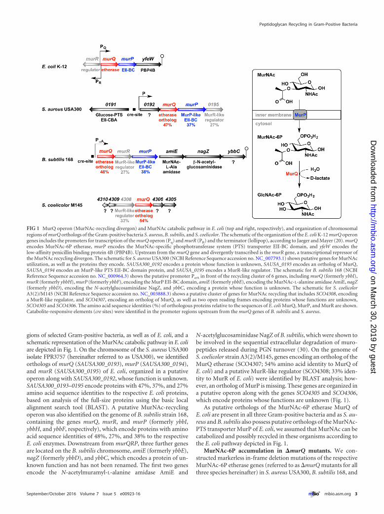
gions of selected Gram-positive bacteria, as well as of E. coli, and aschematic representation of the MurNAc catabolic pathway in E. coliare depicted in Fig. 1. On the chromosome of the S. aureus USA300isolate FPR3757 (hereinafter referred to as USA300), we identifiedorthologs of murQ (SAUSA300_0193), murP (SAUSA300_0194),and murR (SAUSA300_0195) of E. coli, organized in a putativeoperon along with SAUSA300_0192, whose function is unknown.SAUSA300_0193–0195 encode proteins with 47%, 37%, and 27%amino acid sequence identities to the respective E. coli proteins,based on analysis of the full-size proteins using the basic localalignment search tool (BLAST). A putative MurNAc-recyclingoperon was also identified on the genome of B. subtilis strain 168,containing the genes murQ, murR, and murP (formerly ybbI,ybbH, and ybbF, respectively), which encode proteins with aminoacid sequence identities of 48%, 27%, and 38% to the respectiveE. coli enzymes. Downstream from murQRP, three further genesare located on the B. subtilis chromosome, amiE (formerly ybbE),nagZ (formerly ybbD), and ybbC, which encodes a protein of un-known function and has not been renamed. The first two genesencode the N-acetylmuramyl-L-alanine amidase AmiE and
N-acetylglucosaminidase NagZ of B. subtilis, which were shown tobe involved in the sequential extracellular degradation of muro-peptides released during PGN turnover (30). On the genome ofS. coelicolor strain A3(2)/M145, genes encoding an ortholog of theMurQ etherase (SCO4307; 54% amino acid identity to MurQ ofE. coli) and a putative MurR-like regulator (SCO4308; 33% iden-tity to MurR of E. coli) were identified by BLAST analysis; how-ever, an ortholog of MurP is missing. These genes are organized ina putative operon along with the genes SCO4305 and SCO4306,which encode proteins whose functions are unknown (Fig. 1).
As putative orthologs of the MurNAc-6P etherase MurQ ofE. coli are present in all three Gram-positive bacteria and as S. au-reus and B. subtilis also possess putative orthologs of the MurNAc-PTS transporter MurP of E. coli, we assumed that MurNAc can becatabolized and possibly recycled in these organisms according tothe E. coli pathway depicted in Fig. 1.
MurNAc-6P accumulation in �murQ mutants. We con-structed markerless in-frame deletion mutations of the respectiveMurNAc-6P etherase genes (referred to as �murQ mutants for allthree species hereinafter) in S. aureus USA300, B. subtilis 168, and
FIG 1 MurQ operon (MurNAc-recycling divergon) and MurNAc catabolic pathway in E. coli (top and right, respectively), and organization of chromosomalregions of murQ orthologs of the Gram-positive bacteria S. aureus, B. subtilis, and S. coelicolor. The schematic of the organization of the E. coli K-12 murQ operongenes includes the promoters for transcription of the murQ operon (Pq) and murR (PR) and the terminator (lollipop), according to Jaeger and Mayer (20). murQencodes MurNAc-6P etherase, murP encodes the MurNAc-specific phosphotransferase system (PTS) transporter EII-BC domain, and yfeW encodes thelow-affinity penicillin binding protein 4B (PBP4B). Upstream from the murQ gene and divergently transcribed is the murR gene, a transcriptional repressor ofthe MurNAc recycling divergon. The schematic for S. aureus USA300 (NCBI Reference Sequence accession no. NC_007793.1) shows putative genes for MurNAcutilization, as well as the proteins they encode. SAUSA300_0192 encodes a protein whose function is unknown, SAUSA_0193 encodes an ortholog of MurQ,SAUSA_0194 encodes an MurP-like PTS EII-BC domain protein, and SAUSA_0195 encodes a MurR-like regulator. The schematic for B. subtilis 168 (NCBIReference Sequence accession no. NC_000964.3) shows the putative promoter P�A in front of the recycling cluster of 6 genes, including murQ (formerly ybbI),murR (formerly ybbH), murP (formerly ybbF), encoding the MurP EII-BC domain, amiE (formerly ybbE), encoding the MurNAc-L-alanine amidase AmiE, nagZ(formerly ybbD), encoding the N-acetylglucosaminidase NagZ, and ybbC, encoding a protein whose function is unknown. The schematic for S. coelicolorA3(2)/M145 (NCBI Reference Sequence accession no. NC_003888.3) shows a putative cluster of genes for MurNAc recycling that includes SCO4308, encodinga MurR-like regulator, and SCO4307, encoding an ortholog of MurQ, as well as two open reading frames encoding proteins whose functions are unknown,SCO4305 and SCO4306. The amino acid sequence identities (%) of orthologous proteins relative to the sequences of E. coli MurQ, MurP, and MurR are shown.Catabolite-responsive elements (cre sites) were identified in the promoter regions upstream from the murQ genes of B. subtilis and S. aureus.
Peptidoglycan Recycling in Gram-Positive Bacteria
September/October 2016 Volume 7 Issue 5 e00923-16 ® mbio.asm.org 3
on March 30, 2019 by guest
http://mbio.asm
.org/D
ownloaded from
S. coelicolor strain M145 to investigate whether these Gram-positive bacteria recycle MurNAc derived from their own PGN,involving MurNAc-6P etherase. We expected that if recycling ofthe MurNAc content of their PGN occurred, �murQ cells wouldaccumulate MurNAc-6P intracellularly. In addition, we generatedan S. aureus USA300 deletion mutant lacking the entire putativemurQPR operon (�SAUSA_0192– 0195) and, in addition, aB. subtilis 168 �murQRP deletion mutant. Both of these mutantslack murQ and the genes encoding the putative MurNAc PTStransporters. Thus, these mutants should not be able to take upMurNAc released from the PGN wall and therefore should notaccumulate MurNAc-6P. Gene deletions on the chromosomewere confirmed by PCR (see Fig. S1 in the supplemental material)and sequencing.
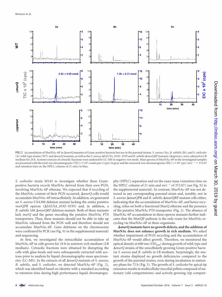
At first, we investigated the intracellular accumulation ofMurNAc-6P in cells grown for 24 h in nutrient-rich medium (LBmedium). Cytosolic fractions were obtained by disrupting thecells with glass beads and were subsequently extracted with ace-tone prior to analysis by liquid chromatography-mass spectrom-etry (LC-MS). In the extracts of all �murQ mutants of S. aureus,B. subtilis, and S. coelicolor, we detected MurNAc-6P (Fig. 2),which was identified based on identity with a standard accordingto retention time during high-performance liquid chromatogra-
phy (HPLC) separation and on the exact mass (retention time onthe HPLC column of 21 min and m/z�1 of 372.07) (see Fig. S2 inthe supplemental material). In contrast, MurNAc-6P was not de-tected in any corresponding parental strain and, notably, not inS. aureus �murQPR and B. subtilis �murQRP mutant cells either,indicating that the accumulation of MurNAc-6P, and hence recy-cling, relies on both a functional MurQ etherase and the presenceof the putative MurNAc PTS transporter (Fig. 2). The absence ofMurNAc-6P accumulation in these operon mutants further indi-cates that the MurQP pathway is the only route for MurNAc re-cycling via MurNAc-6P in these organisms.
�murQ mutants have no growth defects, and the addition ofMurNAc does not enhance growth in rich medium. We askedwhether impairment of PGN recycling and the accumulation ofMurNAc-6P would affect growth. Therefore, we monitored theoptical density at 600 nm (OD600) during growth of wild-type and�murQ strains of the unicellularly growing Gram-positive bacte-ria S. aureus and B. subtilis in LB medium. Surprisingly, the mu-tant strains displayed no growth deficiencies compared to thegrowth of the parental strains, even during incubation in station-ary phase for 72 h (Fig. 3). The growth of S. coelicolor by apical tipextension results in multicellular mycelial pellets composed of sta-tionary (old compartments) and actively growing (tip compart-
FIG 2 Accumulation of MurNAc-6P in �murQ mutants of Gram-positive bacteria but not in the parental strains. S. aureus (Sa), B. subtilis (Bs), and S. coelicolor(Sc) wild-type strains (WT) and �murQ mutants, as well as the S. aureus �SAUSA_0192– 0195 and B. subtilis �murQRP mutants (�operon), were cultured in LBmedium for 24 h. Acetone extracts of cytosolic fractions were analyzed by LC-MS in negative-ion mode. Mass spectra of MurNAc-6P in the investigated samplesare presented with the total-ion chromatograms (TIC) (�105 counts per s [cps]) in gray and the extracted-ion chromatograms (EIC) (�103 cps) (m/z�1 � 372.07and retention time on the HPLC column of 21 min) in blue.
Borisova et al.
4 ® mbio.asm.org September/October 2016 Volume 7 Issue 5 e00923-16
on March 30, 2019 by guest
http://mbio.asm
.org/D
ownloaded from
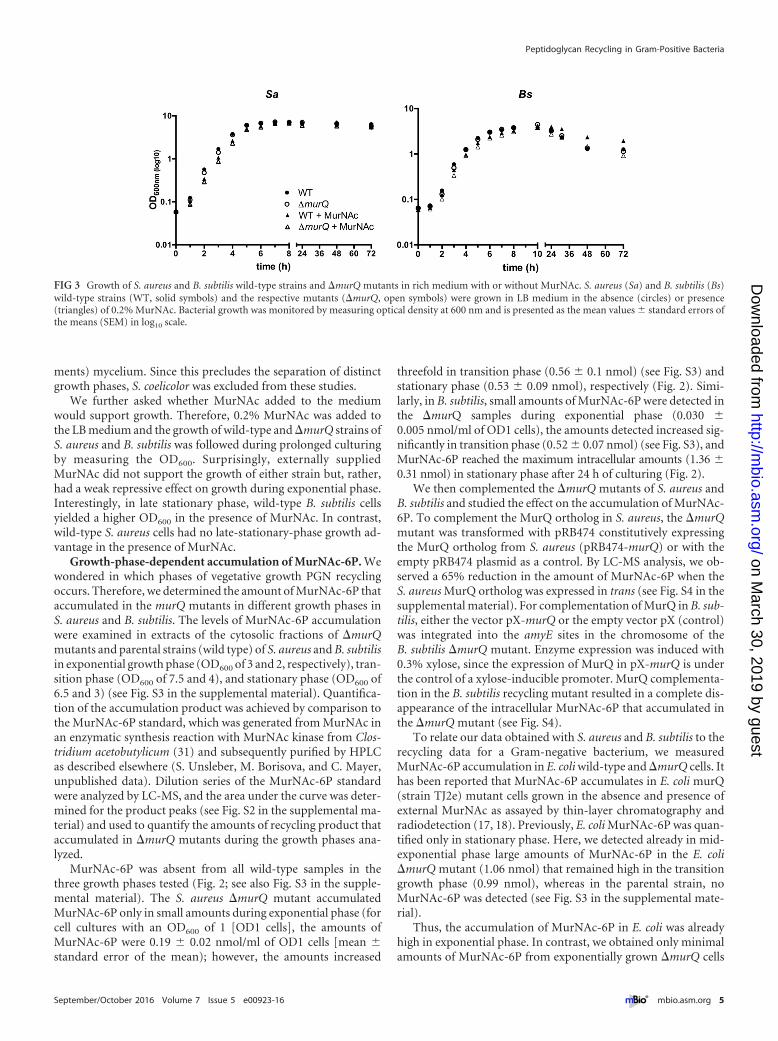
ments) mycelium. Since this precludes the separation of distinctgrowth phases, S. coelicolor was excluded from these studies.
We further asked whether MurNAc added to the mediumwould support growth. Therefore, 0.2% MurNAc was added tothe LB medium and the growth of wild-type and �murQ strains ofS. aureus and B. subtilis was followed during prolonged culturingby measuring the OD600. Surprisingly, externally suppliedMurNAc did not support the growth of either strain but, rather,had a weak repressive effect on growth during exponential phase.Interestingly, in late stationary phase, wild-type B. subtilis cellsyielded a higher OD600 in the presence of MurNAc. In contrast,wild-type S. aureus cells had no late-stationary-phase growth ad-vantage in the presence of MurNAc.
Growth-phase-dependent accumulation of MurNAc-6P. Wewondered in which phases of vegetative growth PGN recyclingoccurs. Therefore, we determined the amount of MurNAc-6P thataccumulated in the murQ mutants in different growth phases inS. aureus and B. subtilis. The levels of MurNAc-6P accumulationwere examined in extracts of the cytosolic fractions of �murQmutants and parental strains (wild type) of S. aureus and B. subtilisin exponential growth phase (OD600 of 3 and 2, respectively), tran-sition phase (OD600 of 7.5 and 4), and stationary phase (OD600 of6.5 and 3) (see Fig. S3 in the supplemental material). Quantifica-tion of the accumulation product was achieved by comparison tothe MurNAc-6P standard, which was generated from MurNAc inan enzymatic synthesis reaction with MurNAc kinase from Clos-tridium acetobutylicum (31) and subsequently purified by HPLCas described elsewhere (S. Unsleber, M. Borisova, and C. Mayer,unpublished data). Dilution series of the MurNAc-6P standardwere analyzed by LC-MS, and the area under the curve was deter-mined for the product peaks (see Fig. S2 in the supplemental ma-terial) and used to quantify the amounts of recycling product thataccumulated in �murQ mutants during the growth phases ana-lyzed.
MurNAc-6P was absent from all wild-type samples in thethree growth phases tested (Fig. 2; see also Fig. S3 in the supple-mental material). The S. aureus �murQ mutant accumulatedMurNAc-6P only in small amounts during exponential phase (forcell cultures with an OD600 of 1 [OD1 cells], the amounts ofMurNAc-6P were 0.19 � 0.02 nmol/ml of OD1 cells [mean �standard error of the mean); however, the amounts increased
threefold in transition phase (0.56 � 0.1 nmol) (see Fig. S3) andstationary phase (0.53 � 0.09 nmol), respectively (Fig. 2). Simi-larly, in B. subtilis, small amounts of MurNAc-6P were detected inthe �murQ samples during exponential phase (0.030 �0.005 nmol/ml of OD1 cells), the amounts detected increased sig-nificantly in transition phase (0.52 � 0.07 nmol) (see Fig. S3), andMurNAc-6P reached the maximum intracellular amounts (1.36 �0.31 nmol) in stationary phase after 24 h of culturing (Fig. 2).
We then complemented the �murQ mutants of S. aureus andB. subtilis and studied the effect on the accumulation of MurNAc-6P. To complement the MurQ ortholog in S. aureus, the �murQmutant was transformed with pRB474 constitutively expressingthe MurQ ortholog from S. aureus (pRB474-murQ) or with theempty pRB474 plasmid as a control. By LC-MS analysis, we ob-served a 65% reduction in the amount of MurNAc-6P when theS. aureus MurQ ortholog was expressed in trans (see Fig. S4 in thesupplemental material). For complementation of MurQ in B. sub-tilis, either the vector pX-murQ or the empty vector pX (control)was integrated into the amyE sites in the chromosome of theB. subtilis �murQ mutant. Enzyme expression was induced with0.3% xylose, since the expression of MurQ in pX-murQ is underthe control of a xylose-inducible promoter. MurQ complementa-tion in the B. subtilis recycling mutant resulted in a complete dis-appearance of the intracellular MurNAc-6P that accumulated inthe �murQ mutant (see Fig. S4).
To relate our data obtained with S. aureus and B. subtilis to therecycling data for a Gram-negative bacterium, we measuredMurNAc-6P accumulation in E. coli wild-type and �murQ cells. Ithas been reported that MurNAc-6P accumulates in E. coli murQ(strain TJ2e) mutant cells grown in the absence and presence ofexternal MurNAc as assayed by thin-layer chromatography andradiodetection (17, 18). Previously, E. coli MurNAc-6P was quan-tified only in stationary phase. Here, we detected already in mid-exponential phase large amounts of MurNAc-6P in the E. coli�murQ mutant (1.06 nmol) that remained high in the transitiongrowth phase (0.99 nmol), whereas in the parental strain, noMurNAc-6P was detected (see Fig. S3 in the supplemental mate-rial).
Thus, the accumulation of MurNAc-6P in E. coli was alreadyhigh in exponential phase. In contrast, we obtained only minimalamounts of MurNAc-6P from exponentially grown �murQ cells
FIG 3 Growth of S. aureus and B. subtilis wild-type strains and �murQ mutants in rich medium with or without MurNAc. S. aureus (Sa) and B. subtilis (Bs)wild-type strains (WT, solid symbols) and the respective mutants (�murQ, open symbols) were grown in LB medium in the absence (circles) or presence(triangles) of 0.2% MurNAc. Bacterial growth was monitored by measuring optical density at 600 nm and is presented as the mean values � standard errors ofthe means (SEM) in log10 scale.
Peptidoglycan Recycling in Gram-Positive Bacteria
September/October 2016 Volume 7 Issue 5 e00923-16 ® mbio.asm.org 5
on March 30, 2019 by guest
http://mbio.asm
.org/D
ownloaded from
of Gram-positive bacteria, particularly in B. subtilis, but muchlarger amounts from cells in transition and stationary phases. Weaimed to clarify whether the apparent delay of PGN recycling inGram-positive bacteria is due to a low activity or abundance ofMurQ and MurP orthologs or to the small amounts of MurNAcprovided from the breakdown of cell wall turnover products.Therefore, we added MurNAc (0.02%) to the culture medium anddetermined the intracellular concentrations of MurNAc-6P inS. aureus and B. subtilis wild-type and �murQ cells at differentgrowth phases (Fig. 3; see also Fig. S5 in the supplemental mate-rial). MurNAc-6P was generally absent in the cytosolic fractions ofwild-type cells grown in LB medium supplemented with 0.02%MurNAc, except for small amounts measured in the S. aureus cellsafter 24 h of growth (see Fig. S5). However, in S. aureus �murQ,the intracellular concentrations of MurNAc-6P in the presence ofMurNAc increased in exponential phase to 2.33 � 0.32 nmol/mlof OD1 cells and in transition phase to 6.21 � 0.66 nmol/ml ofOD1 cells, and they reached a maximum in stationary growthphase of 11.56 � 0.39 nmol/ml of OD1 cells (see Fig. S5). Thus,S. aureus cells tolerated the very high intracellular concentrationsof MurNAc-6P without showing any obvious growth defect, evenwith 0.2% exogenous MurNAc (Fig. 3). In B. subtilis �murQ cells,the addition of MurNAc caused large increases in the intracellu-lar MurNAc-6P levels, particularly in exponentially growing cells.When MurNAc was added to the medium, MurNAc-6P concen-trations of 1.05 � 0.05 nmol were determined in exponentialphase. During transition phase, adding MurNAc to the me-dium caused the levels of MurNAc-6P to increase to 5.21 �0.73 nmol/ml of OD1 cells, and adding MurNAc in stationaryphase caused the levels to increase to 3.11 � 0.13 nmol/ml OD1cells. Surprisingly, the levels of MurNAc-6P dropped from transi-tion to stationary phase in B. subtilis �murQ cells grown in LBwith MurNAc (see Fig. S5). Reevaluation of the MS data revealedthat the reduction of the amount of MurNAc-6P in stationary-phase cells correlated with an increase in MurNAc. This is mostlikely caused by an unknown phosphatase in B. subtilis that isresponsible for dephosphorylation of MurNAc-6P, thus yieldingMurNAc (data not shown).
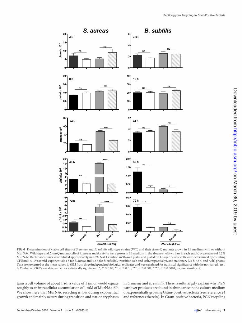
Cell wall recycling is crucial for survival in stationary phase.Although large amounts of MurNAc-6P accumulate in �murQcells of S. aureus and B. subtilis grown in LB and even more in thepresence of MurNAc, the growth rates determined by optical den-sity measurements were more or less identical for wild-type andmutant cells for at least 24 h of growth (Fig. 3). However, inlate stationary phase in LB medium supplemented with 0.2%MurNAc, B. subtilis wild-type cultures remained at a higher OD(Fig. 3). We supposed that, although the impairment of recoveryof the MurNAc of the peptidoglycan wall had no effect on growth,it might still affect cell survival. To investigate whether MurNAcrecycling provides a survival benefit, we analyzed the viability ofcells by determining the CFU counts (CFU/ml) (Fig. 4; see alsoTable S3 in the supplemental material). Similar cell numbers weredetermined for wild-type and �murQ strains grown to exponen-tial phase and transition phase in medium with and without addedMurNAc. Intriguingly, in stationary phase (24 h of culturing),only the wild-type S. aureus cell cultures supplemented with ex-ogenous MurNAc maintained high cell numbers; in all othercases, the CFU counts dropped dramatically. This is not a growthbut a survival effect, since the cell numbers did not increase be-tween 8 h and 24 h of incubation of wild-type S. aureus cell cul-
tures in medium with MurNAc but did remain at a high level,whereas cell numbers of the �murQ strain dropped by twofold.Notably, prolonged incubation of S. aureus cells in medium withMurNAc for 48 h and 72 h showed that wild-type cell culturesmaintained seven- and sixfold higher cell numbers (CFU/ml), re-spectively, than did mutant cell cultures. A slight but significantsurvival advantage of S. aureus wild-type cells compared to thesurvival of mutant cells was also observed upon culturing for 48 hand 72 h in LB medium without MurNAc. Thus, prolonged incu-bation in the culture medium revealed a defect in the survival ofthe MurNAc-recycling-mutant cells compared to the survival ofwild-type cells (Fig. 4; see also Table S3).
Similar data were obtained for B. subtilis. We observed no sig-nificant differences in the CFU counts of wild-type and mutantcell cultures within the first 24 h of growth in LB either in theabsence or presence of MurNAc. However, the viability of murQmutant cell cultures decreased dramatically during prolonged in-cubation in stationary phase (48 h and 72 h). In the presence ofMurNAc, surprisingly, a dramatic decrease of viability was ob-served, possibly due to cell lysis at the onset of sporulation. Nota-bly, this effect was even enhanced in the recycling mutant (Fig. 4;see also Table S3).
DISCUSSION
We show here that three Gram-positive bacteria, S. aureus, B. sub-tilis, and S. coelicolor, recycle the MurNAc contents of their PGNcell wall during vegetative growth in rich medium, which requiresthe orthologs of the MurQ enzyme of E. coli. Thereby, we providefor the first time direct evidence for PGN recycling in Gram-positive bacteria and proof of MurQ function in these organisms.Since these differently growing species, the elongating rod-shapedB. subtilis, the orthogonally dividing coccoidal S. aureus, and theapically growing filamentous S. coelicolor, all recycle the MurNAcof their PGN, this pathway is presumably a general feature ofGram-positive bacteria. Indeed, orthologs of murQ, initially iden-tified and related to PGN recycling in the Gram-negative modelbacterium E. coli (17, 18), are found on the chromosomes ofnearly all Gram-positive bacteria (an exception is Streptococcusspecies). We directly measured the recycling product MurNAc-6Pthat accumulated in the murQ mutants by applying a massspectrometry-based method, which avoids radioactive prelabelingof cell walls and the subsequent measurement of the radioactivefragments released but still allows sensitive detection and quanti-fication of MurNAc-6P in cell extracts within a large concentra-tion range of 2.5 to 1,250 �M. Since the wild-type strains, as well ascomplemented mutants, contained no MurNAc-6P or greatlyreduced amounts, respectively, it is evident that the MurQ ether-ases are required for MurNAc recycling. The possibility thatMurNAc-6P accumulated in �murQ mutants due to the presenceof MurNAc in the growth medium could be excluded becauseMurNAc is absent from the LB culture medium (24, 32; data notshown). Furthermore, MurNAc-6P accumulation was not de-tected in strains of S. aureus and B. subtilis that were defective inboth MurQ and the putative MurNAc transporter, indicating thatthe MurP MurNAc phosphotransferase transporters are the onlyuptake systems for MurNAc recycling in these organisms.
We determined the levels of MurNAc-6P that accumulated inmurQ mutants of S. aureus and B. subtilis in different growthphases. The amounts of MurNAc-6P are presented as nanomolesper milliliter of OD1 cells. Assuming that 1 ml of OD1 cells con-
Borisova et al.
6 ® mbio.asm.org September/October 2016 Volume 7 Issue 5 e00923-16
on March 30, 2019 by guest
http://mbio.asm
.org/D
ownloaded from
tains a cell volume of about 1 �l, a value of 1 nmol would equateroughly to an intracellular accumulation of 1 mM of MurNAc-6P.We show here that MurNAc recycling is low during exponentialgrowth and mainly occurs during transition and stationary phases
in S. aureus and B. subtilis. These results largely explain why PGNturnover products are found in abundance in the culture mediumof exponentially growing Gram-positive bacteria (see reference 24and references therein). In Gram-positive bacteria, PGN recycling
FIG 4 Determination of viable cell titers of S. aureus and B. subtilis wild-type strains (WT) and their �murQ mutants grown in LB medium with or withoutMurNAc. Wild-type and �murQ mutant cells of S. aureus and B. subtilis were grown in LB medium in the absence (left two bars in each graph) or presence of 0.2%MurNAc. Bacterial cultures were diluted appropriately in 0.9% NaCl solution in 96-well plates and plated on LB agar. Viable cells were determined by countingCFU/ml (�108) at mid-exponential (4 h for S. aureus and 4.5 h for B. subtilis), transition (8 h and 10 h, respectively), and stationary (24 h, 48 h, and 72 h) phases.Data are presented as the mean values � SEM from three independent biological replicates and were analyzed for statistical significance with the nonpaired t test.A P value of �0.05 was determined as statistically significant (*, P � 0.05; **, P � 0.01; ***, P � 0.001; ****, P � 0.0001; ns, nonsignificant).
Peptidoglycan Recycling in Gram-Positive Bacteria
September/October 2016 Volume 7 Issue 5 e00923-16 ® mbio.asm.org 7
on March 30, 2019 by guest
http://mbio.asm
.org/D
ownloaded from
occurs not synchronous with turnover, as in E. coli (2), but after adelay. Turnover of the Gram-positive cell wall occurs by a so-called inside-to-outside growth mechanism (33). The PGN is firstassembled near the plasma membrane and is then graduallymoved to the outside, where it becomes stretched and more sus-ceptible to the activity of autolysins. The small amounts ofMurNAc-6P in exponential-phase cells, particularly in the B. sub-tilis �murQ mutant, can be explained by the delayed release ofMurNAc from the PGN wall due to the slow autolytic processingof larger PGN turnover fragments. It is likely that MurNAc isreleased more slowly from the very long PGN chains present inB. subtilis than from the rather short PGN chains of S. aureus (34).
MurNAc recycling does not sustain the growth of S. aureus andB. subtilis; instead, it appears to be of major relevance for theirsurvival capacity when growth is stalled during nutrient limitationat the onset of stationary phase and recycling may be used toovercome a shortage of nutrients and precursors for cell (wall)differentiation. Our results are in agreement with those of previ-ous studies indicating that the transcription of B. subtilis murQoccurs mainly during the transition to stationary phase (35).Moreover, these results are consistent with previous findingsshowing that the expression of the recycling N-acetylglu-cosaminidase NagZ of B. subtilis is low during exponential phasebut highly elevated in stationary phase (30). The enzyme releasesGlcNAc from the PGN, and it is likely that besides MurNAc, theGlcNAc part of the PGN is also recycled; however, this wouldrequire uptake by a different transport system.
When MurNAc was added to the medium, the accumulation ofMurNAc-6P increased greatly, indicating that the uptake and re-cycling of MurNAc is limited by the availability of the sugar. Asmentioned above, the release of MurNAc depends on autolyticprocessing of PGN, which is growth phase-dependently regulated.In the proximity of murQ and its orthologs on the genomes of thebacteria studied, transcriptional regulator proteins were identifiedthat, similar to the case for E. coli (20), may likely accelerate thetranscription of the MurNAc transporter and MurQ in the pres-ence of MurNAc-6P. Interestingly, in stationary phase, largeamounts of MurNAc and MurNAc-6P were found in the cytosolicfractions of B. subtilis �murQ mutant cells grown in LB mediumsupplemented with MurNAc, which might be due to dephosphor-ylation of MurNAc-6P at high concentrations by a putative phos-phatase. Notably, an enzyme with identical function was recentlyproposed in a Pseudomonas species (13).
To relate our quantitative data on MurNAc-6P accumulationin S. aureus and B. subtilis to its accumulation in the Gram-negative bacterium E. coli, we reexamined and quantified theMurNAc-6P accumulation in the murQ mutant of E. coli usingour mass spectrometry-based method. In E. coli murQ mutantcells, the maximum accumulation of MurNAc-6P occurred al-ready in mid-exponential growth phase and reached an intracel-lular concentration of c. 1 mM (1 nmol/ml of OD1 cells); Wientjesand colleagues reported that the PGN of one E. coli cell contains3.5 � 106 molecules of diaminopimelic acid (36), and presumably,similar numbers of MurNAc molecules are present. As 1 ml of acell culture with an OD600 of 1 (OD1 cells) in exponential phaseamounts to 5 � 108 to 1 � 109 cells, it can be calculated that oneE. coli cell contains 1.7 � 1015 to 3.5 � 1015 molecules of MurNAc.Thus, a recycling rate of 45% per generation would lead to aMurNAc-6P accumulation of about 1.3 to 2.6 mM. The value ofabout 1 mM obtained in exponentially growing E. coli cells in this
study is a little lower than the expected concentration range. In-terestingly, the amounts of the recycling product MurNAc-6P de-termined in E. coli cells during mid-log phase and in the examinedGram-positive bacteria during transition to stationary phase weresimilar. This indicates that, compared to the rates of PGN synthe-sis, the turnover and recycling of PGN appear to be quite similar inboth groups of bacteria. However, related to the thickness of theGram-positive cell wall, with approximately 5- to 10-fold-greaterPGN contents than in E. coli, it can be estimated that only about 5to 10% of the PGN is recycled in S. aureus and B. subtilis pergeneration. These values are in the range of the 8% recycling re-ported for B. subtilis by Pooley (25) but are lower than the rates ofturnover reported elsewhere (22, 26, 27).
The most intriguing finding of our study was that PGN sugarrecycling affects survival fitness. Apparently, the cell wall sugarMurNAc does not serve primarily as an energy source but is pref-erentially utilized for cell wall synthesis. The same presumablyholds for GlcNAc, as the MurNAc and GlcNAc catabolic pathwaysmerge. Amino sugar utilization and its regulation were studied inB. subtilis and in S. aureus (37) and were compared with those ofE. coli in a recent review (38). These studies revealed that S. aureusand B. subtilis use GlcNAc preferentially for cell wall synthesis; inB. subtilis, about 95% of GlcNAc from the medium is usuallyincorporated into PGN. Our findings are consistent with theseobservations. Both cell wall sugars, GlcNAc and MurNAc, are pre-sumably used in S. aureus and B. subtilis for cell wall synthesis, atleast during growth in rich medium. Combined with the datapresented here, one could speculate that in Gram-positive bacte-ria, MurNAc recycled from the PGN, as well as that salvaged fromthe medium, is not used to gain energy or for growth but might beutilized to stabilize the cell wall, and thus, it increases survivalduring prolonged starvation. Although PGN recycling is not es-sential, a huge set of recycling genes are maintained in almost allbacterial genomes, suggesting that the pathway must provide abenefit for bacterial cells. We show here that PGN recycling iscrucial for the survival of Gram-positive bacteria during station-ary phase.
MATERIALS AND METHODSChemicals, enzymes, and oligonucleotides. Enzymes for DNA restric-tion and for cloning were obtained from New England Biolabs (Ipswich,MA) or Thermo Fischer Scientific (Schwerte, Germany). The Gene JETplasmid miniprep kit, PCR purification kit, and Gene Ruler 1-kb markerwere from Thermo Fisher Scientific and Qiagen (Venlo, Netherlands).N-Acetylmuramic acid (MurNAc) was from Bachem (Bubendorf, Swit-zerland), and the DNA dye NonTox was from Applichem (Darmstadt,Germany). Oligonucleotides were purchased from MWG Eurofins (Eber-sberg, Germany) and are listed in Table S1 in the supplemental material.
Bacterial strains, growth conditions, and construction of mutantsand plasmids. The plasmids and bacterial strains used in this study areshown in Table S2 in the supplemental material. The construction ofmutant strains and plasmids is described in Text S1 in the supplementalmaterial. Escherichia coli K-12, Bacillus subtilis 168, and Staphylococcusaureus USA300 were cultured aerobically in lysogeny broth (LB; 5 g/literyeast extract, 10 g/liter tryptone, 10 g/liter NaCl) at 37°C and under con-tinuous shaking at 160 rpm or on solid LB supplemented with 1.5% agar.B. subtilis and S. aureus overnight cultures (~16 h) were used to inoculatefresh LB medium to yield an initial optical density at 600 nm (OD600) of0.05 for growth studies or the determination of intracellular accumulationof N-acetylmuramic acid-6 phosphate (MurNAc-6P) at different growthphases. Tryptic soy broth (TSB; Oxoid) was used to generate the marker-less S. aureus �murQ mutant and for complementation experiments.
Borisova et al.
8 ® mbio.asm.org September/October 2016 Volume 7 Issue 5 e00923-16
on March 30, 2019 by guest
http://mbio.asm
.org/D
ownloaded from
S. coelicolor M145 was grown in LB medium with constant shaking(180 rpm) at 30°C. Antibiotics were used, when appropriate, at the fol-lowing concentrations: ampicillin (100 �g/ml) for E. coli; chlorampheni-col (5 �g/ml) and erythromycin and tetracycline (10 �g/ml) for B. subtilis;erythromycin (5 �g/ml) and chloramphenicol (10 �g/ml) for S. aureus;and apramycin (50 �g/ml) for S. coelicolor. For induction of the PxylA=promoter in B. subtilis, 0.3% xylose was added to the LB growth medium.
Generation of cytosol fractions. Overnight cultures of S. aureus,B. subtilis, and E. coli, including both the wild types and the respective�murQ mutants, were used to inoculate LB medium, yielding an initialOD600 of 0.05, and cells were grown at 37°C. S. aureus cells were harvestedat mid-exponential phase (OD600 of 3 after growth for ~3.5 h in 100-mlcultures), at transition phase (OD600 of 7.5 after growth for 8 h in 40-mlcultures), and at stationary phase (OD600 of 6 after growth for 24 h in50-ml cultures). B. subtilis cultures were harvested at mid-exponentialphase (OD600 of 2 after growth for ~4 h in 150-ml cultures), at earlystationary phase (OD600 of 4 after growth for 10 h in 75-ml cultures), andat stationary phase (OD600 of 3 after growth for 24 h in 100-ml cultures).E. coli cells were harvested at mid-exponential phase (OD600 of 1.5 aftergrowth for ~3 h in 200-ml cultures) and at transition phase (OD600 of 3.5after growth for 6.5 h in 86-ml cultures). Bacteria were centrifuged at3,000 � g for 10 min and washed with 20 ml deionized water, and pelletswere frozen at �80°C. Frozen cell samples were thawed at room temper-ature and suspended in water to yield 1.2-ml cell suspensions with anOD600 of 250.
Approximately 108 spores of S. coelicolor M145 wild type and the�murQ (�SCO4307) mutant were incubated in LB medium (50 ml) andgrown for 24 h in a rotary shaker at 30°C. Fifteen-milliliter amounts of thecultures were harvested by centrifugation, washed, and frozen at �80°Cuntil being dissolved in 1,000 �l water for further analysis.
The whole bacterial samples, suspended in the amount of water indi-cated above, were transferred to new tubes containing ~0.25 g glass beads(0.25 to 0.5 mm; Roth), and cells were disrupted in a FastPrep FP120(Thermo Savant) cell disrupter at speed 6 for 35 s. This was repeated 4times, with cooling on ice for 1 min after the second cycle. Lysates werecooled briefly and subsequently centrifuged for 10 min at maximumspeed in a microcentrifuge. Two hundred microliters of the supernatantwas added to 800 �l of ice-cold acetone to precipitate the remaining pro-teins in the supernatant. After centrifugation (12,000 � g for 10 min), thesupernatant was transferred to a new tube, and samples were dried undervacuum for 2 h at 55°C and finally stored at 4°C prior to LC-MS measure-ments.
Analysis of MurNAc-6P accumulation by LC-MS. Sample analysis ofbacterial cytosolic fractions was conducted using an electrosprayionization-time of flight (ESI-TOF) mass spectrometer (MicrO-TOF II;Bruker), operated in negative-ion mode and connected to the UltiMate3000 high-performance liquid chromatography (HPLC) system (Di-onex). Dried samples were dissolved in 100 �l of water before measure-ment, and 5-�l amounts were injected into a Gemini C18 column (150 by4.6 mm; Phenomenex) at 37°C. The following previously described (12)45-min-gradient program at a flow rate of 0.2 ml/min was used. Fiveminutes of washing with 100% buffer A (0.1% formic acid with 0.05%ammonium formate) was followed by a linear gradient over 30 min to40% buffer B (100% acetonitrile). A 5-min delay and 5 min of reequili-bration completed the method. The mass spectra of the investigated sam-ples are presented as total-ion chromatograms (TIC) and extracted-ionchromatograms (EIC) for MurNAc-6P, created using Data Analysis(Bruker) and Prism 6 (GraphPad) software. To quantify MurNAc-6Pconcentrations in cell extracts (presented as nmol/ml of OD1 cells), wegenerated EIC for MurNAc-6P (m/z�1 �372.07) in each sample and de-termined the area under the curve of the peak obtained by using Prism 6(baseline of 30). A dilution series of a MurNAc-6P standard (1,250 �M to2.45 �M) was also analyzed by LC-MS, and the data are presented as EICbased on the area under the curve of the measurements obtained. The
standard curve was applied to define the intracellular MurNAc-6P con-centrations in the bacterial cytosolic fractions.
SUPPLEMENTAL MATERIALSupplemental material for this article may be found at http://mbio.asm.org/lookup/suppl/doi:10.1128/mBio.00923-16/-/DCSupplemental.
Text S1, DOCX file, 0.1 MB.Figure S1, DOCX file, 1.1 MB.Figure S2, DOCX file, 0.1 MB.Figure S3, DOCX file, 0.4 MB.Figure S4, DOCX file, 0.1 MB.Figure S5, DOCX file, 0.3 MB.Table S1, DOCX file, 0.1 MB.Table S2, DOCX file, 0.1 MB.Table S3, DOCX file, 0.1 MB.
ACKNOWLEDGMENTS
We are very grateful to Roger Plaut, Annette Fagerlund, Ewa Bukowska-Faniband, Claes von Wachenfeldt, and Annette Latus for plasmids andstrains.
This work was financed by grants from the Baden-Württemberg Stif-tung (P-BWS-Glyko11) and the German Research Foundation (DFGgrants MA2436/4, BI1350/1-2, and SFB766/A15, A13, and A6 and grantGRK1708). We acknowledge support by the DFG and Open Access Pub-lishing Fund of University of Tübingen.
FUNDING INFORMATIONThis work, including the efforts of Christoph Mayer, was funded byDeutsche Forschungsgemeinschaft (DFG) (MA2436/4, SFB766/A15, andGRK1708). This work, including the efforts of Markus Bischoff, wasfunded by Deutsche Forschungsgemeinschaft (DFG) (BI1350/1-2). Thiswork, including the efforts of Christoph Mayer, was funded by Baden-Württemberg Stiftung (Baden-Württemberg Foundation) (P-BWS-Glyko11). This work, including the efforts of Friedrich Götz, was fundedby Deutsche Forschungsgemeinschaft (DFG) (SFB766/A6). This work,including the efforts of Günther Muth, was funded by the Deutsche For-schungsgemeinschaft (DFG) (SFB766/A13).
REFERENCES1. Litzinger S, Mayer C. 2010. The murein sacculus, p 3–52. In König H,
Claus H, Varma A (ed), Prokaryotic cell wall compounds—structure andbiochemistry. Springer Verlag, Heidelberg, Germany.
2. Höltje JV. 1998. Growth of the stress-bearing and shape-maintainingmurein sacculus of Escherichia coli. Microbiol Mol Biol Rev 62:181–203.
3. Mayer C. 2012. Bacterial cell wall recycling. eLS. http://dx.doi.org/10.1002/9780470015902.a0021974.
4. Chaloupka J, Krecková P, Rfhová L. 1962. The mucopeptide turnover inthe cell walls of growing cultures of Bacillus megaterium KM. Experientia18:362–363. http://dx.doi.org/10.1007/BF02172250.
5. Doyle RJ, Chaloupka J, Vinter V. 1988. Turnover of cell walls in micro-organisms. Microbiol Res 52:554 –567.
6. Vollmer W, Joris B, Charlier P, Foster S. 2008. Bacterial peptidoglycan(murein) hydrolases. FEMS Microbiol Rev 32:259 –286. http://dx.doi.org/10.1111/j.1574-6976.2007.00099.x.
7. Park JT, Uehara T. 2008. How bacteria consume their own exoskeletons(turnover and recycling of cell wall peptidoglycan). Microbiol Mol BiolRev 72:211–227. http://dx.doi.org/10.1128/MMBR.00027-07.
8. Goodell EW. 1985. Recycling of murein by Escherichia coli. J Bacteriol163:305–310
9. Jacobs C, Huang LJ, Bartowsky E, Normark S, Park JT. 1994. Bacterialcell wall recycling provides cytosolic muropeptides as effectors for�-lactamase induction. EMBO J 13:4684 – 4694.
10. Uehara T, Park JT. 2008. Growth of Escherichia coli: significance of pep-tidoglycan degradation during elongation and septation. J Bacteriol 190:3914 –3922. http://dx.doi.org/10.1128/JB.00207-08.
11. Gisin J, Schneider A, Nägele B, Borisova M, Mayer C. 2013. A cell wallrecycling shortcut that bypasses peptidoglycan de novo biosynthesis. NatChem Biol 9:491– 493. http://dx.doi.org/10.1038/nchembio.1289.
12. Borisova M, Gisin J, Mayer C. 2014. Blocking peptidoglycan recycling in
Peptidoglycan Recycling in Gram-Positive Bacteria
September/October 2016 Volume 7 Issue 5 e00923-16 ® mbio.asm.org 9
on March 30, 2019 by guest
http://mbio.asm
.org/D
ownloaded from
Pseudomonas aeruginosa attenuates intrinsic resistance to fosfomycin. Mi-crob Drug Resist 20:231–237. http://dx.doi.org/10.1089/mdr.2014.0036.
13. Johnson JW, Fisher JF, Mobashery S. 2013. Bacterial cell-wall recycling.Ann N Y Acad Sci 1277:54 –75. http://dx.doi.org/10.1111/j.1749-6632.2012.06813.x.
14. Bertsche U, Mayer C, Götz F, Gust AA. 2015. Peptidoglycan perception-sensing bacteria by their common envelope structure. Int J Med Microbiol305:217–223. http://dx.doi.org/10.1016/j.ijmm.2014.12.019.
15. den Blaauwen T, de Pedro MA, Nguyen-Distèche M, Ayala JA. 2008.Morphogenesis of rod-shaped sacculi. FEMS Microbiol Rev 32:321–344.http://dx.doi.org/10.1111/j.1574-6976.2007.00090.x.
16. Dahl U, Jaeger T, Nguyen BT, Sattler JM, Mayer C. 2004. Identificationof a phosphotransferase system of Escherichia coli required for growth onN-acetylmuramic acid. J Bacteriol 186:2385–2392. http://dx.doi.org/10.1128/JB.186.8.2385-2392.2004.
17. Jaeger T, Arsic M, Mayer C. 2005. Scission of the lactyl ether bond ofN-acetylmuramic acid by Escherichia coli “etherase.” J Biol Chem 280:30100 –30106. http://dx.doi.org/10.1074/jbc.M502208200.
18. Uehara T, Suefuji K, Jaeger T, Mayer C, Park JT. 2006. MurQ etheraseis required by Escherichia coli in order to metabolize anhydro-N-acetylmuramic acid obtained either from the environment or from its owncell wall. J Bacteriol 188:1660 –1662. http://dx.doi.org/10.1128/JB.188.4.1660-1662.2006.
19. Vega D, Ayala JA. 2006. The DD-carboxypeptidase activity encoded bypbp4B is not essential for the cell growth of Escherichia coli. Arch Micro-biol 185:23–27. http://dx.doi.org/10.1007/s00203-005-0057-5.
20. Jaeger T, Mayer C. 2008. The transcriptional factors MurR and cataboliteactivator protein regulate N-acetylmuramic acid catabolism in Escherichiacoli. J Bacteriol 190:6598 – 6608. http://dx.doi.org/10.1128/JB.00642-08.
21. Renner-Schneck M, Hinderberger I, Gisin J, Exner T, Mayer C,Stehle T. 2015. Crystal structure of the N-acetylmuramic acid a-1-phosphate (MurNAc-a-1-P) uridylyltransferase MurU, a minimalsugar nucleotidyltransferase and potential drug target enzyme in gram-negative pathogens. J Biol Chem 290:10804 –10813. http://dx.doi.org/10.1074/jbc.M114.620989.
22. Mauck J, Chan L, Glaser L. 1971. Turnover of the cell wall of Gram-positive bacteria. J Biol Chem 246:1820 –1827.
23. Boothby D, Daneo-Moore L, Higgins ML, Coyette J, Shockman GD.1973. Turnover of bacterial cell wall peptidoglycans. J Biol Chem 248:2161–2169.
24. Reith J, Mayer C. 2011. Peptidoglycan turnover and recycling in Gram-positive bacteria. Appl Microbiol Biotechnol 92:1–11. http://dx.doi.org/10.1007/s00253-011-3486-x.
25. Pooley HM. 1976. Turnover and spreading of old wall during surfacegrowth of Bacillus subtilis. J Bacteriol 125:1127–1138.
26. Blümel P, Uecker W, Giesbrecht P. 1979. Zero order kinetics of cell wallturnover in Staphylococcus aureus. Arch Microbiol 121:103–110. http://dx.doi.org/10.1007/BF00689972.
27. Wong W, Young FE, Chatterjee AN. 1974. Regulation of bacterial cellwalls: turnover of cell wall in Staphylococcus aureus. J Bacteriol 120:837– 843.
28. Pitel DW, Gilvarg C. 1970. Mucopeptide metabolism during growth andsporulation in Bacillus megaterium. J Biol Chem 245:6711– 6717.
29. Boersma MJ, Kuru E, Rittichier JT, VanNieuwenhze MS, Brun YV,Winkler ME. 2015. Minimal peptidoglycan (PG) turnover in wild-typeand PG hydrolase and cell division mutants of Streptococcus pneumoniaeD39 growing planktonically and in host-relevant biofilms. J Bacteriol 197:3472–3485. http://dx.doi.org/10.1128/JB.00541-15.
30. Litzinger S, Duckworth A, Nitzsche K, Risinger C, Wittmann V, MayerC. 2010. Muropeptide rescue in Bacillus subtilis involves sequential hydro-lysis by �-N-acetylglucosaminidase and N-acetylmuramyl-L-alanine ami-dase. J Bacteriol 192:3132–3143. http://dx.doi.org/10.1128/JB.01256-09.
31. Reith J, Berking A, Mayer C. 2011. Characterization of an N-acetylmuramicacid/N-acetylglucosamine kinase of Clostridium acetobutylicum. J Bacteriol193:5386 –5392. http://dx.doi.org/10.1128/JB.05514-11.
32. Jeske O, Schüler M, Schumann P, Schneider A, Boedeker C, Jogler M,Bollschweiler D, Rohde M, Mayer C, Engelhardt H, Spring S, Jogler C.2015. Planctomycetes do possess a peptidoglycan cell wall. Nat Commun6:7116. http://dx.doi.org/10.1038/ncomms8116.
33. Doyle RJ, Koch AL. 1987. The functions of autolysins in the growth anddivision of Bacillus subtilis. Crit Rev Microbiol 15:169 –222. http://dx.doi.org/10.3109/10408418709104457.
34. Hayhurst EJ, Kailas L, Hobbs JK, Foster SJ. 2008. Cell wall peptidoglycanarchitecture in Bacillus subtilis. Proc Natl Acad Sci U S A 105:14603–14608. http://dx.doi.org/10.1073/pnas.0804138105.
35. Botella E, Hübner S, Hokamp K, Hansen A, Bisicchia P, Noone D,Powell L, Salzberg LI, Devine KM. 2011. Cell envelope gene expression inphosphate-limited Bacillus subtilis cells. Microbiology 157:2470 –2484.http://dx.doi.org/10.1099/mic.0.049205-0.
36. Wientjes FB, Woldringh CL, Nanninga N. 1991. Amount of peptidogly-can in cell walls of gram-negative bacteria. J Bacteriol 173:7684 –7691.
37. Komatsuzawa H, Fujiwara T, Nishi H, Yamada S, Ohara M, McCallumN, Berger-Bächi B, Sugai M. 2004. The gate controlling cell wall synthesisin Staphylococcus aureus. Mol Microbiol 53:1221–1231. http://dx.doi.org/10.1111/j.1365-2958.2004.04200.x.
38. Plumbridge J. 2015. Regulation of the utilization of amino sugars byEscherichia coli and Bacillus subtilis: same genes, different control. J MolMicrobiol Biotechnol 25:154 –167. http://dx.doi.org/10.1159/000369583.
Borisova et al.
10 ® mbio.asm.org September/October 2016 Volume 7 Issue 5 e00923-16
on March 30, 2019 by guest
http://mbio.asm
.org/D
ownloaded from
Related Documents


