Peptides as skin penetration enhancers: Mechanisms of action Sunny Kumar a,b , Michael Zakrewsky a,b,1 , Ming Chen a,b,1 , Stefano Menegatti a,b , John A. Muraski c, ⁎, Samir Mitragotri a,b, ⁎ a Center for Bioengineering, University of California, Santa Barbara, CA, United States b Department of Chemical Engineering, University of California, Santa Barbara, CA, United States c Convoy Therapeutics, 405 W Cool Drive, Suite 107, Oro Valley, AZ 85704, United States abstract article info Article history: Received 5 October 2014 Accepted 8 December 2014 Available online 9 December 2014 Keywords: Cell penetrating peptide Cyclosporine Keratin Transporter Topical Transcellular Skin penetrating peptides (SPPs) have garnered wide attention in recent years and emerged as a simple and ef- fective noninvasive strategy for macromolecule delivery into the skin. Although SPPs have demonstrated their potential in enhancing skin delivery, they are still evolving as a new class of skin penetration enhancers. Detailed studies elucidating their mechanisms of action are still lacking. Using five SPPs (SPACE peptide, TD-1, polyarginine, a dermis-localizing peptide and a skin penetrating linear peptide) and a model hydrophobic mac- romolecule (Cyclosporine A, CsA), herein we provide a mechanistic understanding of SPPs. To evaluate the mech- anism and safety of SPPs, their effects on skin lipids, proteins and keratinocyte cells were evaluated. Three SPPs (SPACE, Polyarginine and TD-1) significantly enhanced CsA penetration into the skin. SPPs did not alter the skin lipid barrier as measured by skin resistance, transepidermal water loss (TEWL) and Fourier transform infra- red (FTIR) spectroscopic analysis. In contrast, SPPs interacted with skin proteins and induced changes in skin pro- tein secondary structures (α-helices, β-sheet, random coils and turns), as evaluated by FTIR analysis and confirmed by in-silico docking. SPPs enhanced CsA skin penetration, via a transcellular pathway, enhancing its partitioning into keratin-rich corneocytes through concurrent binding of SPP with keratin and CsA. Interaction between SPP and keratin best correlated with measured CsA skin transport. Many SPPs appeared to be safe as shown by negligible effect on skin integrity, nominal skin irritation potential and cytotoxicity. Among the pep- tides tested, SPACE peptide was found to be least toxic to keratinocytes, and among the most effective at deliv- ering CsA into the skin. © 2014 Elsevier B.V. All rights reserved. 1. Introduction The skin is the largest and most easily accessible organ of the human body for drug delivery [1]. Drug delivery through the skin offers numer- ous advantages and has great implications for pharmaceutical and cos- metic products. However, drug penetration into and across the skin is a serious challenge [1–3]. The skin serves as a natural protective barrier from the external environment and its low permeability severely limits the transport of most pathogens, toxins and drug molecules [4]. The main transport barrier resides in the outermost layer of the skin, the stratum corneum (SC), which is comprised of keratin-rich cells embed- ded in multiple lipid bilayers [4]. To penetrate through the SC, drugs must navigate through the tortuous lipid pathways surrounding the keratin-rich cells, or repeatedly partition between the aqueous, keratin-rich phase and the lipid phase [5]. Therefore, only potent drugs with optimal physicochemical properties (molecular weight b 500 Da, high hydrophobicity, and adequate solubility in aqueous and non-aqueous solvents) can be passively transported through the SC [4,6]. Numerous skin penetration enhancement strategies have been evolved to promote drug delivery across the SC including active and passive methods [7]. Active skin penetration enhancers generally in- clude devices, which are effective but may be difficult to use on large skin areas [7]. In contrast, passive methods such as chemical permeation enhancers (CPEs) are simpler to use and can be applied to large skin areas. Most CPEs enhance skin permeation by affecting the lipid region of the SC either through lipid extraction, fluidization or introduction of other modes of structural reorganization [2,8]. Although CPEs offer high potential in overcoming the skin barrier to enhance transport of drug molecules, their safety as skin penetration enhancers is a poten- tial concern; a balance must often be sought between transport Journal of Controlled Release 199 (2015) 168–178 Abbreviations: SPP, skin penetrating peptide; SPACE, skin penetrating and cell entering; SDS, sodium dodecyl sulfate; Poly-R, poly-arginine; CsA, Cyclosporine A; SC, stratum corneum; FTIR, Fourier transform infrared; Da, Dalton; CPE, chemical penetration enhancer; PBS, phosphate buffer saline; FDC, Franz diffusion cells; TEWL, transepidermal water loss; HEKa, human epidermal keratinocytes; LP-12, Linear Peptide-12mer; DLP, dermis localizing peptide. ⁎ Corresponding authors. E-mail addresses: [email protected] (J.A. Muraski), [email protected] (S. Mitragotri). 1 Equal contribution. http://dx.doi.org/10.1016/j.jconrel.2014.12.006 0168-3659/© 2014 Elsevier B.V. All rights reserved. Contents lists available at ScienceDirect Journal of Controlled Release journal homepage: www.elsevier.com/locate/jconrel

Welcome message from author
This document is posted to help you gain knowledge. Please leave a comment to let me know what you think about it! Share it to your friends and learn new things together.
Transcript
Journal of Controlled Release 199 (2015) 168–178
Contents lists available at ScienceDirect
Journal of Controlled Release
j ourna l homepage: www.e lsev ie r .com/ locate / j conre l
Peptides as skin penetration enhancers: Mechanisms of action
Sunny Kumar a,b, Michael Zakrewsky a,b,1, Ming Chen a,b,1, Stefano Menegatti a,b,John A. Muraski c,⁎, Samir Mitragotri a,b,⁎a Center for Bioengineering, University of California, Santa Barbara, CA, United Statesb Department of Chemical Engineering, University of California, Santa Barbara, CA, United Statesc Convoy Therapeutics, 405 W Cool Drive, Suite 107, Oro Valley, AZ 85704, United States
Abbreviations:SPP, skinpenetratingpeptide;SPACE, skSDS, sodium dodecyl sulfate; Poly-R, poly-arginine; Cscorneum;FTIR,Fouriertransforminfrared;Da,Dalton;CPE,PBS, phosphate buffer saline; FDC, Franz diffusion cells; TEHEKa,humanepidermalkeratinocytes;LP-12,LinearPeptidpeptide.⁎ Corresponding authors.
E-mail addresses: [email protected] (J.A. [email protected] (S. Mitragotri).
1 Equal contribution.
http://dx.doi.org/10.1016/j.jconrel.2014.12.0060168-3659/© 2014 Elsevier B.V. All rights reserved.
a b s t r a c t
a r t i c l e i n f oArticle history:Received 5 October 2014Accepted 8 December 2014Available online 9 December 2014
Keywords:Cell penetrating peptideCyclosporineKeratinTransporterTopicalTranscellular
Skin penetrating peptides (SPPs) have garnered wide attention in recent years and emerged as a simple and ef-fective noninvasive strategy for macromolecule delivery into the skin. Although SPPs have demonstrated theirpotential in enhancing skin delivery, they are still evolving as a new class of skin penetration enhancers. Detailedstudies elucidating their mechanisms of action are still lacking. Using five SPPs (SPACE peptide, TD-1,polyarginine, a dermis-localizing peptide and a skin penetrating linear peptide) and a model hydrophobic mac-romolecule (Cyclosporine A, CsA), hereinwe provide amechanistic understanding of SPPs. To evaluate themech-anism and safety of SPPs, their effects on skin lipids, proteins and keratinocyte cells were evaluated. Three SPPs(SPACE, Polyarginine and TD-1) significantly enhanced CsA penetration into the skin. SPPs did not alter theskin lipid barrier as measured by skin resistance, transepidermal water loss (TEWL) and Fourier transform infra-red (FTIR) spectroscopic analysis. In contrast, SPPs interactedwith skin proteins and induced changes in skin pro-tein secondary structures (α-helices, β-sheet, random coils and turns), as evaluated by FTIR analysis andconfirmed by in-silico docking. SPPs enhanced CsA skin penetration, via a transcellular pathway, enhancing itspartitioning into keratin-rich corneocytes through concurrent binding of SPP with keratin and CsA. Interactionbetween SPP and keratin best correlated with measured CsA skin transport. Many SPPs appeared to be safe asshown by negligible effect on skin integrity, nominal skin irritation potential and cytotoxicity. Among the pep-tides tested, SPACE peptide was found to be least toxic to keratinocytes, and among the most effective at deliv-ering CsA into the skin.
© 2014 Elsevier B.V. All rights reserved.
1. Introduction
The skin is the largest andmost easily accessible organ of the humanbody for drug delivery [1]. Drug delivery through the skin offers numer-ous advantages and has great implications for pharmaceutical and cos-metic products. However, drug penetration into and across the skin isa serious challenge [1–3]. The skin serves as a natural protective barrierfrom the external environment and its low permeability severely limitsthe transport of most pathogens, toxins and drug molecules [4]. Themain transport barrier resides in the outermost layer of the skin, the
inpenetratingandcellentering;A, Cyclosporine A; SC, stratumchemicalpenetrationenhancer;WL, transepidermalwater loss;e-12mer;DLP,dermislocalizing
ki),
stratum corneum (SC), which is comprised of keratin-rich cells embed-ded in multiple lipid bilayers [4]. To penetrate through the SC, drugsmust navigate through the tortuous lipid pathways surrounding thekeratin-rich cells, or repeatedly partition between the aqueous,keratin-rich phase and the lipid phase [5]. Therefore, only potentdrugs with optimal physicochemical properties (molecular weightb500 Da, high hydrophobicity, and adequate solubility in aqueous andnon-aqueous solvents) can be passively transported through the SC[4,6].
Numerous skin penetration enhancement strategies have beenevolved to promote drug delivery across the SC including active andpassive methods [7]. Active skin penetration enhancers generally in-clude devices, which are effective but may be difficult to use on largeskin areas [7]. In contrast, passivemethods such as chemical permeationenhancers (CPEs) are simpler to use and can be applied to large skinareas. Most CPEs enhance skin permeation by affecting the lipid regionof the SC either through lipid extraction, fluidization or introduction ofother modes of structural reorganization [2,8]. Although CPEs offerhigh potential in overcoming the skin barrier to enhance transportof drugmolecules, their safety as skin penetration enhancers is a poten-tial concern; a balance must often be sought between transport
169S. Kumar et al. / Journal of Controlled Release 199 (2015) 168–178
enhancement and skin irritation [2]. A major reason behind CPE-associated skin toxicity is that a large number of CPEs (such as azone de-rivatives, fatty acids, alcohols, esters, sulphoxides, pyrrolidones, glycols,surfactants and terpenes) are small molecules (b500 Da) that can pen-etrate the skin in significant quantities and can cause skin irritation, cy-totoxicity or irreversibly alter the skin barrier [8,9].
To realize the benefits of transdermal/dermal delivery in the clinic,skin penetration enhancers must not only overcome the barrier proper-ties of the skin but also meet safety and patient-compliance require-ments. Hence, the search continues for novel skin penetrationenhancers that are effective, non-invasive, non-toxic and non-irritating to the skin. Recently, small peptides (1000–1500 Da) havebeen identified as safer alternatives to enhance the delivery of smalland large molecules into and across the skin [2–11]. Introduced just adecade ago, skin-penetrating peptides (SPPs) are still evolving as aclass of skin penetration enhancers [4]. The use of SPPs for transdermaldrug delivery is particularly intriguing because the SC presents a formi-dable, non-specific barrier to the penetration of peptides (N500 Da)[10–14]; hence, the ability of peptides to act as penetration enhancersis unexpected. Several studies have reported the use of SPPs [10–23],and a few have hypothesized potential mechanisms ranging from tran-sient pore formation in appendages [10] to interactions with skin lipidsor keratin in the skin [11–13]; however, a comprehensive investigationof the primary underlying mechanisms responsible for SPP-mediatedskin penetration enhancement has yet to be undertaken for several re-ported SPPs in a single concerted study.
Herein, we report the first study aimed at understanding the mech-anism by which SPPs, as a group, mediate skin penetration enhance-ment of macromolecules. To this end, we evaluated, using fivedifferent peptides (Fig. 1, Table 1), skin penetration enhancement of amodel macromolecule (Cyclosporine A, CsA), structural changes in themicroscopic domains of the skin SC barrier, and cellular toxicity. Ofthe five peptides studied here, three have been previously reported in
Fig. 1. Structures of skin penetrating peptides (SPPs). 3D conformations of SPPs predi
the literature and shown to enhance skin delivery (SPACE peptide [11,17,18], polyarginine (Poly-R) [14,20,22,23] and TD-1 [10,15,16,19,21]).One peptide has been reported before but not well-studied (dermis lo-calizing peptide, DLP [11]) and one peptide is reported here for the firsttime (Linear Peptide-12 mer, LP-12). Molecular properties of all fivepeptides are reported in Table 1. The results of this study suggest thatSPPs, as a class, enhance transport of CsA through a single mechanism:transcellular partitioning of drug through binding of SPPs to keratin.Moreover, SPPs appeared to interactwith keratinwithout causing irrita-tion. Taken together, this study suggests that transport of hydrophobicmacromolecules may be enhanced by pairing with a keratin-bindingpeptide and further establishes that SPPs are an advantageous alterna-tive delivery strategy for topical and transdermal products.
2. Materials and methods
2.1. Chemicals
The peptides were chemically synthesized by Genscript Inc.(Piscataway, NJ, USA). Cyclosporine A (CsA) was purchased fromAbcam (Cambridge, MA, USA). 3H-Cyclosporine A was purchased fromPerkinElmer (Waltham, MA, USA). All other common chemicals re-quired for the experiments were obtained from Fisher Scientific (FairLawn, NJ, USA).
2.2. In-vitro skin penetration study
Full thickness porcine skinwas procured (Lampire Biological Labora-tories, Pipersville, PA, USA) and processed as reported in our earlierstudies [17]. The skin integrity was determined by measuring the skinconductivity [24]. In-vitro skin penetration studies were performedusing Franz diffusion cells (FDCs) under occlusive condition at 37 ± 1 °C.The effective penetration area and receptor cell volume were 1.77 cm2
cted using Pep-Fold 1.5: (i) SPACE, (ii) DLP, (iii) LP-12, (iv) TD-1, and (v) Poly-R.
Table 1Molecular properties of SPPs.
Name Sequence Isoelectric point Charge at pH 7 Hydrophilicity Hydrophobicity Mol. wt.
SPACE ACTGSTQHQCG 7.26 0 9% 9% 1092.18Poly-R RRRRRRR 13.28 7 100% – 1111.33TD-1 ACSSSPSKHCG 8.24 1 18% 18% 1063.18DLP ACKTGSHNQCG 8.24 1 18% 9% 1105.22LP-12 HIITDPNMAEYL 4.18 −1.9 25% 50% 1416.62
170 S. Kumar et al. / Journal of Controlled Release 199 (2015) 168–178
and 12.0 mL, respectively. The receptor compartment was filled withpH 7.4 phosphate buffered saline (PBS). Test formulations used inthe study were CsA alone (5 mg/mL) or CsA (5 mg/mL) with a SPP(25 mg/mL) dissolved in (45%, v/v) ethanol/PBS solution. Penetrationof CsA wasmeasured in the presence of SPPs. SPACE, DLP and TD-1 for-mulations were prepared using pH 8.0 PBS (50 mM). Penetration datafor CsA alone (control) and CsA with SPACE peptide (25 mg/mL) wastaken from Ref. [25]. Penetration of CsA in the presence of Poly-R, TD-1 and LP-12 was measured in this study. Poly-R and LP-12 peptidewere prepared using at pH 7.0 PBS (50 mM) and pH 10.0 PBS(50 mM), respectively. LP-12 peptide was discovered by performing aphage display peptide library screening on full thickness porcine skin,following a previously published method [11], using a 12-mer phagedisplay peptide library (PhD12 library, New England Biolabs, Ipswich,MA). Test formulations were spiked with 3H-CsA (25 μCi/mL) for thepurpose of quantitation. Test formulations (100 μL) were loaded intothe donor compartment of FDCs (n = 3) and incubated for 24 h at37 °C with moderate stirring. After 24 h of incubation, the amount ofCsA that penetrated into different layers of the skin and the amount ofCsA that permeated across the skin were quantified. To measure CsApenetration into different layers (SC, epidermis and dermis) of theskin, the tape-stripping technique was used as described in our earlierstudies [17]. To measure CsA that permeated across the skin, 3 mL ofsample was withdrawn from the receptor compartment. Further, tissueand receptor chamber samples were then incubated with 5mL of aque-ous based tissue solubilizer, (Soluble, PerkinElmer, Inc., Walthan, MA)overnight at 60 °C. The following day, 5mLof liquid scintillation cocktail(Ultima Gold, PerkinElmer, Inc., Walthan, MA), was added and 3H-CsAin each sample was quantified using a liquid scintillation counter(TRI-CARB 2100TR, Packard Instrument Company, Downers Grove, IL).
2.3. Change in skin resistance and transepidermal water loss (TEWL)
The TEWL and skin resistance were measured before and after SPPtreatment. Tomeasure the TEWL (gm/m2/h), a vapometer (Delfin Tech-nologies, Kuopio, Finland) was placed on the donor chamber of FDCs[26]. Formeasuring the skin resistance, an electric potentialwas appliedusing a constant power supply unit (Phoresor II, Iomed, Inc., Salt LakeCity, UT) through platinum electrodes (Fisher Scientific). The current(I) across the skin was measured using a multimeter (Fluke, Everett,WA) and the skin resistance (R) was calculated from Ohm's law (V =IR) [26,27]. The ratios representing change in skin resistance andTEWL were calculated respectively by dividing skin resistance andwater loss values obtained after SPP treatmentwith values obtained be-fore SPP treatment.
2.4. Fourier transform infrared (FTIR) spectroscopy measurements
Full thickness porcine skin was purchased from Lampire BiologicalLaboratories (Pipersville, PA) and stored at −80 °C. Skin was thawedat room temperature for 30 min prior to use. Hair was clipped usingscissors and the skin was submerged in a 60 °C water bath for 90 s toloosen the epidermis from the dermis. The epidermis was then separat-ed from the dermis using forceps. Next, the epidermis-SCwasfloated on0.25% trypsin solution with the SC facing up. Trypsin digestion wasallowed to occur for 24 h at room temperature. This step has been
shown sufficient to digest the epidermis without disrupting the SC mi-crostructure [28]. Isolated SC was then washed in PBS and allowed todry at room temperature for 72 h. The SC was then cut into individual15 mm diameter samples and a pre-treatment FTIR spectrum was col-lected for each sample. SC samples were then incubated with 200 μLof test formulation (25 mg/mL peptide dissolved in 45% v/v ethanol inPBS) in a 24-well culture plate (Corning Inc., Corning, NY) for 24 h.45% v/v ethanol in PBS was also tested as a negative control, and eachtest condition and control condition were performed in quadruplicate.After treatment, the SC was thoroughly washed with PBS and allowedto dry for 72 h. Post-treatment FTIR spectrawere collected for each sam-ple and compared to the spectra before treatment to assess the effects ofSPPs on skin microstructure. Spectra were collected using a NicoletMagna 850 spectrometer with a resolution of 2 cm−1 and averagedover 100 scans. Spectra were baseline corrected, smoothed, and ana-lyzed in Origin Pro.
2.5. In-silico docking analysis
The coordinate files for the skin-penetrating peptides (SPPs) weregenerated using the structure prediction server PEP-FOLD. The coordi-nate file for the 2B regions from the central coiled-coil domains ofhumankeratin5 and keratin14, expressed in the keratinocytes of epider-mis, was obtained from the RCSB Protein Data Bank (PDB, 3TNU). Thesolvent accessible residues on keratin were defined as “active” andused as target for ligand docking. All active residues exhibit a relative sol-vent accessibility higher than 40%, as defined by the program NACCESS.Molecular modeling was performed using the program HADDOCK (ver-sion 2.1). Default HADDOCK parameters (e.g., temperatures for heating/cooling steps, and number of molecular dynamics sets per stage) wereused in the docking procedure. The resulting docked structures weregrouped in clusters by assigning a minimum cluster size of 4 and anRMSD (root-mean-square-distance) lower than 2.5 Å using the programProFit (http://www.bioinf.org.uk/software/profit/). All the clusters se-lected for each sequence based on visual inspection of the lowest energydocked solution were analyzed according to the in-silico binding score(HPScore, HMScore, HSScore, − log(Kd), and DiG). The structures usedfor analysis were the most energetically favored docked pairs fromeach cluster. Clusters were analyzed using built-in scoring functions,which comprise empirical scoring functions that estimate the free ener-gy of binding, and hence the affinity, of a given protein–ligand complexof known three-dimensional structure. These functions account for vanderWaals interactions, hydrogen bonding, deformation penalty, and hy-drophobic effects, atomic contact energy, softened van der Waals inter-actions, partial electrostatics, and additional estimations of the bindingfree energy, and dipole–dipole interactions. The rankings were thencompiled, each listing the sequences ordered based on the scoringvalue obtained according to the respective function. These rankingswere finally totaled and averaged to obtain a final list of sequences,where lower (more negative) score indicates higher affinity. The coordi-natefiles for the SPPs, as obtainedusing the server PEP-FOLD, and the co-ordinates for Cyclosporine A (PDB, 1CsA) were used to generate therespective SPP/CsA couples using the docking program HADDOCK. Allresidues of both peptides were defined as active for this docking.These dipeptide couples were in turn docked against human keratin(PDB, 3TNU) to obtain SPP/CsA/keratin complexes. Docking parameters
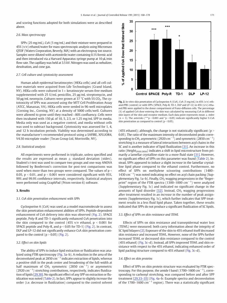
Fig. 2. In-vitro skin penetration of Cyclosporine A (CsA). CsA (5 mg/mL) in 45% (v/v) eth-anol/PBS (control) or with SPPs (SPACE, Poly-R, TD-1, DLP and LP-12) in 45% (v/v) etha-nol/PBS were applied to the donor compartment of Franz-diffusion cells. The percentage(%) of applied CsA dose entering the skin was calculated by measuring CsA in differentskin layers of the skin and receptor medium. Each data point represents mean ± stdev(n = 3). The asterisks (***p b 0.001 and *p b 0.05) indicate significantly higher % CsAskin penetration as compared to control (p b 0.05).
171S. Kumar et al. / Journal of Controlled Release 199 (2015) 168–178
and scoring functions adopted for both simulations were as describedabove.
2.6. Mass spectroscopy
SPPs (25mg/mL), CsA (5mg/mL) and their mixture were prepared in45% (v/v) ethanol/water formass spectroscopic analysis usingMicromassQTOF (Waters Corporation, Beverly, MA)with an electrospray ion source.Samples were dilutedwith acetonitrile/water containing 0.1% formic acidand then introduced via a Harvard Apparatus syringe pump at 10 μL/minflow rate. The capillarywas held at 3.5 kV.Nitrogenwas used as nebulizer,desolvation, and cone gas.
2.7. Cell culture and cytotoxicity assessment
Human adult epidermal keratinocytes (HEKa cells) and all cell cul-ture materials were acquired from Life Technologies (Grand Island,NY). HEKa cells were cultured in 1× keratinocyte serum-free mediumsupplemented with 25 U/mL penicillin, 25 μg/mL streptomycin, and50 μg/mL neomycin. Cultures were grown at 37 °C with 5% CO2. The cy-totoxicity of SPPs was assessed using the MTT Cell Proliferation Assay(ATCC, Manassas, VA). HEKa cells were seeded in 96-well microplates(Corning Inc., Corning, NY) at a density of 5000 cells/well. Cultureswere allowed to grow until they reached ~80% confluency. Cells werethen incubated with 150 μL of 10, 5, 2.5, or 1.25 mg/mL SPP in media.Media only was used as a negative control, and media without cellswas used to subtract background. Cytotoxicity was assessed for 1, 4,and 12 h incubation periods. Viability was determined according tothe manufacturer's recommended protocol using a SAFIRE, XFLUOR4,V4.50 microplate reader (Tecan Group Ltd, Morrisville, NY).
2.8. Statistical analysis
All experiments were performed in triplicate, unless specified andthe results are expressed as mean ± standard deviation (stdev).Student's t-test was used to compare two groups and one-way ANOVAfollowed by Bonferroni's correction for post-test comparisons wasused when more than two groups were compared. The values of p b
0.05, p b 0.01, and p b 0.001 were considered significant with 95%,99% and 99.9% confidence intervals, respectively. Statistical analyseswere performed using GraphPad (Prism version 6) software.
3. Results
3.1. CsA skin penetration enhancement with SPPs
Cyclosporine A (CsA) was used as a model macromolecule to assessthe skin penetration enhancement abilities of SPPs. Peptide-dependentenhancement of CsA delivery into skin was observed (Fig. 2). SPACEpeptide, Poly-R and TD-1 significantly enhanced CsA penetration intothe skin compared to the control (45% v/v ethanol, p b 0.001 forSPACE peptide and Poly-R, and p b 0.05 for TD-1) (Fig. 2). In contrast,DLP and LP-12 did not significantly enhance CsA skin penetration com-pared to the control (p N 0.05) (Fig. 2).
3.2. Effect on skin lipids
The ability of SPPs to induce lipid extraction or fluidizationwas ana-lyzed using FTIR spectroscopy (Fig. 3a–b). A reduction in the area of thedeconvoluted peak at 2850 cm−1 indicates extraction of lipids, whereasa positive shift in the peak center and broadening of the full-width athalf maximum of CH2 symmetric (2850 cm−1) or asymmetric(2920 cm−1) stretching contributions, respectively, indicates fluidiza-tion of lipids [29,30]. No significant effect of any SPP on extraction or flu-idizationwas noted (Table 2). In fact, SPPs appear to slightly increase theorder (i.e. decrease in fluidization) compared to the control solvent
(45% ethanol); although, the change is not statistically significant (p N
0.05). The ratio of themaximum intensity of deconvoluted peaks corre-sponding to CH2 asymmetric (2920 cm−1) and symmetric (2850 cm−1)stretching is ameasure of lateral interactions between acyl chains in theSC and is another indicator of lipid fluidization [31]. An increase in thisratio (Height2920/2850) indicates a shift in lipid microstructure from pri-marily a lamellar crystalline-state to a more fluid state [31]. However,no significant effect of SPPs on this parameter was found (Table 2). In-stead, SPPs appeared to induce a slight increase in the lamellar crystal-line lipid phase compared to the ethanol control. Furthermore, noeffect of SPPs on methylene scissoring contributions (1480–1430 cm−1) was noted indicating no effect on acyl chain packing (Sup-plementary Fig. 1a–b). Finally, CH2 wagging progressions in the “finger-print” region of the FTIR spectra (1300–1000 cm−1) were analyzed(Supplementary Fig. 1c) and indicated no significant change in theamounts of lipid disorder [32]. Instead, CH2 wagging progressionsafter treatment resulted in an increase in the number of peak assign-ments (Supplementary Fig. 1c), which further indicates that SPP treat-ment results in a less fluid lipid phase. Taken together, these resultsindicated that SPPs do not produce a significant fluidization of SC lipids.
3.3. Effect of SPPs on skin resistance and TEWL
Effects of SPPs on skin resistance and transepidermal water loss(TEWL) were measured; both carry information about the integrity ofSC lipid bilayers [2]. Exposure of the skin to 45% ethanol itself decreasedskin resistance and increased TEWL. However, none of the SPPs furtherincreased TEWL or decreased skin resistance compared to the control(45% ethanol) (Fig. 3c–d). Instead, all SPPs improved TEWL and skin re-sistance with respect to the 45% ethanol, indicating enhanced order oflipid packing/structure compared to 45% ethanol (Fig. 3c–d).
3.4. Effect on skin proteins
Effect of SPPs on skin protein structure was evaluated by FTIR spec-troscopy. For this purpose, the amide I band (1700–1600 cm−1), corre-sponding to carbonyl stretching, was compared before and after SPPtreatment [29,33–35] (Fig. 4a–b: Example spectra and deconvolutionof the 1700–1600 cm−1 region). There was a statistically significant
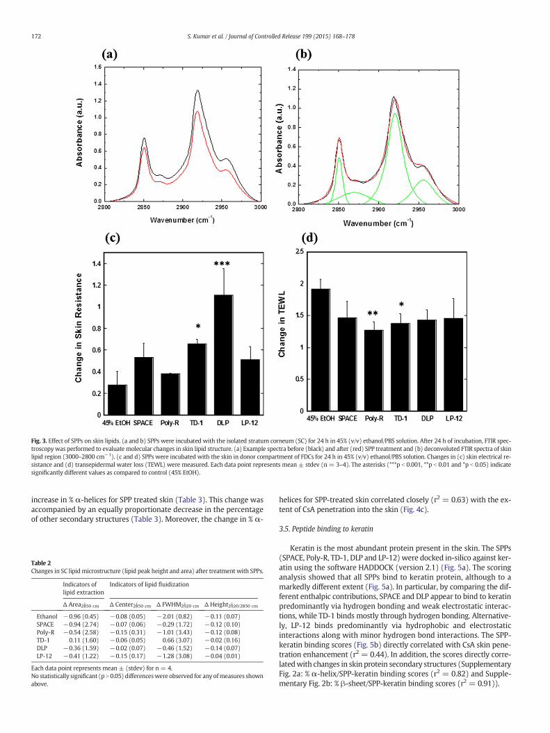
Fig. 3. Effect of SPPs on skin lipids. (a and b) SPPs were incubated with the isolated stratum corneum (SC) for 24 h in 45% (v/v) ethanol/PBS solution. After 24 h of incubation, FTIR spec-troscopywas performed to evaluate molecular changes in skin lipid structure. (a) Example spectra before (black) and after (red) SPP treatment and (b) deconvoluted FTIR spectra of skinlipid region (3000–2800 cm−1). (c and d) SPPs were incubated with the skin in donor compartment of FDCs for 24 h in 45% (v/v) ethanol/PBS solution. Changes in (c) skin electrical re-sistance and (d) transepidermal water loss (TEWL) were measured. Each data point represents mean ± stdev (n = 3–4). The asterisks (***p b 0.001, **p b 0.01 and *p b 0.05) indicatesignificantly different values as compared to control (45% EtOH).
172 S. Kumar et al. / Journal of Controlled Release 199 (2015) 168–178
increase in % α-helices for SPP treated skin (Table 3). This change wasaccompanied by an equally proportionate decrease in the percentageof other secondary structures (Table 3). Moreover, the change in % α-
Table 2Changes in SC lipid microstructure (lipid peak height and area) after treatment with SPPs.
Indicators oflipid extraction
Indicators of lipid fluidization
Δ Area2850 cm−1 Δ Center2850 cm
−1 Δ FWHM2920 cm−1 Δ Height2920/2850 cm
−1
Ethanol −0.96 (0.45) −0.08 (0.05) −2.01 (0.82) −0.11 (0.07)SPACE −0.94 (2.74) −0.07 (0.06) −0.29 (1.72) −0.12 (0.10)Poly-R −0.54 (2.58) −0.15 (0.31) −1.01 (3.43) −0.12 (0.08)TD-1 0.11 (1.60) −0.06 (0.05) 0.66 (3.07) −0.02 (0.16)DLP −0.36 (1.59) −0.02 (0.07) −0.46 (1.52) −0.14 (0.07)LP-12 −0.41 (1.22) −0.15 (0.17) −1.28 (3.08) −0.04 (0.01)
Each data point represents mean ± (stdev) for n = 4.No statistically significant (p N 0.05) differenceswere observed for any ofmeasures shownabove.
helices for SPP-treated skin correlated closely (r2 = 0.63) with the ex-tent of CsA penetration into the skin (Fig. 4c).
3.5. Peptide binding to keratin
Keratin is the most abundant protein present in the skin. The SPPs(SPACE, Poly-R, TD-1, DLP and LP-12) were docked in-silico against ker-atin using the software HADDOCK (version 2.1) (Fig. 5a). The scoringanalysis showed that all SPPs bind to keratin protein, although to amarkedly different extent (Fig. 5a). In particular, by comparing the dif-ferent enthalpic contributions, SPACE and DLP appear to bind to keratinpredominantly via hydrogen bonding and weak electrostatic interac-tions, while TD-1 binds mostly through hydrogen bonding. Alternative-ly, LP-12 binds predominantly via hydrophobic and electrostaticinteractions along with minor hydrogen bond interactions. The SPP-keratin binding scores (Fig. 5b) directly correlated with CsA skin pene-tration enhancement (r2 = 0.44). In addition, the scores directly corre-latedwith changes in skin protein secondary structures (SupplementaryFig. 2a: % α-helix/SPP-keratin binding scores (r2 = 0.82) and Supple-mentary Fig. 2b: % β-sheet/SPP-keratin binding scores (r2 = 0.91)).
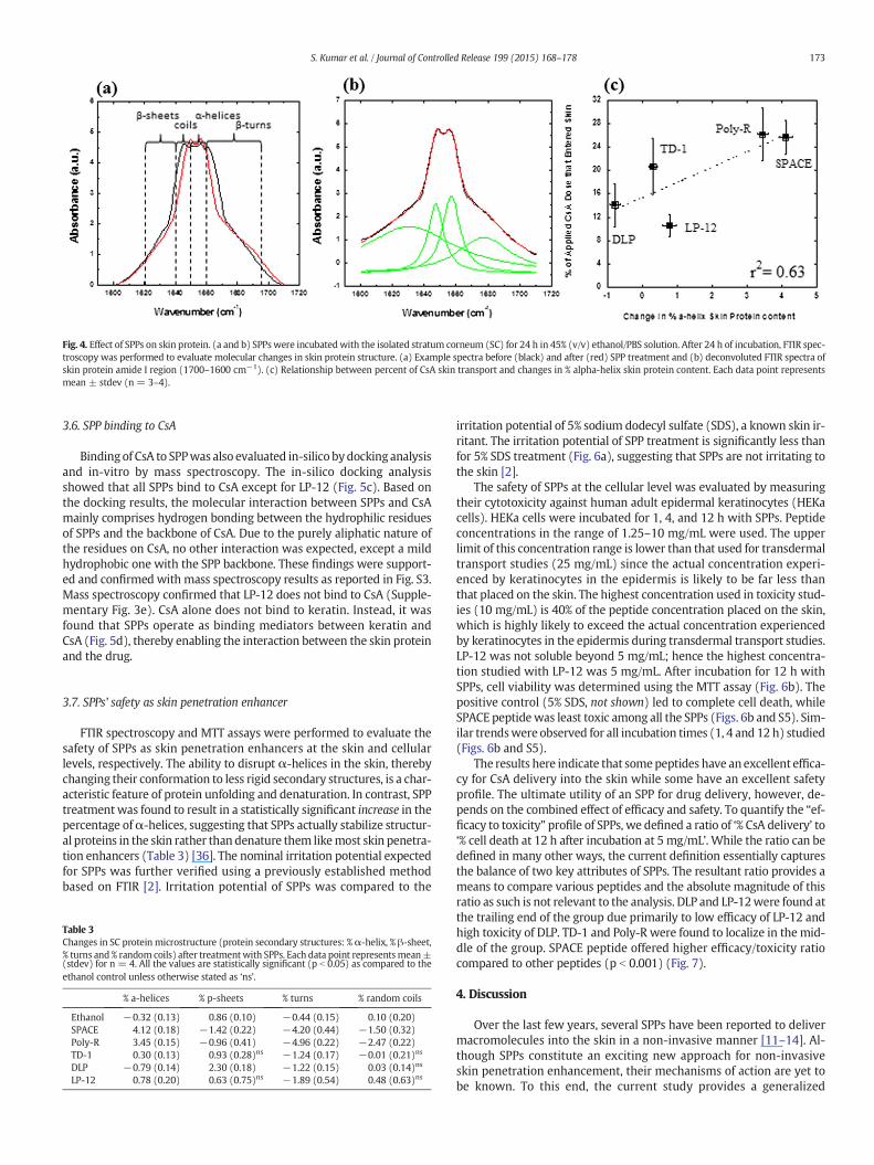
Fig. 4. Effect of SPPs on skin protein. (a and b) SPPswere incubatedwith the isolated stratum corneum (SC) for 24 h in 45% (v/v) ethanol/PBS solution. After 24 h of incubation, FTIR spec-troscopy was performed to evaluate molecular changes in skin protein structure. (a) Example spectra before (black) and after (red) SPP treatment and (b) deconvoluted FTIR spectra ofskin protein amide I region (1700–1600 cm−1). (c) Relationship between percent of CsA skin transport and changes in % alpha-helix skin protein content. Each data point representsmean ± stdev (n = 3–4).
173S. Kumar et al. / Journal of Controlled Release 199 (2015) 168–178
3.6. SPP binding to CsA
Binding of CsA to SPPwas also evaluated in-silico bydocking analysisand in-vitro by mass spectroscopy. The in-silico docking analysisshowed that all SPPs bind to CsA except for LP-12 (Fig. 5c). Based onthe docking results, the molecular interaction between SPPs and CsAmainly comprises hydrogen bonding between the hydrophilic residuesof SPPs and the backbone of CsA. Due to the purely aliphatic nature ofthe residues on CsA, no other interaction was expected, except a mildhydrophobic one with the SPP backbone. These findings were support-ed and confirmed with mass spectroscopy results as reported in Fig. S3.Mass spectroscopy confirmed that LP-12 does not bind to CsA (Supple-mentary Fig. 3e). CsA alone does not bind to keratin. Instead, it wasfound that SPPs operate as binding mediators between keratin andCsA (Fig. 5d), thereby enabling the interaction between the skin proteinand the drug.
3.7. SPPs' safety as skin penetration enhancer
FTIR spectroscopy and MTT assays were performed to evaluate thesafety of SPPs as skin penetration enhancers at the skin and cellularlevels, respectively. The ability to disrupt α-helices in the skin, therebychanging their conformation to less rigid secondary structures, is a char-acteristic feature of protein unfolding and denaturation. In contrast, SPPtreatmentwas found to result in a statistically significant increase in thepercentage ofα-helices, suggesting that SPPs actually stabilize structur-al proteins in the skin rather than denature them likemost skin penetra-tion enhancers (Table 3) [36]. The nominal irritation potential expectedfor SPPs was further verified using a previously established methodbased on FTIR [2]. Irritation potential of SPPs was compared to the
Table 3Changes in SC protein microstructure (protein secondary structures: %α-helix, % β-sheet,% turns and % random coils) after treatmentwith SPPs. Each data point representsmean±(stdev) for n = 4. All the values are statistically significant (p b 0.05) as compared to theethanol control unless otherwise stated as ‘ns’.
% a-helices % p-sheets % turns % random coils
Ethanol −0.32 (0.13) 0.86 (0.10) −0.44 (0.15) 0.10 (0.20)SPACE 4.12 (0.18) −1.42 (0.22) −4.20 (0.44) −1.50 (0.32)Poly-R 3.45 (0.15) −0.96 (0.41) −4.96 (0.22) −2.47 (0.22)TD-1 0.30 (0.13) 0.93 (0.28)ns −1.24 (0.17) −0.01 (0.21)ns
DLP −0.79 (0.14) 2.30 (0.18) −1.22 (0.15) 0.03 (0.14)ns
LP-12 0.78 (0.20) 0.63 (0.75)ns −1.89 (0.54) 0.48 (0.63)ns
irritation potential of 5% sodium dodecyl sulfate (SDS), a known skin ir-ritant. The irritation potential of SPP treatment is significantly less thanfor 5% SDS treatment (Fig. 6a), suggesting that SPPs are not irritating tothe skin [2].
The safety of SPPs at the cellular level was evaluated by measuringtheir cytotoxicity against human adult epidermal keratinocytes (HEKacells). HEKa cells were incubated for 1, 4, and 12 h with SPPs. Peptideconcentrations in the range of 1.25–10 mg/mL were used. The upperlimit of this concentration range is lower than that used for transdermaltransport studies (25 mg/mL) since the actual concentration experi-enced by keratinocytes in the epidermis is likely to be far less thanthat placed on the skin. The highest concentration used in toxicity stud-ies (10 mg/mL) is 40% of the peptide concentration placed on the skin,which is highly likely to exceed the actual concentration experiencedby keratinocytes in the epidermis during transdermal transport studies.LP-12 was not soluble beyond 5 mg/mL; hence the highest concentra-tion studied with LP-12 was 5 mg/mL. After incubation for 12 h withSPPs, cell viability was determined using the MTT assay (Fig. 6b). Thepositive control (5% SDS, not shown) led to complete cell death, whileSPACE peptidewas least toxic among all the SPPs (Figs. 6b and S5). Sim-ilar trendswere observed for all incubation times (1, 4 and 12 h) studied(Figs. 6b and S5).
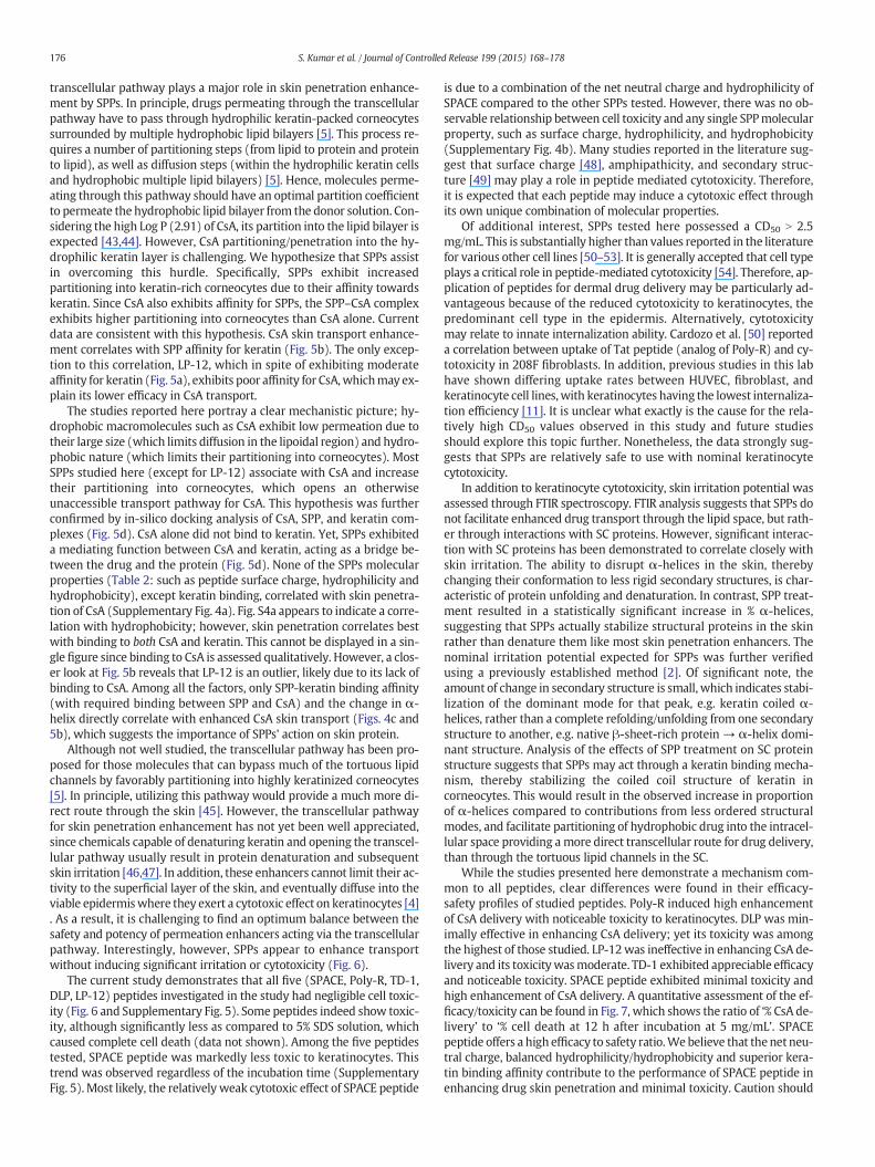
The results here indicate that somepeptides have anexcellent effica-cy for CsA delivery into the skin while some have an excellent safetyprofile. The ultimate utility of an SPP for drug delivery, however, de-pends on the combined effect of efficacy and safety. To quantify the “ef-ficacy to toxicity” profile of SPPs, we defined a ratio of ‘% CsA delivery’ to‘% cell death at 12 h after incubation at 5 mg/mL’. While the ratio can bedefined in many other ways, the current definition essentially capturesthe balance of two key attributes of SPPs. The resultant ratio provides ameans to compare various peptides and the absolute magnitude of thisratio as such is not relevant to the analysis. DLP and LP-12were found atthe trailing end of the group due primarily to low efficacy of LP-12 andhigh toxicity of DLP. TD-1 and Poly-R were found to localize in themid-dle of the group. SPACE peptide offered higher efficacy/toxicity ratiocompared to other peptides (p b 0.001) (Fig. 7).
4. Discussion
Over the last few years, several SPPs have been reported to delivermacromolecules into the skin in a non-invasive manner [11–14]. Al-though SPPs constitute an exciting new approach for non-invasiveskin penetration enhancement, their mechanisms of action are yet tobe known. To this end, the current study provides a generalized
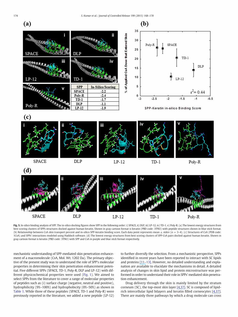
Fig. 5. In-silico binding analysis of SPP. The in-silico docking figures show SPP in the following order: i) SPACE, ii) DLP, iii) LP-12, iv) TD-1, v) Poly-R. (a) The lowest energy structures frombest scoring clusters of SPPs structures docked against human keratin. Shown in gray cartoon format is keratin (PBD code: 3TNU) with peptide structures shown in blue stick format.(b) Relationship between CsA skin transport percent and in-silico SPP-keratin binding score. Each data point represents mean ± stdev (n = 3–4). (c) Structures of CsA (PDB code:1CsA) and SPPs' interactions modeled using Haddock software. (d) The lowest energy structures from best scoring clusters of SPP-CsA pairs docked against human keratin. Shown ingray cartoon format is keratin (PBD code: 3TNU) with SPP and CsA in purple and blue stick format respectively.
174 S. Kumar et al. / Journal of Controlled Release 199 (2015) 168–178
mechanistic understanding of SPP-mediated skin penetration enhance-ment of a macromolecule (CsA, Mol. Wt. 1202 Da). The primary objec-tive of the present study was to understand the role of SPP's molecularproperties in determining their skin penetration enhancement poten-tial. Five different SPPs (SPACE, TD-1, Poly-R, DLP and LP-12) with dif-ferent physicochemical properties were used (Fig. 1). We aimed toselect SPPs from the literature to cover a range of molecular propertiesof peptides such as (i) surface charge (negative, neutral and positive),hydrophilicity (9%–100%) and hydrophobicity (0%–50%) as shown inTable 1. While three of these peptides (SPACE, TD-1 and Poly-R) werepreviously reported in the literature, we added a new peptide (LP-12)
to further diversify the selection. From a mechanistic perspective, SPPsidentified in recent years have been reported to interact with SC lipidsand proteins [11–13]. However, no detailed understanding and expla-nation are available to elucidate the mechanisms in detail. A detailedanalysis of changes in skin lipid and protein microstructure was per-formed in order to understand their role in SPPs'mediated skin penetra-tion enhancement.
Drug delivery through the skin is mainly limited by the stratumcorneum (SC), the top-most skin layer [4,37]. SC is composed of lipid-rich intercellular lipid bilayers and keratin filled corneocytes [4,37].There are mainly three pathways by which a drug molecule can cross
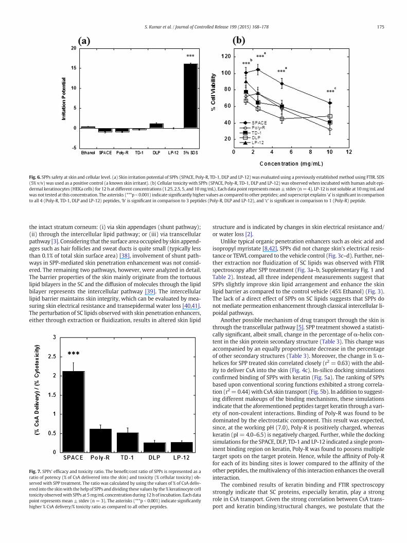
Fig. 6. SPPs safety at skin and cellular level. (a) Skin irritation potential of SPPs (SPACE, Poly-R, TD-1, DLP and LP-12)was evaluated using a previously establishedmethod using FTIR. SDS(5% v/v) was used as a positive control (a known skin irritant). (b) Cellular toxicity with SPPs (SPACE, Poly-R, TD-1, DLP and LP-12) was observedwhen incubated with human adult epi-dermal keratinocytes (HEKa cells) for 12 h at different concentrations (1.25, 2.5, 5, and 10mg/mL). Each data point representsmean± stdev (n=4). LP-12 is not soluble at 10mg/mL andwas not tested at this concentration. The asterisks (***p b 0.001) indicate significantly higher values as compared to other peptides; and superscript explains ‘a’ is significant in comparisonto all 4 (Poly-R, TD-1, DLP and LP-12) peptides, ‘b’ is significant in comparison to 3 peptides (Poly-R, DLP and LP-12), and ‘c’ is significant in comparison to 1 (Poly-R) peptide.
175S. Kumar et al. / Journal of Controlled Release 199 (2015) 168–178
the intact stratum corneum: (i) via skin appendages (shunt pathway);(ii) through the intercellular lipid pathway; or (iii) via transcellularpathway [3]. Considering that the surface area occupied by skin append-ages such as hair follicles and sweat ducts is quite small (typically lessthan 0.1% of total skin surface area) [38], involvement of shunt path-ways in SPP-mediated skin penetration enhancement was not consid-ered. The remaining two pathways, however, were analyzed in detail.The barrier properties of the skin mainly originate from the tortuouslipid bilayers in the SC and the diffusion of molecules through the lipidbilayer represents the intercellular pathway [39]. The intercellularlipid barrier maintains skin integrity, which can be evaluated by mea-suring skin electrical resistance and transepidermal water loss [40,41].The perturbation of SC lipids observedwith skin penetration enhancers,either through extraction or fluidization, results in altered skin lipid
Fig. 7. SPPs' efficacy and toxicity ratio. The benefit/cost ratio of SPPs is represented as aratio of potency (% of CsA delivered into the skin) and toxicity (% cellular toxicity) ob-served with SPP treatment. The ratio was calculated by using the values of % of CsA deliv-ered into the skinwith the help of SPPs anddividing these values by the % keratinocyte celltoxicity observedwith SPPs at 5mg/mL concentration during 12h of incubation. Each datapoint represents mean ± stdev (n= 3). The asterisks (***p b 0.001) indicate significantlyhigher % CsA delivery/% toxicity ratio as compared to all other peptides.
structure and is indicated by changes in skin electrical resistance and/or water loss [2].
Unlike typical organic penetration enhancers such as oleic acid andisopropyl myristate [8,42], SPPs did not change skin's electrical resis-tance or TEWL compared to the vehicle control (Fig. 3c–d). Further, nei-ther extraction nor fluidization of SC lipids was observed with FTIRspectroscopy after SPP treatment (Fig. 3a–b, Supplementary Fig. 1 andTable 2). Instead, all three independent measurements suggest thatSPPs slightly improve skin lipid arrangement and enhance the skinlipid barrier as compared to the control vehicle (45% Ethanol) (Fig. 3).The lack of a direct effect of SPPs on SC lipids suggests that SPPs donot mediate permeation enhancement through classical intercellular li-poidal pathways.
Another possible mechanism of drug transport through the skin isthrough the transcellular pathway [5]. SPP treatment showed a statisti-cally significant, albeit small, change in the percentage of α-helix con-tent in the skin protein secondary structure (Table 3). This change wasaccompanied by an equally proportionate decrease in the percentageof other secondary structures (Table 3). Moreover, the change in % α-helices for SPP treated skin correlated closely (r2 = 0.63) with the abil-ity to deliver CsA into the skin (Fig. 4c). In-silico docking simulationsconfirmed binding of SPPs with keratin (Fig. 5a). The ranking of SPPsbased upon conventional scoring functions exhibited a strong correla-tion (r2=0.44)with CsA skin transport (Fig. 5b). In addition to suggest-ing different makeups of the binding mechanisms, these simulationsindicate that the aforementioned peptides target keratin through a vari-ety of non-covalent interactions. Binding of Poly-R was found to bedominated by the electrostatic component. This result was expected,since, at the working pH (7.0), Poly-R is positively charged, whereaskeratin (pI = 4.0–6.5) is negatively charged. Further, while the dockingsimulations for the SPACE, DLP, TD-1 and LP-12 indicated a single prom-inent binding region on keratin, Poly-R was found to possess multipletarget spots on the target protein. Hence, while the affinity of Poly-Rfor each of its binding sites is lower compared to the affinity of theother peptides, themultivalency of this interaction enhances the overallinteraction.
The combined results of keratin binding and FTIR spectroscopystrongly indicate that SC proteins, especially keratin, play a strongrole in CsA transport. Given the strong correlation between CsA trans-port and keratin binding/structural changes, we postulate that the
176 S. Kumar et al. / Journal of Controlled Release 199 (2015) 168–178
transcellular pathway plays a major role in skin penetration enhance-ment by SPPs. In principle, drugs permeating through the transcellularpathway have to pass through hydrophilic keratin-packed corneocytessurrounded by multiple hydrophobic lipid bilayers [5]. This process re-quires a number of partitioning steps (from lipid to protein and proteinto lipid), as well as diffusion steps (within the hydrophilic keratin cellsand hydrophobic multiple lipid bilayers) [5]. Hence, molecules perme-ating through this pathway should have an optimal partition coefficientto permeate the hydrophobic lipid bilayer from the donor solution. Con-sidering the high Log P (2.91) of CsA, its partition into the lipid bilayer isexpected [43,44]. However, CsA partitioning/penetration into the hy-drophilic keratin layer is challenging. We hypothesize that SPPs assistin overcoming this hurdle. Specifically, SPPs exhibit increasedpartitioning into keratin-rich corneocytes due to their affinity towardskeratin. Since CsA also exhibits affinity for SPPs, the SPP–CsA complexexhibits higher partitioning into corneocytes than CsA alone. Currentdata are consistent with this hypothesis. CsA skin transport enhance-ment correlates with SPP affinity for keratin (Fig. 5b). The only excep-tion to this correlation, LP-12, which in spite of exhibiting moderateaffinity for keratin (Fig. 5a), exhibits poor affinity for CsA,whichmay ex-plain its lower efficacy in CsA transport.
The studies reported here portray a clear mechanistic picture; hy-drophobic macromolecules such as CsA exhibit low permeation due totheir large size (which limits diffusion in the lipoidal region) and hydro-phobic nature (which limits their partitioning into corneocytes). MostSPPs studied here (except for LP-12) associate with CsA and increasetheir partitioning into corneocytes, which opens an otherwiseunaccessible transport pathway for CsA. This hypothesis was furtherconfirmed by in-silico docking analysis of CsA, SPP, and keratin com-plexes (Fig. 5d). CsA alone did not bind to keratin. Yet, SPPs exhibiteda mediating function between CsA and keratin, acting as a bridge be-tween the drug and the protein (Fig. 5d). None of the SPPs molecularproperties (Table 2: such as peptide surface charge, hydrophilicity andhydrophobicity), except keratin binding, correlated with skin penetra-tion of CsA (Supplementary Fig. 4a). Fig. S4a appears to indicate a corre-lation with hydrophobicity; however, skin penetration correlates bestwith binding to both CsA and keratin. This cannot be displayed in a sin-gle figure since binding to CsA is assessed qualitatively. However, a clos-er look at Fig. 5b reveals that LP-12 is an outlier, likely due to its lack ofbinding to CsA. Among all the factors, only SPP-keratin binding affinity(with required binding between SPP and CsA) and the change in α-helix directly correlate with enhanced CsA skin transport (Figs. 4c and5b), which suggests the importance of SPPs' action on skin protein.
Although not well studied, the transcellular pathway has been pro-posed for those molecules that can bypass much of the tortuous lipidchannels by favorably partitioning into highly keratinized corneocytes[5]. In principle, utilizing this pathway would provide a much more di-rect route through the skin [45]. However, the transcellular pathwayfor skin penetration enhancement has not yet been well appreciated,since chemicals capable of denaturing keratin and opening the transcel-lular pathway usually result in protein denaturation and subsequentskin irritation [46,47]. In addition, these enhancers cannot limit their ac-tivity to the superficial layer of the skin, and eventually diffuse into theviable epidermiswhere they exert a cytotoxic effect on keratinocytes [4]. As a result, it is challenging to find an optimum balance between thesafety and potency of permeation enhancers acting via the transcellularpathway. Interestingly, however, SPPs appear to enhance transportwithout inducing significant irritation or cytotoxicity (Fig. 6).
The current study demonstrates that all five (SPACE, Poly-R, TD-1,DLP, LP-12) peptides investigated in the study had negligible cell toxic-ity (Fig. 6 and Supplementary Fig. 5). Some peptides indeed show toxic-ity, although significantly less as compared to 5% SDS solution, whichcaused complete cell death (data not shown). Among the five peptidestested, SPACE peptide was markedly less toxic to keratinocytes. Thistrend was observed regardless of the incubation time (SupplementaryFig. 5). Most likely, the relatively weak cytotoxic effect of SPACE peptide
is due to a combination of the net neutral charge and hydrophilicity ofSPACE compared to the other SPPs tested. However, there was no ob-servable relationship between cell toxicity and any single SPPmolecularproperty, such as surface charge, hydrophilicity, and hydrophobicity(Supplementary Fig. 4b). Many studies reported in the literature sug-gest that surface charge [48], amphipathicity, and secondary struc-ture [49] may play a role in peptide mediated cytotoxicity. Therefore,it is expected that each peptide may induce a cytotoxic effect throughits own unique combination of molecular properties.
Of additional interest, SPPs tested here possessed a CD50 N 2.5mg/mL. This is substantially higher than values reported in the literaturefor various other cell lines [50–53]. It is generally accepted that cell typeplays a critical role in peptide-mediated cytotoxicity [54]. Therefore, ap-plication of peptides for dermal drug delivery may be particularly ad-vantageous because of the reduced cytotoxicity to keratinocytes, thepredominant cell type in the epidermis. Alternatively, cytotoxicitymay relate to innate internalization ability. Cardozo et al. [50] reporteda correlation between uptake of Tat peptide (analog of Poly-R) and cy-totoxicity in 208F fibroblasts. In addition, previous studies in this labhave shown differing uptake rates between HUVEC, fibroblast, andkeratinocyte cell lines,with keratinocytes having the lowest internaliza-tion efficiency [11]. It is unclear what exactly is the cause for the rela-tively high CD50 values observed in this study and future studiesshould explore this topic further. Nonetheless, the data strongly sug-gests that SPPs are relatively safe to use with nominal keratinocytecytotoxicity.
In addition to keratinocyte cytotoxicity, skin irritation potential wasassessed through FTIR spectroscopy. FTIR analysis suggests that SPPs donot facilitate enhanced drug transport through the lipid space, but rath-er through interactions with SC proteins. However, significant interac-tion with SC proteins has been demonstrated to correlate closely withskin irritation. The ability to disrupt α-helices in the skin, therebychanging their conformation to less rigid secondary structures, is char-acteristic of protein unfolding and denaturation. In contrast, SPP treat-ment resulted in a statistically significant increase in % α-helices,suggesting that SPPs actually stabilize structural proteins in the skinrather than denature them like most skin penetration enhancers. Thenominal irritation potential expected for SPPs was further verifiedusing a previously established method [2]. Of significant note, theamount of change in secondary structure is small, which indicates stabi-lization of the dominant mode for that peak, e.g. keratin coiled α-helices, rather than a complete refolding/unfolding from one secondarystructure to another, e.g. native β-sheet-rich protein → α-helix domi-nant structure. Analysis of the effects of SPP treatment on SC proteinstructure suggests that SPPs may act through a keratin binding mecha-nism, thereby stabilizing the coiled coil structure of keratin incorneocytes. This would result in the observed increase in proportionof α-helices compared to contributions from less ordered structuralmodes, and facilitate partitioning of hydrophobic drug into the intracel-lular space providing a more direct transcellular route for drug delivery,than through the tortuous lipid channels in the SC.
While the studies presented here demonstrate a mechanism com-mon to all peptides, clear differences were found in their efficacy-safety profiles of studied peptides. Poly-R induced high enhancementof CsA delivery with noticeable toxicity to keratinocytes. DLP was min-imally effective in enhancing CsA delivery; yet its toxicity was amongthe highest of those studied. LP-12was ineffective in enhancing CsA de-livery and its toxicitywasmoderate. TD-1 exhibited appreciable efficacyand noticeable toxicity. SPACE peptide exhibited minimal toxicity andhigh enhancement of CsA delivery. A quantitative assessment of the ef-ficacy/toxicity can be found in Fig. 7, which shows the ratio of ‘% CsA de-livery’ to ‘% cell death at 12 h after incubation at 5 mg/mL’. SPACEpeptide offers a high efficacy to safety ratio.We believe that the net neu-tral charge, balanced hydrophilicity/hydrophobicity and superior kera-tin binding affinity contribute to the performance of SPACE peptide inenhancing drug skin penetration and minimal toxicity. Caution should
177S. Kumar et al. / Journal of Controlled Release 199 (2015) 168–178
be taken while reaching generalized conclusions about the efficacy andsafety of SPPs from the results in Fig. 7 since the relative efficacy of pep-tides in enhancing drug penetration could vary from drug to drug, espe-cially when the drug properties are substantially different compared toCsA. For example, TD-1 has been used to systemically deliver insulin, amolecule significantly different compared to CsA. Further, the ratio of ef-ficacy to toxicity could depend on the concentration of peptides sinceefficacy and toxicity may depend non-linearly on their concentration.Further, the assessment of toxicity in this study was made based onkeratinocyte cultures and the relative toxicity behavior of peptidescould be different in vivo. In addition, some of these peptides havebeen used in different forms, for example, chemical conjugation tocargo in case of Poly-R or ethosomal formulations in case of SPACE pep-tide. The efficacy/toxicity ratios of peptides could be different for differ-ent formulations.
5. Conclusion
The current study demonstrates the molecular level understandingof peptides acting as skin penetration enhancers. The SPPs examinedin this study appear to act as mediators between CsA and keratin, pro-viding a mechanism for partitioning of drugs into keratin-richcorneocytes through concurrent binding interactions between keratinand SPP, and, SPP andCsA. The SPPs appeared to be safe skin penetrationenhancers as shown by negligible effect on skin integrity, nominal skinirritation and cytotoxicity. As observed with in-silico keratin bindingand FTIR analysis of α-helix content, SPPs' interactions with skin pro-tein, directly correlated with enhanced drug delivery into the skin.
Acknowledgments
This work was supported by the Convoy Therapeutics Inc.(SB120129) The MRL Shared Experimental Facilities are supported bytheMRSEC Programof theNSFunderAwardNo. DMR1121053; amem-ber of the NSF-funded Materials Research Facilities Network. SM is ashareholder and scientific advisor of Convoy Therapeutics.
Appendix A. Supplementary data
Supplementary data to this article can be found online at http://dx.doi.org/10.1016/j.jconrel.2014.12.006.
References
[1] B.W. Barry, Breaching the skin's barrier to drugs, Nat. Biotechnol. 22 (2004)165–167.
[2] P. Karande, A. Jain, K. Ergun, V. Kispersky, S. Mitragotri, Design principles of chemicalpenetration enhancers for transdermal drug delivery, Proc. Natl. Acad. Sci. U. S. A.102 (2005) 4688–4693.
[3] M.R. Prausnitz, S. Mitragotri, R. Langer, Current status and future potential of trans-dermal drug delivery, Nat. Rev. Drug Discov. 3 (2004) 115–124.
[4] P. Karande, S. Mitragotri, Enhancement of transdermal drug delivery via synergisticaction of chemicals, Biochim. Biophys. Acta 1788 (2009) 2362–2373.
[5] G.M. ElMaghraby, B.W. Barry, A.C.Williams, Liposomes and skin: from drug deliveryto model membranes, Eur. J. Pharm. Sci. 34 (2008) 203–222.
[6] J. Zhang, M. Liu, H. Jin, L. Deng, J. Xing, A. Dong, In vitro enhancement of lactate es-ters on the percutaneous penetration of drugs with different lipophilicity, AAPSPharmSciTech 11 (2010) 894–903.
[7] M.R. Prausnitz, R. Langer, Transdermal drug delivery, Nat. Biotechnol. 26 (2008)1261–1268.
[8] B.W. Barry, Mode of action of penetration enhancers in human skin, J. Control. Re-lease 6 (1987) 85–97.
[9] V.V. Venuganti, O.P. Perumal, Effect of poly(amidoamine) (PAMAM) dendrimer onskin permeation of 5-fluorouracil, Int. J. Pharm. 361 (2008) 230–238.
[10] Y. Chen, Y. Shen, X. Guo, C. Zhang, W. Yang, M. Ma, S. Liu, M. Zhang, L.P. Wen, Trans-dermal protein delivery by a coadministered peptide identified via phage display,Nat. Biotechnol. 24 (2006) 455–460.
[11] T. Hsu, S. Mitragotri, Delivery of siRNA and other macromolecules into skin and cellsusing a peptide enhancer, Proc. Natl. Acad. Sci. U. S. A. 108 (2011) 15816–15821.
[12] Y.C. Kim, P.J. Ludovice, M.R. Prausnitz, Transdermal delivery enhanced by magaininpore-forming peptide, J. Control. Release 122 (2007) 375–383.
[13] S. Kumar, P. Sahdev, O. Perumal, H. Tummala, Identification of a novel skin penetra-tion enhancement peptide by phage display peptide library screening, Mol. Pharm.9 (2012) 1320–1330.
[14] J.B. Rothbard, S. Garlington, Q. Lin, T. Kirschberg, E. Kreider, P.L. McGrane, P.A.Wender, P.A. Khavari, Conjugation of arginine oligomers to cyclosporin A facilitatestopical delivery and inhibition of inflammation, Nat. Med. 6 (2000) 1253–1257.
[15] N.M. Carmichael, J.O. Dostrovsky, M.P. Charlton, Peptide-mediated transdermal de-livery of botulinum neurotoxin type A reduces neurogenic inflammation in theskin, Pain 149 (2010) 316–324.
[16] M. Chang, X. Li, Y. Sun, F. Cheng, Q. Wang, X. Xie, W. Zhao, X. Tian, Effect of cationiccyclopeptides on transdermal and transmembrane delivery of insulin, Mol. Pharm.10 (2013) 951–957.
[17] M. Chen, V. Gupta, A.C. Anselmo, J.A. Muraski, S. Mitragotri, Topical delivery ofhyaluronic acid into skin using SPACE-peptide carriers, J. Control. Release 173(2014) 67–74.
[18] M. Chen, M. Zakrewsky, V. Gupta, A.C. Anselmo, D.H. Slee, J.A. Muraski, S. Mitragotri,Topical delivery of siRNA into skin using SPACE-peptide carriers, J. Control. Release179 (2014) 33–41.
[19] C.M. Lin, K. Huang, Y. Zeng, X.C. Chen, S. Wang, Y. Li, A simple, noninvasive and ef-ficient method for transdermal delivery of siRNA, Arch. Dermatol. Res. 304 (2012)139–144.
[20] P.P. Shah, P.R. Desai, D. Channer, M. Singh, Enhanced skin permeation usingpolyarginine modified nanostructured lipid carriers, J. Control. Release 161 (2012)735–745.
[21] T. Zhang, H. Qu, X. Li, B. Zhao, J. Zhou, Q. Li, M. Sun, Transmembrane delivery and bi-ological effect of human growth hormone via a phage displayed peptide in vivo andin vitro, J. Pharm. Sci. 99 (2010) 4880–4891.
[22] G. Candan, H. Michiue, S. Ishikawa, A. Fujimura, K. Hayashi, A. Uneda, A. Mori, I.Ohmori, T. Nishiki, H. Matsui, K. Tomizawa, Combining poly-arginine with the hy-drophobic counter-anion 4-(1-pyrenyl)-butyric acid for protein transduction intransdermal delivery, Biomaterials 33 (2012) 6468–6475.
[23] Y.H. Wang, C.P. Chen, M.H. Chan, M. Chang, Y.W. Hou, H.H. Chen, H.R. Hsu, K. Liu, H.J.Lee, Arginine-rich intracellular delivery peptides noncovalently transport proteininto living cells, Biochem. Biophys. Res. Commun. 346 (2006) 758–767.
[24] P. Karande, A. Jain, S. Mitragotri, Relationships between skin's electrical impedanceand permeability in the presence of chemical enhancers, J. Control. Release 110(2006) 307–313.
[25] M. Chen, S. Kumar, A.C. Anselmo, V. Gupta, D.H. Slee, J.A. Muraski, S. Mitragotri,Topical delivery of Cyclosporine A into the skin using SPACE-peptide, J. Control.Release (2014).
[26] V.V. Venuganti, P. Sahdev, M. Hildreth, X. Guan, O. Perumal, Structure–skin perme-ability relationship of dendrimers, Pharm. Res. 28 (2011) 2246–2260.
[27] S.K. Rastogi, J. Singh, Lipid extraction and transport of hydrophilic solutes throughporcine epidermis, Int. J. Pharm. 225 (2001) 75–82.
[28] L.C. Morejohn, J.N. Pratley, Differential effects of trypsin on the epidermis of Ranacatesbeiana, Cell Tissue Res. 198 (1979) 349–362.
[29] Y. Obata, S. Utsumi, H. Watanabe, M. Suda, Y. Tokudome, M. Otsuka, K. Takayama,Infrared spectroscopic study of lipid interaction in stratum corneum treated withtransdermal absorption enhancers, Int. J. Pharm. 389 (2010) 18–23.
[30] A. Jadoul, J. Doucet, D. Durand, V. Préat, Modifications induced on stratum corneumstructure after in vitro iontophoresis: ATR-FTIR and X-ray scattering studies, J. Con-trol. Release 42 (1996) 165–173.
[31] M. Osada, M. Gniadecka, H.C. Wulf, Near-infrared Fourier transform Raman spectro-scopic analysis of proteins, water and lipids in intact normal stratum corneum andpsoriasis scales, Exp. Dermatol. 13 (2004) 391–395.
[32] L. Senak, D. Moore, R. Mendelsohn, Methylene wagging progressions as IR probes ofslightly disordered phospholipid acyl chain states, J. Phys. Chem. 96 (1992)2749–2754.
[33] T.M. Greve, K.B. Andersen, O.F. Nielsen, ATR-FTIR, FT-NIR and near-FT-Raman spec-troscopic studies of molecular composition in human skin in vivo and pig ear skinin vitro, Spectrosc. Int. J. 22 (2008) 437–457.
[34] B. Barry, H. Edwards, A. Williams, Fourier transform Raman and infrared vibrationalstudy of human skin: assignment of spectral bands, J. Raman Spectrosc. 23 (1992)641–645.
[35] Z. Movasaghi, S. Rehman, D.I. ur Rehman, Fourier transform infrared (FTIR) spec-troscopy of biological tissues, Appl. Spectrosc. Rev. 43 (2008) 134–179.
[36] P. Karande, A. Jain, S. Mitragotri, Discovery of transdermal penetration enhancers byhigh-throughput screening, Nat. Biotechnol. 22 (2004) 192–197.
[37] D. Chantasart, S.K. Li, Structure enhancement relationship of chemical penetration en-hancers in drug transport across the stratum corneum, Pharmaceutics 4 (2012) 71–92.
[38] J. Lademann, F. Knorr, H. Richter, U. Blume-Peytavi, A. Vogt, C. Antoniou, W. Sterry,A. Patzelt, Hair follicles—an efficient storage and penetration pathway for topicallyapplied substances. Summary of recent results obtained at the Center of Experimen-tal and Applied Cutaneous Physiology, Charite -Universitatsmedizin Berlin,Germany, Skin Pharmacol. Physiol. 21 (2008) 150–155.
[39] R.O. Potts, R.H. Guy, Predicting skin permeability, Pharm. Res. 9 (1992) 663–669.[40] L. Barbosa-Barros, C. Barba, M. Cocera, L. Coderch, C. Lopez-Iglesias, A. de la Maza, O.
Lopez, Effect of bicellar systems on skin properties, Int. J. Pharm. 352 (2008)263–272.
[41] D. Shcharbin, A. Drapeza, V. Loban, A. Lisichenok, M. Bryszewska, The breakdown ofbilayer lipid membranes by dendrimers, Cell. Mol. Biol. Lett. 11 (2006) 242–248.
[42] O. Pillai, V. Nair, R. Panchagnula, Transdermal iontophoresis of insulin: IV. Influenceof chemical enhancers, Int. J. Pharm. 269 (2004) 109–120.
[43] A.I. Lauerma, C. Surber, H.I. Maibach, Absorption of topical tacrolimus (FK506)in vitro through human skin: comparison with cyclosporin A, Skin Pharmacol. 10(1997) 230–234.

178 S. Kumar et al. / Journal of Controlled Release 199 (2015) 168–178
[44] A. Fahr, P.v. Hoogevest, S. May, N. Bergstrand, Leigh M.L.S., Transfer of lipophilicdrugs between liposomal membranes and biological interfaces: Consequences fordrug delivery, Eur. J. Pharm. Sci. 26 (2005) 251–265.
[45] R.O. Potts, M.L. Francoeur, Lipid biophysics of water loss through the skin, Proc. Natl.Acad. Sci. 87 (1990) 3871–3873.
[46] K.P. Ananthapadmanabhan, D.J. Moore, K. Subramanyan, M. Misra, F. Meyer, Cleans-ing without compromise: the impact of cleansers on the skin barrier and the tech-nology of mild cleansing, Dermatol. Ther. 17 (Suppl. 1) (2004) 16–25.
[47] A.M. Kligman, Topical pharmacology and toxicology of dimethyl sulfoxide—part 1,JAMA 193 (1965) 796–804.
[48] S.W. Jones, R. Christison, K. Bundell, C.J. Voyce, S.M.V. Brockbank, P. Newham, M.A.Lindsay, Characterisation of cell-penetrating peptide-mediated peptide delivery,Br. J. Pharmacol. 145 (2005) 1093–1102.
[49] E. Eiriksdottir, K. Konate, U. Langel, G. Divita, S. Deshayes, Secondary structure ofcell-penetrating peptides controls membrane interaction and insertion, Biochim.Biophys. Acta 1798 (2010) 1119–1128.
[50] A.K. Cardozo, V. Buchillier, M. Mathieu, J. Chen, F. Ortis, L. Ladriere, N. Allaman-Pillet,O. Poirot, S. Kellenberger, J.S. Beckmann, D.L. Eizirik, C. Bonny, F. Maurer, Cell-permeable peptides induce dose- and length-dependent cytotoxic effects, Biochim.Biophys. Acta Biomembr. 1768 (2007) 2222–2234.
[51] K. Kilk, R. Mahlapuu, U. Soomets, U. Langel, Analysis of in vitro toxicity of five cell-penetrating peptides by metabolic profiling, Toxicology 265 (2009) 87–95.
[52] B.R. Liu, J.F. Li, S.W. Lu, H.J. Lee, Y.W. Huang, K.B. Shannon, R.S. Aronstam, Cellular in-ternalization of quantum dots noncovalently conjugated with arginine-rich cell-penetrating peptides, J. Nanosci. Nanotechnol. 10 (2010) 6534–6543.
[53] T. Sugita, T. Yoshikawa, Y. Mukai, N. Yamanada, S. Imai, K. Nagano, Y. Yoshida, H.Shibata, Y. Yoshioka, S. Nakagawa, H. Kamada, S.I. Tsunoda, Y. Tsutsumi, Compara-tive study on transduction and toxicity of protein transduction domains, Br. J.Pharmacol. 153 (2008) 1143–1152.
[54] R. Trehin, H.P. Merkle, Chances and pitfalls of cell penetrating peptides for cellulardrug delivery, Eur. J. Pharm. Biopharm. 58 (2004) 209–223.
Related Documents