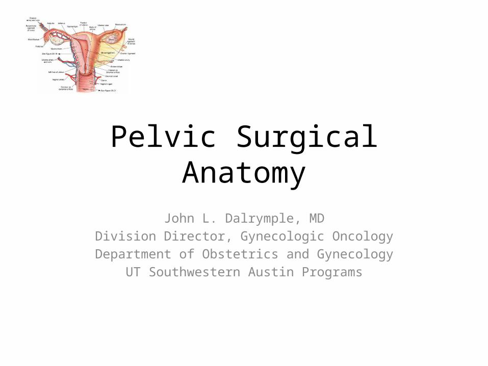
Pelvic Surgical Anatomy John L. Dalrymple, MD Division Director, Gynecologic Oncology Department of Obstetrics and Gynecology UT Southwestern Austin Programs

Pelvic Surgical Anatomy John L. Dalrymple, MD Division Director, Gynecologic Oncology Department of Obstetrics and Gynecology UT Southwestern Austin Programs.
Dec 14, 2015
Welcome message from author
This document is posted to help you gain knowledge. Please leave a comment to let me know what you think about it! Share it to your friends and learn new things together.
Transcript
Pelvic Surgical Anatomy
John L. Dalrymple, MDDivision Director, Gynecologic Oncology
Department of Obstetrics and GynecologyUT Southwestern Austin Programs

I have nothing to disclose.
Objectives
• Describe basic abdominal and pelvic anatomy related to common gynecologic surgical procedures
• Name common potential pitfalls and complications that can occur during gynecologic pelvic surgery
• Describe the challenges related to anatomical distortions from pelvic pathology, patient body habitus, and complex procedures
• List the physiologic changes related to anatomical changes from pelvic surgery (optional)
Why Anatomy is important• Backbone of understanding clinical conditions
– What’s normal– What’s abnormal– Why it’s abnormal– How to manage the problem
• Surgery is all about anatomy• Obstetrics AND Gynecology is loaded with
anatomical clinical correlations
General Considerations
• Preparation for the OR (PRE-OP)– Review basic/relevant anatomy:
• What organs are being removed/corrected/altered?• What anatomy must be traversed to get there?
– Understand indications for surgery:• Why is procedure being done/what are goals of surgery?• What alternatives are there and have they been considered?
General Considerations
• In the OR (INTRA-OP)– Perform the EUA (pelvic AND abdominal exam)
• What anatomical distortions are present?• Does this affect the route of surgery?• How will you position the patient?
– Performing the procedure:• What are the abdominal wall and pelvic floor anatomical
landmarks?• Is the anatomy distorted by the disease process or prior
procedures?• Does the patient’s body habitus affect her anatomy?• What potential complications can you expect?
General Considerations
• After the OR (POST-OP)– Anticipate physiologic changes:
• What will the patient/you expect acutely and chronically from anatomical changes (reproductive, GI, GU, sexually, physically, etc)?
– Manage complications:• What anatomic/physiologic changes will you expect from
common complications (bowel, bladder, vascular, nerve injuries)?• What are the expected postoperative pelvic and abdominal
anatomic changes that occur after surgery?
General Considerations
• In the OR (INTRA-OP)– Perform the EUA (pelvic AND abdominal exam)
• What anatomical distortions are present?• Does this affect the route of surgery?• How will you position the patient?
– Performing the procedure:• What are the abdominal wall and pelvic floor anatomical
landmarks?• Is the anatomy distorted by the disease process or prior
procedures?• Does the patient’s body habitus affect her anatomy?• What potential complications can you expect?
Case Studies
• Relevant surgical anatomy• Special points of consideration
– danger areas and potential complications• Physiologic outcomes
Case Study 1
• 38 yo G2P2 female with symptomatic menometrorrhagia, dysmenorrhea and anemia. Prior cesarean section x 2.
• Examination: BMI – 28; Pelvic – 16 wk fibroid uterus – palpable midway to the umbilicus on abdominal exam
• Ultrasound: multiple leiomyomas (>6) measuring in size from 4 to 8 cm, located in fundal, posterior/anterior and lateral uterus.
• EMB – proliferative; UPT – neg; Hgb – 8 mg/dL
Surgical Approach
• Preop Dx: Symptomatic Leiomyoma• Planned Procedure: Exploratory laparotomy,
Total abdominal hysterectomy (TAH)• Relevant Surgical Anatomy
– Abdominal and pelvic examination– Layers of the abdominal wall– Abdominal structures– Pelvic structures
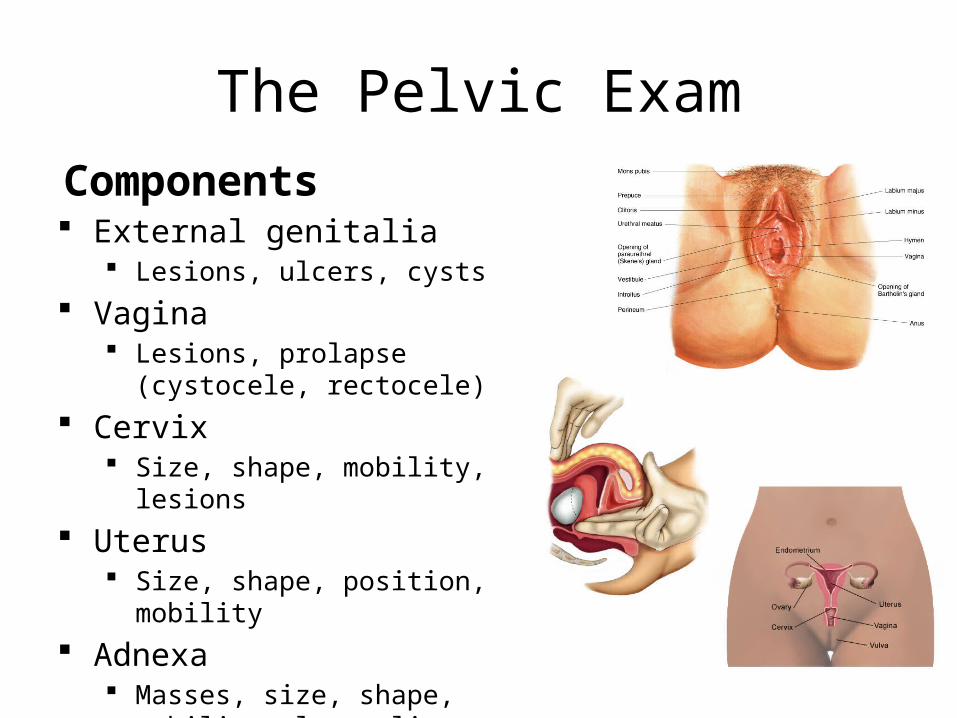
The Pelvic ExamComponents External genitalia
Lesions, ulcers, cysts Vagina
Lesions, prolapse (cystocele, rectocele) Cervix
Size, shape, mobility, lesions Uterus
Size, shape, position, mobility Adnexa
Masses, size, shape, mobility, laterality
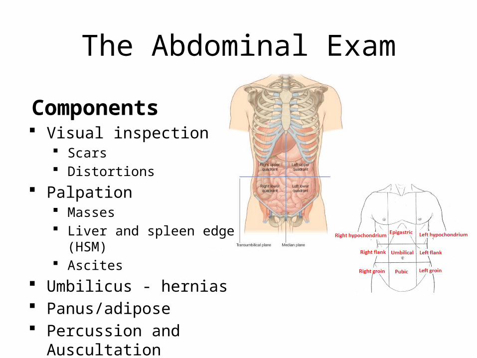
The Abdominal Exam
Components Visual inspection
Scars Distortions
Palpation Masses Liver and spleen edge (HSM) Ascites
Umbilicus - hernias Panus/adipose Percussion and Auscultation
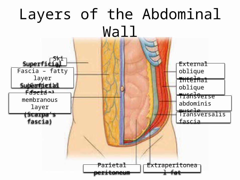
Layers of the Abdominal Wall
SkinSuperficial Fascia –
fatty layer(Camper’s fascia)
Superficial Fascia – membranous layer
(Scarpa’s fascia)
Extraperitoneal fat
External oblique muscleInternal oblique muscleTransverse abdominis muscle
Transversalis fascia
Parietal peritoneum
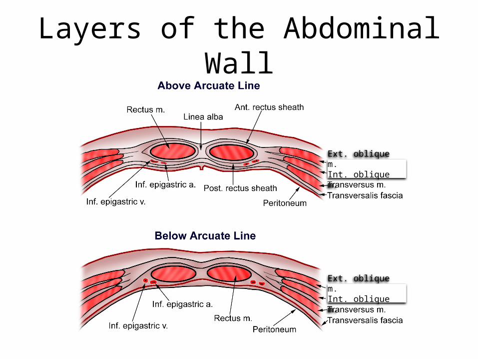
Layers of the Abdominal Wall
Ext. oblique m.Int. oblique m.
Ext. oblique m.Int. oblique m.
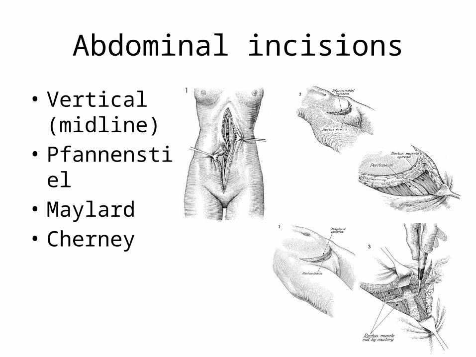
Abdominal incisions
• Vertical (midline)
• Pfannenstiel• Maylard• Cherney

Abdominal Structures
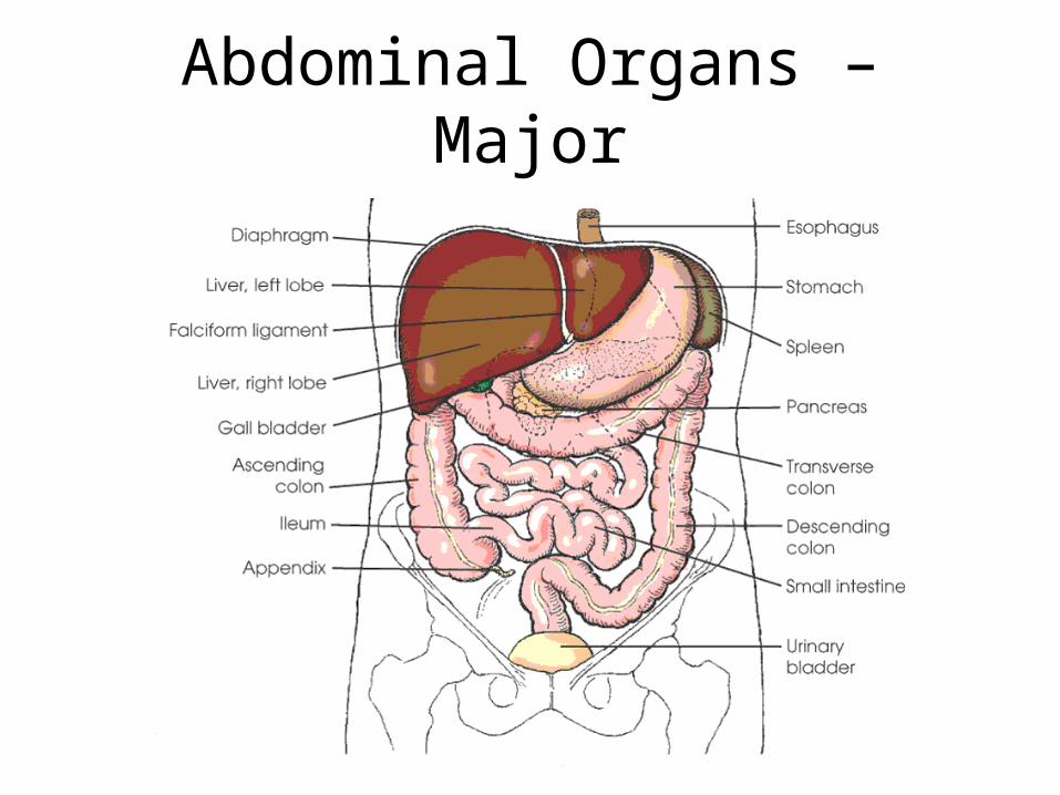
Abdominal Organs – Major
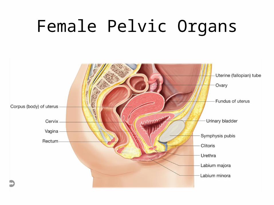
Female Pelvic Organs
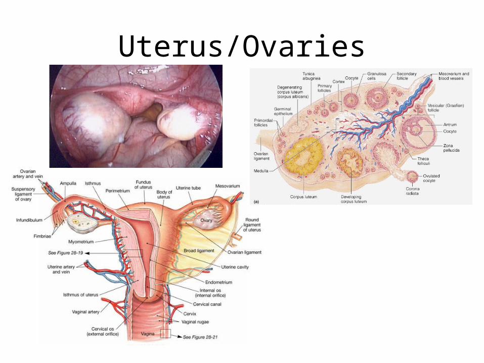
Uterus/Ovaries
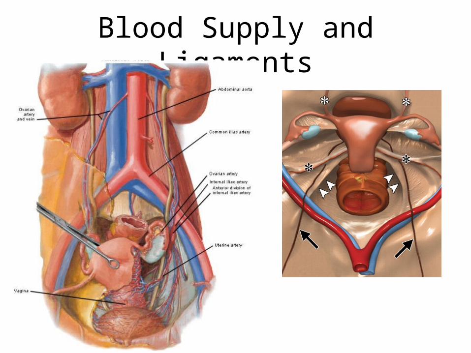
Blood Supply and Ligaments
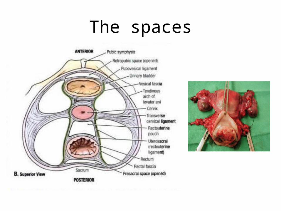
The spaces
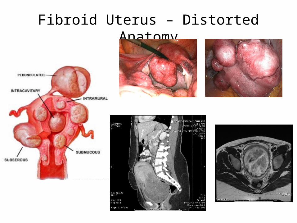
Fibroid Uterus – Distorted Anatomy
Special Points of Consideration
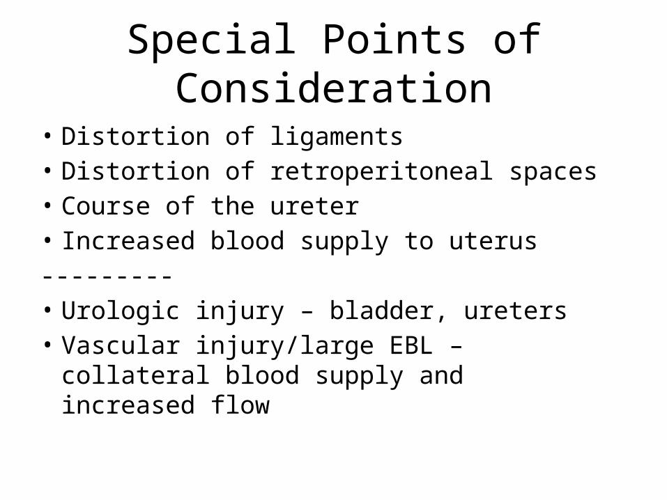
• Distortion of ligaments• Distortion of retroperitoneal spaces• Course of the ureter• Increased blood supply to uterus---------• Urologic injury – bladder, ureters• Vascular injury/large EBL – collateral blood
supply and increased flow
Special Points of Consideration
• Distortion of ligaments• Distortion of retroperitoneal spaces• Course of the ureter• Increased blood supply to uterus---------• Urologic injury – bladder, ureters• Vascular injury/large EBL – collateral blood
supply and increased flow
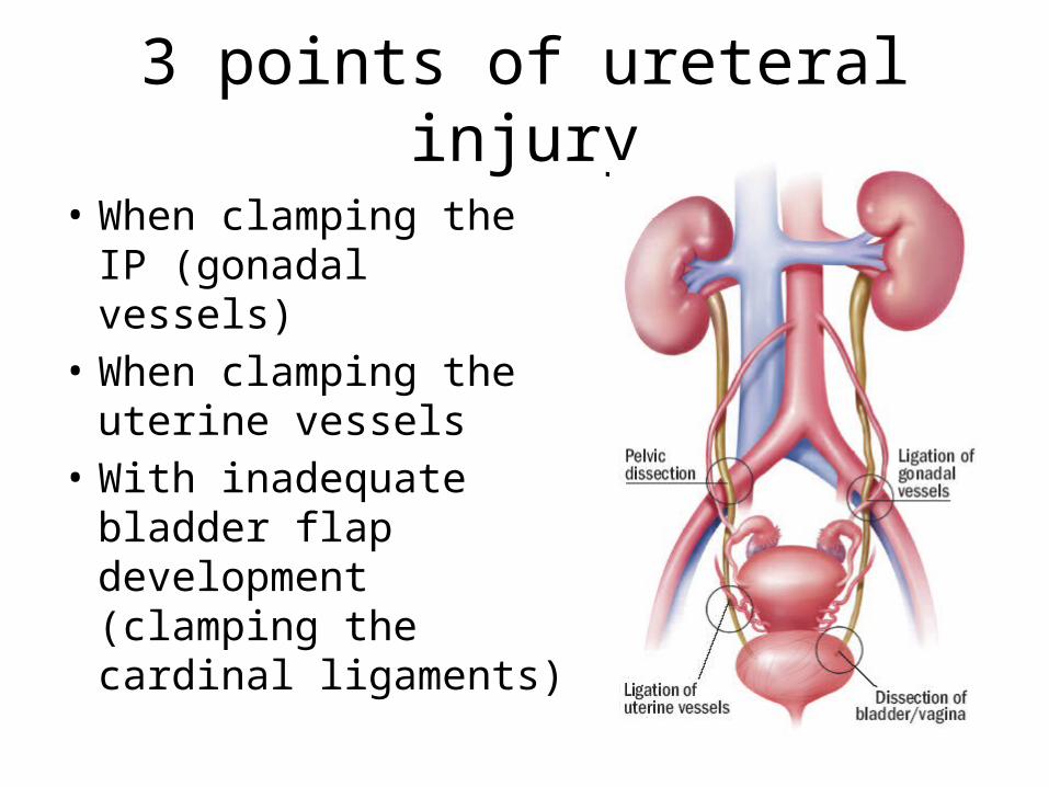
3 points of ureteral injury
• When clamping the IP (gonadal vessels)
• When clamping the uterine vessels
• With inadequate bladder flap development (clamping the cardinal ligaments)
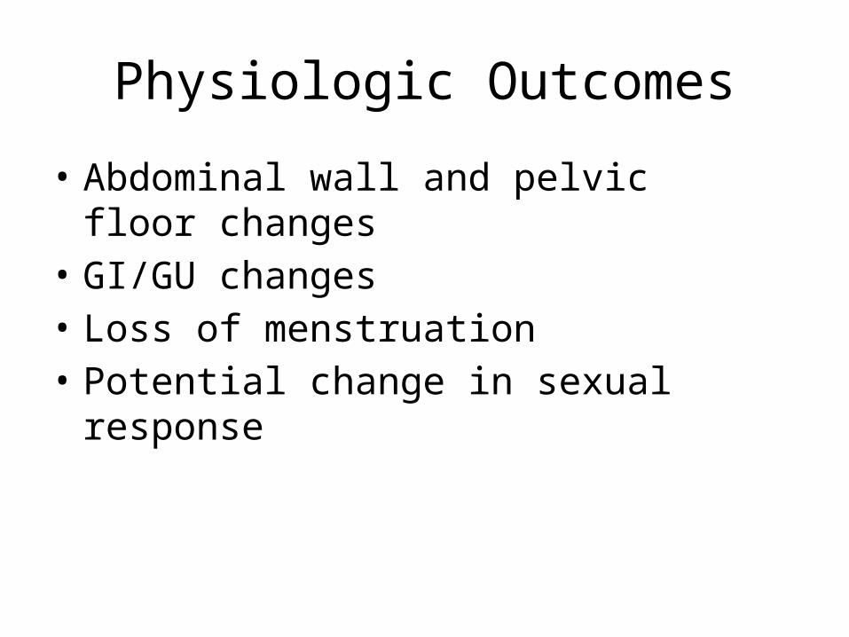
Physiologic Outcomes
• Abdominal wall and pelvic floor changes• GI/GU changes• Loss of menstruation• Potential change in sexual response
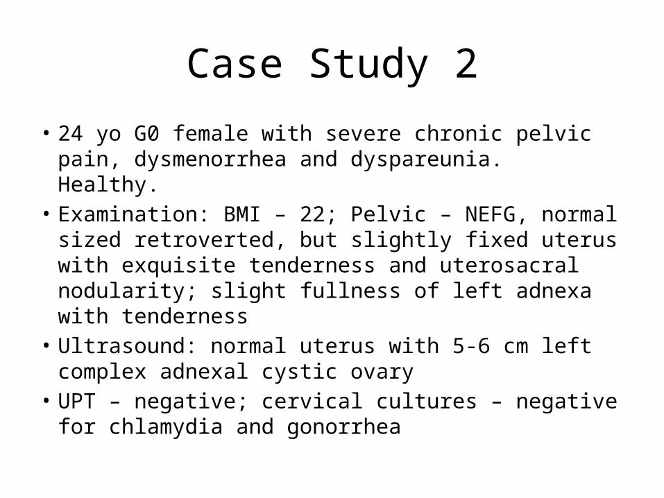
Case Study 2
• 24 yo G0 female with severe chronic pelvic pain, dysmenorrhea and dyspareunia. Healthy.
• Examination: BMI – 22; Pelvic – NEFG, normal sized retroverted, but slightly fixed uterus with exquisite tenderness and uterosacral nodularity; slight fullness of left adnexa with tenderness
• Ultrasound: normal uterus with 5-6 cm left complex adnexal cystic ovary
• UPT – negative; cervical cultures – negative for chlamydia and gonorrhea
Surgical Approach
• Preop Dx: Complex adnexal mass, r/o endometriosis
• Planned Procedure: Diagnostic laparoscopy, left ovarian cystectomy/salpingo-oophorectomy
• Relevant Surgical Anatomy– Abdominal and pelvic examination– Layers of the abdominal wall– Abdominal structures– Pelvic structures
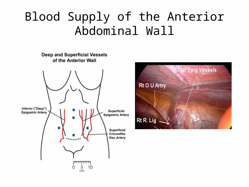
Blood Supply of the Anterior Abdominal Wall
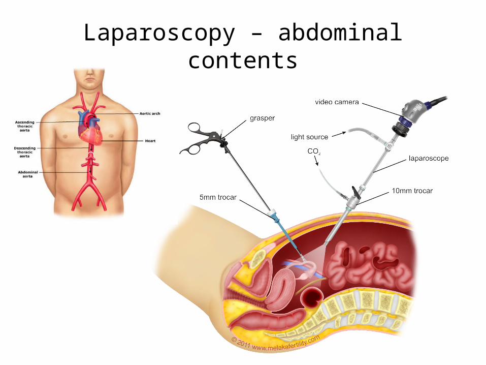
Laparoscopy – abdominal contents
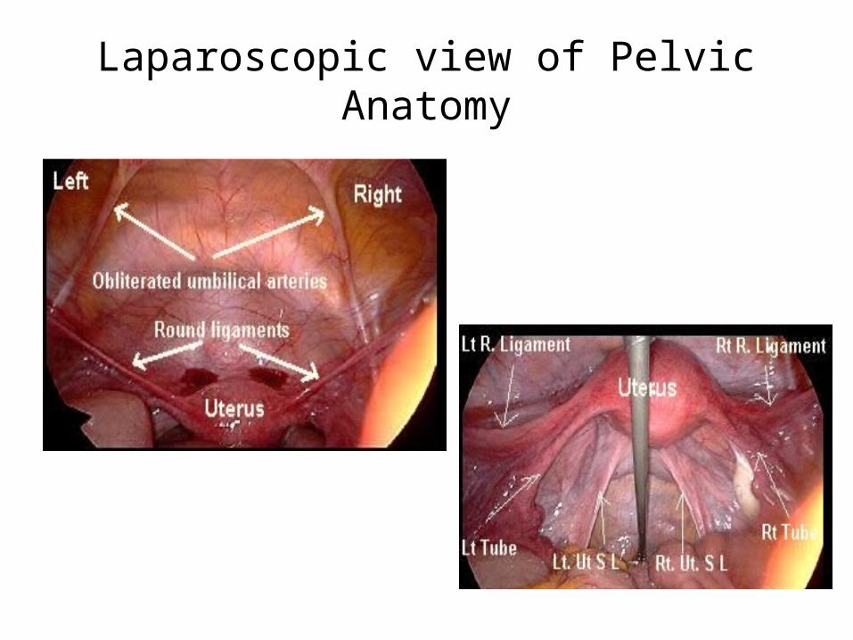
Laparoscopic view of Pelvic Anatomy
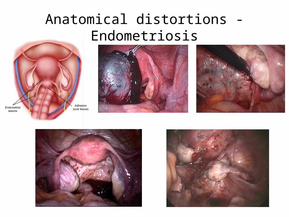
Anatomical distortions - Endometriosis
Special Points of Consideration
• Distortion of uterosacral ligaments• Obliteration of posterior cul-de-sac and ovarian
fossa• Course of the ureter• Blood supply to ovary/tube---------• Ureteral injury• Vascular injury/large EBL• Bowel injury
Physiologic Outcomes
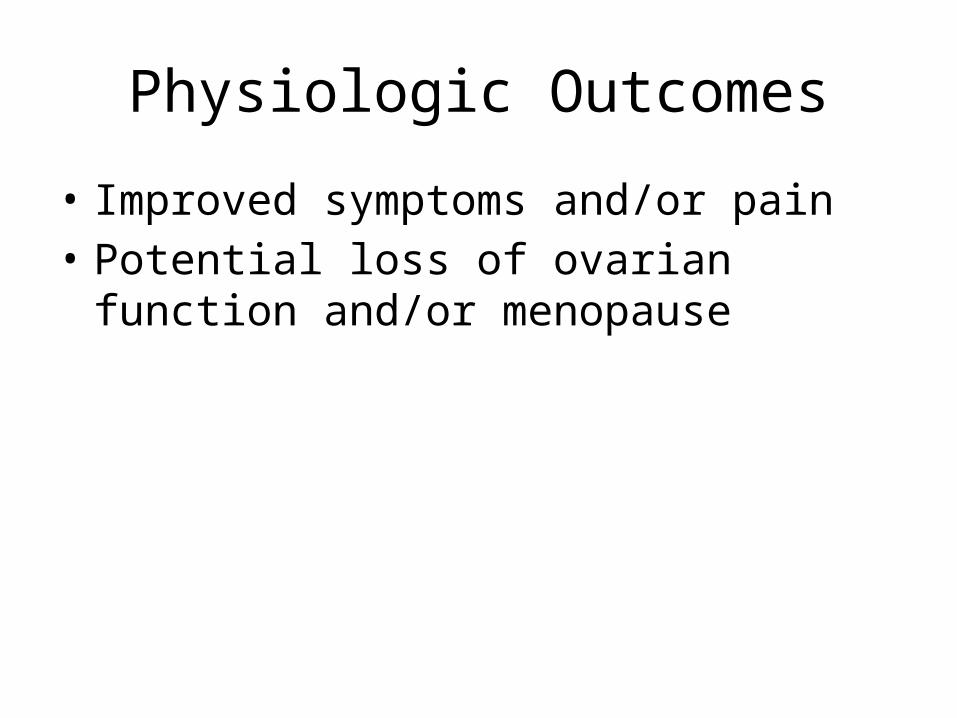
• Improved symptoms and/or pain• Potential loss of ovarian function and/or
menopause
Case Study 3
• 62 yo G4P4 female with pelvic pressure and bulging/protruding mass per vagina
• Examination: Pelvic – near complete uterine prolapse (procidentia)
• Pap smear – negative/normal; U/s – atrophic ovaries; uterus with 3 mm endometrial stripe
Surgical Approach
• Preop Dx: Uterine prolapse• Planned Procedure: Total vaginal hysterectomy
+/- Bilateral salpingo-oophorectomy• Relevant Surgical Anatomy
– Abdominal and pelvic examination– Pelvic structures– Pelvic floor anatomy
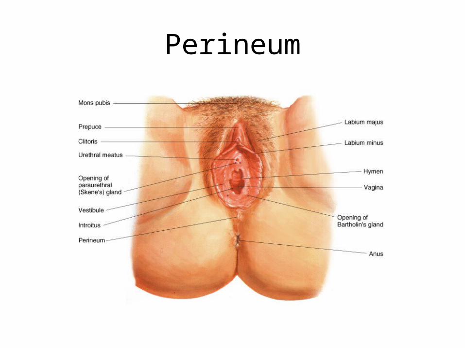
Perineum
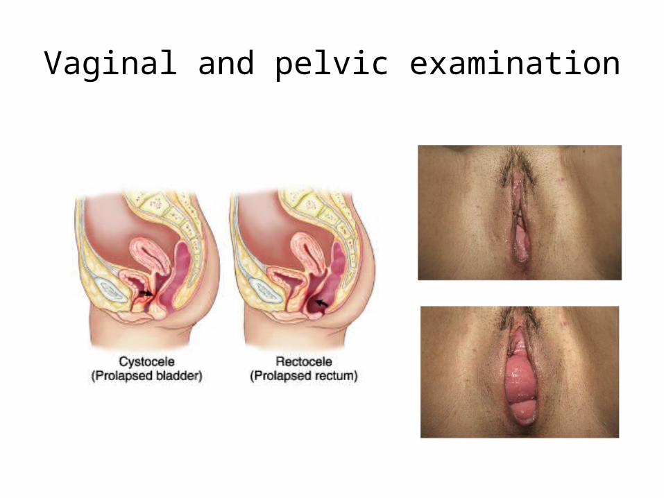
Vaginal and pelvic examination
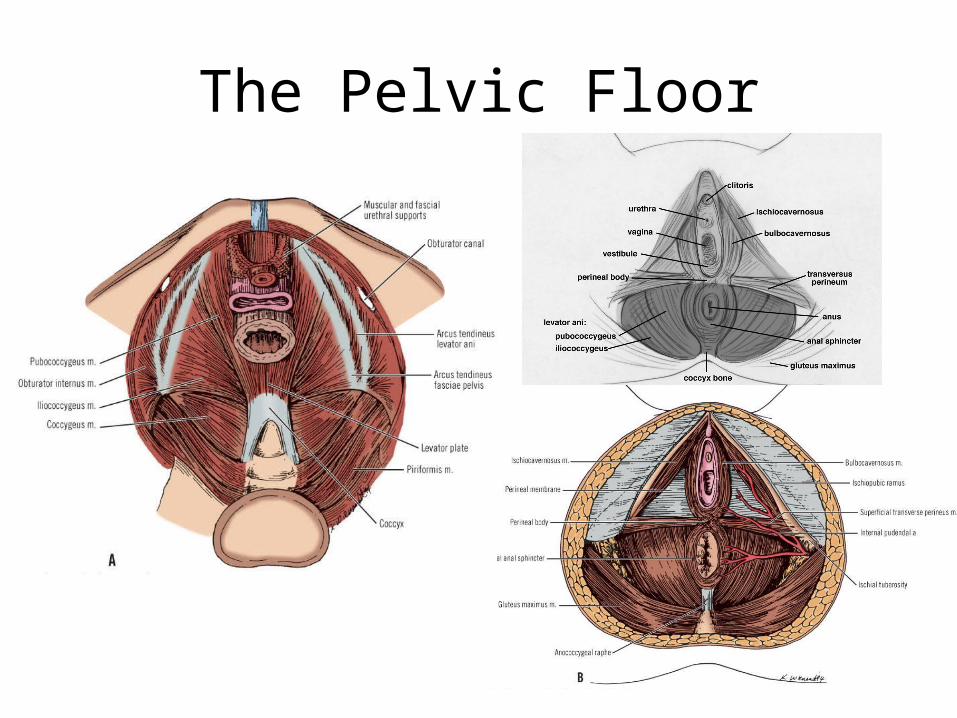
The Pelvic Floor
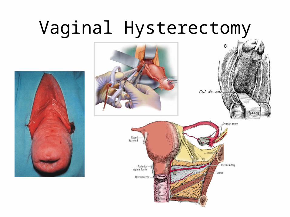
Vaginal Hysterectomy
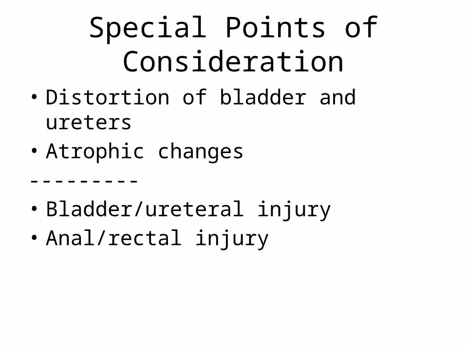
Special Points of Consideration
• Distortion of bladder and ureters• Atrophic changes---------• Bladder/ureteral injury• Anal/rectal injury
Physiologic Outcomes
• Improved pelvic pressure/bulging• Improved GI/GU function
Conclusions
• Pelvic anatomy is generally preserved and knowledge of key abdominal and pelvic anatomical landmarks is essential for any pelvic surgeon
• Complications can best be avoided by anticipating the pathologic changes that result in anatomic alterations as a result of pelvic disease
• Knowledge of pelvic and abdominal anatomy is crucial for successful surgical management that will lead to improved patient outcomes

Congratulations &Welcome to OBGYN!
Related Documents