Welcome message from author
This document is posted to help you gain knowledge. Please leave a comment to let me know what you think about it! Share it to your friends and learn new things together.
Transcript

MDEDGE.COM/OBGYN | VOL 32, NO 2 | FEBRUARY 2020
Follow us on Facebook and Twitter
A member of the Network
Pelvic organ prolapse
A roundtable including expert tips
for managementFeaturing
John B. Gebhart, MD, MS; Mickey M. Karram, MD;
Beri M. Ridgeway, MD; and Mark D. Walters, MD
Progestin-only HT for hot �ashes
Robert L. Barbieri, MD
The role of MRI in women with dense breasts
Mark D. Pearlman, MD
ERAS for cesarean delivery: Intraoperative care
Follow us on Facebook
Update on fertilityp. 11
ObGyn liability risk in 2020
Aromatase inhibitors and breast cancer risk after 5 years of use
WHAT’S YOUR DIAGNOSIS?
Numerous papules in the groin area
C1 0220.indd 1 1/31/20 3:03 PM

11281954 PCP_JournalAd-A-Size M9FR
Client:Product:Client Code:Release Date:Releasing as:WF Issue #
Live/Safety:Trim/Final:Bleed:Gutter:
Flat Size:Finishing:
Colors:
Producer:AD:AE:Production:QC:Digital Artist:FR Spellcheck:
EXACT SCIENCESCOLOGUARD HCPUS.CG.2610-12020-01-03PDFx1A5613490
6.75" x 9.5"8.25" x 10.875"8.5" x 11.125"NA
8.25" x 10.875"None
4 color two page ad (No brief summary)
Ibrahim RumTamer OnayChristine BeckStacy HobdyNoneCannock, George (NYC-FCB)Milene Fernandez
Job info Team
Special Instructions
Gotham (Book, Bold, Medium, Book Italic)
Fonts
Images
Inks
PREPARED BY
Additional Information
February 2020 Issue
None
Cyan, Magenta, Yellow, Black
NY_EXAC_A055724_4C.tif (CMYK; 300 ppi; 100%)
Scale: 1" = 1"
BleedTrimViewingLive
11.125" h x 8.5" w 11.125" h x 8.5" w10.875" h x 8.25" w 10.875" h x 8.25" w10.5" h x 7.75" w 10.5" h x 7.75" w9.5" h x 6.75" w 9.5" h x 6.75" w
Path: PrePress:Exact_Sciences:Cologu...281954_PCP_JrnlAd_A-Size_M9FR.indd
Cologuard is intended to screen adults aged 45 years and older at average risk for CRC.
~44 million patients remain unscreened for colorectal cancer (CRC).1-7* Some of them may even be in your practice.
TAKE ON SUBOPTIMAL SCREENING RATES
ONE YES AT A TIME
In a prospective, head-to-head, point-in-time, 90-site, pivotal study of 10,000 patients aged 50-84 years at average risk for CRC, published in The New England Journal of Medicine, Cologuard demonstrated8†:
In detecting CRC stages I to IV8‡
92%SENSITIVITYOVERALL
In detecting CRC stages I to II8,9‡
94%SENSITIVITYIN EARLY CRC
In patients with nonadvanced adenomas, nonneoplastic findings, or negative colonoscopy results8§
87%SPECIFICITY OVERALL
If a patient received a negative test result, there was a 99.94% chance that there was no CRC8II
99.94%NEGATIVE PREDICTIVE VALUE
Indication and Important Risk InformationCologuard is intended for the qualitative detection of colorectal neoplasia associated DNA markers and for the presence of occult hemoglobin in human stool. A positive result may indicate the presence of colorectal cancer (CRC) or advanced adenoma (AA) and should be followed by diagnostic colonoscopy. Cologuard is indicated to screen adults of either sex, 45 years or older, who are at typical average risk for CRC. Cologuard is not a replacement for diagnostic colonoscopy or surveillance colonoscopy in high-risk individuals.
Cologuard is not for high-risk individuals, including patients with a personal history of colorectal cancer and adenomas; have had a positive result from another colorectal cancer screening method within the last 6 months; have been diagnosed with a condition associated with high risk for colorectal cancer such as IBD, chronic ulcerative colitis, Crohn’s disease; or have a family history of colorectal cancer, or certain hereditary syndromes.
Positive Cologuard results should be referred to diagnostic colonoscopy. A negative Cologuard test result does not guarantee absence of cancer or advanced adenoma. Following a negative result, patients should continue participating in a screening program at an interval and with a method appropriate for the individual patient.
False positives and false negatives do occur. In a clinical study, 13% of patients without colorectal cancer or advanced adenomas received a positive result (false positive) and 8% of patients with cancer received a negative result (false negative). The clinical validation study was conducted in patients 50 years of age and older. Cologuard performance in patients ages 45 to 49 years was estimated by sub-group analysis of near-age groups.
Cologuard performance when used for repeat testing has not been evaluated or established. Rx only.
S:6.75"S:9.5"
ST:7.75"ST:10.5"
LT:8.25"LT:10.875"
B:8.5"B:11.125"
US_CG_2610-1_11281954_PCP_JrnlAd_A-Size_M9FR.indd 1 1/7/20 5:31 PM
Ad Place-new.indd 6 1/14/2020 3:39:27 PM

11281954 PCP_JournalAd-A-Size M9FR
Client:Product:Client Code:Release Date:Releasing as:WF Issue #
Live/Safety:Trim/Final:Bleed:Gutter:
Flat Size:Finishing:
Colors:
Producer:AD:AE:Production:QC:Digital Artist:FR Spellcheck:
EXACT SCIENCESCOLOGUARD HCPUS.CG.2610-12020-01-03PDFx1A5613490
6.75" x 9.5"8.25" x 10.875"8.5" x 11.125"NA
8.25" x 10.875"None
4 color two page ad (No brief summary)
Ibrahim RumTamer OnayChristine BeckStacy HobdyNoneCannock, George (NYC-FCB)Milene Fernandez
Job info Team
Special Instructions
Gotham (Light, Book, Bold, Book Italic), Minion Pro (Regular)
Fonts
Images
Inks
PREPARED BY
Additional Information
February 2020 Issue
None
Cyan, Magenta, Yellow, Black
NY_EXAC_A055724_4C.tif (CMYK; 300 ppi; 100%), ES_logo_color_pos_cmyk.eps (14.11%), cologuardlogo_horizontal_4C_LG®.ai (33.94%, 40.73%)
Scale: 1" = 1"
BleedTrimViewingLive
11.125" h x 8.5" w 11.125" h x 8.5" w10.875" h x 8.25" w 10.875" h x 8.25" w10.5" h x 7.75" w 10.5" h x 7.75" w9.5" h x 6.75" w 9.5" h x 6.75" w
Path: PrePress:Exact_Sciences:Cologu...281954_PCP_JrnlAd_A-Size_M9FR.indd
EXACT SCIENCES CORPORATION | 441 Charmany Drive, Madison, WI 53719 ExactSciences.com | ExactLabs.com | 1-844-870-8870Cologuard is a registered trademark of Exact Sciences Corporation.©2019 Exact Sciences Corporation. All rights reserved. US.CG.2610-1-December 2019
*Estimate based on the US population aged 45 to 74 years as of 2018, adjusted for the reported rates of high-risk conditions and prior screening history for CRC.
†In the pivotal study, screening colonoscopy was the reference method.8 ‡Cologuard sensitivity, per stage of cancer: I: 90% (n=29); II: 100% (n=21); III: 90% (n=10); IV: 75% (n=4).8 §Cologuard specificity: 87% overall specificity, excluding CRC and advanced adenomas, and including all nonadvanced adenomas, nonneoplastic
findings, and negative results on colonoscopy. There was 90% specificity in participants with no lesions biopsied on colonoscopy.8 ||Negative predictive value (NPV) is defined as the probability that disease is absent in those with a negative result; it is highly dependent on
the prevalence of the disease. NPV was derived from the patient population evaluated in the lmperiale et al publication.8
References: 1. Annual estimates of the resident population for selected age groups by sex for the United States: April 1, 2010 to July 1, 2018. United States Census Bureau website. https://factfinder.census.gov/faces/tableservices/jsf/pages/productview.xhtml?pid=PEP_2018_ PEPAGESEX&prodType=table. Updated June 2019. Accessed November 12, 2019. 2. SEER cancer statistics review 1975-2016. Howlader N, Noone AM, Krapcho M, et al, eds. National Cancer Institute website. https://seer.cancer.gov/csr/1975_2016/browse_csr.php?section SEL=6&pageSEL=sect_06_table.10. Updated September 5, 2019. Accessed November 12, 2019. 3. Henrikson NB, Webber EM, Goddard KA, et al. Family history and the natural history of colorectal cancer: systematic review. Genet Med. 2015;17(9):702-712. 4. Loftus EV Jr. Update on the incidence and prevalence of inflammatory bowel disease in the United States. Gastroenterol Hepatol (NY). 2016;12(11):704-707. 5. Colorectal Cancer Facts & Figures 2017-2019. American Cancer Society website. https://www.cancer.org/content/dam/cancer-org/ research/cancer-facts-and-statistics/colorectal-cancer-facts-and-figures/colorectal-cancer-facts-and-figures-2017-2019.pdf. Accessed November 12, 2019. 6. Fedewa SA, Siegel RL, Jemal A. Are temporal trends in colonoscopy among young adults concordant with colorectal cancer incidence? J Med Screen. 2019;26(4):179-185. 7. Use of Colorectal Cancer Screening Tests: 2018 Behavioral Risk Factor Surveillance System. Centers for Disease Control and Prevention website. https://www.cdc.gov/cancer/colorectal/statistics/use-screening-tests-BRFSS.htm. Updated October 22, 2019. Accessed November 12, 2019. 8. Imperiale TF, Ransoho¬ DF, Itzkowitz SH, et al. Multitarget stool DNA testing for colorectal-cancer screening. N Engl J Med. 2014;370(14):1287-1297. 9. Ahlquist DA. Multi-target stool DNA test: a new high bar for noninvasive screening. Dig Dis Sci. 2015;60(3):623-633.
Visit cologuardhcp.com
O�er a highly e�ective, noninvasive CRC screening option to your appropriate average-risk patients aged 45 years or older.
S:6.75"S:9.5"
ST:7.75"ST:10.5"
LT:8.25"LT:10.875"
B:8.5"B:11.125"
US_CG_2610-1_11281954_PCP_JrnlAd_A-Size_M9FR.indd 2 1/7/20 5:31 PM
Ad Place-new.indd 6 1/14/2020 3:40:36 PM

2 OBG Management | February 2020 | Vol. 32 No. 2 mdedge.com/obgyn
Enhancing the quality of women’s health care and the professional development of ObGyns and all women’s health care clinicians†
mdedge.com/obgyn
Arnold P. Advincula, MDVice Chair and Levine Family Professor of Women’s Health, Department of Obstetrics & Gynecology, Columbia University Medical Center; Chief of Gynecologic Specialty Surgery, Sloane Hospital for Women, New York-Presbyterian Hospital/Columbia University, New York, New York
Linda D. Bradley, MDProfessor of Surgery and Vice Chairman, Obstetrics, Gynecology, and Women’s Health Institute, and Vice Chair for Diversity and Inclusion for the Women’s Health Institute; and Director, Center for Menstrual Disorders, Fibroids, & Hysteroscopic Services, Cleveland Clinic, Cleveland, Ohio
Amy L. Garcia, MDMedical Director, Garcia Sloan Centers; Center for Women’s Surgery; and Clinical Assistant Professor, Department of Obstetrics and Gynecology, University of New Mexico, Albuquerque, New Mexico
Steven R. Goldstein, MD, NCMP, CCDProfessor, Department of Obstetrics and Gynecology, New York University School of Medicine; Director, Gynecologic Ultrasound, and Co-Director, Bone Densitometry, New York University Medical Center, New York, New York
Cheryl B. Iglesia, MDDirector, Section of Female Pelvic Medicine and Reconstructive Surgery, MedStar Health; Professor, Departments of ObGyn and Urology, Georgetown University School of Medicine, Washington, DC
Andrew M. Kaunitz, MD, NCMP, Section EditorUniversity of Florida Term Professor and Associate Chairman, Department of Obstetrics and Gynecology, University of Florida College of Medicine-Jacksonville; Medical Director and Director of Menopause and Gynecologic Ultrasound Services, UF Women’s Health Specialists at Emerson, Jacksonville, Florida
Barbara Levy, MDClinical Professor, Obstetrics and Gynecology,�e George Washington University School of Medicine and Health Sciences, Washington DC; Principal, �e Levy Group LLC
David G. Mutch, MDIra C. and Judith Gall Professor of Obstetrics and Gynecology, and Vice Chair, Department of Obstetrics and Gynecology, Washington University School of Medicine, St. Louis, Missouri
Errol R. Norwitz, MD, PhD, MBA, Section EditorChief Scienti�c O�cer, Tufts Medical Center; Louis E. Phaneuf Professor and Chairman, Department of Obstetrics & Gynecology, Tufts University School of Medicine, Boston, Massachusetts
JoAnn V. Pinkerton, MD, NCMPProfessor, Department of Obstetrics and Gynecology, and Director, Midlife Health, University of Virginia Health System, Charlottesville, Virginia; Executive Director Emeritus, �e North American Menopause Society, Pepper Pike, Ohio
John T. Repke, MDProfessor Emeritus, Obstetrics and Gynecology, Penn State University College of Medicine, Hershey, Pennsylvania
Joseph S. San�lippo, MD, MBAProfessor, Department of Obstetrics, Gynecology, and Reproductive Sciences, University of Pittsburgh; Academic Division Director, Reproductive Endocrinology and Infertility, Magee-Womens Hospital, Pittsburgh, Pennsylvania
James A. Simon, MD, CCD, IF, NCMPClinical Professor, Department of Obstetrics and Gynecology, George Washington University; Medical Director, IntimMedicine™ Specialists, Washington, DC
EDITOR IN CHIEF
Robert L. Barbieri, MDChief, Department of Obstetrics and Gynecology
Brigham and Women’s Hospital Kate Macy Ladd Professor of Obstetrics, Gynecology, and Reproductive Biology
Harvard Medical SchoolBoston, Massachusetts
BOARD OF EDITORS
*Source: Kantar Media, Medical Surgical Study December 2019, Obstetrics/Gynecology Combined Office & Hospital Readers.†OBG MANAGEMENT recognizes the importance of addressing the reproductive health of gender-diverse individuals.
EditBoard 0220.indd 2 1/31/20 3:58 PM

NDC DESCRIPTION SIZEAMERI-SOURCE BERGEN
CARDINAL MCKESSON MORRIS DICKSON
17478-711-31 Lidocaine Hydrochloride Jelly USP, 2% 5 mL x 10 910822 5568183 3987286 788026
17478-711-30 Lidocaine Hydrochloride Jelly USP, 2% 30 mL 10054621 3498367 2465839 509901
SIZEMATTERS
That’s why we made
Lidocaine Hydrochloride
Jelly USP, 2% available in
two convenient sizes,
5 mL and 30 mL.
Exclusively from Akorn.
1925 West Field Court, Suite 300 • Lake Forest, IL 60045 • 800-932-5676 • akorn.com ©2019 Akorn, Inc. All rights reserved.
LidocaineLIDOCAINE HYDROCHLORIDE JELLY USP, 2%
5 mL
30 mLAND
JA006 Rev 12/19
JA006_OBG_Management.indd 1 1/21/20 12:41 PMAd Place-new.indd 6 1/21/2020 2:15:47 PM

4 OBG Management | February 2020 | Vol. 32 No. 2 mdedge.com/obgyn
Follow us on Facebook and on
Twitter @MDedgeObGyn
OBG MANAGEMENT (ISSN 1044-307x) is published monthly by Frontline Medical Communications Inc, 7 Century
Drive, Suite 302, Parsippany, New Jersey 07054. The contents of this publication may not be reproduced in whole
or part without the written consent of the owner. 2020 subscription rates (includes full-text access to mdedge.com
/obgyn): United States: $162.00; elsewhere: $211.00. Single copy orders must be prepaid: United States: $27.00;
Canada/Mexico: $33.00; other: $38.00. Periodicals postage paid at Parsippany, NJ, and additional mailing of�c-
es. Orders and Claims: OBG Management, Subscription Service, P.O. Box 3000, Denville, NJ 07834-3000, phone
(833) 836-2705, or e-mail [email protected]. POSTMASTER: Please send address changes to OBG
MANAGEMENT Subscription Service, 10255 W. Higgins Road, Suite 280, Rosemont, IL 60018-9914.
COVER IMAGE : ILLUSTRATION: KIM MARTENS
FAST TRACK is a system to enable you as a reader to move quickly through each issue of OBG MANAGEMENT, identifying articles or sections of articles to read in depth.
FAST TRACK
HT for hot �ashes
6
40
What’s Your Diagnosis?
19
Exploring options for POP treatment: Patient selection, surgical approaches, and ways to manage risks
Four expert gynecologic surgeons offer tips on diagnosis, surgical and nonsurgical treatment approaches, and patient factors to consider
EXPERT PANEL FEATURING JOHN B. GEBHART, MD, MS; MICKEY M. KARRAM, MD; BERI M. RIDGEWAY, MD; AND MARK D. WALTERS, MD
9 Examining the Evidence
Breast cancer chemoprophylaxis in high-risk women: How persistent is the impact of an aromatase inhibitor after 5 years of use?ANDREW M. KAUNITZ, MD, NCMP
11 Update
FertilityACOG guidelines on preconception genetic carrier screening, AI and embryo selection, and the hidden dangers of environmental toxicants and ways to mitigate them
G. DAVID ADAMSON, MD, AND M. MAX EZZATI, MD
19 What’s Your Diagnosis?
Numerous papules in the groin areaPENELOPE J. KALLIS, MD; STEPHANIE J. CARSTENS, MD; AND ANDREW M. KAUNITZ, MD, NCMP
26 Commentary
Should supplemental MRI be used in otherwise average risk women with extremely dense breasts?MARK D. PEARLMAN, MD
30 What’s the Verdict?
ObGyn malpractice liability risk: 2020 developments and probabilities STEVEN R. SMITH, MS, JD, AND JOSEPH S. SANFILIPPO, MD, MBA
6 EDITORIAL
Progestin-only systemic hormone therapy for menopausal hot �ashesROBERT L. BARBIERI, MD
49 OBG MARKETPLACEThe of�cial job board of OBG MANAGEMENT
51 INDEX OF ADVERTISERS
52 ERAS for cesarean delivery: Intraoperative care
FEBRUARY 2020 | VOL 32, NO 2
TOC 0220.indd 4 1/31/20 3:07 PM

mdedge.com/obgyn Vol. 32 No. 2 | February 2020 | OBG Management 5
at mdedge.com/obgyn
EDITORIAL STAFF
EDITOR Lila O’Connor
SENIOR EDITOR Kathy Christie
WEB EDITOR Christina Manago
EDITOR EMERITUS
Janelle Yates
CONTRIBUTING EDITORS
Katherine T. Chen, MD, MPH New York, New York
Lucia DiVenere, MA Washington, DC
Neal M. Lonky, MD, MPH Anaheim, California
Mark D. Pearlman, MD Ann Arbor, Michigan
Steven R. Smith, MS, JD San Diego, California
ART, WEB, PRODUCTION
CREATIVE DIRECTOR Louise Koenig
ART DIRECTOR John J. DeNapoli
DIRECTOR, JOURNAL MANUFACTURING SERVICES Michael Wendt
PRODUCTION MANAGER Donna Pituras
PUBLISHING STAFF
GROUP PUBLISHER Dianne Reynolds
ACCOUNT MANAGER, WEST Judy Harway
DIGITAL ACCOUNT MANAGER Alison Paton
ACCOUNT MANAGER, SPECIAL EVENTS Guy Pawlak
SUBSCRIPTION INQUIRIES [email protected]
CORPORATE
SVP, FINANCE Steven Resnick
VP, SALES Mike Guire
VP, DIGITAL CONTENT & STRATEGY Amy Pfeiffer
PRESIDENT, CUSTOM SOLUTIONS JoAnn Wahl
VP, HUMAN RESOURCES & FACILITY OPERATIONS Carolyn Caccavelli
DATA MANAGEMENT DIRECTOR Mike Fritz
CIRCULATION DIRECTOR Jared Sonners
DIRECTOR, CUSTOM SOLUTIONS Patrick Finnegan
IN AFFILIATION WITH GLOBAL ACADEMY FOR MEDICAL EDUCATION, LLC
PRESIDENT, EVENTS David J. Small, MBA
Reader services. Address correspondence to OBG Management®, 7 Century Drive, Suite 302, Parsippany, NJ 07054.
Copyright. Copyright Frontline Medical Communications Inc., 2020. All rights reserved. No part of this publication may be reproduced, stored in a retrieval system, or transmitted in any form or by any means, mechanical, computer, photocopying, electronic recording, or otherwise, without the prior written permission of Frontline Medical Communications Inc. �e copyright law of the Unted States (Title 17, U.S.C., as amended) governs the making of photocopies or other reproductions of copyrighted material.
Photocopy rights. Authorization to photocopy items from OBG Management for personal or internal use, or for the personal or internal use of speci�c clients, is granted by Frontline Medical Communications Inc., on the condition that the base fee of $3.00 per copy of each ar-ticle or department is paid to the Copyright Clearance Center, 222 Rosewood Drive, Danvers, MA 01923. �is consent does not extend to other kinds of copying, such as general distribu-tion, resale, advertising, or promotional purposes, or for creating new collective works.
Reprint requests. For article reprint requests in the United States and Canada, please contact Wright’s Media, toll free: 877-652-5295, ext. 102; [email protected]. For those outside the US/Canada, contact Content Ed Net, at 267-895-1758; [email protected].
Marketplace advertising. For direct orders and inquiries, contact Tim LaPella at: telephone 484-291-5001; fax 973-206-9378; [email protected].
Subscriber services. To subscribe or to communicate questions or changes related to your paid subscription, please contact OBG Management Subscription Service, P.O. Box 3000, Denville, NJ 07834-3000, phone 833-836-2705, or e-mail [email protected].
Disclaimer. Statements and opinions expressed herein are those of the author(s) and are not necessarily those of the editor or publisher. Neither the editor nor publisher guarantees, warrants, or endorses any product, service, or claim advertised in this journal.
7 Century Drive, Suite 302Parsippany, NJ 07054-4609www.mdedge.com
WEB EXCLUSIVE
Prenatal exposure to pollutants consumed through diet found to be associated with decreased fetal growth
Visit us online for daily news
VIDEO LIBRARY
Laparoscopic techniques for Essure device removalLINDA C. YANG, MD; LINDSAY MCALARNEN, MD, MSC; AND MARY MCKENNA, MD
Brought to you by the Society of Gynecologic Surgeons
A clitoral cyst of “epidermal” proportionsANGELA DICARLO-MEACHAM, LCDR, MC, USN; KATHERINE L. DENGLER, MAJ, MC, USA; ANDREA N. SNITCHLER, CDR, MC, USA; AND DANIEL D. GRUBER, COL, MC, USAF
Brought to you by the Society of Gynecologic Surgeons
Watch these, and more expert surgical technique and commentary videos in the EXPLORE: Multimedia section online
GYNECOLOGIC SURGEONS UNSCRUBBEDA serial podcast in collaboration with the Society of Gynecologic Surgeons
Host Cara King interviews Nancy Petersen on a patient movement around endometriosis
Listen to this podcast in the EXPLORE: Multimedia section online
TOC 0220.indd 5 1/31/20 3:08 PM
6 OBG Management | February 2020 | Vol. 32 No. 2 mdedge.com/obgyn
Robert L. Barbieri, MDEditor in Chief, OBG MANAGEMENT Chair, Obstetrics and Gynecology Brigham and Women’s Hospital Boston, MassachusettsKate Macy Ladd Professor of Obstetrics, Gynecology and Reproductive Biology Harvard Medical School
Progestin-only systemic hormone therapy for menopausal hot �ashesClinicians treating postmenopausal hot �ashes often recommend “systemic estrogen treatment.” However, progestin-only therapy also can effectively treat hot �ashes and is an option for women with a contraindication to estrogen therapy.
T he �eld of menopause medi-cine is dominated by studies documenting the e�ective-
ness of systemic estrogen or estro-gen-progestin hormone therapy for the treatment of hot �ashes caused by hypoestrogenism. �e e�ective-ness of progestin-only systemic hormone therapy for the treatment of hot �ashes is much less studied and seldom is utilized in clinical practice. A small number of stud-ies have reported that progestins, including micronized progesterone, medroxyprogesterone acetate, and norethindrone acetate, are e�ective treatment for hot �ashes. Proges-tin-only systemic hormone therapy might be especially helpful for post-menopausal women with moderate to severe hot �ashes who have a con-traindication to estrogen treatment.
Micronized progesteroneMicronized progesterone (Prome-trium) 300 mg daily taken at bed-time has been reported to e�ectively treat hot �ashes in postmenopausal
women. In one study, 133 postmeno-pausal women with an average age of 55 years and approximately 3 years from their last menstrual period were randomly assigned to 12 weeks of treatment with placebo or micronized progesterone 300 mg daily taken at bedtime.1 Mean serum progesterone levels were 0.28 ng/mL (0.89 nM) and 27 ng/mL (86 nM) in the women taking placebo and micronized progesterone, respectively. Compared with placebo, micronized progesterone reduced day-time and nighttime hot �ash frequency and severity. In addition, compared with placebo, micronized progester-one improved the quality of sleep.1
Most reviews conclude that micronized progesterone has mini-mal cardiovascular risk.2 Micronized progesterone therapy might be espe-cially helpful for postmenopausal women with moderate to severe hot �ashes who have a contraindication to estrogen treatment such as those at increased risk for cardiovascular dis-ease or women with a thrombophilia. Many experts believe that systemic estrogen therapy is contraindicated
in postmenopausal women with an American Heart Association risk score greater than 10% over 10 years.3
Additional contraindications to sys-temic estrogen include women with cardiac disease who have a throm-bophilia, such as the Factor V Leiden mutation.4
For women who are at high risk for estrogen-induced cardiovascu-lar events, micronized progesterone may be a better option than systemic estrogen for treating hot �ashes. Alternatively, in these women at risk of cardiovascular disease a selective serotonin reuptake inhibitor, such as escitalopram, 10 mg to 20 mg daily, may be a good option for treating postmenopausal hot �ashes.5
Medroxyprogesterone acetateMedroxyprogesterone acetate, at a dosage of 20 mg daily, is an e�ective treatment for hot �ashes. In a ran-domized clinical trial 27 postmeno-pausal women with hot �ashes were randomly assigned to treatment with
EDITORIAL
Editorial 0220.indd 6 1/31/20 3:09 PM
mdedge.com/obgyn Vol. 32 No. 2 | February 2020 | OBG Management 7
CONTINUED ON PAGE 8
DIG
ITA
L I
LL
US
TR
AT
ION
: JO
HN
J.
DE
NA
PO
LI
IM
AG
ES
: ©
AN
A B
LA
ZIC
PA
VL
OV
IC/V
AD
YM
NE
CH
YP
OR
EN
KO
/SH
UT
TE
RS
TO
CK
placebo or medroxyprogesterone acetate 20 mg daily for 4 weeks. Vaso-motor �ushes were decreased by 26% and 74% in the placebo and medroxy-progesterone groups, respectively.6
Depot medroxyprogesterone acetate injections at dosages from 150 mg to 400 mg also have been reported to e�ectively treat hot �ashes.7,8 In a trial comparing the e�ectiveness of estrogen monother-apy (conjugated equine estrogen 0.6 mg daily) with progestin monotherapy (medroxyprogesterone acetate 10 mg daily), both treatments were equally e�ective in reducing hot �ashes.9
Micronized progesterone vs medroxyprogesterone acetateExperts in menopause medicine have suggested that in postmeno-pausal women micronized pro-gesterone has a better pattern of bene�ts and fewer risks than med-roxyprogesterone acetate.10,11 For example, in the E3N observational study of hormones and breast can-cer risk, among 80,377 French post-menopausal women followed for a mean of 8 years, the combina-tion of transdermal estradiol plus oral micronized progesterone was associated with no signi�cantly increased risk of breast cancer (rela-tive risk [RR], 1.08, 95% con�dence interval [CI], 0.89–1.31) compared with never users of postmenopausal hormone therapy.12 By contrast, the combination of oral estrogen plus medroxyprogesterone acetate was associated with an increased risk of breast cancer (RR, 1.48; 95% CI, 1.02–
2.16) compared with never users of postmenopausal hormone therapy. �e E3N study indicates that micron-ized progesterone may have a more favorable breast health pro�le than medroxyprogesterone acetate.12
Norethindrone acetateNorethindrone acetate monotherapy is not commonly prescribed for the treatment of menopausal hot �ashes. However, a large clinical trial has dem-onstrated that norethindrone acetate e�ectively suppresses hot �ashes in women with endometriosis treated with depot leuprolide acetate (LA). In one trial 201 women with endome-triosis were randomly assigned to 12 months of treatment with13: • LA plus placebo pills • LA plus norethindrone acetate
(NEA) 5 mg daily • LA plus NEA 5 mg daily plus conju-
gated equine estrogen (CEE) 0.625 mg daily, or
• LA plus NEA 5 mg daily plus CEE 1.25 mg daily.
�e median number of hot �ashes in 24 hours was 6 in the LA plus placebo
group and 0 in both the LA plus NEA 5 mg daily group and the LA plus NEA 5 mg plus CEE 1.25 mg daily group. �is study demonstrates that NEA 5 mg daily is an e�ective treatment for hot �ashes.
In the same study, LA plus placebo was associated with a sig-ni�cant decrease in lumbar spine bone mineral density. No signi�cant decrease in bone mineral density was observed in the women who received LA plus NEA 5 mg daily. �is �nding indicates that NEA 5 mg reduces bone absorption caused by hypoestrogen-ism. In humans, norethindrone is a substrate for the aromatase enzyme system.14 Small quantities of ethinyl estradiol may be formed by aroma-tization of norethindrone in vivo,15,16
contributing to the e�ectiveness of NEA in suppressing hot �ashes and preserving bone density.
Progestin: The estrogen alternative to hot �ashes For postmenopausal women with moderate to severe hot �ashes, estro-gen treatment reliably suppresses hot �ashes and often improves sleep qual-ity and mood. For postmenopausal women with a contraindication to estrogen treatment, progestin-only treatment with micronized progester-one or norethindrone acetate may be an e�ective option.
Dr. Barbieri reports no �nancial rela-tionships relevant to this article.
References1. Hitchcock CL, Prior JC. Oral micronized proges-
terone for vasomotor symptoms—a placebo-controlled randomized trial in healthy postmeno-pausal women. Menopause. 2012;19:886-893.
2. Spark MJ, Willis J. Systematic review of progester-
one use by midlife menopausal women. Maturitas 2012; 72: 192-202.
3. Manson JE, Ames JM, Shapiro M, et al. Algorithm and mobile app for menopausal symptom man-agement and hormonal/nonhormonal therapy
decision making: a clinical decision-suport tool from �e North American Menopause Society. Menopause. 2015;22:247-253.
4. Herrington DM, Vittingho� E, Howard TD, et al. Factor V Leiden, hormone replacement therapy,
Editorial 0220.indd 7 1/31/20 3:09 PM

EDITORIAL
8 OBG Management | February 2020 | Vol. 32 No. 2 mdedge.com/obgyn
and risk of venous thromboembolic events in women with coronary disease. Arterioscler �romb Vasc Biol. 2002;22:1012-1017.
5. Ensrud KE, Jo�e H, Guthrie KA, et al. E�ect ofescitalopram on insomnia symptoms and sub-jective sleep quality in healthy perimenopausaland postmenopausal women with hot �ashes:a randomized controlled trial. Menopause. 2012;19:848-855.
6. Schi� I, Tulchinsky D, Cramer D, et al. Oral medroxy-progesterone in the treatment of postmenopausalsymptoms. JAMA. 1980;244:1443-1445.
7. Bullock JL, Massey FM, Gambrell RD Jr. Use ofmedroxyprogesterone acetate to prevent meno-pausal symptoms. Obstet Gynecol. 1975;46:165-168.
8. Loprinzi CL, Levitt R, Barton D, et al. Phase IIIcomparison of depot medroxyprogesteroneacetate to venlafaxine for managing hot �ashes:
North Central Cancer Treatment Group Trial N99C7. J Clin Oncol. 2006;24:1409-1414.
9. Prior JC, Nielsen JD, Hitchcock CL, et al. Med-roxyprogesterone and conjugated oestrogen areequivalent for hot �ushes: 1-year randomizeddouble-blind trial following premenopausal ova-riectomy. Clin Sci (Lond). 2007;112:517-525.
10. L’hermite M, Simoncini T, Fuller S, et al. Could trans-dermal estradiol + progesterone be a safer postmen-opausal HRT? A review. Maturitas. 2008;60:185-201.
11. Simon JA. What if the Women’s Health Initiativehad used transdermal estradiol and oral proge-sterone instead? Menopause. 2014;21:769-783.
12. Fournier A, Berrino F, Clavel-Chapelon F. Unequal risks for breast cancer associated with di�erenthormone replacement therapies: results fromthe E3N cohort study. Breast Cancer Res Treat. 2008;107:103-111.
13. Hornstein MD, Surrey ES, Weisberg GW, etal. Leuprolide acetate depot and hormonaladd-back in endometriosis: a 12-month study.Lupron Add-Back Study Group. Obstet Gynecol.1998;91:16-24.
14. Barbieri RL, Canick JA, Ryan KJ. High-a�nity ste-roid binding to rat testis 17 alpha-hydroxylase andhuman placental aromatase. J Steroid Biochem.1981;14:387-393.
15. Chu MC, Zhang X, Gentzschein E, et al. Forma-tion of ethinyl estradiol in women during treat-ment with norethindrone acetate. J Clin Endocri-nol Metab. 2007;92:2205-2207.
16. Chwalisz K, Surrey E, Stanczyk FZ. �e hormonal pro�le of norethindrone acetate: rationale foradd-back therapy with gonadotropin-releasinghormone agonists in women with endometriosis.Reprod Sci. 2012;19:563-571.
CONTINUED FROM PAGE 7
Editorial 0220.indd 8 1/31/20 3:09 PM
Examining the EVIDENCE
mdedge.com/obgyn Vol. 32 No. 2 | February 2020 | OBG Management 9
FAST TRACK
Breast cancer chemoprophylaxis in high-risk women: How persistent is the impact of an aromatase inhibitor after 5 years of use?
In a long-term follow-up of the IBIS-II trial, investigators found that anastrozole use substantially reduced the incidence of all breast cancer, including invasive breast cancer and ductal carcinoma in situ
Among postmenopausal women at high risk for breast cancer N = 3,864), those treated with anastrozole (N = 1,920) compared with placebo (N = 1,944) for 5 years had a 49% reduction in breast cancer (85 vs 165 cases; hazard ratio [HR], 0.51; 95% con�dence interval [CI], 0.39–0.66; P<.0001) after a median follow-up of 131 months. The reduction was larger in the �rst 5 years but remained signi�cant after 5 years. Although the risk reduction from this endogenous estrogen-minimizing medication was persistent, no mortality bene�t was observed.
Cuzick J, Sestak I, Forbes JF, et al; IBIS-II Investigators.
Use of anastrozole for breast cancer prevention (IBIS-II):
long-term results of a randomised controlled trial. Lancet.
2020;395;117-122.
EXPERT COMMENTARYAndrew M. Kaunitz, MD, NCMP, is University of Florida Term Professor and Associate Chairman, Department of Obstetrics and Gynecology, University of Florida College of Medicine–Jacksonville; Medical Director and Director of Menopause and Gynecologic Ultrasound Services, UF Women’s Health Specialists at Emerson, Jacksonville. Dr. Kaunitz serves on the OBG MANAGEMENT Board of Editors.
A manufacturer-sponsored trial initi-ated in 2003, IBIS-II (International Breast Cancer Intervention Study II)
included 3,864 menopausal women (mean age at baseline, 59.4 years) at elevated risk
for breast cancer. �e women were ran-domly assigned to 5-year treatment with either placebo (N = 1,944) or the aromatase inhibitor anastrozole 1 mg daily (N = 1,920).1
Reporting on the long-term follow-up results of the trial, Cuzick and colleagues found that anastrozole use substantially reduced the incidence of all breast cancer, including invasive breast cancer and ductal carcinoma in situ. Key adverse events associ-ated with anastrozole were fractures, arthral-gias, and menopausal symptoms (vasomotor symptoms and vaginal dryness).
To determine whether anastrozole had any persistent impact, the investigators con-tinued to follow participants for all breast cancers and other outcomes.2
Details of the study�is randomized controlled trial that included 3,864 postmenopausal women had
Dr. Kaunitz reports serving on advisory boards for P�zer.
Evidence Kaunitz 0220.indd 9 1/31/20 3:12 PM
Examining the EVIDENCE
10 OBG Management | February 2020 | Vol. 32 No. 2 mdedge.com/obgyn
a median overall follow-up of 131 months; the primary outcome was all breast can-cer. Random assignment to anastrozole use (1,920 women) was associated with a 49% reduction in all breast cancer (85 cases vs 165 cases in the placebo group [N = 1,944]; HR, 0.51; 95% CI, 0.39–0.66; P<.0001).
In the �rst 5 years, risk reduction was 61% with anastrozole (P<.0001 for overall and the �rst 5 years of follow-up). Subse-quently, the magnitude of the risk reduction attenuated to 37% (P = .014). With 12 years of follow-up, the estimated risk of being diagnosed with breast cancer was 8.8% and 5.3% in the placebo and anastrozole groups,
respectively. �e number needed to treat for 5 years to prevent 1 breast cancer was 29.
With anastrozole, prevention of estro-gen–receptor positive tumors was substan-tially more robust at 54% (HR, 0.46; 95% CI, 0.33–0.65; P<.0001) than for estrogen–recep-tor negative tumors at 27% (HR, 0.77; 95% CI, 0.41–1.44; P = .41).
Over the course of the long-term study, the incidence of fractures and cardiovascular events was similar in the placebo and anas-trozole groups. Arthralgias and menopausal symptoms were not assessed after the trial’s initial 5 years. Overall, the number of deaths (all cause as well as breast cancer related) were similar in the placebo and anastrozole groups.
Study strengths and limitations�e authors noted that this updated analy-sis of the IBIS-II trial data o�ers further support for the use of anastrozole in breast cancer prevention for high-risk postmeno-pausal women. �e extended posttreatment follow-up showed a signi�cant continuing reduction in breast cancer, and there was no evidence of new late adverse e�ects. A limita-tion of the analysis, however, is that very few deaths from breast cancer occurred during the study timeframe. �us, additional follow-up would be needed to assess anastrozole’s e�ect on breast cancer mortality.
WHAT THIS EVIDENCE MEANS FOR PRACTICE
The breast cancer chemoprophylactic ef�cacy of anastrozole compares favorably with that of tamoxifen. Furthermore, in women with an intact uterus, the increased risks of gynecologic problems, including endometrial cancer, associated with tamoxifen do not occur with aromatase inhibitors. However, the lack of any obvious mortal-ity bene�t means the ultimate value of estrogen deprivation breast cancer chemoprophylaxis continues to be uncertain, especially given other risks, including bone loss. In view of these new data, it will be important for high-risk women considering aromatase inhibitor prophylaxis to understand that these medications have not been associated with a mortality bene�t.
ANDREW M. KAUNITZ, MD, NCMP
References1. Cuzick J, Sestak I, Forbes JF, et al; IBIS-II Investigators.
Anastrozole for prevention of breast cancer in high-riskpostmenopausal women (IBIS-II): an international,double-blind, randomised placebo-controlled trial. Lancet.2014;383:1041-1048.
2. Cuzick J, Sestak I, Forbes JF, et al; IBIS-II Investigators. Use ofanastrozole for breast cancer prevention (IBIS-II): long-term results of a randomised controlled trial. Lancet. 2020;395;117-122.
Evidence Kaunitz 0220.indd 10 1/31/20 3:12 PM

UPDATEFertility
IN THISARTICLE
mdedge.com/obgyn Vol. 32 No. 2 | February 2020 | OBG Management 11
ACOG guidelines on preconception genetic carrier screening, AI and embryo selection, and the hidden dangers of environmental toxicants and ways to mitigate them
Genetic carrier screening
this page
AI and embryo selection
page 13
The risk of environmental
toxicantspage 14
A lthough we are not able to cover all of the important developments in fertil-ity medicine over the past year, there
were 3 important articles published in the past 12 months that we highlight here. First, we discuss an American College of Obste-tricians and Gynecologists (ACOG) com-mittee opinion on genetic carrier screening that was rea�rmed in 2019. Second, we explore an interesting retrospective analysis of time-lapse videos and clinical outcomes of more than 10,000 embryos from 8 IVF
clinics, across 4 countries. �e authors assessed whether a deep learning model could predict the probability of pregnancy with fetal heart from time-lapse videos in the hopes that their research can improve priori-tization of the most viable embryo for single embryo transfer. Last, we consider a review of the data on obstetric and reproductive health e�ects of preconception and prenatal exposure to several environmental toxicants, including heavy metals, endocrine-disrupt-ing chemicals, pesticides, and air pollution.
G. David Adamson, MDDr. Adamson is Founder and CEO of Advanced Reproductive Care, Inc (ARC Fertility); Clinical Professor, ACF, at Stanford University School of Medicine; and Associate Clinical Professor at the University of California, San Francisco. He is also Director of Equal3 Fertility, APC in Cupertino, California.
M. Max Ezzati, MDDr. Ezzati is a Board-certi�ed reproductive endocrinology and infertility (REI) specialist and the Medical Director of Department of Reproductive Endocrinology and Infertility at Palo Alto Medical Foundation Fertility Physicians of Northern California.
�e authors report no �nancial relationships relevant to this article.
Preconception genetic carrier screening: Standardize your counseling approachAmerican College of Obstetricians and Gynecolo-
gists Committee on Genetics. Committee Opinion No.
690: carrier screening in the age of genomic medicine.
Obstet Gynecol. 2017;129:e35–e40.
With the rapid development of advanced and high throughput platforms for DNA sequenc-
ing in the past several years, the cost of
genetic testing has decreased dramatically. Women’s health care provid-ers in general, and fertility specialists in particular, are uniquely positioned to take advantage of these novel and yet a�ordable technologies by counseling prospective parents during the preconception coun-seling, or early prenatal period, about the availability of genetic carrier screening and
Update 0220.indd 11 1/31/20 5:02 PM
UPDATE fertility
12 OBG Management | February 2020 | Vol. 32 No. 2 mdedge.com/obgyn
The uptake of genetic carrier screening has been shown to be signi�cantly higher when offered in the preconception period versus during pregnancy
FAST TRACK
its potential to provide actionable informa-tion in a timely manner. �e ultimate objec-tive of genetic carrier screening is to enable individuals to make an informed decision regarding their reproductive choices based on their personal values. In a study by Larsen and colleagues, the uptake of genetic carrier screening was signi�cantly higher when o�ered in the preconception period (68.7%), compared with during pregnancy (35.1%), which highlights the signi�cance of early counseling.1
Based on the Centers for Disease Con-trol and Prevention’s Birth/Infant Death Data set, birth defects a�ect 1 in every 33 (about 3%) of all babies born in the United States each year and account for 20% of infant mor-tality.2 About 20% of birth defects are caused by single-gene (monogenic) disorders, and although some of these are due to dominant conditions or de novo mutations, a signi�-cant proportion are due to autosomal reces-sive, or X-chromosome linked conditions that are commonly assessed by genetic car-rier screening.
ACOG published a committee opinion on “Carrier Screening in the Age of Genomic Medicine” in March 2017, which was reaf-�rmed in 2019.3 Residual risk. Several points discussed in this document are of paramount impor-tance, including the need for pretest and posttest counseling and consent, as well as a discussion of “residual risk.” Newer plat-forms employ sequencing techniques that potentially can detect most, if not all, of the disease-causing variants in the tested genes, such as the gene for cystic fibrosis and, therefore, have a higher detection rate compared with the older PCR-based
techniques for a limited number of specific mutations included in the panel. Due to a variety of technical and biological limita-tions, however, such as allelic dropouts and the occurrence of de novo mutations, the detection rate is not 100%; there is always a residual risk that needs to be estimated and provided to individuals based on the existing knowledge on frequency of gene, penetrance of phenotype, and prevalence of condition in the general and specific ethnic populations. Expanded vs panethnic screening. Fur-thermore, although sequencing technology has made “expanded carrier screening” for several hundred conditions, simultaneous to and independent of ethnicity and family history, more easily available and a�ordable, ethnic-speci�c and panethnic screening for a more limited number of conditions are still acceptable approaches. Having said this, when the �rst partner screened is identi�ed to be a carrier, his/her reproductive partners must be o�ered next-generation sequenc-ing to identify less common disease-causing variants.4
A cautionary point to consider when expanded carrier screening panels are requested is the signi�cant variability among commercial laboratories with regard to the conditions included in their panels. In addi-tion, consider the absence of a well-de�ned or predictable phenotype for some of the included conditions.
Perhaps the most important matter when it comes to genetic carrier screening is to have a standard counseling approach that is persistently followed and o�ers the opportunity for individuals to know about their genetic testing options and available reproductive choices, including the use of donor gametes, preimplantation genetic testing for monogenic disease (PGT-M, for-merly known as preimplantation genetic diagnosis, or PGD), prenatal testing, and pregnancy management options. For couples and/or individuals who decide to proceed with an a�ected pregnancy, ear-lier diagnosis can assist with postnatal management.
WHAT THIS EVIDENCE MEANS FOR PRACTICE
The preconception period is the perfect time to have a discussion about genetic carrier screening; it offers the opportunity for timely interventions if desired by the couples or individuals.
Update 0220.indd 12 1/31/20 3:37 PM

mdedge.com/obgyn Vol. 32 No. 2 | February 2020 | OBG Management 13
Early use of time-lapse imaging for embryo selection has not shown clinical bene�t, but early methods were dependent on embryologists’ subjective assessment of features of embryo development
FAST TRACK
Medicolegal responsibility. Genetic carrier screening also is of speci�c relevance to the �eld of fertility medicine and assisted repro-ductive technology (ART) as a potential liabil-ity issue. Couples and individuals who are undergoing fertility treatment with in vitro fertilization (IVF) for a variety of medical or personal reasons are a speci�c group that certainly should be o�ered genetic carrier
screening, as they have the option of “adding on” PGT-M (PGD) to their existing treatment plan at a fraction of the cost and treatment bur-den that would have otherwise been needed if they were not undergoing IVF. After counsel-ing, some individuals and couples may ulti-mately opt out of genetic carrier screening. �e counseling discussion needs to be clearly documented in the medical chart.
Arti�cial intelligence and embryo selection Tran D, Cooke S, Illingworth PJ, et al. Deep learning
as a predictive tool for fetal heart pregnancy following
time-lapse incubation and blastocyst transfer. Hum
Reprod. 2019;34:1011-1018.
W ith continued improvements in embryo culture conditions and cryopreservation technology,
there has been a tremendous amount of interest in developing better methods for embryo selection. �ese e�orts are aimed at encouraging elective single embryo transfer (eSET) for women of all ages, thereby low-ering the risk of multiple pregnancy and its associated adverse neonatal and obstetric outcomes—without compromising the preg-nancy rates per transfer or lengthening the time to pregnancy.
One of the most extensively studied methods for this purpose is preimplanta-tion genetic testing for aneuploidy (PGT-A, formerly known as PGS), but emerging data from large multicenter randomized clinical trials (RCTs) have again cast signi�-cant doubt on PGT-A’s e�cacy and utility.5
Meanwhile, alternative methods for embryo selection are currently under investigation, including noninvasive PGT-A and morpho-kinetic assessment of embryo development via analysis of images obtained by time-lapse imaging.
The potential of time-lapse imagingDespite the initial promising results from time-lapse imaging, subsequent RCTs have not shown a signi�cant clinical bene�t.6
However, these early methods of morphoki-netic assessment are mainly dependent on the embryologists’ subjective assessment of individual static frames and “annotation” of observed spatial and temporal features of embryo development. In addition to being a very time-consuming task, this process is subject to signi�cant interobserver and intraobserver variability.
Considering these limitations, even machine-based algorithms that incorporate these annotations along with such other clinical variables as parental age and prior obstetric history, have a low predictive power for the outcome of embryo transfer, with an area under the curve (AUC) of the ROC curve of 0.65 to 0.74. (An AUC of 0.5 represents completely random prediction and an AUC of 1.0 suggests perfect prediction.)7
A recent study by Tran and colleagues has employed a deep learning (neural net-work) model to analyze the entire raw time-lapse videos in an automated manner without prior annotation by embryologists. After analysis of 10,638 embryos from 8 dif-ferent IVF clinics in 4 di�erent countries,
Update 0220.indd 13 1/31/20 3:37 PM

UPDATE fertility
14 OBG Management | February 2020 | Vol. 32 No. 2 mdedge.com/obgyn
Environmental toxicant exposure has signi�cant implications for fertility, infertility, pregnancy, perinatal health, childhood development, adult diseases, and later generational reproduction
FAST TRACK
they have reported an AUC of 0.93 (95% con-�dence interval, 0.92–0.94) for prediction of
fetal heart rate activity detected at 7 weeks of gestation or beyond. Although these data are very preliminary and have not yet been vali-dated prospectively in larger datasets for live birth, it may herald the beginning of a new era for the automation and standardization of embryo assessment with arti�cial intelli-gence—similar to the rapidly increasing role of facial recognition technology for various applications.
WHAT DOES THIS EVIDENCE MEAN FOR PRACTICE?
Improved standardization of noninvasive embryo selection with growing use of arti�cial intelligence is a promising new tool to im-prove the safety and ef�cacy of ART.
Environmental toxicants: �e hidden danger
Segal TR, Giudice LC. Before the begin-
ning: environmental exposures and repro-
ductive and obstetrical outcomes. Fertil Steril.
2019;112:613-621.
We receive news daily about the existential risk to humans of cli-mate change. However, a risk that
is likely as serious goes almost unseen by the public and most health care providers. �at risk is environmental toxicants.8
More than 80,000 chemicals are regis-tered in the United States, most in the last 75 years. �ese chemicals are ubiquitous. All of us are continuously exposed to and suf-fused with these toxicants and their metabo-lites. Air pollution adds insult to injury. Since this exposure has especially signi�cant impli-cations for fertility, infertility, pregnancy, peri-natal health, childhood development, adult diseases, and later generational reproduction, it is imperative that reproductive health pro-fessionals take responsibility for helping miti-gate this environmental crisis.
The problem is exceptionally complicated �e risks posed by environmental toxicants are much less visible than those for climate change, so the public, policymakers, and
providers are largely unaware or may even seem uncaring. Few health professionals have su�cient knowledge to deliver care in this area, know which questions to ask, or have adequate information/medical record tools to assist them in care—and what are the possible interventions?
Addressing risk posed by individual toxicantsAddressing the problem clinically requires asking patients questions about exposure and recommending interventions. Toxicant chemicals include the neurotoxin mercury, which can be addressed by limiting intake of �sh, especially certain types.
Lead was used before 1978 in paint, it also was used in gas and in water pipes. Peo-ple living in older homes may be exposed, as well as those in occupations exposed to lead. Others with lead exposure risk include immigrants from areas without lead regula-tions and people using pica- or lead-glazed pottery. Lead exposure has been associated with multiple pregnancy complications and permanently impaired intellectual develop-ment in children. If lead testing reveals high levels, chelation therapy can help.
Cadmium is a heavy metal used in rechargeable batteries, paint pigment, and
Update 0220.indd 14 1/31/20 3:37 PM
mdedge.com/obgyn Vol. 32 No. 2 | February 2020 | OBG Management 15
CONTINUED ON PAGE 16
plastic production. Exposure results from food intake, smoking, and second-hand smoke. Cadmium accumulates in the liver, kidneys, testes, ovaries, and placenta. Expo-sure causes itai-itai disease, which is char-acterized by osteomalacia and renal tubular dysfunction as well as epigenetic changes in placental DNA and damage to the reproduc-tive system. Eating organic food and reduc-ing industrial exposure to cadmium are preventive strategies.
Pesticides are ubiquitous, with 90% of the US population having detectable levels. Exposure during the preconception period can lead to intrauterine growth restriction, low birth weight, subsequent cancers, and other problems. Eating organic food can reduce risk, as can frequent hand washing when exposed to pesticides, using protective gear, and removing shoes in the home.
Endocrine-disrupting chemicals (EDCs) are chemicals that can mimic or block endog-enous hormones, which leads to adverse health outcomes. In addition to heavy met-als, 3 important EDCs are bisphenol A (BPA), phthalates, and polybrominated diethyl ethers (PBDEs). Exposure is ubiquitous from indus-trial food processing, personal care products, cosmetics, and dust. Phthalates and BPA have short half-lives of hours to days, while PBDEs can persist in adipose tissue for months. Abnormal urogenital and neurologic develop-ment and thyroid disruption can result. Eating organic food, eating at home, and decreasing processed food intake can reduce exposure.
BPA is used in plastics, canned food lin-ers, cash register receipts, and epoxy resins. Exposure is through inhalation, ingestion, and dermal absorption and a�ects semen quality, fertilization, placentation, and early reproduction. Limiting the use of plastic con-tainers, not microwaving food in plastic, and avoiding thermal paper cash register receipts can reduce exposure.
Phthalates are synthetically derived and used as plasticizers in personal and medical products. �e major source of phthalate expo-sure is food; exposure causes sperm, egg, and DNA damage. Phthalate avoidance involves replacing plastic bottles with glass or stainless
steel, avoiding reheating food in plastic con-tainers, and choosing “fragrance free” products.
PBDEs are used in �ame retardants on upholstery, textiles, carpeting, and some electronics. Most PBDEs have been replaced by alternatives; however, their half-life is up to 12 years. Complications caused by PBDEs include thyroid disruption, resulting in abnormal fetal brain development. Avoid-ing dust and furniture that contain PBDEs, as well as hand washing, reduces exposure risk.
Air pollutants are associated with adverse obstetric outcomes and lower cognitive func-tion in children. Avoiding areas with heavy tra�c, staying indoors when air is heavily pol-luted, and using a HEPA �lter in the home can reduce chemicals from air pollution.
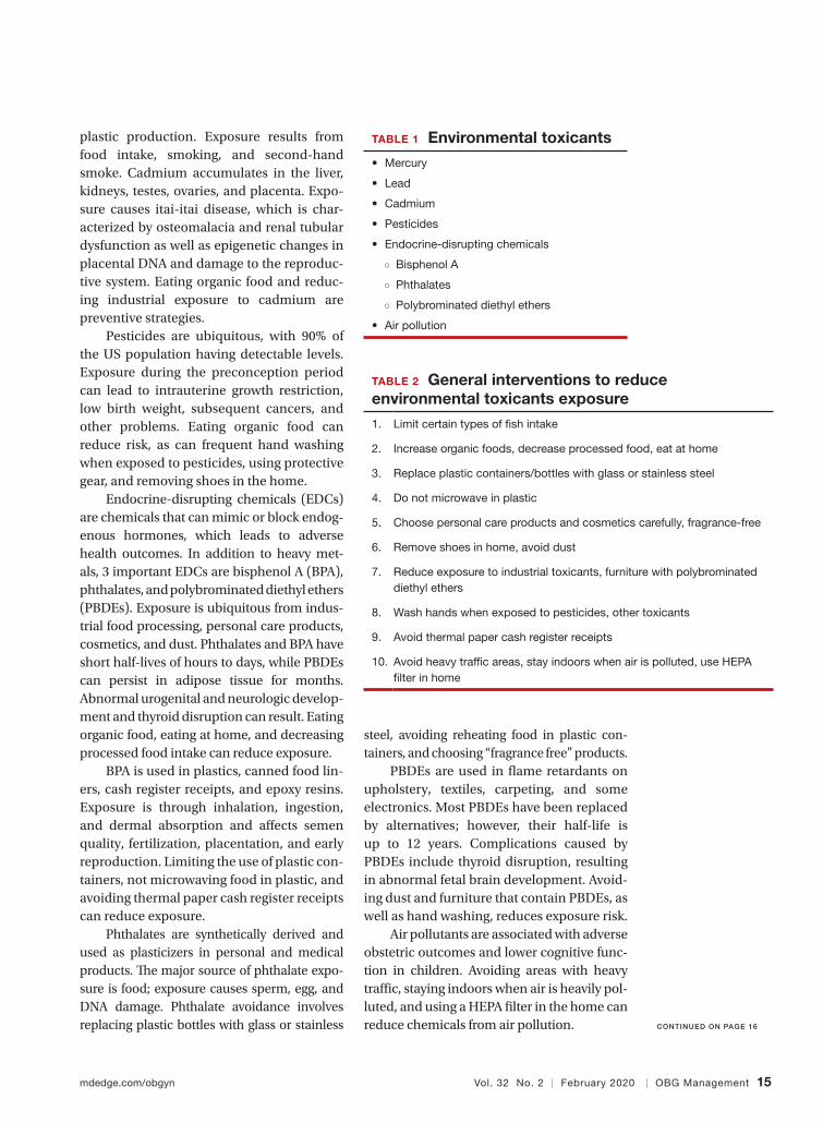
TABLE 1 Environmental toxicants
• Mercury
• Lead
• Cadmium
• Pesticides
• Endocrine-disrupting chemicals
○ Bisphenol A
○ Phthalates
○ Polybrominated diethyl ethers
• Air pollution
TABLE 2 General interventions to reduce environmental toxicants exposure
1.
2.
3.
4.
5.
6.
7.
8.
9.
10.
Limit certain types of �sh intake
Increase organic foods, decrease processed food, eat at home
Replace plastic containers/bottles with glass or stainless steel
Do not microwave in plastic
Choose personal care products and cosmetics carefully, fragrance-free
Remove shoes in home, avoid dust
Reduce exposure to industrial toxicants, furniture with polybrominated diethyl ethers
Wash hands when exposed to pesticides, other toxicants
Avoid thermal paper cash register receipts
Avoid heavy traf�c areas, stay indoors when air is polluted, use HEPA �lter in home
Update 0220.indd 15 1/31/20 3:37 PM
UPDATE fertility
16 OBG Management | February 2020 | Vol. 32 No. 2 mdedge.com/obgyn
CONTINUED FROM PAGE 15
Recommendations�e magnitude of the problem that environ-mental toxicant exposure creates requires health care providers to take action. �e
table in the publication by Segal and Giu-dice can be used as a tool that patients can answer �rst themselves before review by their provider.2 It can be added to your electronic health record and/or patient portal. Even making general comments to raise awareness, asking questions regarding exposure, and making recommendations can be helpful (TABLES 1 AND 2, page 15). When possible, we also should advocate for public awareness and policy changes that address this signi�cant health issue.
WHAT THIS EVIDENCE MEANS FOR PRACTICE
Environmental toxicants are a signi�cant health problem that can be effectively mitigated through patient questions and recommended interventions.
References1. Larsen D, Ma J, Strassberg M, et al. �e uptake of pan-ethnic
expanded carrier screening is higher when o�ered duringpreconception or early prenatal genetic counseling. Prenat Diagn. 2019;39:319-323.
2. Matthews TJ, MacDorman MF, �oma ME. Infant Mortality Statistics From the 2013 Period Linked Birth/Infant DeathData Set. Natl Vital Stat Rep. 2015;64:1-30.
3. American College of Obstetricians and Gynecologists Com-mittee on Genetics. Committee Opinion No. 690: carrierscreening in the age of genomic medicine. Obstet Gynecol. 2017;129:e35-e40.
4. Gregg AR, Edwards JG. Prenatal genetic carrier screening inthe genomic age. Semin Perinatol. 2018;42:303-306.
5. Munné S, Kaplan B, Frattarelli JL, et al; STAR Study Group.Preimplantation genetic testing for aneuploidy versusmorphology as selection criteria for single frozen-thawedembryo transfer in good-prognosis patients: a multicenter
randomized clinical trial. Fertil Steril. 2019;112:1071-1079.e7.
6. Goodman LR, Goldberg J, Falcone T, et al. Does the additionof time-lapse morphokinetics in the selection of embryos for transfer improve pregnancy rates? A randomized controlled trial. Fertil Steril. 2016;105:275-285.e10.
7. Blank C, Wildeboer RR, DeCroo I, et al. Prediction ofimplantation after blastocyst transfer in in vitro fertilization:a machine-learning perspective. Fertil Steril. 2019;111:318-326.
8. �e American College of Obstetricians and GynecologistsCommittee on Health Care for Underserved Women; Ameri-can Society for Reproductive Medicine Practice Commit-tee; �e University of California, San Francisco Program on Reproductive Health and the Environment. ACOG Com-mittee Opinion No. 575. Exposure to environmental toxicagents. Fertil Steril. 2013;100:931-934.
Update 0220.indd 16 1/31/20 3:38 PM

“ T h e t r u t h i s o n t h e m a r c h a n d n o t h i n g w i l l s t o p i t .”
É m i l e Z o l a
Ad Place-new.indd 6 1/29/2020 3:18:03 PM

STAY TUNED For exciting � ndings from the Pelvic Anatomy and Gynecologic Surgery Symposium
DON’T MISS our exclusive columns> Drugs, Pregnancy, & Lactation
> Gynecologic Oncology Consult
> Master Class
GET BREAKING NEWS at mdedge.com/obgyn
2
20
17CYTOMEGALOVIRUS
Valacyclovir cuts vertical transmission,
Dr. Keren Shahar-Nissan reports. EDITORIAL ADVISORY BOARD
Dr. Angela Martin joins the Ob.Gyn. News
team.
DYSMENORRHEA
Dr. Charles E. Miller outlines treatment for
endometriosis-associated pain.
Keeping you informed. Saving you time. A member of the Network.
Vol. 54 ■ No. 12 ■ DECEMBER 2019
MDedge.com/ObGyn
4How should we monitor
for recurrences?
By Dr. Emma Rossi
OVARIAN CANCER
3 PRESURGICAL STAGING Ultrasound can distinguish early- from late-stage endometriosis. ■ 3 CERVICAL CANCER Laparoscopic staging can improve outcomes.
WEIGHT GAIN
BY MITCHEL L. ZOLER
REPORTING FROM OBESITY WEEK 2019
LAS VEGAS – Contrary to current U.S. dietary rec-
ommendations, women with obesity should not
increase their energy intake during pregnancy to
achieve the current recommended level of ges-
tational weight gain, based on findings from an
intensive assessment of 54 women with obesity
during weeks 13-37 of pregnancy.
To achieve the gestational weight gain of 11-20
pounds (5-9.1 kg) recommended by the Institute of
Medicine, women with obesity ‒ those with a body
mass index of 30 kg/m2 or greater ‒ had an average
energy intake during the second and third trimes-
ters of 125 kcal/day less than their energy expen-
diture, Leanne M. Redman, PhD, said at a meeting
presented by the Obesity Society and the American
Society for Metabolic and Bariatric Surgery.
However, women in the study who had inade-
quate gestational weight gain had a daily calorie
deficit that was only slightly larger, an average of
262 kcal/day below their energy expenditure. As a
consequence, Dr. Redman believes the take-home
message from her findings is that pregnant wom-
HYSTERECTOMY
VS. MYOMECTOMY
Outcomes for fibroids
similar at 6-12 weeks
BY JEFF CRAVEN
REPORTING FROM ASRM 2019
PHILADELPHIA – Women who underwent either hys-
terectomy or myomectomy had similar short-term
outcomes between 6 weeks and 12 weeks after
surgery despite different baseline characteristics,
according to results from the COMPARE-UF study
presented at the annual meeting of the American
Society for Reproductive Medicine.
“Both hysterectomy and myomectomy can
substantially improve women’s quality of life
scores and substantially reduce symptom sever-
ity,” reported Wanda K. Nicholson, MD, MPH,
lead investigator for COMPARE-UF and profes-
sor of general obstetrics and gynecology at the
University of North Carolina at Chapel Hill.
Researchers included 1,295 women in the COM-
PARE-UF study who were at least 30 years old,
not attempting pregnancy, and undergoing hyster-
ectomy or myomectomy for treatment of fibroids.
Overall, 727 patients underwent hysterectomy, and
568 patients underwent myomectomy.
The researchers measured quality of life and
symptom severity using the Uterine Fibroid Scale-
QoL, the EQ-5D, and Visual Analog Scale (VAS).
The UFS-QoL contained subscales for concern, ac-
tivities, energy and mood, control, self-conscious-
ness, and sexual function, while the EQ-5D had
subscales for mobility, self-care, usual activities,
pain or discomfort, and anxiety or depression.
See FIBROIDS on page 18
See OBESITY on page 11
Mit
chel
L.
Zole
r/M
Ded
ge N
ews
Oral corticosteroids
This treatment during pregnancy for
rheumatoid arthritis, inflammatory bowel
disease, or asthma may increase the risk of
preterm birth.See page 16
Dr. Leanne M. Redman
Women with obesity need not
boost calories during pregnancy
01_11_18_19_OB19_12.indd 1
12/9/19 2:27 PM
GET BREAKING NEWS
Ad Place-new.indd 6 1/24/2020 12:28:54 PM
What’s Your DIAGNOSIS?
mdedge.com/obgyn Vol. 32 No. 2 | February 2020 | OBG Management 19
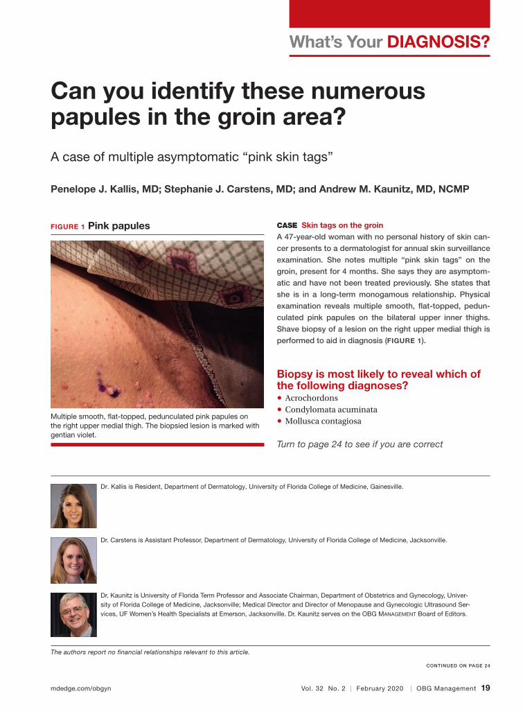
Can you identify these numerous papules in the groin area?
A case of multiple asymptomatic “pink skin tags”
Penelope J. Kallis, MD; Stephanie J. Carstens, MD; and Andrew M. Kaunitz, MD, NCMP
CASE Skin tags on the groinA 47-year-old woman with no personal history of skin can-
cer presents to a dermatologist for annual skin surveillance
examination. She notes multiple “pink skin tags” on the
groin, present for 4 months. She says they are asymptom-
atic and have not been treated previously. She states that
she is in a long-term monogamous relationship. Physical
examination reveals multiple smooth, �at-topped, pedun-
culated pink papules on the bilateral upper inner thighs.
Shave biopsy of a lesion on the right upper medial thigh is
performed to aid in diagnosis (FIGURE 1).
Biopsy is most likely to reveal which of the following diagnoses?• Acrochordons• Condylomata acuminata• Mollusca contagiosa
Turn to page 24 to see if you are correct
FIGURE 1 Pink papules
Multiple smooth, �at-topped, pedunculated pink papules on the right upper medial thigh. The biopsied lesion is marked with gentian violet.
Dr. Kallis is Resident, Department of Dermatology, University of Florida College of Medicine, Gainesville.
Dr. Carstens is Assistant Professor, Department of Dermatology, University of Florida College of Medicine, Jacksonville.
Dr. Kaunitz is University of Florida Term Professor and Associate Chairman, Department of Obstetrics and Gynecology, Univer-sity of Florida College of Medicine, Jacksonville; Medical Director and Director of Menopause and Gynecologic Ultrasound Ser-vices, UF Women’s Health Specialists at Emerson, Jacksonville. Dr. Kaunitz serves on the OBG MANAGEMENT Board of Editors.
The authors report no �nancial relationships relevant to this article.
CONTINUED ON PAGE 24
WYD Kaunitz 0220.indd 19 1/31/20 3:40 PM
Copyright © 2019 Merck Sharp & Dohme B.V., a subsidiary of Merck & Co., Inc. All rights reserved.
US-XPL-00588 05/19
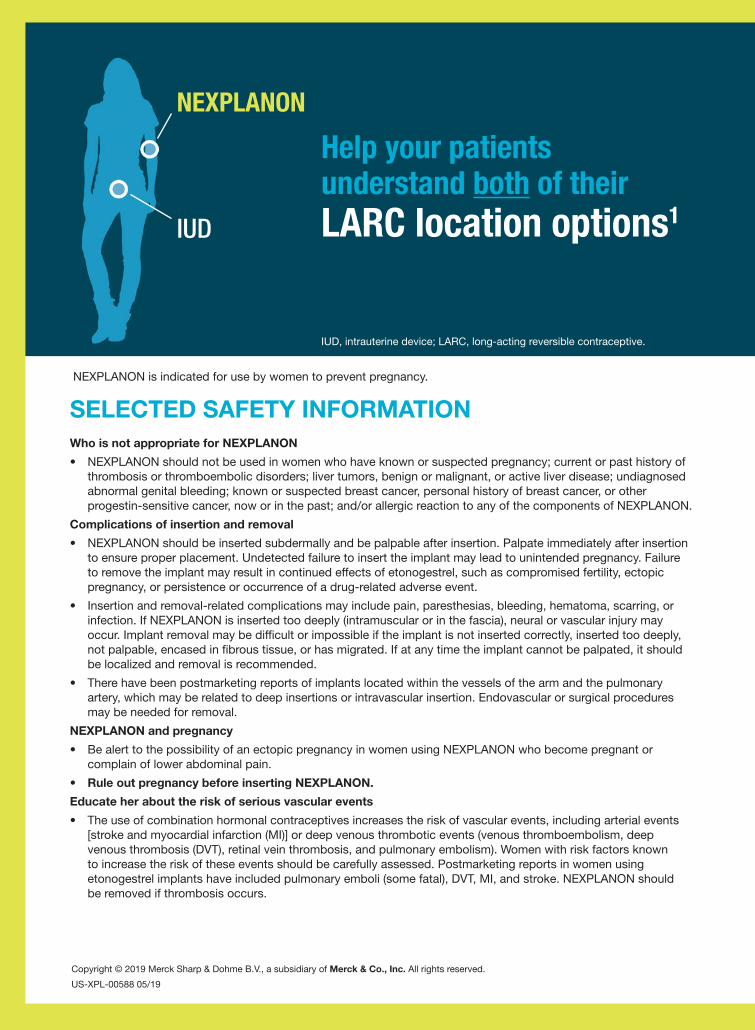
Help your patientsunderstand both of their LARC location options1
NEXPLANON is the onlynon-uterine LARC
NEXPLANON is indicated for use by women to prevent pregnancy.
SELECTED SAFETY INFORMATIONWho is not appropriate for NEXPLANON
• NEXPLANON should not be used in women who have known or suspected pregnancy; current or past history ofthrombosis or thromboembolic disorders; liver tumors, benign or malignant, or active liver disease; undiagnosedabnormal genital bleeding; known or suspected breast cancer, personal history of breast cancer, or otherprogestin-sensitive cancer, now or in the past; and/or allergic reaction to any of the components of NEXPLANON.
Complications of insertion and removal
• NEXPLANON should be inserted subdermally and be palpable after insertion. Palpate immediately after insertionto ensure proper placement. Undetected failure to insert the implant may lead to unintended pregnancy. Failureto remove the implant may result in continued effects of etonogestrel, such as compromised fertility, ectopicpregnancy, or persistence or occurrence of a drug-related adverse event.
• Insertion and removal-related complications may include pain, paresthesias, bleeding, hematoma, scarring, orinfection. If NEXPLANON is inserted too deeply (intramuscular or in the fascia), neural or vascular injury mayoccur. Implant removal may be diffi cult or impossible if the implant is not inserted correctly, inserted too deeply,not palpable, encased in fi brous tissue, or has migrated. If at any time the implant cannot be palpated, it shouldbe localized and removal is recommended.
• There have been postmarketing reports of implants located within the vessels of the arm and the pulmonaryartery, which may be related to deep insertions or intravascular insertion. Endovascular or surgical proceduresmay be needed for removal.
NEXPLANON and pregnancy
• Be alert to the possibility of an ectopic pregnancy in women using NEXPLANON who become pregnant orcomplain of lower abdominal pain.
• Rule out pregnancy before inserting NEXPLANON.
Educate her about the risk of serious vascular events
• The use of combination hormonal contraceptives increases the risk of vascular events, including arterial events[stroke and myocardial infarction (MI)] or deep venous thrombotic events (venous thromboembolism, deepvenous thrombosis (DVT), retinal vein thrombosis, and pulmonary embolism). Women with risk factors knownto increase the risk of these events should be carefully assessed. Postmarketing reports in women usingetonogestrel implants have included pulmonary emboli (some fatal), DVT, MI, and stroke. NEXPLANON shouldbe removed if thrombosis occurs.
IUD, intrauterine device; LARC, long-acting reversible contraceptive.
SELECTED SAFETY INFORMATION (continued)• Due to the risk of thromboembolism associated with pregnancy and immediately following delivery, NEXPLANON
should not be used prior to 21 days postpartum.
• Women with a history of thromboembolic disorders should be made aware of the possibility of a recurrence. Consider removing the NEXPLANON implant in case of long-term immobilization due to surgery or illness.
Counsel her about changes in bleeding patterns
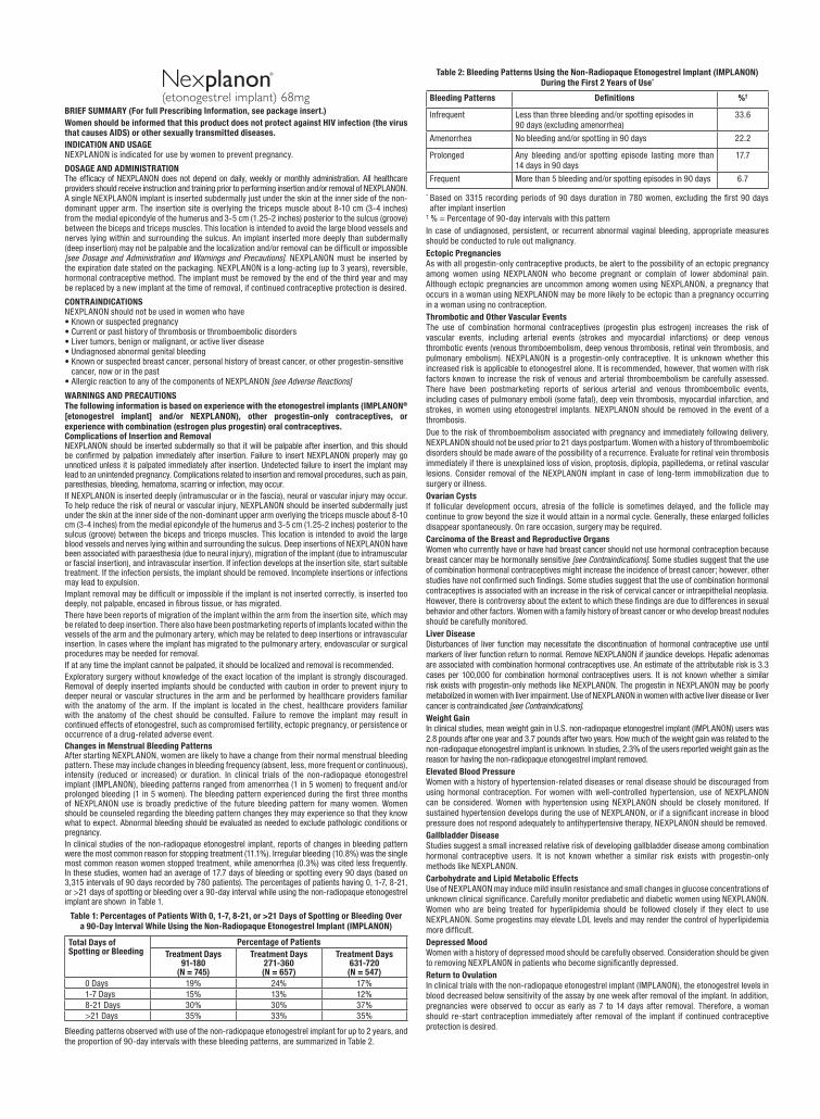
• Women are likely to have changes in their menstrual bleeding pattern with NEXPLANON, including changes in frequency, intensity, or duration. Abnormal bleeding should be evaluated as needed to exclude pathologic conditions or pregnancy. In clinical studies of the non-radiopaque etonogestrel implant, changes in bleeding pattern were the most common reason reported for stopping treatment (11.1%). Counsel women regarding potential changes they may experience.
Be aware of other serious complications, adverse reactions, and drug interactions
• Remove NEXPLANON if jaundice occurs.
• Remove NEXPLANON if blood pressure rises significantly and becomes uncontrolled.
• Prediabetic and diabetic women using NEXPLANON should be carefully monitored.
• Carefully observe women with a history of depressed mood. Consider removing NEXPLANON in patients who become significantly depressed.
• The most common adverse reactions (≥10%) reported in clinical trials were headache (24.9%), vaginitis (14.5%), weight increase (13.7%), acne (13.5%), breast pain (12.8%), abdominal pain (10.9%), and pharyngitis (10.5%).
• Drugs or herbal products that induce enzymes, including CYP3A4, may decrease the effectiveness of NEXPLANON or increase breakthrough bleeding.
• The efficacy of NEXPLANON in women weighing more than 130% of their ideal body weight has not been studied. Serum concentrations of etonogestrel are inversely related to body weight and decrease with time after implant insertion. Therefore, NEXPLANON may be less effective in overweight women.
• Counsel women to contact their health care provider immediately if, at any time, they are unableto palpate the implant.
• NEXPLANON does not protect against HIV or other STDs.
Please read the adjacent Brief Summary of the Prescribing Information.
Reference:
Up to 3 yearsof pregnancy prevention*
Placed subdermally just under the skin in the inner upper arm
*NEXPLANON must be removed by the end of the third year and may be replaced by another NEXPLANON at the time of removal, if continued contraceptive protection is desired.†Less than 1 pregnancy per 100 women who used NEXPLANON for 1 year.
(Actual implant shown; actual implant is 4 cm)
1. American College of Obstetricians and Gynecologists Committee on Practice Bulletins—Gynecology. ACOG Practice Bulletin No. 186: Long-acting reversible contraception: implants and intrauterine devices. Obstet Gynecol. 2017;130(5):e251–e269.
NEXPLANON
IUD>99%effective† Reversible
if her plans change

Copyright © 2019 Merck Sharp & Dohme B.V., a subsidiary of Merck & Co., Inc. All rights reserved.
US-XPL-00588 05/19
Help your patientsunderstand both of their LARC location options1
NEXPLANON is the onlynon-uterine LARC
NEXPLANON is indicated for use by women to prevent pregnancy.
SELECTED SAFETY INFORMATIONWho is not appropriate for NEXPLANON
• NEXPLANON should not be used in women who have known or suspected pregnancy; current or past history of thrombosis or thromboembolic disorders; liver tumors, benign or malignant, or active liver disease; undiagnosed abnormal genital bleeding; known or suspected breast cancer, personal history of breast cancer, or other progestin-sensitive cancer, now or in the past; and/or allergic reaction to any of the components of NEXPLANON.
Complications of insertion and removal
• NEXPLANON should be inserted subdermally and be palpable after insertion. Palpate immediately after insertion to ensure proper placement. Undetected failure to insert the implant may lead to unintended pregnancy. Failure to remove the implant may result in continued effects of etonogestrel, such as compromised fertility, ectopic pregnancy, or persistence or occurrence of a drug-related adverse event.
• Insertion and removal-related complications may include pain, paresthesias, bleeding, hematoma, scarring, or infection. If NEXPLANON is inserted too deeply (intramuscular or in the fascia), neural or vascular injury may occur. Implant removal may be difficult or impossible if the implant is not inserted correctly, inserted too deeply, not palpable, encased in fibrous tissue, or has migrated. If at any time the implant cannot be palpated, it should be localized and removal is recommended.
• There have been postmarketing reports of implants located within the vessels of the arm and the pulmonary artery, which may be related to deep insertions or intravascular insertion. Endovascular or surgical procedures may be needed for removal.
NEXPLANON and pregnancy
• Be alert to the possibility of an ectopic pregnancy in women using NEXPLANON who become pregnant or complain of lower abdominal pain.
• Rule out pregnancy before inserting NEXPLANON.
Educate her about the risk of serious vascular events
• The use of combination hormonal contraceptives increases the risk of vascular events, including arterial events [stroke and myocardial infarction (MI)] or deep venous thrombotic events (venous thromboembolism, deep venous thrombosis (DVT), retinal vein thrombosis, and pulmonary embolism). Women with risk factors known to increase the risk of these events should be carefully assessed. Postmarketing reports in women using etonogestrel implants have included pulmonary emboli (some fatal), DVT, MI, and stroke. NEXPLANON should be removed if thrombosis occurs.
IUD, intrauterine device; LARC, long-acting reversible contraceptive.
SELECTED SAFETY INFORMATION (continued)• Due to the risk of thromboembolism associated with pregnancy and immediately following delivery, NEXPLANON
should not be used prior to 21 days postpartum.
• Women with a history of thromboembolic disorders should be made aware of the possibility of a recurrence.Consider removing the NEXPLANON implant in case of long-term immobilization due to surgery or illness.
Counsel her about changes in bleeding patterns
• Women are likely to have changes in their menstrual bleeding pattern with NEXPLANON, including changesin frequency, intensity, or duration. Abnormal bleeding should be evaluated as needed to exclude pathologicconditions or pregnancy. In clinical studies of the non-radiopaque etonogestrel implant, changes in bleedingpattern were the most common reason reported for stopping treatment (11.1%). Counsel women regardingpotential changes they may experience.
Be aware of other serious complications, adverse reactions, and drug interactions
• Remove NEXPLANON if jaundice occurs.
• Remove NEXPLANON if blood pressure rises signifi cantly and becomes uncontrolled.
• Prediabetic and diabetic women using NEXPLANON should be carefully monitored.
• Carefully observe women with a history of depressed mood. Consider removing NEXPLANON in patients whobecome signifi cantly depressed.
• The most common adverse reactions (≥10%) reported in clinical trials were headache (24.9%), vaginitis (14.5%),weight increase (13.7%), acne (13.5%), breast pain (12.8%), abdominal pain (10.9%), and pharyngitis (10.5%).
• Drugs or herbal products that induce enzymes, including CYP3A4, may decrease the effectiveness ofNEXPLANON or increase breakthrough bleeding.
• The effi cacy of NEXPLANON in women weighing more than 130% of their ideal body weight has not beenstudied. Serum concentrations of etonogestrel are inversely related to body weight and decrease with time afterimplant insertion. Therefore, NEXPLANON may be less effective in overweight women.
• Counsel women to contact their health care provider immediately if, at any time, they are unableto palpate the implant.
• NEXPLANON does not protect against HIV or other STDs.
Please read the adjacent Brief Summary of the Prescribing Information.
Reference:
Up to 3 yearsof pregnancy prevention*
Placed subdermally just under the skin in the inner upper arm
*NEXPLANON must be removed by the end of the third year and may be replaced by anotherNEXPLANON at the time of removal, if continued contraceptive protection is desired.†Less than 1 pregnancy per 100 women who used NEXPLANON for 1 year.
(Actual implant shown; actual implant is 4 cm)
1. American College of Obstetricians and Gynecologists Committee on Practice Bulletins—Gynecology. ACOG Practice Bulletin No. 186: Long-acting reversible contraception: implants and intrauterine devices. Obstet Gynecol. 2017;130(5):e251–e269.
NEXPLANON
IUD>99%effective† Reversible
if her plans change
BRIEF SUMMARY (For full Prescribing Information, see package insert.)Women should be informed that this product does not protect against HIV infection (the virus that causes AIDS) or other sexually transmitted diseases.INDICATION AND USAGENEXPLANON is indicated for use by women to prevent pregnancy.
DOSAGE AND ADMINISTRATIONThe ef�cacy of NEXPLANON does not depend on daily, weekly or monthly administration. All healthcare providers should receive instruction and training prior to performing insertion and/or removal of NEXPLANON. A single NEXPLANON implant is inserted subdermally just under the skin at the inner side of the non-dominant upper arm. The insertion site is overlying the triceps muscle about 8-10 cm (3-4 inches) from the medial epicondyle of the humerus and 3-5 cm (1.25-2 inches) posterior to the sulcus (groove) between the biceps and triceps muscles. This location is intended to avoid the large blood vessels and nerves lying within and surrounding the sulcus. An implant inserted more deeply than subdermally (deep insertion) may not be palpable and the localization and/or removal can be dif�cult or impossible [see Dosage and Administration and Warnings and Precautions]. NEXPLANON must be inserted by the expiration date stated on the packaging. NEXPLANON is a long-acting (up to 3 years), reversible, hormonal contraceptive method. The implant must be removed by the end of the third year and may be replaced by a new implant at the time of removal, if continued contraceptive protection is desired.
CONTRAINDICATIONSNEXPLANON should not be used in women who have• Known or suspected pregnancy• Current or past history of thrombosis or thromboembolic disorders• Liver tumors, benign or malignant, or active liver disease• Undiagnosed abnormal genital bleeding• Known or suspected breast cancer, personal history of breast cancer, or other progestin-sensitive
cancer, now or in the past• Allergic reaction to any of the components of NEXPLANON [see Adverse Reactions]
WARNINGS AND PRECAUTIONS The following information is based on experience with the etonogestrel implants (IMPLANON® [etonogestrel implant] and/or NEXPLANON), other progestin-only contraceptives, or experience with combination (estrogen plus progestin) oral contraceptives.Complications of Insertion and RemovalNEXPLANON should be inserted subdermally so that it will be palpable after insertion, and this should be con�rmed by palpation immediately after insertion. Failure to insert NEXPLANON properly may go unnoticed unless it is palpated immediately after insertion. Undetected failure to insert the implant may lead to an unintended pregnancy. Complications related to insertion and removal procedures, such as pain, paresthesias, bleeding, hematoma, scarring or infection, may occur. If NEXPLANON is inserted deeply (intramuscular or in the fascia), neural or vascular injury may occur. To help reduce the risk of neural or vascular injury, NEXPLANON should be inserted subdermally just under the skin at the inner side of the non-dominant upper arm overlying the triceps muscle about 8-10 cm (3-4 inches) from the medial epicondyle of the humerus and 3-5 cm (1.25-2 inches) posterior to the sulcus (groove) between the biceps and triceps muscles. This location is intended to avoid the large blood vessels and nerves lying within and surrounding the sulcus. Deep insertions of NEXPLANON have been associated with paraesthesia (due to neural injury), migration of the implant (due to intramuscular or fascial insertion), and intravascular insertion. If infection develops at the insertion site, start suitable treatment. If the infection persists, the implant should be removed. Incomplete insertions or infections may lead to expulsion. Implant removal may be dif�cult or impossible if the implant is not inserted correctly, is inserted too deeply, not palpable, encased in �brous tissue, or has migrated. There have been reports of migration of the implant within the arm from the insertion site, which may be related to deep insertion. There also have been postmarketing reports of implants located within the vessels of the arm and the pulmonary artery, which may be related to deep insertions or intravascular insertion. In cases where the implant has migrated to the pulmonary artery, endovascular or surgical procedures may be needed for removal. If at any time the implant cannot be palpated, it should be localized and removal is recommended. Exploratory surgery without knowledge of the exact location of the implant is strongly discouraged. Removal of deeply inserted implants should be conducted with caution in order to prevent injury to deeper neural or vascular structures in the arm and be performed by healthcare providers familiar with the anatomy of the arm. If the implant is located in the chest, healthcare providers familiar with the anatomy of the chest should be consulted. Failure to remove the implant may result in continued effects of etonogestrel, such as compromised fertility, ectopic pregnancy, or persistence or occurrence of a drug-related adverse event.Changes in Menstrual Bleeding PatternsAfter starting NEXPLANON, women are likely to have a change from their normal menstrual bleeding pattern. These may include changes in bleeding frequency (absent, less, more frequent or continuous), intensity (reduced or increased) or duration. In clinical trials of the non-radiopaque etonogestrel implant (IMPLANON), bleeding patterns ranged from amenorrhea (1 in 5 women) to frequent and/or prolonged bleeding (1 in 5 women). The bleeding pattern experienced during the �rst three months of NEXPLANON use is broadly predictive of the future bleeding pattern for many women. Women should be counseled regarding the bleeding pattern changes they may experience so that they know what to expect. Abnormal bleeding should be evaluated as needed to exclude pathologic conditions or pregnancy. In clinical studies of the non-radiopaque etonogestrel implant, reports of changes in bleeding pattern were the most common reason for stopping treatment (11.1%). Irregular bleeding (10.8%) was the single most common reason women stopped treatment, while amenorrhea (0.3%) was cited less frequently. In these studies, women had an average of 17.7 days of bleeding or spotting every 90 days (based on 3,315 intervals of 90 days recorded by 780 patients). The percentages of patients having 0, 1-7, 8-21, or >21 days of spotting or bleeding over a 90-day interval while using the non-radiopaque etonogestrel implant are shown in Table 1.
Table 1: Percentages of Patients With 0, 1-7, 8-21, or >21 Days of Spotting or Bleeding Over a 90-Day Interval While Using the Non-Radiopaque Etonogestrel Implant (IMPLANON)
Bleeding patterns observed with use of the non-radiopaque etonogestrel implant for up to 2 years, and the proportion of 90-day intervals with these bleeding patterns, are summarized in Table 2.
Table 2: Bleeding Patterns Using the Non-Radiopaque Etonogestrel Implant (IMPLANON) During the First 2 Years of Use*
* Based on 3315 recording periods of 90 days duration in 780 women, excluding the �rst 90 daysafter implant insertion
† % = Percentage of 90-day intervals with this patternIn case of undiagnosed, persistent, or recurrent abnormal vaginal bleeding, appropriate measuresshould be conducted to rule out malignancy.Ectopic Pregnancies As with all progestin-only contraceptive products, be alert to the possibility of an ectopic pregnancy among women using NEXPLANON who become pregnant or complain of lower abdominal pain.Although ectopic pregnancies are uncommon among women using NEXPLANON, a pregnancy thatoccurs in a woman using NEXPLANON may be more likely to be ectopic than a pregnancy occurring in a woman using no contraception.Thrombotic and Other Vascular Events The use of combination hormonal contraceptives (progestin plus estrogen) increases the risk ofvascular events, including arterial events (strokes and myocardial infarctions) or deep venousthrombotic events (venous thromboembolism, deep venous thrombosis, retinal vein thrombosis, and pulmonary embolism). NEXPLANON is a progestin-only contraceptive. It is unknown whether thisincreased risk is applicable to etonogestrel alone. It is recommended, however, that women with risk factors known to increase the risk of venous and arterial thromboembolism be carefully assessed. There have been postmarketing reports of serious arterial and venous thromboembolic events,including cases of pulmonary emboli (some fatal), deep vein thrombosis, myocardial infarction, and strokes, in women using etonogestrel implants. NEXPLANON should be removed in the event of athrombosis. Due to the risk of thromboembolism associated with pregnancy and immediately following delivery, NEXPLANON should not be used prior to 21 days postpartum. Women with a history of thromboembolic disorders should be made aware of the possibility of a recurrence. Evaluate for retinal vein thrombosis immediately if there is unexplained loss of vision, proptosis, diplopia, papilledema, or retinal vascular lesions. Consider removal of the NEXPLANON implant in case of long-term immobilization due tosurgery or illness.Ovarian Cysts If follicular development occurs, atresia of the follicle is sometimes delayed, and the follicle maycontinue to grow beyond the size it would attain in a normal cycle. Generally, these enlarged follicles disappear spontaneously. On rare occasion, surgery may be required.Carcinoma of the Breast and Reproductive Organs Women who currently have or have had breast cancer should not use hormonal contraception because breast cancer may be hormonally sensitive [see Contraindications]. Some studies suggest that the use of combination hormonal contraceptives might increase the incidence of breast cancer; however, other studies have not con�rmed such �ndings. Some studies suggest that the use of combination hormonal contraceptives is associated with an increase in the risk of cervical cancer or intraepithelial neoplasia. However, there is controversy about the extent to which these �ndings are due to differences in sexual behavior and other factors. Women with a family history of breast cancer or who develop breast nodules should be carefully monitored.Liver Disease Disturbances of liver function may necessitate the discontinuation of hormonal contraceptive use untilmarkers of liver function return to normal. Remove NEXPLANON if jaundice develops. Hepatic adenomas are associated with combination hormonal contraceptives use. An estimate of the attributable risk is 3.3 cases per 100,000 for combination hormonal contraceptives users. It is not known whether a similarrisk exists with progestin-only methods like NEXPLANON. The progestin in NEXPLANON may be poorly metabolized in women with liver impairment. Use of NEXPLANON in women with active liver disease or liver cancer is contraindicated [see Contraindications].Weight Gain In clinical studies, mean weight gain in U.S. non-radiopaque etonogestrel implant (IMPLANON) users was 2.8 pounds after one year and 3.7 pounds after two years. How much of the weight gain was related to the non-radiopaque etonogestrel implant is unknown. In studies, 2.3% of the users reported weight gain as the reason for having the non-radiopaque etonogestrel implant removed.Elevated Blood Pressure Women with a history of hypertension-related diseases or renal disease should be discouraged from using hormonal contraception. For women with well-controlled hypertension, use of NEXPLANONcan be considered. Women with hypertension using NEXPLANON should be closely monitored. Ifsustained hypertension develops during the use of NEXPLANON, or if a signi�cant increase in blood pressure does not respond adequately to antihypertensive therapy, NEXPLANON should be removed.Gallbladder Disease Studies suggest a small increased relative risk of developing gallbladder disease among combination hormonal contraceptive users. It is not known whether a similar risk exists with progestin-onlymethods like NEXPLANON.Carbohydrate and Lipid Metabolic Effects Use of NEXPLANON may induce mild insulin resistance and small changes in glucose concentrations of unknown clinical significance. Carefully monitor prediabetic and diabetic women using NEXPLANON. Women who are being treated for hyperlipidemia should be followed closely if they elect to useNEXPLANON. Some progestins may elevate LDL levels and may render the control of hyperlipidemia more dif�cult.Depressed Mood Women with a history of depressed mood should be carefully observed. Consideration should be given to removing NEXPLANON in patients who become signi�cantly depressed.Return to Ovulation In clinical trials with the non-radiopaque etonogestrel implant (IMPLANON), the etonogestrel levels in blood decreased below sensitivity of the assay by one week after removal of the implant. In addition, pregnancies were observed to occur as early as 7 to 14 days after removal. Therefore, a womanshould re-start contraception immediately after removal of the implant if continued contraceptiveprotection is desired.
Bleeding Patterns De�nitions %†
Infrequent Less than three bleeding and/or spotting episodes in 90 days (excluding amenorrhea)
33.6
Amenorrhea No bleeding and/or spotting in 90 days 22.2
Prolonged Any bleeding and/or spotting episode lasting more than 14 days in 90 days
17.7
Frequent More than 5 bleeding and/or spotting episodes in 90 days 6.7
Total Days of Spotting or Bleeding
Percentage of PatientsTreatment Days
91-180 (N = 745)
Treatment Days 271-360 (N = 657)
Treatment Days 631-720
(N = 547)0 Days 19% 24% 17%1-7 Days 15% 13% 12%8-21 Days 30% 30% 37%>21 Days 35% 33% 35%
Fluid RetentionHormonal contraceptives may cause some degree of �uid retention. They should be prescribed with caution, and only with careful monitoring, in patients with conditions which might be aggravated by �uid retention. It is unknown if NEXPLANON causes �uid retention.Contact LensesContact lens wearers who develop visual changes or changes in lens tolerance should be assessed by an ophthalmologist.In Situ Broken or Bent ImplantThere have been reports of broken or bent implants while in the patient’s arm. Based on in vitro data, when an implant is broken or bent, the release rate of etonogestrel may be slightly increased. When an implant is removed, it is important to remove it in its entirety [see Dosage and Administration].MonitoringA woman who is using NEXPLANON should have a yearly visit with her healthcare provider for a blood pressure check and for other indicated health care.Drug-Laboratory Test InteractionsSex hormone-binding globulin concentrations may be decreased for the �rst six months after NEXPLANON insertion followed by gradual recovery. Thyroxine concentrations may initially be slightly decreased followed by gradual recovery to baseline.
ADVERSE REACTIONSIn clinical trials involving 942 women who were evaluated for safety, change in menstrual bleeding patterns (irregular menses) was the most common adverse reaction causing discontinuation of use of the non-radiopaque etonogestrel implant (IMPLANON® [etonogestrel implant]) (11.1% of women).Adverse reactions that resulted in a rate of discontinuation of ≥1% are shown in Table 3.
Table 3: Adverse Reactions Leading to Discontinuation of Treatment in 1% or More of Subjects in Clinical Trials of the Non-Radiopaque Etonogestrel Implant (IMPLANON)
* Includes “frequent”, “heavy”, “prolonged”, “spotting”, and other patterns of bleeding irregularity.† Among US subjects (N=330), 6.1% experienced emotional lability that led to discontinuation.‡ Among US subjects (N=330), 2.4% experienced depression that led to discontinuation.
Other adverse reactions that were reported by at least 5% of subjects in the non-radiopaque etonogestrel implant clinical trials are listed in Table 4.
Table 4: Common Adverse Reactions Reported by ≥5% of Subjects in Clinical Trials With the Non-Radiopaque Etonogestrel Implant (IMPLANON)
In a clinical trial of NEXPLANON, in which investigators were asked to examine the implant site after insertion, implant site reactions were reported in 8.6% of women. Erythema was the most frequent implant site complication, reported during and/or shortly after insertion, occurring in 3.3% of subjects. Additionally, hematoma (3.0%), bruising (2.0%), pain (1.0%), and swelling (0.7%) were reported. Effects of Other Drugs on Hormonal ContraceptivesSubstances decreasing the plasma concentrations of hormonal contraceptives (HCs) andpotentially diminishing the ef�cacy of HCs: Drugs or herbal products that induce certain enzymes, including cytochrome P450 3A4 (CYP3A4), may decrease the plasma concentrations of HCs and potentially diminish the effectiveness of HCs or increase breakthrough bleeding.Some drugs or herbal products that may decrease the effectiveness of HCs include efavirenz, phenytoin,barbiturates, carbamazepine, bosentan, felbamate, griseofulvin, oxcarbazepine, rifampicin, topiramate,rifabutin, ru�namide, aprepitant, and products containing St. John’s wort. Interactions between HCsand other drugs may lead to breakthrough bleeding and/or contraceptive failure. Counsel women to usean alternative non-hormonal method of contraception or a back-up method when enzyme inducers areused with HCs, and to continue back-up non-hormonal contraception for 28 days after discontinuing theenzyme inducer to ensure contraceptive reliability.
Substances increasing the plasma concentrations of HCs: Co-administration of certain HCs and strong or moderate CYP3A4 inhibitors such as itraconazole, voriconazole, �uconazole, grapefruit juice, or ketoconazole may increase the serum concentrations of progestins, including etonogestrel.Human Immunode�ciency Virus (HIV)/Hepatitis C Virus (HCV) protease inhibitors and non-nucleoside reverse transcriptase inhibitors: Signi�cant changes (increase or decrease) in the plasma concentrations of progestin have been noted in cases of co-administration with HIV protease inhibitors (decrease [e.g., nel�navir, ritonavir, darunavir/ritonavir, (fos)amprenavir/ritonavir, lopinavir/ritonavir, and tipranavir/ritonavir] or increase [e.g., indinavir and atazanavir/ritonavir])/HCV proteaseinhibitors (decrease [e.g., boceprevir and telaprevir]) or with non-nucleoside reverse transcriptaseinhibitors (decrease [e.g., nevirapine, efavirenz] or increase [e.g., etravirene]). These changes may be clinically relevant in some cases. Consult the prescribing information of anti-viral and anti-retroviral concomitant medications to identify potential interactions.Effects of Hormonal Contraceptives on Other DrugsHormonal contraceptives may affect the metabolism of other drugs. Consequently, plasmaconcentrations may either increase (for example, cyclosporine) or decrease (for example, lamotrigine).Consult the labeling of all concurrently-used drugs to obtain further information about interactions with hormonal contraceptives or the potential for enzyme alterations.
USE IN SPECIFIC POPULATIONSPregnancyRisk SummaryNEXPLANON is contraindicated during pregnancy because there is no need for pregnancy prevention in a woman who is already pregnant [see Contraindications]. Epidemiologic studies and meta-analyseshave not shown an increased risk of genital or non-genital birth defects (including cardiac anomalies and limb-reduction defects) following maternal exposure to low dose CHCs prior to conception or during early pregnancy. No adverse development outcomes were observed in pregnant rats and rabbits with the administration of etonogestrel during organogenesis at doses of 315 or 781 times the anticipated human dose (60 μg/day). NEXPLANON should be removed if maintaining a pregnancy.LactationRisk SummarySmall amounts of contraceptive steroids and/or metabolites, including etonogestrel are present inhuman milk. No signi�cant adverse effects have been observed in the production or quality of breastmilk, or on the physical and psychomotor development of breastfed infants. Hormonal contraceptives,including etonogestrel, can reduce milk production in breastfeeding mothers.This is less likely to occuronce breastfeeding is well-established; however, it can occur at any time in some women. Whenpossible, advise the nursing mother about both hormonal and non-hormonal contraceptive options,as steroids may not be the initial choice for these patients. The developmental and health bene�ts ofbreastfeeding should be considered along with the mother’s clinical need for NEXPLANON and anypotential adverse effects on the breastfed child from NEXPLANON or from the underlying maternalcondition.Pediatric UseSafety and ef�cacy of NEXPLANON have been established in women of reproductive age. Safety and ef�cacy of NEXPLANON are expected to be the same for postpubertal adolescents. However, no clinical studies have been conducted in women less than 18 years of age. Use of this product before menarche is not indicated.Geriatric UseThis product has not been studied in women over 65 years of age and is not indicated in this population.Hepatic ImpairmentNo studies were conducted to evaluate the effect of hepatic disease on the disposition of NEXPLANON. The use of NEXPLANON in women with active liver disease is contraindicated [see Contraindications].Overweight WomenThe effectiveness of the etonogestrel implant in women who weighed more than 130% of their ideal body weight has not been de�ned because such women were not studied in clinical trials. Serum concentrations of etonogestrel are inversely related to body weight and decrease with time after implant insertion. It is therefore possible that NEXPLANON may be less effective in overweight women, especially in the presence of other factors that decrease serum etonogestrel concentrationssuch as concomitant use of hepatic enzyme inducers.
OVERDOSAGEOverdosage may result if more than one implant is inserted. In case of suspected overdose, the implant should be removed.
NONCLINICAL TOXICOLOGYIn a 24-month carcinogenicity study in rats with subdermal implants releasing 10 and 20 mcg etonogestrel per day (equal to approximately 1.8-3.6 times the systemic steady state exposure in women using NEXPLANON), no drug-related carcinogenic potential was observed. Etonogestrel wasnot genotoxic in the in vitro Ames/Salmonella reverse mutation assay, the chromosomal aberrationassay in Chinese hamster ovary cells or in the in vivo mouse micronucleus test. Fertility in rats returned after withdrawal from treatment.PATIENT COUNSELING INFORMATION See FDA-Approved Patient Labeling.• Counsel women about the insertion and removal procedure of the NEXPLANON implant. Provide the
woman with a copy of the Patient Labeling and ensure that she understands the information in the Patient Labeling before insertion and removal. A USER CARD and consent form are included in the packaging. Have the woman complete a consent form and retain it in your records. The USER CARD should be �lled out and given to the woman after insertion of the NEXPLANON implant so that she will have a record of the location of the implant in the upper arm and when it should be removed.
• Counsel women to contact their healthcare provider immediately if, at any time, they are unable to palpate the implant.
• Counsel women that NEXPLANON does not protect against HIV infection (AIDS) or other STDs.• Counsel women that the use of NEXPLANON may be associated with changes in their normal
menstrual bleeding patterns so that they know what to expect.
Adverse Reactions All StudiesN = 942
Bleeding Irregularities* 11.1%
Emotional Lability† 2.3%
Weight Increase 2.3%
Headache 1.6%
Acne 1.3%
Depression‡ 1.0%
Adverse Reactions All Studies N = 942
Headache 24.9%
Vaginitis 14.5%
Weight increase 13.7%
Acne 13.5%
Breast pain 12.8%
Abdominal pain 10.9%
Pharyngitis 10.5%
Leukorrhea 9.6%In�uenza-like symptoms 7.6%
Dizziness 7.2%
Dysmenorrhea 7.2%
Back pain 6.8%
Emotional lability 6.5%
Nausea 6.4%
Pain 5.6%
Nervousness 5.6%
Depression 5.5%
Hypersensitivity 5.4%
Insertion site pain 5.2%
For more detailed information, please read the Prescribing Information.USPI-MK8415-IPTX-1810r020Revised: 10/2018
Copyright © 2019 Merck Sharp & Dohme B.V., a subsidiary of Merck & Co., Inc. All rights reserved.US-XPL-00588 05/19
Manufactured for: Merck Sharp & Dohme Corp., a subsidiary of
MERCK & CO., INC., Whitehouse Station, NJ 08889, USA.

BRIEF SUMMARY (For full Prescribing Information, see package insert.)Women should be informed that this product does not protect against HIV infection (the virus that causes AIDS) or other sexually transmitted diseases.INDICATION AND USAGENEXPLANON is indicated for use by women to prevent pregnancy.
DOSAGE AND ADMINISTRATIONThe ef�cacy of NEXPLANON does not depend on daily, weekly or monthly administration. All healthcareproviders should receive instruction and training prior to performing insertion and/or removal of NEXPLANON.A single NEXPLANON implant is inserted subdermally just under the skin at the inner side of the non-dominant upper arm. The insertion site is overlying the triceps muscle about 8-10 cm (3-4 inches) from the medial epicondyle of the humerus and 3-5 cm (1.25-2 inches) posterior to the sulcus (groove) between the biceps and triceps muscles. This location is intended to avoid the large blood vessels and nerves lying within and surrounding the sulcus. An implant inserted more deeply than subdermally (deep insertion) may not be palpable and the localization and/or removal can be dif�cult or impossible [see Dosage and Administration and Warnings and Precautions]. NEXPLANON must be inserted by the expiration date stated on the packaging. NEXPLANON is a long-acting (up to 3 years), reversible, hormonal contraceptive method. The implant must be removed by the end of the third year and may be replaced by a new implant at the time of removal, if continued contraceptive protection is desired.
CONTRAINDICATIONSNEXPLANON should not be used in women who have• Known or suspected pregnancy• Current or past history of thrombosis or thromboembolic disorders• Liver tumors, benign or malignant, or active liver disease• Undiagnosed abnormal genital bleeding• Known or suspected breast cancer, personal history of breast cancer, or other progestin-sensitive
cancer, now or in the past• Allergic reaction to any of the components of NEXPLANON [see Adverse Reactions]
WARNINGS AND PRECAUTIONSThe following information is based on experience with the etonogestrel implants (IMPLANON®
[etonogestrel implant] and/or NEXPLANON), other progestin-only contraceptives, orexperience with combination (estrogen plus progestin) oral contraceptives.Complications of Insertion and RemovalNEXPLANON should be inserted subdermally so that it will be palpable after insertion, and this shouldbe con�rmed by palpation immediately after insertion. Failure to insert NEXPLANON properly may gounnoticed unless it is palpated immediately after insertion. Undetected failure to insert the implant maylead to an unintended pregnancy. Complications related to insertion and removal procedures, such as pain,paresthesias, bleeding, hematoma, scarring or infection, may occur.If NEXPLANON is inserted deeply (intramuscular or in the fascia), neural or vascular injury may occur.To help reduce the risk of neural or vascular injury, NEXPLANON should be inserted subdermally justunder the skin at the inner side of the non-dominant upper arm overlying the triceps muscle about 8-10cm (3-4 inches) from the medial epicondyle of the humerus and 3-5 cm (1.25-2 inches) posterior to thesulcus (groove) between the biceps and triceps muscles. This location is intended to avoid the largeblood vessels and nerves lying within and surrounding the sulcus. Deep insertions of NEXPLANON havebeen associated with paraesthesia (due to neural injury), migration of the implant (due to intramuscularor fascial insertion), and intravascular insertion. If infection develops at the insertion site, start suitabletreatment. If the infection persists, the implant should be removed. Incomplete insertions or infectionsmay lead to expulsion.Implant removal may be dif�cult or impossible if the implant is not inserted correctly, is inserted too deeply, not palpable, encased in �brous tissue, or has migrated.There have been reports of migration of the implant within the arm from the insertion site, which may be related to deep insertion. There also have been postmarketing reports of implants located within the vessels of the arm and the pulmonary artery, which may be related to deep insertions or intravascular insertion. In cases where the implant has migrated to the pulmonary artery, endovascular or surgical procedures may be needed for removal.If at any time the implant cannot be palpated, it should be localized and removal is recommended.Exploratory surgery without knowledge of the exact location of the implant is strongly discouraged. Removal of deeply inserted implants should be conducted with caution in order to prevent injury to deeper neural or vascular structures in the arm and be performed by healthcare providers familiar with the anatomy of the arm. If the implant is located in the chest, healthcare providers familiar with the anatomy of the chest should be consulted. Failure to remove the implant may result in continued effects of etonogestrel, such as compromised fertility, ectopic pregnancy, or persistence oroccurrence of a drug-related adverse event.Changes in Menstrual Bleeding PatternsAfter starting NEXPLANON, women are likely to have a change from their normal menstrual bleeding pattern. These may include changes in bleeding frequency (absent, less, more frequent or continuous), intensity (reduced or increased) or duration. In clinical trials of the non-radiopaque etonogestrel implant (IMPLANON), bleeding patterns ranged from amenorrhea (1 in 5 women) to frequent and/or prolonged bleeding (1 in 5 women). The bleeding pattern experienced during the �rst three months of NEXPLANON use is broadly predictive of the future bleeding pattern for many women. Women should be counseled regarding the bleeding pattern changes they may experience so that they know what to expect. Abnormal bleeding should be evaluated as needed to exclude pathologic conditions orpregnancy.In clinical studies of the non-radiopaque etonogestrel implant, reports of changes in bleeding patternwere the most common reason for stopping treatment (11.1%). Irregular bleeding (10.8%) was the singlemost common reason women stopped treatment, while amenorrhea (0.3%) was cited less frequently.In these studies, women had an average of 17.7 days of bleeding or spotting every 90 days (based on3,315 intervals of 90 days recorded by 780 patients). The percentages of patients having 0, 1-7, 8-21,or >21 days of spotting or bleeding over a 90-day interval while using the non-radiopaque etonogestrelimplant are shown in Table 1.
Table 1: Percentages of Patients With 0, 1-7, 8-21, or >21 Days of Spotting or Bleeding Over a 90-Day Interval While Using the Non-Radiopaque Etonogestrel Implant (IMPLANON)
Bleeding patterns observed with use of the non-radiopaque etonogestrel implant for up to 2 years, and the proportion of 90-day intervals with these bleeding patterns, are summarized in Table 2.
Table 2: Bleeding Patterns Using the Non-Radiopaque Etonogestrel Implant (IMPLANON)During the First 2 Years of Use*
* Based on 3315 recording periods of 90 days duration in 780 women, excluding the �rst 90 days after implant insertion
† % = Percentage of 90-day intervals with this patternIn case of undiagnosed, persistent, or recurrent abnormal vaginal bleeding, appropriate measuresshould be conducted to rule out malignancy.Ectopic PregnanciesAs with all progestin-only contraceptive products, be alert to the possibility of an ectopic pregnancy among women using NEXPLANON who become pregnant or complain of lower abdominal pain.Although ectopic pregnancies are uncommon among women using NEXPLANON, a pregnancy thatoccurs in a woman using NEXPLANON may be more likely to be ectopic than a pregnancy occurring in a woman using no contraception.Thrombotic and Other Vascular EventsThe use of combination hormonal contraceptives (progestin plus estrogen) increases the risk of vascular events, including arterial events (strokes and myocardial infarctions) or deep venous thrombotic events (venous thromboembolism, deep venous thrombosis, retinal vein thrombosis, andpulmonary embolism). NEXPLANON is a progestin-only contraceptive. It is unknown whether thisincreased risk is applicable to etonogestrel alone. It is recommended, however, that women with riskfactors known to increase the risk of venous and arterial thromboembolism be carefully assessed. There have been postmarketing reports of serious arterial and venous thromboembolic events, including cases of pulmonary emboli (some fatal), deep vein thrombosis, myocardial infarction, and strokes, in women using etonogestrel implants. NEXPLANON should be removed in the event of a thrombosis.Due to the risk of thromboembolism associated with pregnancy and immediately following delivery, NEXPLANON should not be used prior to 21 days postpartum. Women with a history of thromboembolic disorders should be made aware of the possibility of a recurrence. Evaluate for retinal vein thrombosis immediately if there is unexplained loss of vision, proptosis, diplopia, papilledema, or retinal vascular lesions. Consider removal of the NEXPLANON implant in case of long-term immobilization due to surgery or illness.Ovarian CystsIf follicular development occurs, atresia of the follicle is sometimes delayed, and the follicle may continue to grow beyond the size it would attain in a normal cycle. Generally, these enlarged follicles disappear spontaneously. On rare occasion, surgery may be required.Carcinoma of the Breast and Reproductive OrgansWomen who currently have or have had breast cancer should not use hormonal contraception becausebreast cancer may be hormonally sensitive [see Contraindications]. Some studies suggest that the useof combination hormonal contraceptives might increase the incidence of breast cancer; however, otherstudies have not con�rmed such �ndings. Some studies suggest that the use of combination hormonalcontraceptives is associated with an increase in the risk of cervical cancer or intraepithelial neoplasia.However, there is controversy about the extent to which these �ndings are due to differences in sexualbehavior and other factors. Women with a family history of breast cancer or who develop breast nodulesshould be carefully monitored.Liver DiseaseDisturbances of liver function may necessitate the discontinuation of hormonal contraceptive use untilmarkers of liver function return to normal. Remove NEXPLANON if jaundice develops. Hepatic adenomasare associated with combination hormonal contraceptives use. An estimate of the attributable risk is 3.3cases per 100,000 for combination hormonal contraceptives users. It is not known whether a similarrisk exists with progestin-only methods like NEXPLANON. The progestin in NEXPLANON may be poorlymetabolized in women with liver impairment. Use of NEXPLANON in women with active liver disease or livercancer is contraindicated [see Contraindications].Weight GainIn clinical studies, mean weight gain in U.S. non-radiopaque etonogestrel implant (IMPLANON) users was2.8 pounds after one year and 3.7 pounds after two years. How much of the weight gain was related to thenon-radiopaque etonogestrel implant is unknown. In studies, 2.3% of the users reported weight gain as thereason for having the non-radiopaque etonogestrel implant removed.Elevated Blood PressureWomen with a history of hypertension-related diseases or renal disease should be discouraged from using hormonal contraception. For women with well-controlled hypertension, use of NEXPLANONcan be considered. Women with hypertension using NEXPLANON should be closely monitored. If sustained hypertension develops during the use of NEXPLANON, or if a signi�cant increase in blood pressure does not respond adequately to antihypertensive therapy, NEXPLANON should be removed.Gallbladder DiseaseStudies suggest a small increased relative risk of developing gallbladder disease among combination hormonal contraceptive users. It is not known whether a similar risk exists with progestin-only methods like NEXPLANON.Carbohydrate and Lipid Metabolic EffectsUse of NEXPLANON may induce mild insulin resistance and small changes in glucose concentrations of unknown clinical significance. Carefully monitor prediabetic and diabetic women using NEXPLANON. Women who are being treated for hyperlipidemia should be followed closely if they elect to use NEXPLANON. Some progestins may elevate LDL levels and may render the control of hyperlipidemia more dif�cult.Depressed MoodWomen with a history of depressed mood should be carefully observed. Consideration should be given to removing NEXPLANON in patients who become signi�cantly depressed.Return to OvulationIn clinical trials with the non-radiopaque etonogestrel implant (IMPLANON), the etonogestrel levels in blood decreased below sensitivity of the assay by one week after removal of the implant. In addition, pregnancies were observed to occur as early as 7 to 14 days after removal. Therefore, a woman should re-start contraception immediately after removal of the implant if continued contraceptive protection is desired.
Bleeding Patterns De�nitions %†
Infrequent Less than three bleeding and/or spotting episodes in 90 days (excluding amenorrhea)
33.6
Amenorrhea No bleeding and/or spotting in 90 days 22.2
Prolonged Any bleeding and/or spotting episode lasting more than 14 days in 90 days
17.7
Frequent More than 5 bleeding and/or spotting episodes in 90 days 6.7
Total Days of Spotting or Bleeding
Percentage of PatientsTreatment Days
91-180(N = 745)
Treatment Days 271-360(N = 657)
Treatment Days 631-720
(N = 547)0 Days 19% 24% 17%1-7 Days 15% 13% 12%8-21 Days 30% 30% 37%>21 Days 35% 33% 35%
Fluid Retention Hormonal contraceptives may cause some degree of �uid retention. They should be prescribed with caution, and only with careful monitoring, in patients with conditions which might be aggravated by �uid retention. It is unknown if NEXPLANON causes �uid retention.Contact Lenses Contact lens wearers who develop visual changes or changes in lens tolerance should be assessed by an ophthalmologist.In Situ Broken or Bent Implant There have been reports of broken or bent implants while in the patient’s arm. Based on in vitro data, when an implant is broken or bent, the release rate of etonogestrel may be slightly increased. When an implant is removed, it is important to remove it in its entirety [see Dosage and Administration].Monitoring A woman who is using NEXPLANON should have a yearly visit with her healthcare provider for a blood pressure check and for other indicated health care.Drug-Laboratory Test Interactions Sex hormone-binding globulin concentrations may be decreased for the �rst six months after NEXPLANON insertion followed by gradual recovery. Thyroxine concentrations may initially be slightly decreased followed by gradual recovery to baseline.
ADVERSE REACTIONSIn clinical trials involving 942 women who were evaluated for safety, change in menstrual bleeding patterns (irregular menses) was the most common adverse reaction causing discontinuation of use of the non-radiopaque etonogestrel implant (IMPLANON® [etonogestrel implant]) (11.1% of women).Adverse reactions that resulted in a rate of discontinuation of ≥1% are shown in Table 3.
Table 3: Adverse Reactions Leading to Discontinuation of Treatment in 1% or More of Subjects in Clinical Trials of the Non-Radiopaque Etonogestrel Implant (IMPLANON)
* Includes “frequent”, “heavy”, “prolonged”, “spotting”, and other patterns of bleeding irregularity.† Among US subjects (N=330), 6.1% experienced emotional lability that led to discontinuation.‡ Among US subjects (N=330), 2.4% experienced depression that led to discontinuation.
Other adverse reactions that were reported by at least 5% of subjects in the non-radiopaqueetonogestrel implant clinical trials are listed in Table 4.
Table 4: Common Adverse Reactions Reported by ≥5% of Subjects in Clinical Trials With the Non-Radiopaque Etonogestrel Implant (IMPLANON)
In a clinical trial of NEXPLANON, in which investigators were asked to examine the implant site after insertion, implant site reactions were reported in 8.6% of women. Erythema was the most frequent implant site complication, reported during and/or shortly after insertion, occurring in 3.3% of subjects. Additionally, hematoma (3.0%), bruising (2.0%), pain (1.0%), and swelling (0.7%) were reported. Effects of Other Drugs on Hormonal ContraceptivesSubstances decreasing the plasma concentrations of hormonal contraceptives (HCs) and potentially diminishing the ef�cacy of HCs: Drugs or herbal products that induce certain enzymes, including cytochrome P450 3A4 (CYP3A4), may decrease the plasma concentrations of HCs and potentially diminish the effectiveness of HCs or increase breakthrough bleeding.Some drugs or herbal products that may decrease the effectiveness of HCs include efavirenz, phenytoin, barbiturates, carbamazepine, bosentan, felbamate, griseofulvin, oxcarbazepine, rifampicin, topiramate, rifabutin, ru�namide, aprepitant, and products containing St. John’s wort. Interactions between HCs and other drugs may lead to breakthrough bleeding and/or contraceptive failure. Counsel women to use an alternative non-hormonal method of contraception or a back-up method when enzyme inducers are used with HCs, and to continue back-up non-hormonal contraception for 28 days after discontinuing the enzyme inducer to ensure contraceptive reliability.
Substances increasing the plasma concentrations of HCs: Co-administration of certain HCs and strong or moderate CYP3A4 inhibitors such as itraconazole, voriconazole, �uconazole, grapefruit juice, or ketoconazole may increase the serum concentrations of progestins, including etonogestrel.Human Immunode�ciency Virus (HIV)/Hepatitis C Virus (HCV) protease inhibitors and non-nucleoside reverse transcriptase inhibitors: Signi�cant changes (increase or decrease) in the plasma concentrations of progestin have been noted in cases of co-administration with HIV protease inhibitors (decrease [e.g., nel�navir, ritonavir, darunavir/ritonavir, (fos)amprenavir/ritonavir, lopinavir/ritonavir, and tipranavir/ritonavir] or increase [e.g., indinavir and atazanavir/ritonavir])/HCV protease inhibitors (decrease [e.g., boceprevir and telaprevir]) or with non-nucleoside reverse transcriptase inhibitors (decrease [e.g., nevirapine, efavirenz] or increase [e.g., etravirene]). These changes may be clinically relevant in some cases. Consult the prescribing information of anti-viral and anti-retroviral concomitant medications to identify potential interactions.Effects of Hormonal Contraceptives on Other DrugsHormonal contraceptives may affect the metabolism of other drugs. Consequently, plasma concentrations may either increase (for example, cyclosporine) or decrease (for example, lamotrigine).Consult the labeling of all concurrently-used drugs to obtain further information about interactions with hormonal contraceptives or the potential for enzyme alterations.
USE IN SPECIFIC POPULATIONSPregnancy Risk Summary NEXPLANON is contraindicated during pregnancy because there is no need for pregnancy prevention in a woman who is already pregnant [see Contraindications]. Epidemiologic studies and meta-analyses have not shown an increased risk of genital or non-genital birth defects (including cardiac anomalies and limb-reduction defects) following maternal exposure to low dose CHCs prior to conception or during early pregnancy. No adverse development outcomes were observed in pregnant rats and rabbits with the administration of etonogestrel during organogenesis at doses of 315 or 781 times the anticipated human dose (60 μg/day). NEXPLANON should be removed if maintaining a pregnancy. LactationRisk Summary Small amounts of contraceptive steroids and/or metabolites, including etonogestrel are present in human milk. No signi�cant adverse effects have been observed in the production or quality of breast milk, or on the physical and psychomotor development of breastfed infants. Hormonal contraceptives, including etonogestrel, can reduce milk production in breastfeeding mothers.This is less likely to occur once breastfeeding is well-established; however, it can occur at any time in some women. When possible, advise the nursing mother about both hormonal and non-hormonal contraceptive options, as steroids may not be the initial choice for these patients. The developmental and health bene�ts of breastfeeding should be considered along with the mother’s clinical need for NEXPLANON and any potential adverse effects on the breastfed child from NEXPLANON or from the underlying maternal condition. Pediatric Use Safety and ef�cacy of NEXPLANON have been established in women of reproductive age. Safety and ef�cacy of NEXPLANON are expected to be the same for postpubertal adolescents. However, no clinical studies have been conducted in women less than 18 years of age. Use of this product before menarche is not indicated.Geriatric Use This product has not been studied in women over 65 years of age and is not indicated in this population.Hepatic ImpairmentNo studies were conducted to evaluate the effect of hepatic disease on the disposition of NEXPLANON. The use of NEXPLANON in women with active liver disease is contraindicated [see Contraindications].Overweight WomenThe effectiveness of the etonogestrel implant in women who weighed more than 130% of their ideal body weight has not been de�ned because such women were not studied in clinical trials. Serum concentrations of etonogestrel are inversely related to body weight and decrease with time after implant insertion. It is therefore possible that NEXPLANON may be less effective in overweight women, especially in the presence of other factors that decrease serum etonogestrel concentrations such as concomitant use of hepatic enzyme inducers.
OVERDOSAGEOverdosage may result if more than one implant is inserted. In case of suspected overdose, the implant should be removed.
NONCLINICAL TOXICOLOGYIn a 24-month carcinogenicity study in rats with subdermal implants releasing 10 and 20 mcg etonogestrel per day (equal to approximately 1.8-3.6 times the systemic steady state exposure in women using NEXPLANON), no drug-related carcinogenic potential was observed. Etonogestrel was not genotoxic in the in vitro Ames/Salmonella reverse mutation assay, the chromosomal aberration assay in Chinese hamster ovary cells or in the in vivo mouse micronucleus test. Fertility in rats returned after withdrawal from treatment.PATIENT COUNSELING INFORMATION See FDA-Approved Patient Labeling.• Counsel women about the insertion and removal procedure of the NEXPLANON implant. Provide the
woman with a copy of the Patient Labeling and ensure that she understands the information in the Patient Labeling before insertion and removal. A USER CARD and consent form are included in the packaging. Have the woman complete a consent form and retain it in your records. The USER CARD should be �lled out and given to the woman after insertion of the NEXPLANON implant so that she will have a record of the location of the implant in the upper arm and when it should be removed.
• Counsel women to contact their healthcare provider immediately if, at any time, they are unable to palpate the implant.
• Counsel women that NEXPLANON does not protect against HIV infection (AIDS) or other STDs.• Counsel women that the use of NEXPLANON may be associated with changes in their normal
menstrual bleeding patterns so that they know what to expect.
Adverse Reactions All Studies N = 942
Bleeding Irregularities* 11.1%
Emotional Lability† 2.3%
Weight Increase 2.3%
Headache 1.6%
Acne 1.3%
Depression‡ 1.0%
Adverse Reactions All Studies N = 942
Headache 24.9%
Vaginitis 14.5%
Weight increase 13.7%
Acne 13.5%
Breast pain 12.8%
Abdominal pain 10.9%
Pharyngitis 10.5%
Leukorrhea 9.6%In�uenza-like symptoms 7.6%
Dizziness 7.2%
Dysmenorrhea 7.2%
Back pain 6.8%
Emotional lability 6.5%
Nausea 6.4%
Pain 5.6%
Nervousness 5.6%
Depression 5.5%
Hypersensitivity 5.4%
Insertion site pain 5.2%
For more detailed information, please read the Prescribing Information. USPI-MK8415-IPTX-1810r020 Revised: 10/2018
Copyright © 2019 Merck Sharp & Dohme B.V., a subsidiary of Merck & Co., Inc. All rights reserved.US-XPL-00588 05/19
Manufactured for: Merck Sharp & Dohme Corp., a subsidiary of
MERCK & CO., INC., Whitehouse Station, NJ 08889, USA.
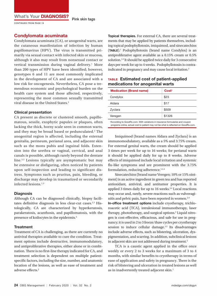
What’s Your DIAGNOSIS?
24 OBG Management | February 2020 | Vol. 32 No. 2 mdedge.com/obgyn
Pink skin tagsCONTINUED FROM PAGE 19
Condylomata acuminataCondylomata acuminata (CA), or anogenital warts, are the cutaneous manifestation of infection by human papillomavirus (HPV). The virus is transmitted pri-marily via sexual contact with infected skin or mucosa, although it also may result from nonsexual contact or vertical transmission during vaginal delivery.1 More than 200 types of HPV have been identified; however, genotypes 6 and 11 are most commonly implicated in the development of CA and are associated with a low risk for oncogenesis. Nevertheless, CA pose a tre-mendous economic and psychological burden on the health care system and those affected, respectively, representing the most common sexually transmitted viral disease in the United States.2
Clinical presentationCA present as discrete or clustered smooth, papillo-matous, sessile, exophytic papules or plaques, often lacking the thick, horny scale seen in common warts, and they may be broad based or pedunculated.2 The anogenital region is affected, including the external genitalia, perineum, perianal area, and adjacent skin such as the mons pubis and inguinal folds. Exten-sion into the urethra or vaginal, cervical, and anal canals is possible, although rarely beyond the dentate line.2,3 Lesions typically are asymptomatic but may be extensive or disfiguring, often noticed by patients upon self-inspection and leading to significant dis-tress. Symptoms such as pruritus, pain, bleeding, or discharge may develop in traumatized or secondarily infected lesions.1,3
DiagnosisAlthough CA can be diagnosed clinically, biopsy facili-tates de�nitive diagnosis in less clear-cut cases.1,3 His-tologically, CA are characterized by hyperkeratosis, parakeratosis, acanthosis, and papillomatosis, with the presence of koilocytes in the epidermis.2
TreatmentTreatment of CA is challenging, as there are currently no antiviral therapies available to cure the condition. Treat-ment options include destructive, immunomodulatory, and antiproliferative therapies, either alone or in combi-nation. �ere is no �rst-line therapy indicated for CA, and treatment selection is dependent on multiple patient-speci�c factors, including the size, number, and anatomic location of the lesions, as well as ease of treatment and adverse e�ects.2
Topical therapies. For external CA, there are several treat-ments that may be applied by patients themselves, includ-ing topical podophyllotoxin, imiquimod, and sinecatechins (TABLE).1 Podophyllotoxin (brand name Condylox) is an antiproliferative agent available as a 0.15% cream or 0.5% solution.1,2 It should be applied twice daily for 3 consecutive days per week for up to 4 weeks. Podophyllotoxin is contra-indicated in pregnancy and may cause local irritation.2
Imiquimod (brand names Aldara and Zyclara) is an immunomodulatory, available as a 5% and 3.75% cream. For external genital warts, the cream should be applied 3 times per week for up to 16 weeks; for perianal warts it should be applied daily for up to 8 weeks. Adverse e�ects of imiquimod include local irritation and systemic �u-like symptoms and are prominent with the 3.75% formulation, reducing adherence.1,2,4
Sinecatechins (brand name Veregen; 10% or 15% oint-ment) is an active ingredient in green tea and has reported antioxidant, antiviral, and antitumor properties. It is applied 3 times daily for up to 16 weeks.2,4 Local reactions may occur and, rarely, severe reactions such as vulvovagi-nitis and pelvic pain, have been reported in women.2,4 In-of�ce treatment options include cryotherapy, trichlo-roacetic acid (TCA), intralesional immunotherapy, laser therapy, phototherapy, and surgical options.2 Liquid nitro-gen is cost-e�ective, e�cacious, and safe for use in preg-nancy; it is used in 2 to 3 freeze/thaw cycles per cryotherapy session to induce cellular damage.1,2 Its disadvantages include adverse e�ects, such as blistering, ulceration, dys-pigmentation, and scarring. In addition, subclinical lesions in adjacent skin are not addressed during treatment.2
TCA is a caustic agent applied in the o�ce once weekly or every 2 to 3 weeks for a maximum of 3 to 4 months, with similar bene�ts to cryotherapy in terms of ease of application and safety in pregnancy. �ere is the risk of blistering and ulceration in treated lesions as well as in inadvertently treated adjacent skin.1
TABLE Estimated cost of patient-applied medications for anogenital warts
Medication (Brand name) Costa
Condylox $23
Aldara $17
Zyclara $509
Veregen $1326
a According to GoodRx.com. With variations in insurance formularies and coupon programs online, actual cost to patient may not be what is re�ected on GoodRx.com.
WYD Kaunitz 0220.indd 24 1/31/20 3:40 PM
mdedge.com/obgyn Vol. 32 No. 2 | February 2020 | OBG Management 25
Intralesional immunotherapy with Candida antigen (brand name Candin) is used in 3 sessions 4 to 6 weeks apart and is safe, with minimal adverse e�ects.2
Laser therapy treatment options include carbon dioxide laser therapy and ND:YAG laser. �eir use is lim-ited, however, by availability and cost.1,2
CA may be removed surgically via shave excision, scissor excision, curettage, and electrosurgery. �ese procedures can be painful, however, requiring local anes-thesia and having a prolonged healing course.1,2
CA recurrenceCA unfortunately has a high rate of recurrence despite treatment, and patients require extensive counseling. Patients should be screened for other sexually transmit-ted infections and advised to notify their sexual partners. If followed properly, safe sexual practices, including condom use and limiting sexual partners, may prevent further transmission.1 �e quadrivalent HPV vaccine
(e�ective for the prevention of infection with HPV geno-types 6, 11, 16, and 18 in unexposed individuals) is inef-fective in treating patients with pre-existing CA but can protect against the acquisition of other HPV genotypes included in the vaccine.1,5
Arriving at the diagnosisAcrochordons are a common skin �nding in the groin, but the onset is more gradual and the individual lesions tend to be more pedunculated. Molluscum is also on the di�erential and can a�ect the genitalia. Molluscum lesions have a characteristic central dimple or dell, which is absent in CA.
CASE Treatment courseThe patient was treated with successive sessions of cryo-
therapy in combination with a course of topical imiquimod
followed by several injections with Candida antigen, with
persistence of some lesions as well as recurrence.
References1. Steben M, Garland SM. Genital warts. Best Prac Res Clin Obstet Gynaecol.
2014;28:1063-1073.2. Fathi R, Tsoukas MM. Genital warts and other HPV infections: established and
novel therapies. Clin Dermatol. 2014;32:299-306.3. Lynde C, Vender R, Bourcier M, et al. Clinical features of external genital warts.
J Cutan Med Surg. 2013;17 (suppl 2):S55-60.
4. Scheinfeld N. Update on the treatment of genital warts. Dermatol Online J.2013;19:18559.
5. Markowitz LE, Dunne EF, Saraiya M, et al; Centers for Disease Control andPrevention (CDC); Advisory Committee on Immunization Practices (ACIP).Quadrivalent Human Papillomavirus Vaccine: recommendations of the Advisory Committee on Immunization Practices (ACIP). MMWR Recomm Rep. 2007;56:1-24.
WYD Kaunitz 0220.indd 25 1/31/20 3:40 PM
COMMENTARY
26 OBG Management | February 2020 | Vol. 32 No. 2 mdedge.com/obgyn
Should supplemental MRI be used in otherwise average-risk women with extremely dense breasts? Recent data show that supplemental MRI screening in women with extremely dense breasts and negative screening mammograms decreases the rate of interval breast cancers. It remains unclear, however, whether supplemental MRIs will improve other outcomes, such as breast cancer mortality.
W hile the frequency of dense breasts decreases with age, approximately
10% of women in the United States have extremely dense breasts (Breast Imaging, Reporting, and Data Sys-tem [BI-RADS] category D), and another 40% have heterogeneously dense breasts (BI-RADS category C).1 Women with dense breasts have both an increased risk for developing breast cancer and reduced mammo-graphic sensitivity for breast cancer detection compared with women who have nondense breasts.2
�ese 2 observations have led the majority of states to pass legis-lation requiring that women with dense breasts be informed of their breast density, and most require that providers discuss these results with
their patients. �oughtful clinicians who review the available literature, however, will �nd sparse evidence on which to counsel patients as to next steps.
Now, a recent trial adds to our knowledge about supplemental magnetic resonance imaging (MRI) breast screening in women with extremely dense breasts.
DENSE trial offers high-quality dataBakker and colleagues studied women aged 50 to 74 who were par-ticipating in a Netherlands popula-tion-based biennial mammography screening program.3 �ey enrolled average-risk women with extremely dense breasts who had a negative screening digital mammogram into the Dense Tissue and Early Breast Neoplasm Screening (DENSE)
multicenter trial. �e women were randomly assigned to receive either continued biennial digital mammog-raphy or supplemental breast MRI.
�e primary outcome was the between-group di�erence in the development of interval breast can-cers—that is, breast cancers detected by women or their providers between rounds of screening mammography. Interval breast cancers were chosen as the primary outcome for 2 reasons:• interval cancers appear to be more
aggressive tumors than thosecancers detected by screeningmammography
• interval cancers can be identi-�ed over a shorter time interval,making them easier to study thanoutcomes such as breast cancermortality, which typically requiremore than a decade to identify.
�e DENSE trial’s secondary outcomes included recall rates from
Mark D. Pearlman, MDDirector, Cancer Genetics and Breast Health S. Jan Behrman Professor, Department of Obstetrics and Gynecology University of Michigan Medical School Ann Arbor, Michigan Committee Member, National Comprehensive Cancer Network Writing Group for Breast Cancer Screening and Diagnosis OBG MANAGEMENT Contributing Editor
The author reports no �nancial relationships rel-evant to this article.
Commentary Pearlman 0220.indd 26 1/31/20 3:42 PM
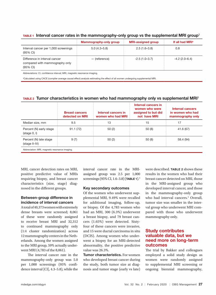
mdedge.com/obgyn Vol. 32 No. 2 | February 2020 | OBG Management 27
MRI, cancer detection rates on MRI, positive predictive value of MRIs requiring biopsy, and breast cancer characteristics (size, stage) diag-nosed in the di�erent groups.
Between-group difference in incidence of interval cancersA total of 40,373 women with extremely dense breasts were screened; 8,061 of these were randomly assigned to receive breast MRI and 32,312 to continued mammography only (1:4 cluster randomization) across 12 mammography centers in the Neth-erlands. Among the women assigned to the MRI group, 59% actually under-went MRI (4,783 of the 8,061).
�e interval cancer rate in the mammography-only group was 5.0 per 1,000 screenings (95% con�-dence interval [CI], 4.3–5.8), while the
interval cancer rate in the MRI-assigned group was 2.5 per 1,000 screenings (95% CI, 1.6–3.8) (TABLE 1).3
Key secondary outcomesOf the women who underwent sup-plemental MRI, 9.49% were recalled for additional imaging, follow-up, or biopsy. Of the 4,783 women who had an MRI, 300 (6.3%) underwent a breast biopsy, and 79 breast can-cers (1.65%) were detected. Sixty-four of these cancers were invasive, and 15 were ductal carcinoma in situ (DCIS). Among women who under-went a biopsy for an MRI-detected abnormality, the positive predictive value was 26.3%.Tumor characteristics. For women who developed breast cancer during the study, both tumor size at diag-nosis and tumor stage (early vs late)
were described. TABLE 2 shows these results in the women who had their breast cancer detected on MRI, those in the MRI-assigned group who developed interval cancer, and those in the mammography-only group who had interval cancers.3 Overall, tumor size was smaller in the inter-val group who underwent MRI com-pared with those who underwent mammography only.
Study contributes valuable data, but we need more on long-term outcomes�e trial by Bakker and colleagues employed a solid study design as women were randomly assigned to supplemental MRI screening or ongoing biennial mammography,
TABLE 1 Interval cancer rates in the mammography-only group vs the supplemental MRI group3
Mammography-only group MRI-assigned group If all had MRIa
Interval cancer per 1,000 screenings (95% CI)
5.0 (4.3–5.8) 2.5 (1.6–3.8) 0.8
Difference in interval cancer compared with mammography only (95% CI)
— (reference) -2.5 (1.0–3.7) -4.2 (2.0–6.4)
Abbreviations: CI, con�dence interval; MRI, magnetic resonance imaging.
aCalculated using CACE (complier average causal effect) analysis estimating the effect of all women undergoing supplemental MRI.
TABLE 2 Tumor characteristics in women who had mammography only vs supplemental MRI3
Breast cancers detected on MRI
Interval cancers in women who had MRI
Interval cancers in women who were
assigned to but did not have MRI
Interval cancers in women who had mammography only
Median size, mm 9.5 13 15 17
Percent (N) early stage (stage 0, I)
91.1 (72) 50 (2) 50 (8) 41.6 (67)
Percent (N) late stage (stage II–IV)
9 (7) 50 (2) 50 (8) 58.4 (94)
Abbreviation: MRI, magnetic resonance imaging.
Commentary Pearlman 0220.indd 27 1/31/20 3:42 PM
COMMENTARY
28 OBG Management | February 2020 | Vol. 32 No. 2 mdedge.com/obgyn
and nearly all cancers were identi-�ed in the short-term of follow-up. In addition, very few women were lost to follow-up, and secondary out-comes, including false-positive rates, were collected to help providers and patients better understand some of the potential downsides of supple-mental screening.
�e substantial reduction in interval cancers (50% in the intent-to-screen analysis and 84% in the women who actually underwent sup-plemental MRI) was highly statisti-cally signi�cant (P<.001). While there were substantially fewer interval cancers in the MRI-assigned group, the interval cancers that did occur were of similar stage as those in the women assigned to the mammogra-phy-only group (TABLE 2).
Data demonstrate that interval cancers appear to be more aggressive than screen-detected cancers.4 While reducing interval cancers should be a good thing overall, it remains unproven that using supplemental MRI in all women with dense breasts would reduce breast cancer speci�c mortality, all-cause mortality, or the risk of more invasive treatments (for example, the need for chemotherapy or requirement for mastectomy).
On the other hand, using routine supplemental breast MRI in women with extremely dense breasts would result in very substantial use of resources, including cost, radiologist time, provider time, and machine time. In the United States, approxi-mately 49 million women are aged 50 to 74.5 Breast MRI charges commonly range from $1,000 to $4,000. If the 4.9 million women with extremely dense breasts underwent supple-mental MRI this year, the approxi-mate cost would be somewhere between $4.9 and $19.5 billion for imaging alone. �is does not include callbacks, biopsies, or provider
time for ordering, interpreting, and arranging for follow-up.
While the reduction in interval cancers seen in this study is promis-ing, more assurance of improvement in important outcomes—such as reduced mortality or reduced need for more invasive breast cancer treat-ments—should precede any routine change in practice.
Unanswered questions�is study did not address a num-ber of other important questions, including:Should MRI be done with every round of breast cancer screen-ing given the possibility of preva-lence bias? Prevalence bias can be de�ned as more cancers detected in the �rst round of MRI screen-ing with possible reduced bene�t in future rounds of screening. �e study authors indicated that they will con-tinue to analyze the study results to see what occurs in the next round of screening.Is there a similar impact on decreased interval cancers in women undergoing annual mam-mography or in women screened between ages 40 and 49? �is study was conducted in women aged 50 to 74 undergoing mammography every 2 years. In the United States, annual mammography in women aged 40 to 49 is frequently recommended.What effect does supplemental MRI screening have in women with heterogeneously dense breasts, which represents 40% of the population? �e US Food and Drug Administration recommends that all women with dense breasts be counseled regarding options for management.6
Do these results translate to the more racially and ethnically diverse populations of the United States? In the Netherlands, where this study
was conducted, 85% to 90% of women are either Dutch or of western Euro-pean origin. Women of di�erent racial and ancestral backgrounds have bio-logically di�erent breast cancers and cancer risk (for example, higher rates of triple-negative breast cancers in African American women; 10-fold higher rates of BRCA pathogenic vari-ants in Ashkenazi Jewish women).
Use validated tools to assess risk comprehensivelyWomen aged 50 to 74 with extremely dense breasts have reduced interval cancers following a normal biennial mammogram if supplemental MRI is o�ered, but the long-term bene�t of identifying these cancers earlier is unclear. Until more data are available on important long-term outcomes (such as breast cancer mortality and need for more invasive treatments), providers should consider breast density in the context of a more comprehensive assessment of breast cancer risk using a validated breast cancer risk assessment tool.
I prefer the modi�ed version of the International Breast Cancer Intervention Study (IBIS) tool, which is readily available online (https://ibis.ikonopedia.com/).7 �is tool incorporates several breast cancer risk factors, including reproductive risk factors, body mass index, BRCA gene status, breast density, and fam-ily history. �e tool takes 1 to 2 min-utes to complete and provides an estimate of a woman’s 10-year risk and lifetime risk of breast cancer.
If the lifetime risk exceeds 20%, I o�er the patient supplemental MRI screening, consistent with current recommendations of the National Comprehensive Cancer Network and the American Cancer Society.8,9 I generally recommend starting breast
Commentary Pearlman 0220.indd 28 1/31/20 3:42 PM
mdedge.com/obgyn Vol. 32 No. 2 | February 2020 | OBG Management 29
imaging screening 7 to 10 years prior to the youngest breast cancer occur-rence in the family, with mammog-raphy starting no earlier than age 30
and MRI no earlier than age 25. Other validated tools also can be used.10-13
Incorporating breast density and other important risk factors allows a
more comprehensive analysis upon which to counsel women about the value (bene�ts and harms) of breast imaging.8
References1. Sprague BL, Gagnon RE, Burt V, et al. Prevalence
of mammographically dense breasts in the UnitedStates. J Natl Cancer Inst. 2014;106:dju255. doi:10.1093/jcni/dju255.
2. Boyd NF, Guo H, Martin LJ, et al. Mammographic density and the risk and detection of breast can-cer. N Engl J Med. 2007;356:227-236.
3. Bakker MF, de Lange SV, Pijnappel RM, et al; for the DENSE Trial Study Group. Supplemental MRIscreening for women with extremely dense breast tissue. N Engl J Med. 2019;381:2091-2102.
4. Drukker CA, Schmidt MK, Rutgers EJT, et al.Mammographic screening detects low-risk tumorbiology breast cancers. Breast Cancer Res Treat.2014;144:103-111.
5. Statista website. Resident population of theUnited States by sex and age as of July 1, 2018.
https://www.statista.com/statistics/241488/population-of-the-us-by-sex-and-age. Accessed January 6, 2020.
6. US Food and Drug Administration website. Mam-mography: what you need to know. https://www .fda.gov/consumers/consumer-updates/mam mography-what-you-need-know. Accessed Janu-ary 13, 2020.
7. IBIS (International Breast Cancer InterventionStudy) website. Online Tyrer-Cuzick Model Breast Cancer Risk Evaluation Tool. ibis.ikonopedia.com. Accessed January 13, 2020.
8. Bevers TB, Anderson BO, Bonaccio E, et al;National Comprehensive Cancer Network. Breast cancer screening and diagnosis: NCCN practiceguidelines in oncology. JNCCN. 2009;7:1060-1096.
9. Saslow D, Boetes C, Burke W, et al. American Can-
cer Society guidelines for breast screening with MRI as an adjunct to mammography. CA Cancer J Clin. 2007;57:75-89.
10. Antoniou AC, Cunningham AP, Peto J, et al. �eBOADICEA model of genetic susceptibility tobreast and ovarian cancers: updates and exten-sions. Br J Cancer. 2008;98:1457-1466.
11. Claus EB, Risch N, �ompson WD. Autosomal domi-nant inheritance of early-onset breast cancer: impli-cations for risk prediction. Cancer. 1994;73:643-651.
12. Parmigiani G, Berry D, Aguilar O. Determiningcarrier probabilities for breast cancer-suscepti-bility genes BRCA1 and BRCA2. Am J Hum Genet. 1998;62:145-158.
13. Tyrer J, Du�y SW, Cuzick J. A breast cancer predic-tion model incorporating familial and personalrisk factors. Stat Med. 2004;23:1111-1130.
Commentary Pearlman 0220.indd 29 1/31/20 3:42 PM
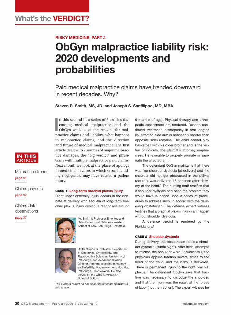
What’s the VERDICT?
IN THISARTICLE
30 OBG Management | February 2020 | Vol. 32 No. 2 mdedge.com/obgyn
Malpractice trendspage 31
Claims payoutspage 32
Claims data observationspage 37
RISKY MEDICINE, PART 2
ObGyn malpractice liability risk: 2020 developments and probabilities
Paid medical malpractice claims have trended downward in recent decades. Why?
Steven R. Smith, MS, JD, and Joseph S. San�lippo, MD, MBA
In this second in a series of 3 articles dis-cussing medical malpractice and the ObGyn we look at the reasons for mal-
practice claims and liability, what happens to malpractice claims, and the direction and future of medical malpractice. �e �rst article dealt with 2 sources of major malprac-tice damages: the “big verdict” and physi-cians with multiple malpractice paid claims. Next month we look at the place of apology in medicine, in cases in which error, includ-ing negligence, may have caused a patient injury.
CASE 1 Long-term brachial plexus injuryRight upper extremity injury occurs in the neo-
nate at delivery with sequela of long-term bra-
chial plexus injury (which is diagnosed around
6 months of age). Physical therapy and ortho-
pedic assessment are rendered. Despite con-
tinued treatment, discrepancy in arm lengths
(ie, affected side arm is noticeably shorter than
opposite side) remains. The child cannot play
basketball with his older brother and is the vic-
tim of ridicule, the plaintiff’s attorney empha-
sizes. He is unable to properly pronate or supi-
nate the affected arm.
The defendant ObGyn maintains that there
was “no shoulder dystocia [at delivery] and the
shoulder did not get obstructed in the pelvis;
shoulder was delivered 15 seconds after deliv-
ery of the head.” The nursing staff testi�es that
if shoulder dystocia had been the problem they
would have launched upon a series of proce-
dures to address such, in accord with the deliv-
ering obstetrician. The defense expert witness
testi�es that a brachial plexus injury can happen
without shoulder dystocia.
A defense verdict is rendered by the
Florida jury.1
CASE 2 Shoulder dystociaDuring delivery, the obstetrician notes a shoul-
der dystocia (“turtle sign”). After initial attempts
to release the shoulder were unsuccessful, the
physician applies traction several times to the
head of the child, and the baby is delivered.
There is permanent injury to the right brachial
plexus. The defendant ObGyn says that trac-
tion was necessary to dislodge the shoulder,
and that the injury was the result of the forces
of labor (not the traction). The expert witness for
Mr. Smith is Professor Emeritus and Dean Emeritus at California Western School of Law, San Diego, California.
Dr. San�lippo is Professor, Department of Obstetrics, Gynecology, and Reproductive Sciences, University of Pittsburgh, and Academic Division Director, Reproductive Endocrinology and Infertility, Magee-Womens Hospital, Pittsburgh, Pennsylvania. He also serves on the OBG MANAGEMENT Board of Editors.
The authors report no �nancial relationships relevant to this article.
WTV 0220.indd 30 1/31/20 3:43 PM
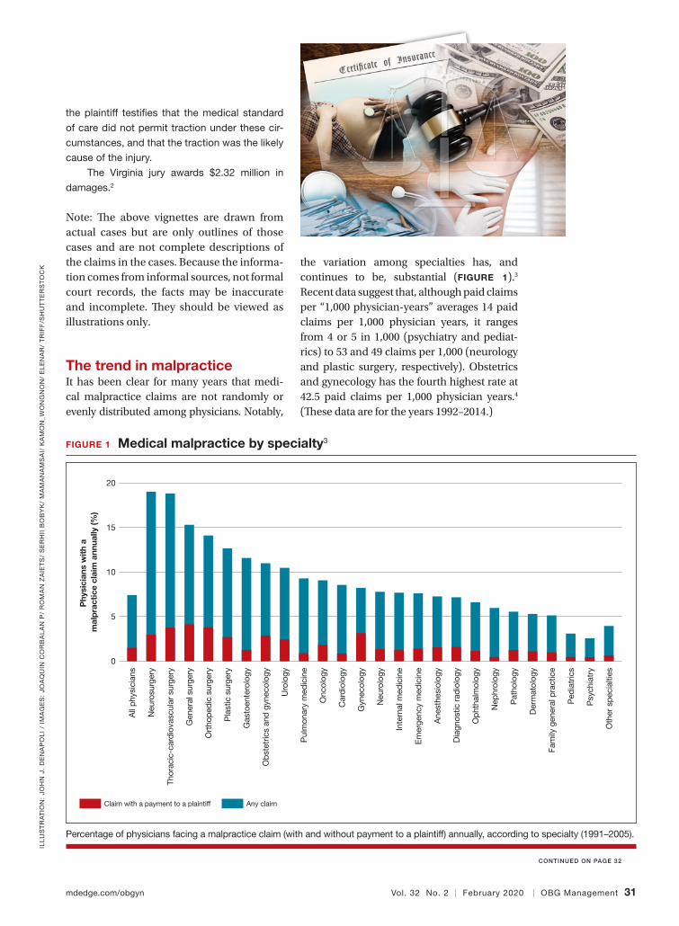
mdedge.com/obgyn Vol. 32 No. 2 | February 2020 | OBG Management 31
ILL
US
TR
AT
ION
: JO
HN
J.
DE
NA
PO
LI
/ IM
AG
ES
: JO
AQ
UIN
CO
RB
AL
AN
P/
RO
MA
N Z
AIE
TS
/ S
ER
HII
BO
BY
K/
MA
MA
NA
MS
AI/
KA
MO
N_W
ON
GN
ON
/ E
LE
NA
R/
TR
IFF
/SH
UT
TE
RS
TO
CK
CONTINUED ON PAGE 32
the plaintiff testi�es that the medical standard
of care did not permit traction under these cir-
cumstances, and that the traction was the likely
cause of the injury.
The Virginia jury awards $2.32 million in
damages.2
Note: �e above vignettes are drawn from actual cases but are only outlines of those cases and are not complete descriptions of the claims in the cases. Because the informa-tion comes from informal sources, not formal court records, the facts may be inaccurate and incomplete. �ey should be viewed as illustrations only.
The trend in malpractice It has been clear for many years that medi-cal malpractice claims are not randomly or evenly distributed among physicians. Notably,
the variation among specialties has, and continues to be, substantial (FIGURE 1).3
Recent data suggest that, although paid claims per “1,000 physician-years” averages 14 paid claims per 1,000 physician years, it ranges from 4 or 5 in 1,000 (psychiatry and pediat-rics) to 53 and 49 claims per 1,000 (neurology and plastic surgery, respectively). Obstetrics and gynecology has the fourth highest rate at 42.5 paid claims per 1,000 physician years.4
(�ese data are for the years 1992–2014.)
FIGURE 1 Medical malpractice by specialty3
Claim with a payment to a plaintiff
Phy
sici
ans
with
a
mal
pra
ctic
e cl
aim
ann
ually
(%)
Any claim
20
15
10
5
0
All
phys
icia
ns
Neu
rosu
rger
y
Thor
acic
-car
diov
ascu
lar
surg
ery
Gen
eral
sur
gery
Ort
hope
dic
surg
ery
Pla
stic
sur
gery
Gas
toen
tero
logy
Obs
tetr
ics
and
gyne
colo
gy
Uro
logy
Pul
mon
ary
med
icin
e
Onc
olog
y
Car
diol
ogy
Gyn
ecol
ogy
Neu
rolo
gy
Inte
rnal
med
icin
e
Em
erge
ncy
med
icin
e
Ane
sthe
siol
ogy
Dia
gnos
tic r
adio
logy
Oph
thal
mol
ogy
Nep
hrol
ogy
Pat
holo
gy
Der
mat
olog
y
Fam
ily g
ener
al p
ract
ice
Ped
iatr
ics
Psy
chia
try
Oth
er s
peci
altie
s
Percentage of physicians facing a malpractice claim (with and without payment to a plaintiff) annually, according to specialty (1991–2005).
WTV 0220.indd 31 1/31/20 3:43 PM
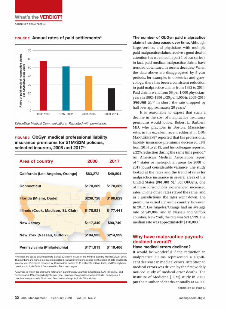
What’s the VERDICT?
32 OBG Management | February 2020 | Vol. 32 No. 2 mdedge.com/obgyn
CONTINUED FROM PAGE 31
CONTINUED ON PAGE 34
The number of ObGyn paid malpractice claims has decreased over time. Although large verdicts and physicians with multiple paid malpractice claims receive a good deal of attention (as we noted in part 1 of our series), in fact, paid medical malpractice claims have trended downward in recent decades.5 When the data above are disaggregated by 5-year periods, for example, in obstetrics and gyne-cology, there has been a consistent reduction in paid malpractice claims from 1992 to 2014. Paid claims went from 58 per 1,000 physician-years in 1992–1996 to 25 per 1,000 in 2009–2014 (FIGURE 2).4,6 In short, the rate dropped by half over approximately 20 years.4
It is reasonable to expect that such a decline in the cost of malpractice insurance premiums would follow. Robert L. Barbieri, MD, who practices in Boston, Massachu-setts, in his excellent recent editorial in OBG Management6 reported that his professional liability insurance premiums decreased 18% from 2014 to 2019, and his colleague reported a 22% reduction during the same time period.6
An American Medical Association report of 7 states or metropolitan areas for 2008 to 2017 found considerable variance. �e study looked at the rates and the trend of rates for malpractice insurance in several areas of the United States (FIGURE 3).7 For ObGyns, one of these jurisdictions experienced increased rates; in one other, rates stayed the same, and in 5 jurisdictions, the rates went down. �e premiums varied across the country, however. In 2017, Los Angeles/Orange had an average rate of $49,804, and in Nassau and Su�olk counties, New York, the rate was $214,999. �e median rate was approximately $170,000.7
Why have malpractice payouts declined overall?Have medical errors declined? It would be wonderful if the reduction in malpractice claims represented a signi�-cant decrease in medical errors. Attention to medical errors was driven by the �rst widely noticed study of medical error deaths. �e Institute of Medicine (IOM) study in 2000, put the number of deaths annually at 44,000
FIGURE 2 Annual rates of paid settlements6
Rat
es o
f pai
d m
edic
al m
alp
ract
ice
clai
ms
per
1,0
00 p
hysi
cian
-yea
rs
70
60
50
40
30
20
10
01992–1996 1997–2002 2003–2008 2009–2014
©Frontline Medical Communications. Reprinted with permission.
FIGURE 3 ObGyn medical professional liability insurance premiums for $1M/$3M policies, selected insurers, 2008 and 2017a,7
Area of country
California (Los Angeles, Orange)
Connecticut
Florida (Miami, Dade)
Illinois (Cook, Madison, St. Clair)
New Jersey
New York (Nassau, Suffolk)
Pennsylvania (Philadelphia)
2008
$63,272
$170,389
$238,728
$178,921
$117,340
$194,935
$171,813
2017
$49,804
$170,389
$190,829
$177,441
$90,749
$214,999
$119,466
aThe data are based on Annual Rate Survey (October) Issues of the Medical Liability Monitor, 2008–2017. The numbers are manual premiums reported by a liability insurer selected on the basis of data availability in every year. Premiums reported for Connecticut pertain to $1 million/$4 million limits, and Pennsylvania premiums include Patient Compensation Fund surcharges.
bCounties to which the premiums refer are in parentheses. Counties in California (CA), Illinois (IL), and Pennsylvania (PA) changed slightly over time. However, CA counties always include Los Angeles, IL counties always include Cook, and PA counties always include Philadelphia.
WTV 0220.indd 32 1/31/20 3:43 PM

Enhancing patient outcomes, managing costs, and optimizing delivery of care.
©2019 Laboratory Corporation of America® Holdings All rights reserved. 19875-0119
LabCorp’s NuSwab® portfolio combines cost-e�ective, clinically relevant, research-driven testing with the convenience of a single-swab collection and is a targeted approach to testing for bacterial vaginosis, yeast (Candida), and four common STDs.
Ampli�ed molecular target testing (NuSwab) has been shown to exceed non-ampli�ed testing by:1
• Identifying an additional 20-25% of symptomaticpatients with suspected vulvovaginitis (VV). Which in turn helps:
• Lower odds of preterm labor or delivery forsymptomatic pregnant patients by 15%
• Lower per patient expenditure of $268 duringfollow up period
• Lower odds of all-cause hospitalization by 22%
Value beyond testing.
For more information, please visitwww.labcorp.com/value-care-vaginitis
The value of care:VAGINITIS TESTING
1. Ackerman SJ, Wahl PM, Knight T, Cartwright, CP. Healthcare Resource Utilization and Costs of Ampli�ed Versus Non-ampli�ed Molecular Probe Testing for Vaginitis/Vaginosis: A U. S. Commercial Payer Perspective. LabCorp.
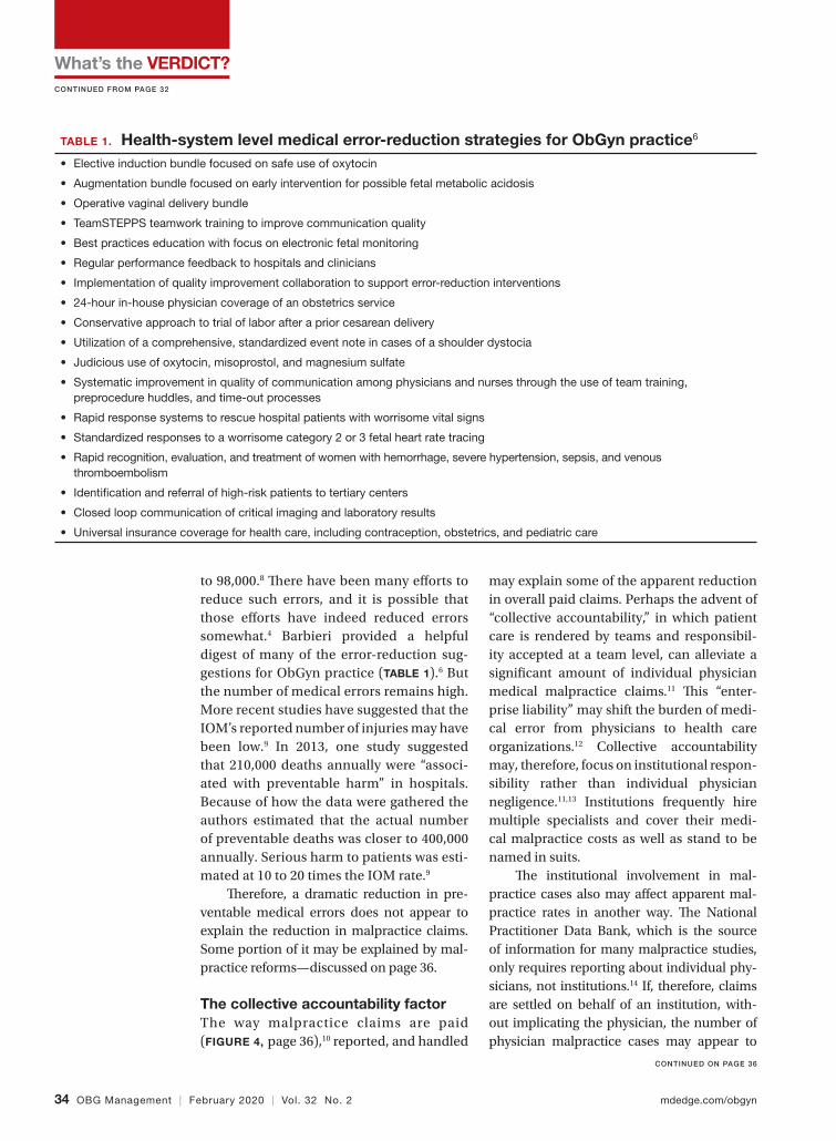
What’s the VERDICT?
34 OBG Management | February 2020 | Vol. 32 No. 2 mdedge.com/obgyn
CONTINUED FROM PAGE 32
CONTINUED ON PAGE 36
to 98,000.8 �ere have been many e�orts to reduce such errors, and it is possible that those e�orts have indeed reduced errors somewhat.4 Barbieri provided a helpful digest of many of the error-reduction sug-gestions for ObGyn practice (TABLE 1).6 But the number of medical errors remains high. More recent studies have suggested that the IOM’s reported number of injuries may have been low.9 In 2013, one study suggested that 210,000 deaths annually were “associ-ated with preventable harm” in hospitals. Because of how the data were gathered the authors estimated that the actual number of preventable deaths was closer to 400,000 annually. Serious harm to patients was esti-mated at 10 to 20 times the IOM rate.9
�erefore, a dramatic reduction in pre-ventable medical errors does not appear to explain the reduction in malpractice claims. Some portion of it may be explained by mal-practice reforms—discussed on page 36.
The collective accountability factorThe way malpractice claims are paid (FIGURE 4, page 36),10 reported, and handled
may explain some of the apparent reduction in overall paid claims. Perhaps the advent of “collective accountability,” in which patient care is rendered by teams and responsibil-ity accepted at a team level, can alleviate a signi�cant amount of individual physician medical malpractice claims.11 �is “enter-prise liability” may shift the burden of medi-cal error from physicians to health care organizations.12 Collective accountability may, therefore, focus on institutional respon-sibility rather than individual physician negligence.11,13 Institutions frequently hire multiple specialists and cover their medi-cal malpractice costs as well as stand to be named in suits.
�e institutional involvement in mal-practice cases also may a�ect apparent mal-practice rates in another way. �e National Practitioner Data Bank, which is the source of information for many malpractice studies, only requires reporting about individual phy-sicians, not institutions.14 If, therefore, claims are settled on behalf of an institution, with-out implicating the physician, the number of physician malpractice cases may appear to
TABLE 1. Health-system level medical error-reduction strategies for ObGyn practice6 • Elective induction bundle focused on safe use of oxytocin
• Augmentation bundle focused on early intervention for possible fetal metabolic acidosis
• Operative vaginal delivery bundle
• TeamSTEPPS teamwork training to improve communication quality
• Best practices education with focus on electronic fetal monitoring
• Regular performance feedback to hospitals and clinicians
• Implementation of quality improvement collaboration to support error-reduction interventions
• 24-hour in-house physician coverage of an obstetrics service
• Conservative approach to trial of labor after a prior cesarean delivery
• Utilization of a comprehensive, standardized event note in cases of a shoulder dystocia
• Judicious use of oxytocin, misoprostol, and magnesium sulfate
• Systematic improvement in quality of communication among physicians and nurses through the use of team training,preprocedure huddles, and time-out processes
• Rapid response systems to rescue hospital patients with worrisome vital signs
• Standardized responses to a worrisome category 2 or 3 fetal heart rate tracing
• Rapid recognition, evaluation, and treatment of women with hemorrhage, severe hypertension, sepsis, and venousthromboembolism
• Identi�cation and referral of high-risk patients to tertiary centers
• Closed loop communication of critical imaging and laboratory results
• Universal insurance coverage for health care, including contraception, obstetrics, and pediatric care
WTV 0220.indd 34 1/31/20 3:43 PM

OPPORTUNITIES FOR CME AND MOC IN 2020
MARCH 16-19, 2020LAS VEGAS
MISS-CME.org
Earn up to 30 CME Credits +
MOC
To register and for complete information, including accreditation information, please see our websites.
MARCH 27-28, 2020WASHINGTON, DC
DDNAcme.com
Earn up to 14.25 CME/CE
Credits + MOC
APRIL 2-3, 2020 | LA JOLLA, CAAcuteLeukemiaForum.com
Earn up to 9.5 AMA PRA
Category 1 Credits™
SKIN DISEASE EDUCATION FOUNDATION PRESENTS
16TH ANNUAL
GlobalAcademyCME.com/WPD
JUNE 19-20, 2020 • Newport Beach, CA
CPAACP-CME.com
MARCH 26–28, 2020 OPTIONAL POST-CONFERENCE WORKSHOPS MARCH 28
CHICAGO, IL
Earn up to 22 CME Credits +
MOC
SAVE THE
DATE!
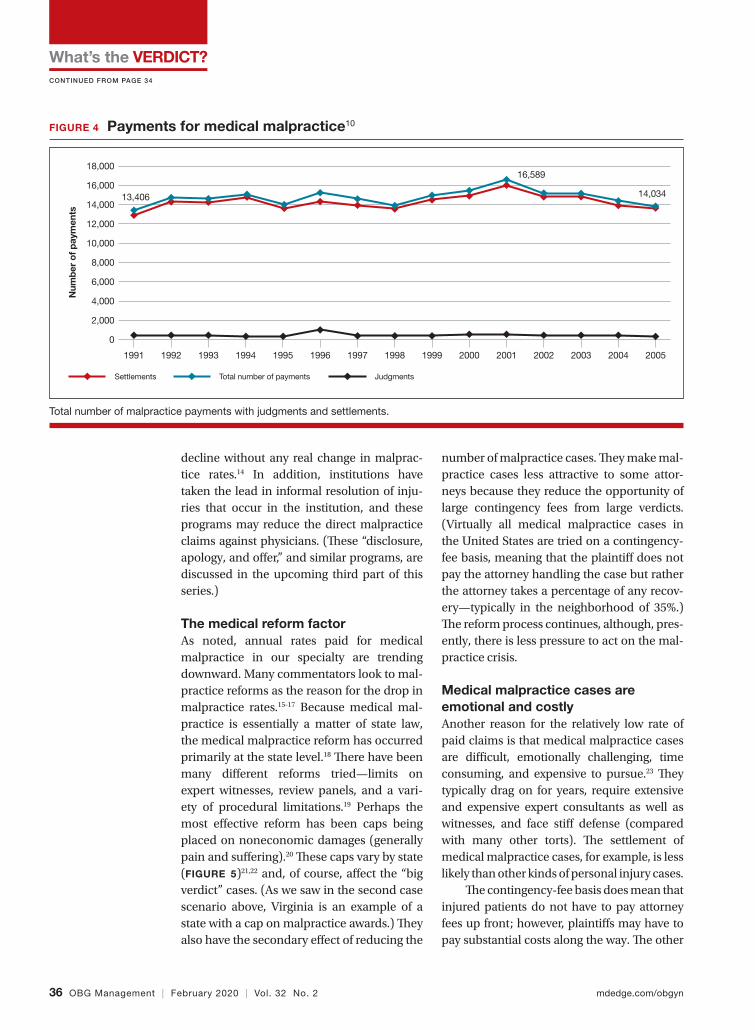
What’s the VERDICT?
36 OBG Management | February 2020 | Vol. 32 No. 2 mdedge.com/obgyn
CONTINUED FROM PAGE 34
decline without any real change in malprac-tice rates.14 In addition, institutions have taken the lead in informal resolution of inju-ries that occur in the institution, and these programs may reduce the direct malpractice claims against physicians. (�ese “disclosure, apology, and o�er,” and similar programs, are discussed in the upcoming third part of this series.)
The medical reform factorAs noted, annual rates paid for medical malpractice in our specialty are trending downward. Many commentators look to mal-practice reforms as the reason for the drop in malpractice rates.15-17 Because medical mal-practice is essentially a matter of state law, the medical malpractice reform has occurred primarily at the state level.18 �ere have been many di�erent reforms tried—limits on expert witnesses, review panels, and a vari-ety of procedural limitations.19 Perhaps the most e�ective reform has been caps being placed on noneconomic damages (generally pain and su�ering).20 �ese caps vary by state (FIGURE 5)21,22 and, of course, a�ect the “big verdict” cases. (As we saw in the second case scenario above, Virginia is an example of a state with a cap on malpractice awards.) �ey also have the secondary e�ect of reducing the
number of malpractice cases. �ey make mal-practice cases less attractive to some attor-neys because they reduce the opportunity of large contingency fees from large verdicts. (Virtually all medical malpractice cases in the United States are tried on a contingency-fee basis, meaning that the plainti� does not pay the attorney handling the case but rather the attorney takes a percentage of any recov-ery—typically in the neighborhood of 35%.) �e reform process continues, although, pres-ently, there is less pressure to act on the mal-practice crisis.
Medical malpractice cases are emotional and costlyAnother reason for the relatively low rate of paid claims is that medical malpractice cases are di�cult, emotionally challenging, time consuming, and expensive to pursue.23 �ey typically drag on for years, require extensive and expensive expert consultants as well as witnesses, and face sti� defense (compared with many other torts). �e settlement of medical malpractice cases, for example, is less likely than other kinds of personal injury cases.
�e contingency-fee basis does mean that injured patients do not have to pay attorney fees up front; however, plainti�s may have to pay substantial costs along the way. �e other
FIGURE 4 Payments for medical malpractice10
Settlements
Num
ber
of p
aym
ents
1991 1992 1993 1994 1995 1996 1997 1998 1999 2000 2001 2002 2003 2004 2005
18,000
16,000
14,000
12,000
10,000
8,000
6,000
4,000
2,000
0
Total number of payments Judgments
13,406
16,589
14,034
Total number of malpractice payments with judgments and settlements.
WTV 0220.indd 36 1/31/20 3:43 PM

mdedge.com/obgyn Vol. 32 No. 2 | February 2020 | OBG Management 37
side of this coin is that lawyers can be reluctant to take malpractice cases in which the dam-ages are likely to be small, or where the legal uncertainty reduces the odds of achieving any damages. �us, many potential malpractice cases are never �led.
A word of caution �e news of a reduction in malpractice paid claims may not be permanent. �e num-bers can conceivably be cyclical, and political reforms achieved can be changed. In addi-tion, new technology will likely bring new kinds of malpractice claims. �at appears to be the case, for example, with electronic health records (EHRs). One insurer reports that EHR malpractice claims have increased over the last 8 years.24 �e most common injury in these claims was death (25%), as well as a magnitude of less serious injuries. EHR-related claims result from system failures, copy-paste
inaccuracies, faulty drop-down menu use, and uncorrected “auto-populated” �elds. Obstet-rics is tied for �fth on the list of 14 specialties with claims related to EHRs, and gynecology is tied for eighth place.24
A federal court ruled that a hospital that changed from paper records to EHRs for test results had a duty to “‘implement a reason-able procedure during the transition phase’ to ensure the timely delivery of test results” to health care providers.25 We will address this in a future “What’s the Verdict?”.
Rates of harm, malpractice cases, and the disposition of cases�ere are many surprises when looking at medical malpractice claims data generally. �e �rst surprise is how few claims are �led rel-ative to the number of error-related injuries.
FIGURE 5 An overview of noneconomic medical malpractice caps by state21,22
No cap
Ohio has a sliding scale, with awards ranging from $250k–$500k depending on different circumstances
$250k–$400k $500k–$1M $1M+
CONTINUED ON PAGE 38
WTV 0220.indd 37 1/31/20 3:43 PM
What’s the VERDICT?
38 OBG Management | February 2020 | Vol. 32 No. 2 mdedge.com/obgyn
Given the estimate of 210,000 to 400,000 deaths “associated with preventable harm” in hospitals, plus 10 to 20 times that number of serious injuries, it would be reasonable to expect claims of many hundreds of thou-sands per year. Compare the probability of a malpractice claim from an error-related injury, for example, with the probability of other personal injuries—eg, of tra�c deaths associated with preventable harm.
�e second key observation is how many of the claims �led are not successful—even when there was evidence in the record of errors associated with the injury. Studies slice the data in di�erent ways but collectively sug-gest that only a small proportion of malprac-tice claims �led (a claim is generally regarded
as some written demand for compensation for injuries) result in payments, either through settlement or by trial. A 2006 study by Studdert and colleagues determined that 63% of formal malpractice claims �led did involve injuries resulting from errors.26 �e study found that in 16% of the claims (not injuries) there was no payment even though there was error. In 10% of the claims there was payment, even in the absence of error.
Overall, in this study, 56% of the claims received some compensation.26 �at is higher than a more recent study by Jena and others, which found only 22% of claims resulted in compensation.3
How malpractice claims are decided is also interesting. Jena and colleagues found that only 55% of claims resulted in litigation.27
Presumably, the other 45% may have resulted in the plainti� dropping the case, or in some form of settlement. Of the claims that were liti-gated, 54% were dismissed by the court, and another 35% were settled before a trial ver-dict. �e cases that went to trial (about 10%), overwhelmingly (80%) resulted in verdicts for the defense.3,27 A di�erent study found that
TABLE 2 Goals of tort law
1. Compensation: Provide money to cover the costs of those who were
injured by the carelessness of others
2. Deterrence: Reduce injuries (caused by carelessness) by requiring those
harming others to pay for the damages they cause. It is appropriate to
ask, “How effective and just is this system in the United States?”
Why did the 2 opening case vignettes come out differently?
The two vignettes described at the beginning, with similar injuries (shoulder dystocia), had disparate outcomes. In one there was a defense verdict and in the other a verdict for the plaintiffs of more than $2 million. The differences explain a number of important elements related to malpractice claims. (We have only very abbreviated and incomplete descriptions of the cases, so this discussion necessarily assumes facts and jumps to conclusions that may not be entirely consistent with the actual cases.)
These vignettes are unusual in that they went to trial. As we have noted, only a small percentage of malpractice cases are tried. And the verdict for the plaintiff-patient (in the second case) is unusual among those cases that go to trial, where plaintiffs seldom prevail.
From the facts we have, one signi�cant difference in the 2 cases is that the plaintiff’s expert witness speci�cally testi�ed in the second case that the “medical standard of care did not permit traction under these circumstances.” That is an essential element of a successful plaintiff’s malpractice case. In this case, the expert could also draw a connection between that breach of standard of care and harm to the child. In the case without liability, the nursing staff was able to testify that there was no shoulder dystocia because if there had been such an injury, they would have immediately launched into special action, which did not happen. By contrast, in the liability case, there seemed to be critical gaps in the medical record.
It is also important to remember that these cases were tried in different states, with different laws. The juries and judges in the 2 cases were different. Finally, the quality of the attorneys representing the plaintiffs and defendants were different. We mention these factors to point out that medical malpractice is not an exact science. It depends on many human elements that make the outcome of cases somewhat unpredictable. This unpredictability is one reason why parties and attorneys like to settle cases.
CONTINUED FROM PAGE 37
WTV 0220.indd 38 1/31/20 3:43 PM
mdedge.com/obgyn Vol. 32 No. 2 | February 2020 | OBG Management 39
only 9% of cases went to trial, and 87% were a defense verdict.28 �e high level of defense verdicts may suggest that malpractice defense lawyers, and their client physicians, do a good job of assessing cases they are likely to lose, and settling them before trial.
ObGyns generally have larger numbers of claims and among the largest payment amounts when there is payment. Fewer of their cases are dismissed by the courts, so more go to trial. At trial, however, ObGyns prevail at a remarkably high rate.27 As for the probability of payment of a malpractice claim for ObG-yns, one study suggested that there is approxi-mately a 16% annual probability of a claim being �led, but only a 3% annual probability of a payment being made (suggesting about a 20% probability of payment per claim).3
The purposes and effects of the medical malpractice system�e essential goals of tort law (including medical malpractice) include compensation for those who are injured and deterrence of future injuries (TABLE 2). What are the over-all e�ects to the medical malpractice sys-tem? Unfortunately, the answer is that the law delivers disappointing results at best. It has a fairly high error rate. Many people who deserve some compensation for their injuries never seek compensation, and many deserv-ing injured patients fail in e�orts to receive
compensation. At the same time, a few of the injured receive huge recoveries (even wind-falls), and at least a small fraction receive compensation when there was no medical error. In addition to the high error rate, the system is ine�cient and very expensive. Both defendants (through their insurance carri-ers) and plainti�s spend a lot of money, years of time, and untold emotional pain dealing with these cases. �e system also exacts high emotional and personal costs on plainti�s and defendants.
Malpractice reform has not really addressed these issues—it has generally been focused on ways to reduce the cost of malpractice insurance. �e most e�ective reform in reducing rates—caps—has had the e�ect of compensating the most seriously injured as though they were more modestly injured, and dissuading attorneys from tak-ing the cases of those less seriously injured.
�e medical and legal professions exist to help patients (the public). It does not seem that we have arrived at a system that does that very fairly or e�ciently when a patient is injured because of preventable medical error.
Watch for the third and �nal article in this series next month, as we are going to look at “apology in medicine and a proactive response” to communication regarding a complication.
References 1. Shoulder dystocia—Florida defense verdict. Medical
Malpractice: Verdicts, Settlements & Experts. 2019;35(1):18.2. Shoulder dystocia improperly managed—$2.320 million
Virginia verdict. Medical Malpractice: Verdicts, Settlements &Experts. 2019;35(2):13.
3. Jena AB, Seabury S, Lakdawalla D, et al. Malpractice riskaccording to physician specialty. N Engl J Med. 2011;365:629-636.
4. Scha�er AC, Jena AB, Seabury SA, et al. Rates andcharacteristics of paid malpractice claims amongUS physicians by specialty, 1992–2014. JAMA InternMed. 2017;177:710-718.
5. Lowes R. Malpractice premiums trail in�ation for somephysicians. Medscape. December 16, 2016. https://www .medscape.com/viewarticle/873422. Accessed January 10, 2020.
6. Barbieri RL. Good news for ObGyns: medical liability claimsresulting in payment are decreasing! OBG Manag. 2019;31: 10-13.
7. Guardado JR. Medical professional liability insurancepremiums: an overview of the market from 2008 to 2017. AMA Policy Research Perspectives, 2018. https://www.ama-assn.org/sites/ama-assn.org/files/corp/media-browser/public
/government/advocacy/policy-research-perspective-liability-insurance-premiums.pdf. Accessed January 10, 2020.
8. Institute of Medicine Committee on Quality Health Care inAmerica; Kohn LT, Corrigan JM, Donaldson MS, eds. To Erris Human: Building a Safer Health System. Washington, DC:National Academies Press; 2000.
9. James JT. A new, evidence-based estimate of patient harmsassociated with hospital care. J Patient Saf. 2013;9:122-128.https://journals.lww.com/journalpatientsafety/Fulltext/2013/09000/A_New,_Evidence_based_Estimate_of_Patient_Harms.2.aspx. Accessed January 10, 2020.
10. Public Citizen Congress Watch. �e great medical malpractice hoax: NPDB data continue to show medical liability systemproduces rational outcomes. January 2007. https://www.citizen.org/wp-content/uploads/npdb_report_final.pdf. Accessed January 23, 2020.
11. Bell SK, Delbanco T, Anderson-Shaw L, et al. Accountability for medical error: moving beyond blame to advocacy. Chest.2011;140:519-526.
12. Ramanathan T. Legal mechanisms supporting accountablecare principles. Am J Public Health. 2014;104:2048-2051.
13. Kachalia A, Kaufman SR, Boothman R, et al. Liability claims CONTINUED ON PAGE 48
WTV 0220.indd 39 1/31/20 3:43 PM

40 OBG Management | February 2020 | Vol. 32 No. 2 mdedge.com/obgyn
ILLU
STR
ATIO
N: K
IMB
ER
LY M
AR
TEN
S F
OR
OB
G M
AN
AG
EM
EN
T
Roundtable 0220.indd 40 1/31/20 3:55 PM
mdedge.com/obgyn Vol. 32 No. 2 | February 2020 | OBG Management 41
Nonsurgical options for POP
this page
Choosing a surgical approach
page 43
Sacrocolpopexy tips
page 47
IN THISARTICLE
ROUNDTABLE
Exploring options for POP treatment: Patient selection, surgical approaches, and ways to manage risksFour expert gynecologic surgeons offer tips on diagnosis, surgical and nonsurgical treatment approaches, and patient factors to consider
Expert panel featuring John B. Gebhart, MD, MS; Mickey M. Karram, MD; Beri M. Ridgeway, MD; and Mark D. Walters, MD
A number of presentations at the 2019 Pelvic Anatomy and Gynecologic Sur-gery (PAGS) Symposium (Las Vegas,
Nevada, December 12-14, 2019) focused on pelvic organ prolapse (POP) repair, includ-ing anatomic considerations, the evolution of surgical procedures, and transvaginal repair. OBG Management caught up with John B. Gebhart, MD, MS, and 3 other experts in gynecologic surgery for a discussion on cur-rent approaches for diagnosing and treat-ing POP, including an exchange on the removal of the mesh option for transvaginal prolapse repair.
Nonsurgical approaches for POP: A good option for the right patientJohn B. Gebhart, MD, MS: What are the nonsurgical options for POP?Mark D. Walters, MD: Women who have prolapse could, of course, choose to continue to live with the prolapse. If they desire treat-ment, however, the main nonsurgical option is a combination of pessary use, possibly with some estrogen, and possibly with pelvic mus-cle exercises. Women who have a well-�tting pessary can be managed satisfactorily for years. If possible, women should be taught to take the pessary in and out on a regular basis to minimize their long-term complications.
Dr. Gebhart: How can nonsurgical treatment options be maximized?Beri M. Ridgeway, MD: It depends on patient commitment. �is is important to assess at the �rst visit when you are making management decisions, because if someone is not going to attend physical therapy or not going to continue to do the exercises, the expectation for the outcome is not going to be great.
Also, if a patient feels very uncomfortable using a pessary and really does not want it, I am �ne proceeding with surgery as a �rst-line treatment. If the patient is committed, the ideal is to educate her and connect her with the right people, either a pelvic �oor physical therapist or someone in your o�ce who will encourage her and manage pessary use.Dr. Gebhart: It goes back to assessing patient goals and expectations.Mickey M. Karram, MD: If you have a patient who is a good candidate for a pes-sary—say she has a well-supported distal vagina and maybe a cervical prolapse or an apical prolapse—and you can �t a small pes-sary that will sit in the upper vagina in a com-fortable fashion, it is worthwhile to explain to the patient that she is a really good candidate for this option. By contrast, someone who has a wide genital hiatus and a large rectocele will not have good success with a pessary.Dr. Gebhart: �at is important: Choose your
Roundtable 0220.indd 41 1/31/20 3:55 PM

Exploring options for POP treatment
42 OBG Management | February 2020 | Vol. 32 No. 2 mdedge.com/obgyn
nonsurgical patients well, those who will respond to therapy and maybe not get frus-trated with it.Dr. Walters: A problem I see is that some people are good at �tting a pessary, but they do not teach how to use it very well. When I see the patient back, she says, “What’s my long term on the pessary?” I say, “If we teach you to take it in and out, you are less likely to have any problems with it, and then you can manage it for years that way. Otherwise, you have to keep visiting a practitioner to change it and that is not necessarily a good long-term option.” At the very �rst visit, I teach them what a pes-sary is, its purpose, and how to maintain it themselves. I think that gives patients the best chance for long-term satisfaction.Dr. Gebhart: Surgery is always an option if pessary management is not satisfactory.
Dr. Ridgeway: I also tell patients, espe-cially those uncertain about using a pes-sary, “Worst case, you spend a little time to �gure this out, but if it works, you can avoid surgery. If it doesn’t—the risks are very low and you perhaps wasted some time—but at least you’ll know you tried the conservative management.”Dr. Gebhart: Mickey made an excellent point earlier that it can be a diagnostic treatment strategy as well.Dr. Karram: If you are concerned about the prolapse worsening or negatively impacting a functional problem related to the bladder or bowel, it is good to place a pessary for a short period of time. �is can potentially give you an idea of how your surgery will impact a patient’s bladder or bowel function.
Decisions to make before choosing a surgical approachDr. Gebhart: Would you elaborate on the surgical options for managing POP?Dr. Walters: For women with prolapse who decide they want to have surgery, the woman and the surgeon need to make a number of decisions. Some of these include whether the uterus, if present, needs to be removed; whether the woman would like to maintain sexual function or not; whether the repair would best be done vaginally only with native tissue suturing, vaginally with some aug-mentation (although that is not likely in the United States at this time), or through the abdomen, usually laparoscopically or roboti-cally with a mesh-augmented sacrocolpopexy repair.
Also, we must decide whether to do addi-tional cystocele and rectocele repairs and whether to add slings for stress incontinence, which can coexist or could develop after the prolapse repair. A lot of di�erent decisions need to be made when choosing a prolapse repair for di�erent women.Dr. Ridgeway: It is shared decision-making with the patient. You need to understand her goals, the degree of prolapse, whether she has contraindications to uterine preservation, and how much risk she is willing to take.
OBG MANAGEMENT Expert Panel
John B. Gebhart, MD, MSProfessorObstetrics and GynecologyMayo ClinicRochester, Minnesota
Mickey M. Karram, MDDirector of UrogynecologyThe Christ HospitalVolunteer Professor of Ob/GynUniversity of CincinnatiCincinnati, Ohio
Beri M. Ridgeway, MDDepartment Chair, Regional Ob/GynCleveland ClinicAssociate ProfessorCleveland Clinic Lerner College of MedicineCleveland, Ohio
Mark D. Walters, MDProfessorDepartment of Obstetrics and GynecologyCleveland Clinic
The authors report no �nancial relationships relevant to this article.
Roundtable 0220.indd 42 1/31/20 3:55 PM
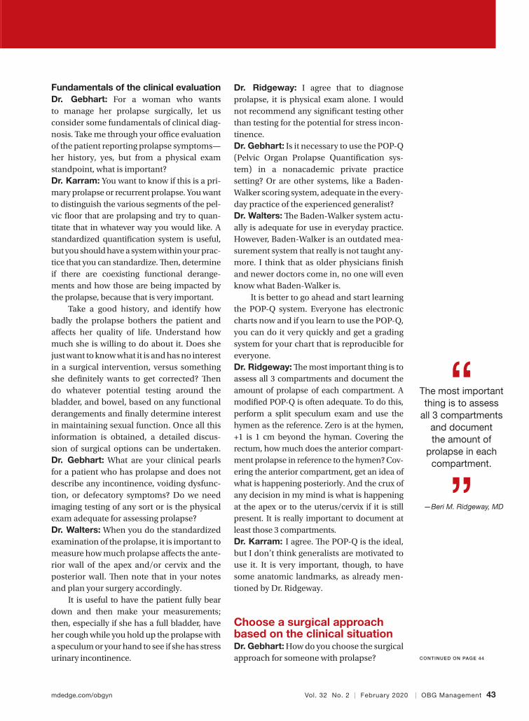
Exploring options for POP treatment
mdedge.com/obgyn Vol. 32 No. 2 | February 2020 | OBG Management 43
The most important thing is to assess
all 3 compartments and document the amount of
prolapse in each compartment.
—Beri M. Ridgeway, MD
Fundamentals of the clinical evaluationDr. Gebhart: For a woman who wants to manage her prolapse surgically, let us consider some fundamentals of clinical diag-nosis. Take me through your o�ce evaluation of the patient reporting prolapse symptoms—her history, yes, but from a physical exam standpoint, what is important?Dr. Karram: You want to know if this is a pri-mary prolapse or recurrent prolapse. You want to distinguish the various segments of the pel-vic �oor that are prolapsing and try to quan-titate that in whatever way you would like. A standardized quanti�cation system is useful, but you should have a system within your prac-tice that you can standardize. �en, determine if there are coexisting functional derange-ments and how those are being impacted by the prolapse, because that is very important.
Take a good history, and identify how badly the prolapse bothers the patient and a�ects her quality of life. Understand how much she is willing to do about it. Does she just want to know what it is and has no interest in a surgical intervention, versus something she de�nitely wants to get corrected? �en do whatever potential testing around the bladder, and bowel, based on any functional derangements and �nally determine interest in maintaining sexual function. Once all this information is obtained, a detailed discus-sion of surgical options can be undertaken. Dr. Gebhart: What are your clinical pearls for a patient who has prolapse and does not describe any incontinence, voiding dysfunc-tion, or defecatory symptoms? Do we need imaging testing of any sort or is the physical exam adequate for assessing prolapse?Dr. Walters: When you do the standardized examination of the prolapse, it is important to measure how much prolapse a�ects the ante-rior wall of the apex and/or cervix and the posterior wall. �en note that in your notes and plan your surgery accordingly.
It is useful to have the patient fully bear down and then make your measurements; then, especially if she has a full bladder, have her cough while you hold up the prolapse with a speculum or your hand to see if she has stress urinary incontinence.
Dr. Ridgeway: I agree that to diagnose prolapse, it is physical exam alone. I would not recommend any signi�cant testing other than testing for the potential for stress incon-tinence.Dr. Gebhart: Is it necessary to use the POP-Q (Pelvic Organ Prolapse Quanti�cation sys-tem) in a nonacademic private practice setting? Or are other systems, like a Baden-Walker scoring system, adequate in the every-day practice of the experienced generalist?Dr. Walters: �e Baden-Walker system actu-ally is adequate for use in everyday practice. However, Baden-Walker is an outdated mea-surement system that really is not taught any-more. I think that as older physicians �nish and newer doctors come in, no one will even know what Baden-Walker is.
It is better to go ahead and start learning the POP-Q system. Everyone has electronic charts now and if you learn to use the POP-Q, you can do it very quickly and get a grading system for your chart that is reproducible for everyone.Dr. Ridgeway: �e most important thing is to assess all 3 compartments and document the amount of prolapse of each compartment. A modi�ed POP-Q is often adequate. To do this, perform a split speculum exam and use the hymen as the reference. Zero is at the hymen, +1 is 1 cm beyond the hyman. Covering the rectum, how much does the anterior compart-ment prolapse in reference to the hymen? Cov-ering the anterior compartment, get an idea of what is happening posteriorly. And the crux of any decision in my mind is what is happening at the apex or to the uterus/cervix if it is still present. It is really important to document at least those 3 compartments.Dr. Karram: I agree. �e POP-Q is the ideal, but I don’t think generalists are motivated to use it. It is very important, though, to have some anatomic landmarks, as already men-tioned by Dr. Ridgeway.
Choose a surgical approach based on the clinical situationDr. Gebhart: How do you choose the surgical approach for someone with prolapse?
“
”
CONTINUED ON PAGE 44
Roundtable 0220.indd 43 1/31/20 3:55 PM
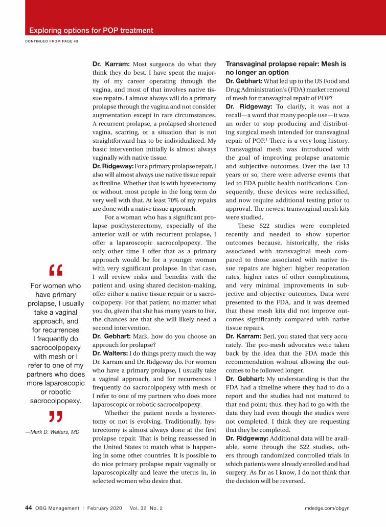
Exploring options for POP treatment
44 OBG Management | February 2020 | Vol. 32 No. 2 mdedge.com/obgyn
Dr. Karram: Most surgeons do what they think they do best. I have spent the major-ity of my career operating through the vagina, and most of that involves native tis-sue repairs. I almost always will do a primary prolapse through the vagina and not consider augmentation except in rare circumstances. A recurrent prolapse, a prolapsed shortened vagina, scarring, or a situation that is not straightforward has to be individualized. My basic intervention initially is almost always vaginally with native tissue.Dr. Ridgeway: For a primary prolapse repair, I also will almost always use native tissue repair as �rstline. Whether that is with hysterectomy or without, most people in the long term do very well with that. At least 70% of my repairs are done with a native tissue approach.
For a woman who has a signi�cant pro-lapse posthysterectomy, especially of the anterior wall or with recurrent prolapse, I o�er a laparoscopic sacrocolpopexy. �e only other time I o�er that as a primary approach would be for a younger woman with very signi�cant prolapse. In that case, I will review risks and bene�ts with the patient and, using shared decision-making, o�er either a native tissue repair or a sacro-colpopexy. For that patient, no matter what you do, given that she has many years to live, the chances are that she will likely need a second intervention.Dr. Gebhart: Mark, how do you choose an approach for prolapse?Dr. Walters: I do things pretty much the way Dr. Karram and Dr. Ridgeway do. For women who have a primary prolapse, I usually take a vaginal approach, and for recurrences I frequently do sacrocolpopexy with mesh or I refer to one of my partners who does more laparoscopic or robotic sacrocolpopexy.
Whether the patient needs a hysterec-tomy or not is evolving. Traditionally, hys-terectomy is almost always done at the �rst prolapse repair. �at is being reassessed in the United States to match what is happen-ing in some other countries. It is possible to do nice primary prolapse repair vaginally or laparoscopically and leave the uterus in, in selected women who desire that.
Transvaginal prolapse repair: Mesh is no longer an optionDr. Gebhart: What led up to the US Food and Drug Administration’s (FDA) market removal of mesh for transvaginal repair of POP?Dr. Ridgeway: To clarify, it was not a recall—a word that many people use—it was an order to stop producing and distribut-ing surgical mesh intended for transvaginal repair of POP.1 �ere is a very long history. Transvaginal mesh was introduced with the goal of improving prolapse anatomic and subjective outcomes. Over the last 13 years or so, there were adverse events that led to FDA public health noti�cations. Con-sequently, these devices were reclassi�ed, and now require additional testing prior to approval. �e newest transvaginal mesh kits were studied.
�ese 522 studies were completed recently and needed to show superior outcomes because, historically, the risks associated with transvaginal mesh com-pared to those associated with native tis-sue repairs are higher: higher reoperation rates, higher rates of other complications, and very minimal improvements in sub-jective and objective outcomes. Data were presented to the FDA, and it was deemed that these mesh kits did not improve out-comes signi�cantly compared with native tissue repairs.Dr. Karram: Beri, you stated that very accu-rately. �e pro-mesh advocates were taken back by the idea that the FDA made this recommendation without allowing the out-comes to be followed longer.Dr. Gebhart: My understanding is that the FDA had a timeline where they had to do a report and the studies had not matured to that end point; thus, they had to go with the data they had even though the studies were not completed. I think they are requesting that they be completed.Dr. Ridgeway: Additional data will be avail-able, some through the 522 studies, oth-ers through randomized controlled trials in which patients were already enrolled and had surgery. As far as I know, I do not think that the decision will be reversed.
For women who have primary
prolapse, I usually take a vaginal approach, and for recurrences I frequently do
sacrocolpopexy with mesh or I
refer to one of my partners who does more laparoscopic
or robotic sacrocolpopexy.
—Mark D. Walters, MD
“
”
CONTINUED FROM PAGE 43
Roundtable 0220.indd 44 1/31/20 3:55 PM
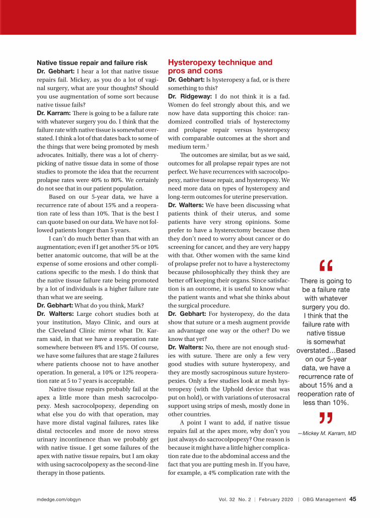
Exploring options for POP treatment
mdedge.com/obgyn Vol. 32 No. 2 | February 2020 | OBG Management 45
Native tissue repair and failure riskDr. Gebhart: I hear a lot that native tissue repairs fail. Mickey, as you do a lot of vagi-nal surgery, what are your thoughts? Should you use augmentation of some sort because native tissue fails?Dr. Karram: �ere is going to be a failure rate with whatever surgery you do. I think that the failure rate with native tissue is somewhat over-stated. I think a lot of that dates back to some of the things that were being promoted by mesh advocates. Initially, there was a lot of cherry-picking of native tissue data in some of those studies to promote the idea that the recurrent prolapse rates were 40% to 80%. We certainly do not see that in our patient population.
Based on our 5-year data, we have a recurrence rate of about 15% and a reopera-tion rate of less than 10%. �at is the best I can quote based on our data. We have not fol-lowed patients longer than 5 years.
I can’t do much better than that with an augmentation; even if I get another 5% or 10% better anatomic outcome, that will be at the expense of some erosions and other compli-cations speci�c to the mesh. I do think that the native tissue failure rate being promoted by a lot of individuals is a higher failure rate than what we are seeing.Dr. Gebhart: What do you think, Mark?Dr. Walters: Large cohort studies both at your institution, Mayo Clinic, and ours at the Cleveland Clinic mirror what Dr. Kar-ram said, in that we have a reoperation rate somewhere between 8% and 15%. Of course, we have some failures that are stage 2 failures where patients choose not to have another operation. In general, a 10% or 12% reopera-tion rate at 5 to 7 years is acceptable.
Native tissue repairs probably fail at the apex a little more than mesh sacrocolpo-pexy. Mesh sacrocolpopexy, depending on what else you do with that operation, may have more distal vaginal failures, rates like distal rectoceles and more de novo stress urinary incontinence than we probably get with native tissue. I get some failures of the apex with native tissue repairs, but I am okay with using sacrocolpopexy as the second-line therapy in those patients.
Hysteropexy technique and pros and consDr. Gebhart: Is hysteropexy a fad, or is there something to this?Dr. Ridgeway: I do not think it is a fad. Women do feel strongly about this, and we now have data supporting this choice: ran-domized controlled trials of hysterectomy and prolapse repair versus hysteropexy with comparable outcomes at the short and medium term.2
�e outcomes are similar, but as we said, outcomes for all prolapse repair types are not perfect. We have recurrences with sacrocolpo-pexy, native tissue repair, and hysteropexy. We need more data on types of hysteropexy and long-term outcomes for uterine preservation.Dr. Walters: We have been discussing what patients think of their uterus, and some patients have very strong opinions. Some prefer to have a hysterectomy because then they don’t need to worry about cancer or do screening for cancer, and they are very happy with that. Other women with the same kind of prolapse prefer not to have a hysterectomy because philosophically they think they are better o� keeping their organs. Since satisfac-tion is an outcome, it is useful to know what the patient wants and what she thinks about the surgical procedure.Dr. Gebhart: For hysteropexy, do the data show that suture or a mesh augment provide an advantage one way or the other? Do we know that yet?Dr. Walters: No, there are not enough stud-ies with suture. �ere are only a few very good studies with suture hysteropexy, and they are mostly sacrospinous suture hystero-pexies. Only a few studies look at mesh hys-teropexy (with the Uphold device that was put on hold), or with variations of uterosacral support using strips of mesh, mostly done in other countries.
A point I want to add, if native tissue repairs fail at the apex more, why don’t you just always do sacrocolpopexy? One reason is because it might have a little higher complica-tion rate due to the abdominal access and the fact that you are putting mesh in. If you have, for example, a 4% complication rate with the
There is going to be a failure rate with whatever
surgery you do. I think that the failure rate with
native tissue is somewhat
overstated…Based on our 5-year
data, we have a recurrence rate of about 15% and a reoperation rate of
less than 10%.
—Mickey M. Karram, MD
“
”
Roundtable 0220.indd 45 1/31/20 3:55 PM
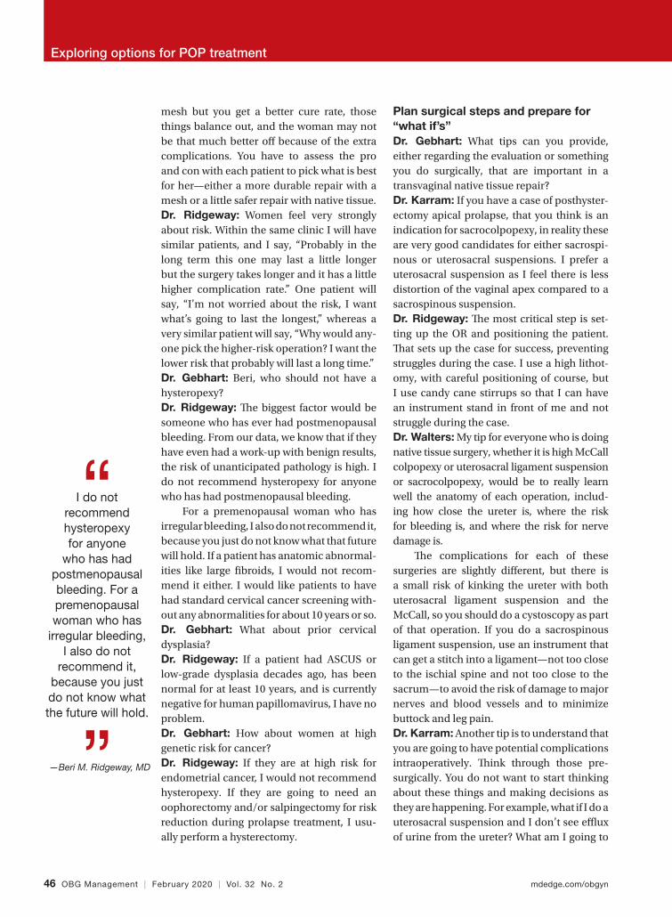
Exploring options for POP treatment
46 OBG Management | February 2020 | Vol. 32 No. 2 mdedge.com/obgyn
mesh but you get a better cure rate, those things balance out, and the woman may not be that much better o� because of the extra complications. You have to assess the pro and con with each patient to pick what is best for her—either a more durable repair with a mesh or a little safer repair with native tissue.Dr. Ridgeway: Women feel very strongly about risk. Within the same clinic I will have similar patients, and I say, “Probably in the long term this one may last a little longer but the surgery takes longer and it has a little higher complication rate.” One patient will say, “I’m not worried about the risk, I want what’s going to last the longest,” whereas a very similar patient will say, “Why would any-one pick the higher-risk operation? I want the lower risk that probably will last a long time.”Dr. Gebhart: Beri, who should not have a hysteropexy?Dr. Ridgeway: �e biggest factor would be someone who has ever had postmenopausal bleeding. From our data, we know that if they have even had a work-up with benign results, the risk of unanticipated pathology is high. I do not recommend hysteropexy for anyone who has had postmenopausal bleeding.
For a premenopausal woman who has irregular bleeding, I also do not recommend it, because you just do not know what that future will hold. If a patient has anatomic abnormal-ities like large �broids, I would not recom-mend it either. I would like patients to have had standard cervical cancer screening with-out any abnormalities for about 10 years or so. Dr. Gebhart: What about prior cervical dysplasia?Dr. Ridgeway: If a patient had ASCUS or low-grade dysplasia decades ago, has been normal for at least 10 years, and is currently negative for human papillomavirus, I have no problem.Dr. Gebhart: How about women at high genetic risk for cancer?Dr. Ridgeway: If they are at high risk for endometrial cancer, I would not recommend hysteropexy. If they are going to need an oophorectomy and/or salpingectomy for risk reduction during prolapse treatment, I usu-ally perform a hysterectomy.
Plan surgical steps and prepare for “what if’s”Dr. Gebhart: What tips can you provide, either regarding the evaluation or something you do surgically, that are important in a transvaginal native tissue repair?Dr. Karram: If you have a case of posthyster-ectomy apical prolapse, that you think is an indication for sacrocolpopexy, in reality these are very good candidates for either sacrospi-nous or uterosacral suspensions. I prefer a uterosacral suspension as I feel there is less distortion of the vaginal apex compared to a sacrospinous suspension.Dr. Ridgeway: �e most critical step is set-ting up the OR and positioning the patient. �at sets up the case for success, preventing struggles during the case. I use a high lithot-omy, with careful positioning of course, but I use candy cane stirrups so that I can have an instrument stand in front of me and not struggle during the case.Dr. Walters: My tip for everyone who is doing native tissue surgery, whether it is high McCall colpopexy or uterosacral ligament suspension or sacrocolpopexy, would be to really learn well the anatomy of each operation, includ-ing how close the ureter is, where the risk for bleeding is, and where the risk for nerve damage is.
�e complications for each of these surgeries are slightly di�erent, but there is a small risk of kinking the ureter with both uterosacral ligament suspension and the McCall, so you should do a cystoscopy as part of that operation. If you do a sacrospinous ligament suspension, use an instrument that can get a stitch into a ligament—not too close to the ischial spine and not too close to the sacrum—to avoid the risk of damage to major nerves and blood vessels and to minimize buttock and leg pain.Dr. Karram: Another tip is to understand that you are going to have potential complications intraoperatively. �ink through those pre-surgically. You do not want to start thinking about these things and making decisions as they are happening. For example, what if I do a uterosacral suspension and I don’t see e�ux of urine from the ureter? What am I going to
I do not recommend hysteropexy for anyone
who has had postmenopausal bleeding. For a premenopausal woman who has
irregular bleeding, I also do not
recommend it, because you just do not know what the future will hold.
—Beri M. Ridgeway, MD
“
”
Roundtable 0220.indd 46 1/31/20 3:55 PM
Exploring options for POP treatment
mdedge.com/obgyn Vol. 32 No. 2 | February 2020 | OBG Management 47
do, and how long am I going to wait before I intervene? If I do a sacrospinous and I start to see a lot of bleeding from that area, what am I going to do? My plan would be, “I will pack the area, get extra suction, etc.” �inking these ideas through before they occur is very helpful.Dr. Gebhart: �at is critical, to have an algo-rithm or a scheme in your mind. You want to think through it before it occurs because you are not always thinking as clearly when things are not going well.
I would say get good at physical exami-nation skills in the o�ce, then have a plan for the OR based on what you see in the o�ce. If what is going on with the prolapse is not com-pletely investigated and other issues are not addressed, then failure results because you did not make the diagnosis. Certainly, mod-ify the procedure according to what you �nd intraoperatively, but follow through.
Indications and tips for sacrocolpopexyDr. Gebhart: What are the indications for sacrocolpopexy?Dr. Ridgeway: Indications include recurrent apical prolapse, posthysterectomy prolapse, or severe prolapse in someone quite young. It is a fantastic operation with overall low risks, but this needs to be discussed with the patient. Dr. Walters: �ere are some unusual cir-cumstances—for example, the woman has a short prolapsed vagina, usually after a prior surgery—in which the best repair is a bridg-ing piece of mesh, usually done laparoscopi-cally, because those operations cannot be done very well vaginally to obtain a durable result.Dr. Karram: I agree. I do not think that all recurrent prolapses mandate a sacrocolpo-pexy. You need to individualize, but in gen-eral the short prolapsed vagina and patients who are very young are at high risk for a recurrence.Dr. Gebhart: An older patient might be a very good candidate, even if she had recur-rence from another vaginal repair.
Beri, does the patient with a high body
mass index need augmentation?Dr. Ridgeway: �at is a great question, and this has to be individualized because, while heavier patients can bene�t from augmen-tation, in a very heavy patient, getting into that abdomen has its own set of challenges. Anatomically they get a better repair with a mesh-augmented repair like a sacrocolpo-pexy, but they do have increased risks. �at is important to acknowledge and clarify with the patient.Dr. Gebhart: Any surgical tip you might o�er on sacrocolpopexy?Dr. Ridgeway: Perform the operation in the same way you would an open procedure. Meaning, use the same materials, the same sutures, the same placement, and the same type of dissection in order to obtain results similar to those with an open operation. Using your assistants to manipulate the vagina and rectum is important, as well as exposure and typical careful surgical technique.Dr. Gebhart: What is important about the placement of sutures on the anterior longitu-dinal ligament, and what do you need to be cognizant of?Dr. Ridgeway: Be careful of that left common iliac vein that is a little more medial than you would expect and of the middle sacral artery, and try to di�erentiate between L5 and S1. In an ideal circumstance, place the suture at S1 or L5 but not the inner disc space, which is the area to avoid placement.
Historically, the recommendation is S1. Some people do L5 because of some pull out strength studies, but also because it is easier, and sometimes in that area of the anterior longitudinal ligament is much better. �e key is to do enough dissection and use haptic feedback, especially with conventional lapa-roscopy or an open approach, to avoid plac-ing sutures through the disc space, as there is some concern that it increases the risk for discitis or osteomyelitis in that area.Dr. Gebhart: We also have found that if you have a combined surgery with colorectal col-leagues, like a rectal prolapse repair, there is a little higher risk of discitis.Dr. Ridgeway: In my own practice I saw a combined case with a rectopexy in
Get good at physical
examination skills in the of�ce, then have a plan for the OR based
on what you see in the of�ce…
Certainly, modify the procedure according to what you �nd
intraoperatively, but follow through.
—John B. Gebhart, MD, MS
“
”
Roundtable 0220.indd 47 1/31/20 3:55 PM
Exploring options for POP treatment
48 OBG Management | February 2020 | Vol. 32 No. 2 mdedge.com/obgyn
someone who had a biologic mesh erosion. When we reviewed the literature, a number of reported cases of discitis had either an early post-op or concurrent urinary tract infection or vaginal infection that likely predisposed them to an infection that trav-eled up the material.Dr. Karram: My �nal comment is that a sacrocolpopexy is not a few stitches or a lit-tle mesh right at the apex. If the patient has
an isolated enterocele, okay, but it is a wide mesh for a reason and it should connect to the endopelvic fascia anteriorly, posteriorly. It is a mistake to suture just a little bit of the cu� and grab it and think, “I’ve done a col-popexy” when the procedure has not been executed as it should be.Dr. Gebhart: I want to thank our expert panel and OBG Management for providing this discussion opportunity. �ank you.
Reference1. US Food and Drug Administration. FDA takes action to
protect women’s health, orders manufacturers of surgicalmesh intended for transvaginal repair of pelvic organ prolapse to stop selling all devices. https://www.fda.gov/news-events/press-announcements/fda-takes-action-protect-womens-health-orders-manufacturers-surgical-mesh-intended-
transvaginal. April 16, 2019. Accessed January 14, 2020.2. Detollenaere RJ, den Boon J, Stekelenburg J, et al. Sacrospinous
hysteropexy versus vaginal hysterectomy with suspension of the uterosacral ligaments in women with uterine prolapsestage 2 or higher: multicentre randomised non-inferioritytrial. BMJ. 2015;351:h3717.
What’s the VERDICT?CONTINUED FROM PAGE 39
and costs before and after implementation of a medical error disclosure program. Ann Intern Med. 2010;153:213-221.
14. National Practitioner Data Bank web site. What you mustreport to the NPDB. https://www.npdb.hrsa.gov/hcorg /whatYouMustReportTo�eDataBank.jsp. Accessed January10, 2020.
15. Bovbjerg RR. Malpractice crisis and reform. Clin Perinatol. 2005;32:203-233, viii-ix.
16. Viscusi WK. Medical malpractice reform: what works andwhat doesn’t. Denver Law Rev. 2019;96:775-791. https://static1.squarespace.com/static/5cb79f7efd6793296c0eb738 /t/5d5f4ffabd6c5400011a12f6/1566527483118/Vol96_ Issue4_Viscusi_FINAL.pdf. Accessed January 10, 2020.
17. National Conference of State Legislatures. Medicalmalpractice reform. Health Cost Containment andE�ciencies: NCSL Briefs for State Legislators. 2011;(16).http://www.ncsl.org/research/health/medical-malpractice-reform-health-cost-brief.aspx. Accessed January 10, 2020.
18. Kass JS, Rose RV. Medical malpractice reform: historicalapproaches, alternative models, and communication andresolution programs. AMA J Ethics. 2016;18:299-310.
19. Boehm G. Debunking medical malpractice myths: unraveling the false premises behind “tort reform”. Yale J Health PolicyLaw Ethics. 2005;5:357-369.
20. Hellinger FJ, Encinosa WE. �e impact of state laws limitingmalpractice damage awards on health care expenditures. Am J Public Health. 2006;96:1375-1381.
21. Perry G. Medical malpractice caps by state [infographic].
January 3, 2013. https://www.business2community.com/infographics/medical-malpractice-caps-by-state-infographic-0368345. Accessed January 23, 2020.
22. Goguen D. State-by-state medical malpracticedamages caps. An in-depth look at state laws limitingcompensation for medical malpractice plaintiffs. https://www.nolo.com/legal-encyclopedia/state-state-medical-malpractice-damages-caps.html. Accessed January 23,2020.
23. Berlin L. Medical errors, malpractice, and defensivemedicine: an ill-fated triad. Diagnosis (Berl). 2017;4:133-139.
24. Ranum D. Electronic health records continue to lead tomedical malpractice suits. �e Doctors Company. August2019. https://www.thedoctors.com/articles/electronic-health- records-continue-to-lead-to-medical-malpractice-suits/. Accessed January 10, 2020.
25. Mangalmurti SS, Murtagh L, Mello MM. Medical malpracticeliability in the age of electronic health records. N Engl J Med. 2010;363:2060-2067.
26. Studdert DM, Mello MM, Gawande AA, et al. Claims,errors, and compensation payments in medical malpractice litigation. N Engl J Med. 2006;354(19):2024-2033.
27. Jena AB, Chandra A, Lakdawalla D, et al. Outcomes of medical malpractice litigation against US physicians. Arch Intern Med. 2012;172:892-894.
28. Glaser LM, Alvi FA, Milad MP. Trends in malpractice claims for obstetric and gynecologic procedures, 2005 through 2014.Am J Obstet Gynecol. 2017;217:340.e1-340.e6.
Roundtable 0220.indd 48 1/31/20 3:56 PM

mdedge.com/obgyn Vol. 32 No. 2 | February 2020 | OBG Management 49
THE OFFICIAL JOB BOARD OF OBG MANAGEMENTMEDJOBNETWORK comOBG MARKETPLACE THE OFFICIAL JOB BOARD OF OBG MANAGEMENTMEDJOBNETWORK comOBG MARKETPLACE
Learn all you need to know about successfully bringing “Genital Plastics” into your o�ce!
Labiaplasty; hood reduction; Perineoplasty, Vaginoplasty/Vaginal Reconstruction Training Program
taught by Dr. Michael Goodman Cash Model
Learn from the Best!�e Program: �is is one of the premier programs in the world, having thus far trained ~ 100 surgeons in the U.S. and >10 foreign countries in the art of female genital plastic/cosmetic surgery. �is program is accredited for up to 14.5 AMA Category 1 CME credits from Medical Education Resources. In this 2-day course you will thoroughly learn:
1. �e techniques and the “RULES” for successful linear and V-Wedge labiaplasty + hood reduction.
2. �e techniques and the “RULES” for successful vaginal tightening surgery (“Vaginoplasty.”)
3. How to set-up and perform in-o�ce, “local” anesthesia.
4. How to fully train your o�ce sta� to interact with potential patients.
5. Marketing techniques for success.
6. Instruction in O-Shot™ and other uses for PRP. Uses of fractional CO2 laser and RF.
7. Full-length real-time professional surgical videos of all procedures. Animal lab. Live surgery option. Limited to 10 participants/class. Close interaction with instructors!
www.labiaplastytraining.com for full prospectus, info on instructors, and registration documentsOr contact “Nicole” at (530) 753-2787, [email protected]
Classifieds 0220.indd 49 1/31/20 4:01 PM

50 OBG Management | February 2020 | Vol. 32 No. 2 mdedge.com/obgyn
THE OFFICIAL JOB BOARD OF OBG MANAGEMENTMEDJOBNETWORK comOBG MARKETPLACE
“This product does everything right. It aids physicians with providing care
to patients while optimizing patient comfort and maintaining cleanliness.”
—Dr. Elizabeth Ramsey
Resilient® Professional Foot Supports by comenityMED®
www.comenitymed.com
The most comprehensive online databank of domestic, international, and online CME and non-accredited medical meetings
Search by date, specialty, location, and keyword. Updated daily, each listing contains sponsoring organization, topic or title of the meeting, credits available, registration fee, recreational activities, and special events for attendees, contact information, and registration opportunities.
PTMG.com
SEARCH 1000s OF JOBS AND APPLY IN 1 CLICKAnd get FREE bene�ts including…• Access to 30+ medical web sites• E-Alert and Newsletters on your smart phone• Online CME and MD-IQ Quizzes• Coverage of over 200 meetings
MEDJOBNETWORK com
Tim Lapella Senior Sales DirectorPhone: 484-291-5001E-mail: [email protected]
FOR ADVERTISING OPPORTUNITIES CONTACT:
Classifieds 0220.indd 50 1/31/20 4:01 PM
mdedge.com/obgyn Vol. 32 No. 2 | February 2020 | OBG Management 51
Course Name: Fellowship in Cosmetic Gynecology Step 1 and Step 2Dates: June 26th & 27th (Step 1)
June 28th & 29th (Step 2)Address: Step 1 and Step 2: American Aesthetic Association
120 Cedar Grove Lane Somerset, NJ 08873
Course features:• Introduction to Cosmetic Gynecology and Anatomy• Different indication of laser and radiofrequency in the Cosmetic Gynecology• Botox and dermal �ller for the Gynecologist• Different uses of the PRP• Techniques for safe labioplasty and how to avoid complications• Vaginoplasty/clitoral hood reduction• Labia majora reduction and augmentation• Autologous fat transfer• Genital bleaching• Safety and legal issues, informed consent, and of�ce setting
Live surgery and access to surgical procedure. Limited to 10 participants/class. Close interactions with instructors.
Contact — [email protected] Phone: 610-333-4030 American Aesthetic Association 120 Cedar Grove Lane Somerset, NJ 08873
AKORN PharmaceuticalsLidocaine Hydrochloride Jelly, USP . . . . . . . . . . . . . . . P 3
Exact SciencesCologuard . . . . . . . . . . . . . . . . . . . . . . . . . . . . . . . . .C2, P1
HologicRapid fFN . . . . . . . . . . . . . . . . . . . . . . . . . . . . . . . . . . . .C4
LabCorpThe Value of Care: Vaginitis Testing . . . . . . . . . . . . . . P 33
Merck & Co.Nexplanon . . . . . . . . . . . . . . . . . . . . . . . . . . . . . . PP 20-23
INDEX OF ADVERTISERS
Classifieds 0220.indd 51 1/31/20 4:01 PM
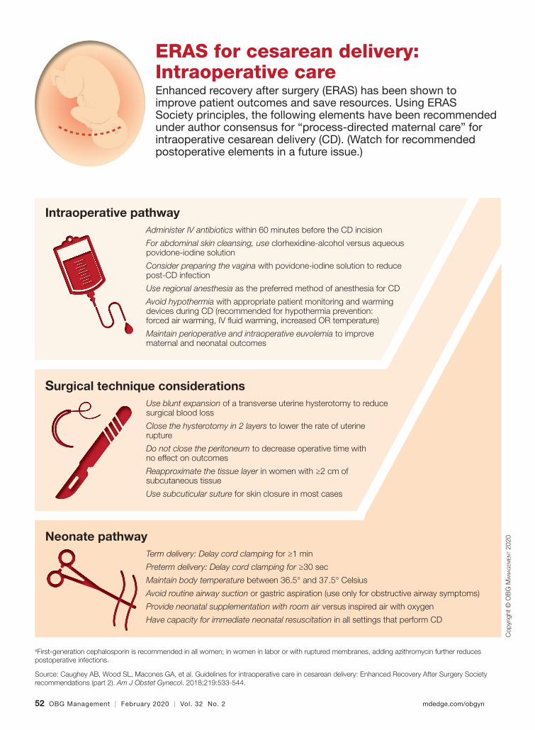
52 OBG Management | February 2020 | Vol. 32 No. 2 mdedge.com/obgyn
Antenatal pathwaya
Inform patients about procedures as best as possible before, during, and after scheduled or unscheduled CD
CD without indication should not be recommended without preadmission evaluation of harms and bene�ts to mother and baby
ERAS for cesarean delivery: Intraoperative careEnhanced recovery after surgery (ERAS) has been shown to improve patient outcomes and save resources. Using ERAS Society principles, the following elements have been recommended under author consensus for “process-directed maternal care” for intraoperative cesarean delivery (CD). (Watch for recommended postoperative elements in a future issue.)
Cop
yrig
ht ©
OB
G M
ANAGEMENT
2020
Intraoperative pathwayAdminister IV antibiotics within 60 minutes before the CD incision
For abdominal skin cleansing, use clorhexidine-alcohol versus aqueous povidone-iodine solution
Consider preparing the vagina with povidone-iodine solution to reduce post-CD infection
Use regional anesthesia as the preferred method of anesthesia for CD
Avoid hypothermia with appropriate patient monitoring and warming devices during CD (recommended for hypothermia prevention: forced air warming, IV �uid warming, increased OR temperature)
Maintain perioperative and intraoperative euvolemia to improve maternal and neonatal outcomes
Surgical technique considerationsUse blunt expansion of a transverse uterine hysterotomy to reduce surgical blood loss
Close the hysterotomy in 2 layers to lower the rate of uterine rupture
Do not close the peritoneum to decrease operative time with no effect on outcomes
Reapproximate the tissue layer in women with ≥2 cm of subcutaneous tissue
Use subcuticular suture for skin closure in most cases
Neonate pathwayTerm delivery: Delay cord clamping for ≥1 min
Preterm delivery: Delay cord clamping for ≥30 sec
Maintain body temperature between 36.5° and 37.5° Celsius
Avoid routine airway suction or gastric aspiration (use only for obstructive airway symptoms)
Provide neonatal supplementation with room air versus inspired air with oxygen
Have capacity for immediate neonatal resuscitation in all settings that perform CD
aFirst-generation cephalosporin is recommended in all women; in women in labor or with ruptured membranes, adding azithromycin further reduces postoperative infections.
Source: Caughey AB, Wood SL, Macones GA, et al. Guidelines for intraoperative care in cesarean delivery: Enhanced Recovery After Surgery Society recommendations (part 2). Am J Obstet Gynecol. 2018;219:533-544.
ERAS Pre Cesarean Infographic 0220.indd 52 1/31/20 4:18 PM

Of course, current and past articles are still there for you—in full text and as convenient, downloadable PDF �les. And you’ll �nd an easy way to contact us.
Have you visited mdedge.com/obgyn
this week?
Plus, visit us online for daily news!
Prenatal exposure to pollutants consumed through diet found to be associated with decreased fetal growth
Is elagolix effective at reducing HMB for women with varying �broid sizes and types?
ERAS for cesarean delivery: Antenatal and preoperative care
Laparoscopic techniques for Essure device removalBrought to you by the Society of Gynecologic Surgeons
OTC hormonal contraception: An important goal in the �ght for reproductive justiceAbby L. Schultz, MD, and Megan L. Evans, MD, MPH
Considerations on the mode of delivery for pregnant women with hepatitis C infection Morgan Brazel, BA, and Patrick Duff, MD
Does planned early delivery make sense in women with preterm preeclampsia?Sarosh Rana, MD, MPH
A clitoral cyst of “epidermal” proportionsBrought to you by the Society of Gynecologic Surgeons
Does BSO status affect health outcomes for women taking estrogen for menopause?Q&A with JoAnn Manson, MD, DrPH
Persistent vulvar itch: What is the diagnosis? Kerrie G. Satcher, MD, Stephanie J. Carstens, MD, and Andrew M. Kaunitz, MD, NCMP
OBGM_0220_WebAD.indd 3 1/31/20 4:04 PM

Reference: 1. Rapid fFN for the TliIQ System [package insert]. AW-04196, Sunnyvale, CA: Hologic, Inc.; 2017. 2. McLaren JS, et al. Prediction of spontaneous preterm birth using quantitative fetal fibronectin after recent sexual intercourse. Am J Obstet Gynecol. 2015; 212:89e1-5.
ADS-02754-001 Rev. 001 © 2019 Hologic, Inc. All rights reserved. Hologic, Rapid fFN and associated logos are trademarks and/or registered trademarks of Hologic, Inc. and/or its subsidiaries in the United States and/or other countries.
Even if a patient has had sex in the prior 24 hours:
• A negative result is valid
• A positive result may not be valid& should be confirmed > 24 hours1
The majority of patients will test negative, even if they’ve had sex in the prior 24 hours.2
Sex last night? A negative fFN result is valid.
Ad Place-new.indd 6 1/23/2020 2:02:34 PM
Related Documents