Page1 MacPeds Pediatric ECG Survival Guide First Edition 2018 Editors: Ahmad Jaafar Dragos Predescu This guide is dedicated to my fellow MacPeds residents!

Welcome message from author
This document is posted to help you gain knowledge. Please leave a comment to let me know what you think about it! Share it to your friends and learn new things together.
Transcript

Pag
e1
MacPeds
Pediatric ECG Survival Guide First Edition
2018
Editors:
Ahmad Jaafar
Dragos Predescu
This guide is dedicated to my fellow MacPeds residents!

Pag
e2
Table of Contents:
Approach of ECG interpretation: Page 3
Summary of normal ECG findings in the pediatric population: Page 3
Chest electrode positions: Page 4
ECG Components:
- Calibration and paper speed Page 5
- Heart rate Page 5
- Cardiac axis Page 6
- Rhythm Page 6
- Waves, segments and intervals Page 7
- Denominations of QRS complex Page 7
- P wave (atrial enlargement) Page 7
- PR interval Page 7-8
- Q wave Page 8
- QRS duration Page 9
- QRS amplitude & R/S ratio Page 10
- ST segment Page 11
- T wave Page 12
- U wave Page 12
- QT interval Page 13
- JT Interval Page 13
Specific cardiac conditions:
- Pericarditis Page 14
- Myocarditis Page 14
- Ventricular enlargement Page 15
- Myocardial ischemia/infarction Page 16
- Hypo/hypercalcemia Page 16
- Hypo/hyperkalemia Page 17
- WPW syndrome Page 17
- Brugada syndrome Page 18
- Long QT syndrome Page 18
- ECG and athletes Page 19-20
References: Page 21
Helpful recourses: Page 21
Pag
e3
Approach of ECG Interpretation:
Various approaches of ECG interpretation exist; listed below is a common one.
Approach:
1. Identification information: Name, age, date, indication of the ECG.
2. Calibration and paper speed.
3. Heart rate.
4. Rhythm.
5. Cardiac axis.
6. Intervals.
7. Wave amplitude.
8. Morphology.
9. Repolarization phase (ST segment & T wave).
Normal ECG variations in pediatrics:
ECGs of the normal pediatric population are different from those of normal adults.
Many differences are due to the right ventricular dominance in infants, and the evolution to
adult dynamics.
Listed below are the features that you may encounter in pediatric ECGs in comparison to
adult ECGs:
- Faster heart rate.
- Sinus arrhythmia.
- Rightward QRS axis (up to 3 months and again in adolescence).
- T wave inversions in the right precordial leads (RPLs).
- Dominant R wave in V1.
- RSR’ pattern in V1.
- Shorter PR interval and QRS duration.
- Slightly longer QTc.
Pag
e4
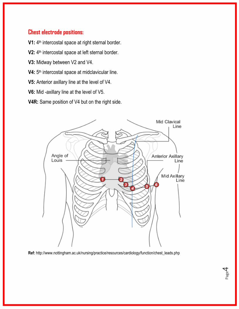
Chest electrode positions:
V1: 4th intercostal space at right sternal border.
V2: 4th intercostal space at left sternal border.
V3: Midway between V2 and V4.
V4: 5th intercostal space at midclavicular line.
V5: Anterior axillary line at the level of V4.
V6: Mid -axillary line at the level of V5.
V4R: Same position of V4 but on the right side.
Ref: http://www.nottingham.ac.uk/nursing/practice/resources/cardiology/function/chest_leads.php

Pag
e5
Calibration and paper speed:
Ref: http://a-fib.com/treatments-for-atrial-fibrillation/diagnostic-tests/the-ekg-signal/
Heart rate:
Calculation:
- Regular rhythm:
o 300/number of large squares between 2 consecutive R waves.
o 1500/number of small squares between 2 consecutive R waves.
- Irregular rhythm:
o Multiply the number of QRS complexes on the rhythm strip by 6.
Age HR
Newborn ≤ 1 wk 120-160
Newborn ≤ 1 mo 120-160
Infant ≤1 yr 110-140
Toddler 1-3 yr 90-130
Preschool 3-5 yr 90-120
Child 6-12 yr 80-110
Adolescent >12 yr 70-100
Adult >18 yr 60-100
Ref: MacPeds Survival Guide, 2017
Standard ECG recording speed: 25 mm/sec.
Standard ECG calibration: 10 mm/mV.

Pag
e6
Cardiac (QRS) axis:
▪ Full-term newborn: +135° (range +58° to +168°).
▪ 1 week – 1 month: +110° (range +65° to +159°).
▪ 1 month – 3 months: +75° (range +31° to +115°).
▪ 3 months – 6 months: +60° (range +7° to +105°).
▪ 6 months – 1 year: +54° (range +7° to +98°).
▪ 1 year – 3 years: +55° (range +8° to +100°).
▪ 3 years – 5 years: +55° (range +7° to +104°).
▪ 5 years – 8 years: +66° (range +10° to +140°).
▪ 8 years – 12 years: +61° (range +9° to +115°).
▪ 12 years – 16 years: + 58° (range +11° to +133°).
Ref: Davignon et al, 1980.
LAD causes: AVSD, tricuspid atresia, LBBB, left anterior hemiblock, WPW (type b), LVH, ccTGA.
RAD causes: RBBB, RVH.
The normal QRS-T axis angle is 0 to +90:
➢ If the angle >90 primary T wave abnormality (e.g., myocardial ischemia).
➢ If the angel is normal secondary T wave abnormality (e.g., BBB, ventricular hypertrophy).
Rhythm:
Regular: Constant RR interval on the rhythm strip.
Sinus (all of the below criteria should be met):
- P wave proceeding each QRS complex (i.e., QRS complex after every P wave).
- Constant PR interval.
- Normal P wave axis (0° to +90°), i.e., upright P waves in leads I and aVF.
1. Use lead I and aVF to locate a quadrant.
2. Find a lead with an equiphasic QRS
complex (height of R equal to depth of S).
The QRS axis will be perpendicular to this
in the previously determined quadrant.
Pag
e7
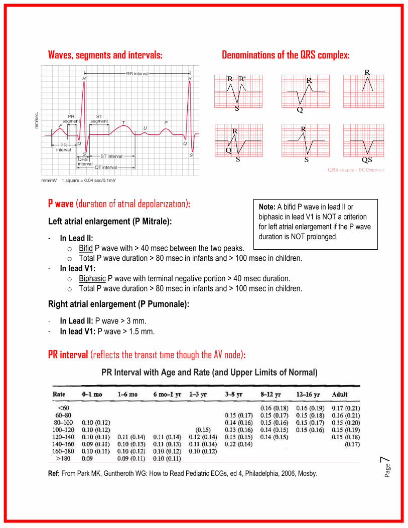
Waves, segments and intervals: Denominations of the QRS complex:
P wave (duration of atrial depolarization):
Left atrial enlargement (P Mitrale):
- In Lead II: o Bifid P wave with > 40 msec between the two peaks. o Total P wave duration > 80 msec in infants and > 100 msec in children.
- In lead V1: o Biphasic P wave with terminal negative portion > 40 msec duration. o Total P wave duration > 80 msec in infants and > 100 msec in children.
Right atrial enlargement (P Pumonale):
- In Lead II: P wave > 3 mm.
- In lead V1: P wave > 1.5 mm.
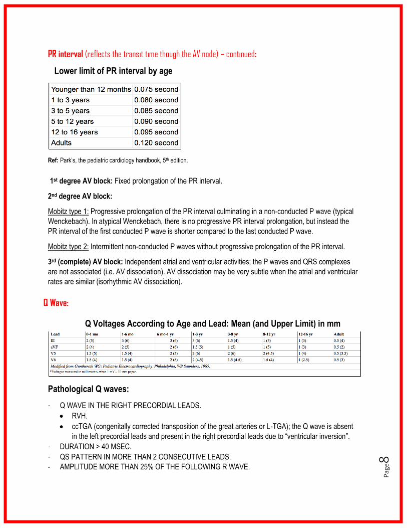
PR interval (reflects the transit time though the AV node):
PR Interval with Age and Rate (and Upper Limits of Normal)
Ref: From Park MK, Guntheroth WG: How to Read Pediatric ECGs, ed 4, Philadelphia, 2006, Mosby.
Note: A bifid P wave in lead II or
biphasic in lead V1 is NOT a criterion
for left atrial enlargement if the P wave
duration is NOT prolonged.
Pag
e8
PR interval (reflects the transit time though the AV node) – continued:
Lower limit of PR interval by age
Ref: Park’s, the pediatric cardiology handbook, 5th edition.
1st degree AV block: Fixed prolongation of the PR interval.
2nd degree AV block:
Mobitz type 1: Progressive prolongation of the PR interval culminating in a non-conducted P wave (typical
Wenckebach). In atypical Wenckebach, there is no progressive PR interval prolongation, but instead the
PR interval of the first conducted P wave is shorter compared to the last conducted P wave.
Mobitz type 2: Intermittent non-conducted P waves without progressive prolongation of the PR interval.
3rd (complete) AV block: Independent atrial and ventricular activities; the P waves and QRS complexes
are not associated (i.e. AV dissociation). AV dissociation may be very subtle when the atrial and ventricular
rates are similar (isorhythmic AV dissociation).
Pathological Q waves:
- Q WAVE IN THE RIGHT PRECORDIAL LEADS.
• RVH.
• ccTGA (congenitally corrected transposition of the great arteries or L-TGA); the Q wave is absent
in the left precordial leads and present in the right precordial leads due to “ventricular inversion”.
- DURATION > 40 MSEC.
- QS PATTERN IN MORE THAN 2 CONSECUTIVE LEADS.
- AMPLITUDE MORE THAN 25% OF THE FOLLOWING R WAVE.
Q Voltages According to Age and Lead: Mean (and Upper Limit) in mm
Q Wave:
Q
Pag
e9
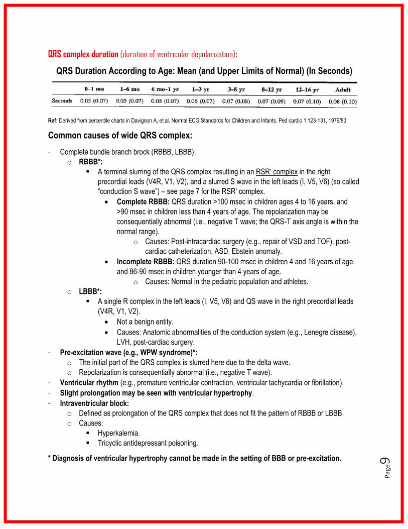
QRS complex duration (duration of ventricular depolarization):
Ref: Derived from percentile charts in Davignon A, et al. Normal ECG Standards for Children and Infants. Ped cardio 1:123-131, 1979/80.
Common causes of wide QRS complex:
- Complete bundle branch brock (RBBB, LBBB):
o RBBB*:
▪ A terminal slurring of the QRS complex resulting in an RSR’ complex in the right
precordial leads (V4R, V1, V2), and a slurred S wave in the left leads (I, V5, V6) (so called
“conduction S wave”) – see page 7 for the RSR’ complex.
• Complete RBBB: QRS duration >100 msec in children ages 4 to 16 years, and
>90 msec in children less than 4 years of age. The repolarization may be
consequentially abnormal (i.e., negative T wave; the QRS-T axis angle is within the
normal range).
o Causes: Post-intracardiac surgery (e.g., repair of VSD and TOF), post-
cardiac catheterization, ASD, Ebstein anomaly.
• Incomplete RBBB: QRS duration 90-100 msec in children 4 and 16 years of age,
and 86-90 msec in children younger than 4 years of age.
o Causes: Normal in the pediatric population and athletes.
o LBBB*:
▪ A single R complex in the left leads (I, V5, V6) and QS wave in the right precordial leads
(V4R, V1, V2).
• Not a benign entity.
• Causes: Anatomic abnormalities of the conduction system (e.g., Lenegre disease),
LVH, post-cardiac surgery.
- Pre-excitation wave (e.g., WPW syndrome)*:
o The initial part of the QRS complex is slurred here due to the delta wave.
o Repolarization is consequentially abnormal (i.e., negative T wave).
- Ventricular rhythm (e.g., premature ventricular contraction, ventricular tachycardia or fibrillation).
- Slight prolongation may be seen with ventricular hypertrophy.
- Intraventricular block:
o Defined as prolongation of the QRS complex that does not fit the pattern of RBBB or LBBB.
o Causes:
▪ Hyperkalemia.
▪ Tricyclic antidepressant poisoning.
* Diagnosis of ventricular hypertrophy cannot be made in the setting of BBB or pre-excitation.
QRS Duration According to Age: Mean (and Upper Limits of Normal) (In Seconds)
Pag
e10
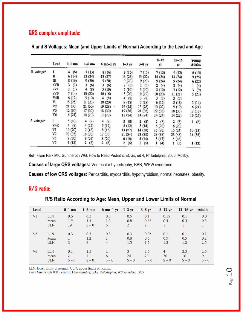
QRS complex amplitude:
Ref: From Park MK, Guntheroth WG: How to Read Pediatric ECGs, ed 4, Philadelphia, 2006, Mosby.
Causes of large QRS voltages: Ventricular hypertrophy, BBB, WPW syndrome.
Causes of low QRS voltages: Pericarditis, myocarditis, hypothyroidism, normal neonates, obesity.
R/S ratio:
R/S Ratio According to Age: Mean, Upper and Lower Limits of Normal
R and S Voltages: Mean (and Upper Limits of Normal) According to the Lead and Age
f
Pag
e11
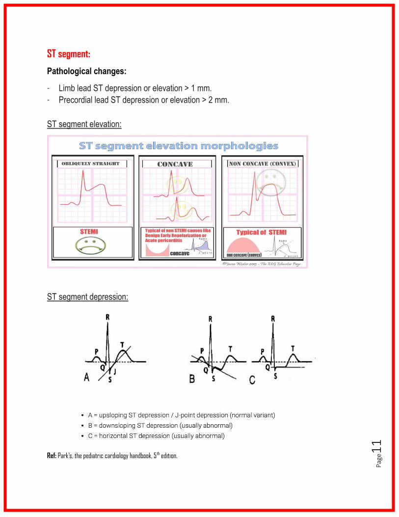
ST segment:
Pathological changes:
- Limb lead ST depression or elevation > 1 mm.
- Precordial lead ST depression or elevation > 2 mm.
ST segment elevation:
ST segment depression:
Ref: Park’s, the pediatric cardiology handbook, 5 th edition.
Pag
e12
T waves:
✓ For the first week of life, the T waves in V1 may be positive, in V2-V4 are usually
negative, in V5 are variable but usually positive, and in V6 are always positive.
✓ After the first week of life, the T waves become inverted in V1-4.
o They will start becoming positive starting with V4, then V3, then V2 and lastly V1.
This process does not skip leads, so if there are negative T waves in-between
positive ones or vice-versa, this is abnormal. Most commonly, this is due to lead
misplacement or chest electrodes touching each other.
o The T waves should be positive in V5 and V6.
✓ T waves usually become positive in V1 when the child becomes 7 years and older.
✓ However, inverted T waves in V1 can persist into adolescence and early adulthood.
✓ Negative T waves in V2 after the age of 14 years is a minor criterion for ARVC
(arrhythmogenic right ventricular hypertrophy) in a suggestive clinical context.
✓ T wave inversion in leads V1-V4 in black/African athletes is normal.
Key point: Upright T waves in V1 in children >7 days to <7 years are suggestive of RVH
(pressure overload such as in pulmonary stenosis or TOF).
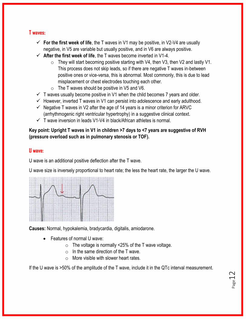
U wave:
U wave is an additional positive deflection after the T wave.
U wave size is inversely proportional to heart rate; the less the heart rate, the larger the U wave.
Causes: Normal, hypokalemia, bradycardia, digitalis, amiodarone.
• Features of normal U wave:
o The voltage is normally <25% of the T wave voltage.
o In the same direction of the T wave.
o More visible with slower heart rates.
If the U wave is >50% of the amplitude of the T wave, include it in the QTc interval measurement.
Pag
e13
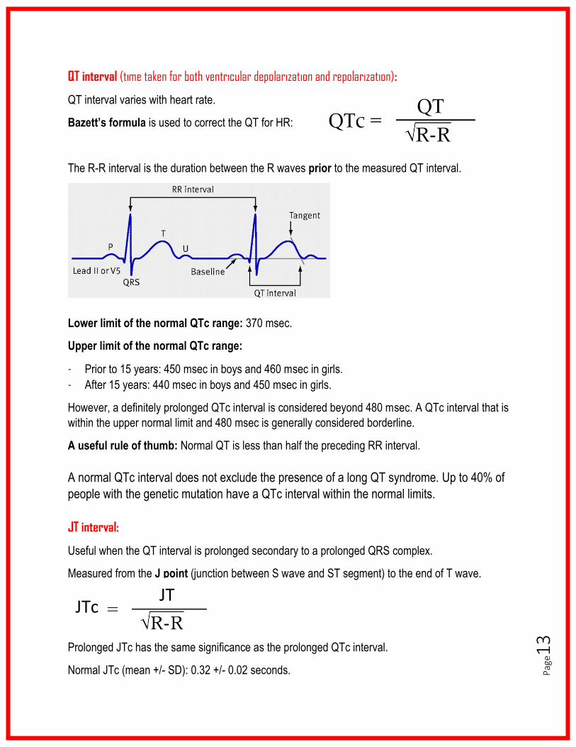
QT interval (time taken for both ventricular depolarization and repolarization):
QT interval varies with heart rate.
Bazett’s formula is used to correct the QT for HR:
The R-R interval is the duration between the R waves prior to the measured QT interval.
Lower limit of the normal QTc range: 370 msec.
Upper limit of the normal QTc range:
- Prior to 15 years: 450 msec in boys and 460 msec in girls.
- After 15 years: 440 msec in boys and 450 msec in girls.
However, a definitely prolonged QTc interval is considered beyond 480 msec. A QTc interval that is
within the upper normal limit and 480 msec is generally considered borderline.
A useful rule of thumb: Normal QT is less than half the preceding RR interval.
A normal QTc interval does not exclude the presence of a long QT syndrome. Up to 40% of
people with the genetic mutation have a QTc interval within the normal limits.
JT interval:
Useful when the QT interval is prolonged secondary to a prolonged QRS complex.
Measured from the J point (junction between S wave and ST segment) to the end of T wave.
Prolonged JTc has the same significance as the prolonged QTc interval.
Normal JTc (mean +/- SD): 0.32 +/- 0.02 seconds.
JTc JT

Pag
e14
Characteristic ECG patterns for particular conditions:
Pericarditis:
- Stage 1 – widespread ST elevation and PR depression with reciprocal (apposite)
changes in aVR (occurs during the first two weeks).
- Stage 2 – Generalized T wave flattening (1 to 3 weeks).
- Stage 3 – flattened T waves become inverted (3 to several weeks).
- Stage 4 – ECG returns to normal (several weeks onwards).
Less than 50% of patients progress through all four classical stages and evolution of
changes may not follow this typical pattern.
Pericardial effusion may produce QRS voltages ≤5 mm in all limb leads.
Myocarditis:
- AV conduction disturbances, ranging from PR prolongation to complete AV dissociation.
- Low QRS voltages (5 mm or less in all limb leads).
- Decreased T wave amplitude, negative or flat T waves.
- QT prolongation.
- Tachyarrhythmias including SVT and VT.
- ‘Pseudo-infarction’ pattern with deep Q waves and poor R wave progression in
precordial leads.
- ST segment elevation that may vary in magnitude with the symptoms.
Pag
e15
Ventricular enlargement:
Right ventricular enlargement (RVH):
- Axis:
o RAD for the patients age.
- Voltages:
o Tall R waves (greater than limits for age) in right-sided leads (V1, V2, aVR).
o rSR’ (small R, tall S, tall R’) with R’ >15 mm before the age of 1 year or >10 mm after that
age (provided that there is no RBBB).
o Deep S waves (greater than limits for age) in left-sided leads (V5, V6, I).
- R/S ratio:
o R/S ratio in V1 and V2 is more than the upper limits of normal for age.
- T waves:
o Upright T waves in V1 in children >7 days to <7 years (provided that T waves are upright
in the left precordial leads, i.e. upright in V5 and V6). This is enough to diagnose RVH.
- Q waves:
o qR (small Q, tall R) or qRs (small Q, tall R, small S) pattern in V1.
Left ventricular enlargement (LVH):
- Axis:
o LAD for the patients age.
- Voltages:
o Tall R waves (greater than limits for age) in the left-sided leads (V5, V6, I).
o Deep S waves (greater than limits for age) in the right-sided leads (V1, V2, aVR).
- R/S ratio:
o R/S ratio in V1 and V2 is less than the lower limits of normal for age.
- T waves:
o Inverted T waves in lead I & aVL AND left precordial leads (LV strain pattern).
o Tall, symmetrical T waves, particularly in mid-precordial leads, may suggest apical HCM.
- Q waves:
o Abnormal Q waves in V5 and V6 (≥5 mm), coupled with tall symmetric T waves (volume-
overload type).
Biventricular enlargement:
- Positive voltage criteria for RVH and LVH (with normal QRS duration).
- Positive voltage criteria for RVH or LVH and normal voltages for the other ventricle.
- Large equiphasic QRS complexes in two or more limb leads and in mid-precordial leads (V2-
V4) – called “Katz-Wachtel phenomenon”.
Pag
e16
Myocardial ischemia/infarction:
- Infarction (death of cardiac tissue): o ST segment elevation in contiguous leads with reciprocal ST segment depression elsewhere.
- Injury (prolonged oxygen deficiency, usually >20 minutes): o Horizontal ST segment depression.
- Ischemia (oxygen deficiency for a shorter period, usually <20 minutes): o Flat/negative T waves with QRS-T axis angle >90 degrees.
Correlation between ECG leads and coronary artery territory affected:
Hypocalcaemia and hypercalcemia:
- Hypocalcaemia prolongs the ST segment with resulting prolongation of the QTc.
- Hypercalcaemia shortens the ST segment and the QTc.
Ref: Park’s, the pediatric cardiology handbook, 5th edition.
Pag
e17
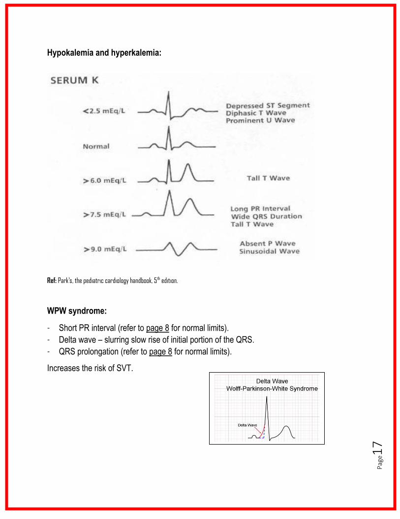
Hypokalemia and hyperkalemia:
Ref: Park’s, the pediatric cardiology handbook, 5 th edition.
WPW syndrome:
- Short PR interval (refer to page 8 for normal limits).
- Delta wave – slurring slow rise of initial portion of the QRS.
- QRS prolongation (refer to page 8 for normal limits).
Increases the risk of SVT.
Pag
e18
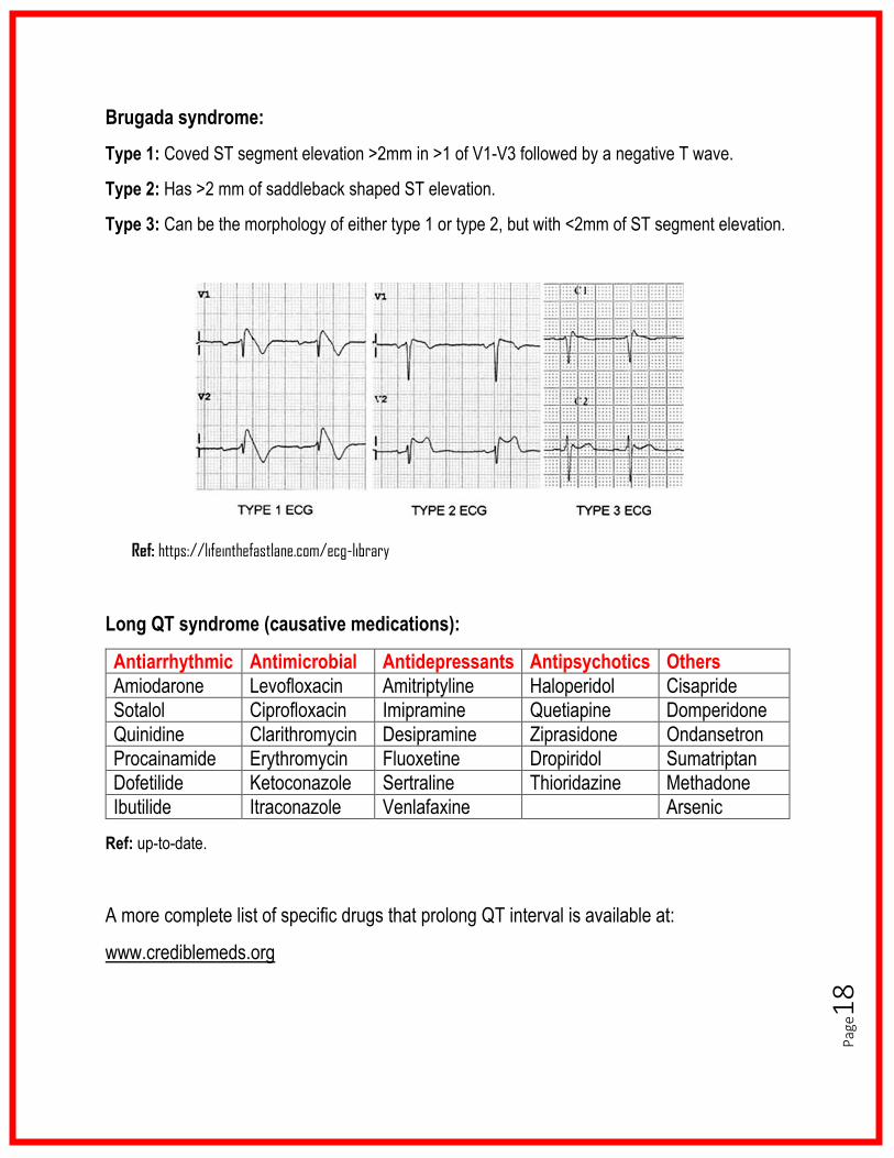
Brugada syndrome:
Type 1: Coved ST segment elevation >2mm in >1 of V1-V3 followed by a negative T wave.
Type 2: Has >2 mm of saddleback shaped ST elevation.
Type 3: Can be the morphology of either type 1 or type 2, but with <2mm of ST segment elevation.
Ref: https://lifeinthefastlane.com/ecg-library
Long QT syndrome (causative medications):
Antiarrhythmic Antimicrobial Antidepressants Antipsychotics Others
Amiodarone Levofloxacin Amitriptyline Haloperidol Cisapride
Sotalol Ciprofloxacin Imipramine Quetiapine Domperidone
Quinidine Clarithromycin Desipramine Ziprasidone Ondansetron
Procainamide Erythromycin Fluoxetine Dropiridol Sumatriptan
Dofetilide Ketoconazole Sertraline Thioridazine Methadone
Ibutilide Itraconazole Venlafaxine Arsenic
Ref: up-to-date.
A more complete list of specific drugs that prolong QT interval is available at:
www.crediblemeds.org
Pag
e19
Normal ECG findings in athletes:
1. Sinus bradycardia (≥ 30 bpm).
2. Sinus arrhythmia.
3. Ectopic atrial rhythm.
4. Junctional escape rhythm.
5. 1° AV block (PR interval>200 msec).
6. Mobitz Type I (Wenckebach) 2° AV block.
7. Incomplete RBBB.
8. Isolated QRS voltage criteria for LVH:
o Except: QRS voltage criteria for LVH occurring with any non-voltage criteria
for LVH such:
i. Left atrial enlargement.
ii. Left axis deviation.
iii. ST segment depression.
iv. T-wave inversion.
v. Pathological Q waves.
9. Early repolarisation:
o ST elevation, J-point elevation, J-waves or terminal QRS slurring.
10. Convex (‘domed’) ST segment elevation combined with T-wave inversion in leads
V1–V4 in black/African athletes.
These common training-related ECG alterations are physiological adaptations to regular exercise,
considered normal variants in athletes and do not require further evaluation in asymptomatic
athletes.
Ref: Jonathan A Drezner, et al. Electrocardiographic interpretation in athletes: the ‘Seattle Criteria’. BMJ. 7th Dec.
2012.
Pag
e20
Abnormal ECG findings in athletes (refined criteria):
Left atrial enlargement*: Negative portion of the P wave in V1 ≥0.1 mV in depth and ≥40 msec in duration.
Right atrial enlargement*: P wave amplitude ≥2.5 mm in II, II and aVF.
Left QRS axis deviation*: -30° to -90°.
Right QRS axis deviation*: >115°.
RV hypertrophy*: Sum of R wave in V1 and S wave in V5 or V6 ≥ 1.05 mV.
Corrected QT interval**: >470 msec in women and >480 msec in men.
Complete LBBB*: QRS duration ≥120 msec with predominantly negative QRS complex in V1 (QS or rS) and upright monomorphic R wave in lead I and V6.
Complete RBBB*: RSR’ pattern in anterior precordial leads with QRS ≥120 msec.
Interventricular conduction delay*: Any QRS >120 msec in duration including RBBB and LBBB.
Pathological Q waves: ≥40 msec in duration or ≥25% of the ensuring R wave.
Significant T wave inversion**: >1 mm in depth in 2 or more of the leads V2-V6, II and aVF or I and aVL (excludes V1, III and aVR).
ST segment depression*: ≥0.5 mm in ≥2 leads.
Ventricular preexcitation**: PR interval <120 msec with a delta wave.
* European Society of Cardiology ** Seattle Criteria
When to proceed with further cardiovascular evaluation:
As per the Refined Criteria, Athletes would not receive further cardiovascular evaluation when presenting with the following recognized training-related ECG changes in isolation:
1. Left atrial enlargement. 2. Right atrial enlargement. 3. Left axis deviation. 4. Right axis deviation. 5. Sokolow-Lyon voltage criteria for RVH or LVH.
However, importantly, the presence of two or more of the above ECG patterns would warrant secondary investigation.
Also, T wave inversion preceded by convex ST segment elevation in leads V1-V4 in asymptomatic black athletes do not require further evaluation.
Ref: Riding N. et al. Comparison of three current sets of electrocardiographic interpretation criteria for use in screening athletes. Heart 2015.
Pag
e21
Abbreviations:
LVH (left ventricular hypertrophy); RVH (right ventricular hypertrophy); LBBB (left bundle
branch block); RBBB (right bundle branch block); WPW (Wolf-Parkinson-White syndrome);
QTc (corrected QT interval); PVC (premature ventricular contraction); AV (atrioventricular);
bpm (beats per minute); msec or ms (milliseconds); sec (seconds); mo (month); yr (year);
TOF (tetralogy of Fallot); HCM (hypertrophic cardiomyopathy); RPL (right precordial leads);
LPL (left precordial leads); SVT (supraventricular tachycardia).
Acknowledgment:
We would like to express our gratitude to Jennifer Klowak and all our cardiologists at
McMaster Children’s Hospital for their significate contribution to this booklet.
References:
- Park’s, the pediatric cardiology handbook, 5th edition
- https://lifeinthefastlane.com/ecg-library
- Pediatrics Review Core Curriculum, 7th edition, 2016-2017
Helpful ECG resources:
Please, visit the cardiology section of the MacPeds website
(https://macpeds.com/cardiology.html) for important ECG resources, including review
articles of common ECG arrhythmias.
You can find in the website an ECG Reporting Record to help you report and document
ECG findings in a systematic approach.
Related Documents