Pediatric Anesthesia and Malignant Hyperthermia By: Ashley Evick, BSN, SRNA

Pediatric Anesthesia and Malignant Hyperthermia By: Ashley Evick, BSN, SRNA.
Dec 22, 2015
Welcome message from author
This document is posted to help you gain knowledge. Please leave a comment to let me know what you think about it! Share it to your friends and learn new things together.
Transcript
Pediatric Anesthesia and Malignant Hyperthermia
By: Ashley Evick, BSN,
SRNA
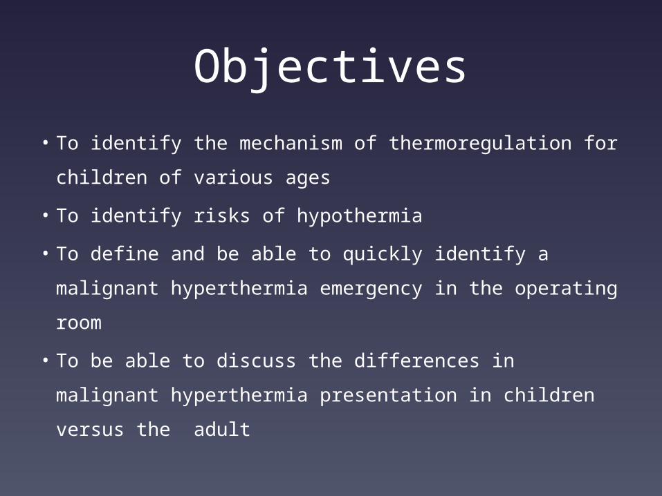
Objectives
• To identify the mechanism of thermoregulation for
children of various ages
• To identify risks of hypothermia
• To define and be able to quickly identify a malignant
hyperthermia emergency in the operating room
• To be able to discuss the differences in malignant
hyperthermia presentation in children versus the
adult

Our patients…
Children are NOT little
adults
They are a unique
patient population
Age groups:
• Neonate: less than 30
days
• Infant: 1-12 months
• Toddler: 13-23 months
• Preschool: 2-5 years
• School age: 6-11 years
• Adolescent: 12-18 years

Thinking about our care…
• You must consider a child’s
developmental stage and
the unique features of
each stage
• Care must be appropriate
to each developmental
age
• Unique physiological
aspects of each age group
and patient must also be
considered
What are our major anesthesia differences?

Airway
• Children have larger tongues
• Larger heads and shorter
necks, larger occiput
• Larynx is at C 3-4 level
• Larger epiglottis, that is
narrower and elongated
• Infant’s vocal cords slanted
posterior and cephalad
• Anterior airway more prone
to injury
• Narrowest part is cricoid
cartilage (until 5 years old)


Major Lung Differences• Rapid breathing rate and increased
alveolar ventilation
• Prone to rapid desaturation due to high
oxygen consumption
• Small FRC, fast inhalation induction
• Prone to atelectasis closing capacity may
exceed FRC
• Lung matures at 8 years old
• Increased chest compliance
• Herring-Breuer reflex- deep breath and
kids stop breathing and vagal negative
feedback loop via vagus nerve

Major Cardiac Differences
• High CO
• HR dependent
• Non-compliant heart
• Avoid air bubbles
due to possible PFO
• Vagal dominant and
unopposed
Thermoregulation
Hypothermia
• Defined as a core
temperature below
35 degrees
centigrade
• Asystole occurs at
30 degrees
centigrade
Pediatric Thermoregulation
• Adults use shivering
to increase heat
production
(increases O2
consumption, CO2
production, and CO)
• Shivering is inefficient
in young children
• Non-shivering
thermogenesis

Non-shivering thermogenesis• Cold induced O2
consumption and heat
production
• Primary means in infants to
produce heat
• Utilize brown fat- rich in
mitochondria, dense capillary
network, and innervated by
SNS nerve endings
• Brown fat is 6% of neonates
total body weight

How it works…..
• When norepinephrine
is stimulated by SNS,
triglycerides are
hydrolyzed to fatty
acids and glycerol with
heat being released
from enhanced
oxygen consumption

Other Differences
• Body surface area
(BSA) to body mass is
very high
• Infant’s head is 20%
of BSA and
contributes to 40% of
heat loss
• Rapid heat loss

Mechanisms of Heat Loss
• Evaporation
• Conduction
• Convection
• Radiation
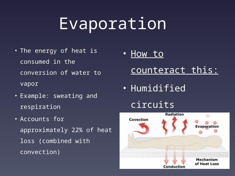
Evaporation
• The energy of heat is
consumed in the conversion
of water to vapor
• Example: sweating and
respiration
• Accounts for approximately
22% of heat loss (combined
with convection)
• How to counteract
this:
• Humidified circuits
• Run lower gas
flows

Conduction • The transfer of heat energy
due to a temperature
gradient
• Example: skin touching metal
OR table
• Accounts for approximately
15% of heat loss (combined
with convection)
• Pediatric patients have a
thinner layer of subQ fat so
more heat is lost though
conduction
• Ways to counteract
this loss:
• No skin to metal
contact
• Irrigation solution
warmed
• Warm IV fluids
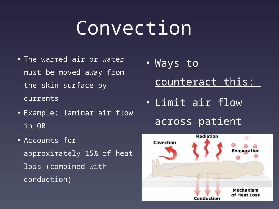
Convection
• The warmed air or water
must be moved away from
the skin surface by currents
• Example: laminar air flow in
OR
• Accounts for approximately
15% of heat loss (combined
with conduction)
• Ways to counteract
this:
• Limit air flow
across patient
• Warming blankets
above and below
patient
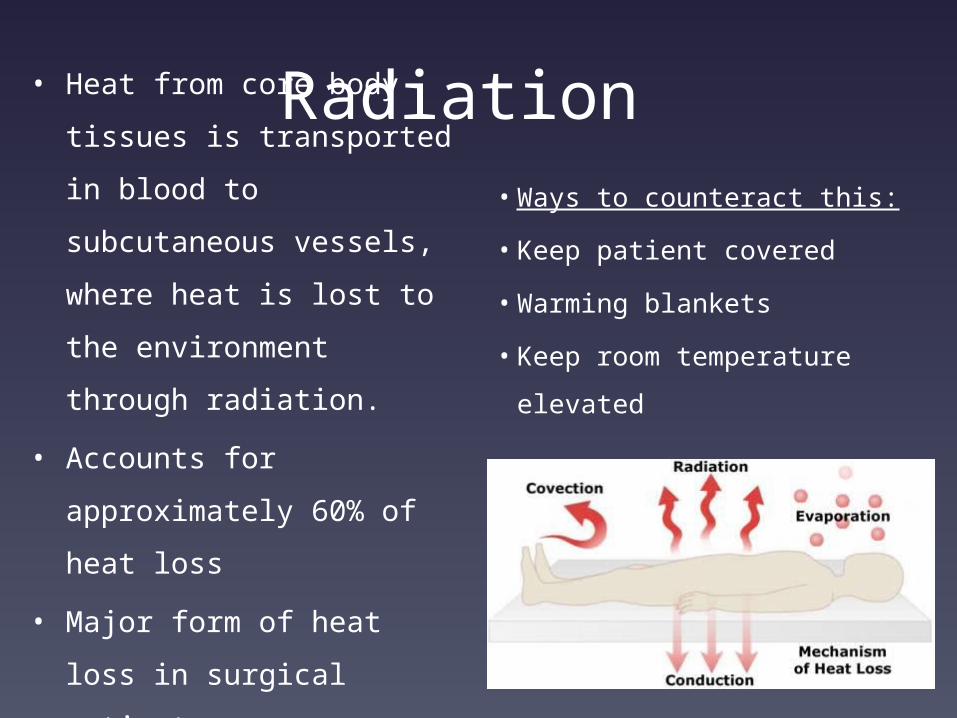
Radiation • Heat from core body
tissues is transported in
blood to subcutaneous
vessels, where heat is lost
to the environment
through radiation.
• Accounts for
approximately 60% of
heat loss
• Major form of heat loss in
surgical patients
• Ways to counteract this:
• Keep patient covered
• Warming blankets
• Keep room temperature
elevated
•
The research says….
• Best to use a warmer
ambient room
temperature and
warming blankets
• Pre-warming proved
beneficial in studies
• Esophagus,
nasopharynx or rectum
(highly perfused tissues,
the temperature of
which is uniform and
high in comparison with
the rest of the body)
best for measurement
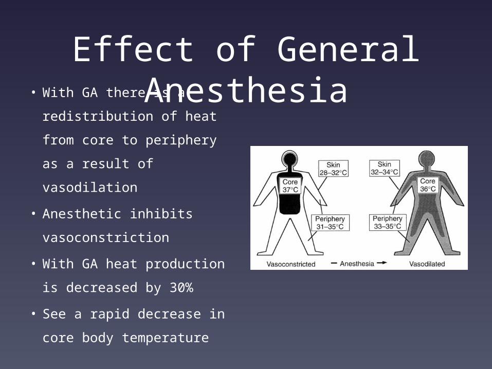
Effect of General Anesthesia• With GA there is a
redistribution of heat
from core to periphery as
a result of vasodilation
• Anesthetic inhibits
vasoconstriction
• With GA heat production
is decreased by 30%
• See a rapid decrease in
core body temperature
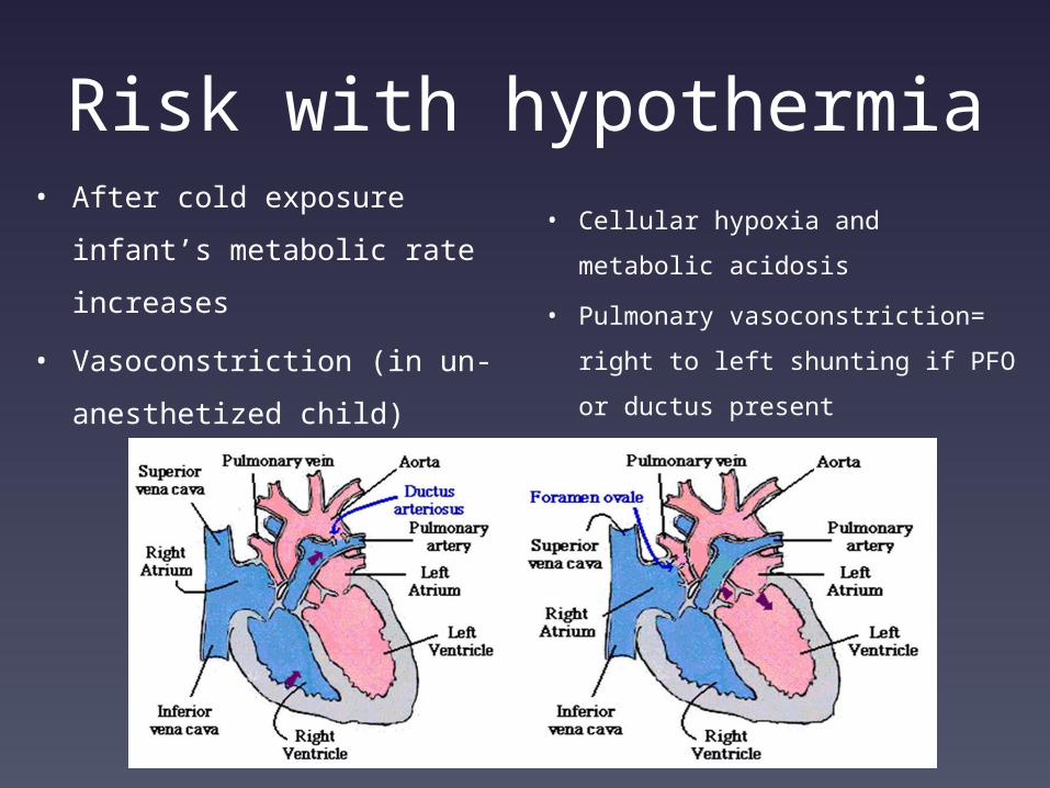
Risk with hypothermia• After cold exposure infant’s
metabolic rate increases
• Vasoconstriction (in un-
anesthetized child)
• Cellular hypoxia and metabolic
acidosis
• Pulmonary vasoconstriction=
right to left shunting if PFO or
ductus present
• Worsening hypoxia
Additional Risks with Hypothermia
• Adverse cardiac events
• Prolonged stay in the recovery room and
hospital
• Delayed surgical wound healing and higher
infection rates
• Cold-induced coagulation dysfunction
• Prolonged drug metabolism

Hyperthermia
• Elevated body
temperature due to
failure of
thermoregulation or
other disorder
• Heat stroke
• Adverse reaction to
drugs, such as malignant
hyperthermia

Case Discussion
History
• 4 month old male
• Wt. 6.48 kg
• NKDA
• No past surgical HX
• No medications
• HX of trigonocephaly
and premature birth
• No family HX of surgery

Surgical procedure
• Craniosynostosis
• GA with ETT
• Anticipation of large blood loss
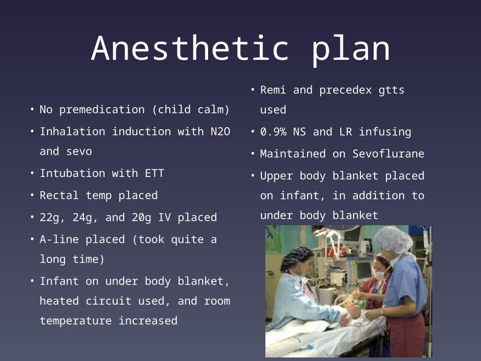
Anesthetic plan• No premedication (child
calm)
• Inhalation induction with N2O
and sevo
• Intubation with ETT
• Rectal temp placed
• 22g, 24g, and 20g IV placed
• A-line placed (took quite a
long time)
• Infant on under body blanket,
heated circuit used, and
room temperature increased
• Remi and precedex gtts
used
• 0.9% NS and LR infusing
• Maintained on Sevoflurane
• Upper body blanket placed
on infant, in addition to
under body blanket
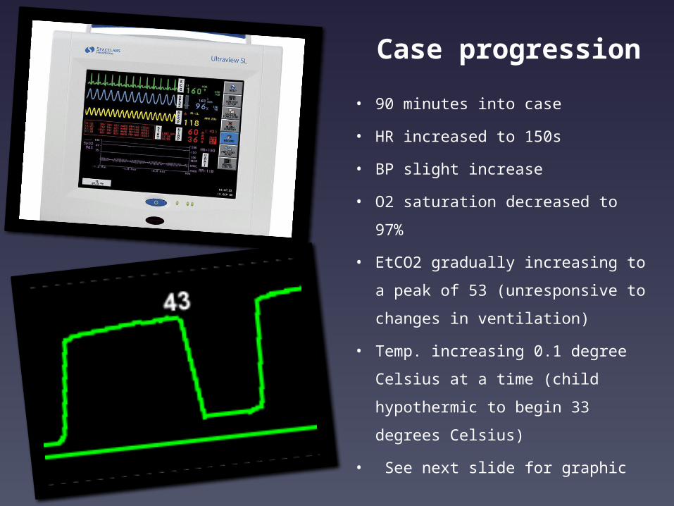
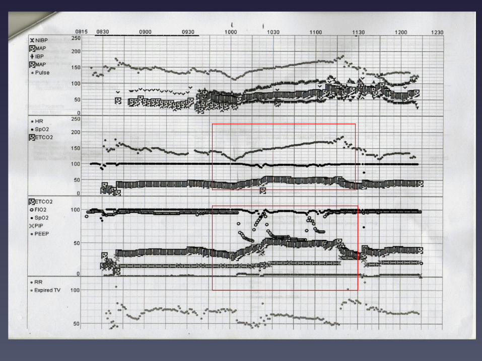
Case progression
• 90 minutes into case
• HR increased to 150s
• BP slight increase
• O2 saturation decreased to 97%
• EtCO2 gradually increasing to a
peak of 53 (unresponsive to
changes in ventilation)
• Temp. increasing 0.1 degree
Celsius at a time (child
hypothermic to begin 33
degrees Celsius)
• See next slide for graphic

Differential Diagnosis
• Gave fentanyl and
remi boluses to assure
child was not too light
• No change in EtCO2 in
ventilation changes
• ETT in good position
and not obstructed
• MALIGNANT
HYPERTHERMIA!!!!

Labs time 1013 1112 1129 1200 1448
ph 7.29 7.17 7.29 7.21 7.39
CO2 48 60 39 51 38
O2 97 71 250 393 419
K 3.6 5.1 5.1 3.7 4.0
temp 33.5 38.1 37.2 35.0
Bicarb 22.3 19.4 19.3 19.3 23.6
FiO2 60 100 100 100 100
Ca 1.26 1.24 1.55 1.40 1.45

time 1130 1357 1911 0116 0500 0841 2350 0438
CK 155 192 197 245 199 202 213 83
Myoglobin in urine: negative

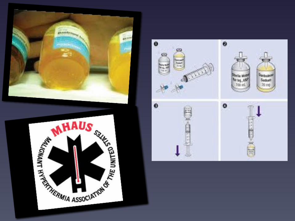
Treatment• Call for help!!!!
• Sevo stopped, flows increased
• CO2 absorber and circuit
changed
• Ice applied to infant, warming
blankets turned off, and room
temp decreased
• Dantrolene 2.5 mg/kg initial
dose
• Insulin R
• Dextrose
• Gtts changed to plasma-lyte
• Remi and precedex gtts ran
as anesthetic agents
• Calcium chloride given
• MHAUS called and assisted in
treatment plan
• Emergency algorithm guide
used
• Versed given
• Subsequent doses of
Dantrolene given at 1.5
mg/kg then 1 mg/kg
• Child transferred to PICU and
remained on ventilator

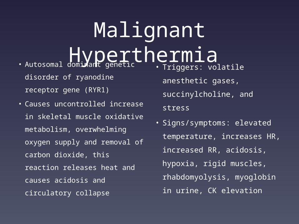
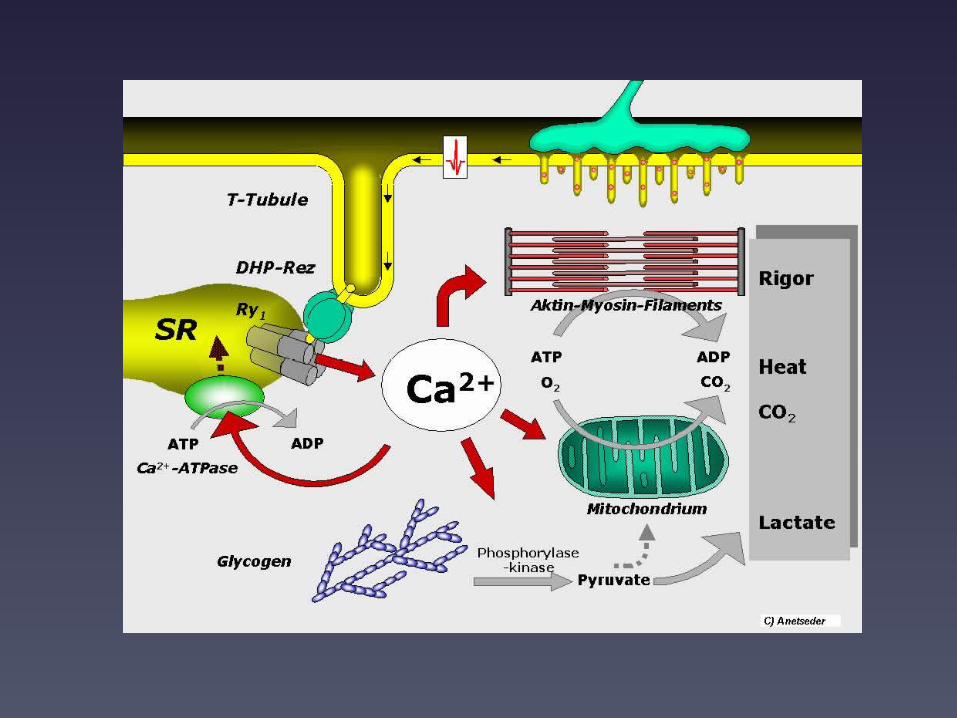
Malignant Hyperthermia • Autosomal dominant genetic
disorder of ryanodine receptor
gene (RYR1)
• Causes uncontrolled increase
in skeletal muscle oxidative
metabolism, overwhelming
oxygen supply and removal of
carbon dioxide, this reaction
releases heat and causes
acidosis and circulatory
collapse
• Triggers: volatile
anesthetic gases,
succinylcholine, and stress
• Signs/symptoms: elevated
temperature, increases HR,
increased RR, acidosis,
hypoxia, rigid muscles,
rhabdomyolysis, myoglobin
in urine, CK elevation
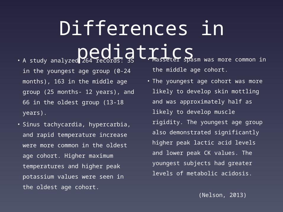
Differences in pediatrics • A study analyzed 264 records: 35 in
the youngest age group (0-24
months), 163 in the middle age
group (25 months- 12 years), and
66 in the oldest group (13-18
years).
• Sinus tachycardia, hypercarbia, and
rapid temperature increase were
more common in the oldest age
cohort. Higher maximum
temperatures and higher peak
potassium values were seen in the
oldest age cohort.
• Masseter spasm was more common
in the middle age cohort.
• The youngest age cohort was more
likely to develop skin mottling and
was approximately half as likely to
develop muscle rigidity. The
youngest age group also
demonstrated significantly higher
peak lactic acid levels and lower
peak CK values. The youngest
subjects had greater levels of
metabolic acidosis.
(Nelson,
2013)

A Published Case ReportThe Case:
• 7-year-old boy with cholesteatomas
underwent tympanoplasty.
• Three previous anesthetics with
sevoflurane induction and
maintenance with propofol infusion
were not associated with MH
symptoms.
• No family history of MH or muscle
disease
• A minor rise of end tidal CO2
• Increased rectal temperature
• Rhabdomyolysis and his father’s
positive IVCT results
Discussion:
• MH-susceptible patient responds
differently to various agents
• Atypical MH forms are problematic
• It is possible that the speed of onset
reflects the rate of increase of the
intracellular Ca2+ concentration, which
depends on the particular drug used, its
concentration in muscles and any
number of physiological variables that
dictate the efficacy of Ca2+
homeostatic processes in each patient.
(BONCIU, 2007)
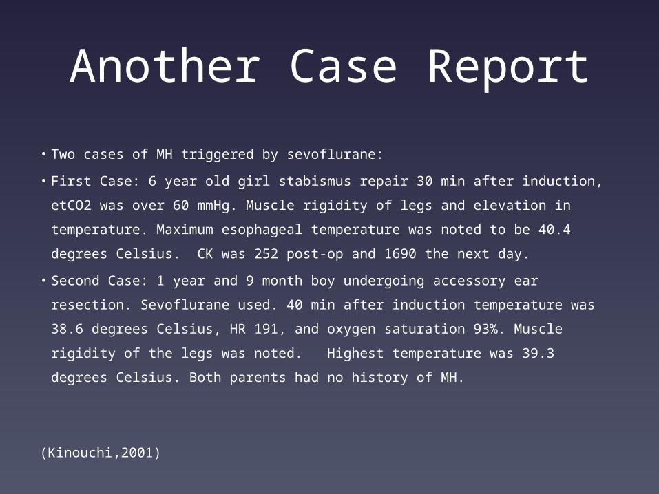
Another Case Report
• Two cases of MH triggered by sevoflurane:
• First Case: 6 year old girl stabismus repair 30 min after induction,
etCO2 was over 60 mmHg. Muscle rigidity of legs and elevation in
temperature. Maximum esophageal temperature was noted to be 40.4
degrees Celsius. CK was 252 post-op and 1690 the next day.
• Second Case: 1 year and 9 month boy undergoing accessory ear
resection. Sevoflurane used. 40 min after induction temperature was
38.6 degrees Celsius, HR 191, and oxygen saturation 93%. Muscle
rigidity of the legs was noted. Highest temperature was 39.3 degrees
Celsius. Both parents had no history of MH.
(Kinouchi,2001)

Take Home Message
• Kids can present with
MH differently than
adults
• If MH is suspected treat
with MH protocols
• Early interventions have
the best outcomes

References• CASSEY , J., KING, R., & ARMSTRONG , P. (2009). Is there thermal benefit from preoperative
warming in children?. Pediatric Anesthesia, 20(1), 63-71. Retrieved from
http://onlinelibrary.wiley.com/doi/10.1111/j.1460-9592.2009.03204.x/abstract
• Díaz, M., & Becker, D.(2010). Thermoregulation: Physiological and clinical considerations
during sedation and general anesthesia. Anesthesia Progress, 57(1), 25-33. Retrieved from
http://www.ncbi.nlm.nih.gov/pmc/articles/PMC2844235/
• Pearce, B., Christensen, R., & Voepel-Lewis, T. (n.d.). Perioperative hypothermia in the
pediatric population: Prevalence, risk factors and outcomes. Journal of Anesthesia & Clinical
Research, 1(1), 1-4. Retrieved from http://www.omicsonline.org/2155-6148/2155-6148-1-102.
• Sessler, D. (2011). Temperature monitoring: Consequences and prevention of mild
perioperative hypothermia. American Society of Anesthesiologists, 109, 1-7.

References • BONCIU, M., DE LA CHAPELLE, A., DELPECH, H., DEPRET, T., KRIVOSIC-HORBER,
R., & AIMÉ, M. (2007). Minor increase of endtidal CO2 during sevoflurane-induced
malignant hyperthermia. Pediatric Anesthesia, 17(2), 180-182.
doi:10.1111/j.1460-9592.2006.02051.
• Kinouchi, K., Okawa, M., Fukumitsu, K., Tachibana, K., Kitamura, S., & Taniguchi,
A. (2001). [Two pediatric cases of malignant hyperthermia caused by
sevoflurane]. Masui. The Japanese Journal Of Anesthesiology, 50(11), 1232-1235.
• Nelson, P., & Litman, R. (2013). Malignant Hyperthermia in Children: An Analysis
of the North American Malignant Hyperthermia Registry. Anesthesia And
Analgesia.

Thank You
Questions?
Related Documents


![Malignant hyperthermia [final]](https://static.cupdf.com/doc/110x72/58ceb1b71a28abb2218b5123/malignant-hyperthermia-final.jpg)


![[4]Scientific Advances in Malignant Hyperthermia](https://static.cupdf.com/doc/110x72/577d1cf31a28ab4e1e8b45c6/4scientific-advances-in-malignant-hyperthermia.jpg)
