LOCSU Community Services Primary Eyecare Assessment and Referral Service (PEARS) Pathway Issued by Local Optical Committee Support Unit December 2008 [Revised November 2013]

Welcome message from author
This document is posted to help you gain knowledge. Please leave a comment to let me know what you think about it! Share it to your friends and learn new things together.
Transcript

LOCS
U C
omm
unit
y Se
rvic
es
Primary Eyecare Assessment and Referral Service (PEARS) Pathway
Issued by Local Optical Committee Support Unit December 2008
[Revised November 2013]

LOCSU PEARS Pathway. Copyright © LOC Central Support Unit. Dec 2008. All Rights Reserved. [Rev Nov 2013]. Page 2 of 26
Contents Page
Outline description ............................................................................................................ 3
Key Drivers ............................................................................................................................ 3
Purpose of Service ............................................................................................................ 3
Description ........................................................................................................................... 4
Criteria for inclusion ...................................................................................................... 4
Same day referral .......................................................................................................... 5
Exclusions .......................................................................................................................... 5
Outcomes .......................................................................................................................... 5
Supply of therapy ........................................................................................................... 6
Record keeping ............................................................................................................... 6
Special requirements: Equipment ............................................................................. 7
Special requirements: Competencies ...................................................................... 7
Patient information ......................................................................................................... 8
The Welsh Experience .................................................................................................... 8
Primary presenting symptoms and outcome table ............................................. 9
Appropriateness of Optometric Management table .......................................... 10
Patient satisfaction .......................................................................................................... 11
Flashes and floaters management guidelines ...................................................... 12
Flashes and floaters pathway ...................................................................................... 16
Age-related Macular Degeneration guidelines .................................................... 17
Maculopathy referral pathway .................................................................................... 21
Red Eye pathway ................................................................................................................. 22
Referral forms (Word versions are available separately)
Appendix: Referral to Optometrist ................................................................................. 23
Appendix: First Optometric Attendance ....................................................................... 24
Appendix: Follow-up Attendance ................................................................................... 25

LOCSU PEARS Pathway. Copyright © LOC Central Support Unit. Dec 2008. All Rights Reserved. [Rev Nov 2013]. Page 3 of 26
Outline Description A PEARS examination will provide a timely assessment of the needs of a patient presenting with an eye condition. This will be undertaken by an accredited optometrist within suitably equipped premises who will manage the patient appropriately and safely. Management will be maintained within the primary care setting for as many patients as possible, thus avoiding unnecessary referrals to hospital services. Where referral to secondary care is required it will be to a suitable specialist with appropriate urgency.
Patients can self refer or be referred by GPs, pharmacists or other optometrists.
Key Drivers The national key drivers include:
• Equity & Excellence: liberating the NHS (2010) • Right Care: Increasing Value – Improving Quality (June 2010) • NHS 2010-15 ; from good to great • Operating framework for the NHS in England 2010/11 • Quality Innovation Productivity & Prevention (QIPP) agenda • HM Treasury (2010) The Spending Review Framework • Creating a patient-led NHS: Delivering the NHS Improvement Plan (March 2005) • Commissioning Framework for 2007-8 • Implement care closer to home; convenient quality care for patients (April 2007) • Commissioning Framework for health and well-being (March 2007) • Trust, Assurance and Safety – the Regulation of Health Professionals (February
2007) • Safeguarding patients ( February 2007) • The UK Vision Strategy
Purpose of Service Using the skills of primary care optometrists to triage, manage and prioritise patients presenting with an eye condition, patient care will be improved by:
• Improving access • Refining referrals • Ensuring referrals are appropriate and timely • Retaining patients in primary care where appropriate • Signposting to other appropriate services • Improve eye health in line with the UK Vision Strategy
LOCSU PEARS Pathway. Copyright © LOC Central Support Unit. Dec 2008. All Rights Reserved. [Rev Nov 2013]. Page 4 of 26
Description Patients can be referred into the service by their own GP or the practice nurse or surgery receptionist). There is a list of participating optometrists for the patient to choose from. Optometrists must, within reason, be able to offer an acute PEARS examination within 48 hours of the day that the appointment has been requested by the GP or pharmacist (excluding weekends and public holidays) unless it is for routine assessment. Where this is not possible, the patient should be directed to a colleague nearby. For acute potentially sight threatening eye conditions the optometrist should arrange to see the patient on the same day or refer directly to Eye Casualty. All referrals should be read and prioritised within 24 working hours. An appointment for a routine assessment should be offered within 2 weeks. The level of examination should be appropriate to the reason for referral. All procedures are at the discretion of the optometrist. Guidelines for the commonest eye conditions listed below. It is recommended that practitioners utilise the College of Optometrists’ Clinical Management Guidelines which can be found on their website www.college-optometrists.org/en/professional-standards/clinical_management_guidelines/index.cfm and on the Medicines Support Unit website for optometrists www.med-support.org.uk. A GOS sight test or private eye examination may also be required but it would be unusual for this to be carried out at the same time as a PEARS examination. Practitioners should at all times respect the patient’s loyalty to their usual optometrist and not solicit the provision of services that fall outside the scope of the service. The patient’s details should NOT be added to the practice reminder system for the purpose of sending recall letters for regular eye examinations, unless the patient expressly requests it. Children under 17 years of age should be accompanied by a responsible adult. The criteria for inclusion of patients may include the following:
• Loss of vision including transient loss • Ocular pain • Systemic disease affecting the eye • Differential diagnosis of the red eye • Foreign body and emergency contact lens removal (not by the fitting practitioner) • Dry eye • Epiphora (watery eye) • Trichiasis (in growing eyelashes)

LOCSU PEARS Pathway. Copyright © LOC Central Support Unit. Dec 2008. All Rights Reserved. [Rev Nov 2013]. Page 5 of 26
• Differential diagnosis of lumps and bumps in the vicinity of the eye • Recent onset of Diplopia • Flashes/floaters • Retinal lesions • Field defects • GP referral
The following cases should be referred directly to the nearest Eye Casualty:
• Severe ocular pain requiring immediate attention • Suspect Retinal detachment • Retinal artery occlusion • Chemical injuries • Penetrating trauma • Orbital cellulitis • Temporal arteritis • Ischaemic optic neuropathy
Other conditions excluded from the service:
• Diabetic retinopathy • Adult squints, long standing diplopia
Outcomes Outcomes resulting from the consultation are likely to be one of the following:
• The optometrist decides to manage the condition, and offers the patient advice and/or prescribes/recommends medication. A follow-up consultation may be necessary.
• The optometrist carries out a minor clinical procedure e.g. eyelash removal or foreign body removal. A follow-up consultation may be necessary.
• The optometrist diagnoses the condition and suggests/prescribes appropriate medication or the GP is requested to prescribe
• The optometrists makes a tentative diagnosis and refers the patient urgently/non-urgently into the Hospital Eye Service using the usual channels of communication
• The optometrist reassures the patient and discharges him/her • The examining optometrist recommends an NHS or private sight test.
All procedures undertaken and advice given to the patient should be recorded on a patient record card or electronic device, and stored in a safe retrieval system.

LOCSU PEARS Pathway. Copyright © LOC Central Support Unit. Dec 2008. All Rights Reserved. [Rev Nov 2013]. Page 6 of 26
Supply of therapy Registered Optometrists may sell or supply all pharmacy medicines (P) or general sale list medicines (GSL) in the course of their professional practice, including 0.5% Chloramphenicol antibiotic eye drops or 1% eye ointment. Optometrists may give the patient a written (signed) order for the patient to obtain the above from a registered pharmacist, as well as the following prescription only medicines (POMs):
• Chloramphenicol • Cyclopentolate hydrochloride • Fusidic Acid • Tropicamide
In making the supply to the patient the optometrist must ensure:
• Sufficient medical history is obtained to ensure that the chosen therapy is not contra-indicated in the patient
• All relevant aspects, in respect of labelling of medicine outlined in the Medicine Act 1968 are fully complied with
• The patient has been fully advised on the method and frequency of administration of the product
In general, supply via a pharmacist is preferred. The College of Optometrists has produced guidelines on the use & supply of drugs as part of its ‘Code of Ethics & Guidelines for Professional Conduct’ section K1: www.college-optometrists.org/en/professional-standards/Ethics_Guidelines/index.cfm. If the patient is exempt from prescription charges, supply of appropriate treatments could be covered by Group Prescribing Directives and/or by Minor Ailment Services in accordance with The National Pharmacy Enhanced Service Plan already in existence. Independent prescribing optometrists may be able to issue FP10s depending on local agreements.
Record keeping On conclusion of a PEARS examination the optometrist must complete a PEARS report form, entering the information on the IT system where applicable for audit purposes & report to the referring GP, and to the hospital eye service, should an onward referral be necessary.

LOCSU PEARS Pathway. Copyright © LOC Central Support Unit. Dec 2008. All Rights Reserved. [Rev Nov 2013]. Page 7 of 26
Special requirements – equipment All practices contracted to supply the service will be expected to employ an accredited optometrist and have the following equipment available:
• Access to the Internet • Means of indirect ophthalmoscopy (Volk/headset indirect ophthalmoscope) • Slit lamp • Applanation Tonometer • Distance test chart (Snellen/logmar) • Near test type • Equipment for epilation • Threshold fields equipment to produce a printed report • Amsler Charts • Equipment for FB removal • Appropriate ophthalmic drugs
o Mydriatic o Anaesthetic o Staining agents
Special requirements – competencies All participating optometrists will have the core competencies as defined by the GOC and may require some extra training or updating of skills. In addition the following apply:
• Aware of own limitations. • Does not compromise patient safety.
Training and accreditation for participating optometrists to perform within PEARS will include demonstrating the ability to identify and manage a range of ocular abnormalities and proficiency in the use of certain elements of the above-mentioned equipment. Participating optometrists must complete the Cardiff University/LOCSU PEARS Distance Learning modules (Part 1) and the associated Practical Skills Demonstration (Part 2). In order to progress to the second element of the accreditation process, a candidate must have successfully passed the first.

LOCSU PEARS Pathway. Copyright © LOC Central Support Unit. Dec 2008. All Rights Reserved. [Rev Nov 2013]. Page 8 of 26
An optometrist who has a relevant higher qualification and experience may be exempt from the PEARS Distance Learning and/or the Practical Skills Assessment at the discretion of the Clinical Lead. Participating optometrists will also be expected to keep their knowledge and skills up to date.
Patient information Examples of leaflets that will be available and will be handed to patients as appropriate are:
• Mydriatic drops - warning re pupil dilation • Tear Dysfunction/Dry eye • Blepharitis • Conjunctivitis • Trichiasis • Epiphora • Foreign body removal • Flashes & floaters • Age related macular degeneration • Glaucoma • Public Health Messages e.g.
o Smoking cessation o Obesity o Alcohol abuse
The Welsh Experience Generally there is little data on referrals into ophthalmology on the basis of condition and there is no differentiation between optometry referral and GP referral. However using the figures from the PEARS evaluation in Wales1 it can be seen that a significant number of cases can be either:
• Discharged at first visit or managed in practice or • Referred to GP for appropriate care
1 Br J Ophthalmol. 2009 Apr;93(4):435-8. Epub 2008 Nov 21. Novel optometrist-led all Wales primary eye-care services: evaluation of a prospective case series. Sheen NJ, Fone D, Phillips CJ, Sparrow JM, Pointer JS, Wild JM. Cardiff School of Optometry and Vision Sciences, Cardiff University, Maindy Road, Cardiff CF24 4LU, UK. [email protected]

LOCSU PEARS Pathway. Copyright © LOC Central Support Unit. Dec 2008. All Rights Reserved. [Rev Nov 2013]. Page 9 of 26
In addition, of the patients self-referring for an acute eye problem, 27% were discharged at the first visit, 36% were managed in practice, 15% were referred to the GP and only 22% were referred to the HES. In England, a recent evaluation2 undertaken by Somerset CCG showed that, over an 18 month period, 86% of presentations were managed within the community by optometrists. Between 38-58% patients would have had to have been seen by secondary care if they were not seen under the ACES scheme. The threshold stated by the CCG to make ACES economic was 40%, and so the review supported the cost benefit of ACES. The table below lists the most common reasons for patients to present to a PEARS accredited optometrist.
Primary symptom and outcome of the 4881 patients presenting for a PEARS examination
Symptom reported Referred to HES
Managed in practice or discharged
Referred to GP
Total number
Acute vision loss 160 27 1 188
Chronic vision loss 132 376 3 511
Distorted vision 64 49 0 113
Diplopia 23 2 3 28
Headaches 15 83 116 214
Unilateral red eye 140 888 388 1416
Bilateral red eye 15 99 2 116
Discomfort / Irritation 87 590 309 986
Ocular discharge, only 12 2 0 14
Unusual lid appearance 11 4 5 20
Flashes + floaters 190 403 8 601
Floaters, only 34 39 2 75
Trauma 18 34 15 67
Unclassified 96 428 8 532
2 Evaluation of the ACES Somerset Service. 2009-2011. East Quay Vision – [email protected] with support from the College of Optometrists, LOC, NHS Somerset & East Quay Medical Centre
LOCSU PEARS Pathway. Copyright © LOC Central Support Unit. Dec 2008. All Rights Reserved. [Rev Nov 2013]. Page 10 of 26
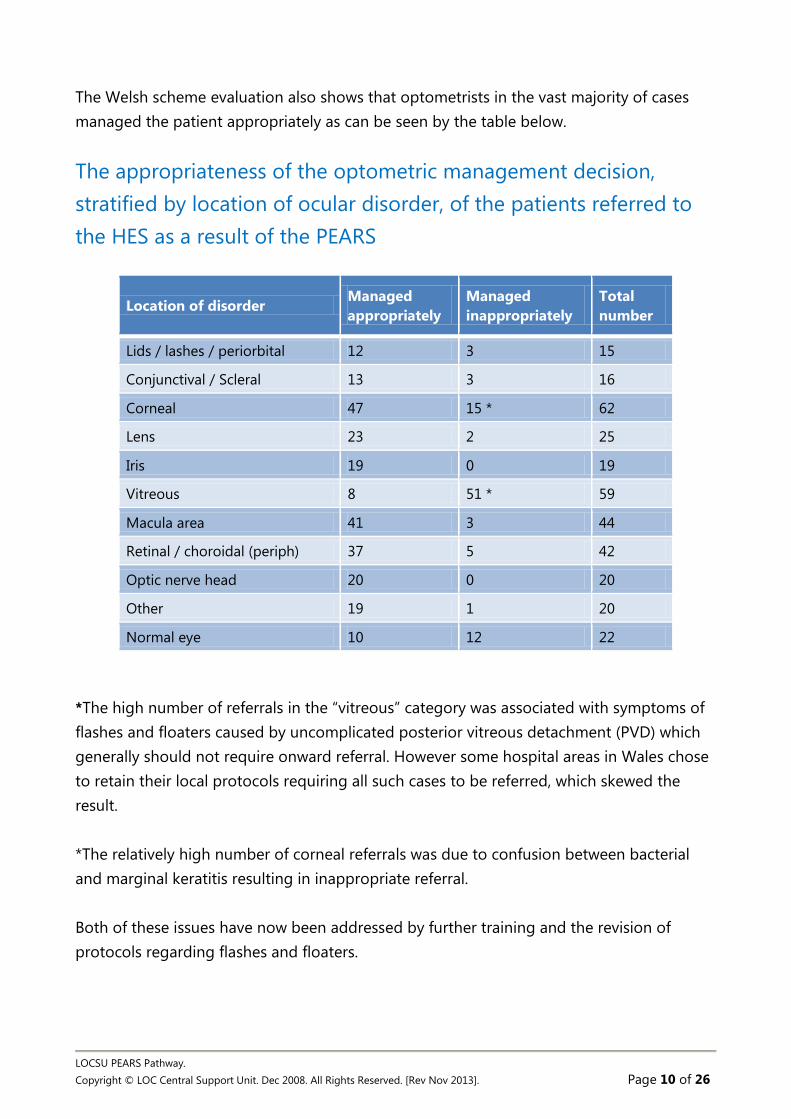
The Welsh scheme evaluation also shows that optometrists in the vast majority of cases managed the patient appropriately as can be seen by the table below.
The appropriateness of the optometric management decision, stratified by location of ocular disorder, of the patients referred to the HES as a result of the PEARS
Location of disorder Managed appropriately
Managed inappropriately
Total number
Lids / lashes / periorbital 12 3 15
Conjunctival / Scleral 13 3 16
Corneal 47 15 * 62
Lens 23 2 25
Iris 19 0 19
Vitreous 8 51 * 59
Macula area 41 3 44
Retinal / choroidal (periph) 37 5 42
Optic nerve head 20 0 20
Other 19 1 20
Normal eye 10 12 22
*The high number of referrals in the “vitreous” category was associated with symptoms of flashes and floaters caused by uncomplicated posterior vitreous detachment (PVD) which generally should not require onward referral. However some hospital areas in Wales chose to retain their local protocols requiring all such cases to be referred, which skewed the result. *The relatively high number of corneal referrals was due to confusion between bacterial and marginal keratitis resulting in inappropriate referral. Both of these issues have now been addressed by further training and the revision of protocols regarding flashes and floaters.

LOCSU PEARS Pathway. Copyright © LOC Central Support Unit. Dec 2008. All Rights Reserved. [Rev Nov 2013]. Page 11 of 26
Patient satisfaction The Evaluation in Wales also asked patients how they had found the service:
• 84.1% thought that their optometrist ‘seemed to know what their eye problem was’ • 86.5% also thought that ‘their optometrist knew what to do for their eye problem,’ • 92.7% also thought that ‘they had the chance to tell their optometrists everything
they wanted to about their eye problem’, • 94.5% also thought that ‘their optometrist understood them,’ • 98.3% considered that ‘they got on well with their optometrist,’ • 94.8% were “very satisfied” and the remaining 5.2% were “fairly satisfied”. No
interviewees were “very dissatisfied”, “fairly dissatisfied” or “neither”.

LOCSU PEARS Pathway. Copyright © LOC Central Support Unit. Dec 2008. All Rights Reserved. [Rev Nov 2013]. Page 12 of 26
Flashes and Floaters Management Guidelines Terminology
The following terms are important in this text:
Retinal break This is a retinal hole or tear Retinal detachment This is any type of retinal detachment including rhegmatogenous, traction or exudative
Optometric Assessment
History and symptoms
A full and thorough history and symptoms is essential. In addition to the normal history and symptoms, careful attention must also be given to the following:
History: • Age • Myopia • Family history of retinal break or detachment • Previous ocular history of break or detachment • Systemic disease • History of recent ocular trauma, surgery or inflammation
Symptoms:
• Loss or distortion of vision (a curtain / shadow / veil over vision) • Floaters • Flashes
For symptoms of floaters these additional questions should be asked:
• Are floaters of recent onset? • What do they look like? • How many are there? • Which eye do you see them in? • Any flashes present

LOCSU PEARS Pathway. Copyright © LOC Central Support Unit. Dec 2008. All Rights Reserved. [Rev Nov 2013]. Page 13 of 26
For symptoms of flashes these additional questions should be asked:
• Describe the flashes? • How long do they last? • When do you notice them?
For symptoms of a cloud, curtain or veil over the vision these additional questions should be asked:
• Where in the visual field is the disturbance? • Is it static or mobile? • Which eye? • Does it appear to be getting worse?
Symptoms of less concern:
• Long term stable flashes and floaters • Symptoms >2 months • Normal vision
Clinical examination
All patients presenting for a PEARS examination with symptoms indicative of a potential retinal detachment should have the following investigations (in addition to such other examinations that the optometrist feels are necessary):
• Tests of pupillary light reaction including swinging light test for Relative Afferent Pupil Defect (RAPD), prior to pupil dilation
• Visual acuity recorded and compared to previous measures • Contact tonometry, noting IOP discrepancy between eyes • Visual Field examination at discretion of optometrist • Slit lamp bio microscopy of the anterior and posterior segments, noting:
o Pigment cells in anterior vitreous, 'tobacco dust' (Shafer’s sign) o Vitreous haemorrhage o Cells in anterior chamber (mild anterior uveitic response)
• Dilated pupil fundus examination with slit lamp binocular indirect ophthalmoscopy using a Volk or similar fundus lens (wide field fundus lens optimal) asking the patient to look in the 8 cardinal directions of gaze and paying particular attention to the superior temporal quadrant as about 60% of retinal breaks occur in that area. Noting:
o Status of peripheral retina, including presence of retinal tears, holes, detachments or lattice degeneration
o Presence of vitreous syneresis or Posterior Vitreous Detachment (PVD)

LOCSU PEARS Pathway. Copyright © LOC Central Support Unit. Dec 2008. All Rights Reserved. [Rev Nov 2013]. Page 14 of 26
Management
If local protocols for the referral of retinal detachment are in place, then these should be followed. If not, you should note that some HES ophthalmology departments will not have RD surgery facilities. In these cases it is best to telephone the department first to find out what procedures to follow. Symptoms requiring assessment within 24 hours:
1. Sudden increase in number of floaters, patient may report as "numerous", "too many to count" or “sudden shower or cloud of floaters” Suggests blood cells, pigment cells, or pigment granules (from the retinal pigment epithelium) are present in the vitreous. Could be signs of retinal break or detachment present
2. Cloud, curtain or veil over the vision. Suggests retinal detachment or vitreous haemorrhage – signs of retinal break or detachment should be present
Signs requiring referral within 24 hours:
1. Retinal detachment with good vision unless there is imminent danger that the fovea is about to detach i.e. detachment within 1 disc diameter of the fovea especially a superior bulbous detachment, when urgent surgery is required.
2. Vitreous or pre-retinal haemorrhage 3. Pigment 'tobacco dust' in anterior vitreous 4. Retinal tear/hole with symptoms
Signs requiring referral ASAP next available clinic appointment:
Retinal detachment with poor vision (macula off) unless this is long standing Retinal hole/tear without symptoms Lattice degeneration with symptoms of recent flashes and/or floaters
Require discharge with SOS advice (verbal advice and a leaflet):
1. Uncomplicated PVD without signs and symptoms listed above 2. Signs of lattice degeneration without symptoms listed above
Explain the diagnosis and educate the patient on the early warning signals of further retinal traction and possible future retinal tear or detachment:
• Give the patient a Retinal Detachment warning leaflet • Instruct the patient to return immediately or go to A&E if flashes or floaters
worsen

LOCSU PEARS Pathway. Copyright © LOC Central Support Unit. Dec 2008. All Rights Reserved. [Rev Nov 2013]. Page 15 of 26
Referral letters
Patients requiring referral for retinal breaks or detachment must have the following noted on the referral form to the ophthalmologist. Letters should be typed whenever possible and may be faxed or sent with the patient in urgent cases.
• A clear indication of the reason for referral e.g. Retinal tear in superior temporal periphery of Right eye
• A brief description of any relevant history and symptoms • A description of the location of any retinal break / detachment / area of lattice • In the case of retinal detachment whether the macula is on or off. • The urgency of the referral

LOCSU PEARS Pathway. Copyright © LOC Central Support Unit. Dec 2008. All Rights Reserved. [Rev Nov 2013]. Page 16 of 26
Flashes and Floaters Patient Pathway
Positive signs
Refer Urgent 24hrs Soon – next available clinic Routine
Patient presents via PEARS to Optometrist
Investigations as per protocol
Negative signs
Discharge SOS advice Explain / educate on RD Given written warnings
Symptoms of less concern
• Stable flashes and floaters • Symptoms >2 months • Normal vision
Clinically significant symptoms
• Recent onset • Increasing flashes and/or floaters • Less than 6 weeks duration • Field loss • Cloud, curtain or veil over vision

LOCSU PEARS Pathway. Copyright © LOC Central Support Unit. Dec 2008. All Rights Reserved. [Rev Nov 2013]. Page 17 of 26
Age-related Macular Degeneration Management Guidelines
Terminology The following terms are important in this text & for differential diagnosis:
Wet (exudative) AMD This can progress very rapidly causing loss of central vision & metamorphopsia (distortion). It is characterised by sub retinal neovascular membrane, macular haemorrhages & exudates.
Dry (atrophic) AMD A slowly progressive disease characterized by drusen & retinal pigment epithelial changes
Optometric Assessment
History and symptoms
A full and thorough history and symptoms is essential. In addition to the normal history and symptoms, careful attention must also be given to the following: History
• Age • Family history of maculopathy • Previous ocular history • Systemic disease eg hypertension, diabetes • History of ocular surgery- cataract extraction, retinal detachment repair • Myopia • Medication e.g. chloroquine derivatives, tamoxifen • Smoking status • Excessive exposure to sunlight/UV
Symptoms
• Loss of central vision • Spontaneously reported distortion of vision
LOCSU PEARS Pathway. Copyright © LOC Central Support Unit. Dec 2008. All Rights Reserved. [Rev Nov 2013]. Page 18 of 26
These additional questions should be asked:
• Is loss of vision of recent onset? • In which eye are symptoms present? • Has the loss of vision occurred suddenly or gradually?
Clinical examination All patients presenting for a PEARS examination with symptoms indicative of a potential macular degeneration should have the following investigations (in addition to such other examinations that the optometrist feels are necessary):
• Tests of pupillary light reaction including swinging light test for Relative Afferent Pupil Defect (RAPD), prior to pupil dilation
• Visual acuity recorded and compared to previous measures • Refraction as a hyperopic shift can be indicative of macular oedema • Amsler grid or similar assessment of central vision • Dilated pupil fundus examination with slit lamp binocular indirect ophthalmoscopy
using a Volk or similar fundus lens noting:
o Status of macula, including presence of drusen(&size), haemorrhages, pigment epithelial changes ie hyper or hypo pigmentation, exudates,, oedema, signs of sub retinal neovascular membrane
Management If local protocols for the referral of AMD are in place, then these should be followed. If not, you should note that some HES ophthalmology departments will not have the facilities to deal with wet age related macular degeneration. In these cases it is best to telephone the department first to find out what procedures to follow. Symptoms requiring referral ASAP next available clinic appointment:
1. Sudden deterioration in vision + VA better than 3/60 in affected eye 2. Spontaneously reported distortion in vision + VA better than 3/60
Signs requiring referral ASAP next available clinic appointment:
1. Sub retinal neovascular membrane 2. Macular haemorrhage 3. Macular oedema
LOCSU PEARS Pathway. Copyright © LOC Central Support Unit. Dec 2008. All Rights Reserved. [Rev Nov 2013]. Page 19 of 26
Requiring routine referral: 1. Patient eligible and requesting certification of visual impairment 2. Patients requesting a home visit from Social Services to help them manage their
visual impairment in their home. 3. Patients who require an assessment for LVA 4. Patients likely to benefit from an intra-ocular Galilean telescope system
Low Vision Aids may be available in the community or hospital eye service - this varies in different areas. Require routine follow up but provide an Amsler chart, verbal advice and a leaflet – see sheet appended).
• Dry AMD, drusen and / or pigment epithelial changes • Explain the diagnosis and educate the patient on the early warning signs of wet
AMD. • Give stop smoking advice via leaflet if appropriate + advice on healthy diet +
protection from blue light • Use 4 point scale to assess risk of AMD progression. Count one point for large
drusen of 125 microns or larger (about the size of a vein at the disc margin) and one point for any pigmentary change. Score each eye separately and then add them together for a score out of 4. A full score of 4 points means a 50% chance of progressing to advanced AMD in the next 5 years. 3 points gives a 25% chance, 2 points a 12% chance and with 1 point the risk is just 3%.
• For those at intermediate risk of AMD progression give information on AREDS findings & leaflet on anti-oxidant supplements
• Give information on local services for the visually impaired- public and third sector.
• Give appropriate information on national voluntary agencies e.g. RNIB, Macular Disease Society
• Instruct the patient to inform the practice or GP immediately if vision suddenly deteriorates or becomes distorted.

LOCSU PEARS Pathway. Copyright © LOC Central Support Unit. Dec 2008. All Rights Reserved. [Rev Nov 2013]. Page 20 of 26
Referral letters Patients requiring referral for macular degeneration must have the following noted on the referral form to the ophthalmologist. Letters should be typed whenever possible and may be faxed or sent with the patient in urgent cases. The Royal College of Ophthalmologists fast track referral form for wet AMD can be used: www.college-optometrists.org/en/utilities/document-summary.cfm/docid/81143450-07B2-4A16-BA3ED6F3F7A86D77
• A clear indication of the reason for referral e.g. macular haemorrhage • A brief description of any relevant history and symptoms • A description of the type of macular degeneration or signs of drusen, pigment
epithelial changes, sub retinal neovascular membrane, haemorrhages, exudates, macular oedema.
• The urgency of the referral
Differential diagnosis Macular hole
This is a hole at the macula caused by tangential vitreo-retinal traction at the fovea. Causes impaired central vision & typically affects elderly females
Macular epiretinal membrane Can be divided into cellophane maculopathy & macular pucker
Central Serous Retinopathy Typically sporadic, self-limited disease of young or middle-aged adult males. Unilateral localised detachment of sensory retina at the macula causing unilateral blurred vision.
Cystoid Macular Oedema An accumulation of fluid at the macula most commonly due to retinal vascular disease, intra-ocular inflammatory disease or post cataract surgery,
Myopic Maculopathy Chorio retinal atrophy can occur with high myopia, usually > 6.00D, which can involve the macula.

LOCSU PEARS Pathway. Copyright © LOC Central Support Unit. Dec 2008. All Rights Reserved. [Rev Nov 2013]. Page 21 of 26
Solar Maculopathy
Due to the effects of solar radiation from looking at the sun causing circumscribed retinal pigment epithelium mottling or a lamellar hole at the macula.
Drug Induced Maculopathies Antimalarials e.g chloroquine, hydroxychloroquine Phenothiazines eg thioridazine (melleril), chlorpromazine (Largactil) Tamoxifen

LOCSU PEARS Pathway. Copyright © LOC Central Support Unit. Dec 2008. All Rights Reserved. [Rev Nov 2013]. Page 22 of 26
Maculopathy Referral Pathway
Positive signs
Refer Soon – next available clinic Routine Inform Social Services
Patient presents via PEARS to Optometrist
Investigations as per protocol
Negative signs
Discharge • SOS advice • Explain / educate on types of
maculopathy • Give Amsler grid • Advice on:
smoking cessation Blue light Vitamin supplements
Symptoms of less concern • Longstanding loss of vision • Gradual deterioration in vision • Normal vision
Clinically significant symptoms • Loss of vision of recent onset • Spontaneously reported visual
distortion

LOCSU PEARS Pathway. Copyright © LOC Central Support Unit. Dec 2008. All Rights Reserved. [Rev Nov 2013]. Page 23 of 26
Red Eye Pathway
Patient presents to PEARS Optometrist
Optometrist takes History and symptoms and examines patient and makes initial diagnosis
Manage in practice • Bacterial conjunctivitis • Allergic conjunctivitis • Non-herpetic viral conjunctivitis • Subconjunctival haemorrhage • Tear Dysfunction (Dry eye) • Episcleritis • Marginal keratitis • Superficial abrasions • Recurrent epithelial erosion • Small corneal foreign bodies
• Remove • In-growing eyelash
• Remove
Treat and advise • Antimicrobials • Mast cell stabilisers • Ocular lubricants • Artificial tears • Topical antihistamines • Ibuprofen
Follow up Generally none expected Exceptions • Repeated in-growing
lashes • Dry eye
No improvement? Refer to secondary care
Urgent telephone referral • Infective keratitis • Anterior uveitis • Posterior uveitis • Scleritis
Complete record and report to GP
Follow up in Secondary care
LOCSU PEARS Pathway Copyright © LOC Central Support Unit. Dec 2008. [Revised June 2012]. All Rights Reserved Page 24 of 26
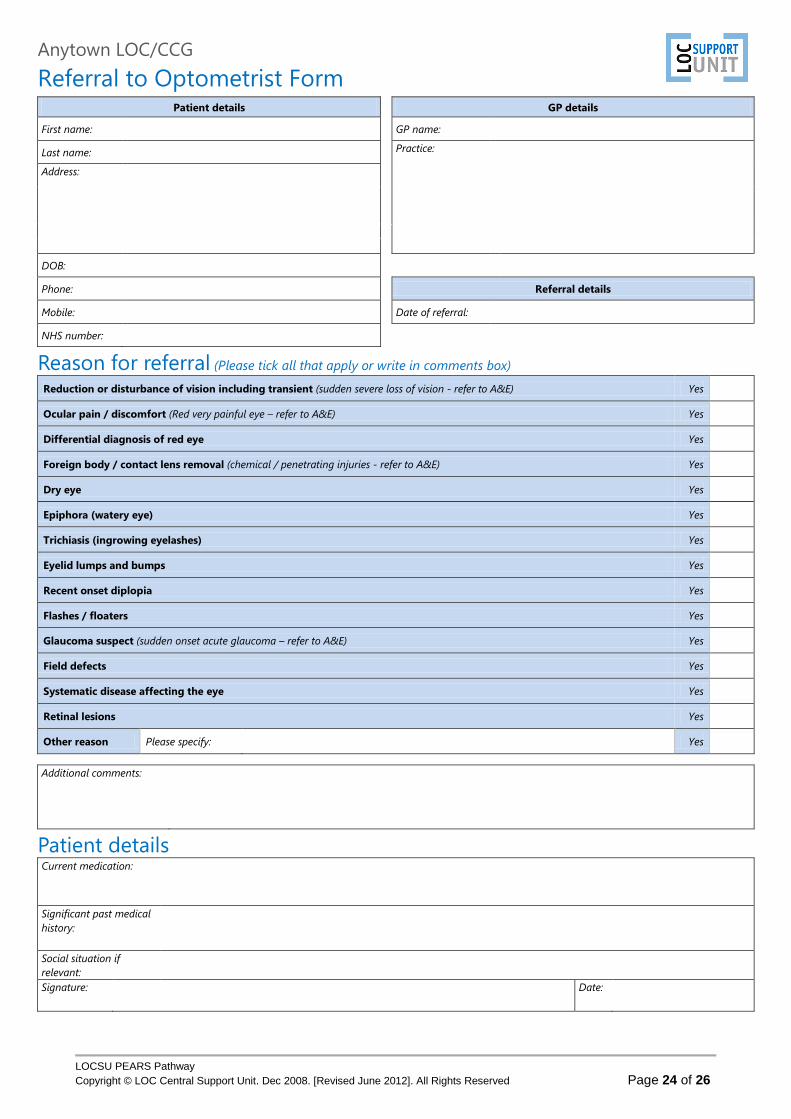
Anytown LOC/CCG
Referral to Optometrist Form Patient details GP details
First name: GP name:
Last name: Practice:
Address:
DOB:
Phone: Referral details
Mobile: Date of referral:
NHS number:
Reason for referral (Please tick all that apply or write in comments box) Reduction or disturbance of vision including transient (sudden severe loss of vision - refer to A&E) Yes
Ocular pain / discomfort (Red very painful eye – refer to A&E) Yes
Differential diagnosis of red eye Yes
Foreign body / contact lens removal (chemical / penetrating injuries - refer to A&E) Yes
Dry eye Yes
Epiphora (watery eye) Yes
Trichiasis (ingrowing eyelashes) Yes
Eyelid lumps and bumps Yes
Recent onset diplopia Yes
Flashes / floaters Yes
Glaucoma suspect (sudden onset acute glaucoma – refer to A&E) Yes
Field defects Yes
Systematic disease affecting the eye Yes
Retinal lesions Yes
Other reason Please specify: Yes
Additional comments:
Patient details Current medication:
Significant past medical history:
Social situation if relevant:
Signature: Date:

LOCSU PEARS Pathway. Copyright © LOC Central Support Unit. Dec 2008. All Rights Reserved. [Rev Nov 2013]. Page 25 of 26
Anytown LOC/CCG
PEARS – First Attendance Patient’s Details Optometrist / Practice
First name: Optometrist: Last name: OPL number: DOB: Practice: NHS number: Address:
Phone: Patient’s GP
Phone: GP name: Mobile: Practice: Email:
Referral info Date referred: Date seen: Referred by GP Patient Optometrist Sight test Carried out Not carried out Not carried out but advised
Reason for referral Headache Loss of vision Ocular discomfort Other (specify):
Flashes/floaters Red eye Trauma
Diagnosis
Conjunctiva
Resolved
Anterior uveitis
KP
Retinal lesions
?Melanoma Pinguecula Flare Naevus
Eyelid lumps and bumps
Resolved Allergic CL injection BRVO/CRVO Concretions Bacterial Synechiae CRAO/BRAO
Papilloma Viral Macular degeneration
Drusen Isolated haem Cyst Tumour Dry Other (specify below) Stye Episcleritis Wet
Field defects
Physiological ?Malignant Scleritis
Maculopathy Serious ret Artefact
Tear Dysfunction
Dry eye
Cornea
Foreign body Cellophane Longstanding Conjunctivitis Pterygium Macular hole Glaucoma
Tear duct Keratoconus
Flashes/ floaters
Weiss ring/PVD Neurological Lid laxity Marginal keratitis Tobacco dust
Diplopia Resolved
Ectropion Dystrophy Retinal hole/tear Recent onset Lid and Lash problems
Trichiasis Herpes keratitis Ret.Detachment refractive Entropion Microbial keratitis non-PVD floaters
Systematic disease affecting eye
Hypertension Blepharitis Other keratitis Diabetes
MS
Other diagnosis /
comments: Thyroid Arteritis
Dist. VA Right:
Action taken
Discharged Additional comments: Left: Epilated
Smoker? Yes Foreign body removed
Recent ex Lubricated No Lid hygene
Dilated? Yes Rx requested by GP (specify) No Follow up (specify interval)
Tonometry
Right: Refer back to GP
Left: Refer to secondary care (specify routine, urgent,
emergency)
Signature: Date:
STATEMENT: The reason for this referral has been explained to the patient or guardian who agrees to it. The patient or guardian also consents to information being exchanged between the Hospital Eye Service, their General Medical Practitioner, and optometrist or ophthalmic medical practitioner (delete any not consented to).

LOCSU PEARS Pathway. Copyright © LOC Central Support Unit. Dec 2008. All Rights Reserved. [Rev Nov 2013]. Page 26 of 26
Anytown LOC/CCG
PEARS – Follow-up Attendance Patient’s Details Optometrist / Practice
First name: Optometrist:
Last name: OPL number:
DOB: Practice:
NHS number:
Address:
Phone:
Patient’s GP
Phone: GP name:
Mobile: Practice:
Email:
Follow-up info Date last seen: Date seen:
Diagnosis
Conjunctiva
Resolved
Anterior uveitis
KP
Retinal lesions
?Melanoma
Pinguecula Flare Naevus
Eyelid lumps and bumps
Resolved Allergic CL injection BRVO/CRVO
Concretions Bacterial Synechiae CRAO/BRAO
Papilloma Viral Macular degeneration
Drusen Isolated haem
Cyst Tumour Dry Other (specify below)
Stye Episcleritis Wet
Field defects
Physiological
?Malignant Scleritis
Maculopathy
Serious ret Artefact
Tear Dysfunction
Dry eye
Cornea
Foreign body Cellophane Longstanding
Conjunctivitis Pterygium Macular hole Glaucoma
Tear duct Keratoconus
Flashes/ floaters
Weiss ring/PVD Neurological
Lid laxity Marginal keratitis Tobacco dust
Diplopia
Resolved
Ectropion Dystrophy Retinal hole/tear Recent onset
Lid and Lash problems
Trichiasis Herpes keratitis Ret.Detachment refractive
Entropion Microbial keratitis non-PVD floaters
Systematic disease affecting eye
Hypertension
Blepharitis Other keratitis Diabetes
MS
Other diagnosis / comments:
Thyroid
Arteritis
Dist. VA Right:
Action taken
Discharged Additional comments:
Left: Epilated
Smoker?
Yes Foreign body removed
Recent ex Lubricated
No Lid hygene
Dilated? Yes Rx requested by GP (specify)
No Follow up (specify interval)
Tonometry Right: Refer back to GP
Left: Refer to secondary care
(specify routine, urgent, emergency)
Signature: Date:
Related Documents