Israel Journal of Veterinary Medicine Vol. 68 (2) June 2013 111 Total Leukocyte Count in Birds INTRODUCTION Non-mammalian vertebrates are used for food production, are increasingly more popular as pets and are kept in many zoos and conservation facilities, thereby increasing the de- mand for high standard veterinary care (1). A complete blood count (CBC) with differential leukocyte count (DLC) is part of the minimum database for evaluation of ill animals, and of routine evaluation of healthy animals, and influence their diagnostic workup and treatment. In mammals, a reliable CBC is obtained routinely using automatic hematology an- alyzers, and some analyzers with flow-cytometry capability provide reliable DLCs (2). In contrast, in non-mammalian vertebrates, reliable automatic methods to perform the CBC are unavailable because their erythrocytes and thrombocytes contain nuclei (1, 3). In such species, CBC and DLC are al- most exclusively performed manually, although an automated total erythrocyte count can be made (1, 3). Microscopic evaluation of non-mammalian vertebrate blood is time-consuming, requiring familiarity with blood cell morphology in many different species, which present higher leukocyte variability compared to mammals (4). A Novel Modified Semi-direct Method for Total Leukocyte Count in Birds Aroch, I., 1 * Targan, N. 1 and Gancz. A.Y. 2 1 Koret School of Veterinary Medicine, Hebrew University of Jerusalem, P.O. Box 12, Rehovot 76100, Israel. 2 e Exotic Clinic, 26 Ben Gurion St., Herzliya 46785, Israel. * Corresponding author: Prof. Itamar Aroch, Koret School of Veterinary Medicine, Hebrew University of Jerusalem, P.O. Box 12, Rehovot 76100, Israel. E-mail: [email protected]. Tel: +972-3-9688556; Fax: +972-3-9604079 ABSTRACT e complete blood count and differential leukocytes count (DLC) are performed in non-mammalian species manually using several staining solutions and by direct or indirect hemocytometer counting. Some of these methods are time consuming and/or the solutions are not readily available to the clinician (e.g., Natt and Herrick's staining solution). A popular kit for non-mammalian indirect leukocyte count (Unopette 5877 kit) has recently become unavailable. Semi-quantitative leukocyte evaluation can be done on Romanowsky- stained blood smears, but is less accurate. is study evaluated a modified semi-direct method for total leukocyte count (MSDTLC) in avian blood, using a diluted commercially-available eosin-based dye. Blood samples were collected from 13 birds. Blood cell staining was evaluated microscopically by comparison of blood cells stained with the tested dye and with a Romanowsky stain. Leukocyte counts were calculated based on hemocytometer counting of cells stained with the tested dye with a DLC done in Romanowsky- stained blood smears. e precision of the MSDTLC was assessed by several methods. Dilution of the tested dye solution to 1:10 with distilled water showed the optimal results, allowing easy identification of heterophils and eosinophils. e agreement and correlation between counts was high (interclass absolute agreement correlation [ICC] 0.93, P<0.001 and r=0.872, P<0.001, respectively).e agreement and correlation between counts in each side of the hemocytometer were high (ICC 0.92, P<0.001 and r=0.940, P<0.001, respectively). However, results differed significantly (mean 22%, P<0.01). Differences were <21% in 56% of the counts. e MSDTLC using the readily-available diluted (1:10) tested eosin-based stain, performed well, with high precision, allowing reliable counting of both heterophils and eosinophils in avian blood samples. Key Words: Hematology, Avian, Heterophil, Parrot, Eosin

Welcome message from author
This document is posted to help you gain knowledge. Please leave a comment to let me know what you think about it! Share it to your friends and learn new things together.
Transcript

Israel Journal of Veterinary Medicine Vol. 68 (2) June 2013 111 Total Leukocyte Count in Birds
INTRODUCTION
Non-mammalian vertebrates are used for food production, are increasingly more popular as pets and are kept in many zoos and conservation facilities, thereby increasing the de-mand for high standard veterinary care (1). A complete blood count (CBC) with differential leukocyte count (DLC) is part of the minimum database for evaluation of ill animals, and of routine evaluation of healthy animals, and influence their diagnostic workup and treatment. In mammals, a reliable CBC is obtained routinely using automatic hematology an-
alyzers, and some analyzers with flow-cytometry capability provide reliable DLCs (2). In contrast, in non-mammalian vertebrates, reliable automatic methods to perform the CBC are unavailable because their erythrocytes and thrombocytes contain nuclei (1, 3). In such species, CBC and DLC are al-most exclusively performed manually, although an automated total erythrocyte count can be made (1, 3).
Microscopic evaluation of non-mammalian vertebrate blood is time-consuming, requiring familiarity with blood cell morphology in many different species, which present higher leukocyte variability compared to mammals (4).
A Novel Modified Semi-direct Method for Total Leukocyte Count in BirdsAroch, I.,1* targan, n.1 and Gancz. A.Y.2
1 Koret School of Veterinary Medicine, Hebrew University of Jerusalem, P.O. Box 12, Rehovot 76100, Israel.2 The Exotic Clinic, 26 Ben Gurion St., Herzliya 46785, Israel.
* Corresponding author: Prof. Itamar Aroch, Koret School of Veterinary Medicine, Hebrew University of Jerusalem, P.O. Box 12, Rehovot 76100, Israel. E-mail: [email protected]. Tel: +972-3-9688556; Fax: +972-3-9604079
ABSt RACtThe complete blood count and differential leukocytes count (DLC) are performed in non-mammalian species manually using several staining solutions and by direct or indirect hemocytometer counting. Some of these methods are time consuming and/or the solutions are not readily available to the clinician (e.g., Natt and Herrick's staining solution). A popular kit for non-mammalian indirect leukocyte count (Unopette 5877 kit) has recently become unavailable. Semi-quantitative leukocyte evaluation can be done on Romanowsky-stained blood smears, but is less accurate. This study evaluated a modified semi-direct method for total leukocyte count (MSDTLC) in avian blood, using a diluted commercially-available eosin-based dye. Blood samples were collected from 13 birds. Blood cell staining was evaluated microscopically by comparison of blood cells stained with the tested dye and with a Romanowsky stain. Leukocyte counts were calculated based on hemocytometer counting of cells stained with the tested dye with a DLC done in Romanowsky-stained blood smears. The precision of the MSDTLC was assessed by several methods. Dilution of the tested dye solution to 1:10 with distilled water showed the optimal results, allowing easy identification of heterophils and eosinophils. The agreement and correlation between counts was high (interclass absolute agreement correlation [ICC] 0.93, P<0.001 and r=0.872, P<0.001, respectively).The agreement and correlation between counts in each side of the hemocytometer were high (ICC 0.92, P<0.001 and r=0.940, P<0.001, respectively). However, results differed significantly (mean 22%, P<0.01). Differences were <21% in 56% of the counts. The MSDTLC using the readily-available diluted (1:10) tested eosin-based stain, performed well, with high precision, allowing reliable counting of both heterophils and eosinophils in avian blood samples.
Key Words: Hematology, Avian, Heterophil, Parrot, Eosin

Israel Journal of Veterinary Medicine Vol. 68 (2) June 2013Aroch, I.112
Therefore, many veterinarians refer blood samples to refer-ence laboratories. However, leukocytes may distort if analysis is delayed (5, 6). In such cases, prolonged exposure to eth-ylenediamine tetraacetic acid (EDTA) anticoagulant may cause erythrolysis, viscosity changes and leukocyte morphol-ogy alterations in blood samples of crowned cranes, crows, jays, brush turkeys, hornbills, magpies and some ratites (5, 7).In-clinic blood smear evaluation has several advantages: it is cost-efficient, samples are fresh, avoiding time-dependent deterioration and morphologic artifacts (5) and results are obtained and interpreted immediately, thereby promoting early diagnosis and treatment.
The DLC in non-mammalian species is performed by counting 100-200 leukocytes in stained blood smears, and has been studied mostly in the domestic chicken (Gallus gal-lus) (1). In non-mammalian species, differentiation between eosinophils and heterophils, small lymphocytes and throm-bocytes, and large lymphocytes and monocytes is often dif-ficult (4). Eosinophil granule color varies between species of parrots and iguanas, from red to light blue, purple or grey and depends on the staining type and technique (1, 8, 9, 10).
The total leukocyte count (TLC) in non-mammalian vertebrates can be counted using three methods; 1) direct hemocytometer counting with Natt and Herrick's or tolu-idine blue stain solutions (1, 7, 11, 12); 2) semi-direct he-mocytometer count with phloxine-B dye or the (now dis-continued) commercial eosinophil Unopette 5877 (Becton - Dickinson, Rutherford, NJ) (1, 3, 8, 11-14), combined with manual DLC in Romanowsky-stained blood smears (4, 516, 17); 3) Semi-quantitative TLC evaluation in Romanowsky-stained blood smears, which is considered less accurate (1, 3). The latter can be done by averaging leukocyte number in 10 monolayer, X400 fields and multiplying it by 2000, yielding the TLC in cell/µL. Alternatively, one can deter-mine the leukocyte average number in five X1000 monolayer fields, multiply it by 3,500,000 (the approximate number of erythrocytes in 1 µL of blood in birds when the packed cell volume (PCV) is 35-55%), and dividing the result by 1000 (the average number of erythrocytes in 5 X1000 monolayer fields). The result should be corrected if PCV is abnormally high or low (1, 3, 4).
Direct leukocyte count is a complex, time-consuming procedure, involving preparation of the stain solution, and is complicated by the need to differentiate lymphocytes from thrombocytes, and with presence of stained erythrocytes
in the hemocytometer (1, 4, 5). The semi-direct counting method relies on the positive staining of heterophils and eo-sinophils by the dye, allowing their counting in the hemocy-tometer. Calculating their total number per microliter, and then the TLC is achieved by performing a DLC using a Romanowsky-stained blood smear. This method is more pre-cise and less time consuming than the direct hemocytometer count (1, 3, 4, 8); however, it becomes progressively less ac-curate for estimation of the TLC with increasing proportion of mononuclears to granulocytes (1). Currently, the Unopette 5877 kit has been discontinued (Ady Gancz, personal com-munication), while the other staining solutions mentioned are not readily available in most veterinary clinics. In con-trast, Romanowsky-based quick staining solutions are readily available and are used in many veterinary clinics for blood smear staining. Based on the staining affinity of heterophils and eosinophils, eosin-based staining solutions are expected to stain these cells similarly as phloxine-B.
The aim of the present study was to evaluate a modified semi-direct method for TLC (MSDTLC) in non-mam-malian blood, using a diluted eosin-based dye, included in a Romanowsky-based quick stain ( J-322A-2, Jorgensen Laboratories Inc. Loveland, Colorado, USA), which is read-ily available, instead of phloxine-B as the staining diluent, and to assess its accuracy and variability.
MAtERIALS And MEthodS
Selected animals and blood samples collectionBlood samples were collected from the jugular or brachi-al veins in lithium-heparin pediatric tubes (MiniCollect, Greiner, Greiner Bio-One North America, Inc., Monroe, NC), from 13 psittacine birds (Table 1) presented to the Hebrew University Veterinary Teaching Hospital, Beit Dagan, and to The Exotic Clinic, Herzeliya. Blood smears were prepared immediately, and later stained using a Romanowsky-based stain (modified Wright's solution).
Assessment of the ideal staining solution dilution and blood cell staining and hemocytometer countingFour different dilutions of the dye ( J-322A-2) were tested, in distilled water, and in saline (1:5, 1:10, 1:25 and 1:50) to stain a single fresh blood sample obtained in lithium-heparin from a parrot which was presented to the hospital at the time the study was initiated. The blood was examined within five
Research Articles

Israel Journal of Veterinary Medicine Vol. 68 (2) June 2013 113 Total Leukocyte Count in Birds
minutes from application of the staining solution. The assess-ment of the staining properties was subjective, and was done using the hemocytometer at X100 and X200 microscopic magnification. The following criteria were used: 1) qual-ity of the contrast between the granulocytes and the back-ground; 2) the intensity of granulocytes staining; 3) staining intensity of other blood cells (i.e., other leukocytes, throm-bocytes and erythrocytes). This staining was deemed unde-sirable for our purposes. In order to assess the granulocytes staining with the tested dye, a blood smear of a parrot was stained initially only with the tested stain, and three fields were microscopically screened and photographed. The same smear was later stained with the methylene-blue-based stain of the Romanowsky-based reagent kit ( J-322-3, Jorgensen Laboratories Inc. Loveland, Colorado, USA), and the same fields were rescreened and photographed for comparison.
The improved Neubauer Bright-Line hemocytometer (Reichert, Buffalo, NY) was used in this study. Four differ-ent dilutions of the stain ( J-322A-2) were used (see above). For each test, 10µL of blood were mixed with 990 µL of the diluted stain and loaded onto the hemocytometer cham-
ber to its full 1 µL capacity. The heterophils and eosinophils were counted within five minutes, in five large squares (the central one and the four in the corners) in both sides of the hemocytometer's grid area. Thus, the total volume of diluted (1:100) blood used for this count constitutes 1 µL (10 squares of 0.1 µL). The formula used to calculate the total num-ber of heterophils and eosinophils (THEC) was as follows: THEC [cells/µL] =100 [i.e., dilution factor] X(heterophils + eosinophils counted in 10 squares of the hemocytometer) (18). The next step was performed using a manual DLC in a Romanowsky-stained blood smear by differentially count-ing 100 leukocytes. The TLC was then calculated as follows:
TLC [cells/µL] = (THEC [cells/µL] X100 [i.e., dilution factor]) / (% heterophils + % eosinophils) (1).
determination of precision of the MSdtLCIn order to assess the effect of the person performing the MSDTLC, 13 samples of different avian species were count-ed twice, each time by a different person (NT and AG) and the agreement and correlation between counts were deter-mined. Additionally, for each of the above 13 avian samples,
table 1: Results of the modified semi-direct total leukocyte count, estimated leukocyte count based on Romanowsky-stained blood smears, differential leukocyte count* and packed cell volume in 13 different birds
Packed cell volume
(%)
B4
(%)M 3
(%)L2
(%)h+E1
(%)
Stained blood smear evaluation
of leukocytes(x103/µL)
MSdtLCcount #2(x103/µL)
MSdtLC count #1 (x103/µL)
Bird speciesBird number
30022126640.836.3626.59Cockatoo (Cacatua galerita), 142015176818.69.419.12Cockatoo (Cacatua galerita) 24927127913.811.6512.53Rose-ringed Parakeet (Psittacula krameri)3402711801513.1312.13Cockatiel (Nymphicus holandicus)45401228606326.3338.83Cockatiel (Nymphicus holandicus) 5501698418.820.8326.90African gray parrot (Psittacus erithacus)63501025657.47.546.92Cockatiel (Nymphicus holandicus)74596335212.25.008.46Eclectus parrot (Eclectus roratus)86511365216.42.867.14Budgerigar, (Melapsittacus undulates)960921521810.366.1115.00Budgerigar (Melapsittacus undulates)104067355224.611.7312.69African gray parrot (Psittacus erithacus)11545952347.83.244.12Cockatiel (Nymphicus holandicus)1243115493519.69.4310.57Sun conure (Aratinga solastitalis)13
* Presented as means of the two counts; 1) Heterophils and Eosinophils; 2) Lymphocytes; 3) Monocytes; 4) Basophils
Research Articles

Israel Journal of Veterinary Medicine Vol. 68 (2) June 2013Aroch, I.114
the TLC was also determined based on the number of het-erophils and eosinophils counted in five squares in one cham-ber of the ruled area of the hemocytometer, as well as based on the number of these cells in the corresponding five squares in its other chamber. This procedure was repeated 15 times for one of the samples, and two to three times for the other 12 samples (a total of 45counts). The following formula was used to calculate the TLC: TLC [cells/µL] = (heterophils + eosinophils counted in five hemocytometer squares) X100 [i.e., dilution factor]) X 2 X 100 / (% heterophils + % eo-sinophils obtained by DLC in Romanowsky-stained blood smears of the bird). The correlation and agreement between the TLC’s calculated, based on each chamber of the hemo-cytometer were determined. Finally, a single sample of avian blood was counted 15 times by a single person (NT) us-ing the MSDTLC and the agreement between counts was determined.
Statistical methodsDescriptive statistics for each continuous variable were per-formed. The distribution of data (normal vs. non-normal) was assessed using Kolmogorov-Smirnov test. Student's t- and Wilcoxon-rank-sum tests were used to determine whether the mean difference of two test results was differ-ent from zero. The one-sample Student's t-test was used to determine if the absolute difference between the mean of two repeated counts was different from zero. Pearson’s cor-relations were calculated between pairs of the test results. An interclass absolute agreement correlation (ICC) between tests was also calculated. An ICC of 0.7 was considered as good agreement, while higher values were considered as very good. All tests were two-tailed, and a P ≤0.05 was consid-ered significant. Statistical analyses were performed using a statistical software package (SPSS 15.0 for Windows, SPSS Inc. Chicago, IL).
RESuLtS
Assessment of the ideal staining solution dilution and blood cell staining Dilution of the stain solution with saline led to intense eryth-rocyte staining compared to the granulocytes, making differ-entiation between them difficult using X200 magnification, and therefore necessitating the use of higher magnifications, and thereby protracting the count. Based on the criteria men-
tioned above, the optimal stain dilution was 1:10. This diluted stain solution was mixed with the birds' blood sample (990 and 10 µL, respectively, thus yielding a final blood dilution of 1:100). At that dilution, the heterophils and eosinophils were readily recognizable, with good contrast and background, while staining of the erythrocytes was weak, allowing easy differentiation of the granulocytes from erythrocytes (Figure 1). A good quality granulocyte staining was also observed using a 1:5 dilution, however, erythrocytes stained more in-tensely, and this slowed the granulocyte count. Dilutions of 1:25 and 1:50 were judged to provide poor, unacceptable re-sults, due to low contrast and difficulty to accurately identify the granulocytes.
Using the 1:10 dilution, the cytoplasm of erythrocytes, heterophils and eosinophils stained clearly, allowing their identification (Figure 2-A2; Figure 3-A1, A2). Lymphocytes, monocytes and thrombocytes did not stain, and only their shadows were apparent, not allowing clear definite identi-fication of the cell type (Figure 2-A1, Figure 3-A3; Figure 4-A1, A2). Several selected microscopic fields were marked using the coordinates of the microscope tray and photo-graphed at different magnifications. The identity of the gran-
Figure 1: Photograph of the hemocytometer field at X200 magnification with a blood sample from bird, diluted with the staining solution at the chosen, best dilution (1:10). The arrows point to heterophils or eosinophils, which are easily recognizable, because their granules absorb the eosin-based dye. The black circles mark area
in which erythrocytes can be seen.
Research Articles

Israel Journal of Veterinary Medicine Vol. 68 (2) June 2013 115 Total Leukocyte Count in Birds
Figure 2: Photographs of a blood smear of an African Grey parrot, stained with the tested eosin-based quick stain ( J-322A-2, Jorgensen Laboratories Inc. Loveland, Colorado, USA), which is included in quick staining kit (A). The same smear was later counter-stained with the methylene blue-based stain ( J-322A-3, Jorgensen Laboratories Inc. Loveland, Colorado, USA) also included in the kit. B: the area of the smear as shown in A, after the counter staining. The areas marked by the green and red boxes (A-1 and A-2, respectively) correspond to the areas marked in the green and red boxes in B (B3 and B4, respectively). These areas are magnified in the corresponding photos A1, A2, B3 and B4. A1: the shadow of a lymphocyte is seen (arrow); B3: the same lymphocyte as in A1 after counter staining (arrow); A2: two heterophils (arrows) can be seen, and can be recognized by absorption of the eosin-based dye by their granules; B4: the same heterophils as in A2 (arrows)
after counter staining.
Figure 3: Photographs of a blood smear of an African Grey parrot, stained with the tested eosin-based quick stain ( J-322A-2, Jorgensen Laboratories Inc. Loveland, Colorado, USA) which is included in quick staining kit (A). The same smear was later counter-stained with the methylene blue-based stain ( J-322A-3, Jorgensen Laboratories Inc. Loveland, Colorado, USA) also included in the kit. B: the area of the smear as shown in A, after the counter staining. The areas marked by the red, green and blueboxes (A-1, A-2 and A-3, respectively) correspond to the areas marked in the red, green and blue boxes in B (B4 and B5 and B6, respectively). These areas are magnified in the corresponding photos A1, A2, A3, B4, B5 and B6.A1: two heterophils (arrows) can be recognized by absorption of the eosin-based dye by their granules; B4: the same heterophils as in A2 (arrows) after counter staining; A2: a heterophil (arrow) can be seen, and can be recognized by absorption of the eosin-based dye by its granules; B4: the same heterophils as in A2 (arrows) after counter staining; A3: the shadow of a lymphocyte is seen (arrow); B5: the same hereophil as in A2 after counter staining; B6: the same lymphocyte as in A3 after counter staining (arrow).
Research Articles
Israel Journal of Veterinary Medicine Vol. 68 (2) June 2013Aroch, I.116
ulocytes, lymphocytes and thrombocytes that were stained with the tested stain was confirmed after counter-staining the smear with the methylene-blue-based component of the quick-stain and re-examining the marked microscopic fields (Figure 2-B3, B4; Figure 3-B4, B5, B6 and Figure 4-B3, B4.
The optimally diluted staining solution was later tested in several samples of different non-mammalian species, with similar staining quality (data not shown).
Precision of the MSdtLC The MSDTLC was repeated twice by two different persons for each of the 13 samples obtained from psittacine birds (Table 1). There were no significant differences between the two MSDTLC’s (P=0.319). The absolute agreement between the two MSDTLC’s was very good (ICC 0.93, P<0.001) and the correlation between the two counts was significant and high (r=0.872, P <0.001). The MSDTLC
was also done using single sides of the hemocytometer. Such counts were repeated 2-15 times per sample for all the avian samples (a total of 70 counts). The results of the counts of each sample on one side of the hemocytometer were compared to those of its other side. The absolute agree-ment between counts on each side of the hemocytometer were very good (ICC 0.92, P<0.001) and the correlation was high (r=0.940, P<0.001). However, there was a signifi-cant (P<0.01) difference between the results, with a mean of 2.9 x 10³/µL (SD 2.7 x 10³/µL, range 0-12.1 x 10³/µL corresponding to 22% (SD 16%, range 0-81%). In 16/70 counts (23%) the difference between counts in the two he-mocytometer chambers was below 10%, and in 39/70 (56%) it was below 21%. In the single avian sample in which the MSDTLC was repeated 15 times, the mean count was 10.8 x 10³/µL with a standard deviation of 1.4 x 10³/µL (range 8.9-13.9 x 10³/µL).
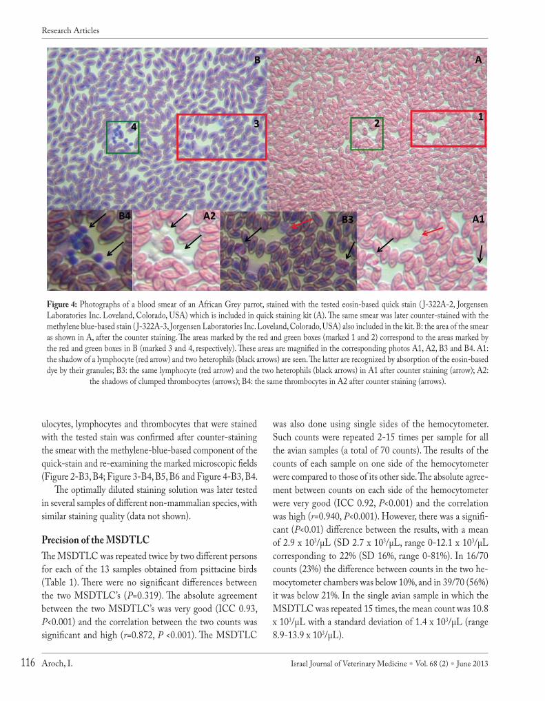
Figure 4: Photographs of a blood smear of an African Grey parrot, stained with the tested eosin-based quick stain ( J-322A-2, Jorgensen Laboratories Inc. Loveland, Colorado, USA) which is included in quick staining kit (A). The same smear was later counter-stained with the methylene blue-based stain ( J-322A-3, Jorgensen Laboratories Inc. Loveland, Colorado, USA) also included in the kit. B: the area of the smear as shown in A, after the counter staining. The areas marked by the red and green boxes (marked 1 and 2) correspond to the areas marked by the red and green boxes in B (marked 3 and 4, respectively). These areas are magnified in the corresponding photos A1, A2, B3 and B4. A1: the shadow of a lymphocyte (red arrow) and two heterophils (black arrows) are seen. The latter are recognized by absorption of the eosin-based dye by their granules; B3: the same lymphocyte (red arrow) and the two heterophils (black arrows) in A1 after counter staining (arrow); A2:
the shadows of clumped thrombocytes (arrows); B4: the same thrombocytes in A2 after counter staining (arrows).
Research Articles

Israel Journal of Veterinary Medicine Vol. 68 (2) June 2013 117 Total Leukocyte Count in Birds
dISCuSSIon
This study has evaluated a novel modified semi-direct tech-nique for TLC for use in non-mammalian vertebrate blood. Originally, it was our intention to assess the MSDTLC with an existing commercial staining system that has been widely used for this purpose that utilizes phloxine-B as the stain-ing reagent (Eosinophil Unopette 5877, Becton-Dickinson, Rutherford, NJ) (12-14). However, just before initiation of the study, production and marketing of this system ceased, exemplifying the need for a simple, cost-efficient and read-ily available staining technique for TLC in non-mammalian blood.
Hemocytometer-based counts have several built-in errors (17). First, the 'error of the field' refers to the random cell setting in the counting area of the chamber, even in properly mixed sample, and is subject to chance. Second, the 'error of the chamber' refers to the counting variations resulting from two separate fillings of the hemocytometer chamber. Third, the 'error of the pipette ' refers to the use of different pipettes for filling the chambers. The total inherent error in the pro-cedure of TLC using a hemocytometer for a TLC of 7,000/µL at two SDs from the mean is ± 21% (17). Such variance is to be expected in clinical settings, although it can be mini-mized when experienced technicians perform the count, and when counts are repeated several times. In contrast, the au-tomated TLC in mammalian blood is much more accurate and is considered a 'gold standard' in both human and vet-erinary medicine. Because such a TLC cannot be performed in non-mammalian blood samples (1, 3), the inherent errors and inaccuracy of the hemocytometer-based TLCs must be accepted. In the present study, the absolute agreement and correlation between counts of the same sample in the two hemocytometer chambers were significant and very high, suggesting that the MSDTLC technique is relatively pre-cise. Additionally, if one accepts the 21% inherent error of hemocytometer-based TLC methods, in 56% of the present counts, the difference between chambers counts was below 21%. Nevertheless, other sources suggest that the difference between chambers should not exceed 10% (1), and in any such event, the count should be repeated. This recommenda-tion was not followed in the present study. Only 23% of the counts complied with the 10% difference recommendation, while the mean difference was 22%. Future assessment of the MSDTLC should probably follow this recommendation and improve its accuracy.
In a clinical setting, repetitions of manual counts is time consuming, and one has to find the optimal balance between maximal accuracy and the time allocated for the count. Performing the MSDTLC 15 times in the same sam-ple has resulted in a clinically acceptable SD of 1.4 x 10³/µL, however, its range was 4.0 x 10³/µL, which is clinically significant, and might influence therapeutic and prognostic considerations. We can thus recommend that the MSDTLC should be done in three repetitions, comparing both cham-bers of the hemocytometer, and if the differences between counts or between chamber counts exceed 20%, the count should be repeated. The final TLC should be the average of all counts. Care should be taken to use the same pipette for both chambers, and incubating and mixing of the blood with the staining solution should be thorough (17).
Semi-direct counts of non-mammalian blood, such as the MSDTLC, are not intended to replace an automated TLC, as this is not commonly available for such species. Rather, the MSDTLC should be an alternative for other semi-di-rect counting methods, such as those based on phloxine-B or toluidine blue, or to the estimation of the TLC based on Romanowsky-stained blood smears. The present results show that the latter and the MSDTLC have a good absolute agreement and a positively significant high correlation. This is probably due to the fact that as for any semi-direct count-ing method, the MSDTL relies on a differential count made in Romanowsky-stained blood smears. Hemocytometer-based counts of avian blood, such as the MSDTLC, are con-sidered more accurate compared to blood smear-based TLC estimations (5).This is because the former are done in a fixed, known blood volume, while when examining blood smears, it is impossible to determine the blood volume used for the count (5). The latter varies considerably, depending on the initial sample volume used to prepare the smear, the length of the smear and the hematocrit (1, 4, 5).
This study has several limitations. First, it included a small number of birds, and the agreement was based on two repetitions of full hemocytometer counts, thereby weakening the power of our statistical analyses. We have tried to par-tially overcome this limitation by introducing comparison of counts between hemocytometer chambers. Second, selected birds were of seven species, which may have introduced some variance due to the different morphological characteristics of the granulocytes. However, we experienced no difficulties in identification of these cells using the tested stain. Ideally
Research Articles

Israel Journal of Veterinary Medicine Vol. 68 (2) June 2013Aroch, I.118
the study should have included a large number of birds, with more birds of each species. Third, we elected to count cells in 10 squares of the hemocytometer, 5 in each chamber), rather than in 18 squares (9 in each chamber) as some veterinary texts recommend (1). The number of squares counted repre-sents a compromise between the goal of getting an accurate result and an attempt to keep the count within a realistic time frame for a busy clinic or laboratory (i.e. <15 minutes per count).
The same methodology is used on a daily basis in refer-ence laboratories. The number of hemocytometer squares to be counted depends on the cellularity of the sample. With low cell counts (e.g. in cerebrospinal fluid) all 9 squares of both sides are required to obtain a reasonably accurate count, while blood samples of healthy avian patients will typically contain 5,000-15,000 white blood cells/µL. As each hemo-cytometer square represents 0.1 µL, this means that the num-ber of cells in 10 squares (at X100 dilution) is expected to be 50-150, thus providing acceptable accuracy.
In conclusion, the preliminary findings of this study showed that MSDTLC performed well using the tested eo-sin-based stain, diluted at 1:10 with distilled water, allow-ing reliable counting of both heterophils and eosinophils in avian blood samples, and showed high precision. Because the tested stain, and similar products are readily available, and the procedure is simple, the method can be applied in non-mammalian blood samples in veterinary practices, as well as in reference laboratories. Further studies, comparing the method with traditional semi-direct methods for TLC, such as phloxine-B, Natt and Herrick's solution and tolu-idine blue-based method should be conducted to assess its performance in such animal species. Such studies should in-clude a larger number of animals and species.
REFEREnCES1. Campbell, T.W.: Hematology of Birds. In: Thrall, A.T, Baker,
D.C., Campbell, T.W., DeNicola, D., Fettman, M., Duane Las-sen, E., Rebar, A. and Weiser G. (Eds.): Veterinary Hematolo-gy and Clinical Chemistry. Baltimore, Lippincott, Williams and Wilkins. pp. 225-258, 2004.
2. Knoll, J.S.: Clinical Automated Hematology Systems. In: Feld-man, B.F., Zinkl, J.G. and Jain, N.C. (Eds): Schalm's Veterinary
Hematology 5th edition. Philadelphia, Lippincott Williams and Wilkins. pp. 3-12, 2000.
3. Wakenel, P.S.: Hematology of Chickens and Turkeys. In: Weiss, D.J. and Wardrop, K.J. (Eds.) Schalm's Veterinary Hematology 6th edition. Ames, Wiley-Blackwell. pp. 958-967, 2010.
4. Zinkl, J.G.: Avian Hematology. In: Jain, N.C. (Ed.). Schalm's Veterinary Hematology 4th edition. Philadelphia, Lea and Fe-biger, pp. 256-273, 1986.
5. Latimer, K.S. and Bienzle, D.: Determination and Interpre-tation of the Avian Leukogram. In: Weiss., D.J. and Wardrop, K.J. (Eds.) Schalm's Veterinary Hematology 6th edition. Ames, Wiley-Blackwell. pp. 345-357, 2010.
6. Lind, P.J., Wolff, P.L., Petrini, K.R. and Keyler, C.W.: Morpholo-gy of the eosinophil in raptors. J. Assoc. Avian Vet. 4: 33-38, 1990.
7. Robertson, G.W. and Maxwell, M.H.: Importance of optimal mixtures of EDTA anticoagulant blood for the preparation of well - stained avian blood smears. Br. Poult. Sci. 34: 615- 617, 1993.
8. Campbell, T.W.: Hematology of Psittacines. In: Weiss, D.J. and Wardrop, K.J. (Eds.) Schalm's Veterinary Hematology 6th edi-tion. Ames, Wiley-Blackwell. pp. 968-976, 2010.
9. Campbell, T.W.: Hematology of Lower Vertebrates. In: 55th An-nual Meeting of the American College of Veterinary Patholo-gists (ACVP) & 39th Annual Meeting of the American Socie-ty of Clinical Pathology (ASVCP). Middleton, American Col-lege of Veterinary Pathologists & American Society for Veteri-nary Clinical Pathology; Internet Publisher: International Veter-inary Information Service, Ithaca, New York, available at: http://www.ivis.org/proceedings/ACVP/2004/Campbell1/ivis.pdf. Ac-cessed at 28.02.12.
10. Campbell, T.W.: Hematology of Reptiles: In: Thrall, A.T, Baker, D.C., Campbell, T.W., DeNicola, D., Fettman, M., Duane Las-sen, E., Rebar, A. and Weiser, G. (Eds.): Veterinary Hematolo-gy and Clinical Chemistry. Baltimore, Lippincott, Williams and Wilkins. pp. 259-276, 2004.
11. Natt, M.P. and Herrick, C.A.: A new blood diluent for counting erythrocytes and leucocytes of the chicken. Poult. Sci. 31:735-738, 1952.
12. Lane, R.: Basic techniques in pet avian clinical pathology. Vet. Clin. North Am. Small Anim. Pract. 21:1157-1179, 1991.
13. Costello, R.T.: A Unopette for Eosinophil counts. Am. J. Clin. Pathol. 54:249-250, 1970.
14. Robertson, G.W. and Maxwell, M.H.: Modified staining tech-niques for avian blood cells. Br. Poult. Sci. 31:881-886, 1990.
15. Lucas, A.M. and Jamoroz, C.: Atlas of Avian Hematology. Ag-ricultural Monograph 25 United States Department of Agricul-ture, Washington DC. p. 271, 1961.
16. Ferris, M. and Bacha, W.J.: A new method for the identification and enumeration of chicken heterophils and eosinophils. Avian Dis. 28: 179-182, 1984.
17. Jain, N.C.: Hematologic Techniques. In: Jain, N.C. (ed.). Schalm's Veterinary Hematology 4th edition. Philadelphia, Lea and Febiger, pp. 21-86, 1986.
Research Articles
Related Documents